molecular cloning and functional characterization of psoralen synthase, the first committed
TRANSCRIPT

Molecular Cloning and Functional Characterization ofPsoralen Synthase, the First Committed Monooxygenaseof Furanocoumarin Biosynthesis*
Received for publication, May 18, 2006, and in revised form, October 25, 2006 Published, JBC Papers in Press, October 26, 2006, DOI 10.1074/jbc.M604762200
Romain Larbat‡1, Sandra Kellner§1, Silvia Specker§, Alain Hehn‡, Eric Gontier‡, Joachim Hans§, Frederic Bourgaud‡,and Ulrich Matern§2
From the §Institut fur Pharmazeutische Biologie, Philipps-Universitat Marburg, Deutschhausstrasse 17A, 35037 Marburg, Germanyand the ‡UMR 1121 Agronomie Environnement INPL-INRA, ENSAIA, 2, av. de la Foret de Haye, 54505 Vandoeuvre-les-Nancy, France
Ammi majus L. accumulates linear furanocoumarins by cyto-chromeP450 (CYP)-dependent conversionof 6-prenylumbellif-erone via (�)-marmesin to psoralen. Relevant activities, i.e. pso-ralen synthase, are induced rapidly from negligible backgroundlevels upon elicitation of A. majus cultures with transient max-ima at 9–10 h and were recovered in labile microsomes.Expressed sequence tags were cloned from elicited Ammi cellsby a nested DD-RT-PCR strategy with CYP-specific primers,and full-size cDNAs were generated from those fragments cor-related in abundance with the induction profile of furanocou-marin-specific activities. One of these cDNAs representing atranscript of maximal abundance at 4 h of elicitation wasassigned CYP71AJ1. Functional expression in Escherichia colior yeast cells initially failed but was accomplished eventually inyeast cells after swapping the N-terminal membrane anchordomainwith thatofCYP73A1.Therecombinantenzymewas iden-tified as psoralen synthase with narrow substrate specificity for(�)-marmesin. Psoralen synthase catalyzes a unique carbon-chaincleavage reaction concomitantly releasing acetone by syn-elimina-tion. Related plants, i.e. Heracleummantegazzianum, are knownto produce both linear and angular furanocoumarins by analogousconversion of 8-prenylumbelliferone via (�)-columbianetin toangelicin, and it was suggested that angelicin synthase has evolvedfrompsoralensynthase.However, (�)-columbianetin failedassub-strate but competitively inhibited psoralen synthase activity. Anal-ogymodelinganddocked solutionsdefined theconditions forhighaffinity substrate binding andpredicted theminimal requirementsto accommodate (�)-columbianetin in the active site cavity. Thestudies suggested that several point mutations are necessary topave the road toward angelicin synthase evolution.
Furanocoumarins are produced by many plants, mostly ofthe Apiaceae, Rutaceae, Moraceae, or the Coronilla and Psora-lea genera of the Fabaceae (1–3). Multiple pharmacologicaleffects have been ascribed to several of thesemetabolites (4–6),
which were included in clinical screenings but received atten-tion also for their inhibitory effect on monooxygenasesinvolved in drug metabolism (7–9) and potential toxicity (10).The (dihydro)furan-substituted 2H-1-benzopyran-2-oneforms the characteristic core structure, and the annulation typedistinguishes the linear furanocoumarins or psoralens from theangular furanocoumarins (Fig. 1). It is noteworthy that plantsaccumulate either psoralens only, e. g. Ammi majus (11–13),Petroselinum crispum (14, 15), and Coronilla sp (16), or bothtypes of compounds, whereas the exclusive production of angu-lar furanocoumarins has never been reported (1). Furanocou-marins often accumulate in the surface wax of tissues and caneasily be collected inwasheswith organic solvents (17, 18). Suchsurface coating was considered to protect the plants from fun-gal invasion or herbivore attack (19), because potent antimy-cotic activity has been documented for furanocoumarins invitro (20), and the few insects utilizing furanocoumarin-pro-ducing plants as an ecological niche, i.e. the black swallowtailbutterfly, depend on a particular detoxifying machinery (21).The toxicity to insects is conceivably a consequence of bothDNAmodification andmonooxygenase inhibition. Furanocou-marins intercalate in double-stranded DNA, and psoralens areknown to cross-link pyrimidine bases under irradiation by [2�2]cycloaddition via their 3,4- and 2�,3�-double bonds, whereasangular furanocoumarins form only monoadducts for stericreasons (22). Although the angular compounds are not aspotent antifeedants as the psoralens (23), the combination ofangular and linear furanocoumarins shows synergisticeffects (24, 25) presumably because the angular compoundsinhibit monooxygenases required for the detoxification ofpsoralens (26, 27). This fact and the relatively limited distri-bution of angular furanocoumarins suggested that the sup-plementary production of angular furanocoumarins hasevolved as a sophisticated means of plants to counter thedetoxification mechanisms of herbivores (28–30). It is con-ceivable that this capability has developed as a modificationof the pathway to linear furanocoumarins at a late stage inthe evolution.The flow of carbon in furanocoumarin biosynthesis is known
since decades from precursor feeding studies (1) and proceedsfrom 4-coumaric acid via umbelliferone, which is prenylated ineither the 6- or 8-position to give demethylsuberosin andosthe-nol, respectively (Fig. 1).Whereas the subcellular compartmentand the precise mode of cyclization of 4-coumaric acid to
* This work was supported in part by the Ministere de la Recherche et del’Enseignement Superieur (to F. B.) and by the Deutsche Forschungsge-meinschaft and Fonds der Chemischen Industrie (to U. M.). The costs ofpublication of this article were defrayed in part by the payment of pagecharges. This article must therefore be hereby marked “advertisement” inaccordance with 18 U.S.C. Section 1734 solely to indicate this fact.
1 These authors contributed equally to the work presented.2 To whom correspondence should be addressed. Tel.: 49-64212822461; Fax:
49-64212826678; E-mail: [email protected].
THE JOURNAL OF BIOLOGICAL CHEMISTRY VOL. 282, NO. 1, pp. 542–554, January 5, 2007© 2007 by The American Society for Biochemistry and Molecular Biology, Inc. Printed in the U.S.A.
542 JOURNAL OF BIOLOGICAL CHEMISTRY VOLUME 282 • NUMBER 1 • JANUARY 5, 2007
by guest on April 4, 2019
http://ww
w.jbc.org/
Dow
nloaded from

umbelliferone have remained elusive (31), the 6-prenyltrans-ferase was assigned to plastids in Ruta graveolens (32), which iscompatible with the mevalonate-independent prenylation ofumbelliferone observed in Apium graveolens (33). An equiva-lent location is likely for the 8-prenyltransferase activity,although this has not been as firmly documented. All subse-quent steps converting demethylsuberosin to psoralen orosthenol to angelicin are supposedly catalyzed by highly analo-gous enzymes (34), but only the linear series has been studied indetail. Enzyme studies conductedmostly with elicitedA. majusor P. crispum cells (35–38) identified marmesin and psoralensynthases as well as psoralen 5-monooxygenase as separatecytochrome P450 (CYP)3 entities for the consecutive conver-sion of demethylsuberosin to bergaptol, which then is methyl-
ated to bergapten (39). These studies became possible, becausefuranocoumarins are completely lacking from the cell suspen-sions propagated in the dark but accumulate rapidly in the cellsand culture fluid upon treatment with fungal elicitor. Neverthe-less, the specific CYP activities of crude microsomes were ratherlow, on average, and varied greatly with the plant type and growthstage of cells. Significant conversion rates were accomplishedwithmicrosomes fromA. majus cells propagated in suspensionno longer than 5 months and treated for 5–10 h with elicitor.Moreover, theCYP activities of isolatedmicrosomes turned outrather labile even on storage at �70 °C, which ruled out anypurification. Overall, the time course of furanocoumarin accu-mulation and the induction of CYP activities suggested a win-dow of about 2–6 h of elicitation formaximal abundance of thecommitted transcripts.Marmesin synthase, psoralen synthase, and psoralen 5-mo-
nooxygenase catalyze very different reactions with psoralensynthase (Fig. 1) catching most the attention. Although oxida-tive carbon-chain cleavage reactions are common in nature,such as in homoterpene (40) or steroid biosynthesis (41), thetrue mechanism has remained under debate (41). The psoralensynthase activity of A. majus microsomes was examined withposition and stereospecifically deuteratedmarmesin substrates(42) and revealed the unique release of acetone as byproduct.The reaction probably initiates at C-3� (Fig. 1), and it is note-worthy that psoralen synthase, in contrast to chemical modelreactions (43), abstracts the hydrogen syn-configurated to theshielding isopropyloxy group (Fig. 1). This is a consequence ofthe radical mode of the reaction requiring that all partners beconfined to a cage (43). The same kind ofmechanism appears tooperate in the pathway to angular furanocoumarin in Hera-cleummantegazzianum, where the conversion of (�)-columbi-anetin to angelicin (Fig. 1) proceeds by syn-elimination (34).These data further supported the assumption that the angularpathway has evolved from psoralen biosynthesis, although therelease of acetone has not been confirmed for Heracleum, andfinal proof must await the alignment of enzyme sequences.In contrast to the basic course of biosynthesis, the molecular
genetics of furanocoumarin formation has remained largelyunresolved due, in part at least, to the lability of the consecu-tively operating P450s. As an initial step, we describe here thecloning and functional expression of the psoralen synthasefrom A. majus, the first furanocoumarin-committedmonooxygenase gene. The recombinant enzyme revealednarrow specificity for (�)-marmesin, whereas (�)-columbi-anetin, which is considered as the analogous precursor of theangular furanocoumarin angelicin acted as a competitiveinhibitor only. Nevertheless, supplemental insight frommolecular modeling and docking approaches seemed to sup-port the idea that psoralen synthase may have evolved alsoangelicin synthase activity through a limited number ofpoint mutations fitting the active site to the metabolism of(�)-columbianetin.
EXPERIMENTAL PROCEDURES
Chemicals—Umbelliferone, psoralen, xanthotoxin, (�)-pu-legone, and cinnamic acids were purchased from Sigma-Aldrich, and xanthotoxol, menthofuran or HPLC-grade 4-cou-
3 The abbreviations used are: CYP, cytochrome P450; SRS, substrate recogni-tion site; C4H, cinnamate 4-hydroxylase; MOPS, 4-morpholinepropanesul-fonic acid; ORF, open reading frame; nt, nucleotide; RACE, rapid amplifica-tion of cDNA ends; UTR, untranslated region; PBD, Protein Data Bank.
FIGURE 1. Pivotal cytochrome P450-dependent reactions in the linearfuranocoumarin pathway converting 6-prenylumbelliferone (demethyl-suberosin) via (�)-marmesin to psoralen and bergaptol or xanthotoxol.The analogous pathway to angular furanocoumarins is shown for compari-son, but has not been established in vitro. The ferryl(IV)-enzyme is likely theoxidizing species, because the one-step elimination of acetone excludes anucleophilic enzyme-peroxide complex for catalysis.
Ammi majus Psoralen Synthase
JANUARY 5, 2007 • VOLUME 282 • NUMBER 1 JOURNAL OF BIOLOGICAL CHEMISTRY 543
by guest on April 4, 2019
http://ww
w.jbc.org/
Dow
nloaded from

maric acid were from Roth (Karlsruhe, Germany) and Fluka(Buchs, Switzerland), respectively. Angelicin, bergapten, andbergaptol were purchased from Extrasynthese (Genay, France).Isoproturon and chlortoluron were from CIL (Paris, France).Demethylsuberosin was synthesized as described previously(36), and (�)-marmesin, 5-hydroxymarmesin, and (�)-3,4,2�,3�-D4-columbianetinwerekindlyprovidedbyG. Innocenti(Faculty of Pharmacy, Padova, Italy) andW. Boland (Max-PlanckInstitut furChemische Okologie, Jena,Germany).Hybond-N� fil-ters and RediprimeTM II Random Prime Labeling kit were fromAmersham Biosciences and [�-32P]dCTP was bought fromMPBiomedicals. TaqDNA polymerase, MMLV-reverse tran-scriptase, restriction endonucleases XhoI, PvuII, BamHI, andPaeI or AccSure DNA polymerase were purchased from MBIFermentas (St. Leon-Rot, Germany) and Bioline (Luckenwalde,Germany), respectively.Cell Cultures and Separation of Coumarins—Cell suspen-
sions of A. majus L. (40-ml B5 cultures in 250-ml flasks) werepropagated continuously in the dark at 21–24 °C, and 7-day-oldcultures were treated with crude Phytophthora sojae cell wallelicitor (5 mg/40 ml, sterilized in 1 ml of water) as describedearlier (35, 36). Control cultures received sterile distilled wateronly. The cells were harvested by vacuum filtration at varioustime intervals up to 32 h, washed twice with sterile distilledwater, frozen in liquid nitrogen, and stored at �70 °C until use.The culture fluid and wash were combined, and coumarinswere separated by HPLC in a solvent system described else-where (44) employing a nucleosil C-18 column (5 �m, 4 � 12.5cm) and a step gradient from 1.5% ortho-phosphoric acid (A)and 80% aqueous acetonitrile (B) starting at 90% A in B,decreasing to 50% A in B over 20 min, and followed by 100% Bfor an additional 10 min. The elution at 1.0 ml/min was moni-tored by ultraviolet absorbance (350 nm), and psoralen (Rt 20.0min) was completely separated under these conditions frombergaptol (18.4 min), xanthotoxol (15.6 min), marmesin (16.0min), demethylsuberosin (26.8 min), umbelliferone (12.0 min),and 4-coumaric acid (11.7 min).cDNA Cloning—Total RNA was isolated from elicitor-in-
duced (1–6 h) and control cells as published elsewhere (45),and cDNA was synthesized with the anchor primer (AP)CCACGCGTCGACTAGTAC(dT)17. First strand cDNA wasamplified with Taq polymerase, and cytochrome P450-relatedESTs were then generated in separate reactions using AP andone of the four primers derived from the conserved PERFmotif(EEFXPER; Fig. 2C) (46). Each incubation was subsequentlydiluted 1:1000 and amplified in a second round of nested PCRusing eight decamer primers inferred from the PFG motif (Fig.2C) in combination with AP (47). The cDNA fragments werecloned, the full-length clones were generated by RNA ligase-mediated RACE (48), and the products were ligated into pCR�4-TOPOVector (Invitrogen) for multiplication in Top10F host(Invitrogen). The cDNA for final sequencing (primer-walking)was amplified by proofreading PCR (0.5 mM primer, 2.0 mMMgSO4, 56 °C annealing temperature) with AccSure DNA po-lymerase-activated (94 °C for 10 min) prior to 35 cycles of 0.5min of denaturation, 1.0min of annealing, and 2.0min of exten-sion. Purification of the cDNAs before RACE and sequencingwas carried out with High Pure PCR Product Purification kit
(Roche Diagnostics, Mannheim, Germany). Sequence analysiswas achieved with advanced WU-Blast2 (EMBL) and align-ments with ClustalW (EMBL-EBI). The induction of specifictranscript abundance was monitored by semiquantitative PCR(Fig. 2B) using end-to-end primers (forward 5�-GCAGAGTG-CAGAGCAATAGAAATGAAGATGC and reverse 5�-GAGC-TGGGAATGAGTATTGACATGG). Genomic DNA was iso-lated by a standard procedure (49) from adult A. majus plantsfreshly harvested from the Botanical Gardens, the CYP71AJ1gene was amplified by PCR employing the end-to-end primers,and the gene was ligated into the pCR�4-TOPO Vector(Invitrogen) for sequencing.Northern and Southern Blotting—Samples of total RNA iso-
lated from the induced cells at various time points of elicitation(up to 32 h) were densitometrically quantified after 1.5% agar-ose gel electrophoresis, and aliquots (4 �g per time point) weredissolved in (30.0 �l) 0.5 MMOPS buffer, pH 7.0, 50% formam-ide containing 2.2 M formaldehyde. Following the denaturationat 68 °C for 15 min, the samples were transferred ontoHybond-N� filters in a Minifold I device (Schleicher & Schull,Dassel, Germany). The filters were dried for 2 h at 80 °C andthen prehybridized at 68 °C for 3 h with 0.025% salmon spermDNA in 5� SSC, 2� Denhardt’s solution containing 0.1% SDS.Full-length CYP71AJ1 cDNA was labeled with [�-32P]dCTPusing the RediprimeTM II Random Prime Labeling kit, purifiedthrough a Sephadex G50 column and employed as a probe inNorthern blotting. The RNA was hybridized at 68 °C for 16 h,the filter washed subsequently twice at 68 °C with 2� SSC con-taining 0.1% SDS and twice with 1� SSC containing 0.1% SDSbefore exposure to an imaging plate. The radioactivity wasspotted by a Bioimager FLA 2000 (Fuji Photo Film, Tokyo,Japan).For Southern blotting, the genomic DNA was isolated from
A. majus leaf tissue (49), resuspended in sterile distilled waterand stored at 4 °C until use. Digestion (20 �g of DNA per incu-bation) was accomplished overnight with endonucleases XhoI,PvuII, BamHI, or PaeI, and the restriction fragments were sep-arated by gel electrophoresis at 70 V for 4 h on 0.8% agarosecontaining 0.4 �g/100 ml ethidium bromide. The DNA wasdepurinated in 0.25 N HCl for about 20min, denatured with 0.5N NaOH containing 1.5 M NaCl for 40 min and neutralizedtwice for 30 min with 0.5 M Tris-HCl, pH 7.4 in the presence of1.5 M NaCl. The DNA was transferred for 20 h onto aHybond-N� filter in 20� SSC. The membrane was dried,washed (2� SSC), prehybridized (5� SSPE, 5� Denhardt’scontaining 0.2% SDS), and the cDNA was labeled with[�-32P]dCTP as described above. The hybridized filter waswashed at 68 °C successively twice with 2� SSC, twice with1� SSC, and twice with 0.1� SSC, all in the presence of 0.1%SDS, and exposed to an imaging plate for Bioimager FLA2000 analysis.ExpressionConstructs—TheORFofCYP71AJ1was amplified
byPCRand introducingKpnI andEcoRI restriction sites proximalupstream to the start ATG and downstream to the stop codon(TGA) employing end-to-end primers (forward 5�-CGGTACCA-TGAAGATGCTGGAACAGAATand reverseGGAATTCTCA-AACATGTGGTCTGGCAACCACCAAGAGAGG). The incu-bation was heated to 95 °C for 5.0 min prior to 30 cycles of 30 s
Ammi majus Psoralen Synthase
544 JOURNAL OF BIOLOGICAL CHEMISTRY VOLUME 282 • NUMBER 1 • JANUARY 5, 2007
by guest on April 4, 2019
http://ww
w.jbc.org/
Dow
nloaded from

denaturation at 95 °C followed by 45 s of annealing at 50 °C and a2.0-min extension at 72 °C. The reaction was finished by an addi-tional 10.0-min extension at 72 °C. The product was purifiedthrough agarose gel electrophoresis, digested with KpnI andEcoRI, and the fragment of 1490 bp was ligated into yeast expres-sion vector pYEDP60 (50). The identity of the expression plasmidwas verified by restriction and sequence analyses prior to thetransformation of yeast cells.Substitution of the N-terminal membrane anchor region
(37 amino acids) in CYP71AJ1 by the corresponding regionof CYP73A1 (33 amino acids) generated the mutantCYP71AJ1mut. A primer was designed corresponding to theamino acid sequence Arg27–Leu33 of Cyp73A1 fused to resi-dues Pro38–Pro45 ofCyp71AJ1 (reverse primer 5�-GGGATAT-TGTGGTGGAGAAGGTGGGAGCTTGAATTTTTTACCG-CGG) and employed for PCR with the CYP73A1 cDNAtemplate in combination with another primer (forward 5�-GGATCCATGGACCTCCTCCTCATAGA) creating a BamHIrestriction site upstream of the ATG. This first amplificationgenerated a product of 123 nt composed of the upstream 99 ntof CYP73A1 followed downstream by 24 nt of CYP71AJ1. Thisproduct was agarose-purified and used in a second PCR as asense primer together with the reverse primer 5-CGAATTCA-AAGCAACTGCAAGTGAAA and CYP71AJ1 cDNA as tem-plate. In both cases, the PCRmixturewas preheated for 5min at95 °C, followed by 10 cycles of 30 s denaturation at 95 °C, 45 sannealing at 60 °C during the first cycle and decreasing by 1 °Cfor each cycle, and a 2.0-min extension at 72 °C. Additional 20cycles were performed with the annealing temperature main-tained at 50 °C, and the PCR was terminated with a final exten-sion of 10 min at 72 °C. The end product CYP71AJ1mut, corre-sponding to a nearly full-size CYP71AJ1 sequence butharboring a replacement of the upstream 111 nt by theupstream 99 nt ofCYP73A1, was flanked in 5� and 3� by BamHIand EcoRI sites, respectively, which were used for ligation intothe pYeDP60 yeast expression vector. The identity of the result-ing plasmid was verified by restriction and sequence analyses.Yeast Expression and Enzyme Assays—Yeast strain WAT11,
engineered to overexpress the P450 reductase isoform ATR1from Arabidopsis thaliana upon galactose induction (50), wastransformed with the various pYeDP60 expression constructsas described elsewhere (51). Propagation of yeast cells andpreparation of microsomes were conducted as described previ-ously (50). Single colonies fromSGI plateswere transferred intoSGI liquid medium (10 ml) and grown at 28 °C for 24 h. Analiquot of the culture (5 ml) was transferred subsequently intoYPGE growth medium (200.0 ml), and growth was continuedfor an additional 24 h at 28 °C. Expression of P450 was theninduced by the addition of galactose (20.0 g/liter growthmedium). The induction was conducted for 18 h at 20 °C,before the cells were harvested, and the microsomal fractioncollected by precipitation in the presence of 0.05 MMgCl2 (52).Protein amounts were determined according to Bradford (53),and the microsomal P450 contents were determined asdescribed by Omura and Sato (54).CYP71AJ1 Substrate Specificity—Each compound listed in
Table 1 was tested as a potential substrate for CYP71AJ1.Briefly, microsomes isolated from yeast CYP71AJ1mut trans-
formants were incubated in 0.1 M sodium phosphate buffer, pH7.0 (150.0 �l total), containing 200 �M of potential substrate, inthe presence of 1.0 mM NADPH or without NADPH (control).The assaymixture was equilibrated for 2.0 min at 27 °C prior tostarting the reaction by the addition ofmicrosomes. After 1 h at27 °C the reaction was terminated by adding 75.0 �l of acetoni-trile/conc. HCl (99:1), and product formation as well as sub-strate consumption was analyzed by reverse-phase HPLC on aPurospher 5�m, 4� 125-mmendcapped column (Merck). Thecolumn was equilibrated in water/acetic acid/acetonitrile (98:1:1) at a flow rate of 0.9 ml/min, and the elution was performedunder diode array detection (220–400 nm) using a convex gra-dient of acetonitrile/methanol (1:1) from 0 to 60% for 25.0 min,followed by 100% acetonitrile/methanol (1:1) for an additional10 min. Bioconversion of (�)-marmesin to psoralen wasdirectly assessed in yeast cultures. Yeast cells transformed withCYP71AJ1, CYP71AJ1mut, or the empty vector were grown inYPGEmediumand induced for P450 expression by the additionof galactose. (�)-Marmesin was added to a final concentrationof 1.0 mM, and the cells were incubated for an additional 4 h at24 °C. The composition of the culture fluid was analyzed sub-sequently by HPLC at 300 nm, using the separation methoddescribed above.Psoralen Synthase Kinetic Analyses—Kinetic assays were
conducted for 6.0min in 0.1 M sodiumphosphate buffer, pH 7.0(200.0 �l total), containing 1.0 mM NADPH and 0.3 pmol ofwild-type or mutant CYP71AJ1, and varying the concentrationof (�)-marmesin. Competitive inhibition assays with (�)-3,4,2�,3�-D4-columbianetin were conducted by adding from10.0 to 300.0 �M of inhibitor to the reaction medium. Theapparent Km was determined by Lineweaver-Burk extrapola-tion. Mechanism-based inactivation assays were performed bypreincubating the microsomes (1.7 pmol of P450) in 0.1 Msodium phosphate buffer, pH 7.0 (60.0 �l total) at 27 °C with0.05 mM of either one of the inhibitor compounds in the pres-ence or in the absence of 1.0mMNADPH, becausemechanism-based inhibition depends on NADPH. P450s are furthermoreknown to undergo partial auto-inactivation in the presence ofNADPH. After 10.0min, aliquots (20.0�l) of the preincubationwere transferred for comparative psoralen synthase assays intothe final incubation mix of 0.1 M phosphate buffer, pH 7.0(200.0-�l total) containing 1.0 mM NADPH and 0.15 mM (�)-marmesin. The reactions were stopped after an additional 10.0min at 27 °C by addition of acetonitrile/conc. HCl (99:1), andpsoralen was quantified by HPLC separation. The residualactivity was expressed as the percentage of the activity meas-ured in control incubations carried out with 1.0mMNADPH inthe absence of inhibitor (100% corresponding to 300 min�1).The activity score recorded for psoralen, 5-methoxypsoralen,8-methoxypsoralen or angelicin after preincubation in thepresence ofNADPH ranged from87� 3% to 95� 2%,while thepreincubation in the absence of NADPH reduced the activity to78 � 8%, which was negligibly affected by the psoralens orangelicin.GC-MS Analysis—The product eluting from HPLC at a
retention time corresponding to psoralenwas collected from20incubations, the solvent was evaporated in an air stream, andthe pellet was resuspended in trichloromethane for GC-MS
Ammi majus Psoralen Synthase
JANUARY 5, 2007 • VOLUME 282 • NUMBER 1 JOURNAL OF BIOLOGICAL CHEMISTRY 545
by guest on April 4, 2019
http://ww
w.jbc.org/
Dow
nloaded from

analysis. The sample (1.0 �l) was injected into a Varian Star3400 CX spectrometer fitted with a Varian factor Four 5MScolumn (15.0 m � 0.25 mm inner diameter, 0.1-�m film) usinghelium at 5.5 psi as carrier gas. The column temperature wasinitially 90 °C for 3 min, ramped to 180 °C at 10 °C � min�1,then to 230 °C at 5 °C � min�1, finally to 250 °C at 10 °C �min�1, and held for 5.0 min. Mass spectra were recorded at 70eV, scanning from 30 to 400 atomic mass units and comparedwith authentic psoralen standard.Site-directed Mutagenesis of CYP71AJ1mut—Site-directed
mutagenesis of CYP71AJ1mut was carried out using a two-stage PCR procedure. In a first round, two separate fragmentswere created, corresponding to the 5�- and 3�-end of the clone,both carrying the desired mutation. To introduce the mutationM120V, the 5�-fragment was amplified using the 73A1ntBamforward primer (5�-GGATCCATGGACCTCCTCCTCA-TAGA-3�) with the M120Vrev primer (5�-GTGTAACGAG-CAAACACGACGTCCTTCCCATTGTAGAAG-3�, mutatedsites are underlined); the 3�-fragmentwas obtained by using theM120Vforw primer (5�-CTTCTACAATGGGAAGGACGTC-GTGTTTGCTCGTTACAC) with the 71AJ1Eco reverseprimer (5�- GGAATTCTCAAACATGTGGTCTGGCAACC-ACCAAGAGAGG-3�). Both reactions were carried out usingPfxDNA polymerase (Invitrogen) with a program consisting ofan initial preheating step of 5 min at 94 °C, followed by 5 cycleswith 30 s denaturation at 94 °C, 30 s annealing at 30 °C, and 90 sextension at 68 °C. Subsequently, 25 cycles were performedwith an annealing temperature of 55 °C. After a final extensionphase for 10 min at 68 °C, both amplified fragments wereagarose-purified using Qiaquick Gel Extraction kit (Qiagen).To merge the 5�- and 3�-ends of the clone, a second round ofPCR (conditions as described above) was carried out usingboth fragments as templates and primers 73A1ntBam and71AJ1Eco. The resultingPCR fragmentwas ligated into thepCR8vector (Invitrogen). Success of the mutation was verified bysequencing. Finally, subcloning into pYeDP60 was performed byligating the 1.5 kb of EcoRI/BamHI Cyp71AJ1mutM120V frag-ment into the linearized vector. Expression of the generatedmutant was performed as described above forCyp71AJ1mut.Enzyme Modeling and Docking Studies—The CYP71AJ1
homology model was built using the MOE program (ChemicalComputing Group, Montreal, Canada) and according toBaudry et al. (55). A multiple sequence alignment betweenCYP71AJ1 and six crystallized P450s (CYP2C5 (PDB: 1DT6),CYP2C9 (PDB: 1OG2), CYP2C8 (PDB: 1PQ2), CYP2B4 (PDB:1PO5), CYP2A6 (PDB: 1Z11), and P450 BM3 (PDB: 1BU7)) wasdone using the Blosum 62 substitution matrix. The sequencealignment was then homology-modeled using the structure ofCYP2C8 (27.5% similarity with CYP71AJ1) as template. Withthe MOE program, 10 models were generated, minimized in afirst round to remove poor Van derWaals contacts and rankedaccording to MOE residue packing quality function. The bestmodel was selected, and the heme coordinates from CYP2C8were added by creating a covalent bond between the heme ironatom, and the sulfur of the conserved cysteine. The structurewas finally minimized using the CHARMm22 force field untilthe final energy gradient was �0.01 kcal/mol�Å. For the dock-ing simulation, marmesin, and columbianetin were created
with the molecular builder application from MOE programand minimized using the CHARMm22 force field. Prior tothe docking simulation, an oxygen atom was added to theheme to represent the iron-oxo intermediate. Docking ofmarmesin and columbianetin was simulated using theMonte Carlo procedure. For each substrate, 25 dockingmodels were created and ranked according to the sum of thesubstrate internal energy and the Van der Waals and theelectrostatic energies. The best ranked model was selected,and the complex substrate/protein was minimized usingMMFF94 force field.Phylogenetic Analysis—Sequenceswere taken from theNCBI
homepage. The phylogenetic analysis was based on alignmentsof psoralen synthase (GenBankTM accession no. AAT06911.1)with translated CYP polypeptide sequences most related topsoralen synthase, whichwere either of undefined classificationfrom Citrus (P450, AAL24049.1) or classified to CYP71 sub-families from soybean (71A9, O81970.1; 71A10, AAB94584.1;71D8, O81974.1; 71D9, O81971.1; 71D10, O48923.1),Solanummelongena (71A2, CAA50645.1; 71A4, CAA50312.1)and Nepeta racemosa (71A5, CAA70575.1; 71A6, CAA70576.1).None of these sequences has been functionally assigned. Men-thofuran synthase from Mentha piperita (accession no.AF346833) was included as the only designated sequence.Furanocoumarins have been reported from Citrus sp.,whereas soybean, Solanum and the Lamiaceae (Nepeta andMentha) may accumulate simple coumarins. The sequenceswere aligned with multalign (Unitary Matrix), and phyloge-netic analyses were carried out using ClustalW by the neigh-bor joining (NJ) method. The distances (percent divergence)between all pairs of sequences were calculated frommultiplealignments, and the NJ method was applied to the distancematrix.
RESULTS
cDNA Cloning—The onset of furanocoumarin accumulationand the induction pattern of relevant enzyme activities inA. majus cell suspensions upon elicitor treatment was recordedpreviously (35–37, 39, 48) and suggested a period of 2–6 h ofelicitation to be sufficient for maximal induction of transcriptabundance. Total RNAwas therefore isolated from cells treatedfor 3–5 h with fungal elicitor and employed as template fordifferential display RT-PCR amplifications of CYP-relatedESTs. A stringent selection was achieved by an RT-PCR strat-egy of two rounds. The initial amplifications used combinationsof an anchor primer and four forward primers deduced fromthe PERF motif (47), which was followed by a second round ofnested PCR employing combinations of the anchor primerwitheight decamer primers deduced from the conserved PFGmotif(46). Distinct bands of 250–500 bp on agarose gel separationwere reproducibly generated representing five divergent ESTs,which were extended to the full-size cDNAs by 3�- and5�-RACE techniques. The cDNAswere sequenced and grouped(56) to CYP families 71 (accession no. AY532370.2), 73(AY219918), 76 (AY532372.1), 82 (AY532373.1), and 98(AY532371.2). CYP73A41 was identified before to encode cin-namate 4-hydroxylase (48), and this report focused onCYP71AJ1, composed of a 1482-bp ORF flanked by 21 and 311
Ammi majus Psoralen Synthase
546 JOURNAL OF BIOLOGICAL CHEMISTRY VOLUME 282 • NUMBER 1 • JANUARY 5, 2007
by guest on April 4, 2019
http://ww
w.jbc.org/
Dow
nloaded from

bp 5�- and 3�-UTRs, respectively (Fig. 2A). The inducibility ofthe selected transcript was initially verified by semiquantitativeRT-PCR employing specific end-to-end oligonucleotide prim-ers (Fig. 2B). The ORF encoded a polypeptide of 494 residuesamounting to a calculated mass of 55891.15 Da with pI 6.49(Fig. 2C). Software analysis predicted anN-terminalmembraneinsertion region and assigned the closest sequence relationship(66% similarity at most) to a few functionally undefinedCYPs71, e.g. from N. racemosa (CYP71A5 and CYP71A6),S. melongena (CAA50312.1; CAA50645.1) or A. thaliana(CYP71A1), and to (�)-menthofuran synthase from pepper-mint (AF346833) (57). None of these plants has been reportedto produce furanocoumarins. The relationship withmentho-furan synthase (45% identity and 65% similarity) was never-theless too low to assign a function to the polypeptide, andmenthofuran has never been reported as a metabolite fromA. majus. The authenticity of the cDNAwas corroborated bycloning of the gene of 1961 bp, which revealed a single intronof 476 bp inserted at position 876 (Fig. 2A). ComplementarySouthern blotting suggested the presence of a small genefamily of at least three copies in the A. majus genome (datanot shown).Functional Expression—Expression of CYP71AJ1 in yeast
strainsWAT11,WR,WAT21 under various regimes of cultureconditions and induction temperature or in E. coli failed asmonitored by enzyme assays with crude extracts, isolatedmicrosomes, and CO difference spectroscopy (58). This wasascribed partly to the in vitro lability of A. majus coumarin-committed CYPs noted previously, presumably resulting frominsufficient membrane association, or to low translational effi-ciency caused by an unfavorable codon bias on yeast expressionof theAmmiCYP71AJ1 sequence (59). Functional expression ofCYPs in yeast cells may critically depend on the quality of theN-terminal anchor sequence as exemplified by CYP73A1
(cinnamate 4-hydroxylase) fromHelianthus tuberosus (60). Thisenzyme was expressed with highactivity in yeast WAT11 strain,and its N-terminal sequence wasemployed to enable or increase theexpression of CYP73A17 andCYP73A15 in yeastmicrosomes (59,61). Obviously, the modification ofthe anchor sequence does not mod-ify the catalytic properties of CYPs(62, 63). Therefore, the N-terminal37 amino acid residues ofCYP71AJ1preceeding the proline-rich regionand spanning the transmembranehelix residues 12–29 were replacedby the corresponding 33-residuemembrane anchor region ofCYP73A1 (Fig. 2C).Swapping of this domain gener-
ated CYP71AJ1mut which wasexpressed in yeast strain WAT11,grown for 24 h at 28 °C and inducedfor 18 h at 20 °C. Under these con-
ditions, low level CYP expression (10.0 pmol/mg) wasobserved in microsomal fractions. Incubation of the isolatedmicrosomes with (�)-pulegone, however, failed to yieldmenthofuran or any other product, and further incubationswere conducted with demethylsuberosin, marmesin or pso-ralen as potential substrates. A product was observed frommarmesin only (Fig. 3A), which required NADPH as a reduc-tant (Fig. 3B) and was lacking in control incubations withmicrosomes from non-transformed yeast cells (Fig. 3C).Absorption spectra (�max 291 nm, broad shoulder at 329 nm)and mass spectra (Fig. 4) showing an M� of 186 as base peakwith major fragments at m/z 158, 130, and 102 (loss of CO)unequivocally identified this product as psoralen. Thus,CYP71AJ1mut encoded psoralen synthase which is the firstfuranocoumarin-committed monooxygenase characterizedat the molecular level after the cloning of bergaptol O-meth-yltransferase had been accomplished recently also fromA. majus (39). Maximal psoralen synthase activity wasobserved in sodium phosphate buffer, pH 7.0 and at a tem-perature ranging from 27 to 35 °C.Microsomes harvested from CYP71AJ1mut yeast transfor-
mants were also unstable upon storage and lost almost all pso-ralen synthase activity within 1 week at �80 °C. The advancedknowledge of assay andhandling parameters prompted anotherapproach to transform yeast cells with wild-type CYP71AJ1,which however failed to yield microsomes showing psoralensynthase activity. Nevertheless, yeast CYP71AJ1 transformantswere employed for precursor feeding studies, and a significantproportion of marmesin added to these cultures (1.0 mM) wasconverted to psoralen during an incubation period of 4 h,whereas yeast cells harboring the empty vector were inactive inthis regard (Fig. 5). Overall, the data demonstrated thatCYP71AJ1 encodes psoralen synthase, which is probably toolabile for detection in isolated yeast microsomes, in accordance
FIGURE 2. A, schematic composition of CYP71AJ1 containing a single intron. B, semiquantitative RT-PCR ofCYP71AJ1 employing end-to-end primers and total RNA as template from A. majus cells elicited for 3 (lane 1a)or 4 h (lane 1b) in comparison to RNA from control cells treated with water for 3 or 4 h (lanes 2a and 2b).Molecular size markers are shown in the left margin. C, polypeptide sequences of CYP73A1 (N terminus) andCYP71AJ1 (full-size). Swapping of the boxed domains generated CYP71AJ1mut. The proline-rich region is under-lined, the I-helix stretch bold-printed, and the conserved Cyt450 motifs (PERF and PFG to delineate primers) areshaded.
Ammi majus Psoralen Synthase
JANUARY 5, 2007 • VOLUME 282 • NUMBER 1 JOURNAL OF BIOLOGICAL CHEMISTRY 547
by guest on April 4, 2019
http://ww
w.jbc.org/
Dow
nloaded from

with previous observations on A. majus microsomes (36, 42).The activity might be stabilized by adjustment of the N-termi-nal anchor sequence.Cell cultures of A. majus accumulate various coumarins
upon elicitation with bergapten as one major stable metabolite(12) and psoralen as an intermediate (Fig. 1). Therefore, thetransient accumulation of psoralen, determined spectro-scopically after HPLC separation, was correlated with therelative CYP71AJ1 transcript abundance (Fig. 6). Theephemeral transcript abundance reached a sharp maximumat 4 h of elicitation followed by themaximal psoralen amountat 9–10 h. The induction patterns fully supported the func-tional significance of the CYP71AJ1 transcript. Moreover,the negligible transcript abundance below 1 h of elicitationemphasized the previously proposed lack of backgroundactivity and de novo induction of this enzyme pivotal to pso-ralen synthesis (Fig. 6).Substrate Specificity andKinetic Analysis—Anumber of cou-
marins, furanocoumarins, phenylpropanoids, and two herbi-cides were examined under standard CYP assay conditions, butnone except marmesin and 5-hydroxymarmesin served as asubstrate (Table 1). The affinity of (�)-marmesin to the recom-binant psoralen synthase determined at an apparent Km of1.5� 0.5�M and kcat 340� 24min�1 is within range (46, 64) or
exceeds (65) the substrate affinities of other enzymes of theCYP71 subfamily involved in plant secondary metabolism.5-Hydroxymarmesin was also converted to bergaptol but withmuch lower affinity (Km of 29.3 � 8.0 �M) and specific activity(kcat 143 � 25 min�1). No product, however, was formed from(�)-columbianetin, the angular analogue of (�)-marmesin(Fig. 1). Further examination revealed significant, albeit weak,inhibition of psoralen synthase activity by (�)-columbianetin(Ki of 225.0 �M), and this effect was competitive with the (�)-marmesin concentration (Fig. 7). Binding of (�)-marmesin and(�)-columbianetin at the same active sitemay suggest that onlyminor differences in the SRS topology distinguish psoralen andangelicin synthases. Various psoralens were reported to act aspotent inhibitors of CYP activities by way of mechanism-basedinhibition (66, 67). Accordingly, kinetic assays were conductedalso in the presence of psoralen or 5- and 8-methoxypsoralen aswell as angelicin. All of these compounds, however, failed toinhibit psoralen synthase in vitro, and this is consistent with theproposed evolutionary context suggesting that CYPs fromfuranocoumarin-producing plants, i. e. CYPs73A (C4Hs), areresistant tomechanism-based inhibition (48, 67). Furthermore,the lack of inhibition of psoralen synthase by angelicin is a pre-requisite to support its suggested role as an inhibitor of psor-alen detoxification by herbivores.Modeling of the CYP71AJ1 Catalytic Site—Alignments with
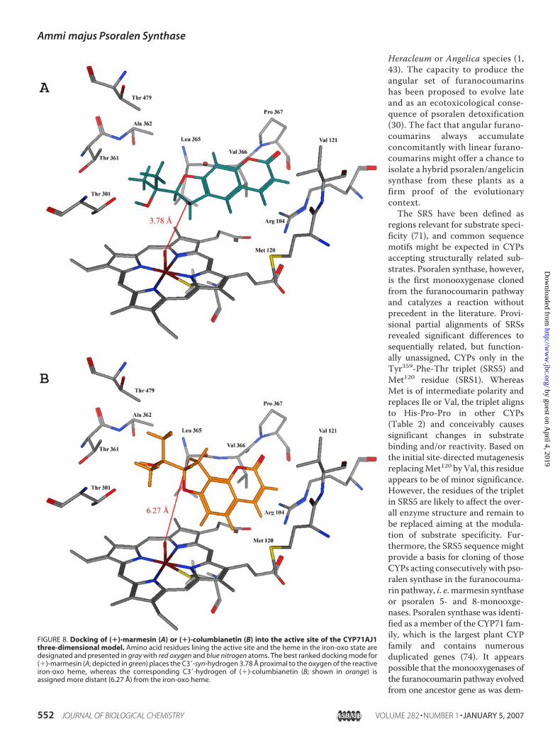
sequences of crystallized P450s (P450 BM3, CYP2C9, C8, C5,A6, andB4)were used to derive a comparativemodel forA. ma-jusCYP71AJ1. Although the accuracy depends critically on thepercentage sequence identity (CYP71AJ1 displays 27.5% simi-larity with CYP2C8) it is possible to build a reliable three-di-mensional model based on the structural cores and severalloop/helix regions that are highly conserved among the CYPfamilies (55, 68–70). This model provided the basis for molec-ular docking of (�)-marmesin or (�)-columbianetin to predictthe binding mode and to assign the SRS sequences (71, 72).Furthermore, the abstraction of hydrogen from C-3� of (�)-marmesin in syn-configuration to the isopropyloxy side chaininitiated by psoralen synthase (Fig. 1) (42, 43) requires the dihy-drofuran moiety in juxtaposition above the iron of the hememoiety. Accordingly, the highest ranked docked solution fitted(�)-marmesin into a 45° angle aligned to the I helix (Ala297 toSer303; Fig. 2C) assigned to SRS4, with the dihydrofuran-ringproximal to residues Ala297 and Thr301 (Fig. 8A). Under thesepremises, the spatial distance of the syn-hydrogen at C-3� of(�)-marmesin to the oxygen atom of the reactive iron-oxoheme approaches 3.78 Å, which likely supports psoralen syn-thase catalysis. The model enzyme-substrate complex revealedsome commonality with CYP proteins such as insect CYP6B8(21) placing Arg104 of SRS1 close to the heme carboxyl groups,probably stabilizing the ring in cooperation with Arg434 in theP450 signaturemotif, and lining the active site cavity above andbelow the coumarin ring system with hydrophobic residuesAla362, Leu365, Val366, Pro367 of SRS5 and Val121, Met120 ofSRS1, respectively. However, hydrophilic residues Thr361(SRS5) and Thr479 (SRS6) were assigned besides Thr301 (SRS4)to surround the dihydrofuran ring with isopropyloxy side chainin the catalytic pocket (Fig. 8A). In addition, Thr301 aligns withthe conserved Thr of the P450 signature ((Ala/Gly)-Gly-X-
FIGURE 3. HPLC separation profile of enzyme assays recorded at 300 nm.The incubations were conducted in 0.1 M sodium phosphate buffer, pH 7.0,with 200.0 �M (�)-marmesin employing: A, microsomes from yeastCYP71AJ1mut transformants in the presence of 1.0 mM NADPH. B, micro-somes as in A but without NADPH. C, microsomes from yeast transformedwith the empty vector pYeDP60. S, (�)-marmesin (substrate); P, product.
Ammi majus Psoralen Synthase
548 JOURNAL OF BIOLOGICAL CHEMISTRY VOLUME 282 • NUMBER 1 • JANUARY 5, 2007
by guest on April 4, 2019
http://ww
w.jbc.org/
Dow
nloaded from

(Asp/Glu)-Thr-(Thr/Ser) that is involved in dioxygen activa-tion (74). The position of Thr301 near the active oxygen of theiron-oxo heme supports this role in CYP71AJ1. The analogousdocked solution for (�)-columbianetin (Figs. 1 and 8B) sug-gested binding at the same topological site, which is compatiblewith competitive inhibition (Fig. 7), but revealed a distance of atleast 6.27Åbetween the 3�-syn-hydrogen and the iron-oxo cen-ter, which appears to exclude catalytic turnover.Psoralen synthase described in this report is the only pub-
lished monooxygenases sequence committed to furanocouma-rin biosynthesis, and alignments are thus limited to less relatedsequences ofmostly undefined functionality. Severalmonooxy-genases degrading furanocoumarins were described frominsects, and pointmutations in their SRS1 regionwere reportedto affect binding (10). These enzymes, however, share very littlesequence homology with CYP71AJ1. Phylogenetic analysis(data not shown) revealed some relationship with a P450 fromCitrus sinensis (62% similarity) or several members of theCYP71 family from Solanummelongena (A2 and A4; 64–65%
similarity) and soybean (Ala9, Ala10, Asp8, Asp9, and Asp10;58–65% similarity) without functional assignment; theseplants produce furanocoumarins (Citrus) or simple coumarinsat least (Solanum, soybean). An equivalent level of similaritycan be ascribed to functionally undefined CYP71A5 (68%) andCYP71A6 (65%) from N. racemosa or menthofuran synthase(65%) fromM. piperita. Taking into account that the substrate-docked model assigned some residues of SRS1 and SRS5, inparticular, to the active site, these sequence motifs were com-pared with the corresponding regions of CYPs from coumarin-producing plants (Table 2). The most peculiar features of pso-ralen synthase are residues Thr361 (SRS5) and Met120 (SRS1).While Met120 substitutes for hydrophobic Ile or Val in otherCYP71 (Table 2), Thr361 in the YFT motif of CYP71AJ1 alignswith the HPP triplet conserved in almost all sequences of theCYP71 family. Replacement of both proline residues must con-siderably affect the spatial configuration of psoralen synthaseand is conceivably involved in positioning Thr361 in the hydro-philic cluster about the isopropyloxy group of (�)-marmesin.
FIGURE 4. MS fragmentation (A) and GC separation profile (C) of the product isolated by incubation of (�)-marmesin with microsomes from yeast CYP71AJ1muttransformants in the presence of NADPH. The fragmentation pattern (B) and GC profile (D) of authentic psoralen were recorded for comparison.
Ammi majus Psoralen Synthase
JANUARY 5, 2007 • VOLUME 282 • NUMBER 1 JOURNAL OF BIOLOGICAL CHEMISTRY 549
by guest on April 4, 2019
http://ww
w.jbc.org/
Dow
nloaded from

Site-directedMutagenesis—To investigate the potential con-tributions of the depicted residues to the catalytic activity,Met120was replaced byVal as the first step. Themutantwas lessreadily expressed in yeast cells than the wild-type enzyme, but
revealed also specificity for (�)-marmesin and did not accept (�)-columbianetin. Psoralen was theonly product observed, and a Km of2.3 �M � 0.39 �M was recorded for(�)-marmesin. This value is inrange with the substrate affinity ofthe wild-type enzyme and suggestsmarginal effects of the M120Vmutation, if any, on the catalyticactivity.
DISCUSSION
Coumarins and furanocoumarinshave been reported from numerousplants and their potential bioactivi-ties have been recognized. Never-theless, the biosynthesis of the1-benzopyran-2-one nucleus andthe molecular details of subsequentsteps have remained incompletelyunderstood (76). Three consecutivecytochrome P450-dependent reac-tions converting demethylsuber-osin to bergaptol (Fig. 1) and identi-fied in Ammi microsomes (35, 36)provided the basis for the differen-tial cloning approach, which fullyconfirmed the previous proposal ofseparate CYP entities (31, 35, 36).Psoralen synthase represents thefirst cloned coumarin-committedmonooxygenase, and CYP71AJ1gives access to a new CYP71 sub-family of enzymes. Functionalexpression suffered from the insta-bility and/or rather low specificactivity of microsomes. This again
confirmed our previous findingswithmicrosomes from elicitedA. majus cells which demanded unusually subtle conditions ofcell propagation and fractionation, and the activitywas lost rap-idly even on storage of microsomes at �70 °C. It is conceivablethat psoralen synthase activity essentially requires the tightassociationwith themembrane and the reductase,which is sup-ported by the beneficial effect of replacing themembrane inser-tion region by that ofCYP73A1, although the activity of isolatedmicrosomes remained unstable upon storage. Recombinantpsoralen synthase showed narrow specificity and high affinity(Km 1.5 �M) for (�)-marmesin (Table 1), compatible with theobservation of (�)-marmesin as an intermediate of psoralenand bergaptol synthesis in elicited A. majus cells (36). An alter-nate pathway to bergaptol, however, appeared possible basedon 5-hydroxylation of (�)-marmesin and subsequent cycliza-tion. Recombinant psoralen synthase accepted 5-hydroxy-marmesin (Table 1), and 5-hydroxymarmesin was includedin an unconfirmed list of A. majus constituents (77). How-ever, none of the original reports on A. majus coumarins (37,75, 78–80) mentioned this compound, and psoralen syn-
FIGURE 5. Bioconversion of (�)-marmesin to psoralen in yeast culture. Yeast cells transformed withCYP71AJ1 (A), CYP71AJ1mut (B), or the empty vector (C) were grown in YPGE medium and induced by theaddition of galactose for P450 expression. (�)-Marmesin was added to a final concentration of 1.0 mM, and thecells were incubated for an additional 4 h at 24 °C. The composition of the culture fluid was analyzed subse-quently by HPLC at 300 nm. M, (�)-marmesin (substrate); P, Psoralen (product).
FIGURE 6. Induction of CYP71AJ1transcript abundance (f) and transientamounts of psoralen (Œ) in A. majus cell cultures following the additionof fungal elicitor.
Ammi majus Psoralen Synthase
550 JOURNAL OF BIOLOGICAL CHEMISTRY VOLUME 282 • NUMBER 1 • JANUARY 5, 2007
by guest on April 4, 2019
http://ww
w.jbc.org/
Dow
nloaded from

thase was shown before to tolerate in vitrominor changes ofsubstrate, such as the linear 2�-acetyl-dihydrofuranocouma-rin (43). This likely excludes the alternate route to bergaptolin A. majus and is corroborated by the data presented in thisreport. The low conversion rate of 5-hydroxymarmesin(kcat/Km 4.9) as compared with (�)-marmesin (kcat/Km 226)assigned 5-hydroxymarmesin as an inefficient substrate forCYP71AJ1 and unlikely precursor for bergaptol in A. majus.
On the assumption that the capacity for angular furanocou-marin formation has evolved from the linear furanocoumarinpathway, related or highly homologous enzymes should beexpected to catalyze the analogous reactions in both pathways.The oxidative chain cleavage reactions of (�)-marmesin and(�)-columbianetin (Fig. 1) appear highly analogous, and syn-elimination has been confirmed also for the conversion of(�)-columbianetin to angelicin (34) suggesting a cyto-chrome P450-dependent mechanism. Moreover, the com-petitive inhibition kinetics documented in this report indi-cate that (�)-columbianetin binds to the active site ofpsoralen synthase, but fails to serve as a substrate. Fitting of(�)-columbianetin into the active site of the CYP71AJ1model may explain this discrepancy, because the highestranked docked solution placed the syn-configurated hydro-gen at carbon-3� 6.27 Å away from the iron-oxo center (Fig.8B), which is much less favorable than the 3.78 Å calculatedin the case of (�)-marmesin (Fig. 8A). Although the data arenot yet sufficient to draw conclusions on the evolution of thepathway to angular furanocoumarins, it is tempting toassume that a limited number of mutations may have trans-formed psoralen synthase to angelicin synthase, concerningprimarily Thr301, Thr361, and the residues Ala362, Leu365, andVal366 in the active site cavity (Fig. 8, A and B). Nevertheless,another major branchpoint in linear versus angular furano-coumarin formation exists at the stage of prenylation ofumbelliferone (Fig. 1). Prenylation reactions at C-6 and O-7,but no C-8-prenylation, of umbelliferone were observed inA. majus (37), and thus more than one hurdle must be over-come to enter the route to angular furanocoumarins. Theseaspects cannot be studied in A. majus because of lack ofangular furanocoumarins, but might be conducted in
FIGURE 7. Competitive inhibition of psoralen synthase (CYP71AJ1mut)activity by (�)-columbianetin. The assays were conducted in the absence(F) and in the presence of (�)-columbianetin fixed at 10.0 (f), 50.0 (Œ), 100.0�M (�), 150 �M (�), and 300 �M (�) concentration. The apparent Km valuesinferred from the Lineweaver-Burk diagram plotted versus the concentrationof (�)-columbianetin (inset) revealed an apparent Ki of about 225.0 �M. Dataare means of triplicate experiments.
TABLE 1Compounds included in psoralen synthase substrate assays
Classification Compound Rt lmax Km kcatmin nm �M min�1
Substrate(�)-Marmesin 19.0 334 1.5 � 0.5 340.0 � 24.05-Hydroxymarmesin 18.0 339 29.3 � 8.0 143.0 � 35.0
No substrateSimple coumarins
Coumarin 12.3 280Herniarin 14.3 323Scopoletin 10.5 346Umbelliferone 10.5 324Demethylsuberosin 28.9 334
FuranocoumarinsPsoralen 20.4 312Bergaptol 14.2 314Bergapten 17.5 312Xanthotoxol 16.9 308Xanthotoxin 15.6 302Isopimpinellin 17.7 315(�)-3,4,2�,3�-D4-Columbianetin 19.8 328Angelicin 21.4 302
Cinnamic acidsCinnamic acid 20.4 2784-Coumaric acid 14.1 3102-Coumaric acid 13.0 283Ferulic acid 12.7 315
Monoterpenes(�)-Pulegone 21.5 258Menthofuran 22.0 288
HerbicidesIsoproturon 23.0 238Chlortoluron 22.0 283
Ammi majus Psoralen Synthase
JANUARY 5, 2007 • VOLUME 282 • NUMBER 1 JOURNAL OF BIOLOGICAL CHEMISTRY 551
by guest on April 4, 2019
http://ww
w.jbc.org/
Dow
nloaded from
Heracleum or Angelica species (1,43). The capacity to produce theangular set of furanocoumarinshas been proposed to evolve lateand as an ecotoxicological conse-quence of psoralen detoxification(30). The fact that angular furano-coumarins always accumulateconcomitantly with linear furano-coumarins might offer a chance toisolate a hybrid psoralen/angelicinsynthase from these plants as afirm proof of the evolutionarycontext.The SRS have been defined as
regions relevant for substrate speci-ficity (71), and common sequencemotifs might be expected in CYPsaccepting structurally related sub-strates. Psoralen synthase, however,is the first monooxygenase clonedfrom the furanocoumarin pathwayand catalyzes a reaction withoutprecedent in the literature. Provi-sional partial alignments of SRSsrevealed significant differences tosequentially related, but function-ally unassigned, CYPs only in theTyr359-Phe-Thr triplet (SRS5) andMet120 residue (SRS1). WhereasMet is of intermediate polarity andreplaces Ile or Val, the triplet alignsto His-Pro-Pro in other CYPs(Table 2) and conceivably causessignificant changes in substratebinding and/or reactivity. Based onthe initial site-directed mutagenesisreplacingMet120 byVal, this residueappears to be of minor significance.However, the residues of the tripletin SRS5 are likely to affect the over-all enzyme structure and remain tobe replaced aiming at the modula-tion of substrate specificity. Fur-thermore, the SRS5 sequence mightprovide a basis for cloning of thoseCYPs acting consecutivelywith pso-ralen synthase in the furanocouma-rin pathway, i. e. marmesin synthaseor psoralen 5- and 8-monooxge-nases. Psoralen synthase was identi-fied as a member of the CYP71 fam-ily, which is the largest plant CYPfamily and contains numerousduplicated genes (74). It appearspossible that the monooxygenases ofthe furanocoumarin pathway evolvedfrom one ancestor gene as was dem-
FIGURE 8. Docking of (�)-marmesin (A) or (�)-columbianetin (B) into the active site of the CYP71AJ1three-dimensional model. Amino acid residues lining the active site and the heme in the iron-oxo state aredesignated and presented in gray with red oxygen and blue nitrogen atoms. The best ranked docking mode for(�)-marmesin (A; depicted in green) places the C3�-syn-hydrogen 3.78 Å proximal to the oxygen of the reactiveiron-oxo heme, whereas the corresponding C3�-hydrogen of (�)-columbianetin (B; shown in orange) isassigned more distant (6.27 Å) from the iron-oxo heme.
Ammi majus Psoralen Synthase
552 JOURNAL OF BIOLOGICAL CHEMISTRY VOLUME 282 • NUMBER 1 • JANUARY 5, 2007
by guest on April 4, 2019
http://ww
w.jbc.org/
Dow
nloaded from

onstrated for theCYPs catalyzing theDIMBOA/DIBOApathwayinmaize (73).
Acknowledgments—We thank Prof. G. Innocenti (Padova, Italy) forsamples of (�)-marmesin and 5-hydroxymarmesin, Prof. W. Boland(Jena, Germany) for the gift of (�)-3,4,2�,3�-D4-columbianetin, andDr. S. Martens for valuable discussions.
REFERENCES1. Murray, R. D. H., Mendez, J., and Brown, S. A. (1982) The Natural Cou-
marins: Occurrence, Chemistry and Biochemistry, Wiley, New York2. Murray, R. D. (1991) Progr. Chem. Org. Natural Prod. 58, 83–3163. Estevez-Braun, A., and Gonzalez, A. G. (1997) Natural Product Rep.
465–4754. Wulff, H., Rauer, H., During, T., Hanselmann, C., Ruff, K., Wrisch, A.,
Grissmer, S., and Haensel, W. (1998) J. Med. Chem. 41, 4542–45495. Kawase, M., Sakagami, H., Motohashi, N., Hauer, H., Chatterjee, S. S.,
Spengler, G., Vigyikanne, A. V., Molnar, A., and Molnar. J. (2005) In Vivo19, 705–711
6. Malaiyandi, V., Sellers, E. M., and Tyndale, R. F. (2005) Clin. Pharmacol.Therap. 77, 145–158
7. De Castro, W. V., Mertens-Talcott, S., Rubner, A., Butterweck, V., andDerendorf, H. (2006) J. Agric. Food Chem. 54, 249–255
8. Wen, Y. H., Sahi, J., Urda, E., Kulkarni, S., Rose, K., Zheng, X., Sinclair, J. F.,Cai, H., Strom, S. C., and Kostrubsky, V. E. (2002) Drug Metabolism 30,977–984
9. Paine,M. F., Criss, A. B., andWatkins, P. B. (2005) J. Pharmacol. Exp. Ther.312, 1151–1160
10. Pan, L., Wen, Z., Baudry, J., Berenbaum, M. R., and Schuler, M. A. (2004)Arch. Biochem. Biophys. 422, 31–41
11. Elgamal. M. H. A., Shalaby, N. M. M., Duddeck, H., and Hiegemann M.(1993) Phytochemistry 34, 819–823
12. Hamerski, D., Beier, R. C., Kneusel, R. E.,Matern,U., andHimmelspach, K.(1990) Phytochemistry 29, 1137–1142
13. Ekiert, H., and Gomolka, E. (2000) Pharmazie 55, 684–68714. Tietjen, K. G., Hunkler, D., and Matern, U. (1983) Eur. J. Biochem. 131,
401–40715. Beier, R. C., Ivie, G. W., and Oertli, E. H. (1994) Phytochemistry 36,
869–87216. Innocenti, G., Cappelletti, E. M., and Caporale, G. (1986) Plantes Medici-
nales et Phytotherapie 4, 313–32217. Zobel, A. M., and Brown, S. A. (1990) J. Chem. Ecol. 16, 1623–163418. Zobel, A. M., and Brown, S. A. (1991) J. Chem. Ecol. 17, 1801–181019. Stadler, E., and Roessingh, P. (1990) Symp. Biol. Hung. 39, 71–8520. Afek, U., Carmeli, S., and Aharoni, N. (1995) Phytochemistry 39,
1347–135021. Li, X., Baudry, J., Berenbaum, M. R., and Schuler, M. A. (2004) Proc. Natl.
Acad. Sci. U. S. A. 101, 2939–2944
22. Kanne, D., Straub, K., Rapoport, H., and Hearst, J. E. (1982) Biochemistry21, 861–871
23. Gruenert, D. C., Ashwood-Smith, M., Mitchell, R. H., and Cleaver, J. E.(1985) Cancer Res. 45, 5394–5398
24. Berenbaum,M. R., Nitao, J. K., and Zangerl, A. R. (1991) J. Chem. Ecol. 17,207–215
25. Berenbaum, M. R., and Zangerl, A. R. (1996) Recent Adv. Phytochem. 30,1–24
26. Ma, R., Cohen, M. B., Berenbaum, M. R., and Schuler, M. A. (1994) Arch.Biochem. Biophys. 310, 332–340
27. Wen, Z., Pan, L., Berenbaum, M. R., and Schuler, M. A. (2003) Insect.Biochem. Mol. Biol. 33, 937–947
28. Berenbaum, M. R., and Feeny, P. (1981) Science 212, 927–92929. Berenbaum, M. R., and Zangerl, A. R. (1993) Oecologia 95, 370–37530. Berenbaum, M. R., and Zangerl, A. R. (1998) Proc. Natl. Acad. Sci. U. S. A.
95, 13743–1374831. Matern, U., Luer, P., and Kreusch, D. (1999) in Comprehensive Natural
Products Chemistry (Sankawa, U., ed), Vol. 1, pp. 623–637. Elsevier,Amsterdam
32. Dhillon, D. S., and Brown, S. A. (1976) Arch. Biochem. Biophys. 177,74–83
33. Stanjek, V., Piel, J., and Boland, W. (1999) Phytochemistry 50, 1141–114534. Stanjek, V., and Boland W. (1998) Helv. Chim. Acta 81, 1596–160735. Hamerski, D., and Matern, U. (1988a) FEBS Lett. 239, 263–26536. Hamerski, D., and Matern, U. (1988b) Eur. J. Biochem. 171, 369–37537. Hamerski, D., Schmitt, D., and Matern, U. (1990) Phytochemistry 29,
1131–113538. Wendorff, H., and Matern, U. (1986) Eur. J. Biochem. 161, 391–39839. Hehmann, M., Lukacin, R., Ekiert, H., and Matern, U. (2004) Eur. J. Bio-
chem. 271, 932–94040. Boland, W., Gabler, A., Gilbert, M., and Feng, Z. (1998) Tetrahedron 54,
14725–1473641. Lieberman, S., and Lin, Y. Y. (2001) Steroid Biochem. Mol. Biol. 78, 1–1442. Stanjek, V., Miksch, M., Luer, P., Matern, U., and Boland, W. (1999) An-
gew. Chemie-Int. Ed. 38, 400–40243. Stanjek, V. (1998) Studien zur Biosynthese der Furanocumarine. PhDThe-
sis, Universitat Bonn44. Hagemeier, J., Batz, O., Schmidt, J.,Wray, V., Hahlbrock, K., and Strack, D.
(1999) Phytochemistry 51, 629–63545. Giuliano, G., Bartley, G. E., and Scolnik, P. A. (1993) Plant Cell 5, 379–38746. Schopfer, C. R., and Ebel, J. (1998)Mol. Gen. Genet. 258, 315–32247. Fischer, T. C., Klattig, J. T., and Gierl, A. (2001) Theor. Appl. Genet. 103,
1014–102148. Hubner, S., Hehmann, M., Schreiner, S., Martens, S., Lukacin, R., and
Matern, U. (2003) Phytochemistry 64, 445–45249. Dellaporta, S. J., Wood, J., and Hick, J. B. (1983) Plant Mol. Biol. Rep. 1,
19–2150. Pompon, D., Louerat, B., Bronne, A., and Urban, B. (1996)Methods Enzy-
mol. 272, 51–6451. Gietz, D., St. Jean, A., Woods, R. A., and Schiestl, R. H. (1992). Nucleic
Acids Res. 20, 142552. Diesperger, H., and Sandermann, H., Jr. (1978) FEBS Lett. 85, 333–33653. Bradford, M. M. (1976) Anal. Biochem. 72, 248–25454. Omura, T., and Sato, R. (1964) J. Biol. Chem. 239, 2370–237855. Baudry, J. Li, W., Pan, L., Berenbaum, M. R., and Schuler, M. A. (2003)
Protein Eng. 16, 577–58756. Specker, S. (2004)Klonierung von Cytochrom P450-abhangigenMonooxy-
genasen aus Ammi majus L. und funktionelle Expression der Zimtsaure4-Hydroxylase. PhD Thesis, Philipps-Universitat, Marburg
57. Croteau, R. B., Davis, E. M., Ringer, K. L., and Wildung, M. R. (2005)Naturwissenschaften 92, 562–577
58. Jefcoate, C. R. (1978)Methods Enzymol. 52, 258–27959. Batard, Y., Hehn A., Nedelkina, S., Schalk, M., Pallett, K., Schaller, H., and
Werck-Reichhart, D. (2000) Arch. Biochem. Biophys. 379, 161–16960. Teutsch, H. G., Hasenfratz, M. P., Lesot, A., Stoltz, C., Garnier, J. M.,
Jeltsch, J. M., Durst, F., and Werck-Reichhart, D. (1993) Proc. Natl. Acad.Sci. U. S. A. 90, 4102–4106
TABLE 2Alignment of partial recognition sites
SRS1 (Arg104–Val121)Psoralen synthase RPYSS VANKI FYNGK DMVP450 Citrus sinensis RPPLI GSGKF TYNYS DIACYP71A9 RPSLY AANRL GYG-S TVSCYP71A10 RRQPT AAKIF GYGCK DVACYP71D8 RPQLL APQFM VYGAT DIACYP71D9 RPYVL AAEIM DYDFK GVACYP71D10 RPDFV LSRIV SYNGS GIV
SRS5 (Leu358–Glu369)Psoralen synthase LYFTA PLLVP REP450 Citrus sinensis LHPPA PLLIA RDCYP71A9 LHPPA PLLVP RECYP71A10 LHPPL PLLIA RECYP71D8 LHPPS QLI-P RECYP71D9 LHPPA PLLLP RECYP71D10 LHPPV PLLVP RV
Ammi majus Psoralen Synthase
JANUARY 5, 2007 • VOLUME 282 • NUMBER 1 JOURNAL OF BIOLOGICAL CHEMISTRY 553
by guest on April 4, 2019
http://ww
w.jbc.org/
Dow
nloaded from

61. Nedelkina, S., Jupe, S. C., Blee, K. A., Schalk,M.,Werck-Reichhart, D., andBolwell, G. P. (1999) Plant Mol. Biol. 39, 1079–1090
62. Richardson, T. H., Jung, F., Griffin, K. J., Wester, M., Raucy, J. D., Kemper,B., Bornheim, L.M., Hasset, C., Omiecinski, C. J., and Johnson, E. F. (1995)Arch. Biochem. Biophys. 323, 87–96
63. Sueyoshi, T., Park, L. J., Moore, R., Juvonen, R. O., and Negishi, M. (1995)Arch. Biochem. Biophys. 332, 265–271
64. Latunde-Dada, A. O., Cabello-Hurtado, F., Czittrich, N., Didierjean, L.,Schopfer, C., Hertkorn, N.,Werck-Reichhart, D., and Ebel, J. (2001) J. Biol.Chem. 276, 1688–1695
65. Takahashi, S., Zhao, Y., O’Maille, P. E., Greenhagen, B. T., Noel, J. P.,Coates, R. M., and Chappell, J. (2005) J. Biol. Chem. 280, 3686–3696
66. Koenigs, I. L., and Trager, W. F. (1998) Biochemistry 37, 13184–1319367. Gravot, A., Larbat, R., Hehn, A., Lievre, K., Gontier, E., Goergen, J-L., and
Bourgaud, F. (2004) Arch. Biochem. Biophys. 422, 71–8068. Kemp, C. A., Marechal, J-D., and Sutcliffe, M. J. (2005) Arch. Biochem.
Biophys. 433, 361–36869. Williams, P. A., Cosme, J., Sridhar, V., Johnson, E. F., and McRee, D. E.
(2000) J. Inorg. Biochem. 81, 183–190
70. Rupasinghe, S., Baudry, J., and Schuler, M. A. (2003) Protein Eng. 16,721–731
71. Gotoh, O. (1992) J. Biol. Chem. 267, 83–9072. Werck-Reichhart, D., and Feyereisen, R. (2000) Genome Biol. 1, 1–973. Frey, M., Huber, K., Park, W. J., Sicker, D., Lindberg, P., Meeley, R. B.,
Simmons, C. R., Yalpani, N., and Gierl, A. (2003) Phytochemistry 62,371–376
74. Werck-Reichhart, D., Bak, S., and Paquette, S. (2002) The ArabidopsisBook, pp. 1–28, The American Society of Plant Biologists, Rockville, MD
75. Herde A. (2005) Untersuchung der Cumarinmuster in Fruchten aus-gewahlter Apiaceae, Ph.D. Thesis, Universitat Hamburg
76. Bourgaud, F., Hehn, A. Y., Larbat, R., Doerper, S., Gontier, E., Kellner, S.,and Matern, U. (2006) Phytochem. Rev. in press
77. Ur-Rahman,A., Said,H.M., andAhmad,V.U. (1986)Encyclopedia PlantaMedica, Vol. 1, p. 373, Hamdard Foundation Press, Karachi, Pakistan
78. Ivie, G. W. (1978) J. Agric. Food Chem. 26, 1394–140279. Krolicka, A., Staniszewska, I., Bielawski, K., Malinski, E., Szafranek, J., and
Lojkowska, E. (2001) Plant Sci. 160, 259–26480. Pande, D., Purohit,M., and Srivastava, P. S. (2002) Plant Sci. 162, 583–587
Ammi majus Psoralen Synthase
554 JOURNAL OF BIOLOGICAL CHEMISTRY VOLUME 282 • NUMBER 1 • JANUARY 5, 2007
by guest on April 4, 2019
http://ww
w.jbc.org/
Dow
nloaded from

Hans, Frederic Bourgaud and Ulrich MaternRomain Larbat, Sandra Kellner, Silvia Specker, Alain Hehn, Eric Gontier, Joachim
First Committed Monooxygenase of Furanocoumarin BiosynthesisMolecular Cloning and Functional Characterization of Psoralen Synthase, the
doi: 10.1074/jbc.M604762200 originally published online October 26, 20062007, 282:542-554.J. Biol. Chem.
10.1074/jbc.M604762200Access the most updated version of this article at doi:
Alerts:
When a correction for this article is posted•
When this article is cited•
to choose from all of JBC's e-mail alertsClick here
http://www.jbc.org/content/282/1/542.full.html#ref-list-1
This article cites 68 references, 12 of which can be accessed free at
by guest on April 4, 2019
http://ww
w.jbc.org/
Dow
nloaded from