molecular modeling and site-directed mutagenesis reveal
TRANSCRIPT
Molecular Modeling and Site-Directed MutagenesisReveal Essential Residues for Catalysis in aProkaryote-Type Aspartate Aminotransferase1[W][OA]
Fernando de la Torre2, Aurelio A. Moya-Garcıa2, Marıa-Fernanda Suarez, Carlos Rodrıguez-Caso,Rafael A. Canas, Francisca Sanchez-Jimenez, and Francisco M. Canovas*
Departamento de Biologıa Molecular y Bioquımica and Instituto Andaluz de Biotecnologıa (F.d.l.T., M.-F.S.,R.A.C., F.M.C.) and Departamento de Biologıa Molecular y Bioquımica and Centro de Investigacion Biomedicaen Red de Enfermedades Raras (A.A.M.-G., C.R.-C., F.S.-J.), Campus Universitario de Teatinos, Universidad deMalaga, 29071 Malaga, Spain
We recently reported that aspartate (Asp) biosynthesis in plant chloroplasts is catalyzed by two different Asp aminotrans-ferases (AAT): a previously characterized eukaryote type and a prokaryote type (PT-AAT) similar to bacterial andarchaebacterial enzymes. The available molecular and kinetic data suggest that the eukaryote-type AAT is involved in theshuttling of reducing equivalents through the plastidic membrane, whereas the PT-AAT could be involved in the biosynthesisof the Asp-derived amino acids inside the organelle. In this work, a comparative modeling of the PT-AAT enzyme from Pinuspinaster (PpAAT) was performed using x-ray structures of a bacterial AAT (Thermus thermophilus; Protein Data Bank accessionnos. 1BJW and 1BKG) as templates. We computed a three-dimensional folding model of this plant homodimeric enzyme thathas been used to investigate the functional importance of key amino acid residues in its active center. The overall structure ofthe model is similar to the one described for other AATenzymes, from eukaryotic and prokaryotic sources, with two equivalentactive sites each formed by residues of both subunits of the homodimer. Moreover, PpAAT monomers folded into one largeand one small domain. However, PpAAT enzyme showed unique structural and functional characteristics that have beenspecifically described in the AATs from the prokaryotes Phormidium lapideum and T. thermophilus, such as those involved in therecognition of the substrate side chain or the “open-to-closed” transition following substrate binding. These predictedcharacteristics have been substantiated by site-direct mutagenesis analyses, and several critical residues (valine-206, serine-207,glutamine-346, glutamate-210, and phenylalanine-450) were identified and functionally characterized. The reported datarepresent a valuable resource to understand the function of this enzyme in plant amino acid metabolism.
Aspartate aminotransferase (AAT; Asp:2-oxogluta-rate aminotransferase; EC 2.6.1.1) catalyzes the revers-ible transamination reaction between Asp and2-oxoglutarate to give Glu and oxaloacetate via aping-pong bi-bi mechanism. AAT enzymes have beenclassified into the aminotransferase family I and thendivided into two subfamilies, Ia and Ib, according totheir amino acid sequence identities (Jensen and Gu,1996). To date, subfamily Ia includes AATs from
eubacteria and eukaryotes, while subfamily Ib in-cludes those from bacteria and archaea. The aminoacid sequence identities between members of subfam-ily Ia (about 40%) is slightly higher than the identitiesbetween members of subfamily Ib (30%–35%). WhenIa and Ib sequences are compared, only about 15%identity can be observed.
Many x-ray crystallographic studies have beenperformed on enzymes of subfamily Ia to elucidatetheir structure, function, and catalytic mechanism.These include AATs from Escherichia coli (Okamotoet al., 1994), chicken (Malashkevich et al., 1995), andpig (Rhee et al., 1997). These studies showed that all ofthese enzymes have a very similar three-dimensional(3D) structures and the same homodimeric quaternarystructure, consisting of identical monomers with amolecular size of about 45 kD (Kallen et al., 1985). Eachpolypeptide chain is folded into a large domain and asmall domain on the basis of the correlated motion ofthe N-terminal and C-terminal parts of the polypep-tide chain upon inhibitor binding. The active sitepocket is located at the interface between both do-mains. The pyridoxal-phosphate (PLP) coenzymeresides at the bottom of the active site and forms aSchiff base with a Lys residue. A large conformational
1 This work was supported by the Ministerio de Ciencia eInnovacion, Spain (grant no. BIO2006–06216), and the Junta deAndalucıa (grant nos. P05–AGR663 and P08–CVI02999 and researchgroups BIO–114 and BIO–267). This work is part of the activities ofthe Andalusian platform for Genomics, Proteomics, and Bioinfor-matics.
2 These authors contributed equally to the article.* Corresponding author; e-mail [email protected] author responsible for distribution of materials integral to the
findings presented in this article in accordance with the policydescribed in the Instructions for Authors (www.plantphysiol.org) is:Francisco M. Canovas ([email protected]).
[W] The online version of this article contains Web-only data.[OA] Open Access articles can be viewed online without a sub-
scription.www.plantphysiol.org/cgi/doi/10.1104/pp.108.134510
1648 Plant Physiology, April 2009, Vol. 149, pp. 1648–1660, www.plantphysiol.org � 2009 American Society of Plant Biologists
change in the small domain toward the large domain(from open to closed form) has been well described inthe AAT subfamily Ia, which occurs when a C4 dicar-boxylic substrate (Asp) binds close to the active site ofthe enzyme (McPhalen et al., 1992; Jager et al., 1994;Okamoto et al., 1994). That means that the catalyticAAT reaction proceeds in the closed structure of theenzyme when Asp is transaminated to 2-oxoglutarate.The closed structure is maintained by electrostatic andhydrophobic interactions between residues located inthe active site and the substrate. Recently, a detailedanalysis of the AAT structure for the reverse reactionusing C5 dicarboxylic substrates suggested that theMichaelis complex with Glu instead of Asp presentsthe open conformation (Islam et al., 2005). Interest-ingly, a very similar 3D structure, based on x-raycrystallography, has been observed for AAT membersof subfamily Ib analyzed by x-ray (Nakai et al., 1998;Kim et al., 2003; PDB 1J32), despite the observeddivergence in primary structures. In the AAT ofThermus thermophilus HB8 (ttAAT), a member of sub-family Ib, a large conformational change from theopen to the closed form has been described (Nakaiet al., 1999).Amino acid sequence comparison clearly shows that
critical residues in the active site are conserved in allAAT enzymes belonging to subfamily Ia. When theprimary structure of AAT enzymes from subfamiliesIa and Ib are compared, most of the residues that areinvolved in the active site and that are essential for thecatalytic mechanism seem to be conserved, despite thelow identity between both AAT types (Nakai et al.,1999). Single residues are implicated in the catalyticmechanism alongwith other essential residues that arenot conserved in subfamily Ib. An example is the aminoacid Arg-292, which is involved in the recognition of thedistal carboxylate of the Asp substrate in subfamily Ia.In subfamily Ib, the same role seems to be carried outby the Lys-109 residue (Nobe et al., 1998).The AAT enzyme is present in plants as a family
composed of at least five different isoenzymes associ-ated with different subcellular compartments (cytosol,chloroplast, mitochondria, and peroxisome). An in-creasing number of cDNA sequences encoding AATisoenzymes have been reported in different plants,such as alfalfa (Medicago sativa; Gantt et al., 1992),Arabidopsis (Arabidopsis thaliana; Schultz and Coruzzi,1995; Wilkie et al., 1995), broomcorn millet (Panicummiliaceum; Taniguchi et al., 1995), carrot (Daucus carota;Turano et al., 1992), lupine (Lupinus albus; Reynoldset al., 1992), and soybean (Glycine max; Wadsworthet al., 1993). Comparative analysis of amino acidsequences indicates that plant AAT isoenzymes arein the same protein family as the vertebrate AATs andbacterial AATs of subfamily Ia (Wadsworth, 1997). Nox-ray crystallographic studies for any plant AAT havebeen performed. Furthermore, only one protein struc-ture has been modeled (Wilkie et al., 1996), theplastidic isoform of eukaryote-type AAT from Arabi-dopsis, which is encoded by the At4g31990 locus.
We recently reported the existence in plants of anovel form of AAT (prokaryote-type AAT [PT-AAT])with a high degree of similarity to the enzymes fromcyanobacteria and archaea (de la Torre et al., 2006,2007). This finding constitutes the first evidence, to ourknowledge, of AAT belonging to subfamily Ib ineukaryotic organisms. The gene encoding PT-AAT ishighly expressed in photosynthetically active tissues,and the enzyme is located in the chloroplast. A puta-tive endosymbiotic origin has been proposed for thisenzyme based on its subcellular localization and se-quence similarities. The aim of this investigation is tofind out the differential role of this PT-AAT withregard to the already described members of the plantAAT gene family. Structural modeling of the enzymefrom Pinus pinaster (PpAAT) and site-direct mutagen-esis experiments should help in understanding therole of critical residues and the mechanism of interac-tion between substrate and enzyme. Knowledge of thestructural and functional characteristics of this novelplant enzyme represents a valuable resource to gainfurther insights into its function in plant amino acidmetabolism.
RESULTS
Amino Acid Sequence Comparison between PlantPT-AAT and Bacterial and Eukaryotic AATs
Nucleotide and protein databases reveal the exis-tence in plants of two types of AAT enzymes. The firsttype corresponds to the eukaryote-type Ia, which is alsofound in animals and some eubacteria. The secondtype, composed solely of a single member in plants,corresponds to the prokaryotic type (de la Torre et al.,2006) and belongs to subfamily Ib of AATs. Numerousfull-length sequences corresponding to type Ib AAThave previously been characterized in different spe-cies of cyanobacteria and other prokaryotes, such asPhormidium lapideum (accession no. AB063283.1), Pro-chlorococcus marinus (BX572097.1), Anabaena variabilis(CP000117.1), Thermosynechococcus elongatus BP-1(NP_683147), and T. thermophilus (NC005835.1). By con-trast, only four full-length sequences belonging to typeIb are available in plants (de la Torre et al., 2006). Theseare in Arabidopsis (AY064152.1), Oryza sativa(AP003235.2), P. pinaster (AJ 628016.1), and Populustrichocarpa (Poptr1: 695900). Nevertheless, similar genesequences are present in the genomes of Vitis viniferaand Carica papaya, and partial sequences with a highlevel of identity have been identified in the publicdatabases for the ongoing genome projects of tomato(Solanum lycopersicum; sgn.cornell.edu) and maize (Zeamays; www.maizegdb.org).
Previous multiple alignments built with AAT se-quences from distant species have highlighted a fewevolutionarily conserved residues. Thus, most of theresidues described as essential to carry out the enzy-matic reactions are conserved between enzymes be-
Structural and Functional Analysis of a Novel AAT Isoform
Plant Physiol. Vol. 149, 2009 1649
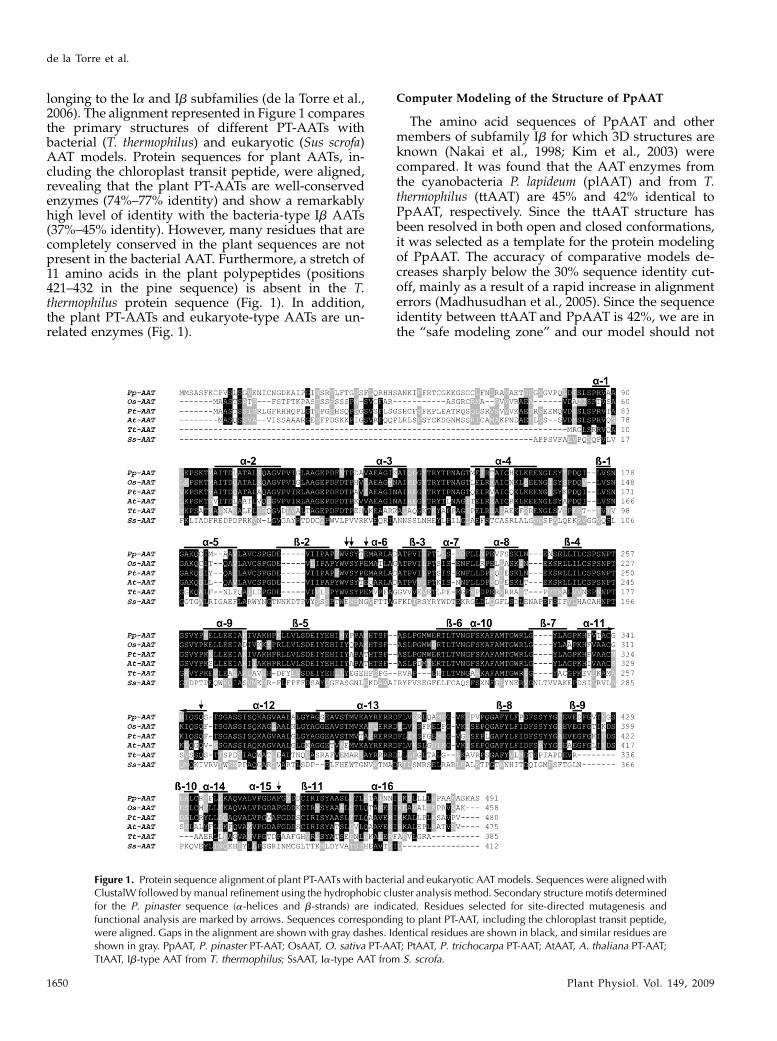
longing to the Ia and Ib subfamilies (de la Torre et al.,2006). The alignment represented in Figure 1 comparesthe primary structures of different PT-AATs withbacterial (T. thermophilus) and eukaryotic (Sus scrofa)AAT models. Protein sequences for plant AATs, in-cluding the chloroplast transit peptide, were aligned,revealing that the plant PT-AATs are well-conservedenzymes (74%–77% identity) and show a remarkablyhigh level of identity with the bacteria-type Ib AATs(37%–45% identity). However, many residues that arecompletely conserved in the plant sequences are notpresent in the bacterial AAT. Furthermore, a stretch of11 amino acids in the plant polypeptides (positions421–432 in the pine sequence) is absent in the T.thermophilus protein sequence (Fig. 1). In addition,the plant PT-AATs and eukaryote-type AATs are un-related enzymes (Fig. 1).
Computer Modeling of the Structure of PpAAT
The amino acid sequences of PpAAT and othermembers of subfamily Ib for which 3D structures areknown (Nakai et al., 1998; Kim et al., 2003) werecompared. It was found that the AAT enzymes fromthe cyanobacteria P. lapideum (plAAT) and from T.thermophilus (ttAAT) are 45% and 42% identical toPpAAT, respectively. Since the ttAAT structure hasbeen resolved in both open and closed conformations,it was selected as a template for the protein modelingof PpAAT. The accuracy of comparative models de-creases sharply below the 30% sequence identity cut-off, mainly as a result of a rapid increase in alignmenterrors (Madhusudhan et al., 2005). Since the sequenceidentity between ttAAT and PpAAT is 42%, we are inthe “safe modeling zone” and our model should not
Figure 1. Protein sequence alignment of plant PT-AATs with bacterial and eukaryotic AAT models. Sequences were aligned withClustalW followed by manual refinement using the hydrophobic cluster analysis method. Secondary structure motifs determinedfor the P. pinaster sequence (a-helices and b-strands) are indicated. Residues selected for site-directed mutagenesis andfunctional analysis are marked by arrows. Sequences corresponding to plant PT-AAT, including the chloroplast transit peptide,were aligned. Gaps in the alignment are shown with gray dashes. Identical residues are shown in black, and similar residues areshown in gray. PpAAT, P. pinaster PT-AAT; OsAAT, O. sativa PT-AAT; PtAAT, P. trichocarpa PT-AAT; AtAAT, A. thaliana PT-AAT;TtAAT, Ib-type AAT from T. thermophilus; SsAAT, Ia-type AAT from S. scrofa.
de la Torre et al.
1650 Plant Physiol. Vol. 149, 2009
have significant errors, assuming a standard align-ment. Nevertheless, a manually created alignment wasused for the initial modeling. The complete sequencefor PpAATwas larger than that of the template ttAAT.Therefore, the plastid-targeting peptide plus 15 resi-dues in the PpAAT N terminus and 10 residues in theC terminus were not included in the model. The regionunder analysis consisted of the amino acid residuesfrom Asp-81 to Leu-478 in the PpAAT primary se-quence. Along this polypeptide fragment, PpAAT andttAATare 42% identical. To achieve a reliable model ofthe quaternary structure for both the apoenzyme andPpAAT in the internal aldimine form, we relaxed thesystem via energy minimization and molecular dy-namics simulations over 1 ns. Based on this, we haveobtained the basic structure that will be used forfurther spectroscopic and mutagenesis studies.There is no resolved structure for plant type IbAAT,
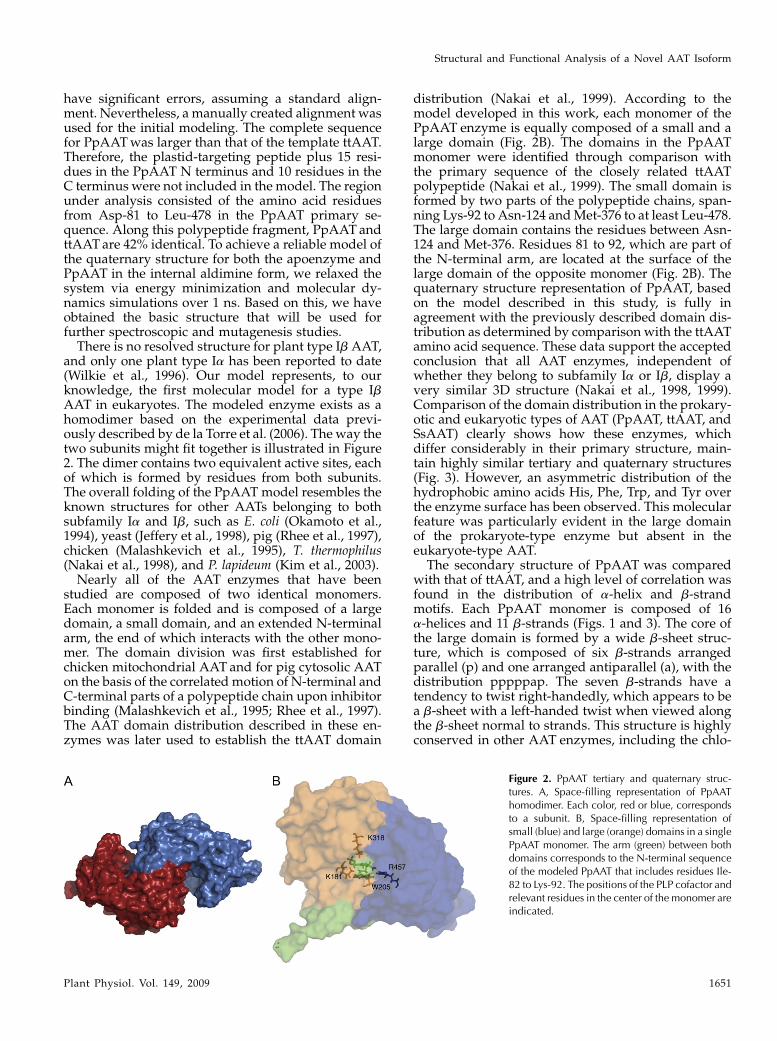
and only one plant type Ia has been reported to date(Wilkie et al., 1996). Our model represents, to ourknowledge, the first molecular model for a type IbAAT in eukaryotes. The modeled enzyme exists as ahomodimer based on the experimental data previ-ously described by de la Torre et al. (2006). The way thetwo subunits might fit together is illustrated in Figure2. The dimer contains two equivalent active sites, eachof which is formed by residues from both subunits.The overall folding of the PpAAT model resembles theknown structures for other AATs belonging to bothsubfamily Ia and Ib, such as E. coli (Okamoto et al.,1994), yeast (Jeffery et al., 1998), pig (Rhee et al., 1997),chicken (Malashkevich et al., 1995), T. thermophilus(Nakai et al., 1998), and P. lapideum (Kim et al., 2003).Nearly all of the AAT enzymes that have been
studied are composed of two identical monomers.Each monomer is folded and is composed of a largedomain, a small domain, and an extended N-terminalarm, the end of which interacts with the other mono-mer. The domain division was first established forchicken mitochondrial AAT and for pig cytosolic AATon the basis of the correlated motion of N-terminal andC-terminal parts of a polypeptide chain upon inhibitorbinding (Malashkevich et al., 1995; Rhee et al., 1997).The AAT domain distribution described in these en-zymes was later used to establish the ttAAT domain
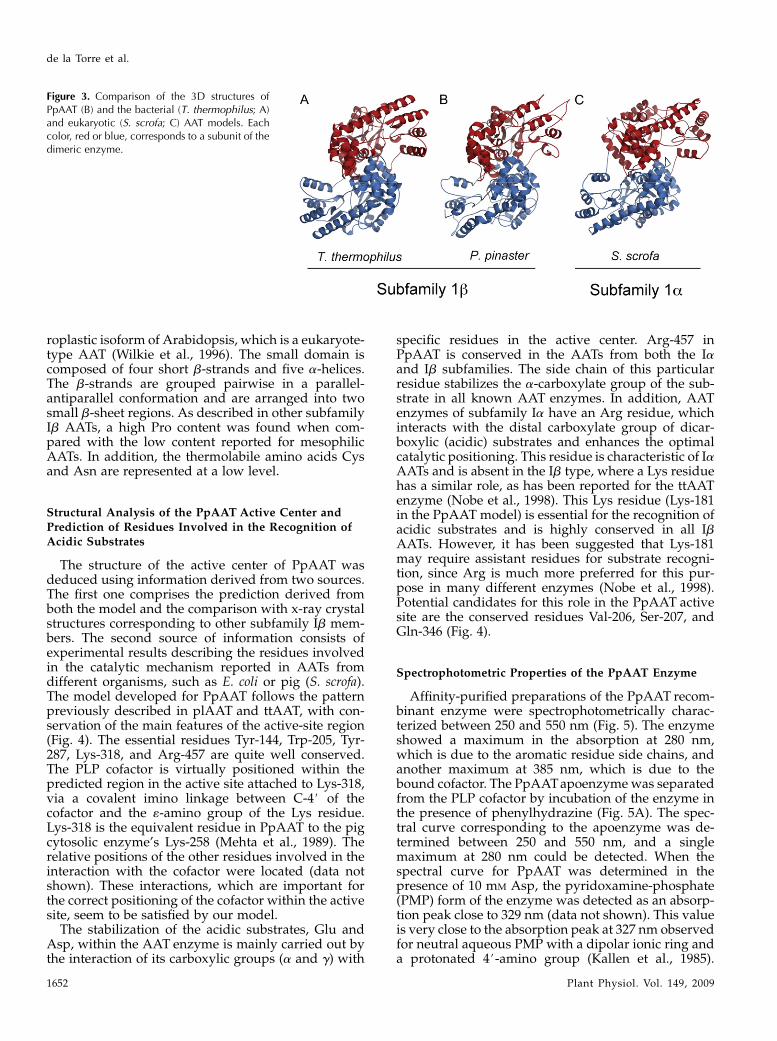
distribution (Nakai et al., 1999). According to themodel developed in this work, each monomer of thePpAAT enzyme is equally composed of a small and alarge domain (Fig. 2B). The domains in the PpAATmonomer were identified through comparison withthe primary sequence of the closely related ttAATpolypeptide (Nakai et al., 1999). The small domain isformed by two parts of the polypeptide chains, span-ning Lys-92 to Asn-124 andMet-376 to at least Leu-478.The large domain contains the residues between Asn-124 and Met-376. Residues 81 to 92, which are part ofthe N-terminal arm, are located at the surface of thelarge domain of the opposite monomer (Fig. 2B). Thequaternary structure representation of PpAAT, basedon the model described in this study, is fully inagreement with the previously described domain dis-tribution as determined by comparison with the ttAATamino acid sequence. These data support the acceptedconclusion that all AAT enzymes, independent ofwhether they belong to subfamily Ia or Ib, display avery similar 3D structure (Nakai et al., 1998, 1999).Comparison of the domain distribution in the prokary-otic and eukaryotic types of AAT (PpAAT, ttAAT, andSsAAT) clearly shows how these enzymes, whichdiffer considerably in their primary structure, main-tain highly similar tertiary and quaternary structures(Fig. 3). However, an asymmetric distribution of thehydrophobic amino acids His, Phe, Trp, and Tyr overthe enzyme surface has been observed. This molecularfeature was particularly evident in the large domainof the prokaryote-type enzyme but absent in theeukaryote-type AAT.
The secondary structure of PpAAT was comparedwith that of ttAAT, and a high level of correlation wasfound in the distribution of a-helix and b-strandmotifs. Each PpAAT monomer is composed of 16a-helices and 11 b-strands (Figs. 1 and 3). The core ofthe large domain is formed by a wide b-sheet struc-ture, which is composed of six b-strands arrangedparallel (p) and one arranged antiparallel (a), with thedistribution pppppap. The seven b-strands have atendency to twist right-handedly, which appears to bea b-sheet with a left-handed twist when viewed alongthe b-sheet normal to strands. This structure is highlyconserved in other AAT enzymes, including the chlo-
Figure 2. PpAAT tertiary and quaternary struc-tures. A, Space-filling representation of PpAAThomodimer. Each color, red or blue, correspondsto a subunit. B, Space-filling representation ofsmall (blue) and large (orange) domains in a singlePpAAT monomer. The arm (green) between bothdomains corresponds to the N-terminal sequenceof the modeled PpAAT that includes residues Ile-82 to Lys-92. The positions of the PLP cofactor andrelevant residues in the center of the monomer areindicated.
Structural and Functional Analysis of a Novel AAT Isoform
Plant Physiol. Vol. 149, 2009 1651
roplastic isoform of Arabidopsis, which is a eukaryote-type AAT (Wilkie et al., 1996). The small domain iscomposed of four short b-strands and five a-helices.The b-strands are grouped pairwise in a parallel-antiparallel conformation and are arranged into twosmall b-sheet regions. As described in other subfamilyIb AATs, a high Pro content was found when com-pared with the low content reported for mesophilicAATs. In addition, the thermolabile amino acids Cysand Asn are represented at a low level.
Structural Analysis of the PpAAT Active Center andPrediction of Residues Involved in the Recognition ofAcidic Substrates
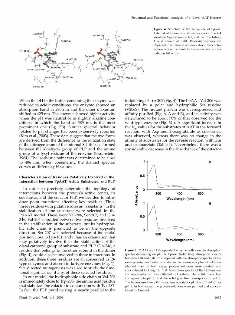
The structure of the active center of PpAAT wasdeduced using information derived from two sources.The first one comprises the prediction derived fromboth the model and the comparison with x-ray crystalstructures corresponding to other subfamily Ib mem-bers. The second source of information consists ofexperimental results describing the residues involvedin the catalytic mechanism reported in AATs fromdifferent organisms, such as E. coli or pig (S. scrofa).The model developed for PpAAT follows the patternpreviously described in plAAT and ttAAT, with con-servation of the main features of the active-site region(Fig. 4). The essential residues Tyr-144, Trp-205, Tyr-287, Lys-318, and Arg-457 are quite well conserved.The PLP cofactor is virtually positioned within thepredicted region in the active site attached to Lys-318,via a covalent imino linkage between C-4# of thecofactor and the «-amino group of the Lys residue.Lys-318 is the equivalent residue in PpAAT to the pigcytosolic enzyme’s Lys-258 (Mehta et al., 1989). Therelative positions of the other residues involved in theinteraction with the cofactor were located (data notshown). These interactions, which are important forthe correct positioning of the cofactor within the activesite, seem to be satisfied by our model.
The stabilization of the acidic substrates, Glu andAsp, within the AAT enzyme is mainly carried out bythe interaction of its carboxylic groups (a and g) with
specific residues in the active center. Arg-457 inPpAAT is conserved in the AATs from both the Iaand Ib subfamilies. The side chain of this particularresidue stabilizes the a-carboxylate group of the sub-strate in all known AAT enzymes. In addition, AATenzymes of subfamily Ia have an Arg residue, whichinteracts with the distal carboxylate group of dicar-boxylic (acidic) substrates and enhances the optimalcatalytic positioning. This residue is characteristic of IaAATs and is absent in the Ib type, where a Lys residuehas a similar role, as has been reported for the ttAATenzyme (Nobe et al., 1998). This Lys residue (Lys-181in the PpAAT model) is essential for the recognition ofacidic substrates and is highly conserved in all IbAATs. However, it has been suggested that Lys-181may require assistant residues for substrate recogni-tion, since Arg is much more preferred for this pur-pose in many different enzymes (Nobe et al., 1998).Potential candidates for this role in the PpAAT activesite are the conserved residues Val-206, Ser-207, andGln-346 (Fig. 4).
Spectrophotometric Properties of the PpAAT Enzyme
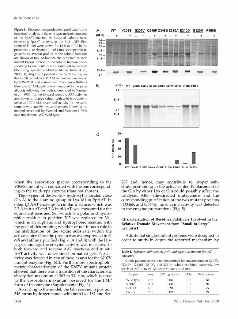
Affinity-purified preparations of the PpAAT recom-binant enzyme were spectrophotometrically charac-terized between 250 and 550 nm (Fig. 5). The enzymeshowed a maximum in the absorption at 280 nm,which is due to the aromatic residue side chains, andanother maximum at 385 nm, which is due to thebound cofactor. The PpAATapoenzymewas separatedfrom the PLP cofactor by incubation of the enzyme inthe presence of phenylhydrazine (Fig. 5A). The spec-tral curve corresponding to the apoenzyme was de-termined between 250 and 550 nm, and a singlemaximum at 280 nm could be detected. When thespectral curve for PpAAT was determined in thepresence of 10 mM Asp, the pyridoxamine-phosphate(PMP) form of the enzyme was detected as an absorp-tion peak close to 329 nm (data not shown). This valueis very close to the absorption peak at 327 nm observedfor neutral aqueous PMP with a dipolar ionic ring anda protonated 4#-amino group (Kallen et al., 1985).
Figure 3. Comparison of the 3D structures ofPpAAT (B) and the bacterial (T. thermophilus; A)and eukaryotic (S. scrofa; C) AAT models. Eachcolor, red or blue, corresponds to a subunit of thedimeric enzyme.
de la Torre et al.
1652 Plant Physiol. Vol. 149, 2009
When the pH in the buffer containing the enzyme wasreduced to acidic conditions, the enzyme showed anabsorption band at 280 nm and the other maximumshifted to 425 nm. The enzyme showed higher activitywhen the pH was neutral or in slightly alkaline con-ditions, in which the band at 385 nm is the mostprominent one (Fig. 5B). Similar spectral behaviorrelated to pH changes has been extensively reported(Kim et al., 2003). These data suggest that the two formsare derived from the difference in the ionization stateof the nitrogen atom of the internal Schiff base formedbetween the aldehyde group of PLP and the aminogroup of a lysyl residue of the enzyme (Braunstein,1964). The isosbestic point was determined to be closeto 406 nm, when considering the distinct spectralcurves at different pH values.
Characterization of Residues Putatively Involved in the
Interaction between PpAAT, Acidic Substrates, and PLP
In order to precisely determine the topology ofinteractions between the protein’s active center, itssubstrates, and the cofactor PLP, we decided to intro-duce point mutations affecting key residues. Thus,three residues with putative roles as “assistants” in thestabilization of the substrate were selected in thePpAAT model. These were Val-206, Ser-207, and Gln-346. Val-206 is located between two residues involvedin the stabilization of the substrate, but its hydropho-bic side chain is predicted to be in the oppositedirection. Ser-207 was selected because of its spatialposition close to Lys-181, and it has an orientation thatmay putatively involve it in the stabilization of thedistal carboxyl group of substrate and PLP. Gln-346, aresidue that belongs to the other subunit in the dimer(Fig. 4), could also be involved in these interactions. Inaddition, these three residues are all conserved in Ib-type enzymes and absent in Ia-type enzymes (Fig. 1).Site-directed mutagenesis was used to study the func-tional significance, if any, of these selected residues.In our model, the hydrophobic side chain of Val-206
is immediately close to Trp-205, the amino acid residuethat stabilizes the cofactor in conjunction with Tyr-287.In fact, the PLP pyridine ring is nearly parallel to the
indole ring of Trp-205 (Fig. 4). The PpAAT Val-206 wasreplaced by a polar and hydrophilic Ser residue(V206S). The mutant protein was overexpressed andaffinity purified (Fig. 6, A and B), and its activity wasdetermined to be about 70% of that observed for thewild-type enzyme (Fig. 6C). A significant increase inthe Km values for the substrates of AAT in the forwardreaction, with Asp and 2-oxoglutarate as substrates,was observed, whereas there was no change in theaffinity of substrates for the reverse reaction, with Gluand oxaloacetate (Table I). Nevertheless, there was aconsiderable decrease in the absorbance of the cofactor
Figure 4. Structure of the active site of PpAAT.External aldimines are shown as sticks. The C4substrate Asp is shown at left, and the C5 substrateGlu is shown at right. Relevant residues aredepicted in wireframe representation. The contri-bution of each subunit to the active site is indi-cated as (A) or (B).
Figure 5. PpAAT is a PLP-dependent enzyme with variable absorptionspectra depending on pH. A, PpAAT (solid line) absorption spectrabetween 250 and 550 nm compared with the absorption spectra of thesame protein previously incubated in the presence of phenylhydrazine(dashed line). In both cases, protein solutions were purified andconcentrated to 1 mg mL21. B, Absorption spectra of the PLP enzymeare represented at two different pH values. The solid black linecorresponds to pH 5, and the solid gray line corresponds to pH 8.The buffers used were 0.1 M sodium acetate for pH 5 and Tris-HCl forpH 8. In both cases, the protein solutions were purified and concen-trated to 1 mg mL21.
Structural and Functional Analysis of a Novel AAT Isoform
Plant Physiol. Vol. 149, 2009 1653
when the absorption spectra corresponding to theV206Smutant was comparedwith the one correspond-ing to the wild-type enzyme (data not shown).
The oxygen of the Ser-207 hydroxyl is located close(2.6 A) to the «-amino group of Lys-181 in PpAAT. Inother Ib AAT enzymes, a similar distance, which was2.7 A in ttAATand 3 A in plAAT, was measured for theequivalent residues. Ser, which is a polar and hydro-philic residue, in position 207 was replaced by Val,which is an aliphatic and hydrophobic residue, withthe goal of determining whether or not it has a role inthe stabilization of the acidic substrate within theactive center. Once the protein was overexpressed in E.coli and affinity purified (Fig. 6, A and B) with the His-tag technology, the enzyme activity was measured inboth forward and reverse AAT reactions and in situAAT activity was determined on native gels. No ac-tivity was detected in any of these assays for the S207Vmutant enzyme (Fig. 6C). Furthermore spectrophoto-metric characterization of the S207V mutant proteinshowed that there was a transition of the characteristicabsorption maximum at 383 to 331 nm, which is closeto the absorption maximum observed for the PMPform of the enzyme (Supplemental Fig. 1).
According to the model, the Gln residue in position346 forms hydrogen bonds with both Lys-181 and Ser-
207 and, hence, may contribute to proper sub-strate positioning in the active center. Replacement ofthe Gln by either Lys or Glu could possibly affect thecatalysis. After site-directed mutagenesis and thecorresponding purification of the two mutant proteins(Q346K and Q346E), no enzyme activity was detectedin the enzyme preparations (Fig. 5).
Characterization of Residues Putatively Involved in theRelative Domain Movement from “Small to Large”in PpAAT
Additional single-mutant proteins were designed inorder to study in depth the reported mechanism by
Figure 6. Recombinant production, purification, andfunctional analysis of the wild type and point mutantsof the PpAAT enzyme. A, Bacterial cultures over-expressing PpAAT proteins in the BL21 DE3 PLysstrain of E. coli were grown for 16 h at 10�C in thepresence (+) or absence (2) of 1 mM isopropylthio-b-galactoside. Protein profiles of the soluble fractionsare shown at top. At bottom, the presence of eachmutant PpAAT protein in the soluble fraction corre-sponding to each culture was confirmed by westernblot using specific antibodies (de la Torre et al.,2006). B, Aliquots of purified enzyme (0.2–2 mg) forthe wild type and each PpAATmutant were separatedby SDS-PAGE and stained with Coomassie BrilliantBlue dye. C, AAT activity was measured in the samealiquots following the method described by Karmenet al. (1955) for the forward reaction. AAT activitiesare shown as relative values, with wild-type activitytaken as 100% (5.6 nkat). AAT activity for the samesamples was equally measured on gels following themethod described by Wendel and Weeden (1989;data not shown). WT, Wild type.
Table I. Substrate affinities (Km) of wild-type and mutant PpAATenzymes
Kinetic parameters were not determined for enzyme mutants S207V,Q346K, Q346E, E210A, and E210K, which exhibited extremely lowlevels of AAT activity. All given values are in mM.
Enzyme L-Asp 2-Oxoglutarate L-Glu Oxaloacetate
Wild type 2.50 0.08 1.0 0.18V206S 14.00 0.60 1.0 0.20E210D 5.3 0.20 1.3 0.55F450S 2.50 0.09 1.0 0.19
de la Torre et al.
1654 Plant Physiol. Vol. 149, 2009
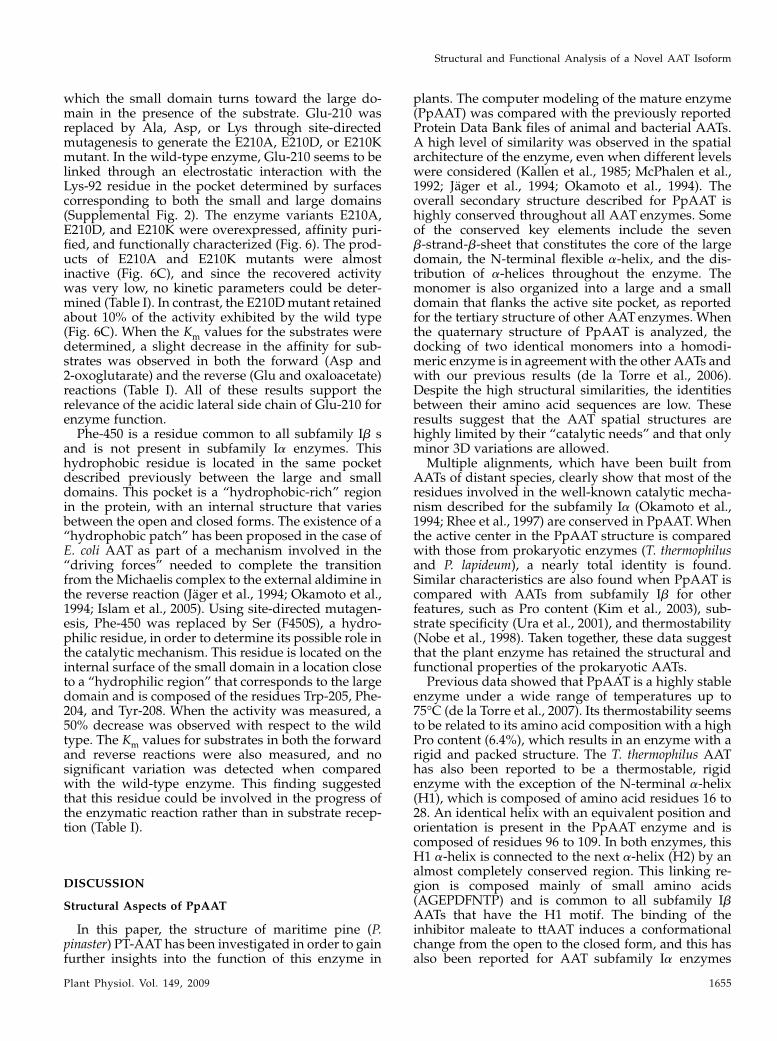
which the small domain turns toward the large do-main in the presence of the substrate. Glu-210 wasreplaced by Ala, Asp, or Lys through site-directedmutagenesis to generate the E210A, E210D, or E210Kmutant. In the wild-type enzyme, Glu-210 seems to belinked through an electrostatic interaction with theLys-92 residue in the pocket determined by surfacescorresponding to both the small and large domains(Supplemental Fig. 2). The enzyme variants E210A,E210D, and E210K were overexpressed, affinity puri-fied, and functionally characterized (Fig. 6). The prod-ucts of E210A and E210K mutants were almostinactive (Fig. 6C), and since the recovered activitywas very low, no kinetic parameters could be deter-mined (Table I). In contrast, the E210Dmutant retainedabout 10% of the activity exhibited by the wild type(Fig. 6C). When the Km values for the substrates weredetermined, a slight decrease in the affinity for sub-strates was observed in both the forward (Asp and2-oxoglutarate) and the reverse (Glu and oxaloacetate)reactions (Table I). All of these results support therelevance of the acidic lateral side chain of Glu-210 forenzyme function.Phe-450 is a residue common to all subfamily Ib s
and is not present in subfamily Ia enzymes. Thishydrophobic residue is located in the same pocketdescribed previously between the large and smalldomains. This pocket is a “hydrophobic-rich” regionin the protein, with an internal structure that variesbetween the open and closed forms. The existence of a“hydrophobic patch” has been proposed in the case ofE. coli AAT as part of a mechanism involved in the“driving forces” needed to complete the transitionfrom the Michaelis complex to the external aldimine inthe reverse reaction (Jager et al., 1994; Okamoto et al.,1994; Islam et al., 2005). Using site-directed mutagen-esis, Phe-450 was replaced by Ser (F450S), a hydro-philic residue, in order to determine its possible role inthe catalytic mechanism. This residue is located on theinternal surface of the small domain in a location closeto a “hydrophilic region” that corresponds to the largedomain and is composed of the residues Trp-205, Phe-204, and Tyr-208. When the activity was measured, a50% decrease was observed with respect to the wildtype. The Km values for substrates in both the forwardand reverse reactions were also measured, and nosignificant variation was detected when comparedwith the wild-type enzyme. This finding suggestedthat this residue could be involved in the progress ofthe enzymatic reaction rather than in substrate recep-tion (Table I).
DISCUSSION
Structural Aspects of PpAAT
In this paper, the structure of maritime pine (P.pinaster) PT-AAT has been investigated in order to gainfurther insights into the function of this enzyme in
plants. The computer modeling of the mature enzyme(PpAAT) was compared with the previously reportedProtein Data Bank files of animal and bacterial AATs.A high level of similarity was observed in the spatialarchitecture of the enzyme, even when different levelswere considered (Kallen et al., 1985; McPhalen et al.,1992; Jager et al., 1994; Okamoto et al., 1994). Theoverall secondary structure described for PpAAT ishighly conserved throughout all AAT enzymes. Someof the conserved key elements include the sevenb-strand-b-sheet that constitutes the core of the largedomain, the N-terminal flexible a-helix, and the dis-tribution of a-helices throughout the enzyme. Themonomer is also organized into a large and a smalldomain that flanks the active site pocket, as reportedfor the tertiary structure of other AAT enzymes. Whenthe quaternary structure of PpAAT is analyzed, thedocking of two identical monomers into a homodi-meric enzyme is in agreement with the other AATs andwith our previous results (de la Torre et al., 2006).Despite the high structural similarities, the identitiesbetween their amino acid sequences are low. Theseresults suggest that the AAT spatial structures arehighly limited by their “catalytic needs” and that onlyminor 3D variations are allowed.
Multiple alignments, which have been built fromAATs of distant species, clearly show that most of theresidues involved in the well-known catalytic mecha-nism described for the subfamily Ia (Okamoto et al.,1994; Rhee et al., 1997) are conserved in PpAAT. Whenthe active center in the PpAAT structure is comparedwith those from prokaryotic enzymes (T. thermophilusand P. lapideum), a nearly total identity is found.Similar characteristics are also found when PpAAT iscompared with AATs from subfamily Ib for otherfeatures, such as Pro content (Kim et al., 2003), sub-strate specificity (Ura et al., 2001), and thermostability(Nobe et al., 1998). Taken together, these data suggestthat the plant enzyme has retained the structural andfunctional properties of the prokaryotic AATs.
Previous data showed that PpAAT is a highly stableenzyme under a wide range of temperatures up to75�C (de la Torre et al., 2007). Its thermostability seemsto be related to its amino acid composition with a highPro content (6.4%), which results in an enzyme with arigid and packed structure. The T. thermophilus AAThas also been reported to be a thermostable, rigidenzyme with the exception of the N-terminal a-helix(H1), which is composed of amino acid residues 16 to28. An identical helix with an equivalent position andorientation is present in the PpAAT enzyme and iscomposed of residues 96 to 109. In both enzymes, thisH1 a-helix is connected to the next a-helix (H2) by analmost completely conserved region. This linking re-gion is composed mainly of small amino acids(AGEPDFNTP) and is common to all subfamily IbAATs that have the H1 motif. The binding of theinhibitor maleate to ttAAT induces a conformationalchange from the open to the closed form, and this hasalso been reported for AAT subfamily Ia enzymes
Structural and Functional Analysis of a Novel AAT Isoform
Plant Physiol. Vol. 149, 2009 1655
(Nakai et al., 1999). This change does not affect thewhole small domain, as only the N-terminal regionthat is mainly composed of the a-helix H1 approachesin order to close the active site (Nakai et al., 1999). Thehighly conserved region linking H1 and H2 is com-posed of small amino acids that allow an efficientrotation due to the low rotation restriction around cand f angles. This region could be involved in the“open-to-closed” movement of the domains. Futuresstudies with substitution by site-directed mutagenesiswill help to determine the role of this region on thecatalytic mechanism.
A significant structural difference betweenmembersof subfamily Ib is the presence of 10 to 11 amino acidresidues in the plant PT-AATs, corresponding to posi-tions 421 to 432 in the PpAATsequence that are absentin bacterial AATs. This particular region was depictedin the model as a short helix that is located outside theactive center (Supplemental Fig. 3). Whether or notthis structural feature has a potential role in the plantPT-AATs will require further studies.
Substrate Recognition in the PpAAT Enzyme
Dicarboxylate substrates are recognized, stabilized,and correctly oriented into the AAT active centerthrough the interactions of their a- and g-carboxylgroups with the side chains of residues that are locatedwithin the “catalytic pocket.” The a-carboxyl group inthe substrate is recognized and stabilized by an Argresidue in all AATs that have been described to date.The interaction between the distal (g) carboxyl groupand the enzyme is carried out in subfamily Ia AATsthrough an Arg residue. Based on previous work thatdescribed the recognition of the distal carboxyl groupof acidic substrates into the T. thermophilus activecenter, it has been proposed that this role is mainlycarried out by a Lys residue that is exclusively foundin subfamily Ib AATs (Nobe et al., 1998). The equiv-alent residue has been determined to be Lys-181 inPpAAT. This residue has been designated as a majordeterminant of the acidic substrate specificity.
The “Ra” type has been described as the mode ofinteraction between an Arg side chain and a sub-strate’s carboxylic groups. This type of interactiondescribes the recognition of a-carboxylic groups in allAATs and the interaction with the distal carboxylicgroups in subfamily Ia. This interaction does not needadditional residues for correct charge compensationand it thought to be the most frequent type of inter-action in enzymes that use carboxylic substrates. Incontrast, the recognition of a carboxyl group through aLys residue has been described as extremely rare. Ithas been proposed that assistant residues would berequired to stabilize substrates in that case (Islam et al.,2005). Site-directed mutagenesis showed that the in-troduction of Arg-292 into the T. thermophilus AAT is akey step in the change of only the acidic substratespecificity in an enzyme that has dual acidic andhydrophobic substrate specificity (Ura et al., 2001).
We modeled the external aldimines for the C4 andC5 dicarboxylic substrates in the closed conformation,since it seems that the reaction proceeds when theenzyme is in its closed form. It is clear that the bindingof the C4 substrate induces a conformational change inthe enzyme from the open to the closed form, and itwas recently reported that, for the C5 substrate, thisconformational transition occurs from the open formin the Michaelis complex to the closed form in theexternal aldimine (Islam et al., 2005). Since our mainobjective was to increase our knowledge about theacidic substrate specificity of PpAAT, the vicinity ofLys-181 was analyzed in order to identify other aminoacid residues in the active center that could be impli-cated in substrate stabilization. The existence of ahydrophobic cavity around the Lys-181 area wasrevealed. The cavity is composed of the residuesPhe-450, Phe-204, Tyr-144 (opposite chain), Tyr-208,Tyr-329, Val-313, Val-188, Val-206, and Trp-205.
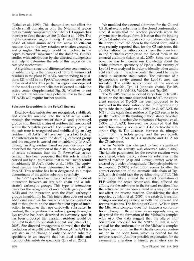
The Val-206 residue is located very close to Trp-205.Based on the structure described for ttAAT, the equiv-alent residue of Trp-205 has been proposed to beinvolved in the stabilization of the PLP pyridine ringby its side chain (Nakai et al., 1999). In E. coli AAT, theN(1) of the equivalent residue of Trp-205 seems to bepartly involved in the binding of the distal carboxylategroup of the dicarboxylic substrates (Hayashi et al.,1990). In PpAAT, however, we observed that thisinteraction mainly occurs with C4 dicarboxylic sub-strates (Fig. 4). The distances between the nitrogenatom from the indole group and the g-carboxylicgroup are 2.9 A for the C4 substrate and 4.1 A forthe C5 substrate.
When Val-206 was changed to Ser, a significantdecrease in the activity was observed (about 30%).The kinetic parameters for the reverse reaction werenot altered, while the Km values for substrates in theforward reaction (Asp and 2-oxoglutarate) were in-creased by 1 order of magnitude. The hydrophobic-to-hydrophilic (V206S) substitution seems to alter thecorrect orientation of the aromatic side chain of Trp-205, which should face the pyridine ring of PLP. Thissubstitution likely altered the correct orientation ofPLP within the active center and, thus, affected theaffinity for the substrates in the forward reaction. If so,the active center has been altered in a way that doesnot affect the reverse reaction. Considering the datareported by Islam et al. (2005), the catalytic transitionchanges are not equivalent in both the forward andreverse reactions. The binding of Glu to AATs to formthe Michaelis complex does not induce a conforma-tional change in the enzyme in an opposite way, asdescribed for the formation of the Michaelis complexwith Asp. Our data suggest that the altered PLPorientation proposed for the V206S mutant is morecritical for the conformation of the Michaelis complexin the closed form than the Michaelis complex confor-mation in the open form, which is needed for thereverse reaction. Another possible explanation for theasymmetric alteration of kinetic parameters can be
de la Torre et al.
1656 Plant Physiol. Vol. 149, 2009
related to the indirect alteration of the orientation ofLys-181 through the movement of Trp-205 that occursas a result of the V206S change. This option suggeststhat the orientation of Lys-181 could be influenced bythe hydrophobic pair Val-206/Trp-205 in the closed(Asp) form but not in the open (Glu) form.The Ser-207 and Gln-346 residues were also selected
as candidates to be analyzed, since they are uniqueresidues that are close to Lys-181 within the active site.Ser-207 is from the same subunit and Gln-346 is fromthe other one. Based on our PpAAT model, the dis-tance between the Ser-207 hydroxyl oxygen and thes-amino nitrogen is 2.7 A for the C4 substrate and 2.9A for the C5 substrate. Gln-346 directly interacts withSer-207 and, therefore, may contribute to substratestabilization. In order to study the importance of thesetwo residues in the stabilization of Lys-181 in thecorrect orientation, Ser-207 was replaced by a Val,whereas Gln-346 was changed to either a basic (Lys) oran acidic (Glu) residue. The replacement of Ser-207 bythe hydrophobic Val residue yielded a mutant proteinthat lacked the target bond that appears to be critical inthe stabilization of Lys-181. Similarly, the ability toestablish hydrogen bonds at position 346 is lacking inthe Q346K and Q346E mutants. The activity of theS207V, Q346K, and Q346E enzymes was completelyabolished in both forward and reverse AAT reactions
(Fig. 6). These data indicate that Ser-207 and Gln-346are essential assistant residues involved in the correctpositioning of Lys-181 and, therefore, involved insubstrate recognition.
The negatively charged residue Glu-210 was alsoselected for functional analysis, again using site-directed mutagenesis. The model structure predictsthat the negatively charged Glu-210 residue can elec-trostatically interact with the positive charge of a Lys-92 residue. These two residues, Glu-210 and Lys-92,are located in the large and small domains, respec-tively. Lys-92 is specifically positioned in the smalldomain, just beside the a-helix (H1) at residues 96 to109. Therefore, it is deeply involved in the open-to-closed conformational change, given that it is posi-tioned in the H6 a-helix of the large domain. Themutant proteins E210A and E210K showed a dramaticdecrease of AAT activity in relation to the wild-typeenzyme, which suggested that the acidic lateral sidechain of Glu-210 is required for enzyme activity. Thereplacement of Glu by Asp further supports the aboveassumption. Thus, the E210D mutant enzyme waspartially active, although its activity was much lowerthan in the wild type, and it exhibited decreasedaffinity for its substrates. Taken together, these datasuggest that alteration in the electrostatic interactionbetween Lys-92 and Glu-210 affects the ability of the
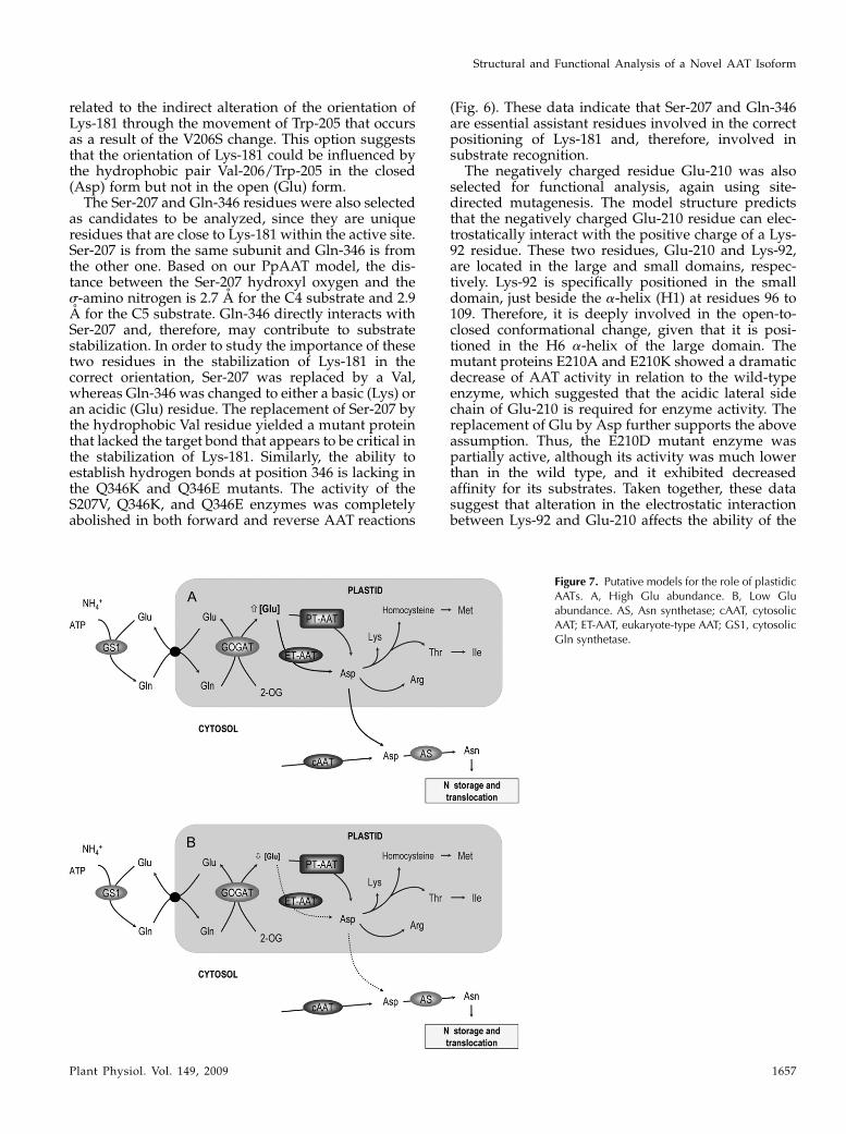
Figure 7. Putative models for the role of plastidicAATs. A, High Glu abundance. B, Low Gluabundance. AS, Asn synthetase; cAAT, cytosolicAAT; ET-AAT, eukaryote-type AAT; GS1, cytosolicGln synthetase.
Structural and Functional Analysis of a Novel AAT Isoform
Plant Physiol. Vol. 149, 2009 1657
enzyme to correctly develop the open-to-closed cata-lytic step. Further functional studies of the residues inthe H1 a-helix will be needed to gain a better under-standing about this critical mechanism that is charac-teristic of the subfamily Ib AATs.
Data derived from the characterization of the F450Smutant suggests that this well-conserved residue has arelevant but not critical role in the enzyme. The re-duced activity of the F450S mutant enzyme and the 3Dlocation of Phe-450 indicates that this hydrophobicresidue seems to be a member of a hydrophobic patchthat is located within the pocket formed between thelarge and small AAT domains. The existence of thesehydrophobic patches has been reported previously inAATenzymes (McPhalen et al., 1992, Jager et al., 1994).
Importance of Structural Studies to Understand theMetabolic Function of PT-AAT and
Biotechnological Applications
The presence of Lys-181, a residue with a shorterside chain than Arg, in the catalytic site of PpAATpossibly favors optimal interactions with the substrateGlu. This could explain why Lys rather than Arg isconserved at this particular position in the subfamilyIb AATs. We propose that Ser-207 and Gln-346 mayfunction as assistants to fix the orientation of the distalcarboxyl groups of the substrates. The structure of amore flexible catalytic site in the subfamily Ib AATenzymes could facilitate the recognition of Glu. Thesestructural features are consistent with the kinetic be-havior of the enzyme. Thus, the kinetic properties ofPpAAT for Asp and 2-oxoglutarate are quite similar tothose reported for the eukaryote-type AAT in plants.The affinity of PpAAT for Glu (Km = 1 mM; de la Torreet al., 2006), however, is much higher than thatreported for plastidic and other eukaryotic AATs,where the Km values range between 10 and 30 mM
(Taniguchi et al., 1995; Wilkie and Warren, 1998).Under conditions of steady-state photosynthesis, theconcentration of Glu in the chloroplast ranges between15 and 50mM (Riens et al., 1991;Winter et al., 1994), whichis well above the Km value of PpAAT for this substrate.In contrast, the concentration of 2-oxoglutarate hasbeen estimated to be in the micromolar range and evenlower in the chloroplast stroma, because the Glnsynthetase/GOGAT cycle represents a strong sink(Weber and Flugge, 2002). Consequently, a high Glu/2-oxoglutarate ratio in the stroma would drive bio-synthetic transaminations that favor Asp biosynthesis.Under these conditions, the eukaryotic and prokary-otic forms of plastidic AATwould be saturated and theflux toward Asp biosynthesis would be very highthrough the catalytic action of both enzymes (Fig. 7A).However, under physiological conditions in whichGlu abundance is low (e.g. limited nitrogen availabil-ity and other stressful conditions), only the PT-AATisoenzyme would ensure the biosynthesis of Asp-derived amino acids and other nitrogen compoundsin the plastids (Fig. 7B). A main pathway of Asp
utilization is the biosynthesis of the Asp-derivedamino acids Lys, Thr, Ile, and Met, all of which arerequired for protein synthesis and produced exclu-sively in the plastid (Azevedo et al., 2006). All of theseamino acids are essential in the nutrition of animals, inwhich PT-AAT does not exist (de la Torre et al., 2006).
In cyanobacteria, inorganic nitrogen is assimilatedthrough the Gln synthetase/GOGAT cycle and turnedinto Glu, which is then utilized as an amino donor byAAT and other transaminases for the biosynthesis ofnitrogen compounds (Muro-Pastor et al., 2005). PT-AAT could play a similar role in the chloroplasts ofhigher plants that utilize Glu for amino acid biosyn-thesis inside that organelle (Canovas et al., 2007). Theresults reported here suggest that the structure of thecatalytic site is adapted for the optimal utilization ofGlu for the biosynthesis of nitrogenous compounds.Molecular and physiological analysis of overexpress-ing and knockout lines of transgenic plants will berequired to determine the role of the enzyme in plantnitrogen metabolism. Pagnussat et al. (2005) reportedthe identification of 130 transposon mutants of Arabi-dopsis that have defects in female gametogenesis andembryo development. One of these lines, a mutantdefective in the At2g22250 locus, was affected in earlyembryo development,which indicates a potential role forPT-AAT in nitrogen metabolism during embryogenesis.
MATERIALS AND METHODS
Computer Modeling of the Spatial Structure of PpAAT
A homology model of Pinus pinasterAAT (residues 81–478) was built using
crystal structures of AAT from Thermus thermophilus as a template in both open
and closed conformations (Protein Data Bank accession nos. 1BJWand 1BKG,
respectively). The PpAAT sequence was isolated from a P. pinaster EST
database of different woody tissues in a previous work (de la Torre et al.,
2006). Alignments were performed with ClustalW (Thompson et al., 1994) and
manually edited. The whole PpAAT polypeptide fragment was modeled with
modeller 9v1 (Sali and Blundell, 1993) on a GNU/Linux box. Structure quality
and minor structural conflicts were evaluated using the Discrete Optimized
Protein Energy statistical potential within modeller (Shen and Sali, 2006). The
quaternary structure of the enzyme was built through comparison with that of
ttAAT following the steps described previously for other PLP-dependent
enzymes (Rodrıguez-Caso et al., 2003; Moya-Garcıa et al., 2005, 2008). External
aldimine was also modeled for both the C4 substrate Asp and the C5 substrate
Glu with PyMOL (DeLano, 2002a, 2002b). Using the structures of malic acid
and PMP as a scaffold, we built the PLP-Glu and PLP-Asp external aldimines
that were placed in the PpAAT active site by manual fitting and comparison
with the positions of PMP and malic acid in the active site of ttAAT x-ray
crystal structure. We built three different enzymatic systems: PpAAT in closed
conformation with PLP-Asp, PpAAT in closed conformation with PLP-Glu,
and PpAAT in open conformation (free enzyme). All of them were energy
minimized and equilibrated using Langevin-Verlet molecular dynamics sim-
ulations (NVT ensemble) over 1 ns at a temperature of 298 K. The FORTRAN
library DYNAMO (Field, 1999) and the OPLS force field were used to perform
every simulation.
Site-Directed Mutagenesis via PCR
The PpAAT sequence used in this study was previously inserted in the
NdeI-BamHI cloning site of pET-11a vector (de la Torre et al., 2006). Single
mutations were introduced into the cloned PpAAT using the QuickChange
site-directed mutagenesis kit (Stratagene). Reactions were carried out
using the following primer pairs: for V206S, 5#-CCAGCTCCATTTTG-
de la Torre et al.
1658 Plant Physiol. Vol. 149, 2009
GTCAAGCTATACTGAAATGGCTCGG-3# (sense) and 5#-CCGAGCCATT-
TCAGTATAGCTTGACCAAAATGGAGCTGG-3# (antisense); for S207V,
5#-CCAGCTCCATTTTGGGTAGTCTATACTGAAATGGCTCGG-3# (sense) and5#-CCGAGCCATTTCAGTATAGCATACCCAAAATGGAGCTGG-3# (antisense);
for E210K, 5#-CCATTTTGGGTAAGCTATACTAAAATGGCTCGGTTAGC-3#(sense) and 5#-GCTAACCGAGCCATTTTAGTATAGCTTACCCAAAATGG-3#(antisense); for E210A, 5#-GGTAAGCTATACTGCAATGGCTCGGTTAGCAG-
ATG-3# (sense) and 5#-CATCTGCTAACCGAGCCATTGCAGTATAGCTTACC-3#(antisense); for E210D, 5#-GGTAAGCTATACTGATATGGCTCGGTTAGCA-
GATG-3# (sense) and 5#-CATCTGCTAACCGAGCCATATCAGTATAGCTTACC-3#(antisense); for F450S, 5#-GTCCCAGGAGATGCCTCCGGCAATGATGACTGC-3#(sense) and 5#-GCAGTCATCATTGCCGGAGGCATCTCCTGGGAC-3# (antisense);for Q346K, 5#-GTGGAAGAATTCAGAGCAAGTCCACATCAGGTGC-3# (sense)and 5#-GCACCTGATGTGGACTTGCTCTGAATTCTTCCAC-3# (antisense); andfor Q346E, 5#-GTGGAAGAATTCAGAGCGAGTCCACATCAGGTGC-3# (sense)and 5#-GCACCTGATGTGGACTCGCTCTGAATTCTTCCAC-3# (antisense). Thepresence of the introduced mutations in the cDNA was confirmed by DNA
sequencing.
Expression and Purification of Recombinant Wild-Typeand Mutant Versions of PpAAT Enzymes
The optimal level of soluble recombinant PpAATwas previously reported
to be obtained with the following procedure. Briefly, the cDNA-coding region
for mature PpAAT was subcloned into the pET11a vector that included an
N-terminal 63His tag. Plasmid constructions were transformed in the Esch-
erichia coli strain BL21-codonPlus-(DE3)-RIL (Stratagene). Transformed cells
were grown at 37�C, with shaking in Luria-Bertani broth containing 100 mg
mL21 ampicillin and 34 mg mL21 chloramphenicol, until a cell density (optical
density at 600 nm) of 0.6 to 0.8 was reached. Flasks containing the cultures
were supplemented with isopropyl-b-D-thiogalactopyranoside at a final con-
centration of 1 mM. Cells were then cultured at 10�C for 14 h with vigorous
shaking. Cells were collected, and pellets were resuspended in a buffer
containing 50 mM Na2HPO4, 300 mM NaCl, and 10 mM imidazole buffer at
pH 8. Cells were lysed by sonication, and cell debris was removed by
centrifugation at 22,000g. Recombinant proteins were purified by affinity
chromatography using a nickel-nitrilotriacetic acid agarose column (Qiagen)
under native conditions. An identical procedure was successfully used to
overexpress and purify the PpAAT mutant versions V206S, S207V, Q346K,
Q346E, E210A, E210D, E210K, and F450S.
Determination of Enzyme Activities
AAT activity was determined for both forward and reverse reactions.
Direct reaction was determined by coupling the production of oxaloacetate
from Asp and 2-oxoglutarate to the oxidation of NADH with malate dehy-
drogenase (Yagi et al., 1985). Reactions were developed in a mix solution
containing 50 mM Tris-HCl, pH 7.8, 50 mM Asp, 10 mM 2-oxoglutarate, 0.07 mM
PLP, 0.1 mM NADH, 2 units of malate dehydrogenase (Roche Farma), and the
appropriate amount of enzyme in a final volume of 700 mL. The reaction was
started by the addition of 2-oxoglutarate. The decrease in the levels of NADH
was measured at 340 nm for a period of 120 s. The reverse reaction was
measured by coupling AAT activity to the reaction catalyzed by Glu dehy-
drogenase in a mixture containing 15 mM Glu, 1 mM oxaloacetate, 0.1 mM
NADH, 5 units of beef Glu dehydrogenase (Roche), 0.07 mM PLP, and 3.1 mM
NH4Cl in 60 mM potassium phosphate buffer, pH 7.5, at a final volume of
1 mL. The reaction was initiated by the addition of Glu and followed by the
decrease of NADH at 340 nm for a period of 120 s.
AAT Activity on Native Gels
AAT recombinant enzymes were electrophoresed under nondenaturing
conditions through a discontinuous, nondenaturing polyacrylamide gel with
a 5% polyacrylamide stacking gel (37.5:1 acrylamide:bisacrylamide, 125 mM
Tris-HCl, pH 6.8) and an 8% separating gel (37.5:1 acrylamide:bisacrylamide,
375 mM Tris-HCl, pH 8.8). The running buffer was 25 mM Tris-HCl and 250 mM
Gly, pH 8.3. The gels were run at 15 mA for 90 min at 4�C. They were then
placed in a bath containing 50 mL of AATsubstrate solution with low shaking
for 5 min. AAT activity was detected when the AAT substrate solution was
supplemented with 1mgmL21 Fast Blue (Sigma). The composition of the AAT
substrate solution (pH 7.4) was 2.2mM 2-oxoglutarate, 8.6 mMAsp, 0.5% (w/v)
polyvinylpyrrolidone-40, 1.7 mM EDTA, and 100 mM Na2HPO4 (Wendel and
Weeden, 1989).
Protein Electrophoresis and Western-Blot Analysis
Proteins were separated by gel electrophoresis under denaturing condi-
tions as described elsewhere (Canovas et al., 1991). Both electroblotting from
SDS gels to nitrocellulose membranes and immunodetection of AAT poly-
peptides were carried out as described elsewhere (de la Torre et al., 2002).
Phenylhydrazine Treatment
Pure PpAAT enzyme preparations were incubated at 50 mM phenylhy-
drazine (pH 7.4) at 37�C for 1 h, followed by gel filtration on a Sephadex G-25
column equilibrated with 50 mM potassium phosphate buffer, pH 7.4. Both
phenylhydrazine-treated and untreated protein samples were characterized
by spectrophotometric absorption between 250 and 550 nm.
Supplemental Data
The following materials are available in the online version of this article.
Supplemental Figure S1. Comparison of the absorption spectra of the
PpAATwild-type and S207V enzymes.
Supplemental Figure S2. Detailed view of the interaction between the
large and small domains in the PpAAT structure.
Supplemental Figure S3. Location in the PpAAT structure of the short
sequence stretch (10–11 amino acid residues) that is absent in bacterial
enzymes.
ACKNOWLEDGMENTS
We are grateful to Dr. S. Martı from the Universitat Jaume I and to Dr. J.
Ruiz-Pernıa from the Universitat de Valencia for their valuable advice. We
also thank the Unidad de Efectos del Medio from the Universitat de Valencia
for computational support.
Received December 17, 2008; accepted January 23, 2009; published January 28,
2009.
LITERATURE CITED
Azevedo RA, Lancien M, Lea PJ (2006) The aspartic acid metabolic
pathway, an exciting and essential pathway in plants. Amino Acids
30: 143–162
Braunstein AE (1964) Binding and reactions of the vitamin-B6 coenzyme
in the catalytic center of aspartate aminotransferase. Vitam Horm 22:
451–484
Canovas FM, Avila C, Canton FR, Canas RA, de la Torre F (2007)
Ammonium assimilation and amino acid metabolism in conifers.
J Exp Bot 58: 2307–2318
Canovas FM, Canton FR, Gallardo F, Garcıa-Gutierrez A, de Vicente A
(1991) Accumulation of glutamine synthetase during early development
of maritime pine (Pinus pinaster) seedlings. Planta 185: 372–378
DeLano WL (2002a) The PyMOL Molecular Graphics System. DeLano
Scientific, San Carlos, CA
DeLano WL (2002b) Unraveling hot spots in binding interfaces: progress
and challenges. Curr Opin Struct Biol 12: 14–20
de la Torre F, De Santis L, Suarez MF, Crespillo R, Canovas FM (2006)
Identification and functional analysis of a prokaryotic-type aspartate
aminotransferase: implications for plant amino acid metabolism. Plant J
46: 414–425
de la Torre F, Garcıa-Gutierrez A, Crespillo C, Canton FR, Avila C,
Canovas FM (2002) Functional expression of two pine glutamine syn-
thetase genes in bacteria reveals that they encode cytosolic isoenzymes
with different molecular and catalytic properties. Plant Cell Physiol 43:
802–809
de la Torre F, Suarez MF, Santis L, Canovas FM (2007) The aspartate am-
Structural and Functional Analysis of a Novel AAT Isoform
Plant Physiol. Vol. 149, 2009 1659
inotransferase family in conifers: biochemical analysis of a prokaryotic-
type enzyme from maritime pine. Tree Physiol 27: 1283–1291
Field MJ (1999) A Practical Introduction to the Simulation of Molecular
Systems. Cambridge University Press, New York
Gantt JS, Larson RJ, Farnham MW, Pathirana SM, Miller SS, Vance CP
(1992) Aspartate aminotransferase in effective and ineffective alfalfa
nodules: cloning of a cDNA and determination of enzyme activity,
protein, and mRNA levels. Plant Physiol 98: 868–878
Jeffery CJ, Barry T, Doonan S, Petsko GA, Ringe D (1998) Crystal
structure of Saccharomyces cerevisiae cytosolic aspartate aminotransfer-
ase. Protein Sci 7: 1380–1387
Hayashi H, Inoue Y, Kuramitsu S, Morino Y, Kagamiyama H (1990) Effects
of replacement of tryptophan-140 by phenylalanine or glycine on the
function of Escherichia coli aspartate aminotransferase. Biochem Biophys
Res Commun 167: 407–412
Islam MM, Goto M, Miyahara I, Ikushiro H, Hirotsu K, Hayashi H (2005)
Binding of C5-dicarboxylic substrate to aspartate aminotransferase:
implications for the conformational change at the transaldimination
step. Biochemistry 44: 8218–8229
Jager J, Moser M, Sauder U, Jansonius JN (1994) Crystal structures of
Escherichia coli aspartate aminotransferase in two conformations: com-
parison of an unliganded open and two liganded closed forms. J Mol
Biol 239: 285–305
Jensen RA, Gu W (1996) Evolutionary recruitment of biochemically spe-
cialized subdivisions of family I within the protein superfamily of
aminotransferases. J Bacteriol 178: 2161–2171
Kallen RG, Korpela T, Martell AE, Matsushima Y, Metzler CM, Metzler
DE, Morozov YV, Ralston IM, Savin FA, Torchinsky YM, et al (1985)
Chemical and spectroscopic properties of pyridoxal and pyridoxar-
amine phosphates. In P Christen, DE Metzler, eds, Transaminases. John
Wiley & Sons, New York, pp 215–234
Karmen A, Wroblewski F, Ladue JS (1955) Transaminase activity in human
blood. J Clin Invest 34: 126–131
Kim H, Ikegami K, Nakaoka M, Yagi M, Shibata H, Sawa Y (2003)
Characterization of aspartate aminotransferase from the cyanobacte-
rium Phormidium lapideum. Biosci Biotechnol Biochem 67: 490–498
Madhusudhan MS, Marti-Renom MA, Eswar N, John B, Pieper U,
Karchin R, Shen MY, Sali A (2005) Comparative protein structure
modeling. In JM Walker, ed, The Proteomics Protocols Handbook.
Humana Press, New York, pp 831–860
Malashkevich VN, Strokopytov BV, Borisov VV, Dauter Z, Wilson KS,
Torchinsky YM (1995) Crystal structure of the closed form of chicken
cytosolic aspartate aminotransferase at 1.9 A resolution. J Mol Biol 247:
111–124
McPhalen CA, Vincent MG, Picot D, Jansonius JN, Lesk AM, Chothia C
(1992) Domain closure in mitochondrial aspartate aminotransferase.
J Mol Biol 227: 197–213
Mehta PK, Hale TI, Christen P (1989) Evolutionary relationships among
aminotransferases: tyrosine aminotransferase, histidinol-phosphate
aminotransferase, and aspartate aminotransferase are homologous pro-
teins. Eur J Biochem 186: 249–253
Moya-Garcıa AA, Medina MA, Sanchez-Jimenez F (2005) Mammalian
histidine decarboxylase: from structure to function. Bioessays 27: 57–63
Moya-Garcıa AA, Ruiz-Pernıa J, Martı S, Sanchez-Jimenez F, Tunon I
(2008) Analysis of the decarboxylation step in mammalian histidine
decarboxylase: a computational study. J Biol Chem 283: 12393–12401
Muro-Pastor MI, Reyes JC, Florencio FJ (2005) Ammonium assimilation in
cyanobacteria. Photosynth Res 83: 135–150
Nakai T, Okada K, Akutsu S, Miyahara I, Kawaguchi S, Kato R,
Kuramitsu S, Hirotsu K (1999) Structure of Thermus thermophilus HB8
aspartate aminotransferase and its complex with maleate. Biochemistry
38: 2413–2424
Nakai T, Okada K, Kawaguchi S, Kato R, Kuramitsu S, Hirotsu K (1998)
Crystallization and preliminary x-ray characterization of aspartate
aminotransferase from an extreme thermophile, Thermus thermophilus
HB8. Acta Crystallogr D Biol Crystallogr 54: 1032–1034
Nobe Y, Kawaguchi S, Ura H, Nakai T, Hirotsu K, Kato R, Kuramitsu S
(1998) The novel substrate recognition mechanism utilized by aspartate
aminotransferase of the extreme thermophile Thermus thermophilusHB8.
J Biol Chem 273: 29554–29564
Okamoto A, Higuchi T, Hirotsu K, Kuramitsu S, Kagamiyama H (1994)
X-ray crystallographic study of pyridoxal 5#-phosphate-type aspartate
aminotransferases from Escherichia coli in open and closed form.
J Biochem 116: 95–107
Pagnussat GC, Yu HJ, Ngo QA, Rajani S, Mayalagu S, Johnson CS,
Capron A, Xie L-F, Ye D, Sundaresan V (2005) Genetic and molecular
identification of genes required for female gametophyte development
and function in Arabidopsis. Development 132: 603–614
Reynolds PH, Smith LA, Dickson JM, Jones WT, Jones SD, Rodber KA,
Carne A, Liddane CP (1992) Molecular cloning of a cDNA encoding
aspartate aminotransferase-P2 from lupin root nodules. Plant Mol Biol
19: 465–472
Rhee S, Silva MM, Hyde CC, Rogers PH, Metzler CM, Metzler DE,
Arnone A (1997) Refinement and comparisons of the crystal structures
of pig cytosolic aspartate aminotransferase and its complex with
2-methylaspartate. J Biol Chem 272: 17293–17302
Riens B, Lohaus G, Heineke D, Heldt HW (1991) Amino acid and sucrose
content determined in the cytosolic, chloroplastic, and vacuolar com-
partments and in the phloem sap of spinach leaves. Plant Physiol 97:
227–233
Rodrıguez-Caso C, Rodrıguez-Agudo D, Moya-Garcıa A, Fajardo I,
Medina MA, Subramaniam V, Sanchez-Jimenez F (2003) Local changes
in the catalytic site of mammalian histidine decarboxylase can affect its
global conformation and stability. Eur J Biochem 270: 4376–4387
Sali A, Blundell TL (1993) Comparative protein modelling by satisfaction
of spatial restraints. J Mol Biol 234: 779–815
Schultz CJ, Coruzzi GM (1995) The aspartate aminotransferase gene
family of Arabidopsis encodes isoenzymes localized to three distinct
subcellular compartments. Plant J 7: 61–75
Shen MY, Sali A (2006) Statistical potential for assessment and prediction
of protein structures. Protein Sci 15: 2507–2524
Taniguchi M, Kobe A, Kato M, Sugiyama T (1995) Aspartate aminotrans-
ferase isozymes in Panicum miliaceum L. and NAD-malic enzyme-type
C4 plant: comparison of enzymatic properties, primary structures and
expression patterns. Arch Biochem Biophys 318: 295–306
Thompson JD, Higgins DG, Gibson TJ (1994) CLUSTALW: improving the
sensitivity of progressive multiple sequence alignment through se-
quence weighting, position-specific gap penalties and weight matrix
choice. Nucleic Acids Res 22: 4673–4680
Turano FJ, Weisemann JM, Matthews BF (1992) Identification and expres-
sion of a cDNA clone encoding aspartate aminotransferase in carrot.
Plant Physiol 100: 374–381
Ura H, Nakai T, Kawaguchi SI, Miyahara I, Hirotsu K, Kuramitsu S (2001)
Substrate recognition mechanism of thermophilic dual-substrate en-
zyme. J Biochem 130: 89–98
Wadsworth GJ (1997) The plant aminotransferase gene family. Physiol
Plant 100: 998–1006
Wadsworth GW, Marmaras SM, Matthews BF (1993) Isolation and char-
acterization of a soybean cDNA clone encoding the plastid form of
aspartate aminotransferase. Plant Mol Biol 21: 993–1009
Weber A, Flugge UI (2002) Interaction of cytosolic and plastidic nitrogen
metabolism in plants. J Exp Bot 53: 865–874
Wendel JF, Weeden NF (1989) Visualization and interpretation of plant
isoenzymes. In DE Soltis, PS Soltis, eds, Isoenzymes in Plant Biology.
Dioscorides Press, Portland, OR, pp 5–45
Wilkie SE, Lambert R, Warren MJ (1996) Chloroplast aspartate amino-
transferase from Arabidopsis thaliana: an examination of the relationship
between the structure of the gene and the spatial structure of the
protein. Biochem J 319: 969–976
Wilkie SE, Roper JM, Smith AG, Warren MJ (1995) Isolation, charac-
terisation and expression of a cDNA clone encoding plastid aspar-
tate aminotransferase from Arabidopsis thaliana. Plant Mol Biol 27:
1227–1233
Wilkie SE, Warren MJ (1998) Recombinant expression, purification, and
characterization of three isoenzymes of aspartate aminotransferase from
Arabidopsis thaliana. Protein Expr Purif 12: 381–389
Winter H, Robinson DG, Heldt HW (1994) Subcellular volumes and
metabolite concentrations in spinach leaves. Planta 193: 530–535
Yagi T, Kagamiyama H, Nozaki M, Soda K (1985) Glutamate-aspartate
transaminase from microorganisms. Methods Enzymol 113: 83–89
de la Torre et al.
1660 Plant Physiol. Vol. 149, 2009