morphogenetic gradients in graptolites and bryozoans - journal · morphogenetic gradients in...
TRANSCRIPT

Morphogenetic gradients in graptolites and bryozoans
ADAM URBANEK
Urbanek, A. 2004. Morphogenetic gradients in graptolites and bryozoans. Acta Palaentologica Polonica 49 (4): 485–504.
Despite independent evolution of coloniality in hemichordates and bryozoans, their colonies show common features. Inboth instances colony is a genet or clonal system composed of zygotic oozooid and a number of blastozooids (= modules)integrated by physical continuity of tissues, sharing a common genotype and subject to common morphogenetic control.In some groups of graptolites and bryozoans, colonies display a regular morphological gradient. Simple graptoloid andbryozoan colonies consist of a proximal zone of astogenetic change and a distal zone of astogenetic repetition. Observedmorphological gradient may be attributed to diffusion, along the colony axis, of a morphogen produced by the oozooid; inthe zone of astogenetic change the morphogen is above certain threshold level and drops below it in the zone of asto−genetic repetition. This model is supported by observations on regeneration of fractured graptoloid colonies. Regenera−tive branch never displays astogenetic change. The same rule is valid for regeneration of fractured bryozoan colonies.While the early astogeny of simple bryozoan colonies may be explained within the framework of the gradient theory, thelate astogeny of more complex ones involves multiple succession of zones of change and repetition, without analogy inastogeny of graptoloids. Thus, late astogeny in bryozoan colonies may be controlled by cyclic somatic/reproductivechanges, probably independent of the primary morphogen. Evolutionary changes in the graptoloid colonies involve boththe spreading of the novelties over a greater number of zooids (penetrance) and an increase in the degree of phenotypicmanifestation of a given character (expressivity). In the phylogeny of bilaterian colonies morphogenetic gradient proba−bly originated as a sort of a side effect of sexual process leading to the appearance of the oozooid. The latter contaminatedthe neighbouring blastozooids with the products of its own morphogenesis. The resulting morphogenetic gradient couldbe used by selective forces to produce various effects of adaptive significance. Morphogens responsible for patterning ofbilaterian colonies are probably related to the products of genes responsible for the anteroposterior control of embryos inall solitary Bilateria (Hox, zootype genes).
Key words: Bilateria, graptolites, bryozoans, colonies, clones, morphogen, Hox genes.
Adam Urbanek [[email protected]], Institute of Palaeobiology, Polish Academy of Sciences, ul. Twarda 51/55,PL−00−818 Warszawa, Poland.
Introduction
Recent interest in the study of coloniality owes much to thework of Beklemishev (English translation 1969, and earlierRussian editions). Two important symposia edited byBoardman et al. (1973) and Larwood and Rosen (1979) fol−lowed many themes suggested by Beklemishev’s work andinitiated some new lines of discussion. Beklemishev (1951,1969) supplied also the modern foundation for studies onthe comparative anatomy and the development of bilateriancolonies.
The aim of the present paper is the integration of data andviews developed independently in the particular fields ofzoological research, concerning the origin and developmentof colonies in selected groups of bilaterian animals. Thestudy is focused on two groups: on one hand it is based onpterobranchs and closely related graptolites and on the otherhand on bryozoans. Pterobranchs indisputably belong toDeuterostomia (Nielsen 2001; Halanych 1995) and usuallyare placed in the phylum Hemichordata, while graptolites arevery closely related to Pterobranchia (Kozłowski 1949,1966; Urbanek and Dilly 2000). Recent authors believe that
both groups should constitute a common class—the Grapto−lithoidea (Beklemishev 1951, 1969; Urbanek 1986; Mie−rzejewski and Kulicki 2002). The systematic position ofBryozoa has been more controversial. They have either beengrouped with the members of Lophophorata at the base ofdeuterostomes (Zimmer 1973), or treated as a group of proto−stomes, displaying transient features to pterobranchs. Theyshare some anatomical similarities with this group of hemi−chordates (see also Stebbing 1970). Molecular data (18SrDNA) indicate, however, that lophophorates are proto−stomes and are related to mollusks and annelids to form aprovisional group of Lophotrochozoa (Halanych et. al 1995).Similarities with pterobranch hemichordates should be re−garded as convergence (Halanych 1996) or a result of paral−lel evolution from the common ancestors of all Bilateria. Re−cently, Nielsen (2002: 687) defined bryozoans as “the mostpuzzling phylum in phylogenetic studies of the Bilateria” butafter evaluation of the entirety of morphological data placedBryozoa with protostomes.
Therefore, it seems safe to conclude that colonialism de−veloped independently in the Graptolithoidea and Bryozoa,each group being related to a different superphylum of Bila−
http://app.pan.pl/acta49/app49−485.pdfActa Palaeontol. Pol. 49 (4): 485–504, 2004

teria. In this paper the Graptolithoidea (Pterobranchia +Graptolithina) are selected as a key model for comparativestudies because they provide the most graphic examples ofmorphogenesis in colonies and an unsurpassed record oftheir evolutionary changes. Some of my ideas on the earlyastogeny of graptolites and bryozoans were presented inNovember, 2000 on a seminar at the Palaeontological Insti−tute, Russian Academy of Sciences in Moscow and laterpublished (Urbanek 2003).
Graptolites as a model groupfor studies on bilaterian coloniesGraptolites are a fossil group of hemichordates that lived in theearly Palaeozoic, appearing 500 million years ago, and were
closely related to still living pterobranchs. Primitive Cambrianrepresentatives of the group were sessile, but their descen−dants, the true graptolites, known as graptoloids, wereplanktic, forming the predominant group of macrozoo−plankton (Figs. 1–3). In the Ordovician and Silurian–EarlyDevonian seas they were ubiquitous, being represented byrapidly evolving and extremely widely distributed species, apriceless tool for stratigraphic subdivision and intercontinentalcorrelation. Each individual (zooid) in the colony produced itsown tube (theca) made of some secreted scleroproteic material(collagen, Towe and Urbanek 1972) displaying a characteris−tic microstructure due to the presence of minute growth bandscalled fuselli. This material is capable of preservation in thefossil state. Such fossil skeletal remains, preserved in the formof carbonized stipes or branches, are the primary source of ourinformation concerning the structure and morphogenesis ofgraptolite colonies.
486 ACTA PALAEONTOLOGICA POLONICA 49 (4), 2004
prosicula
prosicula porus
metasicula
metasiculam
repent tube
repent tube
repent tube
cephalic shield
cephalic shield
lophophore
embrionicvesicle mm contractile stalk
stolon
branchof zooidal tube
stolotheca 1
stolotheca 2
autotheca 2
bitheca 2 mm
node
autothecal stolon
Fig. 1. Astogeny in Graptolithoidea. A–C. Early development of a rhabdopleurid pterobranch and a tuboid graptolite colony. A. Encapsulated larva aftermetamorphosis (A1) and primary zooid in Rhabdopleura compacta Hincks (A2). B, C. Comparison of sicular portions in Recent Rhabdopleura compacta(B) and in Ordovician tuboid graptolite Epigraptus Eisenack (C). D. Sicula (D1) and thecae with underlying stolon system (D2) of an Ordovician dendroidgraptolite Dendrograptus sp. E. Zooidal tubes and internal stolon system in Recent Rhabdopleura normani Allman. Not to scale. A, B, from Stebbing(1970), C–E, from Kozłowski (1949, 1970).

Colonies in the majority of sessile groups of graptolitesare marked with a distinct polymorphism, while the colo−nies of planktic graptolites, the Graptoloidea, are mono−morphic and have colonies composed of a single type of zo−oids and thecae. The basic pattern, which is almost univer−sal in sessile orders of Graptolithina, involves differentia−tion into thecae of three categories: autothecae, bithecae,and stolothecae. Autothecae are the largest and frequentlyhave an apertural apparatus. The bithecae and stolothecaeare much narrower, tubular, and devoid of any aperturalelaborations. The stolothecae carry inside a section of thestolon that divides at certain points within the parentalstolotheca (Fig. 1D2). In this way, the stolon produces shortbranches leading to a bitheca, an autotheca as well as to adaughter stolotheca that contains further extension of thestolon. The bithecae do not contain a stolon and usually areadnate to the adjacent autothecae. Kozłowski (1949) sug−gested probably the most suitable biological interpretationof thecal differentiation, assuming a distinct sexual dimor−phism among zooids: autothecae correspond to fully devel−oped female zooids, and bithecae housing partly reducedmale zooids. In addition to this classical view of Kozłowskione could assume that the male zooid in each triad had itssex phenotypically determined, in response for the presenceof juvenile female zooids in the stolotheca.
The reduction of male zooids observed in some sessilegroups of Graptolithina leads consequently to their elimina−tion in planktic graptoloids. Their thecae are composedsolely of autothecae, fused with their proximal portion corre−sponding to the former stolotheca. Transient forms with col−onies still preserving a few bithecae in their distal part areknown (so−called anisograptids). Elimination of bithecaewas preceded (according to Kozłowski 1949) by transforma−tion of female zooids into hermaphroditic individuals. There−fore, colonies of Graptoloidea were composed of hermaphro−ditic zooids, essentially monomorphic, but displaying to avarious degree the morphological gradient operating alongthe colony axis.
Early development of sessile graptolites (Dendroidea,Tuboidea) was best recognized and interpreted by Kozłow−ski (1949, 1963). The development of the colony starts withthe sicula, the zooidal tube of the founder zooid, which de−veloped from zygote (Fig. 1C, D1). It is composed of twoclearly distinct parts, which differ sharply in their micro−structure: a bottle−shaped or cylindrical prosicula, and a tu−bular metasicula. These differences were ascribed by Koz−łowski, who based his conclusions on bryozoan analogy, tometamorphosis of a free living larva, which produced firstthe prosicula, and later the metasicula. Thus, the siculahoused a siculozooid, the only sexually produced zooid inthe colony (= an oozooid). All remaining zooids originatedby budding from the siculozooid. In sessile graptolites it pro−ceeded from the stolon, which initially originated within theprosicula, and emerged from its cavity through an openingcalled the porus. After this, it penetrated into the initial orsicular stolotheca, inside which occured the first division of
the stolon. In the best studied dendroid and crustoid grapto−lites thecae were produced in triads, each being composed ofan autotheca, a stolotheca, and a bitheca.
The early development of graptolites was compared byKozłowski (1949) with that in rhabdopleurid pterobranchs,which was at that time inadequately known. The presence oftwo portions, homologous to prosicula (“embryonic vesi−cle”) and metasicula respectively was demonstrated, the lat−ter showing a characteristic “fusellar” structure due to com−position of peculiar growth bands. Later studies by Stebbing(1970) and Dilly (1973) provided more details (Fig. 1A). Thelarva encapsulates itself in a completely sealed vesicle madeof skeletal substance. After metamorphosis the juvenileoozooid breaks the wall of the vesicle (so−called perforatory
http://app.pan.pl/acta49/app49−485.pdf
URBANEK—MORPHOGENETIC GRADIENTS IN GRAPTOLITES AND BRYOZOANS 487
1
2
3
4
5
nema
sicula
Fig. 2. Diagram illustrating the structure and terminology of a proximal partin a monograptid colony composed of a series of zooids arranged along a sin−gle axis. Arrow indicates the direction of colony growth. Note the presence ofsicula, the zooidal tube of the oozooid, and a number of zooidal tubes (calledthecae, 1–5), and occupied by asexually produced zooids (blastozooids); thenema is a thread−like prolongation of the apex of sicula, serving as a skeletalaxis for the growing colony. Modified from Urbanek (1973).

budding) and starts to secrete the first growth bands of thezooidal tube. This tube is comparable to the metasicula,while the embryonal vesicle resembles the prosicula of ses−sile graptolites (Fig. 1B, C). In turn the first blastozooid alsobreaks the wall of the vesicle and starts to secrete its ownzooidal tube. The stolon system inside the zooidal tubes ofRhabdopleura is strikingly similar to the stolon systemrecognizad in crustoid, dendroid, and tuboid graptolites (Fig.1D2, E). A more detailed comparison of early developmentalstages in extant Rhabdopleura and tuboid and dendroidgraptolites was given by Urbanek (1986).
Astogeny in most groups of sessile graptolites involves amonotonous iteration of triads (or diads as in Tuboidea), allthecae showing the same size and shape. No polarity or mor−phological gradient has been observed except in Mastigo−graptus, a primitive sessile graptolite, occupying a transitorysystematic position between rhabdopleurid pterobranchs anddendroid graptolites. As revealed by Bates and Urbanek(2003), the stolothecae of Mastigograptus display a morpho−logical gradient in size and shape. However, the only largegroup, which exhibits, as a characteristic feature of its orga−nization, the polar organization of colonies is the plankticGraptoloidea. Thecae (corresponding to the autothecae fusedwith their stolothecal segments, see above) regularly in−crease in size distalwards, until they reach the distal typecharacteristic of zone of astogenetic repetition.
The gradient theory of graptoloidcoloniesThe most simple (although secondarily simplified, which isirrelevant to us) model of graptoloid colonies can be found inSilurian monograptids (Fig. 2). Their colonies consisted of asingle series of individuals interconnected by a string of tis−sue, homologous to the stolon of sessile graptolites but de−prived of a peridermal sheath. They developed by buddingfrom the founder−zooid—the sicula—resembling in essentialfeatures the sicula of sessile forms and composed of pro− andmetasicula. However, the graptoloid prosicula has many de−rived features: instead of producing a basal disc it ends withthe so−called nema, a thread made of skeletal substance andfrequently serving as the axis for the growing stipe. This re−flects a morpho−ecological revolution which occurred aftertransformation of sessile colonies into planktic ones.
There is a striking difference between the theca of this sex−ually−produced founder−zooid (the oozooid, Fig. 2, sicula) andthe thecae of all the remaining zooids, which are its progenyproduced by budding, that is asexually (Fig. 2, 1–5). A grapto−lite colony is therefore a clone. Consequently, all its members,varying in number from a few to several hundred, share thesame genotype. Though this is a fairly obvious conclusion, ithad never been formulated before Urbanek’s paper publishedin 1960. And what is more important, the consequences of theclonal nature of graptolite colonies had been overlooked. An
488 ACTA PALAEONTOLOGICA POLONICA 49 (4), 2004
57
42
35
22
14
10
72
1
5
12
? 30
Fig. 3. Variation of morphological characters of zooidal tubes (thecae)along the colony axis. A. Didymograptus pakrianus Jaanusson, only onebranch of the biramous colony presented. B. Monograptus clingani (Carru−thers). C. “Monograptus” (= Pernerograptus) argenteus (Nicholson). A, Bbelong to uniform type and exhibit mainly size gradient, while C representsa biform type, with distinct differences in morphology of proximal and dis−tal thecae. Not to scale. From Urbanek (1973).

important conclusion following from their clonality is under−standing the remarkable morphological differences within asingle colony merely as variation of the expression of the samegenotype. The same conclusion is tenable for bryozoan colo−nies, which likewise are clonal systems: all zooids representthe progeny of ancestrula produced by budding.
When tracing the degree of expression of a given charac−ter (e.g., the degree of curvature) in the successive thecae of asingle graptoloid colony we observe a regular, gradedchange—the expression is at its highest near the proximal(most frequently, Fig. 3C) or the distal (less frequently, Fig.4A) end of the colony, decreasing gradually towards the op−posite pole (Figs. 3C, 4A). This pattern of change is sugges−tive of a gradient in the distribution of the morphogen, whichcontrols the expression of a given gene or set of genes (Fig.5C, F). Such was the working hypothesis which I advancedfor the first time in my paper published in 1960, and elabo−rated subsequently in later years (Urbanek 1963, 1973; Urba−nek and Uchmański 1990).
All my considerations are based on the remarkable featureof the graptoloid colonies, namely their polar organization: zo−oids in every graptoloid colony gradually increase in sizedistalwards (Figs. 3A and 4B). The proximal portion of thecolony is composed of the smallest zooids, but their size grad−ually increases distalwards, until they reach a maximum at−tainable size in the distal portion of the colony. Further growthconsists of the iteration of the thecae of a uniform size andshape (Urbanek and Uchmański 1990). An immediate com−parison with simply organized bryozoan colonies comes tomind: in both cases astogeny reveals a proximal zone ofastogenetic change and a distal zone of astogenetic repetitionas defined by Boardman and Cheetham (1969). Likewisebryozoan colonies, every stipe of graptoloid colony ends witha growth zone displaying an ontogenetic gradient—growingthecae decrease in size distalwards. It seems that the majorityof monograptid colonies are open ended, i.e., they are capableof an endless addition of new zooids at the growing tip. How−ever, some graptoloids are definitely finite, and composed of adetermined, usually small number of zooids. Finite colonieshave their growth zones arrested at a certain stage, their thecaenever attain size corresponding to the thecae of the repetitionzone. A combination of zone of astogenetic change, with a rel−atively short zone of repetition followed by arrested growthzone, would result in a “foliate” shape of stipe, so characteris−tic of phyllograptid rhabdosomes (Urbanek 1973). In extremecases of colony reduction, exhibited by some retiolitids (e.g.,Holoretiolites), entire colony may be considered as an arrestedgrowth zone. Therefore, the size of the thecae decreasesdistalwards exhibiting a fixed ontogenetic gradient. Paper byKozłowska−Dawidziuk (2004) provides more data on the casein question.
The above mentioned size gradient is usually accompa−nied by a gradual change in the morphology of some thecalstructures, such as different apertural lobes or spines, or vari−ation in the curvature of the apertural part. In some grapto−loids, called technically “biform”, the thecae in the proximal
part of the colony manifest a sharp contrast with those in thedistal portion, but these differences are attained gradually byminute modifications in the structure of the successive thecae(Figs. 3C, 4A). This pattern of thecal variation within a single
http://app.pan.pl/acta49/app49−485.pdf
URBANEK—MORPHOGENETIC GRADIENTS IN GRAPTOLITES AND BRYOZOANS 489
Fig. 4. A. Variation of morphological characters of zooidal tubes (thecae)along the colony axis in Cucullograptus aversus Urbanek (A1), details ofstructure of apertural apparatus within the proximal, medial and distal thecae(A2–A4). B. Pristiograptus dubius (Suess), thecae with growth bands showndiagrammatically. A is a biform type with the strongest expression of charac−ters in the distal part of the colony, while B is a uniform type exhibitingmainly the size gradient. Not to scale. From Urbanek (1973).

graptoloid colony is strongly suggestive of their gradient na−ture. What is valid in this respect for monograptids is largelytenable for all graptoloids. My way of thinking was here in−fluenced by the classical ideas of physiological gradients for−mulated by Child (1915, 1941) and by some later works onthe morphogenesis and regeneration in hydroid colonies(corresponding sources were given in Urbanek 1973, andUrbanek and Uchmański 1990). Gradient interpretation in−troduced a new logic to the understanding of thecal variationin graptoloids, which was otherwise compared with meta−
merism (see Bulman 1958). The present status of morphogengradient theory, examples of best known morphogens andpatterns of their action are discussed in Gordon and Bourillot(2003).
Following the concept of “morphogen and gradient”, I as−sumed that the morphological gradient so distinctly visible ingraptoloid colonies was related to the underlying gradient inthe distribution of the morphogenetically active substance(Urbanek 1960, Fig. 5 herein). Other models are also possible:the gradient may be expressed not by the morphogen alone but
490 ACTA PALAEONTOLOGICA POLONICA 49 (4), 2004
Fig. 5. Diagram illustrating the introduction and spread of new thecal characters in monograptid colonies (A, B, D, E) with attempted biological interpreta−tion of evolutionary changes involved (C, F). A, B. Proximal introduction and distalward spreading (as indicated by an arrow) of a phylogenetic novelty, in−terpreted (in C) as a result of increasing activity of a morphogen produced by the oozooid (siculozoooid) and acting as a stimulator of a given character, ei−ther due to increase of its amount (change from continuous oblique into broken oblique line) at the stable threshold level of the reactivity of the tissue, or dueto increase of reactivity of tissues (lowering of the threshold level from t1 to t2) at the stable amount of morphogen produced. D, E. Distal introduction andspreading toward the proximal end (as indicated by an arrow) of a phylogenetic novelty, explained in F as a result of decreasing activity of a morphogen act−ing in this case as an inhibitor of a given character, either in result of its decreasing amount (change from continuous to broken oblique line) at the stablelevel of the reactivity of tissues (t1) or by the rise of the threshold level (t2) and decrease of their reactivity at the stable of morphogen produced by theoozooid. Note that the expressivity is indicated by the intensity of shading, while penetrance by numbers of zooids affected to any degree. From Urbanek(1960, 1973).

also by the graded change in the position value (competence)of the tissues, there may be two or more morphogens, or evenas recently suggested the gradient might be based on decay ofmRNA in the produced tissues, causing only secondarily agraded drop in the synthetised morphogen (Dubrulle andPourqulé 2004; Schier 2004). However, the model suggestedherein seems most parsimonious, offers minimum of assum−ptions and may serve as a first approximation.
I also argued that the morphogen was probably producedin the egg−cell or tissues of the founder zooid of the colony,the siculozooid (being at the same time the only oozooid inthe colony), and later spread by diffusion producing a gradi−ent effect (see Urbanek 1973, Fig. 6 herein). One of the argu−ments for this assumption has been the almost certain originof the siculozooid from a fertilized egg. As long as 40 yearsago, I suggested that the morphogen or rather its precursormust have been transmitted from the egg cell. One canhypothetize that the morphogenetic agent, which supposedlydefined the spatial organization of graptoloid colonies was
either a regular morphogen of zygotic origin or one of thefactors synthetized in the prezygotic egg cell, similar to thebicoid or caudal gene products in Drosophila and its homo−logues in all Bilateria. The crucial role of these genes andtheir products is widely recognized by contemporary evolu−tionary developmental biology (Wolpert et al. 1998; Carrollet al. 2001; Davidson 2001). Some of them are antero−posterior (A−P) axis organizers and control the expression ofgenes along the A−P axis in embryos of solitary organisms.One can expect that similar genes may control also thephenotypic expression in the series of successive zooidswhich develop by budding from a single parental oozooid insuch clonal systems as graptoloid colonies. However, whileit seems safe to conclude that siculozooid was the source ofmorphogen in graptoloid colonies, considerations concern−ing the intrinsic mechanisms of morphogen nature and actionare beyond the scope of the present paper.
As suggested earlier (Urbanek 1960, 1963, 1973; Urba−nek and Uchmański 1990), such morphogen produced by the
http://app.pan.pl/acta49/app49−485.pdf
URBANEK—MORPHOGENETIC GRADIENTS IN GRAPTOLITES AND BRYOZOANS 491
egg cell (1) cleavage (2)
cross sectionthrough the zooid (3)
induction (4)
sicula
siculozooid
porus
stolon
stolon
th1
blastozooid 1
blastozooid 2
Fig. 6. The gradient theory of organization of graptoloidcolonies explains the origin of morphogen by the fol−lowing course of events: 1–2, prezygotic or postzygoticsynthesis of morphogen precursors (probably RNA)and its direct transmission through cleavage of an eggcell to the tissues of an oozooid (siculozooid), followedby the synthesis of the morphogen proper in the tissuesof an oozooid (3); diffusion (indicated by arrows) ofmorphogen from siculozooid to the tissues of succes−sive blastozooids (B1, B2,...) by interconnections likeporus and stolon and gradual dilution of morphogen asindicated by the intensity of shading; induction (4, thickarcuate arrow) of specific morphological characters inthe thecal tube (stippled area on th1) related to the con−centration of morphogen at a given zooid. Modifiedfrom Urbanek (1960, 1973).

egg cell and/or in the tissues of the siculozooid (oozooid)controls the morphogenesis of the graptoloid colonies.Therefore, a diffusion and gradual decrease in the amountand concentration of the morphogen during the developmentof the colony has an effect of a morphological gradient alongthe stipe. We should assume the long ranged and rather longlasting effects of a morphogen in graptoloid colonies, sur−passing the scale of action recognized in solitary organisms(Gordon and Bourillot 2003).
When a phylogenetic novelty is introduced from theproximal end of the colony, the morphogen stimulates its ex−pression, while in the case of distally introduced new charac−ters the morphogen behaves as an inhibitor of its expression(Fig. 5). In both cases one could expect a direct relationshipbetween the amount of the morphogen available and the de−gree of expressivity of a given trait. Urbanek and Uchmański(1990) presented a mathematical model simulating these re−lations in a growing monograptid colony (see also an attemptat quantification of data by Fortey 1983).
Moreover, the concept of the localization of the mor−phogen source in the siculozooid has found independent andconvincing evidence in the studies on regenerative mor−phoses observed in the fossil record (Figs. 7, 8). Graptoloidcolonies were relatively long tapes suspended in the watercolumn subject to breaking and fragmentation. Some colo−nies which survived such catastrophic events, were capableof regeneration and were preserved in the fossil record.Thanks to “a little bit of luck”, a necessary companion of ev−ery palaeontologist, I collected some instructive instances ofregenerative colonies. They comprise cases of regenerationfrom both the preserved proximal part (a genet) as well as thepreserved distal fragment (a ramet) of the primary colony(Fig. 7A). In the first case, a regenerative colony consists ofthe preserved proximal portion of the primary colony dis−playing a morphological gradient and a regenerative portionmade of uniform robust thecae of the distal type (Figs. 7B,8A, B). Therefore, such colonies display a sharp contrast be−tween the primary part, which developed in the presence ofabundant morphogen, and the regenerative one, which devel−oped after the morphogen content dropped down below thethreshold level. In the second case, the colonies attained a
characteristic bipolar shape, with both the primary and the re−generative part being composed of uniform robust thecae(Figs. 7C, 8C). Such thecae develop under normal conditionsin the distal part of the colonies. Nature itself has providedexperiments comparable with the cutting of the graptoloidcolonies, a method frequently used in laboratory experimentsdesigned to study the morphogenetic potential of a given tis−sue or organism. Hence, we can say that one can apply exper−imental methods in palaeontological studies (Fig. 8 herein,for more data on regeneration of graptoloid colonies seeUrbanek and Uchmański 1990). Moreover, results of thesenatural experiments contradict Dzik’s (1975, 1981) conceptof morphological gradient due to gradual accumulation ofnutrients in the tissues of growing colony, which is expressedin progressively larger size of zooids. Regeneration fromshort proximal fragments that had only a few small zooidsand a very low feeding potential produce large regenerativezooids (Fig. 8A, B, D, E). In our case the results of the experi−ments clearly indicate that the siculozooid was the source ofsome morphogenetic agent, whose activity gradually de−creased to drop eventually below the threshold level of thecompetence (or the position value) of the tissues. In the lightof the entirety of the facts, the gradient theory seems wellsupported by empirical evidence and is in good accord withthe recent theories of morphogenesis.
Further natural experiments are provided by multiramouscolonies, displaying a number of simultaneously growing tips.In the case of Cyrtograptus Carruthers, the colony is com−posed of a main branch, corresponding to the regular mono−graptid stipe, and a number of side branches which developfrom the apertures of its certain thecae (Fig. 9A), while inNeodiversograptus Urbanek additional branches radiate fromthe aperture of the sicula (Fig. 9B). Detailed studies on thesegraptolites (Thorsteinsson 1955; Bulman 1958; Urbanek1963, 1997) show that such compound colonies display a con−comitant growth and thecae near growing tips had the samemorphogenetic potential in spite of the fact that they were situ−ated at different distances from the sicula. Urbanek (1960,1963) interpreted this in the light of the classical studies onplant morphogenesis (Thimann 1932), showing that simulta−neously growing tips must have the same amount of growth
492 ACTA PALAEONTOLOGICA POLONICA 49 (4), 2004
S
S
distal portionproximalportion
proximalfragment
distal fragment
distal regenerativeportion
regenerativeproximalportion
Fig. 7. Diagram showing two patterns of regeneration infragmented graptoloid colonies. A. Primary rhabdosomesubject to fragmentation (as indicated by wavy line) intothe proximal and distal portions. B. Regeneration from theproximal fragment of the primary rhabdosome resulting ina unipolar regenerative morphosis (single arrow), with adistal regenerative portion showing an abrupt increase inthe size of zooidal tubes (thecae). C. Regeneration from thedistal fragment of the primary rhabdosome resulting in abipolarly growing morphosis (two arrows) due to forma−tion of the regenerative proximal portion growing (solid ar−row) simultaneously with the preserved distal tip of the pri−mary rhabdosome (broken arrow); S, scar or traces of frac−ture, regenerated portions obliquely hatched.

http://app.pan.pl/acta49/app49−485.pdf
URBANEK—MORPHOGENETIC GRADIENTS IN GRAPTOLITES AND BRYOZOANS 493
2
0.6
1 2 3 4 5
0.7
0.8
0.9
1.0
Li
mm
0.6
1 2 3 4 5 6 7 8 9 10
0.7
0.8
0.9
1.0
1.1
1.2
1.3
1.4
1.5
Limm
1m
m
1m
m
Fig. 8. Regeneration from preserved proximal (sicular)portion of the primary rhabdosome as seen with SEM inLate Silurian monograptids. A. “Monoclimacis” haupti(Kühne). B. Pristiograptus dubius (Suess), with char−acteristic sudden increase in width immediately behinda distinct scar. C. Instance of regeneration of fracturedcolony from preserved distal portion of the primaryrhabdosome as seen in Late Silurian Linograptus post−humus (Reih. Richter) preserved on the rock surface: abipolar regenerative colony showing contrast in size ofthecae on primary and regenerative branch; d point ofdivergence of two series of zooids. D, E. Graphs show−ing length of thecae in regenerated rhabdosomes as il−lustrated in A and B. The breaking point and directionof regeneration are marked by a broken arrow, asterisksdenote strongly damaged thecae, an arrow in D indi−cates that theca 5 is composed if two portions primaryand regenerative one. Specimens A and B reproducedafter Urbanek and Uchmański (1991) were etched fromthe Mielnik I.G. 1 bore−core: “M.” haupti depth 912.10m, top of C. aversus zone; P. dubius depth 922.10 m, S.leintwardinensis zone; C from Urbanek (1973), Chełmborehole, depth 1554.50 m, Neocolonograptus lochko−vensis Zone, approx. × 15.

hormone (auxin), otherwise growth in one of the tips will beinhibited. Mutatis mutandis, such regularity is tenable forgraptolite colonies (and was named the “Thorsteinsson rule”by Urbanek 1960)—thecae formed at the same time (iso−chronous) have the same size and shape (are isomorphous),because as we can infer, the amount of morphogen availableduring their budding was the same. Hence, the first theca ofthe primary stipe (11) and the first theca of the sicular cladium(12) in Neodiversograptus display a sharp contrast becausethey are formed at a different time (Fig. 9B), while in thephylogenetically more advanced Linograptus Frech both re−semble each other, because both are growing simultaneously(Urbanek 1963, 1996). One can conclude that multiramousgraptoloid colonies were balanced morphogenetic systemsregulated by distribution of some morphogene, which attainedan equal level on concurrently growing tips.
When tracing the phylogeny of gradient organization inGraptolithoidea we see the disparity of this feature of asto−geny. There are no traces of morphological gradients inRhabdopleuroidea, but distinct traces of them were detectedin Mastigograptina. This latter group has transient featuresbetween rhabdopleurids and dendroid graptolites. This mayindicate for a latent polarity of the colonies in all Grapto−lithoidea. This latent polarity was probably created by pro−duction and distribution of some morphogen in the pre−zygotic or zygotic egg−cell. Only in some cases has this un−derlying polarity been expressed phenotypically. This is thecase in the Graptoloidea, where morphological gradients area characteristic feature of astogeny in the entire group. Thereasons for this are not obvious—some authors (e.g., Finney1986) suggested that morphological gradients create distinctdifferences between the proximal (juvenile) and the distal
494 ACTA PALAEONTOLOGICA POLONICA 49 (4), 2004
18
1
1
2
20
15
sicula
virgella
dorsalspine
11
1
1
2
1
2
1
12
sicula
sicula
Fig. 9. Growth relations in compound monograptid colonies. A. Cladial generation in Cyrtograptus rigidus Tullberg, showing successive stages of buddingof lateral cladium from aperture of a mother theca on the main stipe (A1–A5) and a mature rhabdosome with isochronous thecae showing the same size andshape interconneccted by broken lines (A6). B. Delayed formation of a sicular cladium in Neodiversograptus nilssoni (Lapworth), where its first theca (12) isisochronic with thecae 151–201 of the primary stipe (B1) and therefore first theca of the sicular cladium (12) much more robust than the first theca of the pri−mary cladium (11) (B2). Not to scale. A, after Thorsteinsson (1955); B, from Palmer (1971) and Urbanek (1963).

(mature) portion of the rhabdosome. Due to different hydro−dynamic properties this may be an agent enabling the segre−gation of juvenile and mature colonies in the water column.This, however, most probably, was not the only function ofmorphological gradient.
Phenogenetics of astogenyAs already mentioned, an important conclusion followingfrom the clonal nature of graptolite colonies is understandingthe remarkable morphological differences within a singlecolony merely as variation of the expression of the samegenes. This observation was the starting point of my consid−erations (Urbanek 1960, 2001) when I realized that the con−cepts of expressivity and penetrance introduced by Timo−feef−Ressovsky (1931, 1934) may be applied to the evolu−tionary changes observed in graptoloid colonies. Expres−sivity is the measure of the severity of the phenotypic effect,while penetrance is a number of individuals having a geneand expressing it to any measure. One of the elementarychanges in these colonies is the appearance and spreadingthroughout the colony of a new morphological trait in thestructure of the zooidal tubes (thecae). A frequently observednovelty in the thecal characters is curvature displayed as agradual change from a straight tube to a curved (hooked) one.A considerable variation in the degree of expression of thisand many other characters may be seen in successive thecaealong the axis of the colony (Figs. 3, 4). Such changes indi−cate changes in expressivity of certain genes in the develop−ment of a single colony. Moreover, when tracing changes ofa given theca (say, No. 1 or 3) in sequential species (chrono−species) of a lineage, we frequently observe an increase inthe degree of curvature, from almost straight to stronglyhooked, or even coiled. Such changes are suggestive ofchanges in expressivity, as defined by Timofeef−Ressovsky,of a given character (phene) in the course of phylogeny. Highspecificity of many traits (such as position of rostral pro−cesses, left—or right asymmetry of apertural lobes) seems toindicate that the morphological differences in thecal charac−ters within a single colony are merely variation in the expres−sion of the same genes. Thus, the proximal to distal variationin the degree of hypertrophy of the left apertural lobe as ob−served in the astogeny of Cucullograptus hemiaversus (Ur−banek 1960) can be best explained as a different expressionof the same gene or gene set. Argument that the proximal andthe distal thecae may express different genes seems veryunlikely.
As one can expect, such phylogenetic novelties maketheir first appearance either from the proximal or from thedistal end of the colony (Fig. 5A, D). As has been traced innumerous lineages by a great number of students, thenewly−appeared characters are primarily expressed only in afew zooids, situated at one end or the other of the ends of thecolony. Further evolutionary changes involve gradualspreading of the novel character (frequently an apomorhic
feature) distal− or proximalwards, respectively. Therefore itbecomes expressed in a greater number of zooids (Fig. 5B,E). Moreover, the highest observed degree of expression of agiven morphological character also increases (Fig. 5A, B, D,E)). These phenomena, recorded by generations of palae−ontologists engaged in the study of graptolites, provide, inmy opinion, a strong analogy to penetrance, another notiondefined by Timofeef−Ressovsky (1931, 1934). There is a re−markable similarity between graptolite problem and theproblem Timofeef−Ressovsky was facing when studying theexpression of the vti gene affecting the veins on wings in theDrosophila funebris. There was a difference, however. In or−der to ensure that the given recessive gene is really present inall individuals of the studied experimental population Timo−feef−Ressovsky was obliged to use strict inbreeding of F2homozygotes. In graptoloid colonies this condition is given apriori due to the asexual reproduction from a single zooid,playing the role of the founder of the colony.
Another element of the model suggests the presence ofthe threshold effect (Urbanek 1960, 1963, 1973; Urbanekand Uchmański 1990). A drop in the amount of the morpho−gen below a certain level results in the absence of the pheno−typic effect. It is obvious that the position of the threshold de−fines the number of zooids displaying phenotypically a giventrait, and in this way it also determines the penetrance of agiven gene in the graptoloid colony. A direct comparisonwith primary zones of astogenetic change and primary zonesof astogenetic repetition as recognized in bryozoan colonies(see below) is possible—and there is little doubt that theyalso represent a threshold effect.
An analysis of the evolution in numerous graptoloid lin−eages indicates that changes in both expressivity and pene−trance are involved. Moreover, in graptoloid colonies highexpressivity is corelated with high penetrance and vice versa.In other words, expressivity and penetrance display a specialspatial pattern being subordinated to the gradient organiza−tion of the colony. Speaking in terms of modern develop−mental biology, the distribution of the morphogen suppliespositional information to particular zooids defining their wayof expressing their genes.
Moreover, in the light of the recent views on the morpho−genesis of the graptolite skeleton, it should be considered anexternal structure, roughly comparable to honey combs, spi−der webs etc. In other words, like the skeletons in extantpterobranchs, they were secreted by the zooid’s glandular or−gan, the so−called cephalic disc (Crowther 1980). Therefore,we must assume that the morphogen provided merely a sig−nal controlling the secretionary behaviour of the zooids andresulting in a specific and position−depending structure of thezooidal tube.
The morphological gradient hypothesis met with differ−ent opinions from graptolite specialists. Some considered theidea as sufficiently stimulating (Bulman 1970; Berry 1987;Fortey and Bell 1987). Some were criticizing this theory asinadequate (Rickards 1978), probably because the mere con−cept of morphogen was misunderstood by them (see Urbanek
http://app.pan.pl/acta49/app49−485.pdf
URBANEK—MORPHOGENETIC GRADIENTS IN GRAPTOLITES AND BRYOZOANS 495

and Uchmański 1990: 57). Gould (1977) reviewed Urba−nek’s papers (1960, 1966, 1970) and accepted his view thatthecal evolution results from changes in a morphogeneticsubstance produced by the sicula. However, his attempt to re−duce evolution of graptoloid colonies to heterochrony as a“primary determinant” seems misleading—it neglects theclonal nature of graptolite genets. Dzik (1975, 1981) com−mented on the morphogen hypothesis and has suggested anentirely different approach, discussed herein on p. 492 inconnection with regeneration of graptolite colonies. Hammer(2000) discussed the inhibitory interaction between adjacentstipes in multiramous colonies (diffusion of nutrients orpheromones) as a possible mean of morphogenetic controlon colony development—a point raised earlier by Fortey andBell (1987).
Towards a new interpretation ofastogeny in bryozoan coloniesBryozoan colonies differ from the previously discussed colo−nies of Graptolithina in a number of ways. Some of these dif−ferences are an obvious consequence of the systematic posi−tion of both groups (see above), while others are more spe−cific. The latter include an epithelial secretion of the skeleton(instead secretion by the glands of the cephalic disc in ptero−branchs and presumably in graptolites) and its strong miner−alization (as compared with purely organic skeleton in all
hemichordates), the substantial role of chitin as fabric of thecuticle and matrix of the mineral skeleton (as compared withproteinaceous, mostly collageneous, nature of the skeleton inhemichordates, Towe and Urbanek 1972; Armstrong et al.1984). Because of the epithelial mode of skeletal secretionthe morphogenetic signal in bryozoans is directly transmittedto the growing tissues, while in pterobranchs and graptolitesit is transmitted as a positional information via secretionarybehaviour of the zooids.
Polymorphism of zooids in bryozoan colonies is muchmore elaborated as compared with an incipient polymor−phism observed within autothecae in some groups of sessilegraptolites (see for a detailed comparison Urbanek 1986).Because of the polymorphism of most bryozoan colonies,their astogeny is a much more complicated process and is re−flected in a parallel way in particular morphs. In the presentpaper considerations are based on the astogenetic changes inthe autozooecia, where these changes are most distinct andbest known. Also the pattern of budding of succeeding asto−genetic generations is in bryozoan colonies more differenti−ated as compared with colonies of Graptolithoidea. Stolonsare present in some bryozoan groups but it seems that theirmorphological nature differs from that in Graptolithoidea.
Yet in spite of these distinct differences there are also re−markable similarities in the general organization of coloniesin graptolites and bryozoans. They are most expressed in thegeneral pattern of astogeny.
In the normal course of astogeny (Fig. 10), bryozoan colo−nies are commonly founded by a single primary zooid (ance−
496 ACTA PALAEONTOLOGICA POLONICA 49 (4), 2004
primary stolon
1 1
2 2 2
3 3 3 3
ancestrula
ancestrula
Fig. 10. Ancestrulae and early astogeny in bryozoan colonies. A. Idealized diagram showing organization of a gymnolaemate bryozoan colony, 1–3 succes−sive generations of blastozooids. B. Early stage of astogeny in a colony of a Recent ascophoran cheilostomate Metrarabdotos, showing ancestrula and threeautozooecia placed immediately distally to it. C. Ancestrula of a Recent ctenostome Amathia lendigera with a stolon on which all new buds will generate.Not to scale. A, from Cheetham (1986); B, from Cook (1973); C, from Zimmer and Woollacott (1977).

strula), fully comparable with the sicula of Graptolithina in itsorigin from the zygotic egg cell through the larval metamor−phosis. In the Cyclostomata the attached larva (preancestrula),occupying the bottle−shaped part of the prinary zooeciumtransforms into the ancestrula proper which produces the tubu−lar portion of it (Zimmer and Woollacott 1977).
Also fully comparable is the astogenetic role of the pri−mary zooid in both groups, exhibited in the asexual produc−tion of zooids according to various budding patterns. How−ever, the resemblance between the sicula of Graptolithinaand the ancestrula of Bryozoa, although very important forbiological interpretation, cannot be evaluated as homology.In both cases these similarities reflect merely the course ofmetamorphosis of the free living larva, a process which prob−ably developed independently in each group as a result ofparallelism or convergence (Kozłowski 1949).
The ancestrula, not unlike the sicula in graptolites, mor−phologically differs more or less markedly from the asexuallyproduced zooid, which in turn are, mutatis mutandis, fullycomparable with blastozooids in graptolite colonies. In somebryozoans metamorphosis of the oozooid is followed by si−multaneous formation of a pair of primary zooids (double ortwin ancestrula) or even a greater number of first generationzooids morphologically so similar, that none of them could beregarded as ancestrula proper. However, such primary zooids,as it follows from the course of astogeny, play the role of acomposite ancestrula. Generally speaking, bryozoan coloniescorrespond to the genets (as defined by Harper 1981) and dis−play for this reason many features in common with graptolitecolonies which are of the same nature, although their modulesare in each case quite differently organized.
The asexually produced zooids of bryozoan colonies,which develop from ancestrula, usually display more or lessconspicuous generational differences in morphology. Com−monly these astogenetic differences are restricted to theproximal parts of the colony, while its distal parts are charac−terized by several generations of zooids of repeated morphol−ogy. This simple pattern of colony organization, common incheilostome colonies, resembles essentially the pattern ob−served in graptoloid colonies. Moreover, the astogeneticchanges seen within the proximal part of bryozoan coloniesdisplay a distinct gradient, expressed in uniform progressiondistalwards in general size and in a number of structural de−tails of zooecia. In cheilostome colonies best studied in thisrespect, these changes include besides the increase in sizealso the gradient of complexity in number of spines or costae,and in the complexity of oral and other skeletal structures.This part of the colony was termed by Boardman andCheetham (1969) the primary zone (or stage) of astogeneticchange. It includes the primary zooid (or zooids) and thegroup of immediately budded zooids, and therefore com−prises relatively few generations and a small total number ofzooids. The primary zone of astogenetic change is followedby the primary zone of astogenetic repetition (Boardman andCheetham 1969), which consists of zooids with uniform size,morphological characters and pattern of budding. This zone
http://app.pan.pl/acta49/app49−485.pdf
URBANEK—MORPHOGENETIC GRADIENTS IN GRAPTOLITES AND BRYOZOANS 497
prim
ary
zo
ne
of
asto
ge
ne
tic
rep
etitio
n
primary zoneof astogeneticrepetition
primary zoneof astogeneticchange
growingmargin
pro
xim
alg
rad
ien
to
f
on
tog
en
etic
ch
an
ge
dis
talg
rad
ien
to
f
asto
ge
ne
tic
ch
an
ge
gro
win
gtip
s
Fig. 11. Astogeny of bryozoan colonies. A. Diagram based on EscharoidesMilne Edwards and showing morphological variation in relatively simplebryozoan colony. All zooids have originated from the primary oozooid(A, ancestrula) and display a gradient in size and shape of zooecia untilfirst repetitive zooids appear (3). This proximal series of zooids composetogether the primary zone of astogenetic change. Further developmentleads to a series of zooids showing the same size and shape and making theprimary zone of astogenetic repetition. A series of zooids near the growingedge (5–8) display a growth gradient, all of them will reach eventuallymorphology displayed by 4. B. Astogeny in a cheilostomate bryozoanPoricellaria d’Orbigny showing besides primary zone of astogeneticchange and repetition, also cyclically repeating subsequent zones ofchange (S1C, S2C) as well as of repetition (S1R, S2R). While primaryzone of astogenetic change begins with the ancestrula (A) subsequentzones of astogenetic change start with diminutive zooids which are notwholly comparable with ancestrula. In the given zone of astogenetic repeti−tion zooids are alike and their morphology repeats this of the last genera−tion of the preceding zone of astogenetic change. Successive zones ofchange occur over fewer generations, their zooecia become longer, moreasymmetrical, may also change the budding pattern. Modified from Board−man et al. (1969).
comprises many generations and therefore also the vast ma−jority of zooids in the colony. Distally, bryozoan coloniesend with a growth zone composed of zooecia showing anontogenetic gradient (Fig. 11A).
Bryozoan colonies which display this pattern of zonationmay be easily compared with previously described mono−graptid colonies (Fig. 12). Thus, the primary zone of asto−genetic change corresponds to the proximal portion ofgraptoloid colonies showing a distinct morphological gradi−ent in the size and shape of thecae. Both in graptoloids aswell as in bryozoans this gradient may be explained as a cor−relate of the underlying gradient in the distribution ofmorphogen, produced by the oozooid (sicula and ancestrula,respectively). The primary zone of astogenetic change repre−sents this band of bryozoan colonies where the amount (orconcentration) of the presumed morphogen introduced fromthe egg cell via ancestrula is above the threshold level of thereactivity of zooidal tissues. It follows from my reasoningthat the primary zone of astogenetic repetition would repre−sent this band of the bryozoan colony where budding andgrowth of zooids proceeded below the threshold level of themorphogen concentration. This band fully corresponds to themost distal part of graptoloid colonies, produced by iterationof thecae with the same size and shape. As already stated, thezone of astogenetic change in bryozoan colonies is a rathershort interval of colony, comprising only a few generationsof zooids. Bryozoans as compared with graptoloids made inthe evolution of their colonies a rather limited use of theopportunities opened by the morphogenetic gradient.
At the present state of knowledge the presence of morpho−gen produced by an oozooid is purely hypothetical, but itspresence is implied by the comparison of events related to de−velopment of colonies from the sexually produced ancestrulaand through regeneration from colony fragments (ramets). Ineach case, morphological gradients in the proximal part ofnormal colonies appear only in the presence of the ancestrula,which developed from the zygote, while colonies developedfrom fragmented colonies and therefore lacking an ancestrula,do not display morphological gradients. This reflects a re−markable difference between genets and ramets and in turn isin full accord with the observations on fragmentation and re−generation of graptoloid colonies described above (p. 492). Inthe course of regeneration from colony fragments (usuallyfragments from larger zone of astogenetic repetition of suchcolonies) in free−living cheilostomate Cupuladria, most re−generated colonies lack a primary zone of astogenetic change(Boardman and Cheetham 1973). In some cases, however,zooecia of the regenerated part of the colony are initially dis−tinctly smaller attaining only gradually the size typical of theprimary colony (Bałuk and Radwański 1977, 1984). There−fore, an abrupt change into large sized zooecia, as one mightexpect by analogy with monograptid colonies, is not observed.
It seems safe to conclude that in bryozoans the presenceof morphological gradients (= primary zone of astogeneticchange) seems in most cases directly related to the process ofsexual production of the primary zooid of the colony. How−
ever, some bryozoan colonies, which develop asexually fromencapsulated dormant bodies called statoblasts, begin with aprimary zooid that can have some morphological featuresdifferent from those budded from it. Therefore, such coloniesare classified technically by most bryozoologists as having aprimary zone of astogenetic change. However, because thereis no real gradient of changes except for this single dif−ference, the conclusion that they grow without astogeneticchange seems more justified.
Otherwise the variety of astogenetic pattern known in theBryozoa (Boardman 1983; Cheetham and Cook 1983) in−cludes colonies where generational changes are observedthroughout the colony life (as in conescharelliniform bryo−zoans described by Cook and Lagaij 1976). No graptoliteanalogy can be immediately mentioned, except perhaps sec−ondarily reduced (dwarfed) colonies of retiolitids. However,most gymnolaemates display colonies composed of a pri−mary zone of astogenetic change and a primary zone ofastogenetic repetition. The presence of bryozoan colonieswith the above simple pattern of astogeny is especially sig−nificant for interpretation of their morphogenesis. Mostprobably they represent a common pattern of astogenywithin the group, and in my opinion, their development maybe explained in the same way as we try to explain theastogeny of graptoloid colonies.
As bryozoan colonies are clones, they are genetically uni−form and the morphological differences observed in expres−sion of certain characters in the course of astogeny (such assize of autozooecia, number and length of spines) may be hy−pothetically explained in the same way, as it has been sug−gested for graptoloids (change in expressivity and penetranceof the genes within the same genotype). Until now, the avail−able evidence for the appearance of new characters and trendsin the astogeny of bryozoan colonies is inadequate. Moreover,it must be remembered that the bryozoan primary zone ofastogenetic change comprises only a few zooids, and one can−not expect effects comparable with magnificient thecal varia−tion in graptoloids. Because of remarkable polymorphism ofzooids observed in some bryozoan groups, one can supposethat generation of different polymorphs involved different setsof genes from the common genotype. Thus an avicularia or avibraculum may turn on a gene set never turned on in anautozooid. This situation is quite different from the pattern ofthecal variation seen in the graptoloid colonies, where themorphological differences may be ascribed to the variation ofthe expression of the same gene set (p. 495).
It is not surprising then when discussing a possible extrap−olation of Urbanek’s gradient theory onto bryozoan coloniesBoardman and Cheetham (1973) stressed empirical similarityof astogenetic changes observed in graptoloid colonies to thatin bryozoans within the primary zone of astogenetic change.However, they have found a number of difficulties in applica−tion of theoretical explanation suggested by Urbanek to bryo−zoan colonies. First of all, while in graptoloids the morpho−genetic substance is inferred to diffuse from the oozooid dis−tally, resulting in differentiation of morphology between gen−
498 ACTA PALAEONTOLOGICA POLONICA 49 (4), 2004

erations throughout the colony, in Bryozoa, the primary zoneof astogenetic change comprises only a small number of asex−ually produced generations “and thus any morphogenetic sub−stance produced by primary zooids appears not to have beencontinuously diffused throughout colony development”
(Boardman and Cheetham 1973: 130). This conclusion ofBoardman and Cheetham neglects the postulate suggested byUrbanek, namely a gradual attenuation of morphogen, until itreaches the sub−threshold level. In fact, the distal part ofgraptoloid colonies is fully comparable with the primary zoneof astogenetic repetition in the bryozoan colonies, assuming inboth cases a threshold effect in the reactivity of blastozooidtissues. Further, Boardman and Cheetham (1973: 130) arguedthat “in some stenolaemate Bryozoa soft−tissue connections ofthe primary zooids to asexually produced zooids are appar−ently interrupted during calcification”, and this again indicatesa lack of continuing morphogenetic control by the primary zo−oids. However, available data indicate that the time requiredfor the action of a morphogen is of short duration (Gordon andBourillot 2001). Therefore morphological gradients observedin the proximal part of bryozoan colonies in my opinion couldbe most reasonably explained by the action of some morpho−gen diffusing from the ancestrula and transmitted during theshort period of bud formation (blastogeny). There is no need toassume a continuous diffusion of the morphogen during theentire astogeny. Recent studies (Bates and Urbanek 2003) onthe Ordovician sessile graptolite Mastigograptus reveal thatthe sicula in many adult colonies is occluded, yet in spite ofthis they display a distinct morphological gradient in size andshape of stolothecae (an exceptional case among sessilegraptolites). It is obvious that morphogen was released earlier,from still active siculozooid.
Studies by Dzik (1992) on the evolution of Ordovicianrhabdomesid bryozoans provided an instance of applicationof gradient theory assuming an increase of the activity of themorphogen produced by ancestrula. Some authors proposedcertain modification of the gradient theory in order to explainthe patterns of astogenetic variation observed in bryozoancolonies. Thus origin and growths of monticules, being a sortof subcolonies in trepostome bryozoans, were explained bythe presence of the secondary founder zooids. They were as−sumed to reproduce at certain locations within the colony,being the source of growth substances which controlled themorphogenetic activity around the monticular centers. Theastogenetic variation resulting from the activity of ancestrulais considered to be of minor significance (Anstey et al. 1976;Pachut and Anstey 1979).
Cyclicity in late astogenyof bryozoan coloniesHowever, it seems that some patterns of the late astogeny ofbryozoan colonies constitute the main stumbling block onapplication of the morphogen gradient theory to the coloniesof Bryozoa. Thus Boardman and Cheetham (1973) empha−size the common occurrence in Bryozoa of subsequent zonesof astogenetic change, which develop distally of the primaryzone of astogenetic repetition and can appear many times insequential order, being each time succeeded by a corre−
http://app.pan.pl/acta49/app49−485.pdf
URBANEK—MORPHOGENETIC GRADIENTS IN GRAPTOLITES AND BRYOZOANS 499
BU
DD
ING
ZO
NE
sicula basaldisc
ancestrula
ZO
NE
OF
RE
PE
TIT
ION
ZO
NE
OF
CH
AN
GE
Fig. 12. Comparison of simply organized colonies of a monograptid grapto−lite (A) and a cheilostomate bryozoan (B). In both instances colonies origi−nated from an oozooid (sicula or ancestrula) and display a primary zone ofastogenetic change (morphological gradient present), followed by a zone ofprimary astogenetic repetition (no morphological gradient). Colonies endwith a terminal growing tip (A) or growth zone (B). A, original, B, modifiedfrom Boardman and Cheetham (1973).
sponding subsequent zone of astogenetic repetition (Fig.11B). Such zones of subsequent astogenetic change and rep−etition were described in some species of Cheilostomata(Boardman et al. 1970) and in many stenolaemates. This cy−clic pattern is doubtlessly an important feature of astogeny inmany bryozoan colonies but finds no analogy in the develop−ment of colonies in Graptolithoidea.
Moreover, sequential morphological differences betweengenerations in late astogeny, as emphasized by Boardman andCheetham (1973: 130) can not be controlled by the primaryzooids, “which indeed, may no longer have been alive whensubsequent zone of change developed”. The last conclusion iscorrect for the late astogeny, but cannot be regarded as ade−quate for the early astogeny of bryozoan colonies. In my opin−ion, the primary zonation in bryozoan colonies may be bestexplained by assuming certain morphogenetic control by pri−mary zooid, while isochronous zonation observed in laterastogeny may be best interpreted by taking into account so−matic and reproductive cyclicity, so characteristic for the lifeof Bryozoa in general. There is also a considerable literaturedealing with the influence of environmental factors on thegrowth of bryozoan colonies. Evidence for seasonality ofpolypide recycling and sexual reproduction in Antarcticcheilostome bryozoans has been provided by Barnes andClarke (1998), when Pätzold, Ristedt, and Wefer (1987) rec−ognized growth bands made of less calcified zooids and re−flecting water temperature changes in an anascan cheilostomefrom the Irish Sea. Distinct growth check lines were recog−nized by many authors on bryozoan colonies from differentenvironments and are usually ascribed to seasonal cessation ofgrowth. Although at the present state of knowledge thezonation observed in late astogeny of bryozoan colonies can−not be related with certainty to any known instance of somaticor reproductive cycle (which include i.a. such events as oo−genesis, embryogenesis, degeneration and renewal cycles inzooids, Ryland 1979), a correlation of zones of zooids with thesame morphological characteristics with succession of certainphysiological events in the bryozoan colonies is, in myopinion, highly probable.
The most characteristic of such processes is the degener−ation/dormancy—regeneration/renewal cycle. This is clearlyindicated by Abbot (1973) in her studies on repetitions inastogeny of the genus Hippoporina. She has distinguishedtwo types of growth pattern in bryozoan colonies. In Type Acolonies zooids are budded in an uninterrupted, continuousgrowth sequence from ancestrula to the zone of primary rep−etition, and further until death or dormancy. In Type R colo−nies budding proceeds from dormant zooids and is discontin−uous, interrupted by one or more cycles of colony dormancyand regeneration. These events are reflected in the morphol−ogy of zooids in such a way that provides a basis of differen−tiating astogenetic growth phases or zones, which are analo−gous to the sequential zones of change and repetition as de−fined by Boardman and Cheetham (1969, see also theiropinion on Abbot’s results in Boardman and Cheetham1973: 174).
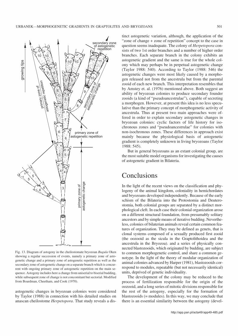
Analysis of factual data provided by Boardman et al.(1970) indicates that category of subsequent zones of asto−genetic change and repetition comprises rather different in−stances of morphogenetic changes which are probably of dif−ferent nature and might imply different causation. First of all,subsequent zones of change and repetition may be eitherwholly sequential or in part concurrent. From the instancesdiscussed by Boardman et al. (1970) the example of Poricel−laria d’Orbigny (Fig. 11B) belongs to the former category,while example of Bugula Oken (Fig. 13) illustrates a changefrom primary repetition into a secondary change only in onebranch, while others continue the budding pattern and mor−phology characteristic of the zone of repetition. The formercase is suggestive of a factor widely distributed in the colonyor in the environmemt why latter indicates its local, sectorial,nature. It seems clear that only a wholly sequential pattern,which implies the change of the entire growing edge, mightbe considered as a normal budding pattern and deserve thename of an astogenetic zone (or stage). In my opinion suchlate astogenetic zones may be ascribed to certain somatic orreproductive cycles in the life of the bryozoan colonies,which affect also the growing edge. Moreover, as recognizedby Boardman and Cheetham (1986: 40) “correlated cyclicgrowth within a group of zooids is not necessarily a result ofcolony control, but may be simultaneous separate responsesto a cyclic environment”.
In contrast the concurrent (non−isochronous) changeswill result in a sectorial, rather than zonal distribution ofmorphogenetic changes, and as such do not imply the exis−tence of a distinct astogenetic stage of the colony. They maybe stimulated by microenvironmental accidents. The lattermay include such factors as crowding, overgrowth, substrateirregularities (Abbot 1973; Boardman and Cheetham 1986).Thus, the local change of growing zooids, from a morphol−ogy characteristic of zone of repetition to that of secondarychange, may be in some cases microenvironmentally in−duced (e.g., by obstruction to growth). Although the zooidwholly comparable to ancestrula is lacking in the subsequentzones of change, nevertheless zooecia resemble the earliestgenerations in the colony, so one can speak on certain rever−sion in morphology. Moreover, when the essential similaritybetween ancestrula and particular zooids appearing late inthe astogeny and initiating a succeeding zone of astogeneticchange is well established (as in the case of anascan cheilo−stome Rhabdozoum wilsoni, described by Cook and Bock(1994) one could perhaps hypothesize on certain effects ofrejuvenation, related probably to the reproductive cycle andinduced by hormonal agents. To my knowledge such pheno−mena are unknown in Graptholithoidea and seem to be aunique feature of astogeny in bryozoan colonies.
It seems that the early and late astogeny in bryozoan colo−nies have different causation: while the former is probablyrelated to the sexual origin of the founder of the colony andas we can suppose to the morphogen produced by it, the latterreflects merely responses to physiological or environmentalcyclicity in life of the colony. However, some new aspects of
500 ACTA PALAEONTOLOGICA POLONICA 49 (4), 2004
astogenetic changes in bryozoan colonies were consideredby Taylor (1988) in connection with his detailed studies onanascan cheilostome Herpetopora. That study reveals a dis−
tinct astogenetic variation, although, the application of the“zone of change + zone of repetition” concept to the case inquestion seems inadequate. The colony of Herpetopora con−sists of two 1st order branches and a number of higher orderbranches. Each separate branch in the colony exhibits anastogenetic gradient and the same is true for the whole col−ony which may perhaps be in perpetual astogenetic change(Taylor 1988: 540). According to Taylor (1988: 546) theastogenetic changes were most likely caused by a morpho−gen released not from the ancestrula but from the parentalzooid of each new branch. This interpretation resembles thatby Amstey et. al. (1976) mentioned above. Both suggest anability of bryozoan colonies to produce secondary founderzooids (a kind of “pseudoancestrulae”), capable of secretinga morphogen. However, at present this idea is no less specu−lative than the primary concept of morphogenetic activity ofancestrula. Thus at present two main approaches were of−fered in order to explain secondary astogenetic changes inbryozoan colonies: cyclic factors of life history for iso−chronous zones and “pseudoancestrulae” for colonies withnon−isochronous zones. These differences in approach existmainly because the physiological basis of astogeneticgradient is completely unknown in living bryozoans (Taylor1988: 545).
But in general bryozoans as an extant colonial group, arethe most suitable model organisms for investigating the causesof astogenetic gradient in Bilateria.
ConclusionsIn the light of the recent views on the classification and phy−logeny of the animal kingdom, coloniality in hemichordatesand bryozoans developed independently. Because of the earlyschism of the Bilateria into the Protostomia and Deutero−stomia, both colonial groups are separated by a distinct mor−phological cleft. In each case their colonial organization aroseon a different structural foundation, from presumably solitaryancestors and by simple means of iterative budding. Neverthe−less, colonies of bilaterian animals reveal certain common fea−tures of organization. They may be defined as genets, that isclonal systems composed of a sexually produced first zooid(the oozooid as the sicula in the Graptolithoidea and theancestrula in the Bryozoa). and a series of physically con−nected blastozooids, which originated by budding, are subjectto common morphogenetic control, and share a common ge−notype. In the light of the theory of modular organization ofanimal colonies advanced by Harper (1981), blastozooids cor−respond to modules, repeatable (but not necessarily identical)units, deprived of genetic individuality.
The development of the colony may be reduced to theprocess of fertilization responsible for the origin of theoozooid, and a long series of mitotic divisions responsible forthe rest of the astogeny, especially for the formation ofblastozooids (= modules). In this way, we may conclude thatthere is an essential similarity between the astogeny (devel−
http://app.pan.pl/acta49/app49−485.pdf
URBANEK—MORPHOGENETIC GRADIENTS IN GRAPTOLITES AND BRYOZOANS 501
10
9
8
gro
win
gtip
s
10
10 1011 11
9
8
9 9
9
10 10
8
7
7
6
6
5 5
55
4 4
3
2
1
A
44
3
prim
ary
zo
ne
of
asto
ge
ne
tic
ch
an
ge
prim
ary
zone
of
asto
genetic
repetition
seco
ndary
zone
of
ast
ogenetic
change
primary zone ofastogenetic repetition
secondary zoneof astogenetic
repetition
Fig. 13. Diagram of astogeny in the cheilostomate bryozoan Bugula Okenshowing a regular succession of events, namely a primary zone of asto−genetic change and a primary zone of astogenetic repetition as well as thesecondary zone of astogenetic change on a separate branch which is concur−rent with ongoing primary zone of astogenetic repetition on the main se−quence. Astogeny includes here a change from uniserial to biserial budding,while subsequent zone of change is not concomitant but sectorial. Modifiedfrom Boardman, Cheetham, and Cook (1970).

opment of the colony) and the ontogeny (individual develop−ment of solitary organisms) in respect of the morphogeneticmechanisms involved. There is little specificity in the organi−zation of the bilaterian genets; in fact, they share a number ofcommon biological properties even with plant genets. Thestructural characteristics of modules are, however, in eachcase highly specific and dependent on the phylum fromwhich the colonial forms were derived. This model of colo−nial organization probably may be extrapolated to coloniesof Entoprocta, which are also clonal systems (genets) com−posed of an oozooid subject to metamorphosis and series ofblastozooids, budding from a type of stolons. However, abetter knowledge of their astogeny is needed for a safeconclusion.
Bilaterian colonies display a latent polar organization,frequently expressed in a regular morphological gradient.This gradient may be explained by diffusion, over the longaxis of the colony, of a morphogen produced by the zygoticfounder−zooid of the genet. The hypothesis that the patternsof budding and graded morphological differences among zo−oids observed in the early astogeny are under control of themorphogen produced by the oozooid seems to be consistentwith the entirety of facts available for hemichordate andbryozoan colonies. In my opinion, the gradient of morpho−gen per se has hardly any adaptive significance. The origin ofthis gradient may be seen as a sort of a side effect of sexualprocess leading to the formation of the founder zooid. Thelatter developing as a regular bilaterian animal contaminatedthe neighboring blastozooids with the products of its ownmorphogenesis, which certainly included products of genescontrolling the body axes. Therefore, we may expect thatmorphogens responsible for patterning of bilaterian coloniesare related to the products of genes responsible for theanteroposterior control of embryos in all solitary Bilateria(Hox, zootype genes). However, once created, the morpho−genetic gradient could be used by selective forces to producevarious effects, i.a., phenetic differences among zooids of thecolony. In a given environmental context such differencesmight attain certain adaptive significance.
Hemichordate and bryozoan colonies are genetically uni−form and all zooids of a single colony share the same geno−type. The genetic uniformity may be considered a commonfeature of colonial organization, with a notable exclusion oftunicate colonies, where zooids with different genotypesmight be present within a single cormus (Sabbadin 1979).Hence, in respect of genetic uniformity bilaterian coloniesmay be divided into genetically uniform (majority of cases)and genetically differentiated (probable exceptional cases).Therefore, astogenetic variation and morphological evolu−tionary change in the majority of bilaterian colonies may bebest described in terms of phenogenetics, that is through theseverity or measure of the relative degree of the expression ofa given character (expressivity) and the number of zooids af−fected by it (penetrance, percentage of phenotypic effect). Inaccordance with the colony’s polar organization, phylogen−etic novelties are introduced from either its proximal or dis−
tant end. Progression of phylogenetic novelties may be inter−preted as increasing penetrance of genetic factors. In the evo−lution of graptoloid colonies changes in penetrance andexpressivity are functions of the gradient in the distributionof the morphogen which supplies positional information tothe growing zooids. A similar set of notions may probably beapplied to the trends observed in bryozoan colonies but theknowledge of relevant details is inadequate.
The hypothesis concerning the source and role of themorphogen was corroborated by observations on regenera−tion of fragmented graptolite and bryozoan colonies. Theyrevealed that the colonies which developed in the course of anormal astogeny (in the presence of an oozooid) and displayan astogenetic change, have their regenerated portions (lack−ing of an oozooid) always devoid of such change. However,it is clear that the problem in question may be satisfactorilysolved only by the use of modern experimental techniqueselaborated within the “Evo−Devo” program.
AcknowledgementsI thank Dr. Denis E.B. Bates (Aberysthwyth) for reading and improvingmy manuscript as well as for his valuable remarks, and two anonymousreviewers for their helpful criticism. Moreover, I am grateful to my col−leagues at the Institute of Palaeobiology (Warsaw), Dr. Anna Kozłow−ska−Dawidziuk, Aleksandra Hołda−Michalska M.Sc., and Dr. CyprianKulicki for their help in preparing illustrations. Thanks are also due toDr. Urszula Hara (State Geological Institute, Warsaw) for her helpfuladvices. My work was financially supported by grant KBN 3 P04C03522 from the Polish Committee for Scientific Research (KBN).
ReferencesAbbot, M.B. 1973. Intra− and intercolony variation in populations of Hippo−
porina Neviani (Bryozoa−Cheilostomata). In: R.S. Boardman, A.H.Cheetham, and W.A. Oliver (eds.), Animal Colonies. Development andFunctions Through Time, 223–245. Dowden, Hutchinson and Ross,Stroudsburg.
Anstey, R.L., Pachut, J.F., and Prezbindowski, D.R. 1976. Morphogeneticgradients in Paleozoic bryozoan colonies. Paleobiology 2: 131–146.
Armstrong, W.G., Dilly, P.N., and Urbanek, A. 1984. Collagen in thepterobranch coenecium and the problem of graptolite affinities. Lethaia17: 145–152.
Bałuk, W. and Radwański, A. 1977. The colony regeneration and life habitat offree−living bryozoans, Cupuladria canariensis (Busk) and C. haidingeri(Reuss), from the Korytnica Clays (Middle Miocene; Holy Cross Moun−tains, Poland). Acta Geologica Polonica 27: 143–156.
Bałuk, W. and Radwański, A. 1984. Middle Miocene (Badenian) free−livingbryozoans from the Vienna Basin. Annalen des Naturhistorisches Mu−seum Wien 86: 13–40.
Barnes, D.K.A. and Clarke, A. 1998. Seasonality of polypide recycling andsexual reproduction in some erect Antarctic bryozoans. Marine Biology131: 647–658
Bates, D.E. and Urbanek, A. 2003. The ultrastructure, development, amdsystematic position of the graptolite genus Mastigograptus. Acta Palae−ontologica Polonica 47: 445–458.
Beklemishev, W.N. [Beklemišev, W.N.] 1951. Towards the systematic classi−fication of animals. Deuterostomia, their origin and composition [in Rus−sian]. Uspehi sovremennoj biologii 32: 256–270.
502 ACTA PALAEONTOLOGICA POLONICA 49 (4), 2004

Beklemishev, W.N. 1969. Principles of Comparative Anatomy of Inverte−brates. 1, Promorphology. 190 pp. Oliver and Boyd, Edinburgh.
Berry, W.B.N. 1987. Phylum Hemichordata (including Graptolithina). In:R.S. Boardman, A.H. Cheetham, and A.J. Rowell (eds.), Fossil Inverte−brates, 612–635. Blackwell Scientific Publications, Palo Alto.
Boardman, R.S. 1983. General features of the class Stenolaemata. In: R.A.Robinson (ed.), Treatise on Invertebrate Paleontology. Part G, Bryo−zoa, vol. 1, 49–137. Geological Society of America and University ofKansas Press, Boulder.
Boardman, R.S. and Cheetham, A.H. 1969. Skeletal growth, intracolonyvariation and evolution in Bryozoa: a review. Journal of Paleontology43: 205–233.
Boardman, R.S. and Cheetham, A.H. 1973. Degrees of colony dominance instenolaemate and gymnolaemate Bryozoa. In: R.S. Boardman, A.H.Cheetham, and W.A. Oliver (eds.), Animal Colonies. Development andFunctions Through Time, 441–514. Dowden, Hutchinson and Ross,Stroudsburg.
Boardman, R.S. and Cheetham, A.H. 1987. Phylum Bryozoa. In: R.D.Boardman, A.H. Cheetham, and A.J. Rowell (eds.), Fossil Inverte−brates, 497–549. Blackwell Scientific Publications, Palo Alto.
Boardman, R.S., Cheetham, A.H., and Cook, P.L. 1970. Intracolony varia−tion and the genus concept in Bryozoa. Proceedings of the North Ameri−can Paleontological Convention, September, 1969, Part C, 294–320.
Boardman, R.S., Cheetham, A.H., and Cook, P.L. 1983. Introduction to theBryozoa. In: R.A. Robinson (ed.), Treatise on Invertebrate Paleontology.Part G, Bryozoa, vol. 1, 3–48. Geological Society of America and Univer−sity of Kansas Press, Boulder.
Boardman, R.S., Cheetham, A.H., and Oliver, W.A. (eds.) 1973. Animal Colo−nies. Development and Functions Through Time. ix + 603 pp. Dowden,Hutchinson, and Ross Inc., Stroudsburg.
Bulman, O.M.B. 1958. Patterns of colonial development in graptolites.Journal of Linnean Society London, Zoology 44: 24–32.
Bulman, O.M.B. 1970. Graptolithina. In: C. Teichert (ed.), Treatise on In−vertebrate Paleontology, Part V, second edition, xxxii +163 pp. Geo−logical Society of America and University of Kansas Press, Lawrence.
Bulman, O.M.B. 1973. Graptolite periderm structure and budding patterns:a resume. In: G.P. Larwood (ed.), Living and Fossil Bryozoa. RecentAdvances in Research, 11–20, Academic Press, London.
Carroll, S.B., Grenier, J.K., and Weatherbee, S.D. 2001. From Genes toDiversity. Molecular Genetics and the Evolution of Animal Design.xv+214 pp. Blackwell Scientific Publishers, Malden Mass.
Cheetham, A.H. and Cook, P.L. 1983. General features of the class Gymno−laemata. In: R.A. Robinson (ed.), Treatise on Invertebrate Paleontol−ogy. Part G, Bryozoa, vol. 1, 138–207. Geological Society of Americaand University of Kansas, Boulder.
Child, C.M. 1915. Individuality in Organisms. 213 pp. University of Chi−cago Press, Chicago.
Child, C.M. 1941. Patterns and Problems of Development. 811 pp. Univer−sity of Chicago Press, Chicago.
Cook, P.L. and Lagaaij, R. 1976. Some Tertiary and Recent conescharellini−form Bryozoa. Bulletin of the British Museum (Natural History), Zool−ogy 29: 320–376.
Cook, P.L. and Bock, P.E. 1994. The astogeny and morphology of Rhabdo−zoum wilsoni Hincks (Anasca, Buguloidea). In: J. Hayward, J.S. Ryland,and P.D. Taylor (eds.), Biology and Paleobiology of Bryozoans, 47–50.Olsen and Olsen, Fredensborg.
Crowther, P.R. 1980. The fine structure of graptolite periderm. Special Pa−pers in Palaeontology 26: 1–116.
Davidson, E.H. 2001. Genomic Regulatory Systems. Development and Evo−lution. xii + 261 pp. Academic Press, London.
Dilly, P.N. 1973. The larva of Rhabdopleura compacta. Marine Biology 18:69–86.
Dubrulle, J. and Pourqulé, O. 2004. FGF8 mRNA decay establishes a gradi−ent that couples axial elongation to patterning in the vertebrate embryo.Nature 427: 419–422.
Dzik, J. 1975. The origin and early phylogeny of the cheilostomatousBryozoa. Acta Palaeontologica Polonica 20: 395–423.
Dzik, J. 1981. Evolutionary relationships of the early “cyclostomatous”Bryozoa. Palaentology 20: 827–861.
Dzik, J. 1992. Early astogeny and relationships of the Ordovician rhabdo−mesine bryozoans. Acta Palaeontologica Polonica 37: 37–54.
Finney, S.C. 1986. Heterochrony, punctuated equilibrium, and graptolitezonal boundaries. In: C.P. Hughes and R.B. Rickards (eds.), Palaeo−ecology and Biostratigraphy of Graptolites. Geological Society SpecialPublication 20: 103–113.
Fortey, R.A. 1983. Geometrical constraints on the construction of graptolitestipes. Paleobiology 9: 116–125.
Gordon, J.B. and Bourillot, P.−Y. 2001. Morphogen gradient interpretation.Nature 413: 797–803.
Halanych, K.M. 1995. The phylogenetic position of pterobranch hemi−chordates based on 18S rDNA sequence data. Molecular Phylogeny andEvolution 4: 72–76.
Halanych, K.M. 1996. Convergence in the the feeding apparatuses oflophophorates and pterobranch henichordates revealed by 18S rDNA:an interpretation. Biological Bulletin 190: 1–5.
Halanych, K.M., Bachelor, J., Aguinaldo, A.M., Liva, S., Hillis, D.M., andLake, J.A.1995. Evidence from18S rDNA that the lophophorates areprotostome animals. Science 267: 1641–1643.
Hammer, O. 2000. Lateral inhibition in the astogeny of conical multistipedgraptolites. Lethaia 33: 39–45.
Harper, J.L. 1981. The concept of population in modular organisms. In:R.M. May (ed.), Theoretical Ecology. Principles and Applications,53–77, Blackwell Scientific Publications, Oxford.
Kozłowska−Dawidziuk A. 2004. Evolution of retiolitid graptolites—a syn−opsis, with description of a new species of Holoretiolites. Acta Palae−ontologica Polonica 49: 505–518.
Kozłowski, R. 1949. Les graptolithes et quelques nouveaux groupes d’animauxdu Tremadoc de la Pologne. Palaeontologia Polonica 3: xii + 235 pp.
Kozłowski, R. 1963. Le dévelopment d’un graptolite tuboide. Acta Palae−ontologica Polonica 8: 103–134.
Kozłowski, R. 1966. On the structure and relationships of graptolites. Jour−nal of Paleontology 40: 489–501.
Kozłowski, R. 1971. Early development stages and mode of life of grapto−lites. Acta Palaeontologica Polonica 16: 313–343.
Larwood, G. and Rosen, B.R. (eds.) 1979. Biology and Systematics ofColonial Organisms. The Systematics Association, Special Volume 11:xxxv +589 pp. Academic Press, London.
Mierzejewski, P. and Kulicki, C. 2002. Discovery of Pterobranchia (Grapto−lithoidea) in the Permian. Acta Palaeontologica Polonica 47: 169–175.
Nielsen, C. 2001. Animal Evolution. 2nd edition. 563 pp. Oxford UniversityPress, Oxford.
Nielsen, C. 2002. The phylogenetic position of Entoprocta, Ectoprocta,Phoronida, and Brachiopoda. Integrative and Comparative Biology 42:685–691.
Pachut, J.H. and Anstey, R.L. 1979. A developmental explanation of stabil−ity—diversity—variation hypotheses: morphogenetic regulation in Or−dovician bryozoan colonies. Paleobiology 5: 168–187.
Pätzold, J., Ristedt, H., and Wefer, G. 1987. Rate of growth and longevity ofa large colony of Pentapora foliacea (Bryozoa) recorded in their oxy−gen isotope profiles. Marine Biology 96: 535–538.
Podell, M.E. and Anstey, R.L. 1979. The interrelationship of early colonydevelopment, monticules and branches in Palaeozoic bryozoans. Palae−ontology 22: 965–982.
Ryland, J.S. 1970. Bryozoans. 176 pp. Hutchinson University Library, Lon−don.
Ryland, J.S. 1979. Structural and physiological aspects of coloniality inBryozoa. In: G. Larwood and B.R. Rosen (eds.), Biology and Systemat−ics of Colonial Organisms, 211–242. Academic Press, London.
Sabbadin, A. 1979. Colonial structure and genetic patterns in ascidians. In:G. Larwood and B.R. Rosen (eds.), Biology and Systematics of ColonialOrganisms, 433–444. Academic Press, London.
Schier, A.F. 2004. Tail of decay. Nature 427: 403–404.Stebbing, A.R.D. 1970. Aspects of the reproduction and the life cycle of
Rhabdopleura compacta (Hemichordata), Marine Biology 5: 205–212.
http://app.pan.pl/acta49/app49−485.pdf
URBANEK—MORPHOGENETIC GRADIENTS IN GRAPTOLITES AND BRYOZOANS 503

Taylor, P.D. 1988. Colony growth pattern and astogenetic gradients in theCretaceous cheilostome bryozoan Herpetopora. Palaeontology 31:519–549.
Thimann, K.V. and Skoog, F. 1934. On the inhibition of bud developmentand other functions of growth substance in Vicia faba. Proceedings ofthe Royal Society of London B 114: 307–339.
Thorsteinsson, R. 1955. The mode of cladial generation in Cyrtograptus.Geological Magazine 92: 37–49.
Timofeef−Ressovsky, N.V. 1931. Gerichtetes variieren in der phänotypischenManifestierung einiger Genovariationen von Drosophila funebris. Natur−wissenschaften 19: 493–497.
Timofeef−Ressovsky, N.V. 1934. Über den Einfluss des genotypischen Mi−lieus und der Aussenbedingungen auf die Realization des Genotyps.Nachrichten des Gesellschaft der Wissenschaften zu Göttingen NF 1:53–106.
Towe, K.M. and Urbanek, A. 1972. Collagen−like structures in Ordoviciangraptolite periderm. Nature 237: 443–445.
Urbanek, A. 1960. An attempt at biological interpretation of evolutionarychanges in graptolite colonies. Acta Palaeontologica Polonica 5: 127–234.
Urbanek, A. 1963. On generation and regeneration of cladia in some UpperSilurian monograptids. Acta Palaeontologica Polonica 8: 135–254.
Urbanek, A. 1973. Organization and evolution of graptolite colonies. In: R.S.Boardman, A.H. Cheetham, and W.A. Oliver, Jr. (eds.), Animal Colonies.Development and Function Through Time, 441–514. Dowden, Hutchin−son and Ross, Stroudsburg, Pennsylvania.
Urbanek, A. 1986. The enigma of graptolite ancestry: a lesson from a phylo−genetic debate. In: A. Hoffman and M.H. Nitecki (eds.), ProblematicFossil Taxa, 184–225. Oxford University Press, New York.
Urbanek, A. 1997. The emergence and evolution of linograptids. In: A.Urbanek and L. Teller (eds.), Silurian Graptolite Faunas of the East Eu−ropean Platform: Stratigraphy and Evolution. Palaeontologia Polonica56: 233–269.
Urbanek, A. 2001. Phenogenetics of graptoloid colonies. In: V.I. Korogo−dina (ed.), Modern Problems of Radiobiology, Radioecology and Evo−lution. Proceedings of the International Conference dedicated to theCentenary of the Birth of N.W. Timofeef−Ressovsky, 326–336. Joint In−stitute of Nuclear Research, Dubna.
Urbanek, A. 2003. Organization and evolution of animal colonies. BiologyBulletin 30: 1–8 (translated from Izvestiâ Akademii Nauk, Seriâ Biolo−gičeskaâ 1: 5–13).
Urbanek, A. and Dilly, P.N. 2000. The stolon system in Rhabdopleuracompacta (Hemichordata) and its phylogenetic implications. Acta Palae−ontologica Polonica 45: 201–226.
Urbanek, A. and Uchmański, J. 1990. Morphogenesis of uniaxiate grapto−loid colonies—a mathematical model. Paleobiology 16: 49–61.
Wolpert, L., Beddington, R., Brockes, J., Jessell, T., Lawrence, P., andMeyerowitz, E. 1998. Principles of Development. xx + 484 pp. OxfordUniversity Press, London.
Zimmer, R.L. 1973. Morphological and developmental affinities of thelophophorates. In: G.P. Larwood (ed.), Living and Fossil Bryozoa. Re−cent Advances in Research, 593–599. Academic Press, London.
Zimmer, R.L. and Woollacott, R.M. 1977. Metamorphosis, ancestrulae andcoloniality in bryozoan life cycles. In: R.M. Woollacott and R.L.Zimmer (eds.), Biology of Bryozoans, 91–142. Academic Press, NewYork.
504 ACTA PALAEONTOLOGICA POLONICA 49 (4), 2004