morphological organization of rat hippocampal...
TRANSCRIPT

THE JOURNAL OF COMPARATIVE NEUROLOGY 307:87-106 (1991)
Morphological Organization of Rat Hippocampal Slice Cultures
MANFRED CAESER AND AD AERTSEN Max-Planck-Institut fur Biologische Kybernetik, D-7400 Tubingen, Germany
ABSTRACT Using various histological methods, we investigated the cellular and morphological
organization of rat hippocampal slice cultures. Many of the typical features of the hippocampus were retained in vitro over a long period of time. The principal cell types of the hippocampus and dentate gyrus, the pyramidal cells and granule cells, were well preserved and matured in vitro. Nonpyramidal cells and y-aminobutyric-acid (GABA) cells were also present in slice cultures and exhibited a strikingly similar dendritic appearance at the light microscopic level. Moreover, GABA-immunoreactive cell bodies and presynaptic terminals could be identified at the electron microscopic level; they expressed typical symmetric synaptic contacts with cell bodies and dendrites. The course of the intrinsic hippocampal fiber pathways-the mossy fibers, Schaffer collaterals, and alveus-was generally retained in vitro. Additional aberrant fiber projections could be identified. Finally, three types of nonneuronal cells could be distinguished on the basis of immunocytochemical methods.
Key words: hippocampus, in vitro, light microscopy, electron microscopy, development, cell types, fiber course
Over the last decades the hippocampus has become one of the most extensively investigated areas of the mammalian brain. Many of these investigations are motivated by ques- tions associated with the neurobiological basis of learning and memory. In particular the investigation of the cellular mechanisms involved requires a preparation in which de- tailed observation and manipulation of the basic anatomi- cal, physiological and pharmacological environment are possible. One answer to this requirement is to resort to in vitro preparations. In order to qualify as an adequate experimental model for the in vivo situation, such a prepa- ration obviously has to satisfy certain minimal anatomical and physiological requirements, In particular, investiga- tions that focus on the processing of information in the natural nervous assembly rely on a faithful representation of the various cell types found in the hippocampus in vivo, as well as on an intact pattern of interneuronal connectiv- ity.
The morphology and physiology of the hippocampus have been described in detail by various authors (e.g., Cajal, '11; Isaacson and Pribram, '75; Seifert, '83). The characteristic morphological organization of the hippocampus is summa- rized schematically in Figure 1. Briefly, the hippocampus (CA1, CA3) and the dentate gyrus (DG) are characterized by their distinct laminated structure and the trisynaptic fiber pathway. The main cell types of the dentate gyrus and the hippocampus are the granule cells and the pyramidal cells, located in the stratum granulosum (sg) and stratum pyramidale (sp), respectively. The afferent fibers terminate
in separate, non-overlapping laminae. These structural features are reflected in the spatiotemporal spreading of electrical activity entering the hippocampus (Andersen et al., '71). Both in situ and in the acute slice preparation the excitation enters the dentate gyrus by the perforant path (pp), runs along the mossy fibers (mf) and the Schaffer collaterals (sc), and leaves the hippocampus via the alveus (a) and the fimbria (0,
The acute slice preparation of the adult rat (Skrede and Westgaard, '71) and cultures of dissociated cells of the fetal hippocampus (Banker and Cowan, '77, '79) are commonly used in vitro preparations for physiological, developmental, and pharmacological studies. The prominent advantage of the acute slices is that they retain the original cellular and connective organization of the hippocampus. Unfortu- nately, their short survival time in vitro and the difficulty in identifying individual cells represents a limitation of this preparation. In contrast, in dissociated cell cultures, individ- ual cells can easily be identified; however, the separation of the cells leads to an artificial neuronal network that lacks the typical structural features of the intact hippocampus.
Organotypic slice cultures, a relatively new technique established by Gahwiler ('81a), combine a number of the
Accepted December 21, 1990 Address reprint requests to Manfred Caeser, who is now at the Brain
Research Institute, August-Fore1 Strasse 1, CH-8029 Zurich, Switzerland.
o 1991 WILEY-LISS, INC.

88 M. CAESER AND A. AERTSEN
Fig. 1. Schematic drawing of the hippocampal formation, demon- strating the main cell types, laminae and subfields. The figure is a modification from Cajal’s (1911) famous original drawing; the individ- ual cells are reproductions from various authors (Gottlieb and Cowan, ’72; Finch et al., ’83; Seress and Ribak, ’85; Tamamaki et al., ’87; Lingenhohl and Finch, ’91). The excitation enters the dentate gyrus
advantages of both the acute slice preparation and the dissociated cell culture technique. By cultivating slices with the “roller tube technique” over several weeks, he could show that the hippocampal tissue retains its “organotypical” features. In a number of studies, Gahwiler and collegues described the general histological organization of hippocam- pal slice cultures, in particular the pyramidal cells and granular cells, in great detail (Gahwiler, ’81a, ’84; Zimmer and Gahwiler, ’84, ’87; Frotscher and Gahwiler, ’88). Much less is known about the survival, development, and morphol- ogy of the nonpyramidal cells, the nonneuronal cells, and about the detailed course of the intrinsic fiber pathways in this preparation.
The aim of the present study was to investigate the morphology of the different cellular components and the course of the intrinsic fiber pathways in hippocampus slice cultures. In order to be able to compare physiological results, in particular regarding the spatio-temporal distribu- tion of electrical activity (Caeser and Aertsen, ’89) as well as findings pertaining to synaptic plasticity (Bonhoeffer et al., ’89) obtained in such slice cultures with the hippocampus in vivo or acute slices, the results of this study are discussed in relation to the morphological organization of the hippocam- pus in vivo and of the other in vitro preparations. The present observations have been reported in abstract form (Caeser, ’88) and form part of a doctoral thesis (Caeser, ’89).
MATERIALS AND METHODS Culturing procedure
For cultivation of hippocampal explants, the roller tube technique as described by Gahwiler (%la, ’84) was used.
(DG) by the perforant path (pp), runs along the mossy fibers (mf) and the Schaffer collaterals (sc) and leaves the hippocampus via the alveus (a) or the fimbria (f). Further abbreviations: (so) stratum oriens; (sp) stratum pyramidale; (sr) stratum radiatum; (sg) stratum granulosum; (sm) stratum moleculare; (H) Hilus; (EC) Entorhinal Cortex; (S) Subiculum.
Briefly, hippocampal slices (350 pm) were cut in a trans- verse plane from 4- to 7-day old rats. The slices were transferred onto cleaned glass coverslips (12 x 24 mm) and embedded into a plasma clot. The clot was formed by 20 pl of heparinized chicken plasma (lyophilized; Difco), which was coagulated by 20 ~1 of a thrombin solution (0.2 mgiml, 20 NIHU/ml, Hoffman LaRoche). Following coagulation of the plasma clot, the slice cultures were placed into plastic culture tubes (16 x 110 mm, Nunclon) and maintained in a roller drum incubator at 36°C in dry air, without CO,/O, control. The explants were fed one to two times per week with culture medium (50% Eagle basal medium (BME), 25% Hanks’ balanced salt solution (HBSS), and 25% horse serum, containing 0.1 mM glutamine and 6.5 mg/ml glu- cose). After 4 days in vitro, antimitotics (5-Fluoro-2- Deoxyuridine, Cytosine-b-d-Arabino-Furanoside, Uridine; Sigma) were added to the culture medium for 24 hours to give a final concentration of 10 pM.
All experiments were carried out on cultures, 2 to 4 weeks in vitro.
Nissl staining For Nissl staining, cultures were fixed with 4% formalde-
hyde in distilled water for 2 hours. The cultures were rinsed in distilled water and stained in an aqueous solution of cresylviolet (1%) for 15 minutes, differentiated in acetic- acid alcohol, dehydrated in alcohol, cleared in xylene and finally embedded in Entellan (Merck, Darmstadt).
Intracellular staining For intracellular staining with Lucifer yellow, cultures
were transferred into a microchamber mounted on the stage of an inverted microscope (Zeiss IM 35) . The cultures

CELLULAR STRUCTURE OF THE HIPPOCAMPUS IN VITRO 89
were continuously perfused at a rate of 30 ml/h with Hanks' balanced salt solution (HBSS) containing 3.2 mM CaCl,. The bath temperature was kept at 30-33°C. Individual cells were penetrated with glass microelectrodes, containing 20% Lucifer yellow (Sigma) in 0.1 M LiCl, (electrical resistance between 100 and 200 MpR) under visual control, using a David Kopf microdrive mounted on a Leitz microma- nipulator. The fluorescent dye was injected by applying positive current pulses of 1 to 3 nA at a frequency of 1 Hz for 5 to 10 minutes. After injecting several cells, the cultures were fixed with 4% paraformaldehyde in 0.1 M phosphate buffer with 10% sucrose, dehydrated in a graded series of alcohols, cleared, and embedded in Entellan. Labeled cells were photographed, using black-and-white film (Ilford HP5,400 ASA).
Golgi impregnation Cultures were fixed in 2% %Cr,O, and 5% glutaraldehyde
for 2 hours. They were then rinsed several times in bidistilled water to wash off the K,Cr,O,. Using an inverted microscope, a small grain of silver nitrate was placed onto the edge of the culture with the aid of a broken micropi- pette. The glass coverslips with the cultures were then placed in a humid chamber and kept in the dark at room temperature for 24 to 48 hours. Cultures containing stained cells were dehydrated in ethanol, transferred to terpineol and xylol, and finally covered with Entellan. For documen- tation, labeled cells were photographed, using black-and- white film (Ilford FP4, 125 ASA) or drawn by camera lucida.
Di I staining For labeling of individual cells and of the intrinsic fiber
pathways of the hippocampus, we used the fluorescent carbocyanine dye Di I (1,1, dioctadecyl-3,3,3,3-tetrameth- ylindocarbocyanine perclorate; Molecular Probes). Di I works both as an anterograde and a retrograde tracer in vital (Honig and Hume, '86) and in fixed tissue (Godement et al., '87).
To stain the cultures, small crystals (20-50 pm in diameter) of the dye were applied under visual control with a glass micropipette onto the surface of the vital explants. Cultures were then fixed by immersion in phosphate- buffered 4% paraformaldehyde, after which they were kept in the dark at room temperature for 3 to 7 weeks before visualization. This long time was necessary because in- tramembraneous diffusion in fixed tissue is extremely slow. Vital cultures, placed back into the culture tubes after staining and cultivated for an additional 1 to 2 days, gave inferior results: the background fluorescence increased and spreading of the dye as well as debris of the dye were a serious problem. With very small crystals (smaller than 20 pm), only a small population of cells could be stained by retrograde intramembraneous diffusion. With larger crys- tals, the intrinsic fiber pathways of the hippocampus could be labeled.
Di I fluoresces red when excited with green light. There- fore, stained cultures were visualized under an epifluores- cence microscope (Zeiss), equipped with a Rhodamine filter set (BP 546, FT 580, LP 590; Zeiss). Stained cells and fiber pathways were documented photographically with Ilford HP5 (800 ASA) black-and-white film.
For drawing Di I stained cells, we used the photo- oxidation method as previously described for Lucifer yellow (Maranto, '82). Stained cultures were first rinsed several
times in phosphate buffer (pH 7.4) to remove the fixative and then placed on a epifluorescence microscope under a fluorescence objective (16x, or 25x). The excess buffer was blotted off and a drop of a buffered DAB solution was placed on the explant. The cultures were illuminated with the excitation wavelength for 20 to 30 minutes, changing the DAB solution (1.5 mgiml) several times. Due to the illumi- nation, the fluorescence faded and was replaced by a brown DAB reaction product. When the reaction was finished, the cultures where rinsed in phosphate buffer, dehydrated in alcohol, cleared in xylene, and embedded in Entellan.
Silver impregnation In addition to the Di I staining, the silver impregnation
technique was used to visualize the intrinsic hippocampal fiber pathways. Explants were fixed with 4% paraformalde- hyde in bidistilled water. After washing, the cultures were incubated in 20% silver nitrate (AgNO,) in bidistilled water for 1 hour at room temperature. They were then rinsed several times in bidistilled water in order to wash off the AgNO,. For intensification, the explants were exposed to an aquous solution of 5% silver nitrate, containing a few drops of ammonia for approximately 1 minute, washed again, and additionally fixed in an aqueous solution containing 1% formaldehyde and 2% sodium citrate. Finally, the cultures were washed in water, incubated in an aqueous solution containing 3% natriumthiosulfat for 1 minute, rinsed again, dehydrated in a graded series of alcohols, cleared in xylol, and embedded in Entellan.
GABA-immunohistochemistry For immunohistochemistry, cultures were transferred
into 4% paraformaldehyde in 0.1 M phosphate buffer (PB, pH 7.4) for 1 to 12 hours at room temperature. Immuno- staining for light microscopy was performed on cultures fixed on glass coverslips; free-floating explants were used for electron microscopy. After being washed (three 10-min rinses) in 0.05 M Tris buffer with 10% Na-metabisulphite and 0.5% Triton X-100, the cultures were first incubated with a rabbit anti-GABA-serum (Dianova) and kept over- night at 4°C in a humid chamber. The GABA antiserum was diluted to 1:1,000 in PB with 0.5% Triton X-100. The cultures were subsequently washed, treated with biotiny- lated anti-rabbit IgG (Vector), washed, and incubated in avidin-biotin-peroxidase complex (Vector). After three 10- minute washes in PB, the cultures were incubated in a 3,3-diaminobenzidine (DAB, Sigma) solution (100 ml PB, 50 mg DAB, 40 pl 30% H,OJ for 5 minutes. After being rinsed in PB, the tissue was dehydrated in alcohols, cleared in xylene, and embedded in Entellan.
For electron microscopy the cultures were fixed in 4% paraformaldehyde with 0.1% glutaraldehyde and 0.1 mg/ml H,O, in 0.1 M PB (pH 7.2) for 2 hours at room temperature. The procedure for immunostaining was similar as described before, except that no Triton X-100 was used to enhance tissue conservation. For electron microscopic observation, the cultures were embedded in araldite and processed in the conventional way.
Immunohistochemistry against nonneuronal cells
For labeling nonneuronal cells, the following antibodies were used: rabbit antiserum to glial fibrillary acid protein (GFAP, Dakopats) as a marker for astrocytes; mouse antiserum to galactocerebroside (GALC, Dakopats) as a

90 M. CAESER AND A. AERTSEN
marker for oligodendrocytes, and mouse antiserum to THY 1 (Serotec) as a marker for fibroblasts. The specific primary antibodies were visualized by indirect immunolabeling us- ing the corresponding secondary antibodies; rhodamin cou- pled goat antirabbit Ig G and fluorescein coupled goat anti-mouse Ig G. All staining was performed on cultures fixed on coverslips. After staining, the cultures were embed- ded in a mixture of PBS and glycerol (1:9) for preservation. Marked cells were viewed and identified using a fluores- cence microscope (Zeiss), equipped with rhodamine (RITC) and fluorescein (FITC) optics, and photographed using a black-and-white film (Ilford HP5,800 ASA).
For double-labeling experiments, the cultures were first incubated with the GALC-antiserum, followed by the GFAP- antiserum or, alternatively, they were first incubated with the GFAP-antiserum, followed by the THY 1-antiserum. Prior to immunostaining, the cultures were fixed with 4% paraformaldehyde in 0.1 M phosphate buffer (PB, pH 7.4) for 30 minutes. All exposures to the antibodies were performed at room temperature.
For GALC-immunostaining, the cultures, after being washed (three 10-min rinses), were kept overnight in a moist chamber at 4°C in PB. Then the explants were incubated in cold (-20°C) methanol for 5 to 10 minutes to maximize antibody penetration, washed, and incubated in the GALE-antiserum (diluted 1:200 in DMEM) for 2 hours and washed again. For visualization, the explants were then incubated with the fluorescein coupled goat anti-mouse Ig G for 2 hours, rinsed again, and embedded. After this procedure, the same cultures were incubated in GFAP- antiserum (diluted 1500 in DMEM) for 2 hours. The cultures were subsequently washed and exposed to appropri- ate rhodamine coupled goat anti-rabbit Ig G. After being rinsed in PB, the explants were embedded for fluorescence microscopy.
For the second double-labeling, the GFAP-immunostain- ing was performed as described before, except that the rinses in methanol were left out. After GFAP-immunolabel- ing and subsequent washes in PB, the explants were exposed to the THY 1-antiserum (diluted 1:200 in DMEM with 10% fetal calf serum) for 2 hours. After the cultures were washed, they were incubated with fluorescein coupled goat anti-mouse antiserum for 2 hours. After this, the cultures were washed again and embedded as described before.
RESULTS Cytoarchitectonic organization
In accordance with previous reports (Gahwiler, '81a, '84; Zimmer and Gahwiler, '841, the slice cultures flattened to a thickness of approximately 30 to 50 pm during the first two weeks in vitro while preserving the typical hippocampal cytoarchitecture. The flattening of the explants is due to the degeneration of cells that are damaged by the trauma of the preparation procedure. Tissue debris will be removed from the explant by macrophages and continuous change of the bath medium. A comparison of the acute slice preparation (Fig. 2a) with the slice culture (Fig. 2b) shows that the lamellar organization of the hippocampus as well as the major hippocampal subfields-the dentate gyrus, the hilar region, and the CA1 and CA3 areas-were generally re- tained. Certain quantitative modifications, however, do occur. The stratum pyramidale of the CA1 and CA3 areas (Fig. 2b) becomes broader than normal (Fig. 2a), presum-
Fig. 2. Cytoarchitecture of the hippocampal formation in vivo and in vitro. Slices were cut in the transverse plane and stained with cresylviolet. A. Hippocampal slice taken from a 6-day-old rat, the time of explantation. At this developmental stage, all hippocampal subfields are well organized. B. Hippocampal slice culture prepared from a 6-day-old rat and cultivated for 27 days. Note the organotypic appear- ance of the explant when compared with P6. (C) Slice culture from a 6-day-old rat and 22 days in culture. Note the extreme broadening of the cell layer and the reduced width of the neuropil layer. Sometimes, the shape of the cultures was also affected by partial destruction (arrow) of the plasma clot surrounding the explant. Bars = 500 bm.
ably due to the culturing procedure and to the flattening of the tissue. This increase is accompanied by a decrease in the width of the neuropil layers, and can occasionally develop quite massive proportions (Fig. 2c). Consequently, the pyramidal cells in the stratum pyramidale become more loosely arranged, and also the intrinsic fiber system under-

CELLULAR STRUCTURE OF THE HIPPOCAMPUS IN VITRO 91
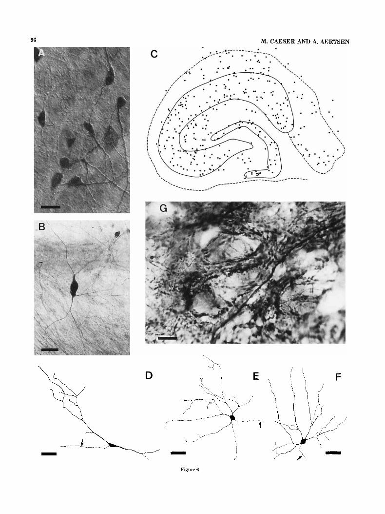
Fig. 3. Morphology of granule cells in hippocampal slice cultures. All explants were taken from rats 5-7 days old and cultivated for 20-27 days. A. Granule cells, retrogradely stained with Di I. The cells were located in the stratum granulosum of the dentate gyrus; they display the typical orientation of cells in this region. The monopolar dendritic tree was always oriented toward the molecular layer, the axons were directed toward the hilar region. B. Lucifer Yellow filled granule cell.
Note the spine-like processes covering the initial segment of the axon (arrow), shown in magnification in the inset of a Golgi impregnated cell. C. Lucifer Yellow filled granule cell at higher magnification. The dendrites are densely covered with spines. D. Camera lucida drawing of a Di I stained granule cell. Note the axonal arborization and the aberrant fiber (arrow) terminating in the molecular layer of the dentate gyrus. Bars in A,B; 50 pm, in C, 10 pm, in D, 25 pm.
goes certain modifications, which are described in detail below.
Also in cultures of longitudinal slices of the hippocam- pus, the layered structure of the explants was generally retained (unpublished observations).
Granule cells and pyramidal cells The principal cell types of the hippocampal formation,
the granule cells and pyramidal cells, differentiated and matured in hippocampal slice cultures as in vivo. This
result is in close agreement with that of previous studies (Gahwiler, '84; Zimmer and Gahwiler, '84; Frotscher and Giihwiler, '88).
Di I staining revealed that the perikarya of the granule cells were located exclusively in the curved structure of the stratum granulosum (Fig. 3a). Granule cells always dis- played the characteristic dendritic appearance of this cell type (Fig. 3b). The monopolar dendritic tree was always oriented toward the stratum moleculare of the dentate gyrus (Fig. 3a,b); the dendrites, and in some cases the

92 M. CAESER AND A. AERTSEN
D \ .i
Fig. 4. Morphology of pyramidal cells in hippocampal slice cultures. All explants were taken from rats 5-7 days old and cultivated for 14-22 days. A. Overview photography of Di I stained CA3 area. The Di I crystal was placed in the stratum pyramidale of the CA1 subfield; the cells were stained by retrograde intramembranous diffusion of the dye. All pyramidal cells are located in the stratum pyramidale and exhibit the typical appearance and orientation of the dendrites. B. Golgi- impregnated CA3 pyramidal cell. C. Golgi-impregnated CA3 pyramidal
perikarya, were covered with spines (Fig. 3c). In cells filled intracellularly with Lucifer Yellow or in Golgi-impregnated cells, the initial segment of the axon was occasionally covered with spine-like processes (Fig. 3b, arrow and inset). A similar observation was made for pyramidal cells.
The axons of the granule cells, the mossy fibers (Fig. 3d), traversed toward the hilar region, where they branched extensively. The main collaterals of the mossy fibers ran along the stratum pyramidale of the CA3 area and stopped before reaching the CA1 area; the same observation was made in Timm-stained cultures by Zimmer and Gahwiler ('84). Apart from this organotypic axonal branching pat- tern, granule cells also displayed various signs of aberrant fiber projections. For example, aberrant branches of the
cells at higher magnification. The basal dendrites are densely covered with spines. D. Camera lucida drawing of a CA1 pyramidal cell, stained retrogradely with Di I. The drawing was made after a photocatalytic DAB reaction. Note the different axonal branches (arrows) projecting toward the subiculum (1) and fimbria (2); other collaterals remained local (3) or projected toward the CA3 area (4) in an aberrant fashion. Bars in A, 75 pm; in B, 30 pm; in C, 20 pm; in D, 50 pm.
mossy fibers, so-called supragranular fibers, ran back to the dentate gyrus and terminated in the stratum moleculare (Fig. 3d, arrow). Moreover but only rarely, additional collaterals could be traced toward the CA1 area to the subiculum and in one case-when two hippocampal ex- plants were cultured side by side-toward the contralateral explant.
The perikarya of the pyramidal cells were located in the stratum pyramidale (Fig. 4a). With their basal dendrites extended into the stratum oriens and their apical dendrites extended into the stratum radiatum, the cells displayed the well-known arrangement (Fig. 1) and morphology of pyra- midal cells (Fig. 4a,b,d). Both basal (Fig. 4c) and apical dendrites (Fig. 4d) were densely covered with spines. In

CELLULAR STRUCTURE OF THE HIPPOCAMPUS IN VITRO 93
pyramidal cells of the CA3 region, intracellularly filled with Lucifer Yellow, somatic spines could be observed. As shown in Figure 4a, the stratum pyramidale of the CA3 subfield was relatively free from distal dendritic processes as well as from axonal processes.
The CA1 and CA3 pyramidal cells in culture displayed a surprisingly extensive ramification of their axons. Similarly to the projection pattern observed in granule cells, most collaterals (Fig. 4d, arrows) traversed along the normal intrinsic hippocampal fiber pathway in an organotypic fashion (see Fig. 4d, for a typical example of a CA1 pyramidal cell), Typically, the axon of a CA3 pyramidal cell originated at the basal pole of the cell body and branched in the stratum oriens. The main axon collaterals crossed the stratum pyramidale and projected toward the CA1 area (the Schaffer collaterals). Other collaterals showed extensive local ramification and a termination pattern inside the stratum pyramidale of the CA3 area. In addition to this organotypic pattern, however, aberrant axon branches of CA3 pyramidal cells in some cases projected toward the dentate gyrus, terminating in the stratum granulosum, or toward the CA1 area, where they terminated in the cell layer and the stratum oriens. CA1 pyramidal cells showed a similar development of both organotypic and aberrant axon branches (Fig. 4d). The main axons of CA1 pyramidal cells were directed toward the stratum oriens, where they branched. Organotypic collaterals projected toward the subiculum (Fig. 4d (1)) and toward the fimbria (Fig. 4d (2)). Further collaterals remained local within the CA1 area (Fig. 4d (3)). By placing a small crystal of Di I into the stratum oriens of the CA3 subfield, CA1 pyramidal cells could sometimes be identified by retrograde diffusion of the dye, indicating an aberrant projection toward the CA3 area (Fig. 4d (4)). The same experimental approach revealed aberrant collaterals of CA1 cells into the dentate gyrus.
Nonpyramidal cells As described by Dietz et al. ('87), approximately 12% of
the hippocampus neurons display a morphology that clearly differs from both pyramidal cells and granule cells. These so-called non-pyramidal cells are essentially involved in the normal physiology and pathophysiology of the hippocampal formation (Schwartzkroin and Prince, '80; Ribak et al., '82; Schwartzkroin and Knowles, '83; Sloviter, '87). We investi- gated non-pyramidal cells in slice cultures with various histological techniques, including Golgi impregnation, Di I staining, and intracellular injection of Lucifer Yellow, the latter only in a small number of cases, due to the relative scarcity of this type of cell.
In hippocampal slice cultures, various classes of non- pyramidal cells survived and matured, each displaying a morphology similar to the corresponding cell type in vivo. We classified the nonpyramidal cells in culture by compari- son with descriptions and designations of such cells in the adult hippocampal formation (Cajal, '11; Lorente de No, '34; Amaral, '78; Ribak and Seress, '83, '88; Schwartzkroin and Kunkel, '85; Seress and Ribak, '85). Typical examples of nonpyramidal cells in culture are shown in Figure 5. Non-pyramidal cells were distributed over the whole ex- plant; they could be observed in all hippocampal subfields and all laminae of the cultures. As shown in Figure 5, the dendritic appearance of non-pyramidal cells is clearly dif- ferent from that of granule cells (Fig. 3) and pyramidal cells (Fig. 4). The dendrites of non-pyramidal cells always lacked spinous processes. Based on the location and orientation of
these cells in the explant and on their dendritic appearance, five types of non-pyramidal cells could be identified. These were the basket cells (Fig. 5a), located in the stratum oriens, the fusiform neurons (Fig. 5b, arrow), situated in the hilus, the pyramidal-like basket cells (Fig. 5c), located in the dentate gyrus in the stratum granulosum, and the multipo- lar cells (Fig. 5d) and bipolar cells (Fig. 5e), both predomi- nantly located in the stratum pyramidale. In addition, a large number of other non-pyramidal cells could be identi- fied. However, since these cells could not be uniquely classified according to the criteria above, they are not shown here. Occasionally, the axons of non-pyramidal cells, stained retrogradely with the fluorescent dye Di I, could be traced over some 100 p,m. The course of the axon-it does not run along the intrinsic fiber pathways-and its branch- ing pattern always differed clearly from that observed in pyramidal cells and in granule cells. A typical example is shown in Figure 5a: the axon of the basket cell, located in the alveus, branched extensively in the stratum pyramidale (arrows), whereas the main axon projected toward the stratum radiatum.
Most non-pyramidal neurons in the adult hippocampus in vivo were described as local interneurons, containing the neurotransmitter GABA (Ribak et al., '78; Knowles and Schwartzkroin, '81b; Schwartzkroin and Knowles, '83; Ottersen and Storm-Mathisen, '84). In order to confirm this finding in hippocampal slice cultures, we stained the inhibitory neurons histochemically with an antiserum against GABA.
GABA-immunohistochemistry In general, the intensity and the quality of the staining
were not uniform; they depended on the degree of flattening of the cultures. In thicker cultures the dendritic appearance of GABA-immunoreactive cells was less clear (Fig. 6a); in cultures flattened to monolayer thickness, the cells often showed a Golgi-like appearance (Fig. 6b). Presumably this limited staining was caused by the lesser degree of flat- tening and by glial cells covering the explant, thereby preventing a better penetration of antibodies. In general, for light microscopy the intensity of staining was increased by prior treatment with Triton X-100. Unspecific staining was not observed in slice cultures. Furthermore, in control cultures no immunostaining could be detected in the ab- sence of the primary antiserum.
Similarly to the non-pyramidal cells, numerous GABA immunoreactive neurons could be observed in all subfields and all laminae of the hippocampal explants. A schematic drawing of a slice culture revealing a typical distribution of GABA-ergic cells is shown in Figure 6c. Notice that only a few immunoreactive cells left the explant by migration. Cell counts from cultures with a good labeling indicated that approximately 6 to 8% of the cells in extensively flattened areas were GABA-immunoreactive.
The dendritic organization of immunoreactive cells was always clearly different from that of granule cells (Fig. 3) and pyramidal cells (Fig. 4), but very similar to that of non-pyramidal cells (Fig. 5). Based on the shape of the dendritic tree and on the location of the cells in the explants, a t least the same classes of GABA-ergic cells could be identified as were reported for nonpyramidal cells. Figure 6 shows some examples: a fusiform cell (Fig. 6d), located in the hilar region, a multipolar neuron (Fig. 6e), located in the stratum pyramidale and a basket cell (Fig.

94 M. CAESER AND A. AERTSEN
Fig. 5. Morphology of nonpyramidal cells in hippocampal slice culture. A. Di I stained basket cell, located in the stratum oriens of the CA1 area. The axon originates from the cell body, runs through the cell layer of the CA1 area and gives off several collaterals (arrows). B. Lucifer yellow injected fusiform neuron (arrow), located in the hilar region. In addition this picture shows a simultaneously filled granule
cell. C. Pyramidal-like basket cell, intracellularly injected with Lucifer Yellow, located in the stratum granulosum of the dentate gyrus. D. Gold-impregnated multipolar cell, located in the cell layer of the CA3 area. E. Gold-stained bipolar cells, located in the stratum pyramidale of the CA3 area. Bars = 40 km.

CELLULAR STRUCTURE OF THE HIPPOCAMPUS IN VITRO 95
60, located in the stratum oriens of the CA1 area. The axons of the cells are marked by arrows.
In general, GABA-immunostaining led to a dense pattern of fiber staining. On their course, fibers expressed a large number of varicose swellings, suggesting the existence of many en passant contacts. At the light microscopic level, numerous GABA-immunoreactive terminal-like puncta could be observed, forming a dense pericellular plexus around non-immunoreactive cell bodies (Fig. 6g) and den- drites in the hippocampus and dentate gyrus. Occasionally, individual GABA-immunoreactive fibers could be followed over a long distance. Interestingly, in co-cultures of two hippocampal explants, some GABA-ergic fibers left the explant and projected toward the co-explant (Caeser, ’89).
In the electron microscope, we found that the hippocam- pal tissue was very dense (Fig. 7a) and without any sign of degeneration. Various GABA-immunoreactive cell struc- tures (Fig. 7a) could be observed in all subfields and layers of the hippocampal explants. Due to the limited penetration of the antibodies, best staining occurred in the upper parts of the hippocampal cultures. Immunolabeling was detected in neuronal perikarya, dendrites, axons, and synaptic termi- nals. Similarly to the light microscopic preparations, no unspecific staining occurred.
GABA-immunoreactive perikarya (Fig. 7b) expressed the typical characteristics of inhibitory neurons. The reaction products were localized diffusely in the cytoplasm, and the cells showed a high density of mitochondria, an invaginated nuclear membrane and an intranuclear rod. Both unla- beled, asymmetric and labeled, symmetric terminals formed synaptic contacts with immunostained cell bodies. GABA- immunoreactive dendrites could also be observed (Fig. 7c), sometimes exhibiting varicose swellings; they were always spineless and received both types of synaptic contacts. Typical GABA-immunoreactive terminals were seen to establish synaptic contacts on perikarya (Fig. 7d) and dendrites (Fig. 7e) of non-immunoreactive cells, presum- ably pyramidal cells in the hippocampus and granule cells in the dentate gyrus. As shown in Figure 7f, symmetric GABA-immunoreactive synaptic contacts were located adja- cent to asymmetric nonimmunostained synaptic contacts.
Nonneuronal cells Indirect double-labeling immunofluorescence was per-
formed to evaluate the survival, morphology and distribu- tion of non-neuronal cells in hippocampal slice cultures. In all cultures three types of non-neuronal cells could be distinguished: astrocytes, oligodendrocytes, and fibro- blasts. In control experiments no unspecific staining could be observed in the absence of the primary antiserum.
Already in the first few days in vitro, non-neuronal cells-presumably fibroblasts and astrocytes-left the hip- pocampal explant by migration, thus generating the out- growth zone. Mitotic inhibitors, added to the culture me- dium for 24 hours to prevent excessive growth of glia cells, had a noticeable effect on the survival of non-neuronal cells by reducing this outgrowth zone.
Astrocytes were stained with an antiserum for glial fibrillary acid protein (GFAP); they were also identified by their characteristic morphology. As revealed with the double- labeling technique, GFAP was only present in astrocytes, not in oligodendrocytes and fibroblasts. GFAP-positive cells were apparent in all subfields of the hippocampal explant, creating a dense glia cell layer (Fig. 8a). They were also the primary constituent of the outgrowth zone surrounding the
explant. In all cultures two types of astrocytes could be distinguished, based on the morphology of GFAP-positive cells. Typical examples of both types are shown in Figures 8b,c. The first type were GFAP-positive cells showing a “neuron-like’’ morphology (fibrous astroglia) with large somata and long thin processes (Fig. 8b). The second type of GFAP-positive cells expressed a morphology similar to fibroblast cells (protoplasmic astroglia): they were large and flat without any fine processes (Fig. 8c). Interestingly, the two types of astrocytes were observed at different locations in the cultures. The neuron-like astrocytes were restricted to the explant proper; this type of cell could not be observed in the dense cell layer surrounding the explant. In contrast, the fibroblast-like astrocytes were exclusively located in the outgrowth zone.
In the optic nerve preparation, Raff et al. (’83), using antibodies against GFAP and A2B5 (a tetrasialoganglio- side), could differentiate two morphological and biochemi- cal distinct types of astrocytes. The morphology of these two types closely resembles that of the two classes of astrocytes we observed in hippocampal slice cultures. How- ever, double-labeling with antiserum against GFAP and A2B5 showed that the A2B5 antiserum is not specific to astrocytes in hippocampal slice cultures. Consequently, although it certainly seems persuasive, it cannot unequivo- cally be confirmed at this stage whether the two morpholog- ically different classes of astrocytes we observed indeed correspond to the two biochemically different types of astrocytes, described by Raff et al. (’83).
The second type of non-neuronal cells present in hippo- campal slice cultures were oligodendrocytes. These could be identified with antiserum against GALC. The GALC- immunohistochemistry was very specific to oligodendro- cytes, no staining could be observed in astrocytes. GALC- positive cells expressed a morphology that is typical for oligodendrocytes: large cells with long, thin, and exten- sively branched processes. Examples are shown in Figure 8d,e. GALC-positive cells made up only a few percent of non-neuronal cells in slice cultures. In not a single case did GALC-positive cells develop such dense cell layers as seen for astrocytes. Oligodendrocytes were located predomi- nantly within the explant, however, they were also observed in the outgrowth zone in some cultures.
The third type of non-neuronal cells in slice cultures were fibroblasts; these were detected with antibodies against THY-1. Fibroblasts were large and flat without fine pro- cesses; they could be observed beyond the border of the outgrowth zone. A typical example of a fibroblast is shown in Figure 8f.
Intrinsic hippocampal fiber pathways To visualize the course of the intrinsic hippocampal fiber
pathways, we used the silver staining and Di I staining techniques. In hippocampal slice cultures, results of Di I staining were clearly superior to those of silver staining. For example, with silver impregnation (Fig. 9a) the com- plete fiber pattern of the whole hippocampal formation was marked. However, neither the projections originating from specific hippocampal subfields nor the axonal arborizations from individual nerve cells could be stained selectively. In contrast, upon local application of small Di I crystals, labeled axons and cells, as well as their terminal distribu- tions were visibly stained by the retrograde and antero- grade intramembraneous diffusion of the dye (Figs. 3a,d, 4a,d, 5a). With the Di I technique it was also possible to
96 M. CAESER AND A. AERTSEN
Figure 6

CELLULAR STRUCTURE OF THE HIPPOCAMPUS IN VITRO 97
stain nerve fibers originating from restricted parts of the explants. The intensity and quality of the staining de- pended directly on the size of the crystal and on the location of dye application. Best results were obtained upon applica- tion of very small dye crystals outside the cell layers of the hippocampus (Fig. 9e, arrow) and dentate gyms.
The major intrinsic fiber projections in slice cultures of the hippocampus generally appeared to be organized as in vivo. In silver stained cultures, the large majority of nerve fibers passed through the stratum radiatum and stratum oriens and avoided the stratum pyramidale (Fig. 9a). After application of small dye I crystals at different locations in the explants, it was always possible to differentiate between the mossy fibers, the Schaffer collaterals, and the alveus, which were separated by the pyramidal cell layer in the CA1 and CA3 regions. Typical results of such experiments are shown in Figure 9d,e and drawn schematically in Figure 9b,c. In addition to the organotypic organization of the major fiber pathways, various amounts of aberrant fiber projections could be observed. These projections expressed a course and a termination pattern which clearly differed from those in vivo.
The course of the intrinsic fiber pathways in hippocampal slice cultures is illustrated in detail by the result of a characteristic Di I experiment in Figure 9d; to facilitate the examination of the fiber projections we show a control photography (normal light microscopy) of the same culture in Figure 9e. A small dye I crystal was placed in the stratum oriens of the CA3 area, next to the hippocampal cell layer (Fig. 9e, arrow); notice the intensive fluorescent area surrounding the dye crystal in Figure 9d. The result was the staining of some CA3 pyramidal cells (Fig. 9d, arrow- heads) and of the axonal projections of CA3 pyramidal cells. In agreement with the situation in vivo (cf. Fig. l), the main axons of CA3 pyramidal cells crossed the stratum pyrami- dale, and passed through the stratum radiatum to the CA1 area and the subicular region. In addition, an aberrant projection pattern was expressed by a small number of Schaffer collaterals. Axonal branches of CA3 pyramidal cells crossed the stratum pyramidale of the CA1 area beyond the transition between the CA3 and CA1 areas (Fig. 9d, arrow) and terminated in the stratum pyramidale and stratum oriens of the CA1 area. Other collaterals projected back toward the hilar region and the dentate gyms where they terminated. The density of such aberrant collaterals depended directly on the degree of broadening of the CA1 pyramidal cell layer: in explants with a highly broadened cell layer (cf. Fig. 2c) we observed an enormous overlap between the fiber path of the Schaffer collaterals and the perikarya of pyramidal cells, even to the extent that no separate fiber paths could be observed anymore.
Fig. 6 . GABA-immunoreactivity in hippocampal slice cultures. A,B. Photomicrographs, demonstrating the dendritic appearance of GABA- ergic neurons located in a thicker explant (A) or in a culture which flattened to monolayer thickness (B). C. Schematic drawing of the distribution of GABA-immunoreactive cells within a hippocampal explant after 25 days in vitro. D-F. Camera Lucida drawings of different kinds of GABA-ergic neurons. The axon of each cell is marked by an arrow. D. Fusiform neuron, located in the hilar region. E. Multipolar neuron, located in the stratum pyramidale of the CA1 subfield. F. Basket cell, located in the stratum oriens of the CA1 subfield. G. Photomicrograph of GABA-immunoreactive puncta sur- rounding pyramidal cells in the CA3 area. Bars in A, 40 pm; in B, 25 pm; in D-F, 60 pm; in G, 10 pm.
In most experiments, small crystals of the fluorescent dye Di I were placed in the stratum oriens of the CA1 or CA3 hippocampal subfields beside the stratum pyramidale. The results of these experiments are shown schematically in Figures 9b,c. The drawing in Figure 9b corresponds to the example shown in Figures 9d,e; the dot indicates the position of the dye crystal.
In general agreement with the situation in vivo, upon application of Di I crystals in the stratum oriens of the CA1 area (Fig. 9c), most fibers curved in the alveus toward the fimbria and subiculum, or crossed the stratum pyramidale and projected toward the subiculum. A certain amount of aberrant fibers could be followed toward the CA3 area and the dentate gyrus. Individual nerve cells, pyramidal cells and non-pyramidal cells, were labeled in the CA1 area around the dye crystal. Occasionally, individual CA3 pyra- midal cells were stained by retrograde, intramembranous diffusion of the dye, caused by aberrant projections of CA3 pyramidal cells terminating in the stratum oriens and stratum pyramidale in the CAI area. For example, a typical retrogradely stained pyramidal cell with a normal and an aberrant axonal branching pattern is shown in Figure 4d. As a general rule, the number of labelled CA3 pyramidal cells in this experimental situation increased with the amount of aberrant projections.
After application of dye crystals in the stratum radiatum near the border between the CA1 and CA3 areas (not shown here), labeled fibers and cells could be observed in all parts of the explants, including the dentate gyrus.
DISCUSSION Cytoarchitectonic organization
Gahwiler and collegues have shown that both the flat- tening of the hippocampal explants and the preservation of the hippocampal formation are the characteristic and prom- inent features of hippocampal slice cultures (Gahwiler, %la, '84; Zimmer and Gahwiler, '84). This was also con- firmed in slice cultures of the cerebellar cortex (Gahwiler, '81b; Jaeger et al., '88) and of the neocortex (Caeser et al., '89). The preservation of cytoarchitecture after experimen- tal manipulation appeared to be an intrinsic property of the hippocampus. It could also be observed in explant cultures prepared by means of the Maximov technique (LaVail and Wolf, '73; Beach et al., '82; Fowler and Crain, '86), and in situ after partial denervation (Zimmer, '73, '76) and trans- plantation (Sunde and Zimmer, '81, '83) of hippocampal tissue.
Granule cells and pyramidal cells The general morphology of the principal cells in the
hippocampus and dentate gyms, the granule cells and the pyramidal cells, has been described previously in a series of papers (Gahwiler, %la, '84; Zimmer and Gahwiler, '84; Frotscher and Gahwiler, '88; Frotscher et al., '90). The morphological findings presented here agree well with these reports. At postnatal day 6 , the time of explantation, pyramidal cells and granule cells are already formed (Alt- man and Das, '65; Schlesinger et al., '75) but exhibit a very immature morphology (Seress and Pokorny, '81; Wenzel et al., '81; Schwartzkroin et al., '82). It is evident from in vitro studies, using either the roller tube technique as was done here (see also Gahwiler, '84; Zimmer and Gahwiler, '84) or the Maximov technique (LaVail et al., '73; Beach et al., '82; Fowler and Crain, '86), that the pyramidal cells and the

98 M. CAESER AND A. AERTSEN
Figure '7

CELLULAR STRUCTURE OF THE HIPPOCAMPUS IN VLTRO 99
granule cells have differentiated and matured after a few weeks in vitro. The cells show a dendritic morphology very similar to that of the corresponding cell type in the adult brain (Gottlieb and Cowan, '72; Seress and Pokorny, '81; Finch et al., '83; Claiborne et al., '86; Tamamaki et al., '87). Also the main axonal branching patterns of the cells correlate with those of granule cells (Gaarskjaer, '85; Claiborne et al., '86) and pyramidal cells (Knowles and Schwartzkroin, '81a; Finch et al., '83; Tamamaki et al., '87) in the intact animal. The morphology of pyramidal cells in dissociated cultures of the fetal hippocampus (Banker and Cowan, '77, '79), however, was clearly different from that observed in situ; presumably this difference is due to the culturing procedure and the time of explantation. In only a few cases did we observe spine-like processes covering the initial axon segment of pyramidal cells and granule cells (Fig. 3b). A possible explanation might be that the spine- like processes were the targets of presynaptic inhibitory terminals as reported in vivo (Kosaka, '80; Somogyi et al., '83).
Next to the surprisingly good preservation and matura- tion, the pyramidal cells and granule cells in slice cultures exhibit certain morphological differences from the corre- sponding cell types in the adult brain. Due to the broaden- ing of the pyramidal cell layer, the apical and basal den- drites of CAl-pyramidal cells develop a much more prominent horizontal orientation than in situ (Frotscher et al., '90). Granule cells and pyramidal cells, stained with Di I (Figs. 3d,4d) or with intracellular HRP (Frotscher and Giihwiler, '88) display a more extensive axonal branching pattern than in situ, occasionally projecting to hippocampal subfields to which they normally do not project. By co- culturing two hippocampal explants side by side, we ob- served that, in addition to the cells that normally contribute to the commissural hippocampal projection (Swanson et al., '80, '€411, also CAl-pyramidal cells and in only one case a granule cell project to the co-explants (Caeser, '89).
Nonpyramidal cells We found that, next to the principal cells of the hippocam-
pus, also non-pyramidal cells show the capacity to survive and differentiate in slice cultures. The distribution and morphological features of the various types of non- pyramidal cells (Fig. 5) correspond well with those of in situ material of the adult rat, studied with Golgi-impregnation and classically described as local circuit neurons (Cajal, '1 1; Lorente de No, '34; Amaral, '78; Seress and Ribak, '85), or with intracellular staining techniques (Misgeld and Frotscher, '86; Schwartzkroin and Kunkel, '85). It is generally accepted that, with only a few exceptions, all non-pyramidal cells in the hippocampus are inhibitory interneurons (Knowles and Schwartzkroin, '81b; Schwartz- kroin and Knowles, '83) with GABA as neurotransmitter
Fig. 7. Electron micrographs showing the GABA-immunoreactivity in hippocampal slice cultures. A. Overview, demonstrating the density of the hippocampal tissue and the spatial distribution of GABA- immunoreactive structures. B. GABA-immunoreactive perikarya. The reaction products are localized in the cytoplasm. Note the invaginated nuclear membrane. C. GABA-immunoreactive dendrite. D,E. Immuno- stained symmetric synaptic contacts with the perikarya (D) and a dendrite (E) of nonimmunostained cells. F. Nonimmunostained den- drite, receiving a symmetric and an asymmetric synaptic contact side by side. Bars in A, 2 Fm; in B, 3.4 pm; in C, 1 pm; in D, 1.4 pm; in E, 0.3 pm; in F, 0.2 km.
(Ribak et al., '78; Ottersen and Storm-Mathisen, '84). Therefore, it was not surprising when we observed that the non-pyramidal cells (Fig. 5) and the GABA-ergic cells (Fig. 6) showed a similar distribution and expressed similar morphological features in vitro. In the adult hippocampus, approximately 12 (Dietz et al., '87) to 20% (Braitenberg and Schuz, '83) of the cells account for non-pyramidal cells. In our study we found that in hippocampal slice cultures only 6-8% of cells are GABA-immunoreactive. A possible expla- nation for this lower level of labeled cells in culture might reside in the restricted staining of GABA-ergic cells due to limited penetration of antibodies.
GABA-immunohistochemistry The distribution and morphology of GABA-immunoreac-
tive neurons presented in this study (cf. Fig. 6) correlates well with previous results on slice cultures using antibodies against GABA (Streit et al., '89) and GAD (Frotscher et al., '90). In the hippocampus, GABA-ergic cells appear between embryonic days 13-17 (Bayer, '80; Amaral and Kurz, '85; Lubbers et al., '85). At postnatal day 6, the time of explantation, GABA-ergic cells are not matured and only reach full maturity 4 weeks after birth (Kunkel et al., '86; Seress and Ribak, '88). The morphological appearance of GABA-immunoreactive neurons and the distribution of cells throughout the whole explant correlates well with those in the adult hippocampus in vivo, using antibodies against GABA (Gamrani et al., '86; Andersen et al., '86; Sloviter and Nilaver, '87) or GAD (Ribak et al., '78; Seress and Ribak, '83). Summarizing, this leads us to conclude that, similar as for pyramidal cells and granule cells, a considerable maturation must also occur for GABA-ergic cells in slice culture. Moreover, GABA-ergic cells were also shown present in dissociated cells cultures (Robain et al., '87), in explant cultures (Beach et al., '82) and in trans- plants (Frotscher and Zimmer, '87; Robain et al., '871, indicating that GABA-immunoreactive cells also survive and differentiate after isolation or experimental manipula- tion of the tissue in other experimental models.
The high density of GABA-immunoreactive puncta, lo- cated around non-immunostained cell bodies (Fig. 6g), corresponds well with the situation in situ (Gamrani et al., '86; Seress and Ribak, '88) and previously observed in slice cultures (Streit et al., '89).
The physiology of the GABA-ergic system in the hippo- campus is characterized by two types of inhibition: feedback inhibition (Knowles and Schwartzkroin, '81b) and feedfor- ward inhibition (Alger and Nicoll, '82; Buzsaki, '84). In the hippocampus, inhibitory action starts a t postnatal day 6 to 7 (Swann et al., '89), just before the time of explantation. Applying electrophysiological and pharmacological meth- ods, Streit et al. ('89) have shown that inhibition is function- ally intact in slice cultures. Using voltage clamp techniques, they revealed that there is no decline of inhibitory action in older cultures as was recently reported by McBain et al. ('89). It remains to be demonstrated, however, that indeed both types of inhibition are expressed in an organotypic fashion. In our study we found an extensive synaptic reorganization and sprouting in slice cultures, which partly leads to the development of aberrant projections (Fig. 9b). For an accurate description of the synaptic organization of the inhibitory system in hippocampal slice cultures as well as for the interpretation of physiological data, it is therefore essential to investigate the expression of the two types of inhibition, both physiologically and morphologically with

100 M. CAESER AND A. AERTSEN
Fig. 8. Immunofluorescence micrographs of the major nonneuronal cells in hippocampal slice cultures. Cell types were recognized with antibodies against GFAF’ (astrocytes), GALC (oligodendrocytes) and THY-1 (fibroblasts). A. Low-power photomicrograph of GFAP positive cells, demonstrating the dense glial cell layer covering the hippocampal explant. B. Cluster of “fibrous” astroglia with fine processes, located in
the CA1 area of the explant. C. “Protoplasmic” astroglia without fine processes, located in the outgrowth zone of the explant. D,E. Oligoden- drocytes, with long, thin and extensively branched processes, located within the explant. F. Fibroblast in the outgrowth zone of the explant. Bars in A, 100 IJ-m; in B,C, 25 IJ-m; in D,E,F, 15 IJ-m.
combined Golgi-EMiGABA-immunohistochemistry. Re- Nonneuronal cells sults from such experiments should be helpful to evaluate the hippocampus slice cultures as a competitive in vitro model €or the study of various physiological and patho- physiological phenomena.
Although application of mitotic inhibitors to the culture medium had a noticeable effect on the survival of non- neuronal cells, they did not eliminate all of them. This is in

CELLULAR STRUCTURE OF THE HIPPOCAMPUS IN VITRO 101
agreement with results recently reported for glial cells in cerebellar slice cultures (Jaeger et al., '88). It should be noted that histological and physiological studies are af- fected to some extent by the dense glial cell layer (Fig. 6a,b), covering the hippocampal explants. First, in immunohis- tochemical studies using antisera against GABA (Fig. 6) or GAD (Frotscher et al., 'go), the penetration of antibodies is partly prevented by the dense layer of astrocytes. Second, although, despite the glial cell layer, slice cultures are a suitable in vitro preparation for physiological and pharma- cological experiments on individual cells in an organotypic neural network using voltage clamp (c.f. Thompson and Gahwiler, '89) or patch clamp (Llano et al., '88) techniques, slices have to be treated with antimitotics and be X-irradi- ated to reduce glial cell proliferation. Finally, in studies on the flow of electrical activity in slice cultures using the optical recording technique (Caeser and Aertsen, '891, it appears to be conceivable that, in addition to the neuronal activity, glial cell activity contributes to the recorded sig- nals.
The morphology of astrocytes was similar to that ob- served in other in vitro models (RafT et al., '79, '83) and in vivo (Kosaka and Hama, '86). In hippocampal slice cultures, we observed two morphologically distinct types of astro- cytes. Fibrous astrocytes (Fig. 8b) were found covering the explant, protoplasmic astrocytes (Fig. 8c) only occurred in the outgrowth zone. Both types were recognized by the GFAP antibody. Astrocytes with a similar morphology and spatial localization could be observed in slice cultures of the neocortex (Caeser, unpublished observations) and the cere- bellum (Jaeger et al., '88). In cultures of the optic nerve, two biochemically distinct types of astrocytes, with a mor- phology similar to those reported here, could be distin- guished using the A2B5 antibody (Raff et al., '83). Since, however, in our preparation, the A2B5 antibody proved not to be specific to astrocytes, we are unable to conclude whether astrocytes in slice culture not only express dif- ferent morphologies, but also represent different classes of astrocytes.
As in other in vitro preparations (Raff et al., '79; Ranscht et al., '82), oligodendrocytes were stained using an antibody against GALC. They showed the characteristic morphologi- cal features of this type of cell (Figs. 8d,e). In an electron microscopic study of neocortical slice cultures, we found myelin sheets, developed by oligodendrocytes around ax- onal processes (Caeser and Schiiz, '89). This indicates that neuron-glia interaction as it normally occurs in vivo during development is also continued in vitro. Different types of such interactions between glial cells and neurons are known to occur in vivo, both during the development of the early postnatal brain and during regeneration after injury (Gage et al., '88) or in vitro (Banker, '80). For example, astrocytes are very important for the morphological expres- sion and orientation of pyramidal cells in the hippocampus (Novakowski and Rakic, '79) and neocortex (Rakic, '71). In a recent study, Steindler and Harrington ('87) reported that astrocytes establish similar morphological features in slice cultures of the neocortex and in the developing neocor- tex. Taken together, these findings strongly suggest that slice cultures of various brain areas may be an appropriate model to study the interactions between neurons and glial cells during development and regeneration.
Intrinsic hippocampal fiber pathways We found the intrinsic hippocampal fiber pathways in
culture (Fig. 9) display an organotypic organization and generally correlate well with that in the adult rat in vivo (Cajal, '11; Lorente de No, '34; Raisman et al., '65; Black- stad et al., '70; Swanson et al., '81; Gaarskjaer, '85). This preservation of the fiber path is instilled by the general lamellar organization of the hippocampus (Andersen et al., '71) and by the plane of the section of the slices being in the transverse orientation. However, the cultivation procedure does lead to some deviations of the normal fiber course, which might have certain physiological consequences. A more detailed knowledge of these morphological changes is evidently a prerequisite in order to compare physiological data obtained in slice cultures with measurements in other experimental models of the hippocampus.
Next to its lamellar structure, the hippocampal forma- tion is also characterized by an orderly laminated organiza- tion of the main extrinsic and intrinsic fiber systems (cf. Cajal, '11; Zimmer, '76; Frotscher, '88). These normally develop a segregated synaptic termination pattern on se- lected parts of the perikarya and dendrites of the pyramidal cells. In the course of the preparation of the tissue, all extrinsic fibers are transected. In the acute slice prepara- tion, the survival of transected fibers is guaranteed by the sealing of membranes for a period of 10 to 15 hours (Frotscher and Misgeld, '89). Therefore, in physiological studies on acute slices, the different extrinsic fiber path- ways can still be selectively stimulated. This is impossible in hippocampal slice cultures, where during the cultivation all extrinsic afferent fibers degenerate. Consequently, all re- maining fibers and synaptic contacts are of intrinsic origin, and only the mossy fibers, the SchafTer collaterals, and the alveus survive. In addition to the main intrinsic fiber systems, which are oriented in the transversal plane, there are additional projections that are oriented in the longitudi- nal direction of the hippocampus (Misgeld and Frotscher, '86; Tamamaki et al., '87; Amaral and Witter, '89). I t should be noted that in culture, due to the orientation of the section plane and to the flattening of the tissue, all hippo- campal projections oriented in longitudinal direction have disappeared. Consequently, in slice cultures the hippocam- pus has become a more or less two-dimensional structure.
As revealed with the silver impregnation technique (Fig. 9a) and with Di I staining (Fig. 9b-e), the mossy fibers, the Schaffer collaterals and the alveus could be clearly identi- fied. Investigations with the Timm staining showed that the mossy fibers are restricted to the regio inferior and stop abruptly at the CAlICA3 border (Zimmer and Gahwiler, '84). The general course of the main fiber systems corre- lates well with that of the main axon branches of individu- ally stained cells presented in the present study (Figs. 3d, 4d) and in previous ones (Zimmer and Gahwiler, '84; Frotscher and Gahwiler, '88). In addition to this surpris- ingly good preservation, the following morphological changes took place in slice cultures. The broadening of the pyrami- dal cell layer (Fig. 2c) is accompanied by a reduction of the width of the neuropil layer. This leads to a certain degree of overlap between the cell layer and the neuropil layer, which is never observed in vivo. I t also induces changes in the dendritic appearance of pyramidal cells (Frotscher et al., '90). Second, the degeneration of the extrinsic afferents causes a considerable broadening of the layer with the Schaffer collaterals (Fig. 9b). In this context it should also

102 M. CAESER AND A. AERTSEN
Fig. 9. Course of the intrinsic hippocampal fiber pathways. A Silver impregnation of a 22-day-old culture, flattened to approximate mono- layer thickness. Part of the CA1 area is shown. Numerous stained nerve fibers can be observed, running in the alveus (a) and the Schaffer collaterals (sc). Note that only a few fibers cross the stratum pyramidale (sp). B,C. Schematic drawings of the course of the intrinsic hippocam- pal projections, as revealed with the Di I staining. In each case the location of the Di I crystal is indicated with a dot. The drawing in B
be mentioned that the entire width of the neuropil layers (stratum oriens, stratum pyramidale) is occupied by the intrinsic fiber systems.
Despite its overall organotypic structure, the hippocam- pal tissue in culture undergoes a considerable amount of synaptic reorganisation and sprouting during the time of cultivation. This is evident from two points. First, in previous, electron microscopic studies on hippocampal cul- tures, Frotscher and Gahwiler ('88) and Frotscher et al. ('90) found a very dense tissue in which numerous synaptic contacts could be observed, indicating that all free synaptic places were occupied by intrinsic terminals. Similar observa- tions were obtained in slice cultures of the neocortex (Caeser and Schiiz, '89) and cerebellum (Jaeger et al., '88). Second, in the present study, we observed aberrant fiber projections of granule cells and pyramidal cells to areas where they do not project normally. Aberrant projections of
corresponds to the example shown in D,E. D,E. Fluorescence photomi- crograph (D), illustrating a typical result of the Di I staining. In order to facilitate the examination of the fiber projections, a phase-contrast photomicrograph of the same culture is shown in (El. Note the stained pyramidal cells in the CA3 area (D, arrowheads), the fibers crossing the stratum pyramidale (D, arrow), and the position of the small dye crystal in the stratum oriens of the CA3 area (E, arrow). Bars in A, 20 km; in D,E, 150 p,m.
granule cells in slice cultures were also reported previously by Zimmer and Gahwiler ('84) on the basis of Timm stained preparations.
The observed formation of aberrant fiber projections in culture obviously has consequences for the interpretation of physiological findings in this preparation. Studies reveal- ing the spatiotemporal spreading of electrical activity in hippocampal cultures showed that upon stimulation of the alveus near to the subiculum, the neuronal activity spreads backward from the CA1 area toward the CA3 area along the Schaffer collaterals (Caeser and Aertsen, '89). The amount of backward spreading seemed to be correlated to the degree of broadening of the cell layer, even to the extent that in cultures with a very much broadened cell layer, all hippo- campal subfield were, in fact, connected to each other. This is never seen in vivo or in acute slices. We propose that this backward spreading upon stimulation in the alveus follows

CELLULAR STRUCTURE OF THE HIPPOCAMPUS IN VITRO 103
Figure 9 continued
104 M. CAESER AND A. AERTSEN
newly developed axon branches, consisting of aberrant projections of individual CA1 pyramidal cells toward the CA3 area. This in turn would imply that stimulation of the Schaf€er collaterals, a typical experiment in the hippocam- pal slice preparation, might lead to antidromic excitation of some of the CA1 pyramidal cells.
The development of aberrant fiber projections in culture has also some other interesting consequences. It is well known that in the hippocampal formation in vivo, after partial denervation, a large amount of synaptic reorganiza- tion and sprouting (Cotman and Nadler, ’78) and dendritic plasticity (Parnavelas et al., ’74; Caceres and Steward, ’83) can be observed. Interestingly, the formation of aberrant projections in slice cultures corresponds well to these reorganization phenomena in vivo after denervation or transplantation. This includes projections of the CA1 area toward the dentate gyrus after denervation (Laurberg and Zimmer, ’81), the existence of supragranular mossy fibers after partial deafferentation (Laurberg and Zimmer, ’80, ’81; Frotscher and Zimmer, ’83) or transplantation (Sunde and Zimmer, ’83) of the dentate gyrus, and, finally, mossy fiber projections toward the CA1 area after partial destruc- tion of the CA3 area (Cook and Crutcher, ’85, ’88) but not seen by Zimmer and Gahwiler (’87). Similar to the intact brain, where a commissural projection of the hippocampus developed after transplantation of hippocampal tissue (Tonder et al., ’88), a “pseudo”-commissural projection could be also observed in hippocampal slice co-cultures (Caeser, ’89). Taken together, this suggests that the slice culture might be a feasible in vitro model to study the development and synaptic reorganisation of nervous tissue after denervation or transplantation.
ACKNOWLEDGMENTS The authors thank Dr. Beat Gahwiler for introducing
them to the slice culture technique, as well as for many constructive discussions and practical support and to Dr. Scott Thompson for critical reading the manuscript. We are also grateful to Drs. Jurgen Bolz, Valentino Braitenberg, Almut Schuz, and Solon Thanos for many helpful discus- sions, as well as for practical advice concerning various aspects of this work. Claudia Martin-Schubert, Annette Munster, and Volker Staiger provided expert technical assistance.
LITERATURE CITED Alger, B.E., and B.E. Nicoll(1982) Feed-forward dendritic inhibition in rat
hippocampal-cells studied in vitro. J. Physiol. 328: 105-123. Altman, J., and G.D. Das (1965) Autoradiographic and histological studies of
postnatal hippocampal neurogenesis in rats. J. Comp. Neurol. 124:319- 336.
Amaral, D.G. (1978) A Golgi study of cell types in the hilar region of the hippocampus in the rat. J. Comp. Neurol. 182:851-914.
Amaral, D.G., and J. Kurz (1985) The time of origin of cells demonstrating glutamic acid decarboxylase-like immunoreactivity in the hippocampal formation of the rat. Neurosci. Lett. 59:33-39.
Amaral, D.G., and M.P. Witter (1989) The three-dimensional organization of the hippocampal formation: A review of anatomical data. Neuroscience 31571-591.
Andersen, P., T.V.P. Bliss, and K.K. Skrede (1971) Lamellar organization of hippocampal excitatory pathways. Exp. Brain Res. 12:222-238.
Anderson, K.J., B.E. Maley, and S.W. Scheff (1986) Immunocytochemical localization of c-aminobutyric acid in the rat hippocampal formation. Neurosci. Lett. 69:7-12.
Banker. G. (1980) Tropic interactions between astrodial cells and hippocam-
Banker, G., and W. Cowan (1977) Rat hippocampal neurons in dispersed cell culture. Brain Res. 126:397-425.
Banker, G., and W. Cowan (1979) Further observations on hippocampal neurons in dispersed cell culture. J. Comp. Neurol. 187:469494.
Bayer, S.A. (1980) Development of the hippocampal region of the rat. 1. Neurogenesis examined with 3H-Thymidine autoradiography. J. Comp. Neurol. 190237-114.
Beach, R.L., S.L. Bathgate, and C.W. Cotman (1982) Identification of cell types in rat hippocampal slices maintained in organotypic cultures. Dev. Brain Res. 3:3-20.
Blackstad, T.W., K. Brink, J. Hem, and B. Jeune (1970) Distribution of hippocampal mossy fibers in the rat. An experimental study with silver impregnation methods. J. Comp. Neurol. 138t433-450.
Bonhoeffer, T., V. Staiger, and A. Aertsen (1989) Synaptic plasticity in rat hippocampal slice cultures: Local “Hebbian” conjunction of pre- and postsynaptic stimulation leads to distributed synaptic enhancement. PNAS 86:8113-8117.
Braitenberg, V., and A. Schiiz (1983) Some anatomical comments on the hippocampus. In W. Seifert ied): Neurobiology of the Hippocampus. New York: Academic Press, pp. 21-37.
Buzsaki, G. (1984) Feed-forward inhibition in the hippocampal formation. Prog. Neurobiology 22:131-153.
Caceres, A., and 0. Steward (1983) Dendritic reorganization in the dener- vated dentate gyms of the rat following entorhinal cortical lesions: A Golgi and electron microscopic analysis. J. Comp. Neurol. 214:387-403.
Caeser, M. (1988) Hippocampus slice cultures as a useful model for physiological studies. In N. Elsner and F.G. Barth (eds): Sense organs. Interfaces between Environment and Behavior. Stuttgart: Georg Thieme Verlag, p. 328.
Caeser, M. (1989) Zellkulturen als In Vitro Model1 in der Neurobiologie. Doctoral thesis, University of Tiibingen.
Caeser. M., and A. Aertsen (1989) Spatio-temporal spreading of electrical activity in hippocampal slice cultures. Eur. J. Neurosci. Suppl. 2273.
Caeser, M., and A. Schiiz (1989) Maturation of neurons in cortical slice cultures. In N. Elsner and W. Singer ieds): Dynamics and Plasticity in Neuronal Systems. Stuttgart: Thieme Verlag, p. 226.
Caeser, M., T. Bonhoeffer, and J. Bolz (1989) Cellular organization and development of slice cultures from rat visual cortex. Exp. Brain Res. 72234-244.
Cajal, R.Y. (1911) Histologie du Systeme Nerveux de 1’Homme et des Vertebres. Vo. 2. Paris: A. Malione.
Claiborne, B.J., D.G. Amaral, and W.M. Cowan (1986) A light and electron microscopic analysis of the mossy fibers of the rat dentate gyrus. J. Comp. Neurol. 246:435-458.
Cook, T.M., and K.A. Crutcher (1985) Extensive target loss during develop- ment results in mossy fibers in the regio superior iCA1) of the rat hippocampal formation. Dev. Brain Res. 21t19-30.
Cook, T.M., and K.A. Crutcher (1988) Lesion-induced CA1 mossy fibers in the rat represent a neoinnervation. Exp. Brain Res. 70:433-436.
Cotman, C.W., and J.V. Nadler (1978) Reactive synaptogenesis in the hippocampus. In C.W. Cotman (ed): Neuronal Plasticity. New York: Raven Press, pp. 227-271.
Dietz, S., M. Frotscher, and K. Abt (1987) Quantitative Untersuchungen zur schichtenspezifischen Verteilung von Neuronen im Hippocampus des Meerschweinchens. Verh. Anat. Ges. 813383484.
Finch, D.M., N.L. Nowlin, and T.L. Babb (1983) Demonstration of axonal projections of neurons in the rat hippocampus and suhiculum by intracellular injection of HRP. Brain Res. 2715201-216.
Fowler, J., and S.M. Crain (1986) Dendritic patterns of granule cells in organotypic explants of fetal and neonatal mouse hippocampal forma- tion. Brain Fks. 368:148-153.
Frotscher, M. (1988) Neuronal elements in the hippocampus and their synaptic connections. In M. Frotscher, P. Kugler, U. Misgeld, and K. Zilles (eds): Neurotransmission in the Hippocampus. Adv. Anat. Em- bryol. Cell. Biol. 111. Berlin: Springer, pp. 2-20.
Frotscher, M., and J. Zimmer (1983) Lesion-induced mossy fibers to the molecular layer of the rat fascia dentata: Identification of postsynaptic granule cells by the Golgi-EM technique. J. Comp. Neurol. 215.299-311.
Frotscher, M., and J. Zimmer (1987) GABAergic nonpyramidal neurons in intracerebral transplants of the rat hippocampus and fascia dentata: A combined light and electron microscopic immunocytochemical study. J. Comp. Neurol. 259:266276.
Frotscher, M., and B.H. Gahwiler (1988) Synaptic organization of intracellu- larly stained CA3 pyramidal neurons in slice cultures of rat hippocam-
I _ _ pal neurons in culture. ~cience~09:1339. pus. Neuroscience 24.341-551.

CELLULAR STRUCTURE OF THE HIPPOCAMPUS IN VITRO 105
Frotscher, M., and U. Misgeld (1989) Characterization of input synapses on intracellularly stained neurons in hippocampal slices: An HRPiEM study. Exp. Brain Res. 75t327-334.
Frotscher, M., B. Heimrich, and H. Schwegler (1990) Plasticity of identified neurons in slice cultures of hippocampus: Combined golgiielectron microscopic and immunocytochemical study. Prog. Brain Res. 83t323- 339.
Gaarskjaer, F.B. (1985) The development of the dentate area and the hippocampal mossy fiber projection of the rat. J. Comp. Neurol. 241.154- 170.
Gage, F.H., P. Olejniczak, and D.M. Armstrong (1988) Astrocytes are important for sprouting in the septohippocampal circuit. Exp. Neurol. 102:2-13.
Gamrani, H., B. Onteniente, P. Seguela, M. Geffard, and A. Calas (1986) Gammaaminobutyric acid-immunoreactivity in the rat hippocampus. A light and electron microscopic study with anti-GABA antibodies. Brain Res. 364t30-38.
Gawiler, B.H. (1981a) Organotypic monolayer cultures of nervous tissue. J. Neurosci. Meth. 4t329-342.
Gkhwiler, B.H. (1981b) Morphological differentiation of nerve cells in thin organotypic cultures derived from rat hippocampus and cerebellum. Proc. R. SOC. Lond. Bio. 21 1:287-290.
Giihwiler, B.H. (1984) Development of the hippocampus in vitro: Cell types, synapses and receptors. Neuroscience I1:751-760.
Godement, P., J. Vanselow, S. Thanos, and F. Bonhoeffer (1987) A study in developing visual systems with a new method of staining neurons and their processes in fixed tissue. Dev. 101:697-713.
Gottlieb, D.I., and W.M. Cowan (1972) On the distribution of axonal terminals containing spheroidal and flattened synaptic vesicles in the hippocampus and dentate g y m s of the rat and cat. Z. Zellforsch. 129:413429.
Honig, M.G., and R.I. Hume (1986) Fluorescent carboqanine dyes allow living neurons of identified origin to be studied in long-term cultures. J. Cell Biol. 103t171-187.
Isaacson, R.L., and K.H. Pribram (1975) (eds): The Hippocampus Vo. 1 Structure and development. Vol. 2 Neurophysiology and Behavior. New York: Plenum Press.
Jaeger, C.B., R. Kapoor, and R. Llinas (1988) Cytology and organization of rat cerebellar organ cultures. Neuroscience 26t509-538.
Knowles, W.D., and P.A. Schwartzkroin (1981a) Axonal ramifications of hippocampal CA1 pyramidal cells. J. Neurosci. lr1236-1251.
Knowles, W.D., and P.A. Schwartzkroin (1981b) Local circuit synaptic interactions in hippocampal brain slices. J. Neurosci. 1:318-322.
Kosaka, T. (1980) The axon initial segment as a synaptic site: Ultrastructure and synaptology of the initial segment of the pyramidal cell in the rat hippocampus (CA3 region). J. Neurocytol. 9,861-882.
Kosaka, T., and K. Hama (1986) Three-dimensional structure of astrocytes in the rat dentate gyrus. J. Comp. Neurol. 249t242-260.
Kunkel, D.D., A.E. Hendrickson, J.-Y. Wu, and P.A. Schwartzkroin (1986) Glutamic acid decarboxylase (GAD) immunocytochemistry of developing rabbit hippocampus. J. Neurosci. 6:541-552.
Laurberg, S., and J. Zimmer (1980) Lesion-induced rerouting of hippocam- pal mossy fibers in developing but not in adult rats. J. Comp. Neurol. 19Ot627-650.
Laurberg, S., and J. Zimmer (1981) Lesion-induced sprouting of hippocam- pal mossy fibers in developing but not in adult rats. J. Comp. Neurol. 200:433459.
LaVail, J.H., H. Jennifer, and M.K. Wolf (1973) Postnatal development of the mouse dentate gyrus in organotypic cultures of the hippocampal formation. Amer. J. Anat. 137t47-66.
Lingenhohl, K., and Finch (1991) Morphological characterization of entorhi- nal neurons in the rat. Exp. Brain Res. (in press).
Llano, I., A. Marty, J.W. Johnson, P. Ascher, and B.H. Giihwiler (1988) Patch clamp recording of amino acid-activated responses in “organotypic” slice cultures. PNAS 85:3221-3225.
Lorente de NO, R. (1934) Studies on the structure of the cerebral cortex. 11. Continuation of the study on the ammonic system. J. Psychol. Neurol. 46t113-177.
Lubbers, K., J.R. Wolff, and M. Frotscher (1985) Neurogenesis of GABAergic neurons in the rat dentate gyrus: A combined autoradiographic and immunocytochemical study. Neurosci. Lett. 62317-322.
McBain, C.J., P. Boden, and R.G. Hill (1989) Rat hiuaocamual slices ‘invitro’
Maranto, A.R. (1982) Neuronal mapping: A photooxidation reaction makes Lucifer yellow useful for electron microscopy. Science 21 7t953-955.
Misgeld, U., and M. Frotscher (1986) Postsynaptic-GABAergic inhibition of non-pyramidal neurons in the guinea-pig hippocampus. Neuroscience 19r193-206.
Nowakowski, R.S., and P. Rakic (1979) The mode of migration of neurons to the hippocampus: A gold and electron microscopic analysis in fetal rhesus monkey. J. Neurocytol. 8t697-718.
Ottersen, O.P., and J. Storm-Mathisen (1984) Neurons containing or accumulating transmitter amino-acids. In A. Bjorklund, T. Hokfelt, and M.J. Kuhar (eds): Handbook of Chemical Neuroanatomy, Vol. 3, Classi- cal Transmitters and Transmitter Receptors in the CNS, Part 11. Amsterdam: Elsevier, pp. 141-246.
Parnavelas, J.G., G. Lynch, N. Brecha, C.W. Cotman, and A. Globus (1974) Spine loss and regrowth in hippocampus following deafferentation. Nature 248t71-73.
RaR, M.C., E.R. Abney, J. Cohen, R. Lindsay, and M. Noble (1983) Two types of astrocytes in cultures of developing rat whit matter: Differences in morphology, surface gangliosides and growth characteristics. J. Neuro- sci. 3:1289-1300.
Raff, M.C., K.L. Fields, S.J. Hakomori, R. Mirsky, R.M. Pruss, and J. Winter (1979) Cell-type specific markers for distinguishing and studying neu- rons and the major classes of glial cells in culture. Brain Res. 174t283- 308.
Raisman, G., W.M. Cowan, and T.P.S. Powell (1965) The extrinsic afferent, commissural and association fibres of the hippocampus. Brain 88~963- 997.
Rakic, P. (1971) Guidance of neurons migrating to the fetal monkey neocortex. Brain Res. 33t471476.
Ranscht, B., P.A. Clapshaw, J. Price, M. Noble, and W. Seifert (1982) Development of oligodendrocytes and swann cells studied with a mono- clonal antibody against galactocerebroside. PNAS 79t2709-2713.
Ribak, C.E., and L. Seress (1983) Five types of basket cells in the hippocam- pal dentate gyrus: A combined golgi and electron microscopic study. J. Neurocytol. 12577-597.
Ribak, C.E., and L. Seress (1988) A golgi-electron microscopic study of fusiform neurons in the hilar region of the dentate gyrus. J. Comp. Neurol. 271167-78.
Ribak, C.E., R.M. Bradburne, and A.B. Harris (1982) A preferential loss of GABAergic, symmetric synapses in epileptic foci: A quantitative ultra- structural analysis of monkey neocortex. J. Neurosci. 21725-1735.
Ribak, C.E., J.E. Vaughn, and K. Saito (1978) Immunocytochemical localiza- tion of glutamic acid decarboxylase in neuronal somata following colchi- cine inhibition of axonal transport. Brain Res. 14Ot315-332.
Robain, O., G. Barbin, Y. Ben-Ari, F. Rozenberg, and A. Prochiantz (1987) GABAergic neurons of the hippocampus: Development in homotopic grafts and dissociated cell cultures. Neuroscience 23r73-86.
Schlesinger, A.R., W.M. Cowan, and D.I. Gottlieb (1975) An autoradio- graphic study of the time of origin and the pattern of granule cell migration in the dentate gyrus of the rat. J. Comp. Neurol. 159t149-176.
Schwartzkroin, P.A., and W.D. Knowles (1983) Local interactions in the hippocampus. TINS 6:88-92.
Schwartzkroin, P.A., and D.D. Kunkel (1985) Morphology of identified interneurons in the CA1 regions of guinea pig hippocampus. J. Comp. Neurol. 232205-218.
Schwartzkroin, P.A., and D.A. Prince (1980) Changes in excitatory and inhibitory synaptic potentials leading to epileptic activity. Brain Res. 183:61-76.
Schwartzkroin, P.A., D.D. Kunkel, and L.H. Mathers (1982) Development of rabbit hippocampus: Anatomy. Dev. Brain Res. 2 4 5 3 4 6 8 .
Seifert, W. (1983) (ed) Neurobiology of the Hippocampus. London: Academic Press.
Seress, L., and J. Pokorny (1981) Structure of the granular layer of the dentate gyrus. A light microscopic and golgi study. J. Anat. 133r181-195.
Seress, L., and C. Ribak (1983) GABAergic cells in the dentate gyrus appear to be local circuit and projection neurons. Exp. Brain Res. 50:173-182.
.. - display spontaneous epileptiform activity following long-term organo- typic culture. J. Neurosci. Meth. 27:35-49. 593.
Seress, L., and C. Ribak (1985) A combined golgi-electron microscopic study of non-pyramidal neurons in the CA1 area of the hippocampus. J. Neurocytol. 14:717-730.
Seress, L., and C. Ribak (1988) The development of GABAergic neurons in the rat hippocampal formation. An immunocytochemical study. Dev. Brain Res. 44t197-209.
Skrede, K.Kr., and R.H. Westgaard (1971) The transverse hippocampal slice: A well-defined cortical structure maintained in vitro. Brain Res. 35.589-

106
Sloviter, R.S. (1987) Decreased hippocampal inhibition and a selective loss of interneurons in experimental epilepsy. Science 235t73-76.
Sloviter, R.S., and G. Nilaver (1987) Immunocytochemical localization of GABA-, cholecystokinin-, vasoactive intestinal polypeptide-, and soma- tostatin-like-immunoreactivity in the area dentata and hippocampus of the rat. J. Comp. Neurol. 256:42-60.
Somogyi, P., A.D. Smith, M.G. Nunzi, A. Gorio, H. Takagi, and J.Y. Wu (1983) Glutamate decarboxylase immunoreactivity in the hippocampus of the cat: Distribution of immunoreactive synaptic terminals with special reference to the axon initial segment of pyramidal neurons. J. Neurosci. 3: 1450-1468.
Steindler, D.A., and K.L. Harrington (1987) Roller tube explant cultures for studies of CNS pattern. SOC. Neurosci. Abstr. 13t35.14.
Streit, P., S.M. Thompson, and B.H. Gahwiler (1989) Anatomical and physiological properties of GABAergic neurotransmission in organotypic slice cultures of rat hippocampus. Eur. J. Neurosci. 1:603-615.
Sunde, N.Aa., and J. Zimmer (1981) Transplantation of central nervous tissue. An introduction with results and implications. Acta Neurol. Scand. 63t323-335.
Sunde, N.Aa., and J. Zimmer (1983) Cellular, histochemical and connective organization of the hippocampus and the fascia dentata transplanted to different regions of immature and adult rat brains. Dev. Brain Res. 8:165-191.
Swann, J.W., R.J. Brady, and D.L. Martin (1989) Postnatal development of GABA-mediated synaptic inhibition in rat hippocampus. Neuroscience 28t551-561.
Swanson, L.W., P.E. Sawchenko, and W.M. Cowan (1980) Evidence that the commissural and septal projections of the regio inferior of the hippocam- pus arise from the same neuron. Brain Res. 197t207-212.
M. CAESER AND A. AERTSEN
Swanson, L.W., P.C. Sawchenko, and W.M. Cowan (1981) Evidence for collateral projections by neurons in ammon’s horn, the dentate gyrus and the subiculum: A multiple retrograde labeling study in the rat. J. Neurosci. lt548-559.
Tamamaki, N., K. Abe, and Y. Nojyo (1987) Columnar organization in the subiculum formed by axon branches originating from single CA1 pyrami- dal neurons in the rat hippocampus. Brain Res. 412:156160.
Thompson, S.M., and B.H. Gahwiler (1989) Activity-dependent disinhibi- tion. I. Repetitive stimulation reduces IPSP driving force and conduc- tance in the hippocampus in vitro. J. Neurophysiol. 6It501-511.
Tonder, N., J.C. Sorensen, E. Bakhum, E. Danielsen, and J. Zimmer (1988) Hippocampal neurons grafted to newborn rats establish efferent commis- sural connections. Exp. Brain Res. 72,577-583.
Wenzel, J., Stender, and G. Duwe (1981) Zur Entwicklung der Neuronen- struktur der Fascia dentata bei der Ratte. Neurohistologisch-morphome- trische, ultrastrukturelle und experimentelle Untersuchungen. J. Hirn- forsch. 22:629-683.
Zimmer, J. (1973) Changes in the Timm sulfide silver staining pattern of the rat hippocampus and fascia dentata following early postnatal deafferen- tation. Brain Res. 64:313-326.
Zimmer, J. (1976) Postlesional remodelling of fiber connections in the hippocampus and fascia dentata: I. Observations on immature rats. Exp. Brain Res. Suppl. lt191-196.
Zimmer, J., and B.H. Gawiler (1984) Cellular and connective organization of slice cultures of the rat hippocampus and fascia dentata. J. Comp. Neurol. 228t432-446.
Zimmer, J., and B.H. Gahwiler (1987) Growth of hippocampal mossy fibers: A lesion and coculture study of organotypic slice cultures. J. Comp. Neurol. 264:l-13.