mosaic expression of atrx in the central nervous system...
TRANSCRIPT

© 2017. Published by The Company of Biologists Ltd.
This is an Open Access article distributed under the terms of the Creative Commons Attribution License
(http://creativecommons.org/licenses/by/3.0), which permits unrestricted use, distribution and reproduction in any medium provided that the original work is properly attributed.
Mosaic expression of Atrx in the central nervous system causes
memory deficits
Renee J. Tamming1,2, Jennifer R. Siu1,3, Yan Jiang1,2, Marco A.M. Prado3,4, Frank
Beier1,3, and Nathalie G. Bérubé1,2,5
1Children’s Health Research Institute, London, Ontario, Canada. 2Departments of
Paediatrics, Biochemistry, and Oncology, Schulich School of Medicine and Dentistry,
the University of Western Ontario, Victoria Research Laboratories, London, Ontario,
Canada. 3Department of Physiology and Pharmacology, Schulich School of Medicine
and Dentistry, the University of Western Ontario, London, Ontario, Canada.
4Department of Anatomy & Cell Biology and Robarts Research Institute, the University
of Western Ontario, London, Ontario, Canada.
5Corresponding author: [email protected]
Key words: ATRX, central nervous system, mouse models, neurobehaviour
Summary statement: Heterozygous expression of the X-linked gene Atrx in the
mouse brain causes deficits in spatial, contextual fear and object recognition memory.
Dis
ease
Mo
dels
& M
echa
nism
s •
DM
M •
Adv
ance
art
icle
http://dmm.biologists.org/lookup/doi/10.1242/dmm.027482Access the most recent version at DMM Advance Online Articles. Posted 12 January 2017 as doi: 10.1242/dmm.027482http://dmm.biologists.org/lookup/doi/10.1242/dmm.027482Access the most recent version at
First posted online on 12 January 2017 as 10.1242/dmm.027482

Abstract
The rapid modulation of chromatin organization is thought to play a critical role
in cognitive processes such as memory consolidation. This is supported in part by the
dysregulation of many chromatin remodeling proteins in neurodevelopmental and
psychiatric disorders. A key example is ATRX, an X-linked gene commonly mutated
in individuals with syndromic and non-syndromic intellectual disability (ID). The
consequences of Atrx inactivation on learning and memory have been difficult to
evaluate due to the early lethality of hemizygous-null animals. In this study we
evaluated the outcome of brain-specific Atrx deletion in heterozygous female mice.
The latter exhibit a mosaic pattern of ATRX protein expression in the CNS due to the
location of the gene on the X chromosome. While the hemizygous male mice die soon
after birth, heterozygous females survive to adulthood. Body growth is stunted in these
animals and they have low circulating levels of insulin growth factor 1 (IGF-1). In
addition, they are impaired in spatial, contextual fear, and novel object recognition
memory. Our findings demonstrate that mosaic loss of ATRX expression in the CNS
leads to endocrine defects, decreased body size and has a negative impact on
learning and memory.
Dis
ease
Mo
dels
& M
echa
nism
s •
DM
M •
Adv
ance
art
icle

Introduction
Alpha thalassemia mental retardation, X-linked, or ATR-X syndrome, is an
intellectual disability (ID) disorder that arises from mutations in the ATRX gene (OMIM
301040). This rare syndrome is characterized by severe developmental delay,
hypotonia, mild α-thalassemia, and moderate to severe ID (Gibbons et al., 1995). A
recent study screened a cohort of nearly 1000 individuals with ID using targeted next-
generation sequencing and identified ATRX variants as one of the most common
cause of ID, reinforcing its importance in cognition (Grozeva et al., 2015). The ATRX
protein is a SWI/SNF-type chromatin remodeler. The N-terminal region of the protein
contains a histone reader domain that mediates interaction of the protein with histone
H3 trimethylated at lysine 9 (H3K9me3) and unmethylated at lysine 4 (H3K4me0)
(Dhayalan et al., 2011). A SWI/SNF2-type helicase domain is located in the C-terminal
half of the protein and confers ATP-dependent chromatin remodeling activity (Aapola
et al., 2000; Gibbons et al., 1997; Picketts et al., 1996). Several proteins have been
shown to interact with ATRX, including MeCP2, HP1, EZH2 and DAXX (Berube et
al., 2000; Cardoso et al., 1998; Nan et al., 2007; Xue et al., 2003). DAXX is a histone
chaperone for histone variant H3.3. In association with ATRX, DAXX deposits H3.3-
containing nucleosomes at telomeres and pericentromeric heterochromatin (Drane et
al., 2010; Lewis et al., 2010).
Several studies have previously implicated ATRX in the regulation of gene
expression through a variety of mechanisms. Chromatin immunoprecipitation (ChIP)
sequencing for ATRX in human erythroblasts showed that the protein tends to bind
GC-rich regions with high tendency to form G-quadruplexes. For example, ATRX was
found to bind tandem repeats within the human α-globin gene cluster and it was
suggested that reduced expression of α-globin might be caused by replication-
dependent mechanisms that would affect the expression of nearby genes (Law et al.,
2010). The induction of replication stress was in fact detected in vivo upon inactivation
of Atrx in either muscle or brain (Leung et al., 2013; Watson et al., 2013). More recently
our group demonstrated that loss of ATRX corresponds to decreased H3.3
incorporation and increased PolII occupancy in GC-rich gene bodies, including
Neuroligin 4, an autism susceptibility gene (Levy et al., 2015).
Dis
ease
Mo
dels
& M
echa
nism
s •
DM
M •
Adv
ance
art
icle

While the mechanisms by which ATRX modulates chromatin and genes is
starting to be resolved, its function in neurons and cognitive processes is still obscure.
To address this question, we generated mice with conditional inactivation of Atrx in the
central nervous system (CNS) starting at early stages of neurogenesis. While
hemizygous male progeny died shortly after birth, heterozygous female mice (here on
called Atrx-cHet) that exhibit mosaic expression of ATRX caused by random X-
inactivation, survived to adulthood, allowing the investigation of neurobehavioural
outcomes upon inactivation of Atrx in the brain.
Results
Survival to adulthood depends on the extent of Atrx deletion in the CNS.
Conditional inactivation of Atrx is required to elucidate its functions in specific
tissues, since general inactivation of the gene is embryonic lethal (Garrick et al., 2006).
We thus generated mice with Cre recombinase-mediated deletion of Atrx floxed alleles
in the CNS using the Nestin-Cre driver line of mice. Hemizygous male mice (Atrx-cKO)
died by postnatal day (P)1 (Figure 1A). Due to random X-inactivation in females, Atrx
is only expressed from one of the alleles in any specific cell, resulting in a mosaic
pattern of expression in the brain of Atrx-cHet mice (e.g. if the floxed allele is the active
allele, these cells are functionally null for Atrx; however, if the floxed allele is the silent
one, cells are functionally wild type for Atrx). This was validated by RT-qPCR with Atrx
primers in exon 17 and the excised exon 18, showing approximately 50% decreased
Atrx expression in the cortex and hippocampus of Atrx-cHet mice compared to
littermate controls (Figure 1B). Moreover, a mosaic pattern of ATRX protein
expression was observed by immunofluorescence staining of the hippocampus and
medial prefrontal cortex (Figure 1C,D). This was quantified in the medial prefrontal
cortex in three pairs of control and cKO animals (Figure 1E). Hematoxylin and eosin
staining of control and Atrx-cHet brain sections did not reveal major histological
alterations in the CA1, CA3 and mPFC regions (Figure 1F). These results
demonstrate that inactivation of Atrx throughout the CNS is perinatal lethal but that
Atrx deletion in approximately half of cells allows survival of the female heterozygous
mice to adulthood.
Dis
ease
Mo
dels
& M
echa
nism
s •
DM
M •
Adv
ance
art
icle

Mosaic inactivation of Atrx in the CNS impedes normal body growth.
The weight of Atrx-cHet mice was measured weekly over the course of the first
24 postnatal weeks. The data show that the Atrx-cHet mice weigh significantly less
than control mice over this time period (F= 17.87, p=0.0003) (Figure 2A,B). Alcian
blue and alizarin red skeletal staining of P17 mice reveal that the Atrx-cHet skeletons
are smaller than those of the control (Figure 2C). Tibia, femur and humerus bones
were also measured and found to be significantly shorter in the Atrx-cHet mice
compared to littermate controls (Figure 2D).
We previously reported that deletion of Atrx in the developing mouse forebrain
and anterior pituitary leads to low circulating levels of IGF-1 and thyroxine (T4)
(Watson et al., 2013). Some evidence suggests that T4 regulates the prepubertal
levels of IGF-1, while after puberty this regulation is largely mediated by growth
hormone (GH) (Xing et al., 2012). Given that the Atrx-cHet mice are smaller than
control mice, we examined the levels of T4, IGF-1 and GH in the blood by ELISA
assays. We observed no significant difference in T4 and GH levels between P17 Atrx-
cHet mice and control littermates. However, there was a large (80%) and significant
decrease in IGF-1 levels (Figure 2E). Thus, reduced body size of the Atrx-cHet mice
correlates with low circulating IGF-1 levels.
Hindlimb clasping phenotype in Atrx-cHet mice.
The Atrx-cHet mice displayed increased hindlimb clasping compared to
controls, with more than 90% exhibiting limb clasping by three months of age
(F=20.78, p<0.0001) (Figure 3A). In the open field test, the distance traveled was not
significantly different between control and Atrx-cHet mice indicating that activity and
locomotion are normal (F=0.20, p=0.66) (Figure 3B). Anxiety levels were also normal,
based on time spent in the centre of the open field apparatus ((F=0.84, p=0.44)
(Figure 3C). Similarly, their performance in the elevated plus maze paradigm revealed
no significant difference in the amount of time control and Atrx-cHet mice spent in the
open vs. closed arms (F=0.68, p=0.41) (Figure 3C,D). We conclude that the Atrx-cHet
mice are not hyper- or hypo-active and do not exhibit excessive anxiety, but the
increased level of hindlimb clasping behaviour is suggestive of neurological defects.
Dis
ease
Mo
dels
& M
echa
nism
s •
DM
M •
Adv
ance
art
icle

Atrx-cHet mice have normal working memory but deficits in object recognition
memory.
Given that ATRX mutations are linked to ID, we next evaluated memory in Atrx-
cHet mice using various established paradigms. We first tested short-term working
memory in the Y-maze task (de Castro et al., 2009). No difference was detected
between control and Atrx-cHet mice in percent alternation, nor in the number of entries
into the arms, suggesting that working memory is normal in Atrx-cHet mice (t=0.05,
p=0.96) (Figure 4A). We then tested the Atrx-cHet mice in the spontaneous novel
object recognition task that mainly involves the prefrontal cortex and hippocampus
(Ennaceur and Delacour, 1988). In rodents, the natural tendency to seek out and
explore novelty leads to a preference for the novel over the familiar object, indicating
recognition memory of the familiar object (Bevins and Besheer, 2006). During the
habituation period, both control and Atrx-cHet mice spent approximately 50% of the
allotted time with each individual object (Figure 4B). In the course of the short-term
memory test (1.5 hours), control mice spent approximately 70% of their time with the
novel object while Atrx-cHet mice still spent ~50% of their time with each object,
suggesting an inability to remember the familiar object (Figure 4B). Similar results
were obtained in the long-term memory test (24 hours). The total amount of time spent
interacting with the objects was unchanged between control and Atrx-cHet mice during
all three tests, ruling out visual or tactile impairment.
Atrx-cHet mice display deficits in contextual fear and spatial memory.
To evaluate contextual fear memory, mice were placed in a box with distinctive
black and white patterns on the sides for 3 minutes and shocked after 2.5 minutes.
Twenty-four hours later they were placed back into the same box with the same
contextual cues and the time spent freezing was measured at 30s intervals. The data
show that the Atrx-cHet mice spent less time freezing than control mice (F=28.57,
p<0.0001), and the total percentage of immobility time was significantly lower for Atrx-
cHet mice, indicating impaired fear memory in these mice (t=5.35, p<0.0001) (Figure
4C).
The Morris water maze paradigm was next utilized to evaluate hippocampal-
dependent spatial memory (Morris, 1984). During the four days of training, the Atrx-
cHet mice took significantly more time finding the target platform while swimming a
Dis
ease
Mo
dels
& M
echa
nism
s •
DM
M •
Adv
ance
art
icle

longer distance compared to control mice (latency F=31.44, p<0.0001; distance
F=12.29, p<0.01) (Figure 5A). The Atrx-cHet mice also swam more slowly than control
mice (F=15.40, p<0.001) (Figure 5A). During testing on the fifth day, the platform was
removed and the time spent in each quadrant was recorded as a measure of spatial
memory. Whereas control mice spent significantly more time in the target quadrant
than the left or opposite quadrant (F=4.70, p<0.01), Atrx-cHet mice showed no
preference for the target quadrant (F=0.75, p=0.53) (Figure 5B). The results suggest
that spatial learning and memory might be impaired in the Atrx-cHet mice. The non-
cued Morris water maze was used to determine whether motivational or sensorimotor
defects contribute to the phenotype seen in the non-cued version of the test. The data
demonstrate that while the control mice quickly learn to correlate the cue with the
platform, the Atrx-cHet mice were unable to do so (F=14.09, p<0.01) (Figure 5C). We
noticed that the mice failed to show normal signs of aversion to water during this task,
preferring to be swimming rather than to climb on the platform during training, even
jumping back into the water after being placed on the platform.
Atrx-cHet mice have normal motor endurance and motor memory.
Given that the Atrx-cHet mice swam slower than controls in the Morris water
maze task, we considered that perhaps the test was confounded by motor skills
deficits. To clarify this issue, we further examined endurance and motor skills in the
mutant mice. We found that motor function and balance measured in the Rotarod task
were not significantly different in Atrx-cHet mice during any of the trials (F=3.02,
p=0.09) (Figure 6A). Atrx-cHet mice also performed similarly to controls in the
treadmill task (t=0.34, p=0.73) (Figure 6B). In contrast, Atrx-cHet mice exhibited
decreased forelimb grip strength, normalized to body weight (t=2.80, p<0.05) (Figure
6C).
Dis
ease
Mo
dels
& M
echa
nism
s •
DM
M •
Adv
ance
art
icle

Discussion
This study demonstrates that deletion of Atrx in the CNS leads to endocrine
defects and behavioural abnormalities. Specifically, we see impairments in spatial
learning and memory in the Morris water maze, in contextual fear conditioning, and in
novel object recognition.
We previously reported that mice lacking ATRX expression in the embryonic
mouse forebrain have an average lifespan of 22 days (Watson et al., 2013). It is thus
not surprising that inactivation of Atrx using the Nestin-Cre driver (which mediates
deletion in the majority of CNS cells) is neonatal lethal. In contrast, the Atrx-cHet
female mice survived to adulthood, likely because roughly half of all Nestin-expressing
cells, as well as their progeny are spared. Mosaic loss of ATRX in Atrx-cHet female
mice still negatively affects development, as the mice are smaller compared to
littermate controls and the length of long bones is decreased. As the NestinCre drive
does not promote Cre expression in bone progenitors (Wiese et al., 2004), this
phenotype might result from the low level of circulating IGF-1 in these mice. The
reason for low IGF-1 is difficult to pinpoint in our mice, It has been shown that mice
expressing Cre under the control of the Nestin promoter are smaller due to a decrease
in mouse GH (Declercq et al., 2015). However, in our hands, GH levels are normal in
the Atrx-cHet mice. Given the normal levels of both T4 and GH, there could be
unanticipated expression of Cre in the liver that affects IGF-1 production. Examining
the potential off-target expression of Cre will be required to elucidate the mechanism
of IGF-1 downregulation in these mice.
The Atrx-cHet mice displayed a variety of behavioural abnormalities. We initially
noticed that the mice display excessive hindlimb clasping, which could indicate
neurological impairment (Guy et al., 2001). This prompted us to perform additional
tests to assess neurobehaviour of the mice. We observed no change in general activity
or anxiety using the open field test and elevated plus maze, respectively, and no
change in working memory in the Y-maze task. The Atrx-cHet mice exhibited
increased latency to reach the platform in the Morris water maze task, which might
indicate a defect in spatial memory. However, the findings are difficult to interpret since
Atrx-cHet mice swam at a lower speed, which could indicate a problem with their ability
to swim rather than with memory. It was previously reported that mice lacking MeCP2
Dis
ease
Mo
dels
& M
echa
nism
s •
DM
M •
Adv
ance
art
icle

protein, an established interactor of ATRX in the brain exhibit navigational difficulties
in the Morris water maze (Stearns et al., 2007). Significant differences in swimming
ability made it difficult to conclude whether the increased latency to the platform was
due to motor or cognitive deficits, similar to our findings with the Atrx-cHet mice. While
we did not observe defective motor skills in the Rotarod or treadmill tests or decreased
activity in the open field test (above), we noticed that the mice failed to show normal
signs of aversion to water during this task, preferring to be swimming rather than to
climb on the platform during training, even jumping back into the water after being
placed on the platform. We attempted to test the mice in the Barnes maze, another
spatial learning and memory test, however the heterozygote mice tended to jump off
the edge of the maze. Based on these observations, it will be important in the future
to perform additional tests that gauge the level of motivation in these mice.
Despite these issues, which might require further experimentation for a full
understanding, we obtained supporting evidence that memory is affected in the Atrx-
cHet mice in the contextual fear the novel object recognition tasks. Additional support
comes from a previous study done in a mouse model of Chudley-Lowry syndrome
associated with reduced expression of ATRX (Nogami et al., 2011). The authors of
that study reported an impairment in contextual fear memory and suggested that
ATRX may play a role in regulation of adult born neurons. Our results show defects
not only in contextual fear memory, but also in novel object recognition and potentially
the Morris water maze task. This may indicate a role for ATRX not only in adult-born
neurons, but perhaps also in the amygdala, hippocampus and the rest of the medial
temporal lobe, structures which are vital for the tasks impaired in the Atrx-cHet mice
(Logue et al., 1997; Phillips and LeDoux, 1992; Wan et al., 1999).
The DAXX protein is a well-established interactor of ATRX. While the behaviour
of DAXX knockout mice has not yet been investigated, a study previously
demonstrated that DAXX binds with ATRX to the promoters of several immediate early
genes upon activation of cortical neuronal cultures (Michod et al., 2012). DAXX was
also shown to be critical for the incorporation of histone H3.3 at these gene promoters,
supporting a potential role for DAXX and ATRX the initial steps of memory
consolidation. EZH2, a member of the PRC2 complex that mediates H3K27
trimethylation is another putative binding partner of ATRX (Cardoso et al., 1998;
Margueron et al., 2009). Inducible deletion of the Ezh2 gene in neural progenitor cells
Dis
ease
Mo
dels
& M
echa
nism
s •
DM
M •
Adv
ance
art
icle

in the adult brain caused impaired spatial learning and memory and contextual fear
memory, suggesting that EZH2 (potentially with ATRX) provides important cues in
adult neural progenitor cells (Zhang et al., 2014).
We emphasize that these mice do not model the ATR-X syndrome, where only
males are affected and females exhibit 100% skewing of X chromosome inactivation
and are therefore largely unaffected. Rather, the Atrx-cHet mice are a useful tool to
probe ATRX function in the CNS. Overall, our study presents compelling evidence that
ATRX is required in the mouse CNS for normal cognitive processes and sets the stage
for additional investigations delving into the mechanisms by which it regulates
chromatin structure and gene expression in neurons in the context of learning and
memory.
Methods
Animal care and husbandry. Mice were exposed to a 12-hour-light/12-hour-dark cycle
and with water and chow ad libitum. The AtrxloxP mice have been described previously
(Berube et al., 2005). AtrxloxP mice (129svj) were mated with mice expressing Cre
recombinase under the control of the Nestin gene promoter (Bl6) (Tronche et al.,
1999). The progeny include hemizygous male mice that produce no full-length ATRX
protein in the CNS (Atrxf/y Cre+) and heterozygous female mice with approximately half
of cells lacking ATRX protein due to the random pattern of X inactivation (Atrxf/+ Cre+).
Male and female littermate floxed mice lacking the Cre allele were used as controls.
Genotyping of tail biopsies for the presence of the floxed and Cre alleles was
performed as described previously (Berube et al., 2005; Seah et al., 2008). All
procedures involving animals were conducted in accordance with the regulations of
the Animals for Research Act of the province of Ontario and approved by the University
of Western Ontario Animal Care and Use Committee (2008-041). Behavioural
assessments started with less demanding tasks (grip force, open field tests) to more
demanding ones (Morris water maze). Experimenters followed ARRIVE guidelines:
mouse groups were randomized, they were blind to the genotypes, and software-
based analysis was used to score mouse performance in most of the tasks. All
experiments were performed between 9:00 AM and 4:00 PM.
Immunofluorescence staining. Mice were perfused and the brain fixed for 72 hours
with 4% paraformaldehyde in PBS and cryopreserved in 30% sucrose/PBS. Brains
Dis
ease
Mo
dels
& M
echa
nism
s •
DM
M •
Adv
ance
art
icle

were flash frozen in Cryomatrix (Thermo Scientific) and sectioned as described
previously (Ritchie et al., 2014). For immunostaining, antigen retrieval was performed
by incubating slides in 10 mM sodium citrate at 95°C for 10 min. Cooled slides were
washed and incubated overnight in anti-ATRX rabbit polyclonal antibody (1:200, H-
300 Santa Cruz Biotechnology Inc. #SC-15408) (Watson et al., 2013) diluted in 0.3%
Triton-X/PBS. Sections were washed and incubated with goat anti-rabbit-Alexa Fluor
594 (Life Technologies) for one hour. Sections were counterstained with DAPI and
mounted with SlowFade Gold (Invitrogen). Cell counts were done in 3 control/KO
littermate-matched pairs was done in a blinded manner.
Microscopy. All images were captured using an inverted microscope (DMI 6000b,
Leica) with a digital camera (ORCA-ER, Hamamatsu). Openlab image software was
used for manual image capture, and images were processed using the Volocity
software (PerkinElmer).
Hematoxylin and eosin staining. Brain cryosections (8 μm) from three month old mice
were rehydrated in 70% ethanol for 2 min then tap water for 5 min. They were then
placed in CAT hematoxylin (Biocare) for 2 min, placed under running tap water for 1
min, and set in filtered Tasha’s Bluing Solution (Biocare) for 30 s. The slides were
placed under running tap water for 10 min and set in filtered Eosin Y (Fisher Scientific)
for 2 min. Immediately after the cells were dehydrated by two baths in 70% ethanol for
30 s each, then one in 90% ethanol for 1 min and two baths in 100% ethanol for 2 min
each. The slides were placed in Xylene 3x for 5 min and mounted with Permount
immediately after.
qRT-PCR. Total RNA was isolated from control and Atrx cHet or control rostral cortex
and hippocampus using the RNeasy Mini Kit (QIAGEN) and reverse transcribed to
cDNA using 1 μg RNA and SuperScript II Reverse Transcriptase (Invitrogen). cDNA
was amplified in duplicate using primers under the following conditions: 95°C for 10 s,
55°C for 20 s, 72°C for 30 s for 35 cycles. Primers detected Atrx exons 17 and 18.
Standard curves were generated for each primer pair. Primer efficiency was calculated
as E = (10-1/slope - 1) * 100%, where a desirable slope is -3.32 and R2 > 0.990. All data
were corrected against β-actin.
ELISA assays. Blood was collected from the inferior vena cava of P17 mice. 100 µL
of 0.5 M EDTA pH 7.0 per mL of blood collected were added to the blood sample and
Dis
ease
Mo
dels
& M
echa
nism
s •
DM
M •
Adv
ance
art
icle

centrifuged at 15,000 rpm for 10 min at 4°C. Plasma supernatant was collected and
kept frozen at -80°C. Plasma IGF-1 level was measured using a mouse IGF-1 ELISA
kit (R&D Systems, #MG100). Plasma growth hormone (GH) (Millipore, #EZRMGH-
45K) and thyroxine (T4) (Calbiotech, #T4044T) were also measured by ELISA
according to the manufacturer’s instructions.
Bone staining and measurements. Skinned and eviscerated P17 mouse carcasses
were fixed overnight in 95% ethanol and transferred to acetone overnight (Ulici et al.,
2009). Fixed skeletons were stained in a 0.05% Alizarin Red, 0.015% Alcian Blue, 5%
acetic acid in 70% ethanol solution for 7 to 14 days. Stained skeletons were cleaned
in decreasing concentrations of potassium hydroxide (2%, 1% and 0.5%) for several
days and stored in 50:50 70% ethanol/glycerol solution. Alcian blue and alizarin red
stained skeletons were imaged using an Olympus SP-57OUZ digital camera. The tibia,
femur and humerus lengths as well as skull widths and foot lengths from at least four
different littermate pairs from both mouse models were imaged using the Zeiss Stereo
Zoom Microscope Stemi SV6 and measured with a ruler accurate to 0.1 mm.
Hindlimb clasping, grip force, rotarod, treadmill, and open field tests. Hindlimb clasping
was measured by lifting mice up by the base of the tail. Clasping was scored on a
scale of 0 (no clasping, limbs splayed) to 2 (clasping, wringing paws).
Grip force, an indicator of muscular strength, was measured using a Grip
Strength Meter (Columbus Instruments) (Solomon et al., 2013). The meter was
positioned horizontally and the mice were held by the base of the tail and lowered
towards the triangular pull bar. Once the mice had gripped the bar, the meter was
calibrated and the mice were gently pulled away from the apparatus. The force applied
to the bar as the mice released it was recorded as peak tension (N). This test was
repeated five times with the highest and lowest value being removed for user error
and the remaining three values were averaged for the final grip strength.
For the Rotarod task, mice were placed on the Rotarod apparatus (San Diego
Instruments) and rotation was increased from 5 rpm to 35 rpm over 5 minutes. Latency
to fall was recorded automatically. Ten trials were performed on the first day and four
were performed on the second day. There was an inter-trial period of 10 min and
during which the mice were placed in their home cage.
Dis
ease
Mo
dels
& M
echa
nism
s •
DM
M •
Adv
ance
art
icle
Training for the treadmill test occurred over four days (3 min/day). On the first
day, the incline was set to 5° and increased by 5° every day to a maximum of 20°. The
initial speed was 8 m/min and the treadmill was accelerated by 1 m min-2. On the
subsequent training days 2, 3, and 4 the initial speed was increased to 10, 11, and 12
m/min respectively with constant acceleration. During testing on the fifth day, the initial
speed was 12 m min-1 and accelerated to 20 m min-1 over the course of 15 min.
Distance to exhaustion was measured and the work performed in Joules (J) was
calculated using the formula: W(J) = body weight (kg) x cos20° x 9.8 (J kg-1 x m) x
distance (m).
In the open field test, locomotor activity was automatically recorded (AccuScan
Instrument). The mice were placed in an arena with an area of 20 cm x 20 cm with 30
cm high walls. Mice were acclimated to the locomotor room for ten minutes prior to
testing. Locomotor activity was measured in 5 min intervals over 2 h, as previously
described (Martyn et al., 2012).
Elevated plus maze, Y-maze, fear conditioning, and novel object recognition.
Animals were placed in the centre of the elevated plus maze (Med Associate
Inc) and their activity was recorded over 5 min. Total time spent in the open and closed
arms was recorded using computer software (AnyMaze). The centre of the mouse
body was used as an indicator of which zone they were in.
Spontaneous Y-maze alternation was measured using a symmetrical three-
armed Y-maze as described (de Castro et al., 2009). Video tracking was performed
using computer software (AnyMaze) and the order and number of entries into each
arm was recorded. Each mouse underwent one trial consisting of five minutes.
Spontaneous alternation was counted when a mouse entered all three arms in a row
without visiting a previous arm.
To measure contextual fear, mice were placed in a 20 cm x 10 cm clear acrylic
enclosure with a metal grid floor and one wall distinct from the others (stripes were
drawn on one of the walls). The chamber was equipped with an electric shock
generator. Videos were recorded using the AnyMaze video tracking software. On Day
1, mice were allowed to explore the enclosure freely and at 150 s the mice were given
a shock (2 mA, 180 V, 2 s). Shock sensitivity was confirmed by vocalization of the
mice. 30 s later the mice were returned to their home cage. After 24 h the mice were
Dis
ease
Mo
dels
& M
echa
nism
s •
DM
M •
Adv
ance
art
icle

placed back into the enclosure for 6 min and freezing time was measured in 30 s
intervals. Freezing was defined as immobility lasting more than 0.5 s.
To test novel object recognition, mice were habituated with no objects in an
open arena (40 cm x 40 cm) for 5 min on both Day 1 and Day 2. On Day 3, mice were
placed in the arena with two identical objects (A, a red plastic ball attached on top of
a yellow cube base) and were allowed to explore for 10 min. Video tracking was used
(AnyMaze). To test short-term memory, 1.5 h after training mice were placed back in
the arena for 5 min with one previous object (A) and one novel object (B, a blue plastic
pyramid attached on top of a green prism base). To test long-term memory, 24 h after
training mice were placed back in the arena for 5 min with one previous object (A) and
one novel object (B). Recognition of previous and novel objects was expressed as
percentage of time spent with each object compared to total time interacting.
Interaction with the object was defined as sniffing or touching the object, but not
leaning against or standing on the object.
Morris Water Maze. The Morris water maze (MWM) was conducted as described
previously (Vorhees and Williams, 2006). Mice were given four trials (90 s) a day for
4 consecutive days with a 15 min inter-trial period. The latency to find the platform was
recorded using video tracking software (AnyMaze). If the mice did not find the platform
during the 90 s, they were gently guided onto the platform. On the fifth and the twelfth
day, the platform was removed and time spent in each quadrant of the maze was
recorded using the video software. The task was performed in a 1.5 m diameter pool
with 25°C water and the platform was submerged 1 cm beneath the water surface.
Spatial cues (shapes printed in black & white) were distributed around the pool. For
the cued version of the Morris water maze, mice were subjected to 4 trials (90 s) per
day for 7 consecutive days with a 30 s inter-trial period. If the mouse did not find the
platform after 90 s, it was gently guided onto the platform. The visible platform and the
mouse starting location changed with each trial so they were unique between trials.
The platform was made visible by placing a red plastic cube on top of the platform
which was wiped with ethanol between each trial.
Statistical analyses. All data were analyzed using GraphPad Prism software or SPSS
with Student’s T test (unpaired, two-tailed) or one or two-way ANOVA with Bonferroni
or Benjamini-Hochburg correction where indicated. All results are depicted as mean
Dis
ease
Mo
dels
& M
echa
nism
s •
DM
M •
Adv
ance
art
icle

+/- SEM unless indicated otherwise. P values of 0.05 or less were considered to
indicate significance.
Acknowledgements
We wish to thank Matthew Cowan at the Robarts Neurobeharioural facility for technical
assistance. We thank Dr. Michael Miller for his help with statistical analyses. We are
also grateful to Doug Higgs and Richard Gibbons for the gift of the Atrxf/f mice.
Competing interests
No competing interests declared.
Author contributions
R.J.T. – design, interpretation of data, execution of experiments, writing of article.
J.R.S. – execution of experiments, interpretation of data. Y.J. – execution of
experiments. M.A.M.P. – interpretation of data, editing of article. F.B. – design,
interpretation of data, editing of article. N.G.B. – conception, design, interpretation of
data, writing of article.
Funding
This work was supported by a studentship from the Department of Paediatrics at the
University of Western Ontario to R.J.T., a Canadian Institutes for Health Research
Masters Scholarship to J.R.L. and a Canadian Institutes for Health Research operating
grant to N.G.B. (MOP#142369).
Dis
ease
Mo
dels
& M
echa
nism
s •
DM
M •
Adv
ance
art
icle

Figures
Figure 1: Mosaic pattern of ATRX expression in the brain of Atrx-cHet mice. (A)
Graph depicting the survival of control male mice (n=20), knockout male mice (n=6),
control female mice (n=14) and heterozygote female mice (n=17) as percent survival
at each time point. (B) Reverse transcriptase-qPCR of Atrx (normalized to Gapdh
expression) in the hippocampus and cortex of Atrx-cHet mice and littermate-matched
controls (mean +/- S.E.M. of n=4 pairs, two-tailed unpaired T-test). Asterisks indicate
Dis
ease
Mo
dels
& M
echa
nism
s •
DM
M •
Adv
ance
art
icle

p<0.05. (C) Immunofluorescence staining of ATRX (red) and DAPI (blue) in the
hippocampus of control and Atrx-cHet mice. Scale bar, 100μM. CA1: Cornus Ammoni
1, CA3: Cornus Ammoni 3, DG: dentate gyrus. (D) Immunofluorescence staining of
ATRX (red) and DAPI (blue) in the medial prefrontal cortex (mPFC) of control and Atrx-
cHet mice. Scale bar, 200μM. (E) The percentage of ATRX positive cells in the mPFC
of control and Atrx-cHet mice (mean of 3 pairs +/- S.E.M., two tailed unpaired T-test).
Asterisk indicates p<0.05. (F) Hematoxylin and eosin staining in the CA1, CA3, and
mPFC of control and Atrx-cHet mice. Scale bar, 200 μM.
Dis
ease
Mo
dels
& M
echa
nism
s •
DM
M •
Adv
ance
art
icle

Figure 2. Atrx-cHet mice have reduced body weight and low circulating IGF-1.
(A) Atrx-cHet female mice are smaller than littermate-matched controls at P17. (B)
Growth curve of mice from 3 to 24 weeks of age (n=13) (p<0.05). Data represented as
mean +/- S.E.M, two-way repeated measures ANOVA with Benjamini-Hochburg post-
hoc test. (C) Skeletal stains of P21 control and Atrx-cHet mice showing cartilage (blue)
and bone (red). (D) Long bone length of Atrx-cHet mice (n=21) are decreased
Dis
ease
Mo
dels
& M
echa
nism
s •
DM
M •
Adv
ance
art
icle

compared to control mice (n=19). (E) Circulating levels of T4, GH and IGF-1 in Atrx-
cHet and control mice (n=3) at P17. Data represented as mean +/- S.E.M., two-tailed,
unpaired T-test. Asterisks indicate p<0.05.
Dis
ease
Mo
dels
& M
echa
nism
s •
DM
M •
Adv
ance
art
icle

Figure 3. Atrx-cHet mice exhibit hindlimb clasping but normal activity and
anxiety levels. (A) Hindlimb clasping was evaluated in control (n=13) and Atrx-cHet
(n=12) female mice and data graphed as the proportion of mice with hindlimb clasping
from 3 to 25 weeks of age. Open field test showed no difference in distance travelled
(B) and time spent in the centre (C) between control (n=11) and Atrx-cHet female mice
(n=14). (D) Elevated plus maze paradigm shows no difference in time spent in the
open and closed arms in control (n=11) and Atrx-cHet (n=13) female mice. The data
is represented as mean +/- S.E.M. and a two-way ANOVA test was performed.
Dis
ease
Mo
dels
& M
echa
nism
s •
DM
M •
Adv
ance
art
icle

Figure 4. Impaired novel object recognition and contextual fear memory in Atrx-
cHet mice. (A) Percent alternation and number of arm entries in the Y maze test by
control (n=14) and Atrx-cHet (n=15) female mice. (B) Control (n=14) and Atrx-cHet
(n=14) mice displayed similar preference for identical objects in the training session of
the Novel Object Recognition task. The Atrx-cHet mice failed to display a preference
for the novel object at 1.5h and 24h later (p<0.05). (C) Atrx-cHet (n=14) mice spent
less time immobile than control mice (n=14) in the fear conditioning paradigm. Total
percent time spent immobile is shown on the right (p<0.0001). Statistical analyses
made use of two-tailed, unpaired T-test or two-way ANOVA; data represented as
mean +/- S.E.M. and asterisks indicate p<0.05.
Dis
ease
Mo
dels
& M
echa
nism
s •
DM
M •
Adv
ance
art
icle
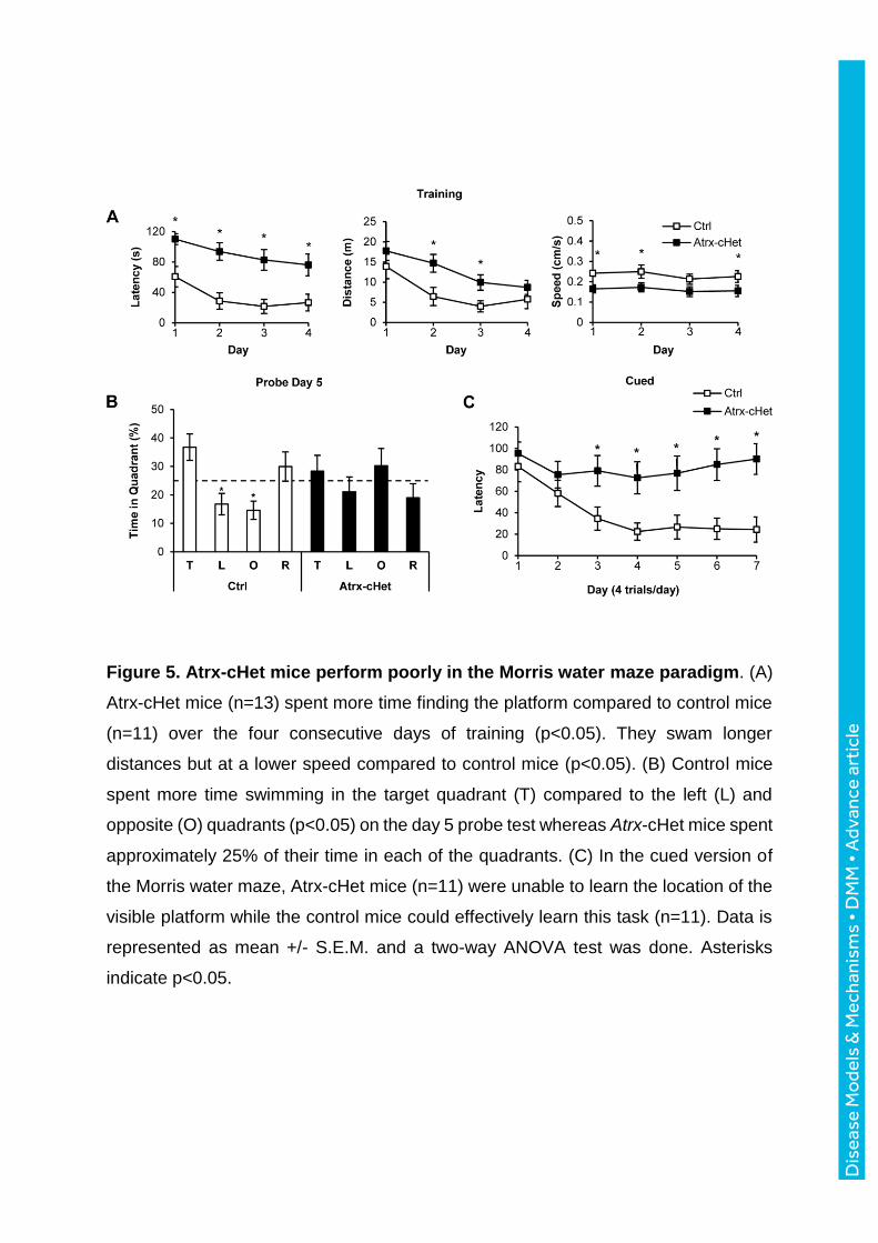
Figure 5. Atrx-cHet mice perform poorly in the Morris water maze paradigm. (A)
Atrx-cHet mice (n=13) spent more time finding the platform compared to control mice
(n=11) over the four consecutive days of training (p<0.05). They swam longer
distances but at a lower speed compared to control mice (p<0.05). (B) Control mice
spent more time swimming in the target quadrant (T) compared to the left (L) and
opposite (O) quadrants (p<0.05) on the day 5 probe test whereas Atrx-cHet mice spent
approximately 25% of their time in each of the quadrants. (C) In the cued version of
the Morris water maze, Atrx-cHet mice (n=11) were unable to learn the location of the
visible platform while the control mice could effectively learn this task (n=11). Data is
represented as mean +/- S.E.M. and a two-way ANOVA test was done. Asterisks
indicate p<0.05.
Dis
ease
Mo
dels
& M
echa
nism
s •
DM
M •
Adv
ance
art
icle

Figure 6. Normal motor memory and endurance but decreased grip strength in
Atrx-cHet mice. (A) Atrx-cHet (n=18) and control (n=16) female mice performed
normally in the rotarod test. (B) Atrx-cHet (n=15) and control (n=15) mice performed
similarly in the treadmill test. (C) Atrx-cHet mice (n=13) exhibit decreased grip strength
compared to control mice (n=11), normalized to body weight. Statistical analyses
made use of two-way ANOVA or two-tailed, unpaired T-test; data represented as
mean +/- S.E.M. and asterisk indicates p<0.05. D
isea
se M
ode
ls &
Mec
hani
sms
• D
MM
• A
dvan
ce a
rtic
le

References:
Aapola, U., Kawasaki, K., Scott, H. S., Ollila, J., Vihinen, M., Heino, M., Shintani, A., Kawasaki, K., Minoshima, S., Krohn, K. et al. (2000). Isolation and initial characterization of a novel zinc finger gene, DNMT3L, on 21q22.3, related to the cytosine-5-methyltransferase 3 gene family. Genomics 65, 293-8. Berube, N. G., Mangelsdorf, M., Jagla, M., Vanderluit, J., Garrick, D., Gibbons, R. J., Higgs, D. R., Slack, R. S. and Picketts, D. J. (2005). The chromatin-remodeling protein ATRX is critical for neuronal survival during corticogenesis. J Clin Invest 115, 258-67. Berube, N. G., Smeenk, C. A. and Picketts, D. J. (2000). Cell cycle-dependent phosphorylation of the ATRX protein correlates with changes in nuclear matrix and chromatin association. Hum Mol Genet 9, 539-47. Bevins, R. A. and Besheer, J. (2006). Object recognition in rats and mice: a one-trial non-matching-to-sample learning task to study 'recognition memory'. Nat Protoc 1, 1306-11. Cardoso, C., Timsit, S., Villard, L., Khrestchatisky, M., Fontes, M. and Colleaux, L. (1998). Specific interaction between the XNP/ATR-X gene product and the SET domain of the human EZH2 protein. Hum Mol Genet 7, 679-84. de Castro, B. M., Pereira, G. S., Magalhaes, V., Rossato, J. I., De Jaeger, X., Martins-Silva, C., Leles, B., Lima, P., Gomez, M. V., Gainetdinov, R. R. et al. (2009). Reduced expression of the vesicular acetylcholine transporter causes learning deficits in mice. Genes Brain Behav 8, 23-35. Declercq, J., Brouwers, B., Pruniau, V. P., Stijnen, P., de Faudeur, G., Tuand, K., Meulemans, S., Serneels, L., Schraenen, A., Schuit, F. et al. (2015). Metabolic and Behavioural Phenotypes in Nestin-Cre Mice Are Caused by Hypothalamic Expression of Human Growth Hormone. PLoS One 10, e0135502. Dhayalan, A., Tamas, R., Bock, I., Tattermusch, A., Dimitrova, E., Kudithipudi, S., Ragozin, S. and Jeltsch, A. (2011). The ATRX-ADD domain binds to H3 tail peptides and reads the combined methylation state of K4 and K9. Hum Mol Genet 20, 2195-203. Drane, P., Ouararhni, K., Depaux, A., Shuaib, M. and Hamiche, A. (2010). The death-associated protein DAXX is a novel histone chaperone involved in the replication-independent deposition of H3.3. Genes Dev 24, 1253-65. Ennaceur, A. and Delacour, J. (1988). A new one-trial test for neurobiological studies of memory in rats. 1: Behavioral data. Behav Brain Res 31, 47-59. Garrick, D., Sharpe, J. A., Arkell, R., Dobbie, L., Smith, A. J., Wood, W. G., Higgs, D. R. and Gibbons, R. J. (2006). Loss of Atrx affects trophoblast development and the pattern of X-inactivation in extraembryonic tissues. PLoS Genet 2, e58. Gibbons, R. J., Bachoo, S., Picketts, D. J., Aftimos, S., Asenbauer, B., Bergoffen, J., Berry, S. A., Dahl, N., Fryer, A., Keppler, K. et al. (1997). Mutations in transcriptional regulator ATRX establish the functional significance of a PHD-like domain. Nat Genet 17, 146-8. Gibbons, R. J., Picketts, D. J., Villard, L. and Higgs, D. R. (1995). Mutations in a putative global transcriptional regulator cause X-linked mental retardation with alpha-thalassemia (ATR-X syndrome). Cell 80, 837-45. Giusti, S. A., Vercelli, C. A., Vogl, A. M., Kolarz, A. W., Pino, N. S., Deussing, J. M. and Refojo, D. (2014). Behavioral phenotyping of Nestin-Cre mice: implications for genetic mouse models of psychiatric disorders. J Psychiatr Res 55, 87-95.
Dis
ease
Mo
dels
& M
echa
nism
s •
DM
M •
Adv
ance
art
icle

Grozeva, D., Carss, K., Spasic-Boskovic, O., Tejada, M. I., Gecz, J., Shaw, M., Corbett, M., Haan, E., Thompson, E., Friend, K. et al. (2015). Targeted Next Generation Sequencing Analysis of 1000 Individuals with Intellectual Disability. Hum Mutat. Guy, J., Hendrich, B., Holmes, M., Martin, J. E. and Bird, A. (2001). A mouse Mecp2-null mutation causes neurological symptoms that mimic Rett syndrome. Nat Genet 27, 322-6. Law, M. J., Lower, K. M., Voon, H. P., Hughes, J. R., Garrick, D., Viprakasit, V., Mitson, M., De Gobbi, M., Marra, M., Morris, A. et al. (2010). ATR-X syndrome protein targets tandem repeats and influences allele-specific expression in a size-dependent manner. Cell 143, 367-78. Leung, J. W., Ghosal, G., Wang, W., Shen, X., Wang, J., Li, L. and Chen, J. (2013). Alpha thalassemia/mental retardation syndrome X-linked gene product ATRX is required for proper replication restart and cellular resistance to replication stress. J Biol Chem 288, 6342-50. Levy, M. A., Kernohan, K. D., Jiang, Y. and Berube, N. G. (2015). ATRX promotes gene expression by facilitating transcriptional elongation through guanine-rich coding regions. Hum Mol Genet 24, 1824-35. Lewis, P. W., Elsaesser, S. J., Noh, K. M., Stadler, S. C. and Allis, C. D. (2010). Daxx is an H3.3-specific histone chaperone and cooperates with ATRX in replication-independent chromatin assembly at telomeres. Proc Natl Acad Sci U S A 107, 14075-80. Logue, S. F., Paylor, R. and Wehner, J. M. (1997). Hippocampal lesions cause learning deficits in inbred mice in the Morris water maze and conditioned-fear task. Behav Neurosci 111, 104-13. Margueron, R., Justin, N., Ohno, K., Sharpe, M. L., Son, J., Drury, W. J., 3rd, Voigt, P., Martin, S. R., Taylor, W. R., De Marco, V. et al. (2009). Role of the polycomb protein EED in the propagation of repressive histone marks. Nature 461, 762-7. Martyn, A. C., De Jaeger, X., Magalhaes, A. C., Kesarwani, R., Goncalves, D. F., Raulic, S., Guzman, M. S., Jackson, M. F., Izquierdo, I., Macdonald, J. F. et al. (2012). Elimination of the vesicular acetylcholine transporter in the forebrain causes hyperactivity and deficits in spatial memory and long-term potentiation. Proc Natl Acad Sci U S A 109, 17651-6. Michod, D., Bartesaghi, S., Khelifi, A., Bellodi, C., Berliocchi, L., Nicotera, P. and Salomoni, P. (2012). Calcium-dependent dephosphorylation of the histone chaperone DAXX regulates H3.3 loading and transcription upon neuronal activation. Neuron 74, 122-35. Morris, R. (1984). Developments of a water-maze procedure for studying spatial learning in the rat. J Neurosci Methods 11, 47-60. Nan, X., Hou, J., Maclean, A., Nasir, J., Lafuente, M. J., Shu, X., Kriaucionis, S. and Bird, A. (2007). Interaction between chromatin proteins MECP2 and ATRX is disrupted by mutations that cause inherited mental retardation. Proc Natl Acad Sci U S A 104, 2709-14. Nogami, T., Beppu, H., Tokoro, T., Moriguchi, S., Shioda, N., Fukunaga, K., Ohtsuka, T., Ishii, Y., Sasahara, M., Shimada, Y. et al. (2011). Reduced expression of the ATRX gene, a chromatin-remodeling factor, causes hippocampal dysfunction in mice. Hippocampus 21, 678-87.
Dis
ease
Mo
dels
& M
echa
nism
s •
DM
M •
Adv
ance
art
icle

Phillips, R. G. and LeDoux, J. E. (1992). Differential contribution of amygdala and hippocampus to cued and contextual fear conditioning. Behav Neurosci 106, 274-85. Picketts, D. J., Higgs, D. R., Bachoo, S., Blake, D. J., Quarrell, O. W. and Gibbons, R. J. (1996). ATRX encodes a novel member of the SNF2 family of proteins: mutations point to a common mechanism underlying the ATR-X syndrome. Hum Mol Genet 5, 1899-907. Ritchie, K., Watson, L. A., Davidson, B., Jiang, Y. and Berube, N. G. (2014). ATRX is required for maintenance of the neuroprogenitor cell pool in the embryonic mouse brain. Biol Open 3, 1158-63. Seah, C., Levy, M. A., Jiang, Y., Mokhtarzada, S., Higgs, D. R., Gibbons, R. J. and Berube, N. G. (2008). Neuronal death resulting from targeted disruption of the Snf2 protein ATRX is mediated by p53. J Neurosci 28, 12570-80. Solomon, L. A., Russell, B. A., Watson, L. A., Beier, F. and Berube, N. G. (2013). Targeted loss of the ATR-X syndrome protein in the limb mesenchyme of mice causes brachydactyly. Hum Mol Genet 22, 5015-25. Stearns, N. A., Schaevitz, L. R., Bowling, H., Nag, N., Berger, U. V. and Berger-Sweeney, J. (2007). Behavioral and anatomical abnormalities in Mecp2 mutant mice: a model for Rett syndrome. Neuroscience 146, 907-21. Tronche, F., Kellendonk, C., Kretz, O., Gass, P., Anlag, K., Orban, P. C., Bock, R., Klein, R. and Schutz, G. (1999). Disruption of the glucocorticoid receptor gene in the nervous system results in reduced anxiety. Nat Genet 23, 99-103. Ulici, V., Hoenselaar, K. D., Agoston, H., McErlain, D. D., Umoh, J., Chakrabarti, S., Holdsworth, D. W. and Beier, F. (2009). The role of Akt1 in terminal stages of endochondral bone formation: angiogenesis and ossification. Bone 45, 1133-45. Vorhees, C. V. and Williams, M. T. (2006). Morris water maze: procedures for assessing spatial and related forms of learning and memory. Nat Protoc 1, 848-58. Wan, H., Aggleton, J. P. and Brown, M. W. (1999). Different contributions of the hippocampus and perirhinal cortex to recognition memory. J Neurosci 19, 1142-8. Watson, L. A., Solomon, L. A., Li, J. R., Jiang, Y., Edwards, M., Shin-ya, K., Beier, F. and Berube, N. G. (2013). Atrx deficiency induces telomere dysfunction, endocrine defects, and reduced life span. J Clin Invest 123, 2049-63. Wiese, C., Rolletschek, A., Kania, G., Blyszczuk, P., Tarasov, K. V., Tarasova, Y., Wersto, R. P., Boheler, K. R. and Wobus, A. M. (2004). Nestin expression--a property of multi-lineage progenitor cells? Cell Mol Life Sci 61, 2510-22. Xing, W., Govoni, K. E., Donahue, L. R., Kesavan, C., Wergedal, J., Long, C., Bassett, J. H., Gogakos, A., Wojcicka, A., Williams, G. R. et al. (2012). Genetic evidence that thyroid hormone is indispensable for prepubertal insulin-like growth factor-I expression and bone acquisition in mice. J Bone Miner Res 27, 1067-79. Xue, Y., Gibbons, R., Yan, Z., Yang, D., McDowell, T. L., Sechi, S., Qin, J., Zhou, S., Higgs, D. and Wang, W. (2003). The ATRX syndrome protein forms a chromatin-remodeling complex with Daxx and localizes in promyelocytic leukemia nuclear bodies. Proc Natl Acad Sci U S A 100, 10635-40. Zhang, J., Ji, F., Liu, Y., Lei, X., Li, H., Ji, G., Yuan, Z. and Jiao, J. (2014). Ezh2 regulates adult hippocampal neurogenesis and memory. J Neurosci 34, 5184-99.
Dis
ease
Mo
dels
& M
echa
nism
s •
DM
M •
Adv
ance
art
icle