motion management for lung and thoracic cancer · motion management for lung and thoracic cancer...
TRANSCRIPT

Motion Management for lung and thoracic cancer
Putipun Puataweepong, MD.
Radiation therapy and oncology unit
Ramathibodi Hospital
The mid annual THASTRO meeting ,10 November 2018

Scope of presentation
o Introduction o Review the motion management techniques
and basic principles of available motion management techniques
o Advantages and disadvantages o Motion management technique for lung SBRT
in Ramathibodi Hospital CyberKnife vs Edge : Based on true story
The results of early stage and locally advanced lung cancer with radiation

2D/3D RT High local failure rates after high-dose RT
Fang LC, 2006
Stage I lung cancer
-Earlier use of 2D-RT planning -Suboptimal dose of RT -Geographic miss

Stage I CA lung SBRT High Local control rate ~ 80-100%
Chi et al, 2010

LA-NSCLC – Standard vs High dose

RTOG 0617: Local Tumor Failure
18-Month
Local
Progression
Rate
34.3%
Lo
ca
l P
rog
ressio
n R
ate
(%
)
0
25
50
75
100
Months since Randomization
0 3 6 9 12 15 18
Patients at RiskStandardHigh dose
213206
205197
187170
165134
137105
113 80
85 62
Fail
6581
HR=1.37 (0.99, 1.89)
Total
213206
p=0.0319
Standard (60 Gy)High dose (74 Gy)
34.3%
25.1%
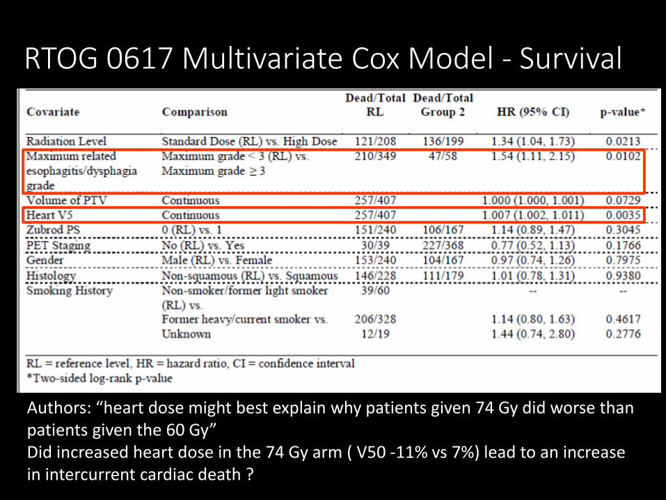
RTOG 0617 Multivariate Cox Model - Survival
Authors: “heart dose might best explain why patients given 74 Gy did worse than patients given the 60 Gy” Did increased heart dose in the 74 Gy arm ( V50 -11% vs 7%) lead to an increase in intercurrent cardiac death ?
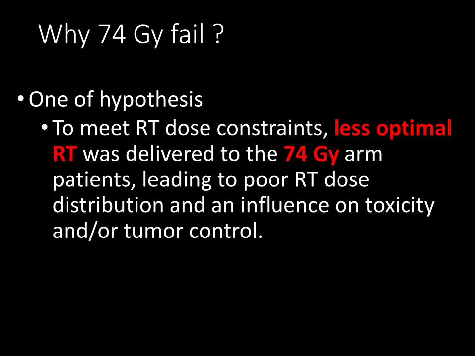
Why 74 Gy fail ?
•One of hypothesis • To meet RT dose constraints, less optimal
RT was delivered to the 74 Gy arm patients, leading to poor RT dose distribution and an influence on toxicity and/or tumor control.

How to improve therapeutic ratio?
Need technologies that allow an increased dose to the tumor while sparing healthy tissue will improve the balance between complications and cure

Advanced radiation therapy
OAR dose
Tumor dose
Target missing risk
Set up errors deletion
Organ motion reduction/
compensation

An important issue in IGRT era •Motion during a fraction •Change in instant geometry •Caused by internal organ motion •Respiratory – much research and
development • Skeletal muscle •Cardiac •Gastrointestinal
Intrafraction motion

Many source of error in RT
Respiratory motion is just one potential source of error in radiotherapy

Lung tumor: Range of motion
Lung – SI- 12 mm on average, up to 50 mm – Largest tumor motion in lower lung

Summary of motion measurement
• No general patterns of respiration
• Many individual characteristics of breathing—quiet versus deep, chest versus abdominal, healthy versus compromised, etc.
• Many motion variations associated with tumor location and pathology
• No association between surrogate ( chest wall or diaphragm ) and tumor or normal structure
Lead to distinct individual patterns in displacement, direction, and phase of tumor motion

When to manage respiratory motion ?
AAPM Task Group 76: respiratory management techniques should be considered if either of the following conditions occur:
• >5 mm range of motion is observed in any direction; or • Significant normal tissue sparing as determined by your clinic can be gained using a respiration management technique.
The management of respiratory motion in radiation oncology report of AAPM Task Group 76

Key component of motion management
Imaging
Slow CT
Breath hold CT
4D-CT
Real-time intervention
Abdominal compression
Breath-hold
Gating
Tracking
Real-time monitoring
Optical
( RPM,
Vision RT)
Electromagnetic ( Calypso)
Hybrid MRI
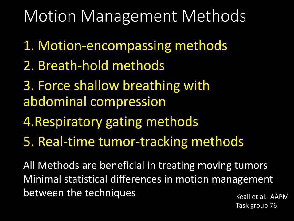
Motion Management Methods
1. Motion-encompassing methods
2. Breath-hold methods
3. Force shallow breathing with abdominal compression
4.Respiratory gating methods
5. Real-time tumor-tracking methods
Keall et al: AAPM Task group 76
All Methods are beneficial in treating moving tumors Minimal statistical differences in motion management between the techniques
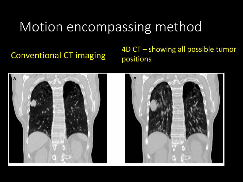
Motion encompassing method
Conventional CT imaging 4D CT – showing all possible tumor positions

First routinely available 4D imaging technique for lung tumors

4D imaging using slow CT scan
Lagerwaard 2001

Slow CT scan
• The imaged structures show the full extent of their movement with respiration
• Disadvantages • Loss of resolution – delineation errors
increase • Not recommended for lesion close to
mediastinum, chest wall or diaphragm, abdominal tumors
Suggested PET*- long acquisition time

Inhale and exhale breath hold CT scan
• CT scan in inhale and exhale breath hold
• Range of motion of target can be determined after fusion
• Combine inhale and exhale GTV to get ITV
• Advantage • Blurring is significantly reduced
• Disadvantages • Increase scan time • Relies on the patient’s ability to hold breath
reproducibility

CT Sim with breath hold

• A process of obtaining image data sets that images are acquired at each couch position for many respiratory phases.
• Other movements/ temporal events (peristalsis, heart beat, organ filling) may or may not be accounted.
• Important to coach patient for periodic breathing as its important for image sorting
• Breathing extent is not the
tumor motion but motion
of the marker
4DCT ( Respiratory correlated CT )

Patient Training
- The ability to achieve reproducible breathing or breath-hold patterns is a requirement for allowing the patient to proceed to simulation and treatment
- Prior to start of simulation, the patient should be made familiar with the equipment and its purpose
- A physicist or trained designee should perform the coaching and evaluation, at least in the initial clinical implementation

Varian RPM (Real‐time positioning management) system
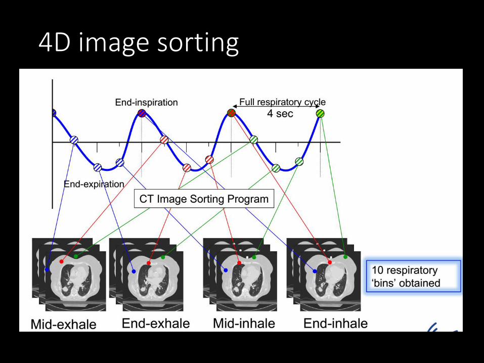
4D image sorting

4DCT • 4D CT not only reduces motion artifacts, but
also gives the tumor/organ motion information
• Irregular respiration will cause artifacts in 4D CT images
• Breathing coaching is always needed
• Radiation exposure from 4DCT is ~ 6 times the dose of a single conventional helical CT scan
• Generation of individualized and usually smaller target volumes derived from 4DCT scans in comparison to standard PTVs justifies this additional radiation exposure
Free breath
4D CT

4D-CT - Significant reduction of the mean PTV from 57.7 cc to 40.7cc (31%) (p< 0.001) , mean lung dose by 17% ( p<0.001)
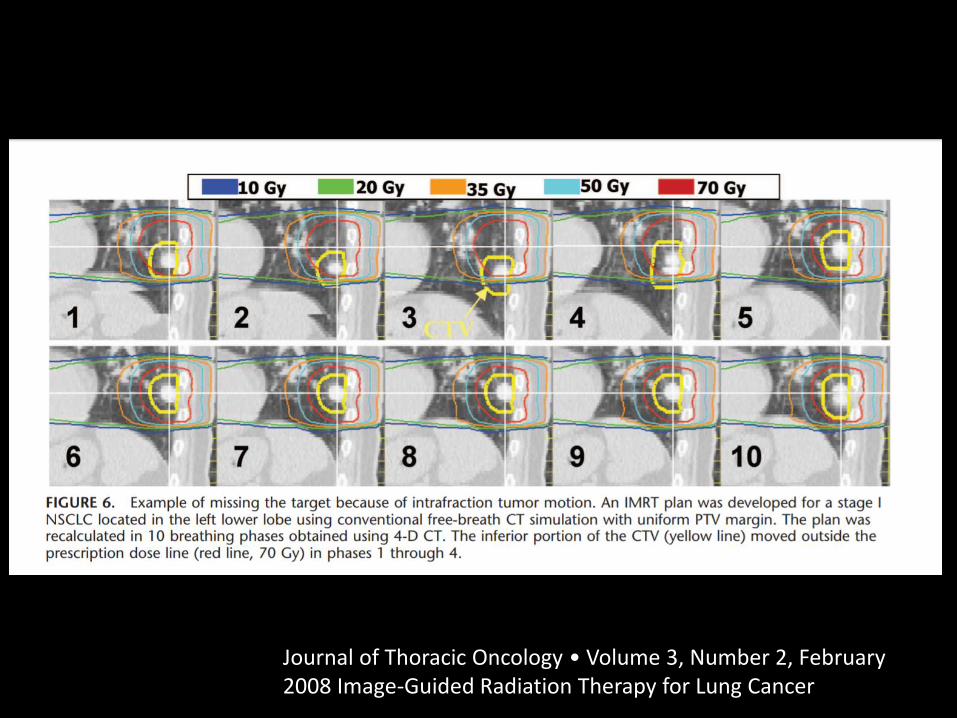
Journal of Thoracic Oncology • Volume 3, Number 2, February 2008 Image-Guided Radiation Therapy for Lung Cancer

4DCT : Key definitions Maximum intensity projection ( MIP)
Maximum HU over all respiratory phases. e.g. Lung tumor. ITV can be estimated by contouring on MIP in lung tumors. Not applicable for GI RT
Minimum intensity projection ( MIN)
Minimum HU over all respiratory phases.
Average intensity projection ( AVG)
Average of all HU at a specific location (often leads to blurred image)
Xi, R & O 2009

Caution when using MIP
Caution with MIP’s for tumors adjacent to mediastinum, diaphragm and atelectasis

RPM with breath hold CT

Motion Management Methods
1. Motion-encompassing methods
2. Breath-hold methods
3. Force shallow breathing with abdominal compression
4.Respiratory gating methods
5. Real-time tumor-tracking methods
Keall et al: The management of respiratory motion in radiation oncology report of AAPM Task group 76

Breath hold methods • Deep‐inspiration
breath hold (DIBH) • Active Breathing
Control (ABC)
Spirometer-monitored technique
Self‐held breath hold with respiratory monitor - RPM - Optical surface - Mechanical devices
tracking the torso - Thermal sensors near
nostril - Fluoroscope images of
implant fiducials - Implants transponders

BH : Inspiration vs Expiration Expiration
• More reproducible and stable
• Prefer in abdominal lesion
Inspiration
• Potential for greater separation between the target and sensitive organs
• Lung volumes are largest and improves lung DVH
• Prefer in thoracic lesion (Lung , breast, mediastinum)
DIBH Heart move away from the chest wall
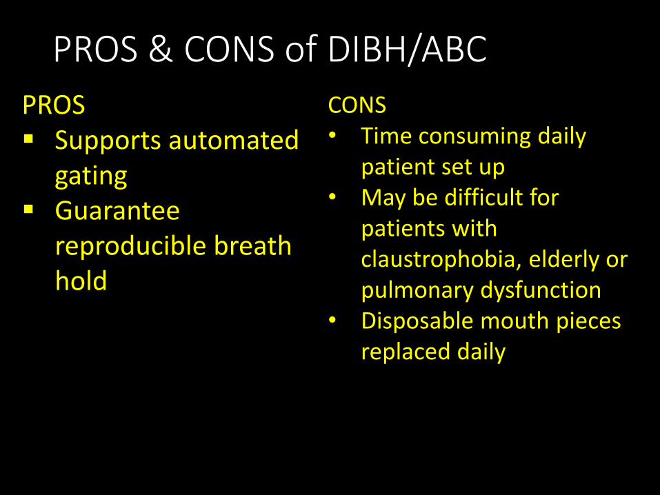
PROS & CONS of DIBH/ABC
PROS Supports automated
gating Guarantee
reproducible breath hold
CONS • Time consuming daily
patient set up • May be difficult for
patients with claustrophobia, elderly or pulmonary dysfunction
• Disposable mouth pieces replaced daily

PROS & CONS of self-held BH
PROS o Patient
respiration is constantly monitored
o Supports automated gating
CONS o Rely heavily on the
patient’s ability and perform a reproducible BH
o Stability of internal anatomy during BH: some pts have been observed to have continuous diaphragm motion during BH, even though they believe they are holding their breath

Motion Management Methods
1. Motion-encompassing methods
2. Breath-hold methods
3. Force shallow breathing with abdominal compression
4.Respiratory gating methods
5. Real-time tumor-tracking methods
Keall et al: The management of respiratory motion in radiation oncology report of AAPM Task group 76

Force shallow breathing with abdominal compression • Motion amplitude of free breathing can be reduced by
mechanical abdominal compression
• Recently, It has been shown to be beneficial only for lower lobe tumors and has no effect or a negative effect on middle and upper lobe tumors
• Whereas the intrafractional amplitude of tumor motion can be reduced by abdominal compression, interfraction motion can even be increased

Lung tumor motion : Free breath vs Abdominal compression
Bouilhol et al. Is abdominal compression useful in lung stereotactic body radiation therapy? A 4DCT and dosimetric lobe-dependent study. Phys Medica. 2013;29:333–340
The most significant impact of AC was obtained in patients with lower lobe tumor. The mean reduction of tumor motion amplitude was 3.5 mm (p = 0.009) for lower lobe tumors and 0.8 mm (p = 0.026) for upper/middle lobe locations. Compression increased motion in 5 cases.

Clinical outcome with or without AC

Motion Management Methods
1. Motion-encompassing methods
2. Breath-hold methods
3. Force shallow breathing with abdominal compression
4.Respiratory gating methods
5. Real-time tumor-tracking methods
Keall et al: The management of respiratory motion in radiation oncology report of AAPM Task group 76

Respiratory gating methods oAIM ‐ irradiate the target volume only when it
moves into a predefined position in the respiratory cycle
o Advantages ‐ Significantly reduces the CTV‐PTV margins ‐ Patient comfortably breathes freely
o Radiation beam is on during only a selected segment (30‐50%) of the respiratory cycle
o Limitation ‐ usually takes longer time to deliver the same prescribed dose

Amplitude based Gating
Breath hold Gating

Respiratory Gating Methods
Gating using internal fiducial markers
Gating using external marker

RPM
- Respiratory pattern of patient during different breathing phases using RPM - Dashed lines indicate upper and lower gating threshold ( 3mm), and determines when gating system turns beam on/off Sung et al, 2014

Respiratory Gating: pro and con
•Reduces margins for motion > 1 cm
•Dosimetric benefits ( lower toxicity)
•Patient-friendly
• Increased room time by 80% and beam-on time by 5.5 x

Motion Management Methods
1. Motion-encompassing methods
2. Breath-hold methods
3. Force shallow breathing with abdominal compression
4.Respiratory gating methods
5. Real-time tumor-tracking methods
Keall et al: The management of respiratory motion in radiation oncology report of AAPM Task group 76

Real-time tumor-tracking methods
Aim: To design radiation beam to follow a moving target by tracking the tumor/organ movement continuously in real‐time Techniques to track tumor position - External respiratory surrogated - Implanted radio-opaque fiducial markers - Surface imaging
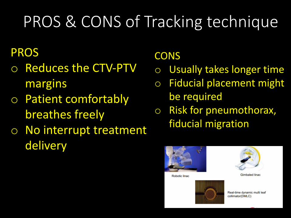
PROS & CONS of Tracking technique
PROS o Reduces the CTV‐PTV
margins o Patient comfortably
breathes freely o No interrupt treatment
delivery
CONS o Usually takes longer time o Fiducial placement might
be required o Risk for pneumothorax,
fiducial migration

Dynamic tracking in Robotic Radiosurgery
-Detect tumor position -Reposition the beam -> 10 years clinical experience

Robotic Radiosurgery: Internal target position
The internal target position can be extracted from: - Fiducials - Tumor density
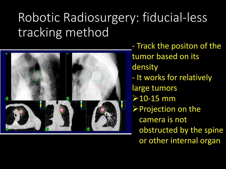
Robotic Radiosurgery: fiducial-less tracking method
- Track the positon of the tumor based on its density - It works for relatively large tumors 10-15 mm Projection on the
camera is not obstructed by the spine or other internal organ

Lung SBRT in Ramathibodi Hospital CyberKnife®
Since 2008
Edge® Since 2016

Eligible criteria for patient
1. Patient accept the risk and benefit of this treatment modality
2. Patient can cooperate and understand all the process of
treatment
Eligible criterial for disease
1. Histological confirm NSCLC, staging T1N0M0 or T2(</=5cm )
N0M0
2. Lung metastasis with known primary malignancy
3. Surgical or medical inoperable or patient refuse surgery or no
further standard treatment
4. Ideally, size of lesion should be < 5 cm and less than 4 lesions,
however, lesion > 5 cm can considered for SBRT, if dose
constraints can be kept within acceptable
SBRT protocol for Lung cancer (primary non-small cell lung cancer, lung metastasis)

RTOG Lung

CyberKnife® Since 2008

3-5 gold (99.9% pure) fiducials placement under CT guidance. For real time tumor tracking ( fiducial as internal surrogate)
immobilization device
undergo a fine-cut 1.5 mm CT scan (full expiration-breath hold phase)
7days (or within 1 month)
start treatment as soon as possible
planning
Workflow for CK SBRT lung
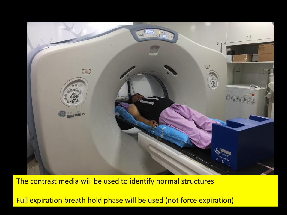
The contrast media will be used to identify normal structures Full expiration breath hold phase will be used (not force expiration)
Target delineation Gross tumor volume (GTV) = gross disease determined from imaging of CT lung windows Clinical target volume (CTV) = GTV Planning target volume (PTV) = GTV + 2 mm. for lateral, anterior-posterior margin + 4 mm. for superior-inferior margin OARs = normal lungs, trachea, esophagus, heart, spinal cord

Planning for this case

Problems with CyberKnife
1. Pneumothorax post fiducial insertion
2. Technical problems with fiducial insertion and rotational
tracking
1.Improper positioning of the fiducials
- track translation – needs at least 1 fiducial
- track rotation - needs 3 fiducials
2. Fiducials migration
- between planning & treatment
- during treatment, inter and intrafraction (CK-long
intrafraction treatment time)

From 21 patients, 32 treatment plans
- > 3 fiducials can be used for planning
13 (61.9%)
- >3 fiducials can be used for intrafraction tracking 6 (28.5%)

Patient has 3 fiducials but only 2 can be used for tracking

Edge® Since 2016

Lung SBRT with Edge system
Target motion > 5 mm and
The patient can hold their breath at least 20 sec ?
Yes No
Free-breath SBRT Self-held breath hold with RPM
monitoring and gating
Vac-lock, arm up, abdominal belt ( lower lobe lesion )
Non-contrast 4D-CT
iGTV – MIP, Organ -average
2 sets of inhale breath hold CT
iGTV – sum of 2 set BH CT
PTV, include iGTV plus 0.5 cm uniformly

Immobilization of Edge : Lung SBRT
Respiratory Belt

Breathing instruction during CT simulation
• Self held inspiration breath hold “Take a breath in, out, in and hold it
• 4DCT
“ Take a regular and slow breathing, breath in, out, in ……
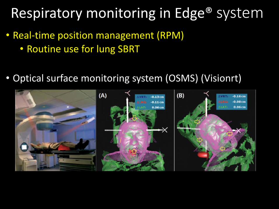
Respiratory monitoring in Edge® system • Real-time position management (RPM)
• Routine use for lung SBRT
• Optical surface monitoring system (OSMS) (Visionrt)
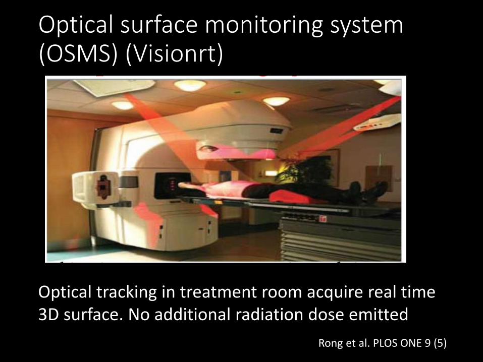
Optical surface monitoring system (OSMS) (Visionrt)
Optical tracking in treatment room acquire real time 3D surface. No additional radiation dose emitted
Rong et al. PLOS ONE 9 (5)

Vision RT schema
During deep inspiration: Fuse planning CT image with real time surface rending at each fraction

During treatment
Real time deltas acquired in 6 degrees of freedom, displayed & if out of user defined tolerances, system gives couch correction to therapist
If patients breath hold is out of tolerance before, or during treatment, beam is manually held until correct breath hold amplitude or positioning acquired

Vision RT schema
Continuous monitoring of patient position during set up and treatment using real time deltas

OSMS in Lung SBRT at our center
• Not routine use for lung SBRT • Problems when monitor with OSMS • Surface image as surrogate, might not
correlated with lung tumor motion • Depend on the selected surface area • If too small – not detect • If too large – easy to out of threshold, beam always turn off
On the day of treatment
• Therapist instructed the patient depend on the technique
• CBCT – Before treatment – physician present at the treatment room to verify CBCT and apply shift
- Middle part of treatment
- End of treatment ( mostly in the fist fraction )
• In BH technique - therapist are instructed to turn on the beam only when the target breath-hold level has been achieved and to stop if the level has fallen below a pre-set tolerance

Inhale BH SBRT with RPM and gating Upper and lower
gating threshold ( 5mm), and determines when gating system turns beam on/off
Free breathing SBRT with RPM, no gating

Comment form real practice • Fiducial tracking
• Pneumothorax risk- accept
• Ideal for manage intrafraction motion
• Patient – friendly
• Staff – less stress if ignore rotation tracking
• Free breathing with ITV concept, no gating
• More friendly to patient and staff
• Inhale breath hold
• Rely heavily on the patient’s ability and perform a reproducible BH
• Introduce more stress to patient and staff

How we select the patient for each technique?

Tumor motion evaluation by 4DCT
Motion <1cm
Regular breathing
Can tolerate long treatment time
Fiducial tracking
Irregular breathing
Poor PS, Comorbid
Can’t tolerate long treatment
ITV in MIP phase
No gating

Tumor motion evaluation
Motion >/= 1 cm
Can tolerate
breath hold
Inhale breath hold
Can’t tolerate breath hold
Regular breathing
Fiducial Tracking
Irregular breathing
ITV in MIP phase

Summary
• Respiratory motion management is beneficial in the reduction of intrafractional motion
• Allows for a decrease in treatment volumes, resulting in a reduction of normal tissue toxicities while giving higher doses to the lesion
• In indirect monitoring it is essential to validate the correlation between surrogate and tumor position during treatment

Motion management methods
Keall et al: The management of respiratory motion in radiation oncology report of AAPM Task group 76
Methods Strategies Techniques Comment
1. Tumor encompassing
Incorporate all movement
-Slow CT -Breath hold CT -4DCT - CINE MRI
-May lead to increased risk of normal tissue toxicity - Imaging dose > 2-15 time
2. Breath hold 3. Force shallow breathing with abdominal compression
Freeze, control motion
-Deep inspiratory breath hold (DIBH) -Active breathing control (ABC)
- Not feasible in all case
4. Respiratory gating
Intercept or control movement
Respiratory as surrogate as tumor position
- Increase treatment time - Difficulty in target verification
5. Real time tumor tracking
Follow or chase tumor
Implanted marker and specialized treatment delivery
- Difficult marker insertion - Increase risk of pneumothorax - Marker migration

Characteristics of proper motion management technique Enables to minimize normal tissue included in the
radiation field Comfortable and reproducible Provides target localization during treatment Not add significant risk Individual patient dependent
Extent of target motion Breathing status Other co-morbidities
Selecting the most appropriate motion management option for each patient requires a good understanding of the available technologies and physic involvement

Thank you for your attention