movement is governed by rotational population dynamics in
TRANSCRIPT

Movement is governed by rotational populationdynamics in spinal motor networks
Henrik Linden,1∗ P. C. Petersen,2, M. Vestergaard3, Rune W. Berg1∗
1 Department of Neuroscience, Faculty of Health and Medical Sciences, University of CopenhagenBlegdamsvej 3, 2200 Copenhagen, Denmark
2 Neuroscience Institute, NYU LangoneNew York University, New York, NY 10016, USA
3 Department of Neuroscience, Max-Delbruck Center for Molecular Medicinein the Helmholtz Association (MDC), Berlin-BuchRobert-Rossle-Strasse 10, 13092 Berlin, Germany
∗Correspondence; E-mail: [email protected] (RWB), [email protected] (HL).
Although the nervous system is elegantly orchestrating movements, the under-
lying neural principles remain unknown. Since flexor- and extensor-muscles
alternate during movements like walking, it is often assumed that the respon-
sible neural circuitry is similarly alternating in opposition. Here, we present
ensemble-recordings of neurons in the turtle lumbar spinal cord that indicate
that, rather than alternation, the population is performing a ”rotation” in neu-
ral space, i.e. the neural activity is cycling through all phases continuously dur-
ing the rhythmic behavior. The radius of rotation correlates with the intended
muscle force. Since existing models of spinal motor control offer inadequate
explanation of this dynamics, we propose a new theory of neural generation
of movement from which rotation and other unresolved issues, such as speed
regulation, force control, and multi-functionalism, are conveniently explained.
1
.CC-BY 4.0 International licenseavailable under a(which was not certified by peer review) is the author/funder, who has granted bioRxiv a license to display the preprint in perpetuity. It is made
The copyright holder for this preprintthis version posted September 1, 2021. ; https://doi.org/10.1101/2021.08.31.458405doi: bioRxiv preprint

The neural circuitry behind movement encompasses several distinct forebrain regions, cere-
bellum and the brainstem. The core executive circuits for movement such as locomotion, how-
ever, reside in the spinal cord (1). These spinal motor circuits, often referred to as central pattern
generators (CPGs), are capable of autonomous generation of rhythmic activity and coordination
of muscles. Although great progress has been made in characterizing the cellular properties of
spinal inter- and motor neurons, including their genetic lineages (2, 3), the network architec-
ture as well as the associated neuronal ensemble dynamics remain elusive. Due to the apparent
right-left and flexor-extensor alternation, it has often been proposed that distinct groups of in-
terneurons, or ‘modules’, are active in a push-pull fashion and that the rhythm is ensured by
cellular pacemaker properties (4, 5). It is unknown if and how such organization and different
motor programs are manifested in ensemble activity of spinal networks.
Here, we examined the activity in spinal motor networks using extracellular multi-electrode
recording in the turtle lumbar spinal cord. This preparation provides mechanical stability, which
allows simultaneous monitoring of large numbers of spinal interneurons in laminae VII-VIII
and motoneurons during the execution of various rhythmic motor programs (6–8). The activity
of individual neurons was rhythmic in relation to the nerves as expected, but the population
activity as a whole appeared rather incomprehensible (Fig. 1A-C). However, when sorting
these neurons according to phase of the motor nerve output we found that the population activity
resembled a continuous sequence, that covered all phases of the cycle (Fig. 1D-E). To better
understand the sequential activity, we performed a principal component analysis (PCA) of both
the neuronal population and the nerve activity. Both the neuronal activity and 6 motor nerves
followed a low-dimensional manifold, i.e. most variance was explained by few components
(Fig. 1D). Whereas the nerve activity appeared entangled, the neuronal activity had a simple
rotation (Fig. 1F). Rotational population activity was observed in all trials and across animals
(Fig. S1-2, Movie S1).
2
.CC-BY 4.0 International licenseavailable under a(which was not certified by peer review) is the author/funder, who has granted bioRxiv a license to display the preprint in perpetuity. It is made
The copyright holder for this preprintthis version posted September 1, 2021. ; https://doi.org/10.1101/2021.08.31.458405doi: bioRxiv preprint

Figure 1: Neuronal population activity in the lumbar spinal cord has rotational dynam-ics. (A) Activity of 3 selected motor nerves (ENG) during rhythmic hindlimb movement. (B)Concurrent ensemble activity in the turtle lumbar spinal cord as raster of spinal neurons (top,n=214) and estimated firing rates (bottom). (C) Sorting the neurons in (B) according to phase(hip flexor) reveals sequential activity. (D) First principal components explains most varianceof ENG activity (green) and neuronal ensemble activity (gray). First two principal componentsof nerve activity (E) and neuronal population (F).
These data indicate that, rather than alternation, the neuronal population is executing a ”ro-
tation”, i.e. the ensemble is cycling through all phases. There did not seem to be any discrete
phase preference as otherwise expected in an alternating modular network (Fig. S1-2). Ro-
tational dynamics has been observed in the motor cortex and elsewhere (9–11), but it has not
been described for spinal circuits previously. Nevertheless, indications can be found as wide
phase-distributions in the scarce literature on spinal population recordings (6, 12–14).
Because conventional CPG models, that are founded on a push-pull organization with in-
trinsically rhythmic modules (15, 16), do not readily explain rotational dynamics, we sought to
conceive a new theory that can accounts for this and other open questions in motor control. In
particular, the mechanisms for generation of rhythms have remained nebulous. Cellular pace-
3
.CC-BY 4.0 International licenseavailable under a(which was not certified by peer review) is the author/funder, who has granted bioRxiv a license to display the preprint in perpetuity. It is made
The copyright holder for this preprintthis version posted September 1, 2021. ; https://doi.org/10.1101/2021.08.31.458405doi: bioRxiv preprint

maker properties has been suggested (4), but decades of research has not been able to pinpoint a
responsible cell type (16). Here, we propose that the rhythm arises from the network rather via
cellular properties. We show that a network, which is close to the transition point of dynamical
instability, can have rhythmogenetic properties without requiring specific cellular properties.
Since the CPG network structure is unknown, our starting point is a parsimonious structure,
where glutamatergic neurons are randomly and recurrently connected. To prevent catastrophic
run-away activity (17, 18) the excitation (E) is balanced by recurrent glycinergic inhibition (I)
(Fig. 2A-B), in line with reports of balanced synaptic input in various motor circuits (19–21).
Balanced networks of this type are known to undergo a phase transition when synaptic weights
are increased beyond a critical value (22, 23). For large networks, activity in this regime is
chaotic (24), whereas finite-sized networks in a dynamical regime close to the transition point
may display more regular activity (25). A linearization of the dynamics close to this point (Sup-
plementary material) shows that finitely-sized networks can generate oscillatory activity if the
leading eigenvalue of the connectivity matrix has a non-zero imaginary part (25). Based on
this idea, we set up a model network of rate-neurons with sparse connectivity where an input
drive, e.g. sensory-related or descending from the brain, could move the eigenvalues of the
connectivity matrix across the stability line due to change in set-point of the firing-rate function
(Fig. 2C-D). As the input was increased beyond a critical level (dashed line) the firing rates
in the network displayed self-sustained rhythmic activity (Fig 2E). Unexpectedly, when sorting
the neurons according to phase, a sequential activity was revealed, i.e. a rotation, similar to the
experimental observations (Fig. 2F-G). We dub the model the ”Balanced Sequence Generator”
(BSG). Both the BSG-model and the experimentally observed rotation are fundamentally differ-
ent from conventional models where the neurons have clustered phase preferences and modules
composed exclusively of excitatory neurons.
To model the output nerve activity from the BSG-network we connected a subset of neurons
4
.CC-BY 4.0 International licenseavailable under a(which was not certified by peer review) is the author/funder, who has granted bioRxiv a license to display the preprint in perpetuity. It is made
The copyright holder for this preprintthis version posted September 1, 2021. ; https://doi.org/10.1101/2021.08.31.458405doi: bioRxiv preprint
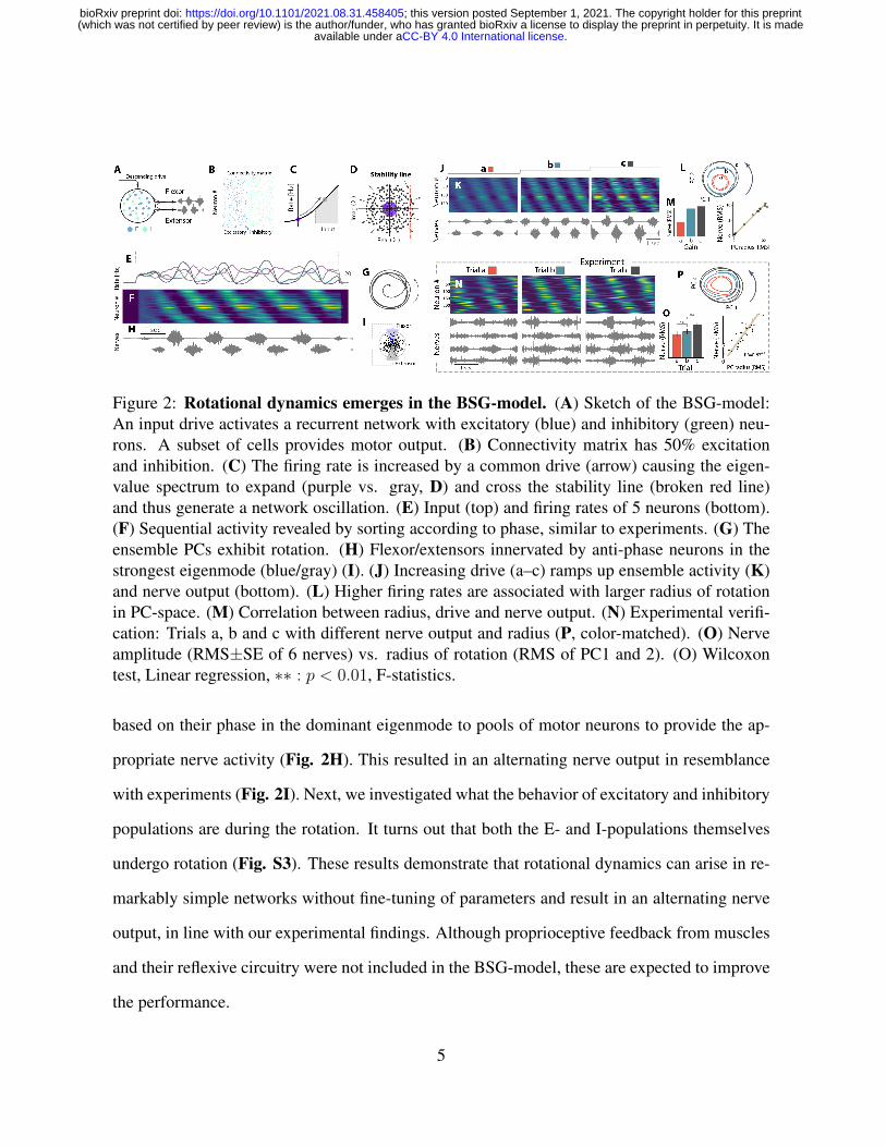
Figure 2: Rotational dynamics emerges in the BSG-model. (A) Sketch of the BSG-model:An input drive activates a recurrent network with excitatory (blue) and inhibitory (green) neu-rons. A subset of cells provides motor output. (B) Connectivity matrix has 50% excitationand inhibition. (C) The firing rate is increased by a common drive (arrow) causing the eigen-value spectrum to expand (purple vs. gray, D) and cross the stability line (broken red line)and thus generate a network oscillation. (E) Input (top) and firing rates of 5 neurons (bottom).(F) Sequential activity revealed by sorting according to phase, similar to experiments. (G) Theensemble PCs exhibit rotation. (H) Flexor/extensors innervated by anti-phase neurons in thestrongest eigenmode (blue/gray) (I). (J) Increasing drive (a–c) ramps up ensemble activity (K)and nerve output (bottom). (L) Higher firing rates are associated with larger radius of rotationin PC-space. (M) Correlation between radius, drive and nerve output. (N) Experimental verifi-cation: Trials a, b and c with different nerve output and radius (P, color-matched). (O) Nerveamplitude (RMS±SE of 6 nerves) vs. radius of rotation (RMS of PC1 and 2). (O) Wilcoxontest, Linear regression, ∗∗ : p < 0.01, F-statistics.
based on their phase in the dominant eigenmode to pools of motor neurons to provide the ap-
propriate nerve activity (Fig. 2H). This resulted in an alternating nerve output in resemblance
with experiments (Fig. 2I). Next, we investigated what the behavior of excitatory and inhibitory
populations are during the rotation. It turns out that both the E- and I-populations themselves
undergo rotation (Fig. S3). These results demonstrate that rotational dynamics can arise in re-
markably simple networks without fine-tuning of parameters and result in an alternating nerve
output, in line with our experimental findings. Although proprioceptive feedback from muscles
and their reflexive circuitry were not included in the BSG-model, these are expected to improve
the performance.
5
.CC-BY 4.0 International licenseavailable under a(which was not certified by peer review) is the author/funder, who has granted bioRxiv a license to display the preprint in perpetuity. It is made
The copyright holder for this preprintthis version posted September 1, 2021. ; https://doi.org/10.1101/2021.08.31.458405doi: bioRxiv preprint

Next, we evaluated whether the BSG-model could embrace previously unsolved issues, such
as independent control of force and speed of the rhythm. The ability to modulate the strength
of the output and speed is key for volitional control, but no mechanisms has been proposed for
controlling these independently to our knowledge. First, we found that collective modulation of
the gain by an input drive could indeed control the amplitude in the BSG-model (Fig. 2J-K).
As the amplitude increased so did the radius of rotation, while the frequency and sequence re-
mained largely unaltered (Fig. 2L-M, S4). To verify this prediction we inspected experimental
trials that, due to the inherent variability, had various radii of rotation (Fig. 2N, S1c). The
radius of rotation had substantial correlation with the motor nerve activity (Fig. 2O, S5), which
provide support for such muscular control in the BSG-model.
Next, we explored the capacity of the BSG-model to control the period of the rhythm. Rather
than collectively adjusting the gain, modulation of the frequency without affecting the amplitude
was indeed possible by selective gain-modulation (Fig. 3). Individual gain-modulation is a
powerful tool in network control (26) and here we tuned a subset of neurons that has most
influence on the period. A systematic perturbation of gain revealed an uneven influence on
the rhythm (Fig. 3A-F). Some neurons had a strong either positive or negative effect, which
we call ”brake-” and ”speed cells”, respectively, while others had small effects. There were
both inhibitory and excitatory neurons among both the speed- and the brake cells (Fig. 3G-H).
Interestingly, cells with a speed-modulating capacity have been demonstrated experimentally
(27,28). Since both excitatory and inhibitory members were found among the brake- and speed
cell categories in our model, we propose this as an experimental prediction. The modulation
capacity is not due to cellular properties, but rather their location in the network. A possible
link between the network location, cell identity, and speed control remains to be assessed.
The ability to execute multiple motor outputs, i.e. a multifunctional output, is the hallmark
of the motor system (29). Contriving a network model that can accommodate the rich reper-
6
.CC-BY 4.0 International licenseavailable under a(which was not certified by peer review) is the author/funder, who has granted bioRxiv a license to display the preprint in perpetuity. It is made
The copyright holder for this preprintthis version posted September 1, 2021. ; https://doi.org/10.1101/2021.08.31.458405doi: bioRxiv preprint

Figure 3: Modulation of frequency in the BSG-model. (A) Adjusting the drive to a neuronchanges the rhythm by moving the eigenvalue up or down (green arrows). (B) Capacity tomodulate the rhythm by an individual neuron is assessed by changing drive (gain). Rankingneurons accordingly reveals ”brake-” and ”speed cells”. (C) When activating brake cells whileimpeding speed cells (activation profile, top left), the rhythm is slowed down compared withneutral (D). Sorted ensemble activity (middle) and nerve output (bottom). Radius of rotation islargely unchanged (PCs top right) indicating a similar amplitude of motor output. (E) Reversedactivation results in faster rhythm (9 Hz). (F) Gradually modulating the speed/brake cells (inset)can either decrease or increase frequency. (G) Capacity to modulate the rhythm has a bell-shaped distribution. Brake and speed cells represent cells with strong modulation capacity, inwhich, both excitatory and inhibitory cells are found. (H) Modulating only excitatory (gray) orinhibitory (orange) cells is sufficient to modulate the frequency.
7
.CC-BY 4.0 International licenseavailable under a(which was not certified by peer review) is the author/funder, who has granted bioRxiv a license to display the preprint in perpetuity. It is made
The copyright holder for this preprintthis version posted September 1, 2021. ; https://doi.org/10.1101/2021.08.31.458405doi: bioRxiv preprint

toire of motor behaviors has so far been a major challenge. For this reason, we explored the
potential of generating multiple motor patterns by selective activation within the BSG-network.
We used two well-known motor behaviors in turtles and investigated these both experimentally
and in the model. These behaviors consist of hind limb movements, where either the knee is
protracted while moving the foot in small circles (pocket scratching) or protracting the foot
with stretched leg (rostral scratching) (30). Indeed, the BSG-model was able to produce quali-
tatively similar patterns. This was accomplished by identifying two distinct neuronal input sets
for gain-modulation in the network (Fig. 4A-B). A small group of output neurons drive the
motor neurons and the consequent movement was induced (Fig. 4C). The output of behav-
ior 1 had knee/hip extensors in phase (‘no shift’, Fig. 4D), whereas the second input pattern
ensured the required phase-shift (Fig. 4E). Interestingly, in spite of the difference in motor
output, the network patterns had marginal visual differences in both behaviors (cf. Fig 4D-E,
Movie S2). The neuronal phase-preference during the two behaviors were broadly scatter near
the unity line (Fig. 4F), which indicates that many neurons kept their timing in the sequence.
Roughly half of the neurons had a change in phase of less than ±45o (48.5 %) (Fig. 4G-H).
These observation were qualitatively similar to experimental observations, where the sequential
activation, although not identical, remained during the two behaviors (Fig. 4I-M). We further
tested whether other distinct motor patterns could be evoked in the BSG-model. A plethora of
patterns or ”gaits” could be induced via different activation profiles, with a diversity conspicu-
ously similar to the diversity of real motor patterns (Fig. S7-8). Generation of a desired motor
pattern is a quest of finding the appropriate combination of neurons to modulate. We suggest
this to be accomplished in trial-and-error-based motor learning.
In this report, we present compelling evidence that, rather than exhibiting push-pull activity,
the spinal motor circuitry performs rotation. This is a low-dimensional dynamics, where the
population continuously cycles through all phases and the radius of rotation correlates with the
8
.CC-BY 4.0 International licenseavailable under a(which was not certified by peer review) is the author/funder, who has granted bioRxiv a license to display the preprint in perpetuity. It is made
The copyright holder for this preprintthis version posted September 1, 2021. ; https://doi.org/10.1101/2021.08.31.458405doi: bioRxiv preprint
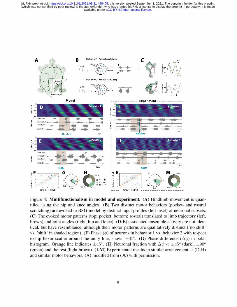
Figure 4: Multifunctionalism in model and experiment. (A) Hindlimb movement is quan-tified using the hip and knee angles. (B) Two distinct motor behaviors (pocket- and rostralscratching) are evoked in BSG-model by distinct input profiles (left inset) of neuronal subsets.(C) The evoked motor patterns (top: pocket, bottom: rostral) translated to limb trajectory (left,brown) and joint angles (right, hip and knee). (D-E) associated ensemble activity are not iden-tical, but have resemblance, although their motor patterns are qualitatively distinct (’no shift’vs. ’shift’ in shaded region). (F) Phase (φ) of neurons in behavior 1 vs. behavior 2 with respectto hip flexor scatter around the unity line, shown ±45o. (G) Phase difference (∆φ) in polarhistogram. Orange line indicates ±45o. (H) Neuronal fraction with ∆φ < ±45o (dark), ±90o
(green) and the rest (light brown). (I-M) Experimental results in similar arrangement as (D-H)and similar motor behaviors. (A) modified from (30) with permission.
9
.CC-BY 4.0 International licenseavailable under a(which was not certified by peer review) is the author/funder, who has granted bioRxiv a license to display the preprint in perpetuity. It is made
The copyright holder for this preprintthis version posted September 1, 2021. ; https://doi.org/10.1101/2021.08.31.458405doi: bioRxiv preprint

intended muscular force. The sequence involved in one behavior is similar to that of a different
behavior (Fig. 4, S7-8). These aspects of motor control have not previously been considered,
and to explain these we had to develop a new theory, the ‘Balanced Sequence Generator’. The
hallmark is sequential activity in a balanced network, which is induced by tonic input from
e.g. descending commands or sensory input. Interestingly, if the drive is brief or targeted,
the model network could also generate non-rhythmic movement sequences, such as stroking
a tennis ball or kicking a soccer ball. Hence, this theory could provide an important bridge
between the motor circuits for rhythmic movement with those for non-rhythmic sequences,
that has previously been absent. We predict such non-rhythmic movements to be generated
by a single cycle of neural rotation, which is sculpted by selective gain modulation the spinal
network via descending commands from the brain or via sensory input.
ACKNOWLEDGMENTS
Funding: This work was supported by The Independent research fund Denmark, and the Carls-
berg foundation. Author contributions: R.W.B. conceived the original experiments. P.C.P.
performed the experiments, collected the data and sorted the spikes. M.W., H.L. and R.W.B.
conceived the original theory. H.L. and R.W.B. designed, and developed the theory, analysed
the experimental data, and wrote the manuscript. Competing interests: None declared. Data and
materials availability: Data analysis code (MATLAB) and model implementation code (Python)
is available on the lab web page (www.berg-lab.net). All (other) data needed to evaluate the con-
clusions in the paper are present in the paper or the supplementary materials.
SUPPLEMENTARY MATERIALS
URL here Materials and Methods Figs. S1 to S9 Table S1 MDAR Reproducibility Checklist
10
.CC-BY 4.0 International licenseavailable under a(which was not certified by peer review) is the author/funder, who has granted bioRxiv a license to display the preprint in perpetuity. It is made
The copyright holder for this preprintthis version posted September 1, 2021. ; https://doi.org/10.1101/2021.08.31.458405doi: bioRxiv preprint

References and Notes
1. S. Arber, R. M. Costa, Science 360 (2018).
2. S. Gosgnach, et al., Journal of Neuroscience 37, 10835 (2017).
3. M. Goulding, Nat Rev Neurosci 10, 507 (2009).
4. S. Grillner, A. El Manira, Physiological Reviews 100, 271 (2020).
5. D. A. McCrea, I. A. Rybak, Brain Res Rev 57, 134 (2008).
6. P. S. G. Stein, S. Daniels-McQueen, J. Neurosci. 22, 6800 (2002).
7. P. C. Petersen, R. W. Berg, eLife 5, e18805 (2016).
8. M. Radosevic, et al., Nature Communications 10, 2937 (2019).
9. M. M. Churchland, et al., Nature 487, 51 (2012).
10. D. Sussillo, M. M. Churchland, M. T. Kaufman, K. V. Shenoy, Nature Neuroscience 18,
1025 (2015).
11. A. M. Bruno, W. N. Frost, M. D. Humphries, eLIFE 6, e27342 (2017).
12. M. B. Berkinblit, T. G. Deliagina, A. G. Feldman, I. M. Gelfand, G. N. Orlovsky, Journal
of neurophysiology 41, 1040 (1978).
13. N. Auyong, K. Ollivier-lanvin, M. A. Lemay, J Neurophysiol 106, 1943 (2011).
14. A. C. Kwan, S. B. Dietz, G. Zhong, R. M. Harris-Warrick, W. W. Webb, J Neurophysiol
104, 3323 (2010).
15. O. Kiehn, et al., Brain Res Rev 57, 56 (2008).
11
.CC-BY 4.0 International licenseavailable under a(which was not certified by peer review) is the author/funder, who has granted bioRxiv a license to display the preprint in perpetuity. It is made
The copyright holder for this preprintthis version posted September 1, 2021. ; https://doi.org/10.1101/2021.08.31.458405doi: bioRxiv preprint

16. V. Rancic, S. Gosgnach, International Journal of Molecular Sciences 22, 1 (2021).
17. G. Hennequin, E. J. Agnes, T. P. Vogels, Annual Review of Neuroscience 40, 557 (2017).
18. R. W. Berg, A. Willumsen, H. Linden, Current Opinion in Physiology 8, 76 (2019).
19. R. W. Berg, A. Alaburda, J. Hounsgaard, Science 315 (2007).
20. T. A. Machado, Probing circuits for spinal motor control, Ph.D. thesis, Columbia University
Academic Commons (2015).
21. J.-M. Ramirez, N. A. Baertsch, Annual Review of neuroscience 41, 475 (2018).
22. K. Rajan, L. F. Abbott, H. Sompolinsky, Physical Review E - Statistical, Nonlinear, and
Soft Matter Physics 82 (2010).
23. G. Hennequin, T. P. Vogels, W. Gerstner, Neuron 82, 1394 (2014).
24. C. van Vreeswijk, H. Sompolinsky, Science 274, 1724 (1996).
25. M. Beiran, S. Ostojic, PLOS Computational Biology 15 (2019).
26. J. P. Stroud, M. A. Porter, G. Hennequin, T. P. Vogels, Nature Neuroscience 21, 1774
(2018).
27. S. Gosgnach, et al., Nature 440, 215 (2006).
28. R. A. Callahan, et al., eLife 8, 1 (2019).
29. A. Berkowitz, A. Roberts, S. R. Soffe, Frontiers in Behavioral Neuroscience 4 (2010).
30. L. I. Mortin, J. Keifer, P. S. Stein, J. Neurophysiol. 53, 1501 (1985).
12
.CC-BY 4.0 International licenseavailable under a(which was not certified by peer review) is the author/funder, who has granted bioRxiv a license to display the preprint in perpetuity. It is made
The copyright holder for this preprintthis version posted September 1, 2021. ; https://doi.org/10.1101/2021.08.31.458405doi: bioRxiv preprint