mr carl baptista, director research & … the development of infection and accelerated healing...
TRANSCRIPT

Mr Carl Baptista, Director Research & Development, ORIGIN Group

Clinical Application in History
• First documented during Napoleonic Wars -“prevented the development of infection and accelerated healing” (Larrey 1832)
• Clinically used during the American Civil War (1861 – 1865)
• Stopped in 1930s - advent of antibiotics. Then, MRSA
• Re-introduced into UK around 1995
• First clinical trial in Singapore @TTSH in 2008 –14 patients (15 wounds, scheduled for
amputation
–Results: Successful outcome for 7 patients

1st Medical Grade Maggots Biomedical Lab in Singapore

LUCILA CUPRINA (SHEEP- BLOW FLY)
I have 10,000 Friends

Where do maggots come from?
Life cycle fly - maggot - fly - maggot…………

Adults emerge on the 8th day from the pupa stage. At about 3-4 weeks, adult females begin laying eggs. After mating, females can lay 2-4 batches of 200 eggs in their lifetime.
Maggots will first form a beige cocoon initially, slowly turning into a darker shade of brown until they mature. This process takes about 7 days.
Eggs are harvested from slivers of meat and allowed to hatch. 1st instar maggots emerge around 18 hours.
1st instar maggot (1mm) takes about 4 days to reach 3rd instar (10mm); feeding on rotting meat. They will then search for a dry environment to pupate. This is known as pre-pupa stage.
18 HOURS
4 DAYS 7 DAYS
21 DAYS
Life Cycle of Lucila Cuprina

Procurement & Production Process
Upon collated orders by 1 pm everyday, Eggs are harvested from slivers of sheep hearts.
Eggs are then delicately separated by hands within a specified time frame.
Eggs are rinsed with tap water and disinfectant solution
After rinsing, the eggs are transferred into a test tube for sterilization
Finally, the eggs are placed into sterile transportation vials; ready for delivery
Each batch of sterilized eggs is inoculated on blood and choc agar for disinfectant control to test for sterility
The eggs are repeatedly rinsed and shaken for 45 minutes

FREE RANGE • Applied for maximum of
48-hrs
• Allows natural mechanical benefit of maggots’ movement
• Extensive coverage
Baggot • Applied for maximum of
72-hrs
• Reduces pain
• Removes ‘Yuk’ factor
• Time management –quick application and removal
Considerations of MediFly Type

Slough
Necrosis
Gangrene

Ulcers Grade 4 –6
Last case
scenario

Maggot Debridement Therapy
the use of live medical grade sterile larvae to effectively clean the wound bed of devitalized tissue which impedes the normal wound healing process

Three commonly recognised phases, which overlap: An orchestrated series of events, with
culmination of these biological processes resulting in the replacement of normal skin structures with fibroblastic mediated scar tissue
Inflammatory phase
Proliferation phase
Maturation phase
Acute (NORMAL) Wound Healing

• Living creatures requiring oxygen and food to survive
• No teeth!
• Chemical factories – move over surface of wound secreting a powerful mixture of proteolytic enzymes which break down dead tissue, liquidizing it.
• The maggots then ‘suck’ up this liquid and ingest it
• Only liquefy devitalized tissue including MRSA
MDT Debridement A Clean Wound Bed

• Ingest and digest the bacteria within the devitalized tissue in the wound, which are killed in their gut (Robinson and Norwood 1933)
• The secretions increase the pH of the wound to around 8 to 8,5 due to the production of ammonia (excreted), inhibiting the growth of some bacteria (Messer and McClellan 1935)
• Secrete chemicals with inherent antimicrobial activity and these help combat infection (Pavillard and Wright 1957)
MDT Debridement A Clean Wound Bed

• ANY CHRONIC ULCER : Pressure Sores, Diabetic Foot Ulcers, Venous, Ischemic, Malignant, Burns….
• MRSA • Devitalized tissue – slough,
necrosis, gangrene • Non-aggressive & quick
debridement • Bio-film formation • Alternative to surgery • Painful adhered Slough
Indications

• Wounds that contain fistulae, or connect with vital organs
• Pseudomonas infections
• Haemophiliac
• Use with caution near exposed blood vessels – monitor wound regularly
• Dry necrotic wounds
• Pain – Baggots
• Osteomyelitis?
Contra-Indications

Osteomyelitis?

What exactly am I applying?
• 1st in-star maggot (approx. 1mm) • They turn into flies in the wound? • Count in and out? • Painful?

How many to apply

•Create ‘cage system’ dressing to contain maggots • Need O2 and food • Allow room for expansion • Consider position of discharge • Off-load
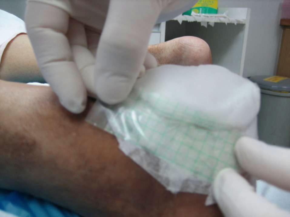
How to Apply?

Step 1: Primary Dressing Frame the wound with Hydrocolloid dressing
Step 2: Primary Dressing Place live maggots onto gauze and invert onto the wound
Step 3: Primary Dressing Encage using gas/air permeable tape e.g. Tagaderm, Opsite
Step 4: Secondary Dressing Place moistened gauze lightly above the Bio Dressing
MDT Application

Application of Maggots in a Bag

Removal –Maggots remain in Bag


• Managing increase in discharge
• Pain / Crawling
• Off-loading
• Regular change of secondary dressings required for effective treatment
MDT Considerations

• Maggots will have grown to approx. 1cm
• Sterile water to irrigate wound clean of maggots
• Photosensitive – create dark environment
• Leave some in, not to be concerned
• Dispose in biohazard waste bag and drown in alcohol based solution. Double bag.
What to expect on removal

Dispose in biohazard and drown alcohol solution

Emergency Escapees!!!!!!
Contact your hospital pest
control company
Contact your nurse clinician
in charge
Discard all escapees as you
would with dressing – biohazard
waste and drown in alcohol.

• 60-Yrs old Male
• Type II Diabetes
• Poor diabetic control
• Smoker
• Successful Revascularization
• Slough adhered –painful for debridement
3 DAYS
POST APPLICATION ONE BAGGOT
• Patient reported minimal pain or discomfort
• Despite advice –Self-discharge for personal reasons • Outpatient –Seen fully granulating
Painful Ischemic Ulcer

Diabetic chronic ischemic ulcer
Skin appearance dorsum and planter dusky, with minor blanching apparent. Necrotic edges dry and adherent. Bone visible within the wound bed – 4th metatarsal region.
• Singapore for third opinion. • Re-vascularisation – pop bypass • Remained questionable to viability of forefoot

Application two free range vials
2 DAYS

Total 6 vials applied = 3 dressings
INITIAL PRESENTATION 11 MAY PRESENTATION 17TH MAY
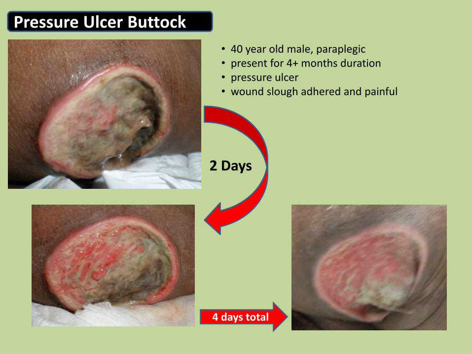
Pressure Ulcer Buttock
• 40 year old male, paraplegic • present for 4+ months duration • pressure ulcer • wound slough adhered and painful
2 Days
4 days total

• 54-Yr old Male, TYPE II Diabetic
• Began as a blister on big toe –quick deterioration and gangrene
• Offered forefoot amputation due to deterioration of all lesser toes
• Re-vascularized successfully
2 DAYS
Post application of one Free Range
• Patient graft surgery same days as removal of MDT
• Discharged 2 days post graft.
Post Forefoot Amputation

2 DAYS 2 DAYS
• 70-Yrs old Female • Type II Diabetes • <30 yrs. duration •Acute Ischemia –
Revascularized •Debridement of non-
viable Achilles Tendon
Pre-Application Post application: 3 Free Range
Post application: 5 Free Range
• No pain reported • Proceed with Negative Pressure dressing • Continues to granulate & decrease in aperture
Apply 3 Free Range
Apply 2 Free Range
Chronic Heel Pressure Sore

Any Chronic Wound…….?

Post removal of MDT

Fungating Breast Wound


• Studies demonstrated maggots can clean wounds in a fraction of the time taken by more conventional dressings (Waymen et al.2001)
• Maggots on average clean wound bed in 5 days; hydrogels 86 days….. Consider long term cost implications not just the short!
• Management of infected wounds containing bacteria that are difficult to kill with more conventional treatments.
• Eliminate MRSA from wounds.
• Reduce need for long antibiotic use – cost effective
Why Maggots to conventional dressings?

Cost Effective – Long Term
In 2008 TTSH approached ORIGIN Scientia to undertake a study to report on our initial experience with MDT, our patients’ perception and to assess factors likely to influence outcome. All patients included on this trial were scheduled for amputations either major or minor. Within only 3 months it was very apparent that MDT was successful in terms of reducing amputation rates significantly. Of the 14 patients, with 15 wounds all scheduled for amputation, 47% had successful outcomes. Ie. 47% did not require amputation

MAGGOTS SUCK! Keep an open mind………