multi-resistance plasmids
TRANSCRIPT

ACTAUNIVERSITATIS
UPSALIENSISUPPSALA
2019
Digital Comprehensive Summaries of Uppsala Dissertationsfrom the Faculty of Medicine 1587
Multi-Resistance Plasmids
Fitness Costs, Dynamics and Evolution
FREDRIKA RAJER
ISSN 1651-6206ISBN 978-91-513-0708-4urn:nbn:se:uu:diva-389855

Dissertation presented at Uppsala University to be publicly examined in Room B:42, BMC,Husargatan 3, Uppsala, Friday, 27 September 2019 at 13:00 for the degree of Doctor ofPhilosophy (Faculty of Medicine). The examination will be conducted in English. Facultyexaminer: Professor Pål Jarle Johnsen (UiT The Arctic University of Norway, Department ofPharmacy).
AbstractRajer, F. 2019. Multi-Resistance Plasmids. Fitness Costs, Dynamics and Evolution. DigitalComprehensive Summaries of Uppsala Dissertations from the Faculty of Medicine 1587.85 pp. Uppsala: Acta Universitatis Upsaliensis. ISBN 978-91-513-0708-4.
Antibiotic resistance is an escalating problem, not only due to less desirable treatment optionsand outcome, but also due to the economic burden to health care caused by resistant pathogens.Since the process of developing new antibiotics is slow, we need to carefully consider the usageof the antibiotics still available. Therefore it is of importance to minimize the development andspread of resistant pathogens. To do so, we need a better understanding of the mechanisms anddynamics underlying the evolution of highly resistant bacteria.
In this thesis I have investigated one of the major drivers of resistance gene disseminationin Gram-negative bacteria, namely multi-resistance plasmids. We show that multi-resistanceplasmids display a dynamic behavior in vivo, where genes can be readily acquired and lost again.Additionally, plasmids can be shared amongst different bacteria, especially in environmentssuch as the human gut. Interestingly, some resistance plasmids confer a fitness disadvantageto their host displayed by decreased growth rate in absence of antibiotics. We could elucidatethat two resistance genes of the multi-resistance plasmid pUUH239.2 were the cause of thelowered growth rate, namely blaCTX-M-15 and tetR/A. In contrast, other resistance genes on theplasmid were cost-free even when overexpressed and likely enable persistence in the bacterialpopulation even under non-selective conditions. Lastly, we studied how the presence of severalβ-lactamase genes on a plasmid affects treatment with different combinations of β-lactam/β-lactamase inhibitors. We found that an efficient mechanism for bacteria to overcome high levelsof antibiotics was by amplification of plasmid-borne resistance genes. This mechanism worksas a stepping-stone for additional mutations giving rise to high-level resistance.
With this work we provide insight into the mechanisms underlying resistance evolution anddissemination due to multi-resistance plasmids. Plasmids enable fast dissemination of multipleresistance genes and therefore simultaneously disable multiple treatment options. Examiningthe effects of resistance genes and antibiotics on strains carrying multi-resistance plasmids willenable us to understand what factors assist or inhibit plasmid spread. Hopefully, this will aid us intreatment design to prevent resistance development to effective antibiotics and have implicationsfor resistance surveillance as well as prediction.
Keywords: multi-resistance plasmids, evolution, fitness cost, dynamics, escherichia coli,ESBL, gene amplification, β-lactamase inhibitor
Fredrika Rajer, Department of Medical Biochemistry and Microbiology, Box 582, UppsalaUniversity, SE-75123 Uppsala, Sweden.
© Fredrika Rajer 2019
ISSN 1651-6206ISBN 978-91-513-0708-4urn:nbn:se:uu:diva-389855 (http://urn.kb.se/resolve?urn=urn:nbn:se:uu:diva-389855)

Till min familj <3


List of Papers
This thesis is based on the following papers, which are referred to in the text by their Roman numerals.
I Brolund A°, Rajer F°, Giske CG, Melefors Ö, Titelman E, Sandegren L. (2019). Dynamics of resistance plasmids in extended-spectrum-β-lactamase-producing Enterobacteriaceae during postinfection colonization. Journal of Antimicrobial Chemotherapy, 63:e02201-18
II Rajer F and Sandegren L. (2019). The role of antibiotic resistance genes in the fitness cost of multi-resistance plasmids. Manuscript
III Rajer F, Allander L, Karlsson P, Sandegren L. (2019). Evolutionary trajectories towards high-level β-lactam/β-lactamase inhibitor re-sistance of a multi-resistance plasmid carrying multiple β-lactamases. Manuscript
° Authors contributed equally to the work.
Reprints were made with permission from the respective publishers. Articles not included in this thesis:
Müller V, Rajer F, Frykholm K, Nyberg LK, Quaderi S, Fritzsche J, Kristiansson E, Ambjörnsson T, Sandegren L, Westerlund F. (2016). Direct identification of antibi-otic resistance genes on single plasmid molecules using CRISPR/Cas9 in combina-tion with optical DNA mapping. Sci. Rep. 6, 37938 Bikkarolla SK, Nordberg V, Rajer F, Müller V, Kabir MH, KK S, Dvirnas A, Ambjörnsson T, Giske CG, Navér L, Sandegren L, Westerlund F. (2019). Optical DNA mapping combined with Cas9-targeted resistance gene identification for rapid tracking of resistance plasmids in a neonatal intensive care unit outbreak. mBio 10:e00347-19. Knopp M, Gudmundsdottir JS, Nilsson T, König F, Warsi O, Rajer F, Ädelroth P, Andersson DI. (2019). De novo emergence of peptides that confer antibiotic re-sistance. mBio 10:e00837-19


Contents
Introduction ................................................................................................... 11The importance of antibiotics ................................................................... 11Antibiotic classes and mechanisms of action ........................................... 12Β-lactam antibiotics and β-lactamase inhibitors .................................. 14
Bacterial antibiotic resistance ................................................................... 20The intrinsic antibiotic resistance ........................................................ 20Acquired antibiotic resistance .............................................................. 21Β-lactam resistance .............................................................................. 25Resistance to β-lactam and β-lactamase inhibitor combinations ......... 31
The role of horizontal gene transfer in antibiotic resistance .................... 32Mobile genetic elements ...................................................................... 34
The Mischievous Plasmids ....................................................................... 38Resistance plasmids ............................................................................. 42Clinical and economic impact of ESBL-producing bacteria ............... 43
Bacterial fitness ........................................................................................ 46The effect of chromosomal alterations on bacterial fitness ................. 47The effect of resistance plasmids on bacterial fitness .......................... 48
Increased resistance by gene duplication and amplification .................... 50
Current Investigations and Future Perspectives ............................................ 53Dynamics of resistance plasmids in extended spectrum β-lactamase-producing Enterobacteriaceae during post-infection colonization ...... 53The role of antibiotic resistance genes in the fitness cost of multi-resistance plasmids ............................................................................... 54Evolutionary trajectories towards high-level β-lactam/β-lactamase inhibitor resistance of a multi-resistance plasmid carrying multiple β-lactamases ......................................................................................... 56
Concluding remarks ...................................................................................... 58
Populärvetenskaplig sammanfattning ........................................................... 60
Populärwissenschaftliche Zusammenfassung ............................................... 62
Acknowledgements ....................................................................................... 64
References ..................................................................................................... 67


Abbreviations
BSI bloodstream infection CZA ceftazidime - avibactam DBO diazabicyclo[3.2.1]octanone DNA deoxyribonucleic acid EPE ESBL-producing Enterobacteriaceae ESBL extended-spectrum β-lactamase GDA gene duplication and amplification HGT horizontal gene transfer HMM high molecular mass Inc incompatibility group IR inverted repeats IRT inhibitor-resistant TEM β-lactamase IS insertion sequence LMM low molecular mass MBL metallo-β-lactamase MIC minimum inhibitory concentration mRNA messenger RNA MRSA methicillin-resistant Staphylococcus aureus OM outer membrane OMP outer membrane protein PBC penicillin binding complex PBP penicillin binding protein PSK post-segregational killing RNA ribonucleic acid SAM ampicillin - sulbactam SNP single nucleotide polymorphism ST sequence type T4CP type IV coupling protein T4SS type IV secretion system TA toxin-antitoxin TCS two-component system TE transposable element tRNA transfer RNA TU translocatable unit TZP piperacillin – tazobactam


11
Introduction
"The thoughtless person playing with penicillin treatment is morally respon-sible for the death of the man who succumbs to infection with the penicillin-resistant organism." - Sir Alexander Fleming, 1945
The importance of antibiotics In the early 20th century common bacterial infections plagued the world. Infections caused by minor scratches and scrapes were potentially deadly, and pneumonia killed otherwise healthy people. In 1928 Sir Alexander Fleming discovered the important antibiotic penicillin1, followed by the sul-fonamides discovered by Gerhard Domagk in 19352. These drugs could fight many common infections, such as pneumonia and sepsis. However, re-sistance towards these drugs developed fast. Penicillin-resistant strains were already found before introduction of the drug3, a warning sign not many took seriously, except for Sir Alexander Fleming himself. The resistance mecha-nisms towards these classes of antibiotics are the same today as the ones that were already found in the end of the 1930s. The modern use of antibiotics has created a more resistant community of bacteria over the last decades. During the golden age of antibiotics, more and more drugs able to treat many kinds of infections were discovered or developed. A common conception was that mankind had eradicated bacterial infectious diseases. Due to the initial success of the antibiotics man thought himself to be spared from death caused by bacteria, which lead to a new primary focus in the field of medi-cine, chemotherapy. There were no new antibiotic classes discovered be-tween the 1960s and 2000s. Even today the discovery of new antibiotics are limited, unfortunately mostly due to economical reasons.
Noteworthy is that antibiotics and antibiotic resistance mechanisms have existed long before the commercial use of antibiotics in treatment of human and animal infections4–6. It is estimated that microorganisms produced anti-biotics as natural products more than 2 billion years ago, indicating that also the resistance mechanisms might be as old5,7,8. The use of antibiotics has enabled these resistance mechanisms to spread from otherwise harmless bacteria to pathogens. The realization that the antibiotic resistance mecha-

12
nisms could be mobile in the 1950s, i.e. move between species, changed the view of the defeat of pathogenic bacteria. Many infections today are due to resistance genes carried on mobile genetic elements, such as plasmids, able to spread across the world in a rapid pace9. Now in the 21st century we stand before the post-antibiotic era where development of new antibiotics is rare and the increase in antibiotic-resistant infections continues, yet again making common infections potentially fatal.
Antibiotic classes and mechanisms of action Antibiotics comprise a diverse group of chemical compounds having differ-ent structures and activity properties against bacteria. These compounds have various ways of interacting with the bacterial cell (Figure 1). Many of the important antibiotics used as treatments mainly act on targets specific to the bacterium and will not affect eukaryotic cells, limiting adverse side ef-fects of the treatment. Antibiotics are usually divided into two classes: (i) bactericidal drugs that will kill the bacterium, and (ii) bacteriostatic drugs that prevent cells from growing, i.e. hindering cell division. However, a drug is rarely purely bacteriostatic or bactericidal. Circumstances, such as growth conditions, bacterial density and drug concentration will influence the effi-ciency of the treatment outcome10. Another property of antibiotics is that they can have different spectra of activity, i.e. they can be efficient against one of the Gram-groups or they can have a broader activity against both Gram-groups, called narrow-spectrum and broad-spectrum antibiotics, re-spectively.
Many drugs inhibit an important biochemical function in the bacterial cell. Fluoroquinolones are a synthetic group of antibiotics inhibiting DNA synthesis by targeting two essential enzymes important for bacterial growth, topoisomerase IV and DNA gyrase11. DNA gyrase uncoils supercoiled dou-ble-stranded DNA, whereas topoisomerase IV acts as a de-catenation en-zyme, i.e. after replication the two chromosomes are interlinked and in order to separate the two topoisomerase IV cleaves and re-ligates the two inde-pendent chromosomes12. However, when quinolones bind to their targets after they have cut the DNA, the enzymes will be trapped and unable to re-join the strands making the polymerases and replication forks unable to pass the trapped complexes leading to accumulation of double stranded breaks13,14, which will cause cell death15. Rifampicin exerts its effect on RNA synthesis by binding to actively transcribing DNA-dependent RNA polymerases inhibiting transcription initiation16. The increasing pool of re-sistant strains towards this antibiotic is alarming making treatment compli-cated17. The group of tetracyclines is a group of broad-spectrum agents orig-inally found as natural products of Streptomyces aureofaciens and S. ri-mosus, but has been modified creating semisynthetic versions18. The mode of

13
action of the tetracyclines is by targeting the protein synthesis of the bacteri-al cell. These agents inhibit the attachment of aminoacyl-tRNA to the ribo-somal acceptor (A) site subsequently in the 30S unit leading to cell death18. Sulfonamides and trimethoprim target the folate production, i.e. they inhibit production of an essential metabolic precursor19. Whereas human can ac-quire folate from the environment bacteria have to synthesize it. These two antibiotics target different enzymes, dihydropteroate synthetase and dihydro-folate reductase, respectively, in the folate pathway. The folate produced by this pathway is important for DNA synthesis19. A combination of these two classes of antibiotics is commonly used since synergistic effects have been displayed20.
Figure 1. Antibiotic targets in the Gram-negative bacterium. Antibiotics can affect replication, transcription and translation. The cell wall can be targeted, where cell wall synthesis or cell membrane is affected. LPS – lipopolysaccharide, OM – outer membrane, PG – peptidoglycan, IM – inner membrane, and PBP – penicillin binding protein.
replication
replicationantibiotic
quinolones topoisomerase rifampicin RNA-polymerase tetracyclinesaminoglycosides
30S subunit30S subunitprecursor enzyme
precursor enzymetrimethoprimsulfonamides
target antibiotic target
transcription
transcription
translation
translation
IM
PG
OMLPS
PBP
antibiotic target
polymyxins membrane
cell membraneantibiotic target
cell wall synthesisantibiotic
ampicillin PBPPBPPBP
piperacillinceftazidime
target

14
Targeting the cell envelope is another mechanism of some antibiotics. Poly-myxins, used today as last line antibiotics, target the Gram-negative cell membrane through electrostatic interactions with the lipopolysaccharide molecules on the outer membrane. This will cause a derangement of the cell membrane and will lead to an increased permeability of the cell envelope, as well as leakage of cell content leading to cell death21,22. The most widely used class of antibiotics in both Gram-positive and Gram-negative infections are the β-lactams, which impede the cell wall synthesis by inhibiting the penicillin binding proteins (PBPs) making them unable to cross-link the peptidoglycan layer. This will lead to arrested growth and eventually cell death23. Since β-lactams are a central part of this thesis, a more detailed de-scription to how this class inhibits bacteria can be found below.
Β-lactam antibiotics and β-lactamase inhibitors Penicillin was the first β-lactam antibiotic discovered in 1928 (Figure 2). It was not until 1946, 18 years later, that penicillin was available as a treatment for bacterial infections to the common man. During this time a new, more potent penicillin was found in Penicillium chrysogenum in the US, later called Penicillin G or Penicillin II. Crystallizations of the two penicillins: Penicillin F (or Penicillin I) from 1928 and Penicillin G (or Penicillin II) showed that both structures have a four-membered ring, called the β-lactam ring23. All β-lactam antibiotics, as well as most β-lactamase inhibitors, con-tain this particular β-lactam ring. Scientists eagerly improved the antibacteri-al potency resulting in Penicillin V23. Since then, semi-synthetic β-lactam compounds have been developed. In the 1960’s, methicillin, the first semi-synthetic penicillin, was introduced on the market. This drug was able to resist the actions of penicillinases, enzymes degrading the earlier generation penicillins24. Natural sources of penicillin continued to be explored, despite the success of semi-synthetic compounds. In the 1970’s the β-lactam antibi-otics ampicillin and amoxicillin were introduced as oral treatment options against Gram-negative pathogens. However, the increasing prevalence of Pseudomonas infections could not be cured with the available β-lactams until carbenicillin was developed. Later on more stable and potent drugs where introduced, piperacillin and ticarcillin, which act as broad-range anti-biotics and target multiple different bacterial organisms, both Gram-negative and Gram-positive25. Due to the occurrence and spread of β-lactamases, able to degrade the β-lactams, the use of penicillins as a monotherapy is limited. Although, the later generation penicillins ampicillin, amoxicillin, ticarcillin and piperacillin in combination with a β-lactamase inhibitor have been and still are to some extent useful25.
In the 1950’s the β-lactam class of cephalosporins was discovered in a strain of Cephalosporium acremonium26. While this group has a slightly different structure than the penicillins they still contain the β-lactam ring. By

15
modification of cephalosporins, improvements have been made expanding the range of bacteria that can be treated with this β-lactam class, including bacteria expressing β-lactamases. The cephalosporins cefotaxime and ceftazidime, also called expanded-spectrum cephalosporins, has increased stability towards TEM-1 and SHV-1 β-lactamases25.
A third class of β-lactams is the group of carbapenems and was discov-ered in the 1970’s. The structure differed slightly from the previously known β-lactams in the sense that the β-lactam ring is fused to a five membered structure containing a carbon instead of a sulfur at 1-position27. Car-bapenems are known to be efficient against β-lactamases, exception being the carbapenemases, and are often used as last line antibiotics.
The last group of β-lactams is the monobactams, which includes the drug aztreonam. The activity of aztreonam is aimed to defeat aerobe enteric bacte-ria and Pseudomonas aeruginosa (P. aeruginosa). The drug is stable against the earlier β-lactamases, but not against ESBLs or the serine carbapenemas-es25.
Figure 2. Different groups of β-lactam antibiotics, including the β-lactamase inhibi-tors.
Due to the spread of the plasmid-encoded β-lactamase TEM-1 into patho-genic bacteria such as N. gonorrhoeae, pharmaceutical companies undertook an investigation for potential inhibitors of this enzyme28. Clavulanic acid,
N
S
COOHO
R
N
S
COOHO
R
R2
penicillins
β-lactamase sensitivee.g. benzylpenicillin
narrow spectrum extended spectrum
1st 2nd 3rd 4th other
anti-staphylococcal e.g. oxacillin
extended spectrume.g. ampicillin
specific targete.g. mecillinam
anti-pseudomonale.g. piperacillin
1st 2nd 3rd 4th 5thβ-lactamase labilee.g. cefalotin
improved β-lactamase stability e.g. cefuroxime
improved β-lactamasestability and increasedactivity e.g. ceftazidime
improved Gram-negative and Gram-positive activity e.g. cefepime
improved β-lactamasestability e.g. ceftolozane
cephalosporins
NO
COOHO
RR2
carbapenems monobactams β-lactamase inhibitors
e.g. imipenem e.g. aztreonam e.g. sulbactam
NO
R
S0 H3
N
SH
O OH
OO
O
16
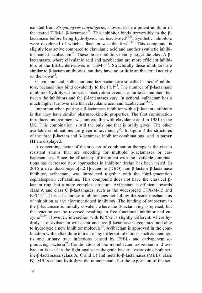
isolated from Streptomyces clavuligerus, showed to be a potent inhibitor of the feared TEM-1 β-lactamase29. This inhibitor binds irreversibly to the β-lactamase before being hydrolyzed, i.e. inactivated28,30. Synthetic inhibitors were developed of which sulbactam was the first31,32. This compound is slightly less active compared to clavulanic acid and another synthetic inhibi-tor named tazobactam33. These three inhibitors mainly target the class A β-lactamases, where clavulanic acid and tazobactam are more efficient inhibi-tors of the ESBL derivatives of TEM-134. Structurally these inhibitors are similar to β-lactam antibiotics, but they have no or little antibacterial activity on their own25.
Clavulanic acid, sulbactam and tazobactam are so called ‘suicide’ inhibi-tors, because they bind covalently to the PBP25. The number of β-lactamase inhibitors hydrolyzed for each inactivation event, i.e. turnover numbers be-tween the inhibitors and the β-lactamases vary. In general, sulbactam has a much higher turnover rate than clavulanic acid and tazobactam35,36.
Important when pairing a β-lactamase inhibitor with a β-lactam antibiotic is that they have similar pharmacokinetic properties. The first combination introduced as treatment was amoxicillin with clavulanic acid in 1981 in the UK. This combination is still the only one that is orally given. The other available combinations are given intravenously25. In figure 3 the structures of the three β-lactam and β-lactamase inhibitor combinations used in paper III are displayed.
A concerning factor of the success of combination therapy is the rise in resistant strains that are encoding for multiple β-lactamases or car-bapenemases. Since the efficiency of treatment with the available combina-tions has decreased new approaches in inhibitor design has been tested. In 2015 a new diazabicyclo[3.2.1]octanone (DBO) non-β-lactam β-lactamase inhibitor, avibactam, was introduced together with the third-generation cephalosporin ceftazidime. This compound does not have the classical β-lactam ring, but a more complex structure. Avibactam is efficient towards class A and class C β-lactamases, such as the widespread CTX-M-15 and KPC-237. This β-lactamase inhibitor does not follow the same mechanisms of inhibition as the aforementioned inhibitors. The binding of avibactam to the β-lactamase is initially covalent where the β-lactam ring is opened, but the reaction can be reversed resulting in free functional inhibitor and en-zyme38,39. However, interaction with KPC-2 is slightly different, where hy-drolysis of avibactam will occur and free β-lactamase is generated and able to hydrolyze a new inhibitor molecule38. Avibactam is approved in the com-bination with ceftazidime to treat many different infections, such as meningi-tis and urinary tract infections caused by ESBL- and carbapenemase-producing bacteria40. Combination of the monobactam aztreonam and avi-bactam is used in the fight against pathogenic bacteria expressing both ser-ine-β-lactamases (class A, C and D) and metallo-β-lactamases (MBLs; class B). MBLs cannot hydrolyze the monobactam, but the expression of the ser-

17
ine β-lactamases will disable the use of this drug. However, avibactam will inhibit the serine β-lactamases and the monobactam can act on the MBLs and thereby lead to successful treatment. This particular combination is now in phase III clinical trials41. Other DBOs are currently being investigated, such as the phase III study of relebactam in combination with the car-bapenem imipenem42. This combination is intended against strains producing KPC or class C β-lactamases, such as carbapenem-resistant Enterobacteri-aceae and P. aeruginosa43. Many more potential inhibitors are being investi-gated giving temporary hope for future treatment options.
Figure 3. Structures of the three β-lactam and β-lactamase inhibitors used in paper III. Ampicillin is administered with sulbactam, piperacillin with tazobactam and ceftazidime with the newer β-lactamase inhibitor avibactam.
The Gram-negative peptidoglycan layer and β-lactam antibiotics Physiological analysis. Already in the 1940’s it was observed that small amounts of penicillin caused elongated and filamentous bacterial cells, indi-cating that the drug is interfering with processes responsible for the cell shape44. By this time it was not known how a bacterial cell was built up. Already decades prior to the discovery of resistant bacteria Hans Christian Gram developed a bacterial staining procedure. Putting stained bacteria un-der a light-microscope allowed for categorization of the bacteria into two groups: Gram-positive and Gram-negative bacteria45. The dye crystal violet colors the sample purple. A decolorizer, for instance acetone, will dehydrate the peptidoglycan. In Gram-positive bacteria, the large shrunken peptidogly-can layer will retain the purple color in the cell. The outer membrane of Gram-negative bacteria and its thinner peptidoglycan layer will not be able to retain the crystal violet and the dye is lost. Counterstaining with safranin will give the Gram-negative cells a red or pink color45.
N
NH
NHS
H2
O OH
OO
ampicillin
N
N
N
NH
NHS
H
O OH
OO
O
piperacillin
N
SH
O OH
OO
O
O
O
sulbactam
N NN
NSH
O OH
OO
O
tazobactam avibactam
N N
NO O
OOH
O OS
N
SNH
HN
O
O
OH
SN
H N2
O O
O
O
ceftazidime
-
+
H N2
β-lactamantibiotics
β-lactamaseinhibitors

18
Figure 4. The actions of PBPs on the peptidoglycan layer of Gram-negative bacteria and the effect of the presence of β-lactam antibiotics. (1) Unlinked pentapeptides attached to NAM-molecules. (2) Removal of the outmost D-alanine before (3) cross-linking the tetrapeptide to an adjacent meso-diaminopimelic acid. (4) β-lactam pre-sent in the bacterial peptidoglycan layer will cause inactivation of the PBP by cova-lent binding (5), causing instability (6) of the peptidoglycan layer and eventually cell arrest.
Biochemical analysis. In the late 40’s and early 50’s it was demonstrated that unknown uridine peptides gathered in the cytoplasm of Staphylococcus au-reus (S. aureus) that had been penicillin-treated. These uridine peptides had a similar composition of amino acids and sugars as the bacterial cell wall. This finding indicated that they were cell wall precursors that were gathered due to penicillin inhibition of the cell wall biosynthesis46. The cell wall is composed of N-acetylglucosamine (GlcNAc) and N-acetylmuramic acid (MurNAc) (Figure 4)47. In Gram-negative bacteria a tetrapeptide, called gly-can strand, consisting of L-alanine, D-glutamic acid, meso-diaminopimelic acid and D-alanine, is attached to the MurNAc. The glycan strand precursor, before cross-linked, has an additional D-alanine at the end of the tetrapep-tide. The inner D-alanine is cross-linked by a transpeptidase with an adjacent meso-diaminopimelic acid that is connected to another MurNAc and the outer D-alanine is removed by a D-alanine carboxypeptidase. These two enzymes are to different extents sensitive to the β-lactam antibiotics. Penicil-lin contains an L-cysteine and D-valine that resembles the D-alanyl-D-alanine from the tetrapeptide of the glycan strand. The β-lactam ring con-tains a reactive CO-N identical to that of the glycan strand’s D-alanyl-D-
IM
PG
PG
PG
OM
1
2
3
MurNAc
GlcNAc
PBP
Complex PBP + β-lactam
β-lactam D- or L-alanine
D- Glutamic acidmeso-diaminopimelic acid
IM
PG
PG
PG
OM
4
5
6
β-lactam

19
alanine. At this site the transpeptidation occurs47. After binding of the β-lactam, the active site serine of the PBP will react with the CO-N of the β-lactam ring causing hydrolysis of the antibiotic, i.e. the β-lactam is covalent-ly bound to the PBP disabling the enzyme to perform its normal cellular function48.
Biophysical analysis. Radioactive penicillin was used to elucidate the site of action of the antibiotic. Penicillin was shown to bind to a target, termed penicillin-binding component (PBC). When penicillin had bound neutral buffers, acids or detergents could not remove it from the PBC indicating that the β-lactam was covalently bound49. It was found that the PBC consisted of several proteins, the penicillin-binding proteins50.
Genetic analysis. PBPs are divided into high-molecular mass (HMM) and low-molecular mass (LMM) groups. HMM PBPs (PBP1-3) are essential, whereas LMM PBPs (PBP4-8, PBP4b, PBP6b and AmpH) are not. In Esch-erichia coli, PBP1a, encoded by mrcA, and PBP1b, encoded by mrcB, have similar functions and are responsible for polymerization of the glycan subu-nits and crosslinking of the muropeptides51. These two can substitute for each other when deleted, but a double deletion is not viable52. PBP1c (pbp1c) in comparison with PBP1a and PBP1b functions only as a transgly-cosylase and cannot substitute the loss of the other PBP1s53. PBP2 is respon-sible for the maintenance of the E. coli rod shape51. Treatment of a cell cul-ture with mecillinam that specifically target PBP2 will render the rod shaped bacteria spherical54. Deletion of the gene responsible for PBP2 is lethal. However, overexpression of ftsZ, an essential cell division protein, makes PBP2 dispensible55. PBP3 (ftsI) functions as a transpeptidase responsible for the crosslinking of the peptidoglycan at the septal ring during cell division. Piperacillin specifically inhibits PBP356. PBP4 (dacB) has DD-endopeptidase activity in addition to DD-carboxypeptidase IB activity and are able to cleave D-alanyl-γ-meso-2,6-diaminopimelyl peptide bond in the crosslinks of the murein as a sort of recycle mechanism of the peptidogly-can57. PBP4b (yfeW) encodes for a putative DD-carboxypeptidase non-essential for the growth of E. coli58. PBP5 (dacA), PBP6 (dacC) and PBP6b (dacD) are DD-alanine carboxypeptidases59,60. PBP5 has a higher specificity than PBP6 to uncross-linked peptidoglycan61. A strain with a mutation in PBP5 show abolished DD-alanine carboxypeptidase IA activity making the bacterial cell more sensitive towards penicillin62. PBP5 concentrations are stable throughout the cell cycle, whereas PBP6 increases two- to ten-fold in stationary phase63. PBP5, PBP6 and PBP6b are non-essential for viability60. PBP8 is a product of PBP7 (pbpG) that has been cleaved by the protease OmpT64. The function of both PBP7 and PBP8 is to act as DD-endopeptidases hydrolyzing D-alanyl-DAP amide bonds within murein crosslinks65, where increased expression gives decreased susceptibility to ceftazidime66. AmpH is a bifunctional enzyme, catalyzing both DD-

20
carboxypeptidase and DD-endopeptidase activity, enabling peptidoglycan remodeling or recycling67.
Different β-lactam antibiotics have different affinity for the PBPs de-scribed68. It is commonly believed that the effect of the β-lactam is to bind the PBPs leading to inability to build the peptidoglycan layer and hence the cell cannot grow. This will ultimately lead to cell death. However, another hypothesis is that the binding of the PBPs will lead to a toxic malfunctioning of the peptidoglycan machinery depleting cellular resources by trying to re-build and recycle the peptidoglycan layer69.
Bacterial antibiotic resistance The discouraging fact that bacteria seem to develop antibiotic resistance faster than mankind can identify new antibiotics is daunting. It is not un-common that shortly after introduction of a certain antibiotic resistant iso-lates are observed in the clinics. For the revolutionizing antibiotic penicillin, resistance was already observed before introducing the drug to clinical use3. Many of the antibiotics we use originate from different environmental spe-cies of bacteria or other microorganisms where the antibiotic inherently may be used as a way to dominate certain environmental niches5,8. However, many of the resistance mechanisms we encounter today have probably evolved in that same bacteria as a protection mechanism against the antibi-otic it produced or in cohabiting bacteria to be able to defend themselves against the toxic substances8.
The immense use of antibiotics in the clinics has led to enrichment of var-ious different resistant bacteria. Not only ancient resistance mechanisms, but also resistance mechanisms to newer, synthetic antibiotics are being spread across diverse species into pathogenic bacteria causing treatment failures70. This has led to a plethora of different resistance mechanisms a bacterium can use to circumvent the harmful actions of antibiotic drugs. If we had known back when the antibiotics were introduced what we know now, a tougher antibiotic usage policy potentially would have enabled us to use the drugs for a longer period.
The intrinsic antibiotic resistance Antibiotic resistance is normally considered a trait that a previously suscep-tible bacterium can achieve, such as by chromosomal mutations or acquisi-tion of resistance genes by horizontal gene transfer. However, all bacterial species are intrinsically more or less susceptible to different antibiotics. This means that some antibiotics can be used to treat some bacteria but not others. An obvious example of this phenomenon is the lack of the antibiotic target in the bacterial cell, as in the case for Mycoplasma pneumoniae. This species

21
lack peptidoglycan and hence β-lactam antibiotics will be ineffective71. An-other example is the lipopeptide antibiotic daptomycin that is efficient in treating Gram-positive infections, but unable to cure infections caused by Gram-negative bacteria. Gram-negative bacteria have a lower proportion of anionic phospholipids in the cytoplasmic membrane than Gram-positive bacteria, which reduces the efficient insertion of daptomycin into the mem-brane and thereby prevents the antibacterial activity of the drug72. In contrast to Gram-positive bacteria the Gram-negative bacteria have an outer mem-brane (OM) that will protect the cell from drugs that are active against many Gram-positives. Since the OM has a low permeability also to many water-soluble nutrients, the bacterium need to take up nutrients through outer membrane proteins (OMPs) or porins. Porins prevent influx of many drugs by size limitation73, hydrophobicity74 and charge repulsion74,75. Active efflux is another reason why many Gram-negative bacteria are less susceptible to some drugs than Gram-positives, since the antibiotic is pumped out of the bacterial cell before it can act on its target. An important class of efflux pumps is the multidrug resistance efflux pumps. These pumps have a broad substrate range and can pump out several classes of antibiotics76. However, active efflux is generally not efficient enough to confer clinical resistance but can act as a stepping stone to increased resistance in combination with other mechanisms77.
Acquired antibiotic resistance There are two fundamentally different ways to acquire antibiotic resistance: by horizontal gene transfer (HGT), or by alteration of the chromosome or previously existing plasmids. Spontaneous resistance, i.e. chromosomal or plasmid alterations, can happen in the patient during treatment78–81. Whereas chromosomal mutations will be vertically inherited mutations on plasmids can be both laterally and vertically disseminated posing a potential problem in future medicine use. The initial effects of chromosomal changes tend to have a greater impact on the fitness of the bacterial cell, i.e. lowered growth rate, than horizontally transferred material82. The reason to why resistance caused by chromosomal mutations often has a detrimental effect on the bac-terial cell is that these mutations often occur in essential or in regulatory genes. Mobile resistance determinants conversely, often encode for enzymes giving rise to antibiotic modifications rather than hindering the binding of the drug target82. Figure 5 shows the most common resistance mechanisms that bacteria employ to escape antibiotic effects.

22
Figure 5. Common antibiotic resistance mechanisms employed by bacteria to cir-cumvent the effects of antibiotics. The most common mechanisms are: decreased import, increased export, target overexpression, drug modification, target bypass and target modification.
Target alteration Many antibiotics bind specifically to their target with high affinity and thereby inhibit the normal function of the target. During selective pressure by antibiotics only the bacteria able to circumvent the toxic environment will be able to survive and spread. Specific mutations in the antibiotic target-encoding gene may render the target less susceptible to the antibiotic but still retain sufficient functionality to exert the original task. Examples of this are mutations in gyrA, encoding for the DNA gyrase, and in parC, DNA topoi-somerase IV, giving rise to high-level resistance towards fluoroquinolones in combination with other mutations83. Both GyrA and ParC have regions called the ‘quinolone-resistance determining region’. Mutations in this re-gion change its affinity of fluoroquinolones and the drug cannot bind effi-ciently anymore84,85. A combination of mutations both in gyrA and parC has proven to yield high-level resistance86. Resistance towards fluoroquinolones is increasing. About 26% of all the invasive clinical E. coli isolates reported to EARS-Net conferred high-level resistance to fluoroquinolones87.
Other target alterations leading to increased antibiotic tolerance can be mutations in the promoter regions of genes, facilitating increased expression of the gene and subsequently increased abundance of the target. For instance
targetoverexpression
target bypass
increased export
A B
decreased import
target modification
drug modification

23
mutations in the promoter region of a PBP can lead to hyper-production of the target88,89. Another mechanism for increased abundance of target is by amplifications, where repeated sequences function as amplification points. The more copies of a particular gene the more target will be produced aiding in the survival of the cell90. Both aforementioned examples will lead to more target proteins in the cell, diluting out the effects of the antibiotic91. Especial-ly the latter mechanism has gotten an increased focus the last years. Gene amplifications are commonly found among bacteria in the clinics. A sub-population of bacteria with amplifications found in an infection can avoid killing and make it harder to treat the infections. They can even to some extent protect the sensitive bacteria from the effects of the antibiotic. This phenomenon is referred to as heteroresistance. Increasing evidence suggest that treatment failures occasionally could be due to gene amplifications92.
Functional inactivation Inactivation of the antibiotic is an efficient way to achieve high-level re-sistance. Resistant strains can encode for enzymes either modifying or de-stroying the structure of the antibiotic93. The most relevant enzymes in Gram-negative bacteria capable of altering the structure of antibiotics are the β-lactamases. The major porins will enable β-lactams to enter into the periplasmic space and the peptidoglycan layer of the bacterial cell, where normally the antibiotic would inhibit the expansion of the cell by binding to PBPs. However, upon entry in cells expressing β-lactamases the antibiotic will be bound to the β-lactamases that reside in the periplasm, where a hy-drolysis procedure will occur rendering the drug inactive. This will lead to the PBPs being able to continue building the peptidoglycan layer and the cell will proliferate23. Since the requirement of hydrolysis is dependent on one water molecule these enzymes can also be excreted outside of the cell, where they can lower the concentrations of the drug before it has entered the cell93. The lowered concentration of antibiotics in the external environment may also facilitate otherwise susceptible bacteria to survive.
There is also an extensive group of transferases that can covalently alter the structure of different antibiotics hindering the binding to the target93. These enzymes operate in the cytosol of the cell where they employ different chemical strategies to ensure target inaccessibility, such as O-acylation, N-acylation, O-phosphorylation, O-nucleotidylation, O-ribosylation, O-glycosylation, and thiol transfer. The inactivation mechanisms are dependent on co-substrates for activity, including ATP, acetyl-CoA, NAD+, UDP glu-cose, or glutathione93. The aminoglycoside antibiotics are often targeted by transferases, which are frequently encoded on plasmids, that will modify the antibiotic in such a way that it cannot bind its target anymore93. This antibi-otic class bind to the A-site of the ribosome and thereby impair the codon-anticodon decoding mechanism leading to synthesis of abnormal proteins, which are toxic to the cell93. These types of modifying enzymes most likely

24
originate from enzymes involved in metabolism and cell signaling. Not until the use of antibiotics as a treatment the enzymes conferring a larger benefit to the bacterium has evolved to functional resistance proteins and have been enriched for, able to cope with the selective environment93.
Alternative enzyme Acquisition and expression of alternative enzymes that are insensitive to the antibiotic is a nifty mechanism that often will lead to high-level resistance. The alternative enzyme can perform the same task as the target that is inhib-ited by the drug. These enzymes are structurally different than the native enzyme in order to lower the binding affinity of the drug. For example, whereas human cells can take up exogenous folate bacteria need to produce the metabolite. Folate is important for the production of cellular compo-nents19. Inhibition of DHFR (dihydrofolate reductase), part of the folate syn-thesis pathway, by trimethoprim will eventually lead to cessation of growth and hence cell death94. By expression of an alternative DHFR, from dhfr-genes often encoded on plasmids, the alternative protein maintains function-ality, whereas the native protein is inhibited. Strains carrying such dhfr-genes also can carry sul-genes, conferring resistance towards sulfonamides, which is another drug targeting the folate synthesis pathway. The co-carriage of dhfr- and sul-genes will disable the use of the combination of trime-thoprim and sulfametoxazole94.
Another example of an alternative enzyme is methicillin resistant S. aure-us. This species has three essential PBPs, PBP1-3, which ensures cell growth. These PBPs are also the targets of many β-lactams, where binding will lead to cell death. However, there is an alternative PBP2, named PBP2a, that can be horizontally transferred between cells making the otherwise sus-ceptible strain resistant to methicillin. This PBP has low affinity to the β-lactams and therefore the cell can grow under antibiotic pressure95.
Decreased intracellular antibiotic levels Eliminating or at least decreasing antibiotic intracellular concentrations is of importance for the bacterium in a selective environment. The expression of efflux pumps will enable rapid expulsion of various drugs and decrease the intracellular levels of the toxic compound enabling survival of the bacte-rium76. The efflux pumps are not only native to some bacterial strain but can also be acquired, such as the tet-pumps76. These pumps specifically transport tetracyclines out of the bacterial cell72. The TetA efflux determinant is en-coded by an operon consisting of two genes, the efflux pump (TetA) and a repressor protein (TetR). In absence of tetracycline the repressor protein binds to the promoter region between the two genes repressing the expres-sion of the efflux pump, but still facilitating expression of the repressor pro-tein. However, when tetracycline is present TetR binds to the antibiotic and

25
the repression of the pump is alleviated, leading to production of the efflux pump18.
Another way of decreasing intracellular concentrations of antibiotics are by hindering the access of the compound to the cell. In Gram-negative bacte-ria this is commonly done by mutations in the porins OmpC or OmpF, or in their regulators OmpR and EnvZ. Mutations reducing or abolishing expres-sion of the porins will not only inhibit the access of the antibiotic to the cell but also hinder important nutrients to enter leading to a slower growth of the bacterium96,97. More about this topic can be found below. It is not uncommon that multi-resistance plasmids carry combinations of the above mentioned resistance mechanisms rendering a bacterium resistant to many different classes of antibiotics98,99. These infections are generally hard-er to treat and in some cases the infections cannot be readily treated leading to higher rates of mortality.
Β-lactam resistance Resistance to penicillin was discovered already before the antibiotic was available for the common man. An enzyme, named penicillinase, was the cause of the decreased susceptibility of S. aureus towards β-lactams100. It took less than a year after the introduction of penicillin in clinical practice until the first treatment failures were reported101. The solution to this prob-lem was to treat the patients with higher doses of penicillin, since the antibi-otic was classified as non-toxic to human and it was easily accessible. The majority of nosocomial S. aureus infections in the United States were re-sistant to penicillin already five years after the introduction23. Ever since the development of ampicillin as treatment of Gram-negative infections in the early 1960’s the prevalence of β-lactam resistance for this Gram-group has increased102. The expansion in resistant isolates spurred the development of newer, enhanced, broader acting β-lactam antibiotics able to treat everything from β-lactamase producing bacteria to infections caused by the opportunis-tic pathogen, P. aeruginosa. The vast numbers of different β-lactam antibiot-ics aimed to slow down resistance development but has led to the evolution of many different β-lactamases23.
There are four main resistance mechanisms towards β-lactam antibiotics in Gram-negative bacteria:
(i) Production of acquired β-lactamases is by far the most common mech-anism to circumvent treatment with β-lactams among Gram-negative bacte-ria25. Not only production, but increased production, due to amplifications of the target gene, will enable the bacterial cell to overcome β-lactams it is otherwise sensitive to103. These enzymes are of extra importance for several projects in this thesis and will be explained in more detail further on.

26
(ii) The outermost part of the cell envelope of Gram-negative bacteria, i.e. the outer membrane, serves as a barrier to the external environment and hence also towards the β-lactams. The antibiotic relies on passive diffusion or influx through outer membrane porins to the periplasm where the penicil-lin binding proteins, the targets of the β-lactams, are located104. Mutations in the porins can result in decreased influx of the drug and decrease the suscep-tibility of the bacterium105–107. This topic will also be discussed in more de-tail later.
(iii) Altered PBPs can lower the binding-affinity of the β-lactam and therefore decrease the susceptibility of the strain108. This is mostly described for Gram-positive bacteria. Another pathway to high-level resistance is the acquisition of the mecA gene by S. aureus. This gene encodes for an alterna-tive PBP2, called PBP2a, that can still create a new cell wall even in pres-ence of high levels of penicillins or cephalosporins due to a low binding affinity to β-lactams109. The potential relevance in antibiotic resistance of mutations in PBPs in Gram-negative bacteria will also be addressed more in depth.
(iv) Efflux pumps, either intrinsic or acquired, actively transport the anti-biotic that enters the periplasm to the outer environment. This will lead to a decreased drug concentration in the cell and hence lead to decreased suscep-tibility. This is especially important in multi-drug resistant P. aeruginosa, where combinations of decreased permeability and increased efflux cause resistance not only to β-lactam antibiotics but also several other antibiotic classes110,111.
The world of β-lactamases The production of β-lactamases is the main resistance mechanism towards β-lactam antibiotics in Gram-negatives. These enzymes are able to hydrolyze the β-lactam ring rendering it inactive (Figure 6). Over 70 years of use of the vast amount of β-lactams available has led to a diverse pool of β-lactamases. It is being speculated that β-lactamases originally evolved from PBPs112,113. Most PBPs and β-lactamases are active site serine enzymes, where both kind of enzymes create an acyl-enzyme intermediate with the β-lactam112. How-ever, only the β-lactamases have evolved a de-acetylation function allowing for the process of hydrolysis of the β-lactam. This would lead to free β-lactamase able to hydrolyze more β-lactam antibiotic112, whereas the PBP will be covalently bound with the β-lactam and inactivated from further ca-talysis.
More than 890 different chromosomally or plasmid-borne β-lactamases have been found this far114. The enzymes can be classified by the functional characteristics of the β-lactamase115 or their primary structure116. In this the-sis the classification by the protein composition will be used, according to the Ambler classification, giving rise to four molecular classes; A, B, C and

27
D116,117. Three of the classes (A, C and D) are active-site serine enzymes, whereas one class (B) contains zinc-dependent β-lactamases.
Figure 6. Overview of the interaction between a β-lactam antibiotic and an active-site serine β-lactamase. The β-lactamase creates an acyl-enzyme complex with the β-lactam antibiotic. After binding, hydrolysis of the β-lactam can occur resulting in an inactivated β-lactam and a free β-lactamase.
Class A, broad-spectrum serine β-lactamases is the largest group of β-lactamases. TEM-1 and SHV-1 belong to this group, two of the most en-countered β-lactamases in Enterobacteriaceae. Even though TEM-1 is mostly associated with ampicillin resistance, increased expression of this β-lactamase can lead to resistance toward cephalosporins and β-lactamase in-hibitor combinations118. All the TEM-enzymes originate from TEM-1 and are plasmid-borne. There are more than 200 derivatives described to date (https://www.ncbi.nlm.nih.gov/pathogens/beta-lactamase-data-resources/). Replacements of amino acids in TEM-1, such as the glycine at position 238 to serine, alanine or aspartic acid, will lead to an ‘expanded’ substrate profile enabling hydrolysis of penicillins and cephalosporins119,120.
The extended-spectrum β-lactamases (ESBLs) of the CTX-M-type has spread worldwide since the first report in the 1980s121. These enzymes are commonly found in E. coli and Klebsiella pneumoniae, but originate from the anaerobe environmental bacteria Kluyvera species122. The acquired CTX-Ms can be divided into five distinct groups; CTX-M-1, CTX-M-2, CTX-M-8, CTX-M-9 and CTX-M-25123, indicating that transfer of CTX-M occurred at least five independent times to pathogenic bacteria. The most widespread CTX-M enzymes are CTX-M-14 (belonging to the CTX-M-9 group) and CTX-M-15 (belonging to the CTX-M-1 group)124. Plasmids from clinical isolates are in addition to CTX-M often encoding for TEM-1 and/or OXA-1 β-lactamases, as well as resistance determinants against aminoglycosides, trimethoprim, sulfonamide and tetracycline98,99,125. Although ESBLs can
OH
ON
ONOH
OHO
N
OHO ON
HO ONHbinding
β-lactamase β-lactam H O2
acylation hydrolysis+

28
hydrolyze a wide range of penicillins and cephalosporins they are usually sensitive to the β-lactamase inhibitors tazobactam and avibactam126.
Serine carbapenemases, such as KPC, can hydrolyze carbapenems that are otherwise very stable against β-lactamases. The most spread serine-carbapenemases are KPC-2 and KPC-3, which mostly are found in K. pneu-moniae but also other Enterobacteriaceae127. In contrast to ESBLs, KPC-types of enzymes are not inhibited by tazobactam, but are by avibactam127.
Class B, metallo-β-lactamases, including VIM and NDM, are different from the other β-lactamases since they have a Zn2+ at the active site128. These en-zymes can hydrolyze most β-lactams, including carbapenems, but they are less active against monobactams. The metal chelator EDTA can inhibit MBLs but to date there are no commercial β-lactamase inhibitors towards these enzymes.
Class C, AmpC-type β-lactamases, confer resistance to most cephalosporins, but to lesser extent towards penicillins and some monobactams129. In some Gram-negative bacteria, such as P. aeruginosa, chromosomal AmpC expres-sion can be induced in response to β-lactams. A constitutive expression of AmpC will also lead to resistance129. AmpC-type β-lactamases can be plas-mid-borne where they can be highly expressed due to strong promoters or high copy number of the plasmid129. A combination of reduced membrane permeability and plasmid-encoded AmpC confers resistance towards car-bapenems in clinical isolates of K. pneumoniae and Salmonella enter-ica130,131.
Class D, OXA-type β-lactamases were conventionally grouped by their oxa-cillin-hydrolyzing characteristics, but have become an expanded group with variable substrate profiles and sequences. This group of β-lactamases share less than 20% of amino acid identity compared to both class A and class C β-lactamases132. One of the first identified OXA-type β-lactamases was OXA-1, encoded on a transposon. Some members can hydrolyze cephalosporins and carbapenems and some can be controlled by the inhibitors tazobactam or avibactam38,133. In recent years the variant OXA-48 with carbapenemase activity has spread throughout Europe134. Plasmid-borne OXA-48 in combi-nation with other β-lactamases could potentially make treatment with all β-lactams ineffective.

29
Figure 7. Porin expression regulation in E. coli. The porins, OmpC and OmpF, stretch over the outer membrane (OM). The two-component regulatory system con-sists of EnvZ, an inner membrane (IM) bound protein, and OmpR, the cytosolic counterpart. Depending on the osmolarity OmpR/EnvZ regulates the expression of OmpC and OmpF differently in combination with the small RNAs micC and micF. During high osmolarity conditions ompC expression is favored, whereas ompF is repressed and vice versa in low osmolarity conditions.
The role of the outer membrane in β-lactam resistance The entry of β-lactam antibiotics is significantly affected by the permeability of the outer membrane96. In E. coli there are three major porins, PhoE, OmpC, and OmpF (Figure 7). PhoE, a phosphate transporting porin, is nor-mally not expressed during laboratory conditions but is induced by high salt or low phosphate135. The other two porins, OmpC and OmpF, are aqueous filled channels used for passive transport of nutrients136,137. These two porins are regulated by the two-component system (TCS) EnvZ-OmpR, as well as two small RNAs micC and micF138,139. This TCS responds to various envi-ronmental stimuli. The two porins are not equally expressed at a given time point, but are differentially regulated depending on the phosphorylation state of OmpR. Upstream of the promoters of ompF and ompC are four (F1, F2, F3 and F4) and three (C1, C2 and C3), respectively, binding sites for phos-phorylated OmpR, OmpR-P140. Under low osmolarity conditions OmpR-P bind to F1, F2 and F3 activating the transcription of ompF. This will lead to an increased influx of nutrients due to its slightly larger pore size96,141. C1 is
IM
OM
OmpC OmpF
P
P
EnvZ
OmpR
osmolarity
specific transporters
high osmolarity low osmolarity
OmpR binding sites ompC
micC
ompFOmpR binding sites
micF

30
always bound by OmpR-P, but this is not sufficient for ompC transcription. However, during high osmolarity OmpR-P becomes more abundant and binds to C2 and C3 as well causing transcription of ompC. During these conditions OmpR-P is proposed to bind the F4 site, creating a loop interact-ing with the F1, F2 and F3 sites inhibiting the transcription of ompF140,142,143.
Since the porins are non-specific, other compounds than nutrients can use this passage and access the periplasm of the cell. For example, hydrophilic antibiotics, such as β-lactam antibiotics, can diffuse through the porins96. Mutations in the genes encoding for the porins or their regulators can lead to decreased uptake of β-lactams and hence will decrease the susceptibility of the strain. Combination of porin deficiencies and other mechanisms giving resistance to β-lactams can have a strong synergistic effect making treatment of clinical infections difficult97,144,145.
Effects of altered penicillin binding proteins After the spread of the penicillin resistant Gram-positive S. aureus-strains in the 1950s the β-lactam methicillin was introduced as a treatment option. Not long after the introduction the first methicillin-resistant S. aureus (MRSA) was discovered146. These isolates are resistant to almost all β-lactams and in addition carry several other resistance determinants. The resistance of MRSA to β-lactams is due to the acquired PBP2a, an alternative to PBP2147,148. PBP2a, encoded by mecA is found together with mecI and mecR1 on a mobile genetic element able to integrate onto the chromo-some149. Mutations in indigenous PBP1, PBP2, and PBP4 can also give a decreased susceptibility to penicillins and cephalosporins88,150.
In Gram-negative organisms, such as H. influenza and Neisseria species, mutations in PBPs are increasing in prevalence among clinical isolates as a resistance mechanism towards β-lactams, including penicillins and extended spectrum cephalosporins151–153. Some Neisseria species have a less suscepti-ble PBP2. Due to natural transformation, i.e. uptake and incorporation sys-tems of foreign DNA, commensal Neisseria species can obtain the less sus-ceptible PBP2 gene and incorporate it on the chromosome creating a mosaic gene154. In E. coli, mutations in PBPs are rare. In one specific case, muta-tions in PBP2 and PBP3 gave rise to resistance towards several β-lactam/β-lactamase inhibitor combinations155. Interestingly, short-term evolution ex-periments with the β-lactam/β-lactamase inhibitor combination ceftazidime-avibactam yielded multiple mutations in different PBPs (Paper III). The effect on resistance of these mutations needs to be further elucidated.

31
Resistance to β-lactam and β-lactamase inhibitor combinations Not unexpectedly, resistance towards the β-lactam/β-lactamase inhibitor combination amoxicillin-clavulanate surfaced a few years after its introduc-tion156. With every commercially available β-lactam/β-lactamase inhibitor combination, mutant clones have been found. The impact of resistance to combination therapy, or antibiotics in general, is that a good treatment out-come is significantly lowered. Especially, serious urinary tract infections, respiratory tract infections and blood stream infections are of relevance157–
159. The routes to resistance towards β-lactam/β-lactamase inhibitors are essen-tially the same as for β-lactams alone:
(i) Hyper-production of chromosomal β-lactamases, such as AmpC in E. coli and SHV-1 in K. pneumoniae158,160, are commonly due to mutations in the promoter or due to amplifications of that particular region. Resistance towards ampicillin-sulbactam (SAM) is sometimes mediated by the hyper-production of TEM-189,91,161,162. Amplification of regions containing re-sistance determinant is not uncommon and could interfere with the outcome of an antibiotic treatment163. TEM-1 producing bacteria are usually suscepti-ble towards the combination piperacillin-tazobactam (TZP), but massive amplifications of this enzyme, sometimes more than 100 copies, will lead to high level resistance not only to SAM but also TZP164. In addition to in-creased copies of one gene, increased copy number of plasmids carrying resistance determinants will also lead to a decreased susceptibility to β-lactam/β-lactamase inhibitors156. We show in paper III that amplifications of different β-lactamases will decrease the susceptibility to certain combina-tions. These amplifications in combination with mutations in CTX-M-15, as well as deficiencies in the outer membrane porins or increased efflux, will turn ESBL-producing strains clinically resistant towards CZA.
(ii) Mutations in the β-lactamase, previously called inhibitor resistant TEM (IRT) β-lactamase, can change the substrate-spectrum it can hydrolyze. There are a wide number of different TEM enzymes giving decreased sus-ceptibility towards β-lactam antibiotics, but also towards the β-lactamase inhibitors162,165–168. For the newer combinations of ceftazidime-avibactam (CZA) mutations in a plasmid-encoded KPC-3 was observed during treat-ment for three patients. The mutations found affected the susceptibility to-wards CZA differently, showing an increased mutational spectrum able to circumvent this combination78. Mutations in KPC-2 were also found to cause resistance to this particular combination169. In vitro selections and evolution of two ESBLs, CTX-M-14 and CTX-M-15, has shown mutations causing a decreased susceptibility to CZA170,171.
(iii) Alterations in PBPs can change the affinity of the β-lactam drug to its target. Specific alterations, by insertion of four amino acids, in PBP3 can

32
make the bacterium less susceptible to not only the combination of aztre-onam with avibactam, but also generally to cephalosporins. However, the potency of carbapenems was not affected155,172. Since the β-lactam antibiot-ics target different PBPs not all PBP alterations will be relevant in the devel-opment of resistance to the different treatments.
(iv) Porin deficiency will also reduce the uptake of some β-lactamase in-hibitors. However, some β-lactamase inhibitors, such as avibactam, can dif-fuse across the membrane in a porin-independent fashion173. Not to forget is the active efflux displayed by many strains reducing the concentration of inhibitor further in a porin deficient bacterial cell.
The role of horizontal gene transfer in antibiotic resistance The ability to exchange genes in the bacterial community is crucial to the evolution of bacteria. All bacterial genomes contain traces of horizontal gene transfer events, displaying the important impact of this mechanism on bacte-rial evolution174,175. Genes facilitating a benefit to the bacterium will most likely be kept and genes posing a disadvantage will gradually be lost176. Considering that before the introduction of antibiotics the disease-causing bacteria did generally not express any resistance, the acquisition of antibiotic resistance determinants by horizontal gene transfer (HGT) must come from non-pathogenic ecological niches177. It is worth noting again that resistance determinants have been found in bacteria already before man existed5. The HGT is somewhat biased, where it is likelier that genes will be shared amongst closely related than more distantly related organisms. This could be due to homology similarities between donor and recipient, facilitating more efficient incorporation of DNA into the genome178. A study in Halobacteria showed that the more distantly related the bacteria were the lower the fre-quency of HGT179. However, there are studies showing DNA sharing amongst commensal or environmental bacteria with pathogenic strains180–182. Horizontal gene transfer has accelerated the spread of resistance mechanisms creating bacteria difficult to treat. Plasmids containing ESBL-determinants has been shown to have spread all over the world in a relatively short peri-od121,124,183. The ability for genes to move between different species of bacte-ria is of great importance since it enables the survival of the bacterium in environments it otherwise would not been able to persist in.
There are three central mechanisms of horizontal gene transfer, which are displayed in Figure 8:

33
Figure 8. Overview of the three major horizontal gene transfer pathways: transfor-mation, transduction and conjugation.
Transformation. Foreign DNA, usually from lysed cells or actively secreted DNA, can be picked up from the environment and then incorporated into the genome of the bacterium184. This process is called natural transformation. Already in the 1950’s observations were made that initially susceptible bac-teria became resistant to penicillin by the addition of sterile extracts of a resistant bacteria185. There are certain features required for natural transfor-mation: (i) extracellular DNA, (ii) the recipient has to encode for transfor-mation mechanisms, and (iii) the exogenous DNA needs to be stabilized to avoid degradation186. Some bacterial species are more prone to pick up ex-ogenous DNA than others, such as the gastritis-causing bacterium Helico-bacter pylori that easily picks up DNA creating a mosaic chromosome187. Interestingly, antibiotic-exposure has been shown to induce competence in many species of bacteria, meaning that antibiotics would not only select for antibiotic resistance but also increase the spread of exogenous DNA188,189. Transduction. Bacteriophages shape the bacterial microbiome in all envi-ronments. Through a process termed transduction the transfer of various genes can be disseminated between strains. Replication of the bacteriophage occurs in the bacterial cell and occasionally erroneous DNA packing results in the incorporation of bacterial DNA into the phage capsids190. The DNA fragments delivered by the bacteriophage to the recipient cell can incorpo-rate into different locations, i.e. into the chromosome or plasmid. Bacterio-phages are generally strain-specific and hence the genes cannot be widely distributed to various species through this mechanism186. However, there are some wide host range bacteriophages that potentially could enable efficient
conjugation
transduction
transformation
free DNA
bacteriophage
plasmid
34
transfer of antibiotic resistance genes to a broader assortment of bacte-ria191,192. The transferred DNA fragments can be anything from chromosomal to plasmid DNA, including resistance determinants193–195. Astonishingly, whole resistance plasmids have been transferred between strains by bacteri-ophages196.
Conjugation. Transfer of DNA via plasmid-mediated conjugation is proba-bly the most important mechanism of HGT for antibiotic resistance dissemi-nation, especially in Gram-negative pathogenic bacteria. This mechanism requires cell-to-cell contact through a passage, or a secretion system, usually expressed by proteins on the plasmid and active transfer of DNA to the re-cipient cell197. This is described in more detail below. Additionally, plasmids that do not encode for their own transfer proteins can use a co-existing con-jugative plasmid for further dissemination into a new host186. Once a re-sistance gene is established on a successful plasmid rapid dissemination to different species is a possible scenario, as can be seen with the CTX-M en-zymes that exist on both broad and narrow host range plasmids122.
These three mechanisms have specific requirements and occur with different probabilities within different bacterial communities. DNA is more stable and less exposed to DNases, enzymes degrading the DNA, in sands and clays198,199 enabling uptake for naturally competent bacteria. Packaging the DNA into vessels, such as phages, and then transporting it to a recipient cell is a more protected environment for the DNA, but phages usually have a very specific host range and will hence most likely only allow for intra-species gene transfer. The DNA is also protected against environmental DNases during conjugation. Some plasmids have a narrow host range, ena-bling intra-species transfer of genes, whereas other plasmids are more pro-miscuous and can transfer DNA to a range of bacterial species200,201. This makes conjugation of plasmids one of the major pathways of spread of anti-biotic resistance genes in various bacterial communities.
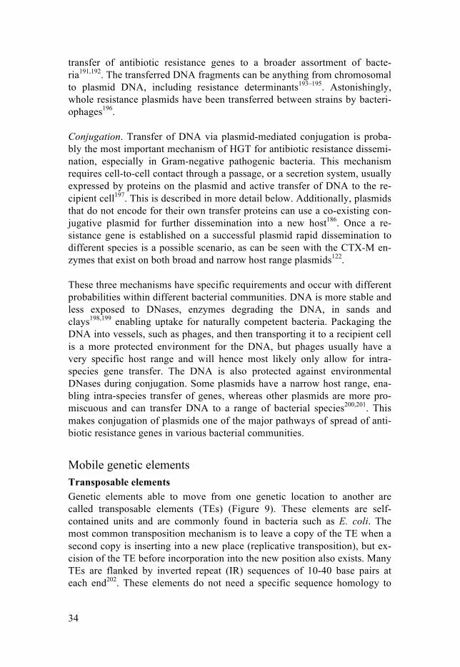
Mobile genetic elements Transposable elements Genetic elements able to move from one genetic location to another are called transposable elements (TEs) (Figure 9). These elements are self-contained units and are commonly found in bacteria such as E. coli. The most common transposition mechanism is to leave a copy of the TE when a second copy is inserting into a new place (replicative transposition), but ex-cision of the TE before incorporation into the new position also exists. Many TEs are flanked by inverted repeat (IR) sequences of 10-40 base pairs at each end202. These elements do not need a specific sequence homology to

35
insert at, however, some sequences seem to be preferred203. Movement of TEs containing resistance determinants between plasmid and chromosome have been observed (Paper I)98,204,205.
Figure 9. Schematic representation of different transposable elements, such as inser-tion sequence (IS)-elements, composite transposons and unit transposons. All these transposable elements are more or less dependent on inverted repeats (IR) for re-combination and transposition.
The simplest transposable element, called insertion sequence (IS) element, encode only for the protein transposase enabling the re-localization. Occa-sionally, resistance genes get trapped between two IS elements, forming a composite transposon, which then can move as a cluster and incorporate onto the chromosome or a plasmid. The most relevant IS-element in the spread of resistance genes in Gram-negative bacteria is the IS26 from the IS6 family. These elements were demonstrated to move by replicative trans-position. However, it is often seen that arrays of resistance genes are sepa-rated by several IS26-elements on plasmids206, including the pUUH239.2 plasmid that has been used in paper II and paper III. Due to this arrange-ment, a second way of movement was described. A single copy of IS26 in combination with an adjacent region, which can be all the way to the next IS26 junction, can insert next to an existing copy of IS26 without replica-tion206. This unit is called a translocatable unit (TU). The frequency of this process is about 50 times higher than the ordinary untargeted replicative transposition206. When an IS26 has incorporated onto the chromosome or the plasmid, acquisition of more copies is likely, building an array of genes206.
A second type of transposon is the unit transposon (Tn). Traditionally these transposons are considered to be elements larger than the IS-elements207. The Tn3 family transposons are also often associated with anti-
transposase gene
IS - element
IS - element
composite transposon
IS - elementX Y Z
IRIR
unit transposon
transposase geneXIR IRY Z

36
biotic resistance. The unit transposons also have terminal inverted repeats, but tend to have a transposase gene (tnpA) that is bigger than those of the IS. A resolvase, tnpR, and a resolution site, res site, are included in the TN3 family transposons207. A replicative process ensures successful duplication of the transposon where the resolvase separates the two copies208. This family of transposons display something called transposition immunity, where a second element is inhibited in the close proximity of the first copy208. A clin-ically important TE from the Tn3 family, Tn21, is often found on multi-resistance plasmids, including pUUH239.2. Tn21 carries a class I integron, encoding for an integrase, that will enable easy acquisition and expression of accessory genes, including resistance genes209.
Since the TEs can incorporate basically anywhere on the chromosome or plasmid, insertions causing a disruption in a gene is not unlikely. The TE can incorporate into an essential gene leading to non-viability of the bacterial cell and hence extinction of that particular cell202. Incorporation could also be beneficial for the bacterium depending on the condition it is faced with. An example is the reduction in uptake of certain β-lactams and β-lactamase inhibitors after disruption of the major porin regulator ompR by TEs (Paper III). Of course, insertion of the transposon can also be neutral to the host. In addition they can cause inversions of smaller or larger pieces of DNA, or amplifications and deletions of genes encoded between two TEs by homolo-gous recombination. Hereby, either increasing expression of proteins from genes included in the amplified region or removing genes that are potentially costly or harmful164,210,211. The transposition frequency of most TEs is greater than the spontaneous mutation rates of the chromosome212. However, most TEs are strictly controlled to minimize transposition, since these events can potentially lead to cell death202.
Integrons Another set of genetic elements of ancient origin are integrons that enable efficient acquisition of exogenous genes and are especially relevant in Gram-negative bacteria213. By targeted recombination mechanisms, the incorpora-tion of a plethora of genes can be achieved (Figure 10). These elements are especially important since they have been found to gather many resistance genes creating an arsenal of determinants making treatment more difficult.
There are three features all integrons share, which together will capture and express the exogenous genes that have been incorporated into the in-tegron214. Firstly, intI, encode for the integrase enzyme (IntI) that catalyzes the recombination between the site attI in the integron and the incoming exogenous gene215. The exogenous genes, or gene cassettes, often only con-tain an open reading frame, a ribosomal binding site and a recombination site, attC. The attC and the attI sites will enable site-specific recombina-tion214. When the exogenous gene is incorporated it will be expressed from the integron-associated promoter, Pc, where the gene cassettes closest to the

37
promoter will be the most efficiently transcribed216. This poses a limitation in how many genes can be incorporated, especially in the clinical class 1 integrons. Genes can be easily excised from the integron again217. There are advantages with these types of systems; (i) since the genes are incorporated at a specific site in the integron the genes pre-existing in the integron will not risk getting inactivated by erroneous incorporation of a new gene, and (ii) the integrated gene does not need an associated promoter itself but will be expressed from the integron promoter.
Integron integrase activation is frequently triggered by the SOS response that will stimulate excision of gene cassettes218. Both transformation of ex-ogenous DNA and plasmid conjugation, as well as antibiotic exposure and other stressors will induce the SOS response, hence up-regulating the activi-ty of the integrase219–222. In other words, when a bacterial population where the bacteria carry different gene cassettes is exposed to various stressors easy sharing and selection of new gene cassettes will enable more resistance genes to be accumulated and disseminated. However, under stable conditions the expression of integron integrases can be deleterious to the bacterial cell223,224. Modeling suggests that selection is needed to retain active inte-grase activity otherwise inactivation will occur224. Almost one third of all intI genes identified in bacterial genomes are in fact disrupted by either an internal stop codon or frameshift mutations rendering the enzyme inactive225. Hence, there are biological and ecological limitations of integrons acquiring and spreading resistance genes across species226.
Figure 10. Schematic representation of an integron and the construction of a gene array.
circular gene cassette
intI mediated recombination
contraction of arrayexpansion of array
intI
Pc
attI attC attC attC
intI
Pc
attI attC attCattC
gene array

38
The Mischievous Plasmids The phenomenon of extra-chromosomal DNA molecules, later named plas-mids, was first described in the late 1940s227 and over 70 years later the prevalence of plasmids in clinical isolates, carrying antibiotic resistance de-terminants as well as virulence genes, are immense228. This mechanism of resistance has had a profound impact on the evolution of the pathogenic bac-terial community we are challenged with today. Plasmids have been found in all branches of the bacterial “tree of life” and they have been found in all communities studied this far228. Plasmids come in all shapes and sizes. Gen-erally, they are circular double stranded DNA molecules, but they also come as linear molecules. One of the biggest linear plasmids is the 1.8Mb plasmid from the Gram-positive soil-bacterium Streptomyces clavulingerus229. The smallest plasmid to date was found in the hyperthermophilic bacterium, Thermotoga sp. strain RQ7 and is only 846bp230. A single bacterial cell can carry a multitude of plasmids. In paper I we found up to 7 plasmids in the same clinical isolate, but even more have been observed231. Many of these isolates carry one or a few larger resistance plasmids, but also multiple smaller plasmids of a few kilo base pairs in size98. These small plasmids encode for genes enabling replication, but often not more than that and are sometimes referred to as cryptic plasmids. Occasionally other genes of un-known function can be found on these entitites98,232,233. The cryptic small plasmids often co-transfer with conjugative plasmids from the same cell and can be spread both horizontally and vertically231. The effect on the bacterial cell of the small cryptic plasmids is currently unknown. Since plasmids are selfish elements and generally do not confer an advantage to the cell under non-selective conditions, but rather cause a burden upon the bacterial cell, the plasmids have evolved complex functions to avoid purging: Replication and incompatibility. Plasmids contain genes necessary for autonomous replication, but they need the host machinery to be able to com-plete the process234. The replication region, also called replicon, consists of an origin of replication (ori) and genes responsible for replication regulation. The well-studied plasmid R1, a low-copy plasmid of the Incompatibility (Inc) FII group, is harbored by E. coli and related organisms234. Many of the plasmids examined in papers I and II belong to this particular group. The replicon of R1 consists of oriR1, repA, tap, copA and copB (Figure 11). By controlling the expression of the plasmid-encoded replication initiator, RepA, the copy number of the plasmid can be regulated. The transcription of repA is mediated by two promoters: PrepA and PcopB. Under conditions where the PrepA is not repressed it is twice as strong as the PcopB promoter. However, the PrepA is normally repressed during steady-state plasmid replication by CopB235–237. The main regulator of replication is an antisense RNA called CopA. The CopA-RNA will bind to the complementary region CopT of the
39
repA-mRNA238, leading to blockage of translation initiation of the tap leader peptide, localized between copA and repA239. Efficient translation of RepA is dependent on the translation of Tap because a stable RNA structure opens up the repA translation initiation site240. Mutations localized to copA have been shown to increase the copy number of the plasmid241. These types of muta-tions can also lead to the inability to bind and hence regulate CopT of a co-resident plasmid with the same replicon. Since the control of antisense RNA works through a negative control circuit any change in plasmid concentra-tion will change the concentration of the antisense RNA leading to changed replication frequencies. When the cell grows the concentration of CopA will be reduced until replication occurs. Increased antisense RNA concentrations, due to increased copy number of the plasmid after replication, will lead to inhibition of replication. It is of importance that this system is fine-tuned to enable persistence of the plasmid. Decreased plasmid copy number can lead to rapid loss of the plasmid242,243, whereas increased copy number, i.e. in-creased replication, will potentially be a burden to the host244.
For single copy plasmids like R1, two plasmids with the same replicon will not be able to stably co-exist in the bacterial cell, since they will be ‘counted’ as double copies of the replicon and therefore not be replicated at cell division. They are incompatible with each other. The definition of plas-mid incompatibility is the inability of two co-resident plasmids to be stably inherited in absence of selection due to the presence of the same replicon245. Therefore, plasmids are categorized into different incompatibility-groups (Inc-groups) by their replicons. If two (or more) plasmids have different replication control mechanisms they can co-exist in the same bacterium245. The replicon or replicons of a plasmid will also affect the host range. In En-terobacteriaceae the incompatibility groups IncA/C, IncL/M and IncN, which are broad host range, and IncI and IncF, narrow host range, are the most clinically relevant plasmids246.
Partitioning systems or random segregation. Replication systems regulat-ing copy number of a plasmid are important for plasmid-maintenance in the bacterial cell. However, successful segregation of the plasmid upon cell divi-sion is also crucial for low copy number plasmids. Systems aiding in the maintenance of plasmids are called partitioning systems247. High copy num-ber plasmids generally do not encode for such systems, but rather rely on random segregation to the two daughter cells. Partitioning systems make sure that low copy number plasmids are positioned in such a way that the two daughter cells will receive one copy each upon cell division. Even though there is diversity amongst the partitioning systems, the organization and the components are largely conserved247. These systems generally con-sist of two proteins, a centromere-binding protein and an NTPase (ATPase or GTPase), and one or several partition sites. The R1 plasmid encodes for the parMRC locus248. ParR binds to parC, a centromere-like site on the

40
plasmid, for auto-regulation of the locus, but also to form a nucleoprotein complex for partitioning. ParM generates actin-like filaments that can inter-act with the ParR/parC complex. When the ParM polymer is capped by ParR/parC, its ATPase activity is stimulated resulting in increased polymeri-zation which in turn will push the plasmids apart ensuring that each daughter cell contains a copy of the plasmid249.
In the random distribution model, high copy number plasmids can diffuse arbitrarily in the cytoplasm of the bacterium. Upon cell division both daugh-ter cells will carry many copies of the plasmid250,251. Plasmids can form di-mers, called multimerization, due to recombination. The natural multi-copy plasmid ColE1 contains a determinant, cer, assisting monomerization of multimerized plasmids enabling stability and inheritance of the plasmid to the daughter cells252.
Figure 11. Replication regulation of the IncFII plasmid, R1. A. RepA is essential for replication initiation of the plasmid. CopB inhibits the transcription of the PrepA pro-moter, regulating the mRNA levels of tap-repA. B. The antisense RNA CopA inter-acts with CopT, encoded in the tap-repA mRNA, inhibiting access of translation of Tap and RepA. If CopA does not bind to CopT the ribosomes can attach to the ribo-somal binding site and translate Tap-RepA. Hereby, activating the replication pro-cess.
CopB TapA
CopA
CopA
CopT
CopT
tap repA
RepA
oriPcopB PrepA
PcopA
A
BCopA/CopT
no ribosome bindingno tap/repA translationno replication
ribosome bindingtap/repA translationreplication

41
Post-segregational killing. Assuming a plasmid comes with a disadvantage to the bacterium, cells without the plasmid would proliferate better than the plasmid-containing cells. To avoid segregational loss of the plasmid, most low copy number plasmids encode for toxin-antitoxin (TA) systems. This system is built upon two plasmid-encoded determinants, a stable toxin (a protein) and a less stable antitoxin (RNA or protein). In a plasmid-containing cell the antitoxin neutralizes the effect of the toxin upon binding and the plasmid-containing cell is viable253. However, if one daughter cell does not receive a plasmid upon cell division, the less stable antitoxin will be degrad-ed leaving the toxin, that was produced before cell division, to exert its kill-ing-effect on the cell. This process is called post-segregational killing (PSK) and ensures inheritance of the plasmid254,255. One of the most characterized TA systems, hok-sok, is constitutively expressed256. The promoters that drive the expression of these two genes are of different strength, where the Sok (antitoxin) promoter is stronger than that of Hok (toxic transmembrane pro-tein). The toxin relies on the expression of mok, which overlaps with the reading frame of hok, for efficient expression and regulation of translation. Sok RNA, a small cis-acting antisense RNA, is complementary to the hok mRNA leader region. In other words, Sok RNA indirectly inhibits transla-tion of hok by preventing mok translation. When a cell cannot produce anti-toxin, i.e. when there is no plasmid present in the cell, de-repression of mok will enable translation of hok resulting in killing of the cell by the toxin256,257. Mobility. Another important feature of plasmids is the ability to spread in-tra- and interspecies enabling the plasmid and all the content it is carrying, such as resistance or virulence genes, to be shared in the bacterial communi-ty. If the plasmid encodes for mating pair formation genes they are consid-ered self-transmissible or conjugative, whereas if the plasmid is using other genetic elements in the bacterial cell for transfer it is called mobilizable. There are also non-mobilizable plasmids, which can be spread by transfor-mation or transduction258.
Low copy number plasmids commonly encode for conjugation systems enabling the spread of the plasmid. In the conjugation machinery of the F plasmid about 40 genes (known as tra-genes) are facilitating the transloca-tion to a naïve cell259. Transfer initiation is started by the assembly of the ‘relaxosome’ to a specific site on the plasmid, called origin of transfer or OriT. The relaxosome consists of one relaxase and accessory genes. The relaxase is responsible for the single strand nicking reaction at OriT at the so-called nic site. This will lead to a reaction causing the relaxase to cova-lently bind a single strand of the plasmid DNA. The relaxosome is important since it unwinds the DNA, but also binds to the type IV secretion system (T4SS) with the help of a type 4 coupling protein (T4CP)260,261. T4CP is also thought to energize the process of DNA transport to the recipient262. Fifteen of the tra-genes are responsible for the build-up of the T4SS creating a pas-

42
sage for the DNA259. After entry of the single strand DNA into the recipient the relaxosome closes the single-stranded plasmid263. The DNA strands in the donor and the recipient strains will be subjected to synthesis creating double stranded DNA. In F plasmids this is done by the DNA polymerase III holoenzyme263.
In addition to the conjugation promoting system there is a system inhibit-ing conjugation initiation, called FinOP. The plasmid-specific unstable anti-sense RNA, finP, is constitutively expressed and will bind to the traJ gene encoding for the conjugation activator TraJ, which is part of the relaxosome. FinO, a polypeptide, will stabilize the FinP antisense formation with traJ and thus inhibit conjugation. Occasionally, traJ escapes the negative repression of FinO and FinP leading to initiation of conjugation with the help of the host-encoded transcriptional regulator ArcA264. When initiated, the expres-sion of tra-genes bursts ensuring transfer to a competent recipient265. Host-encoded factors also help modulate and fine-tune the conjugation, such as stress response elements266–268. Loss-of-function mutations or deletion of finO will lead to de-repressed transfer subsequently leading to a hyper-conjugative strain259.
The different transfer operons have different set ups of genes enabling the transfer and hence can alter the conjugation frequencies259. In paper II we demonstrated that the cost of conjugation, measured as competitive fitness, increased when the ability of conjugation was de-repressed for the pUUH239.2 plasmid. The frequency of conjugation can vary a lot depending on the composition of the plasmid and the host. It has been suggested that the conjugation frequencies are higher in nature than under laboratory condi-tions269. Not to forget, the human gut is an optimal meeting ground for dif-ferent bacteria carrying different resistance determinant promiscuously shar-ing their DNA with each other270.
Resistance plasmids Plasmids have been around for millions of years, even plasmids encoding for resistance mechanisms8. The selection, due to human antibiotic use, has cre-ated a high variability of plasmids, from broad range to narrow range, medi-ating antibiotic resistance in Enterobacteriaceae. There are plasmid-encoded resistance genes against basically all commercially available drugs271. Many antibiotic resistance genes are enclosed by various IS or Tn-elements, ena-bling incorporation, rearrangement and movability. Some plasmid incompat-ibility groups are more prevalent than others among Gram-negative patho-genic bacteria, for instance IncFII, IncA/C, IncL/M, IncN and IncI1245. Some of the plasmid groups appear to be linked to positive selection by antibiotics since they are more prevalent in milieus subjected to antibiotics, an excep-tion being the IncF plasmids that are common in human and animal isolates independently from resistance genes246.

43
One major problem worldwide is Enterobacteriaceae carrying ESBLs, in particular of the CTX-M type, since these are known to cause not only spo-radic infections but also outbreaks122–124,272. Highly virulent and successful clones, such as E. coli sequence type (ST) 131 often carry various plasmids encoding for CTX-M, most commonly CTX-M-15 or CTX-M-14183. ESBL genes are often found on low copy number IncF plasmids with multiple oth-er resistance genes such as the aminoglycoside/fluoroquinolones resistance conferring gene aac(6’)-Ib-cr or the β-lactamase gene blaTEM-1
246. These IncF plasmids vary in size and often carry more than one replicon, where one is strictly necessary for replication of the plasmid, whereas the other is thought to be more flexible able to accumulate mutations and potentially diverge enough to create a whole new replicon erasing the borders of incompatibil-ity273. This would potentially enable IncF plasmids with varying replicons to coexist in the bacterial cell. Resistance plasmids do not only carry antibiotic resistance genes but also virulence and metal resistance genes274–276.
Clinical and economic impact of ESBL-producing bacteria Use and misuse in both human medicine and animal medicine is responsible for the growing problem of antibiotic resistance that we are faced with today. In the U.S. 80% of the prescribed antibiotics are used in agriculture, of which about 70% are of importance to human medicine, since these classes are the same that are being used to treat human infections277. Antibiotics used in agriculture are generally not used for treating sick animals, but to promote growth, increasing meat yields. An extensive use of antibiotics in agriculture may lead to an increased fraction of resistant bacteria, potentially spreading to humans278. Fortunately, many countries in Europe have banned the use of antimicrobials to promote growth of animals. Positive outcomes in restricted use of antibiotic have been observed in Denmark where the re-sistant strains observed in agriculture are decreasing, whereas meat produc-tion is not affected279.
In the clinical settings, infections caused by Gram-negative bacteria are a rising problem, partially due to an increase in transplantation operations, chemotherapy of malignant diseases and a growing senior population280. The trend of ESBLs in clinical E. coli and K. pneumoniae is a worrisome devel-opment since there are limitations in treatment options. As mentioned previ-ously, the ESBLs are often encoded on plasmids enabling further dissemina-tion to other species or strains. Often these types of plasmids carry other resistance determinants, such as towards fluoroquinolones, aminoglycosides and trimethoprim281,282. Treatment options such as carbapenems are still available for these infections but an increase in resistant clones to this class is on the uprise283,284. The rates of resistance to extended-spectrum β-lactams differ from country to country. In Sweden the prevalence of ESBL-producing E. coli from blood and urine isolates is about 7%285. ESBL-

44
infections are correlated with an increased hospital stay and therefore also increased economic impact and increased mortality286,287. It is estimated that infections caused by antibiotic resistant bacteria leads to EUR 1,5 billion in health care costs within the EU every year288. In addition, of the estimated 33,100 deaths due to antibiotic resistance more than a third were due to EPE infections in EU and European Economic Areas289.
A worrisome observation is the asymptomatic carriage of ESBL-producing bacteria, mainly in the gut. This type of carriage often precedes infections, such as urinary tract infections that may develop into blood stream infections290. This would enable spread between family members, citizens in the country, as well as people from other countries. It is not un-common after a visit abroad in a country with endemic carriage of ESBL-producers to bring pathogenic bacteria back to the home country291,292. How-ever, BSIs seems to be caused by more virulent STs than the STs of carri-ers290. Another difference is that the ESBL blaCTX-M-15 is more prevalent in BSIs and blaCTX-M-14 or blaCTX-M-1 are more prevalent in carriers290. These findings are pointing to that certain strains are more capable of causing dan-gerous infections while others might be better at long-term colonization.
If the resistance problem is not being addressed and tackled now it might be too late to change the dystopian future. The term post antibiotic era is already being used to describe the daunting future where antibiotics cannot be used anymore due to the resistance problem we are faced with.
pUUH239.2 Sweden has been relatively spared from outbreaks caused by multi-resistant bacteria but in the years 2005 to 2007 Scandinavia’s biggest extended spec-trum β-lactamase (ESBL)-outbreak occurred at Uppsala University hospi-tal293. Around 310 patients were infected or carriers of the ESBL-strain, mostly elderly patients, from more than 30 different wards294. This outbreak was caused by a single clonal K. pneumoniae carrying a 220kbp big multi-resistance plasmid (Figure 12)99. The plasmid, named pUUH239.2, was also found in unrelated E. coli in the gut of the patients demonstrating in vivo inter-species spread during the outbreak. pUUH239.2 is a hybrid between plasmids originating from K. pneumoniae and E. coli. The backbone is de-rived from the K. pneumoniae-associated pKPN3 plasmid with some addi-tional homologies to genomic regions from Ralstonia as well as from E. coli. An interesting feature with this particular plasmid is the origin of replication because the sequence homology switches from 96% identical to pKPN3 IncFIIK to 100% identical to IncFII of E. coli origin in the middle of the re-plicon. Next to the replicon there is a massive stretch of 40kb with re-sistance-associated genes homologous to E. coli plasmids pEK499 and pC15-1a that are associated with the clonal group of ST131, an E. coli strain causing outbreaks world wide295. In the resistance-associated region genes giving resistance to a wide range of antibiotics were found: two narrow spec-

45
trum β-lactamases, blaTEM-1 and blaOXA-1, the ESBL blaCTX-M-15, a class 1 in-tegron (containing, aadA2, sul1, dhfrVII and qacEΔ1), mph(A), mrx, mphR, aac(6’)-Ib-cr and tetA/R. This plasmid also encode for genes providing re-sistance towards copper, silver and arsenic. The pUUH239.2 plasmid has shown to be stable in K. pneumoniae, but unstable in E. coli, having a loss rate of 0.16% per generation99. In paper II we used this particular plasmid to study the effects of plasmid borne resistance genes on the fitness of the bac-terial cell and in paper III we used pUUH239.2 to study the evolutionary trajectories of resistance against β-lactam/β-lactamase inhibitor combina-tions.
Figure 12. A. Schematic illustration of the pUUH239.2 plasmid with homologies to pKPN3, pC15-1a and pEK499 shown in the outer, middle and inner circles, respec-tively. B. Visualization of the 40 kilo bases resistance cassette. The six IS26-elements are displayed with teal arrows, transposase genes in purple, resistance genes in orange and other genes in grey.
200 kb
180 kb
160 kb
140 kb
120 kb 100 kb
80 kb
60 kb
40 kb
20 kb
0 kb
regi
on
Plasmid transfer region
Ralstonia genomic region
Copper
resistanceResistance cassette
Arsenicresistance
resistanceSilver
E. c
oli g
enom
ic
1600 3600 5600 7600 9600 11600
25600 27600 29600 31600 33600 35600 37600 39600 41600
13600 15600 17600 19600 21600 23600
pemK
blaCTX-M
-15
IS26 (3)
aac(6’)-Ib-cr
blaOXA-1
catB3IS26 (4
)
tnpA
tnpA
tetRtetApecM
tnpAtnpR
IS26 (5)
tnpAIS26 (6
)
tnpAIS26 (1
)
ISEcp1B
tnpRtnpA
blaTEM-1
tnpAtnpR
tnpMint dh
fRaadA2qacEΔ
1
sul1chrA
tnpAmphRmrxmph(A
)
IS26 (2)
A
B

46
Bacterial fitness The fitness of an organism is defined by its ability to replicate, survive and spread in a given environment, possibly consisting of many different types of competitors. Over millions of years bacteria have evolved, increasing fitness and competitiveness, ensuring persistence both in the environment and in the host. A specific trait can have different fitness effects on the bac-terium depending on the environmental conditions296. Plasmid-containing bacteria grown in poor and rich media have shown different fitness effects. A plasmid conferring a significant advantage to the host in rich media can show a significant disadvantage in poor media, and vice versa (unpublished data). In an environment with antibiotics a resistance gene will be beneficial to the bacterium, but during non-selective circumstances the same gene might cause a fitness cost and potentially will be lost from the population. Fitness costs can be displayed as lowered virulence, lower transmission rates or decreased growth rates297. In our studies, papers I-III, we investigate the fitness effects of multi-resistance plasmids on their hosts by growth rate determination in isogenic cultures as well as in competition experiments with isogenic strains.
Measurement techniques for fitness Even though there are numerous ways of measuring fitness, no single meth-od will be able to fully display the comprehensive fitness of a bacterium in different environments or selections. In this thesis two different methods have been applied in the attempt to elucidate growth effects:
Growth rate. Determination of the maximum growth rate by measuring optical density (here OD600) can be used to estimate how fast a bacterium divides, i.e. the doubling time. In this setup, monocultures are being used for easy comparison in different media or other conditions. By comparison be-tween the doubling time of a wild type and a mutant an assessment of the impact of the mutation on fitness can be made. A longer doubling time indi-cates a slower growth and vice versa. One downside to this method is that the sensitivity is 3-4%, disabling visualization of smaller growth differences between wild type and mutant bacteria. Additionally, this method will, by design, only take the exponential growth rate into account and not consider other phases of growth, such as lag or stationary phase.
Competition experiments. A more sensitive approach is to conduct com-petition experiments. In our setup, using isogenic strains, where one strain is tagged with blue fluorescent protein and the other strain is tagged with yel-low fluorescent protein, we can visualize fitness differences down to about 0.5%. Starting with an equal amount of the two strains, the ratio of bacteria with the two fluorescent proteins from mixed bacterial cultures is measured every 10 generation for 30 generations in a MacsQuant flow cytometer using a sample of 100,000 cells. The change in ratio over time can be used to cal-

47
culate the differential fitness of the strains. This method is more complex than just measuring maximum growth rate, since it measures mixed cultures and the whole cell cycle of the bacteria. Additionally, the method is more sensitive and smaller fitness changes can be measured. This method howev-er, would be unfavorable if the bacterial strains affect each other in an ad-verse way.
The effect of chromosomal alterations on bacterial fitness In an environment with antibiotics, the antibiotic resistant bacteria have a fitness advantage to the ones that do not encode such determinants. Howev-er, when the antibiotic pressure is removed, the resistance mechanism facili-tating survival of the bacterium is generally detrimental. This is often dis-played as a decrease in growth rate or a competitive fitness disadvantage.
If resistance mechanisms are costly, why not stop using antibiotics for a while until the resistant bacteria are outcompeted by susceptible bacteria? Theoretical modeling supports the basic concept of a decreased ratio of re-sistant strains by removal of antibiotics from the environment, but there are other processes, such as compensatory evolution and genetic co-selection, that makes the reduction of resistant bacteria less likely298. Rates of de novo and acquired resistance by HGT determine the frequencies of the generation of resistant cells in a bacterial population, including chromosomal mutations, amplifications, reduced expression of the target, altered or acquired drug modifying enzymes, target protection, alternative replacement enzymes and naïve efflux pumps298. Generally, antibiotics target essential functions of the bacterial cell. Hence, changes in these functions are likely to reduce the fit-ness of the bacterium in an antibiotic-free environment. For example, muta-tions in the RNA polymerase (conferring rifampicin resistance) and the ribo-some (conferring streptomycin resistance), such as rpoB S531L and rpsL K42N, respectively, decrease the exponential growth rate approximately between 20 and 40% in different Salmonella species, causing a substantial disadvantage in an antibiotic free environment299. However, there are point mutations in some essential genes without a detectable fitness disad-vantage296. Mutations in regulatory genes can have a wide effect on the fit-ness of a bacterium. Some cause no detectable fitness disadvantage, for ex-ample colistin resistance through the deletion of mgrB299, a small inner membrane protein responsible for negatively regulating PhoQ. Others, such as OmpR mutations in E coli that cause increased tolerance to many antibiot-ics by lowering expression of OmpC and OmpF, cause a substantial cost to the bacterium299, due to limited import of important nutrients.
Not to forget, the cost of different genomic alterations is context depend-ent, i.e. dependent on the environment that particular strain exists in299. A particular deleterious mutation in rich media can have a positive effect in poor media296. Importantly, growth in vitro and in vivo is hard to compare

48
since the bacteria are subjected to different external factors, such as nutrient sources and the immune defense300–302.
The effect of resistance plasmids on bacterial fitness Horizontal gene transfer has also partially been observed to be costly to the host, but compensatory mutations or loss of particular genes have proven to restore growth210,303,304. In general, the resistance plasmid-associated cost is lower than the cost observed from chromosomal changes giving antibiotic resistance82. This is probably due to the fact that chromosomal mutations are de novo mutations and hence the bacterial strain has not yet been able to acquire any mutations compensating for the loss in growth rate. The re-sistance genes expressed on plasmids are usually enzymes that will not affect the normal functions of the bacterial cell. Plasmid-encoded determinants in theory could have adapted to the host through multiple passages reducing the initial cost of plasmid carriage. It is not an unlikely event, that a bacterial strain ‘lost’ a plasmid by segregation, but then retained another copy of the same plasmid by HGT. Thus, compensatory chromosomal mutations that occurred during the first adaptation to the plasmid will still exist in the bacte-rial strain facilitating beneficial bonds upon second entry of the plasmid82. Plasmids have been around for millions of years, most likely longer than most antibiotic resistance genes, hence the backbone of a plasmid has adapted to its host displaying minimal cost. Therefore, it is not surprising that the plasmid backbone of low copy number, narrow host range plasmids, confer low to no cost to the bacterial cell (Paper II). Resistance genes on plasmids are a fairly recent event and have not had sufficient time to co-evolve with the plasmid and host82. Experimental evolution of different re-sistance plasmids have shown plasmid compensation after 280 to 1100 gen-erations of bacterial growth in vitro210,303,305,306. These particular plasmids exerted an initial fitness disadvantage of between 5 and 21% measured by growth rate. The compensation can either be due to compensatory mutations on the chromosome or plasmid, or a combination of both210,303,306. Mathemat-ical modeling suggests that compensatory mutations on the chromosome are more likely to occur due to more sequence space, but has the limitation that these mutations can only be vertically inherited307. Consequently, if compen-sation was purely chromosomal, the entry of the plasmid to a new host would be as starting from square one in terms of adaptation and fitness cost. Plasmid-associated compensatory mutations are less likely to occur, but might have a greater benefit since these mutations might lower the cost in other hosts as well308. Another factor to take into consideration when it comes to fitness cost, loss of plasmids and compensatory mutations, is that the pathogenic bacteria carrying multi-resistance plasmids often exist in environments in close proximity to humans. In these environments sub-inhibitory concentrations of antibiotics will select for resistance determinants

49
retaining them in the bacterial community309. Deliberating the amounts of drugs that can be found in the vicinity of humans the fact that resistance determinants are being selected for at any time is an intimidating thought310.
The existence of multiple resistance genes on the same plasmid will ena-ble co-selection in any environment, especially in environments containing antibiotics. As an example, the prevalence of sulfonamide and trimethoprim resistance, conferred by sul- and dhfr-genes carried on plasmids, in Entero-bacteriaceae is high, most likely due to the combination therapy of these two antibiotic classes311. These two genes are commonly found in class I in-tegrons on plasmids312,313. In paper II we examined the effect of the carriage of a multi-resistance plasmids, pUUH239.2. The fitness cost of the plasmid was measured to be 3% compared to an isogenic plasmid-free strain. Out of 13 resistance determinants only two contributed to the fitness disadvantage, namely blaCTX-M-15 and tetA/R. It is astonishing that out of 225 genes encoded on a 220,824 base pair plasmid, only two regions confer a fitness cost to the bacterium. This would, in combination with other factors, such as stability, ensure the persistence of the plasmid. By comparing the initial isolate and post-treatment isolates in paper I we observed a general fitness increase over the sampling period, either due to chromosomal mutations or occur-rence of particular plasmids. In what context these changes altered the fit-ness of the strains is still unclear.
The magnitude of the associated fitness cost of a resistance determinant will influence its rate of extinction from the bacterial population298. More costly traits will disappear faster during non-selective conditions, whereas less costly might persist for longer. However, after re-introduction of the drug the small fraction of resistant cells that potentially still are present in the population will be able to increase in numbers again298. Another limiting factor for reduction of bacteria carrying multi-resistance plasmids is the fact, that these plasmids often encode for systems preventing loss of the plasmids, such as TA-systems and partitioning systems, as well as conjugation ma-chineries, enabling spread and potentially slowing down or hindering the extinction of the antibiotic resistance genes in an antibiotic-free environ-ment.

50
Increased resistance by gene duplication and amplification A common adaptive process of bacteria is gene duplications and amplifica-tions (GDA), which is playing an important role in genomic variability, as part of a short-term solution90. By increasing the copy number of a relevant gene resulting in increased product bacteria can overcome various stressors, such as altered growth conditions or antibiotics. Rapid formation of ampli-fied genes can enable the bacteria to evade antibiotic treatment (Paper III). Gene duplication and amplification is highly prevalent, but less stable com-pared to genomic mutations or HGT, making GDA comparable to a regula-tory response. Therefore, it is of importance to understand the dynamics of this mechanism163,164,314.
Mechanisms of formation and loss of GDA The two main mechanisms involved in gene amplifications are either RecA-dependent or RecA-independent. Hot spots for RecA-dependent duplications are generally between homologous regions, such as ribosomal RNA oper-ons315, IS or transposable elements commonly found on plasmids99,164, and repetitive extragenic palindromic sequences316. Higher-level amplification can occur between the tandem repeats generated by the initial duplication. Massive amplifications of resistance determinant-encoding regions were observed during evolution experiments giving rise to high-level resistance towards β-lactam/β-lactamase inhibitor combinations (Paper III). The am-plified regions varied between 2- to approximately 50-fold, and the biggest unit amplified was of chromosomal origin of about 600,000 bases. Under certain conditions the amount of DNA in a given cell can be doubled due to extensive amplification317. For RecA-dependent recombination to occur, approximately 50 nucleotides or more need to be homologous to form a re-combinogenic complex318. However, shorter repetitive sequences have been implied to create duplications or deletions leading to an unknown RecA-independent mechanism for homologous recombination319,320. In some cases no homology of the duplication site can be found321. In the same manner as the amplification arose it can also be lost from the bacterial populations. Since the amplifications are dynamic they undergo de-amplification by ho-mologous recombination between the identical repeated amplified regions322. The spontaneous segregational loss rate of any duplication is determined to be between 0.01 and 0.15% per cell per generation with the implication that without selection the duplication is rapidly lost from the population90. The fitness cost associated with duplications does not seem to be size dependent but rather genetic content dependent322, potentially due to overproduction of a given protein having negative effects on the bacterial cell.

51
Figure 13. Illustration of how an amplification event can give increased resistance and act as a stepping-stone for additional resistance mutations. The increased muta-tional target of the amplified gene might lead to mutations increasing the resistance level (yellow star). Mutations in other genes can also confer increased resistance (visualized by the magenta hexagon). After an additional resistance mutation has occurred, the amplified state segregates back to a single gene copy.
Dynamics of GDA and its role in resistance development In a growing bacterial population the frequency of GDA is generally be-tween >10-2 and 10-5 per cell per generation315. These modifications are not limited to one or a couple of locations on the chromosome but can occur basically anywhere, although the likelihood of amplifications are higher between two repeated sequences90. The size of the amplified region can stretch from a few base pairs to several mega bases164,321,323. It has been pro-posed that 10% of a growing bacterial population contains a duplication at any position in the chromosome324. This phenomenon may enable the bacte-rial cell to overcome hurdles, such as nutrient limitations or facilitating de-creased susceptibility towards antibiotics.
The frequency of single base substitutions is many orders of magnitude lower than that of GDA, about 10-10 per cell per generation325. In other words, if antibiotic resistance can develop both through GDA and single nucleotide polymorphisms (SNPs), in selective environments GDA are more likely to occur than SNPs giving rise to potential subpopulations able to cope with antibiotics. GDAs are unstable, since they are potential targets of ho-mologous recombination, in contrast to SNPs. Gene amplifications confer-ring resistance to antibiotics are usually due to amplifications of relevant resistance genes yielding more product able to cope with the selective pres-sure (Figure 13)90. Since the amplifications are so common in a population they can also acquire point mutations creating a new form of resistance pro-
selection
geneamplification
pointmutation
segregation
resistance level
gene X
gene X
gene X gene Xn
gene X gene Xn
gene X gene Xn
gene X

52
tein with properties decreasing the susceptibility to an antibiotic. The addi-tional mutation could also be in a gene unrelated to the amplification itself90. In paper III we observed the populations from an evolution experiment to have various regions of the plasmid amplified, mostly between IS26 ele-ments, in order to evade high levels of antibiotics. In particular it seems that amplification and hence increased production of various β-lactamases are differently important in the resistance development towards the β-lactam/β-lactamase inhibitors tested in the study.
In conclusion, GDA is a common phenomenon for several selective pres-sures creating less susceptible populations. These amplifications pose an increased likelihood of mutations, due to the increased sequence space of the target gene, creating a more resistant strain. These types of genetic altera-tions could also pose a threat to treatment outcome, where a population clas-sified as susceptible in fact contains subpopulations of less susceptible cells.

53
Current Investigations and Future Perspectives
Dynamics of resistance plasmids in extended spectrum β-lactamase-producing Enterobacteriaceae during post-infection colonization The colonization rate of β-lactamase-producing Enterobacteriaceae (EPE) in the gut microbiota has increased drastically the last years causing increased prevalence of antibiotic-resistant infections in the urinary tract and blood stream. Previous studies have shown long-term carriage of pathogenic bacte-ria carrying multiple antibiotic determinants326–330, but few studies have ex-amined the plasmid content of these disease-causing pathogens in depth. In paper I we therefore sought to perform a thorough investigation of the ge-nomes and especially the plasmids of EPEs over time by using isolates col-lected from urine and feces of three patients from a previous study from Titelman et al328. The three patients had initially suffered from UTI caused by EPE and subsequent fecal samples showed that they carried EPE in their guts for at least a year post UTI.
The isolates from patient 2 demonstrated carriage of the same EPE over the sampling period. Interestingly, a chromosomal region containing blaCTX-
M-15 duplicated and inserted onto the plasmid reducing the susceptibility to many β-lactam antibiotics. However, in the last isolate, blaCTX-M-15 was lost by homologous recombination from the plasmid. To our knowledge this patient had not received any β-lactams during this period displaying the dy-namic nature of resistance genes and horizontal gene transfer in vivo even in the absence of antibiotic selection. An insertion sequence, ISEcp1b, is often encoded adjacent to the blaCTX-M-15 β-lactamase gene enabling transposition of the locus. This particular combination of determinants has proven to be very efficient in spreading between different types of bacteria and plasmids belonging to different incompatibility groups and has managed to become one of the most prevalent ESBLs worldwide124,272.
The variability and spread of plasmids in vivo could be exhibited by the isolates from patient 4. Over the sampling period four ESBL-producing E. coli strains were found, three of which belonged to different subgroups of the pathogenic clonal group of ST131. In addition, one ESBL-producing K. pneumoniae was discovered. Many of these isolates shared an identical 33kb plasmid without any resistance genes. Two different ST-groups also shared a 68kb plasmid carrying blaTEM-1. This patient had been traveling in endemic

54
regions for ESBLs, explaining the variety of ESBL-producing strains found in the gut microbiota. An extensive study investigating acquisition of EPEs abroad could conclude that travelling to a country with endemic EPEs in-creased the risk of returning with this type of pathogens291.
The last patient investigated, patient 7, was colonized by the same clone across the sampling period. Interestingly, isolates from this patient differed in plasmid carriage. In some of the isolates one or two other plasmids were found in addition to the ESBL-encoding plasmid indicating a dynamic reser-voir of plasmid-carrying bacteria in the gut, which potentially enable plas-mid transfer to pathogenic bacteria.
Since one clone was taken from each sampling time point, with the excep-tion being when different morphotypes could be observed, we cannot see the collected isolates as a time-line in the evolution of the plasmids but rather as a snap-shot of the particular genetic content at that moment in time. To get a better understanding of the evolution of resistance plasmids in vivo addition-al studies are necessary, with an experimental setup similar to the one Titel-man et al used, but with collection of additional isolates at each sampling time. This would potentially enable the evaluation of HGT frequencies (pa-tient 2), spread of plasmids amongst the different bacteria (patient 4) and occurrence of varying plasmid content (patient 7) at a population level.
The role of antibiotic resistance genes in the fitness cost of multi-resistance plasmids Although it is often postulated that resistance plasmid carriage comes with a fitness disadvantage under non-selective conditions the prevalence of them in nature and in clinical isolates is high. Paper II was aimed at investigating the cost of resistance plasmids and in particular exploring the fitness disad-vantage of one of these plasmids, pUUH239.2. Many studies have explored plasmid costs, where the cost has ranged from astonishing 21% to as little as 1.1%99,210,303,331,332. Evolution experiments have often been used as an at-tempt to elucidate the cause of the fitness cost, where an increased fitness has been achieved by mutations or deletions in the conjugation machin-ery210,303, as well as in certain resistance genes210,333. Amongst our set of clin-ical plasmids transferred to E. coli K-12 MG1655 we observed little to no cost of carriage. In one case the carriage of a plasmid even significantly in-creased the growth rate of the strain. The reason for this is still under inves-tigation. However, one of the plasmids that posed the highest cost was pUUH239.2 with a 3% reduction in maximum growth rate compared to the plasmid-free strain. We chose to investigate this fitness cost in a different way than with evolution experiments, targeting the effect of the resistance region. Site-specific deletions of various regions or resistance genes on the plasmid displayed that out of 225 genes encoded on pUUH239.2 only two of

55
them were responsible for the cost, blaCTX-M-15 and tetR/A. Astonishingly, most of the plasmid backbone including many of the resistance genes were near to cost-free to the bacterium. We observed that expression of tetA in-creased the cost of the pUUH239.2 plasmid, whereas normal expression of tetR without the production of TetA seemed not to have a disadvantage. However, overexpression of TetR decreased the growth of the bacterium significantly. Hence, we hypothesize that the cost conferred by TetA and TetR might be balanced between expression of the two genes. Nonetheless, cause of the cost displayed by TetA and TetR in the current context is not yet fully understood. The cost of blaCTX-M-15 mostly lies in the signal sequence. Almost all proteins exported to the periplasm encode for a signal sequence upstream of the enzyme. This ensures proper transport to the periplasm and is cleaved off after the arrival at the final destination. Previously it has been shown that the cost of another β-lactamase, SME-1, was ameliorated by ex-change of the signal sequence to that of TEM-1333. Indeed, we also observed a decreased fitness disadvantage of blaCTX-M-15 by exchanging its signal se-quence to that of TEM-1. However, the whole cost could not be explained by the signal sequences. To get a better understanding of the global changes in gene expression in the cell when a plasmid is introduced we conducted tran-scriptomics and proteomics analyses of bacteria with and without the pUUH239.2 plasmid, as well as some of the mutants. The transcriptomics analysis showed that blaCTX-M-15 was amongst the top 10 most expressed genes in the bacterium. It also revealed that many genes seem to be down-regulated rather than up-regulated when carrying pUUH239.2 compared to the isogenic plasmid-free strain. However, when looking at the protein abundance only 11 genes show a significant difference, where 7 of these were more abundant in the plasmid containing strain. The affected bacterial pathways have partially been observed before and can be associated with metabolic pathways331. Yet, the exact relationship in terms of gene expres-sion or protein abundance between host and pUUH239.2 has not been fully deciphered yet.
It would be interesting to further study the background of the cost that the signal sequence of blaCTX-M-15 poses. Some hypotheses are that the signal sequence is not readily translated into signal peptide, the signal peptide is not efficiently being transferred to the periplasm, or that the signal peptide has not efficiently been cleaved off. Since CTX-M-15 is one of the costly genes of pUUH239.2 it would be of importance to investigate other plasmids con-taining this gene and determine the fitness impact in those particular back-grounds. Lastly, evolution experiments with pUUH239.2 or even the benefi-cial plasmid we observed in this study would potentially give us clues about cost compensation (pUUH239.2) or beneficial genes.

56
Evolutionary trajectories towards high-level β-lactam/β-lactamase inhibitor resistance of a multi-resistance plasmid carrying multiple β-lactamases Combination therapy of β-lactams and β-lactamase inhibitors is an important treatment option, since the prevalence of β-lactamases is increasing. In pa-per III we examined to what extent a wild-type strain carrying no resistance determinants and a strain carrying the multi-resistance plasmid pUUH239.2 could become resistant to three different combinations: ampicillin-sulbactam (SAM), piperacillin-tazobactam (TZP) and ceftazidime-avibactam (CZA). The strain containing pUUH239.2 already showed a MIC above the clinical breakpoint for SAM, close to the breakpoint for TZP and much below the breakpoint for CZA. Mutants from a fluctuation test showed different pat-terns of increased resistance for the three different combinations. The mu-tants from the wild type parental strain mainly displayed mutations causing porin-deficiencies or increased efflux. For the plasmid-containing strain amplification of blaTEM-1 was observed for SAM, amplifications of blaOXA-1 for TZP and two out of five mutants had amplifications of blaOXA-1 and blaCTX-M-15 for CZA. Previously, it has been shown that amplifications of β-lactamases can cause a decreased susceptibility in clinical isolates164 since there is a linear correlation between the amount of β-lactamase produced and the increasing concentration of β-lactam antibiotic334. Experimentally this has also been tested signifying the importance of increased abundance of β-lactamases on the resistance levels to different β-lactams211,335. However, in vitro evolution experiments conducted with the three combinations showed that in general the region containing blaOXA-1 and blaCTX-M-15 was amplified for all the combinations indicating that massive amplification of blaTEM-1 might not be the optimal mechanism to circumvent the different combination for high-level resistance. The CZA combination was very efficient on the strain with the ESBL-plasmid pUUH239.2, however, serial passage led to increased MIC, above the clinical breakpoint. Around 15 mutations per line-age were observed. The impact of all these genetic changes are still under investigation, but we identified that multiple mutations in PBPs, the outer membrane porins, as well as in the β-lactamase CTX-M-15 contributed to the increased resistance. Other studies examining the resistance-potential of CTX-M-14 and CTX-M-15 have been conducted, where in vitro DNA shuf-fling or a random mutagenesis approach has been used to modify the en-zymes170,171,336. A combination of mutations in the β-lactamase gene and outer membrane deficiencies might be a potential threat to the treatment with newer combinations of β-lactams and β-lactamase inhibitors.
In the future, we plan to further characterize the mutations in CTX-M-15 with regard to the function of the mutations in the enzyme in combination with the antibacterial activity of various β-lactams. Also, the mutant CTX-M-15s need to be evaluated in the terms of fitness cost and stability. Further

57
we want to determine when the mutations occurred during the evolution experiments. This will allow us to elucidate what steps are most important in high-level resistance development. To properly evaluate the contribution of individual mutations to resistance as well as fitness cost, reconstructions of the mutations found at the end-point of the evolution experiments need to be performed. In addition, it would be interesting to separate the evolved plas-mids from the evolved hosts. This way we can move a naïve plasmid into the evolved host and an evolved plasmid into the naïve host and determine the effects of the mutations on stability of the plasmid, as well as the impact on resistance.
58
Concluding remarks
Treatment failure due to multi-resistant strains is becoming a greater prob-lem; not only due to increased mortality, but also in regards to the economic burden it imposes. The outlook of new antibiotics on the market able to treat these kinds of infections is scarce. The previous and current use of different antibiotics has led us down a slippery path where treatment failure associated with a smaller repertoire of treatment options and thereby higher mortality rates will become prevalent. Especially plasmid-borne resistance is an enor-mous problem and it is of great importance to understand the mechanisms in which these multi-resistance plasmids operate and evolve, in the clinics and in the environment.
This thesis addresses the dynamics, the fitness cost and the evolution of plasmids, where in paper I we investigated the plasmid contents from bacte-rial isolates of three patients that carried EPE’s. EPE carriage and their plasmid content can be highly variable. The plasmids contained in these samples display dynamic natures, where incorporation of a resistance gene, as well as loss of it again was a phenomenon that occurred in vivo even un-der non-selective conditions. The bacterial population in the gut consists of many different bacteria, of which all are more or less subjected to horizontal gene transfer, including resistance genes. Therefore, we performed an in-depth analysis of the effects of a resistance plasmid on its host (Paper II). Here we found that the set of plasmids chosen did not impose a substantial fitness cost to the bacterium. Cost-free plasmids can potentially be more persistent in the bacterial populations for longer. The 3% fitness cost of pUUH239.2 carriage could be attributed to blaCTX-M-15 and tetR/A, and most of the cost could be narrowed down to the signal sequence of blaCTX-M-15. The exact mechanism underlying this cost remains unclear. However, this associated cost has not stopped the gene from spreading all over the world, potentially due to the efficient transposase ISEcp1B that is commonly asso-ciated with this β-lactamase. In paper III we investigated how a multi-resistance plasmid containing three β-lactamases (blaTEM-1, blaCTX-M-15 and blaOXA-1) could evolve resistance against three combinations of β-lactam and β-lactamase inhibitors. Here we found that this type of plasmid in vitro easi-ly was able to confer high-level resistance towards some of the combina-tions, such as SAM and TZP. In contrast, high-level resistance to CZA could not be readily achieved. Interestingly, from this study we found that muta-tions in the ESBL blaCTX-M-15 rendered the bacterium less sensitive to the

59
combination of CZA. Mutations in an ESBL in combination with gene am-plifications and outer membrane protein mutations might be a future prob-lem we are faced with when it comes to treatment of certain infections.
The work presented here highlights the plasmid-borne resistance prob-lematic, especially towards the use of β-lactams in combination with β-lactamase inhibitors. The studies particularly addresses three important as-pects of antibiotic resistance evolution: (i) the consequences and dynamics of multi-resistance plasmid acquisition (Paper I), (ii) the genetic and mech-anistic basis of detrimental fitness effects associated with plasmid carriage (Paper II), and (iii) the evolutionary trajectories towards increased β-lactam and β-lactamase inhibitor resistance (Paper III). I am optimistic that these and other studies will aid in predicting and combating dissemination of mul-ti-resistance plasmid-carrying bacteria.

60
Populärvetenskaplig sammanfattning
En av de viktigaste upptäckterna inom medicin är utan tvekan antibiotikan. Denna mirakelmedicin har möjliggjort behandling av vanliga sjukdomar som en gång i tiden kunde leda till döden, så som lunginflammation eller sårin-fektioner. Ett liv utan antibiotika är svårt att föreställa sig, men med största sannolikhet är det ett faktum vi kommer att uppleva. Användandet av antibi-otika har inte enbart en positiv effekt för människan utan även en negativ. Den negativa effekten ligger i att vid användandet av antibiotika berikar man för bakterier som är mindre känsliga för denna sorts behandling. I en popu-lation av bakterier finns det en variation i den genetiska uppsättningen. Med andra ord, vissa bakterier kan ha en mutation eller en gen som gör att bakte-rien kan överleva antibiotikan. Infektioner med dessa så kallade resistenta bakterier är svårare att behandla. Dessutom kan resistenta bakterier spridas mellan människor och djur, och detta sker också i sjukhusmiljöer. Idag är många resistenta bakterier ofta motståndskraftiga mot flera olika klasser av antibiotika vilket gör behandlingen ännu svårare.
Spridningen av de resistenta bakterierna kan ske inom sjukvården, men människor kan även vara så kallade bärare av resistenta bakterier utan att vara sjuka. Att resa till länder som har ett betydande problem med resistenta bakterier innebär att sannolikheten ökar att man förvärvar och bär med sig hem resistenta bakterier i tarmen. Det har visats att man kan bära på dessa bakterier mer än två år, vilket ökar risken för infektioner som orsakas av de resistenta bakterierna. Inom EU dör idag 33,000 människor årligen till följd av infektioner orsakade av resistenta bakterier som inte kan behandlas. Med det ökande resistensproblemet kommer fler människor dö årligen om inget drastiskt görs. Det är därför viktigt att vi sparsamt använder de antibiotika vi har idag, men även att det investeras i att hitta nya. Studierna i denna avhandling angriper ett speciellt område inom antibiotika-resistens, nämligen resistens som sprids med hjälp av mobila DNA moleky-ler, så kallade plasmider. Plasmiderna är till viss del beroende av bakterien för att finnas kvar. I en miljö med antibiotika möjliggör en plasmid, om den innehåller resistensgener, att bakterien överlever. Det som gör plasmider extra problematiska är att de kan föra över en kopia av sig själv till en annan bakterie, vilket gör att en tidigare antibiotika-känslig bakterie blir resistent mot en hel uppsättning av antibiotika som kodas på plasmiden. Vissa re-sistensgener är mer benägna att flyttas och inkorporeras på bakteriens kro-

61
mosom eller på en plasmid. I studie I kunde vi se att en gen ansvarig för resistens mot många β-laktam antibiotika, vilket är den mest använda klassen av antibiotika idag, duplicerades och inkorporerades på en plasmid. Dock, försvann denna kopia igen i ett av de följande isolaten från patienten. Vi kunde också demonstrera spridning av plasmider mellan olika bakterier i tarmen, vilket visar på möjligheten att sprida genetiskt material här i form av plasmider. Detta kan generera en bakteriell population (i detta fall i tarmen) som är mer motståndskraftig mot antibiotika. I studie II kunde vi utröna att vissa resistensgener på plasmider verkar ha en negativ effekt på bakterien vilket gör att den växer långsammare. Dock hade många resistensgener ing-en uppenbar negativ effekt på sin värd, vilket minskar sannolikheten för att de generna går förlorade i populationen. Även om en bakterie är känslig mot ett antibiotikum kan de utveckla förmågan att motverka dess effekt, antingen genom mutationer eller anskaffandet av resistensgener. Bakterier kan ut-trycka ett enzym, β-laktamas, som effektivt inaktiverar β-laktam antibiotika och behandlingen med den här klassen av antibiotika misslyckas. Man kan då istället ge en kombination av β-laktam antibiotika och β-laktamas inhibi-torer. Β-laktamas inhibitorn binder till β-laktamaset och möjliggör därför användandet av β-laktam antibiotikan. Men oroväckande nog finns det mek-anismer som gör bakterien motståndskraftig även mot kombinationen av antibiotika och inhibitor. Genom genamplifieringar kan bakterien bli mer motståndskraftig eftersom fler kopior av en gen kan producera mer av prote-inet som kan öka bakteriens resistensnivå. Detta kunde demonstreras i studie III där resistensen mot β-laktamer och β-laktamas inhibitorer kunde utvidgas på grund av amplifieringar av en olika β-laktamasgener på plasmiden. Det är dessutom lättare att få mutationer i en gen om genen finns i många upplagor. Mutationerna kan öka β-laktamasernas effektivitet och göra de mindre käns-lig mot antibiotika och bidra till en utvidgad resistens även när amplifiering-ar inte finns närvarande.
Arbetena i den här avhandlingen har bidragit till förståelsen av mekan-ismer som kan ligga bakom evolutionen och spridningen av antibiotikare-sistensplasmider. Eftersom plasmider kan bära på många olika resistensgener blir det samtidigt svårare att hitta ett antibiotikum för att bota infektionen. Att undersöka effekterna av resistensgener på plasmider och antibiotikaan-vändningen kommer hjälpa oss förstå vilka faktorer som underlättar eller hämmar spridningen av dessa molekyler. Förhoppningsvis kommer detta bidra till utformningen av effektivare behandling för att förhindra uppkoms-ten av resistens.

62
Populärwissenschaftliche Zusammenfassung
Eine der wichtigsten Entdeckungen im Feld der Medizin ist zweifellos das Antibiotikum. Diese „Wundermedizin“ ermöglicht die Behandlung von übliche Krankheiten, wie Lungenentzündungen oder Wunden, die einst zum sicheren Tod führten. Ein Leben ohne Antibiotika ist schwer vorzustellen, aber mit großer Wahrscheinlichkeit werden wir es erleben. Die Verwendung von Antibiotika hat nicht nur positive Wirkungen für den Menschen sondern auch negative. Die negative Wirkung bei der Verwendung von Antibiotika ist das Anreichern von resistenten Bakterien. In einer Population von Bakte-rien gibt es genetische Variation. Anders ausgedrückt, ein Bakterium kann eine Mutation oder ein Gen aufweisen, welches die Wirkung von Antibiotika unterdrückt. Wenn man in diesem Fall Antibiotika verabreicht, tötet man nur die sensiblen Bakterien, aber resistente Bakterien überleben und vermehren sich. Diese Infektionen sind oft schwer zu behandeln. Die resistenten Bakte-rien sind oft gegen mehrere verschiedene Antibiotika resistent und hierdurch wird die Infektion schwieriger zu behandeln.
Die Ausbreitung von resistenten Bakterien geschieht meistens im Kran-kenhaus, aber diese Keime können auch von Menschen transportiert werden, die keine Symptome zeigen. Das Verreisen in Länder, die ein Problem mit resistenten Bakterien haben, ermöglicht die weitere Verbreitung von gefähr-liche Keimen. Studien zeigen, dass man mehr als zwei Jahre lang Träger von resistenten Bakterien sein kann. Es wird geschätzt, dass 33.000 Men-schen jährlich in der EU sterben, weil man sie nicht mit Antibiotika behan-deln kann. Deshalb ist es wichtig, dass wir vorsichtig mit Antibiotika umge-hen, die noch wirksam gegen diese Keime sind. Diese Doktorarbeit ist einem speziellen Bereich von Antibiotikaresistenz gewidmet: Resistenzgenen, die sich auf speziellen DNA Molekülen, soge-nannten Plasmiden, verbreiten können. Plasmide sind teilweise abhängig von Bakterien und benötigen diese für eine effiziente Vermehrung. Normaler-weise gibt es auch einen Vorteil für das Bakterium, zum Beispiel wenn das Plasmid ein Resistenzgen trägt und das Bakterium hierdurch unempfindlich gegenüber Antibiotika wird. Plasmide sind sehr mobil, das heißt es können identische Kopien hergestellt werden, die zu andere Bakterien übertragen werden und so Resistenzgene verbreiten. Diese Doktorarbeit handelt haupt-sächlich von einer speziellen Gruppe von Antibiotika, den sogenannten β-

63
laktam Antibiotika, die sehr häufig zur Behandlung vieler verschiedener Infektionen benutzt werden.
Die wichtigsten Entdeckungen meiner Doktorarbeit sind, dass Plasmide fremde DNA, einschließlich Resistenzgene, aufnehmen und einfügen können und damit das Arsenal erweitern, welches Resistenz vermittelt. In unserem Magen-Darm-Trakt leben Bakterien nahe beieinander, wodurch sich Plasmi-de zu anderen Bakterien verbreiten können. Das könnte dazu führen, dass eine Population entsteht, die einer erhöhte Resistenz aufweist. Allerdings können diese Plasmide auch verloren gehen, wodurch Bakterien wieder an-fällig gegen Antibiotika werden. Manche der Resistenzgene auf Plasmiden haben zusätzlich einen negativen Effekt auf das Bakterium, wodurch dieses langsamer wächst. Viele Resistenzgene haben jedoch eine neutrale Wirkung was zu einer Beständigkeit der Population führt. Um die Resistenz gegen ein Antibiotikum, wie zum Beispiel β-laktam Antibiotika, zu erweitern oder zu erhöhen, können die verantwortlichen Resistenzgene amplifiziert werden. Wenn es mehr von einem Resistenzgen gibt, gibt es auch mehr von dem kodierten Protein, welches die Resistenz bewirkt. Zusätzlich zu Amplifikati-onen ermöglichen Mutationen in den spezifischen Genen, dass die Resistenz erhöht wird. Dieses mutierte Gen kann sich daraufhin durch Plasmide in andere Bakterien verbreiten.
Diese Doktorarbeit hat zu unserem Verständnis der Mechanismen beige-tragen, der hinter der Evolution und Verbreitung von Antibiotikaresistenz-plasmiden liegen. Plasmide können gleichzeitig mehrere Resistenzgene tra-gen und damit Resistenz zu viele verschiedene Klassen von Antibiotika vermitteln und so die Behandlung von Infektionen behindern. Die Untersu-chung von der Effekte von Resistenzgene, welche man auf Plasmiden finden kann, wird uns helfen die Faktoren zu verstehen, die die Verbreitung dieser Moleküle erleichtern oder hemmen. Zusätzlich könnte dies zu der Entwick-lung von effektiven Behandlungen beitragen, um die Resistenzentstehung zu verhindern.

64
Acknowledgements
Never had I dreamt of pursuing a PhD. All I wanted in life was to do my master thesis and then start a ‘real’ job, earning money. However, after meeting all the wonderful people (and their interesting science projects) that were and are a part of the D7:3 corridor I felt that research was something I wanted to pursue. It has not always been easy but without a doubt one of the best decisions I have made. To the super supervisor Linus – Ten years ago during my bachelor studies you taught us with great excitement that plasmids are these fascinating DNA elements able to spread antibiotic resistance. Your enthusiasm rubbed off and since then I knew that I wanted to work with bacteria and antibiotic re-sistance. I truly feel privileged that I was able to do my PhD with you (and the plasmids, even if they mostly misbehaved). You always had an ear to lend in times of laboratory despair and you even taught me an important life lesson: how to enjoy whisky. I could not have wished for a more dedicated supervisor and I would like to sincerely thank you for the last 5.5 years! Michael – It is impossible to put in words how much you have meant to me since I started as a master student in the corridor. Since the first time I met you until now you have been wholeheartedly supporting me not only in sci-ence but also in life. Weirdly enough, I would even like to thank you for harassing me with all those head calculations! All your help and pushing (a lot of pushing…) has meant the world to me. Without your encouragement and support I would never have achieved this goal. To past and present members of the LS-group: Greta, you did such a great job with your half time. Keep up the good work! Marie, with your bubbly personality it is always a delight to come to work and thank you for sharing all those cute sheep pictures. Valentina and Olivia, I wish you the best of luck in the future. I especially want to thank the devoted bachelor students I got the privilege to supervise: Josefin, Lisa and Philip. Josefin, have fun and good luck with whatever you decide to do in life. Lisa, I wish you all the best with your PhD and play Britney as loud and as often as you can on the lab stereo! Phil-ip, no matter in what science field you’ll eventually land your amazing or-ganizational skills and that quick thought head will get you far.

65
To the members of the DA-group: Dan, it has been fun to occasionally see you around the corridor the last years. I will always remember our dance together. Karin, I am so happy that I got the privilege to share office with you the last 5,5 years. Thank you for all the insightful and fun talks we’ve had! Ulrika, you always have a smile on your face, but yet you can be so scary. Thank you for all the laughter and MiSeq runs! Hervé, please contin-ue harass everyone with whistling Christmas songs in the middle of sum-mer... Jenny J, you have always been hard working, but don’t forget to take some free time from the lab. Po, just do you. Good luck with your studies. Peace! Omar, thank you for always answering my questions. It has been a delight having you as my across-the-gangway-lab-neighbor. Arianne, I wish you all the best with the de novo inserts and take some weekends off! Ro-derich, I wish you an exciting future in Austria. To the newest PhD student Cátia, some parts of science are frustrating but most of the time it is fun! Sweden is a nice place as long as you stay away from the bugs. Jocke, the master in method development, thank you for always helping with things that has not been working. Dear Hind, soon it is your turn to write up your thesis. Have an eye out for the lab ghost… Good luck and don’t forget to spread that laughter! I would also like to thank the other groups of the D7:3 corridor: To my co-supervisor Diarmaid, thank you for giving me great advice and criticism regarding my projects. Linnéa, we started this journey together as master students. I am so happy and proud that I got to walk this path with you. I will certainly miss you and all your faux pas stories! Cao Sha, never stop being honest. It is wonderfully refreshing. Katrin, keep up that sarcastic humor. I wish you the best of luck with your PhD. Doug, it has been nice talking and playing cards at the fika nook. Gerrit, I wish you all the best for your future career in science. Dan, welcome to the best corridor! Lionel, when our paths have been crossing in the corridor it has been fun to share some cynicism. Ravishing Dennis, we have been through thick and thin together. Stay strong, be happy and remember to always be glorious! Isa, that humming and singing in the corridor brightens up the atmosphere at work. Susan, you are witty and that accent of yours is just wonderful. To the newest group in the corridor: Helen, you are probably the most opti-mistic person I have met and I am certain your future is bright. Good luck with starting your own group! To the best French, Tifaine, I can’t believe you came back! Thank you for always cheering me up with that smile, laughter and cute animal pictures. You truly make work (and thesis writing) much more enjoyable. Tomas, I really appreciate the BBQ’s we have had. You are an excellent grill master. Stephan, welcome to the north. I wish you and Betty only the best for the future!

66
To past members: Sweet Nina, never in a million years did I think when you started your master thesis in our corridor that we would become so good friends. I am so lucky to have you in my life and I wish you only the best with your PhD studies! Marius, your humor as well as your work ethics are spot on. You have been missed the latter part of my PhD. Jon, thank you for great times, especially in New Zealand! Jenny F, I wish you all the best for the future with a ‘real’ job. Fabiola, Sweden is a hard place to leave. It has been fun having you in the corridor and I wish you the best of luck wherever you’ll end up. Erik L, I can’t believe we actually survived the ISR course. Thank you for all the times with the MACS when it decided not to work anymore. Erik W-Y, keep that world of warcraft up. Best of luck and enjoy the new job. Lisa A, that laughter of yours is contagious. Lisa T, it has been a pleasure to sit across from you in the lab having insightful conversations. Anna K, my old office and lab bench mate, it has been fun to have you and your enthusiasm about old things around. Sergey, all the best for the future and I hope you’ll soon find a position. Rachel, that scottish humor of yours always crack me up. Thank you for all the recreational times with a glass (or a bottle) of rosé at ‘Stationen’. Vicky and Lisa C, it has been fun and tasty having you in the corridor. Anna P, stay strong. I wish you the best! Tiscar, all that energy and happiness are good traits to hold on to. Eva, Franzi, Jes-sica, Susanna and Kenesha, it has been nice and uplifting having you in the corridor during my PhD. I would also like to thank Bori for all the fun encounters we’ve had during the past 10 years. The traveling, the fikas and the dancing have really made life more enjoyable. To my high school friend Therese, we don’t see each other very often these days, but when we do it feels like yesterday. I wish you all the best with your PhD! To Chris and Madle, thank you for all the fun times, all the beers and the BBQs. Ich möchte auch einen großen Dank ausrichten an alle die aus Deutschland nach Schweden kommen um mit mir diesen speziellen Tag zu feiern: Lisa, Hans, Kathrin, Tobi, Marten, Benni, René, Andy, Michelle und Corinna. Allra mest vill jag tacka min familj: mamma, pappa och bror. Ni har alltid funnits där för mig i så väl roliga som svåra stunder. Tack för att ni finns och alltid stöttar mig i mina beslut!

67
References
1. Fleming, A. On the antibacterial action of cultures of a penicillium, with special reference to their use in the isolation of B. influenzae. 1929. Bull. World Health Organ. 79, 780–790 (2001).
2. Domagk, G. Ein Beitrag zur Chemotherapie der bakteriellen Infektionen. DMW - Dtsch. Med. Wochenschr. 61, 250–253 (1935).
3. Ventola, C. L. The Antibiotic Resistance Crisis. Pharm. Ther. 40, 277–283 (2015).
4. Miteva, V. I., Sheridan, P. P. & Brenchley, J. E. Phylogenetic and Physiological Diversity of Microorganisms Isolated from a Deep Greenland Glacier Ice Core. Appl. Environ. Microbiol. 70, 202–213 (2004).
5. D’Costa, V. M. et al. Antibiotic resistance is ancient. Nature 477, 457–461 (2011).
6. Bhullar, K. et al. Antibiotic Resistance Is Prevalent in an Isolated Cave Micro-biome. PLOS ONE 7, e34953 (2012).
7. Baltz, R. H. Renaissance in antibacterial discovery from actinomycetes. Curr. Opin. Pharmacol. 8, 557–563 (2008).
8. Hall, B. G. & Barlow, M. Evolution of the serine β-lactamases: past, present and future. Drug Resist. Updat. 7, 111–123 (2004).
9. Rossolini, G. M., D’Andrea, M. M. & Mugnaioli, C. The spread of CTX-M-type extended-spectrum β-lactamases. Clin. Microbiol. Infect. 14, 33–41 (2008).
10. Pankey, G. A. & Sabath, L. D. Clinical Relevance of Bacteriostatic versus Bac-tericidal Mechanisms of Action in the Treatment of Gram-Positive Bacterial In-fections. Clin. Infect. Dis. 38, 864–870 (2004).
11. Hooper, D. C. Mode of Action of Fluoroquinolones. Drugs 58, 6–10 (1999). 12. Schoeffler, A. J. & Berger, J. M. DNA topoisomerases: harnessing and con-
straining energy to govern chromosome topology. Q. Rev. Biophys. 41, 41–101 (2008).
13. Hiasa, H., Yousef, D. O. & Marians, K. J. DNA Strand Cleavage Is Required for Replication Fork Arrest by a Frozen Topoisomerase-Quinolone-DNA Ternary Complex. J. Biol. Chem. 271, 26424–26429 (1996).
14. Wentzell, L. M. & Maxwell, A. The Complex of DNA Gyrase and Quinolone Drugs on DNA Forms a Barrier to the T7 DNA Polymerase Replication Com-plex. J. Mol. Biol. 304, 779–791 (2000).
15. Drlica, K., Malik, M., Kerns, R. J. & Zhao, X. Quinolone-Mediated Bacterial Death. Antimicrob. Agents Chemother. 52, 385–392 (2008).
16. Wehrli, W. Rifampin: Mechanisms of Action and Resistance. Rev. Infect. Dis. 5, S407–S411 (1983).
17. Goldstein, B. P. Resistance to rifampicin: a review. J. Antibiot. (Tokyo) 67, 625–630 (2014).
18. Chopra, I. & Roberts, M. Tetracycline Antibiotics: Mode of Action, Applica-tions, Molecular Biology, and Epidemiology of Bacterial Resistance. Microbiol. Mol. Biol. Rev. 65, 232–260 (2001).

68
19. Bermingham, A. & Derrick, J. P. The folic acid biosynthesis pathway in bacte-ria: evaluation of potential for antibacterial drug discovery. BioEssays 24, 637–648 (2002).
20. Masters, P. A., O’Bryan, T. A., Zurlo, J., Miller, D. Q. & Joshi, N. Trime-thoprim-Sulfamethoxazole Revisited. Arch. Intern. Med. 163, 402–410 (2003).
21. Schindler, M. & Osborn, M. J. Interaction of divalent cations and polymyxin B with lipopolysaccharide. Biochemistry 18, 4425–4430 (1979).
22. Newton, B. A. The properties and mode of action of the polymyxins. 20, 14 (1956).
23. Kong, K.-F., Schneper, L. & Mathee, K. Beta-lactam Antibiotics: From Antibio-sis to Resistance and Bacteriology. APMIS Acta Pathol. Microbiol. Immunol. Scand. 118, 1–36 (2010).
24. Fairbrother, R. W. & Taylor, G. Sodium Methicillin in routine therapy. The Lancet 277, 473–476 (1961).
25. Bush, K. & Bradford, P. A. β-Lactams and β-Lactamase Inhibitors: An Over-view. Cold Spring Harb. Perspect. Med. 6, a025247 (2016).
26. Abraham, E. P. & Newton, G. G. F. The structure of cephalosporin C. Biochem. J. 79, 377–393 (1961).
27. Kahan, J. S. et al. Thienamycin, a new β-lactam antibiotic. Discovery, taxono-my, isolation and physical properties. J. Antibiot. (Tokyo) 32, 1–12 (1979).
28. Fisher, J., Charnas, R. L. & Knowles, J. R. Kinetic studies on the inactivation of Escherichia coli RTEM β-lactamase by clavulanic acid. Biochemistry 17, 2180–2184 (1978).
29. Reading, C. & Cole, M. Clavulanic Acid: a Beta-Lactamase-Inhibiting Beta-Lactam from Streptomyces clavuligerus. Antimicrob. Agents Chemother. 11, 852–857 (1977).
30. Charnas, R. L., Fisher, J. & Knowles, J. R. Chemical studies on the inactivation of Escherichia coli RTEM β-lactamase by clavulanic acid. Biochemistry 17, 2185–2189 (1978).
31. Fisher, J., Charnas, R. L., Bradley, S. M. & Knowles, J. R. Inactivation of the RTEM .beta.-lactamase from Escherichia coli. Interaction of penam sulfones with the enzyme. Biochemistry 20, 2726–2731 (1981).
32. English, A. R., Retsema, J. A., Girard, A. E., Lynch, J. E. & Barth, W. E. CP-45,899, a Beta-Lactamase Inhibitor That Extends the Antibacterial Spectrum of Beta-Lactams: Initial Bacteriological Characterization. Antimicrob. Agents Chemother. 14, 414–419 (1978).
33. Gutmann, L., Kitzis, M. D., Yamabe, S. & Acar, J. F. Comparative evaluation of a new beta-lactamase inhibitor, YTR 830, combined with different beta-lactam antibiotics against bacteria harboring known beta-lactamases. Antimicrob. Agents Chemother. 29, 955–957 (1986).
34. Payne, D. J., Cramp, R., Winstanley, D. J. & Knowles, D. J. Comparative activi-ties of clavulanic acid, sulbactam, and tazobactam against clinically important beta-lactamases. Antimicrob. Agents Chemother. 38, 767–772 (1994).
35. Bush, K., Macalintal, C., Rasmussen, B. A., Lee, V. J. & Yang, Y. Kinetic inter-actions of tazobactam with beta-lactamases from all major structural classes. Antimicrob. Agents Chemother. 37, 851–858 (1993).
36. Shapiro, A. B. Kinetics of Sulbactam Hydrolysis by β-Lactamases, and Kinetics of β-Lactamase Inhibition by Sulbactam. Antimicrob. Agents Chemother. 61, (2017).
37. Coleman, K. Diazabicyclooctanes (DBOs): a potent new class of non-β-lactam β-lactamase inhibitors. Curr. Opin. Microbiol. 14, 550–555 (2011).

69
38. Ehmann, D. E. et al. Kinetics of Avibactam Inhibition against Class A, C, and D β-Lactamases. J. Biol. Chem. 288, 27960–27971 (2013).
39. Ehmann, D. E. et al. Avibactam is a covalent, reversible, non–β-lactam β-lactamase inhibitor. Proc. Natl. Acad. Sci. 109, 11663–11668 (2012).
40. Bush, K. & Bradford, P. A. Interplay between β-lactamases and new β-lactamase inhibitors. Nat. Rev. Microbiol. 17, 295 (2019).
41. A Study to Determine the Efficacy, Safety and Tolerability of Aztreonam-Avibactam (ATM-AVI) ± Metronidazole (MTZ) Versus Meropenem (MER) ± Colistin (COL) for the Treatment of Serious Infections Due to Gram Negative Bacteria. https://clinicaltrials.gov/ct2/show/NCT03329092.
42. Imipenem/Relebactam/Cilastatin Versus Piperacillin/Tazobactam for Treatment of Participants With Bacterial Pneumonia (MK-7655A-014). https://clinicaltrials.gov/ct2/show/NCT02493764.
43. Livermore, D. M., Warner, M. & Mushtaq, S. Activity of MK-7655 combined with imipenem against Enterobacteriaceae and Pseudomonas aeruginosa. J. An-timicrob. Chemother. 68, 2286–2290 (2013).
44. Gardner, A. D. Morphological Effects of Penicillin on Bacteria. Nature 146, 837 (1940).
45. Bartholomew, J. W. & Mittwer, T. The Gram Stain. Microbiol. Mol. Biol. Rev. 16, 1–29 (1952).
46. Park, J. T. & Strominger, J. L. Mode of Action of Penicillin: Biochemical Basis for the Mechanism of Action of Penicillin and for Its Selective Toxicity. Science 125, 99–101 (1957).
47. Zeng, X. & Lin, J. Beta-lactamase induction and cell wall metabolism in Gram-negative bacteria. Front. Microbiol. 4, (2013).
48. Frère, J.-M. & Joris, B. Penicillin-sensitive enzymes in peptidoglycan biosynthe-sis. CRC Crit. Rev. Microbiol. 11, (1985).
49. Cooper, P. D., Rowley, D. & Dawson, I. M. Location of Radioactive Penicillin in Staphylococcus aureus after Contact with the Drug. Nature 164, 842 (1949).
50. Spratt, B. G. & Pardee, A. B. Penicillin-binding proteins and cell shape in E. coli. Nature 254, 516 (1975).
51. Cho, H. et al. Bacterial cell wall biogenesis is mediated by SEDS and PBP pol-ymerase families functioning semi-autonomously. Nat. Microbiol. 1, 16172 (2016).
52. Tamaki, S., Nakajima, S. & Matsuhashi, M. Thermosensitive mutation in Esche-richia coli simultaneously causing defects in penicillin-binding protein-1Bs and in enzyme activity for peptidoglycan synthesis in vitro. Proc. Natl. Acad. Sci. U. S. A. 74, 5472–5476 (1977).
53. Schiffer, G. & Höltje, J.-V. Cloning and Characterization of PBP 1C, a Third Member of the Multimodular Class A Penicillin-binding Proteins of Escherichia coli. J. Biol. Chem. 274, 32031–32039 (1999).
54. Blaauwen, T. D., Aarsman, M. E. G., Vischer, N. O. E. & Nanninga, N. Penicil-lin-binding protein PBP2 of Escherichia coli localizes preferentially in the lat-eral wall and at mid-cell in comparison with the old cell pole. Mol. Microbiol. 47, 539–547 (2003).
55. Vinella, D., D’Ari, R., Jaffé, A. & Bouloc, P. Penicillin binding protein 2 is dispensable in Escherichia coli when ppGpp synthesis is induced. EMBO J. 11, 1493–1501 (1992).
56. Botta, G. A. & Park, J. T. Evidence for involvement of penicillin-binding protein 3 in murein synthesis during septation but not during cell elongation. J. Bacteri-ol. 145, 333–340 (1981).

70
57. de Pedro, M. A. & Schwarz, U. Heterogeneity of newly inserted and preexisting murein in the sacculus of Escherichia coli. Proc. Natl. Acad. Sci. U. S. A. 78, 5856–5860 (1981).
58. Vega, D. & Ayala, J. A. The DD-carboxypeptidase activity encoded by pbp4B is not essential for the cell growth of Escherichia coli. Arch. Microbiol. 185, 23–27 (2006).
59. Spratt, B. G. & Strominger, J. L. Identification of the Major Penicillin-Binding Proteins of Escherichia coli as d-Alanine Carboxypeptidase IA. J. Bacteriol. 127, 660–663 (1976).
60. Baquero, M. R., Bouzon, M., Quintela, J. C., Ayala, J. A. & Moreno, F. dacD, an Escherichia coli gene encoding a novel penicillin-binding protein (PBP6b) with DD-carboxypeptidase activity. J. Bacteriol. 178, 7106–7111 (1996).
61. Amanuma, H. & Strominger, J. L. Purification and properties of penicillin-binding proteins 5 and 6 from Escherichia coli membranes. J. Biol. Chem. 255, 11173–11180 (1980).
62. Matsuhashi, M., Tamaki, S., Curtis, S. J. & Strominger, J. L. Mutational evi-dence for identity of penicillin-binding protein 5 in Escherichia coli with the major D-alanine carboxypeptidase IA activity. J. Bacteriol. 137, 644–647 (1979).
63. Buchanan, C. E. & Sowell, M. O. Synthesis of penicillin-binding protein 6 by stationary-phase Escherichia coli. J. Bacteriol. 151, 491–494 (1982).
64. Henderson, T. A., Dombrosky, P. M. & Young, K. D. Artifactual processing of penicillin-binding proteins 7 and 1b by the OmpT protease of Escherichia coli. J. Bacteriol. 176, 256–259 (1994).
65. Romeis, T. & Höltje, J.-V. Penicillin-binding Protein 7/8 of Escherichia coli is a dd-endopeptidase. Eur. J. Biochem. 224, 597–604 (1994).
66. Malouin, F., Chamberland, S., Brochu, N. & Parr, T. R. Influence of growth media on Escherichia coli cell composition and ceftazidime susceptibility. An-timicrob. Agents Chemother. 35, 477–483 (1991).
67. González-Leiza, S. M., de Pedro, M. A. & Ayala, J. A. AmpH, a bifunctional DD-endopeptidase and DD-carboxypeptidase of Escherichia coli. J. Bacteriol. 193, 6887–6894 (2011).
68. Kocaoglu, O. & Carlson, E. E. Profiling of β-Lactam Selectivity for Penicillin-Binding Proteins in Escherichia coli Strain DC2. Antimicrob. Agents Chemoth-er. 59, 2785–2790 (2015).
69. Cho, H., Uehara, T. & Bernhardt, T. G. Beta-Lactam Antibiotics Induce a Lethal Malfunctioning of the Bacterial Cell Wall Synthesis Machinery. Cell 159, 1300–1311 (2014).
70. Holmes, A. H. et al. Understanding the mechanisms and drivers of antimicrobial resistance. The Lancet 387, 176–187 (2016).
71. Taylor-Robinson, D. & Bébéar, C. Antibiotic susceptibilities of mycoplasmas and treatment of mycoplasmal infections. J. Antimicrob. Chemother. 40, 622–630 (1997).
72. Randall, C. P., Mariner, K. R., Chopra, I. & O’Neill, A. J. The Target of Dap-tomycin Is Absent from Escherichia coli and Other Gram-Negative Pathogens. Antimicrob. Agents Chemother. 57, 637–639 (2013).
73. Decad, G. M. & Nikaido, H. Outer membrane of gram-negative bacteria. XII. Molecular-sieving function of cell wall. J. Bacteriol. 128, 325–336 (1976).
74. Nikaido, H., Rosenberg, E. Y. & Foulds, J. Porin channels in Escherichia coli: studies with beta-lactams in intact cells. J. Bacteriol. 153, 232–240 (1983).
75. Cowan, S. W. et al. Crystal structures explain functional properties of two E. coli porins. Nature 358, 727 (1992).

71
76. Blair, J. M. A., Webber, M. A., Baylay, A. J., Ogbolu, D. O. & Piddock, L. J. V. Molecular mechanisms of antibiotic resistance. Nat. Rev. Microbiol. 13, 42–51 (2015).
77. Webber, M. A. & Piddock, L. J. V. The importance of efflux pumps in bacterial antibiotic resistance. J. Antimicrob. Chemother. 51, 9–11 (2003).
78. Shields, R. K. et al. Emergence of Ceftazidime-Avibactam Resistance Due to Plasmid-Borne blaKPC-3 Mutations during Treatment of Carbapenem-Resistant Klebsiella pneumoniae Infections. Antimicrob. Agents Chemother. 61, e02097-16 (2017).
79. Cox, H. S. et al. Emergence of Extensive Drug Resistance during Treatment for Multidrug-Resistant Tuberculosis. N. Engl. J. Med. 359, 2398–2400 (2008).
80. Musher, D. M. et al. Emergence of Macrolide Resistance during Treatment of Pneumococcal Pneumonia. N. Engl. J. Med. 346, 630–631 (2002).
81. Matheeussen, V. et al. Emergence of colistin resistance during treatment of re-current pneumonia caused by carbapenemase producing Klebsiella pneumoniae. Diagn. Microbiol. Infect. Dis. 94, 407–409 (2019).
82. Vogwill, T. & MacLean, R. C. The genetic basis of the fitness costs of antimi-crobial resistance: a meta-analysis approach. Evol. Appl. 8, 284–295 (2015).
83. Garoff, L., Huseby, D. L., Praski Alzrigat, L. & Hughes, D. Effect of aminoacyl-tRNA synthetase mutations on susceptibility to ciprofloxacin in Escherichia coli. J. Antimicrob. Chemother. 73, 3285–3292 (2018).
84. Willmott, C. J. & Maxwell, A. A single point mutation in the DNA gyrase A protein greatly reduces binding of fluoroquinolones to the gyrase-DNA com-plex. Antimicrob. Agents Chemother. 37, 126–127 (1993).
85. Mehla, K. & Ramana, J. Structural signature of Ser83Leu and Asp87Asn muta-tions in DNA gyrase from enterotoxigenic Escherichia coli and impact on quin-olone resistance. Gene 576, 28–35 (2016).
86. Huseby, D. L. et al. Mutation Supply and Relative Fitness Shape the Genotypes of Ciprofloxacin-Resistant Escherichia coli. Mol. Biol. Evol. 34, 1029–1039 (2017).
87. ECDC. Surveillance of antimicrobial resistance in Europe 2017. European Cen-tre for Disease Prevention and Control (2018). Available at: http://ecdc.europa.eu/en/publications-data/surveillance-antimicrobial-resistance-europe-2017.
88. Henze, U. U. & Berger-Bächi, B. Penicillin-binding protein 4 overproduction increases beta-lactam resistance in Staphylococcus aureus. Antimicrob. Agents Chemother. 40, 2121–2125 (1996).
89. Cantón, R., Morosini, M. I., Martin, O., de la Maza, S. & de la Pedrosa, E. G. G. IRT and CMT β-lactamases and inhibitor resistance. Clin. Microbiol. Infect. 14, 53–62 (2008).
90. Sandegren, L. & Andersson, D. I. Bacterial gene amplification: implications for the evolution of antibiotic resistance. Nat. Rev. Microbiol. 7, 578 (2009).
91. Wu, P.-J., Shannon, K. & Phillips, I. Mechanisms of hyperproduction of TEM-1 β-lactamase by clinical isolates of Escherichia coli. J. Antimicrob. Chemother. 36, 927–939 (1995).
92. Andersson, D. I., Nicoloff, H. & Hjort, K. Mechanisms and clinical relevance of bacterial heteroresistance. Nat. Rev. Microbiol. 17, 479 (2019).
93. Wright, G. D. Bacterial resistance to antibiotics: Enzymatic degradation and modification. Adv. Drug Deliv. Rev. 57, 1451–1470 (2005).
94. Eliopoulos, G. M. & Huovinen, P. Resistance to Trimethoprim-Sulfamethoxazole. Clin. Infect. Dis. 32, 1608–1614 (2001).

72
95. Chambers, H. F. Methicillin resistance in staphylococci: molecular and biochem-ical basis and clinical implications. Clin. Microbiol. Rev. 10, 781–791 (1997).
96. Kojima, S. & Nikaido, H. Permeation rates of penicillins indicate that Escherich-ia coli porins function principally as nonspecific channels. Proc. Natl. Acad. Sci. 110, E2629–E2634 (2013).
97. Harder, K. J., Nikaido, H. & Matsuhashi, M. Mutants of Escherichia coli That Are Resistant to Certain Beta-Lactam Compounds Lack the ompF Porin. Anti-microb. Agents Chemother. 20, 549–552 (1981).
98. Brolund, A. et al. Dynamics of Resistance Plasmids in Extended-Spectrum-β-Lactamase-Producing Enterobacteriaceae during Postinfection Colonization. Antimicrob. Agents Chemother. 63, e02201-18 (2019).
99. Sandegren, L., Linkevicius, M., Lytsy, B., Melhus, Å. & Andersson, D. I. Trans-fer of an Escherichia coli ST131 multiresistance cassette has created a Klebsiel-la pneumoniae-specific plasmid associated with a major nosocomial outbreak. J. Antimicrob. Chemother. 67, 74–83 (2012).
100. Abraham, E. P. & Chain, E. An enzyme from bacteria able to destroy penicillin. 1940. Rev. Infect. Dis. 10, 677–678 (1988).
101. Rammelkamp, C. H. & Maxon, T. Resistance of Staphylococcus aureus to the Action of Penicillin. Proc. Soc. Exp. Biol. Med. 51, 386–389 (1942).
102. Finland, M. Changing Ecology of Bacterial Infections as Related to Antibacte-rial Therapy. J. Infect. Dis. 122, 419–431 (1970).
103. Xiang, X., Shannon, K. & French, G. Mechanism and stability of hyperproduc-tion of the extended-spectrum beta-lactamase SHV-5 in Klebsiella pneumoniae. J. Antimicrob. Chemother. 40, 525–532 (1997).
104. Delcour, A. H. Outer Membrane Permeability and Antibiotic Resistance. Bio-chim. Biophys. Acta 1794, 808–816 (2009).
105. Hopkins, J. M. & Towner, K. J. Enhanced resistance to cefotaxime and imipenem associated with outer membrane protein alterations in Enterobacter aerogenes. J. Antimicrob. Chemother. 25, 49–55 (1990).
106. Jacoby, G. A., Mills, D. M. & Chow, N. Role of β-Lactamases and Porins in Resistance to Ertapenem and Other β-Lactams in Klebsiella pneumoniae. Anti-microb. Agents Chemother. 48, 3203–3206 (2004).
107. Oteo, J. et al. Emergence of imipenem resistance in clinical Escherichia coli during therapy. Int. J. Antimicrob. Agents 32, 534–537 (2008).
108. Laible, G., Spratt, B. G. & Hakenbeck, R. Interspecies recombinational events during the evolution of altered PBP 2x genes in penicillin-resistant clinical iso-lates of Streptococcus pneumoniae. Mol. Microbiol. 5, 1993–2002 (1991).
109. Chambers, H. F. Penicillin-Binding Protein-Mediated Resistance in Pneumo-cocci and Staphylococci. J. Infect. Dis. 179, S353–S359 (1999).
110. Li, X.-Z., Zhang, L. & Poole, K. Interplay between the MexA-MexB-OprM multidrug efflux system and the outer membrane barrier in the multiple antibi-otic resistance of Pseudomonas aeruginosa. J. Antimicrob. Chemother. 45, 433–436 (2000).
111. Li, X. Z., Ma, D., Livermore, D. M. & Nikaido, H. Role of efflux pump(s) in intrinsic resistance of Pseudomonas aeruginosa: active efflux as a contributing factor to beta-lactam resistance. Antimicrob. Agents Chemother. 38, 1742–1752 (1994).
112. Massova, I. & Mobashery, S. Kinship and Diversification of Bacterial Penicil-lin-Binding Proteins and β-Lactamases. Antimicrob. Agents Chemother. 42, 1–17 (1998).

73
113. Smith, J. D. et al. Structural Analysis of the Role of Pseudomonas aeruginosa Penicillin-Binding Protein 5 in β-Lactam Resistance. Antimicrob. Agents Chemother. 57, 3137–3146 (2013).
114. Bush, K. Bench-to-bedside review: The role of β-lactamases in antibiotic-resistant Gram-negative infections. Crit. Care 14, 224 (2010).
115. Bush, K., Jacoby, G. A. & Medeiros, A. A. A functional classification scheme for beta-lactamases and its correlation with molecular structure. Antimicrob. Agents Chemother. 39, 1211–1233 (1995).
116. Ambler R. P., Baddiley James & Abraham Edward Penley. The structure of β-lactamases. Philos. Trans. R. Soc. Lond. B Biol. Sci. 289, 321–331 (1980).
117. Ambler, R. P. et al. A standard numbering scheme for the class A beta-lactamases. Biochem. J. 276, 269–270 (1991).
118. Beceiro, A. et al. False extended-spectrum β-lactamase phenotype in clinical isolates of Escherichia coli associated with increased expression of OXA-1 or TEM-1 penicillinases and loss of porins. J. Antimicrob. Chemother. 66, 2006–2010 (2011).
119. Raquet, X. et al. TEM β-Lactamase Mutants Hydrolysing Third-generation Cephalosporins: A Kinetic and Molecular Modelling Analysis. J. Mol. Biol. 244, 625–639 (1994).
120. Knox, J. R. Extended-spectrum and inhibitor-resistant TEM-type beta-lactamases: mutations, specificity, and three-dimensional structure. Antimicrob. Agents Chemother. 39, 2593–2601 (1995).
121. Livermore, D. M. et al. CTX-M: changing the face of ESBLs in Europe. J. Antimicrob. Chemother. 59, 165–174 (2007).
122. Canton, R., Gonzalez-Alba, J. M. & Galán, J. C. CTX-M Enzymes: Origin and Diffusion. Front. Microbiol. 3, (2012).
123. Bonnet, R. Growing Group of Extended-Spectrum β-Lactamases: the CTX-M Enzymes. Antimicrob. Agents Chemother. 48, 1–14 (2004).
124. Cantón, R. & Coque, T. M. The CTX-M β-lactamase pandemic. Curr. Opin. Microbiol. 9, 466–475 (2006).
125. Karim, A., Poirel, L., Nagarajan, S. & Nordmann, P. Plasmid-mediated extend-ed-spectrum β-lactamase (CTX-M-3 like) from India and gene association with insertion sequence ISEcp1. FEMS Microbiol. Lett. 201, 237–241 (2001).
126. Levasseur, P., Girard, A.-M., Miossec, C., Pace, J. & Coleman, K. In Vitro Antibacterial Activity of the Ceftazidime-Avibactam Combination against En-terobacteriaceae, Including Strains with Well-Characterized β-Lactamases. An-timicrob. Agents Chemother. 59, 1931–1934 (2015).
127. Kazmierczak, K. M. et al. Global Dissemination of blaKPC into Bacterial Spe-cies beyond Klebsiella pneumoniae and In Vitro Susceptibility to Ceftazidime-Avibactam and Aztreonam-Avibactam. Antimicrob. Agents Chemother. 60, 4490–4500 (2016).
128. Garau, G., Guilmi, A. M. D. & Hall, B. G. Structure-Based Phylogeny of the Metallo-β-Lactamases. Antimicrob. Agents Chemother. 49, 2778–2784 (2005).
129. Jacoby, G. A. AmpC β-Lactamases. Clin. Microbiol. Rev. 22, 161–182 (2009). 130. Armand-Lefèvre, L. et al. Imipenem Resistance in Salmonella enterica Serovar
Wien Related to Porin Loss and CMY-4 β-Lactamase Production. Antimicrob. Agents Chemother. 47, 1165–1168 (2003).
131. Bradford, P. A. et al. Imipenem resistance in Klebsiella pneumoniae is associ-ated with the combination of ACT-1, a plasmid-mediated AmpC beta-lactamase, and the foss of an outer membrane protein. Antimicrob. Agents Chemother. 41, 563–569 (1997).

74
132. Poirel, L., Naas, T. & Nordmann, P. Diversity, Epidemiology, and Genetics of Class D β-Lactamases. Antimicrob. Agents Chemother. 54, 24–38 (2010).
133. Evans, B. A. & Amyes, S. G. B. OXA β-Lactamases. Clin. Microbiol. Rev. 27, 241–263 (2014).
134. Kazmierczak, K. M., Bradford, P. A., Stone, G. G., Jonge, B. L. M. de & Sahm, D. F. In Vitro Activity of Ceftazidime-Avibactam and Aztreonam-Avibactam against OXA-48-Carrying Enterobacteriaceae Isolated as Part of the Interna-tional Network for Optimal Resistance Monitoring (INFORM) Global Surveil-lance Program from 2012 to 2015. Antimicrob. Agents Chemother. 62, e00592-18 (2018).
135. Poole, K. & Hancock, R. E. Phosphate-starvation-induced outer membrane proteins of members of the families Enterobacteriaceae and Pseudomonodaceae: demonstration of immunological cross-reactivity with an antiserum specific for porin protein P of Pseudomonas aeruginosa. J. Bacteriol. 165, 987–993 (1986).
136. Nikaido, H. Molecular Basis of Bacterial Outer Membrane Permeability Revis-ited. Microbiol. Mol. Biol. Rev. 67, 593–656 (2003).
137. Pagès, J.-M., James, C. E. & Winterhalter, M. The porin and the permeating antibiotic: a selective diffusion barrier in Gram-negative bacteria. Nat. Rev. Mi-crobiol. 6, 893–903 (2008).
138. Mizuno, T., Chou, M. Y. & Inouye, M. A unique mechanism regulating gene expression: translational inhibition by a complementary RNA transcript (mi-cRNA). Proc. Natl. Acad. Sci. 81, 1966–1970 (1984).
139. Chen, S., Zhang, A., Blyn, L. B. & Storz, G. MicC, a Second Small-RNA Reg-ulator of Omp Protein Expression in Escherichia coli. J. Bacteriol. 186, 6689–6697 (2004).
140. Yoshida, T., Qin, L., Egger, L. A. & Inouye, M. Transcription Regulation of ompF and ompC by a Single Transcription Factor, OmpR. J. Biol. Chem. 281, 17114–17123 (2006).
141. Pratt, L. A., Hsing, W., Gibson, K. E. & Silhavy, T. J. From acids to osmZ: multiple factors influence synthesis of the OmpF and OmpC porins in Esche-richia coli. Mol. Microbiol. 20, 911–917 (1996).
142. Aiba, H. & Mizuno, T. Phosphorylation of a bacterial activator protein, OmpR, by a protein kinase, EnvZ, stimulates the transcription of the ompF and ompC genes in Escherichia coli. FEBS Lett. 261, 19–22 (1990).
143. Cai, S. J. & Inouye, M. EnvZ-OmpR Interaction and Osmoregulation in Esche-richia coli. J. Biol. Chem. 277, 24155–24161 (2002).
144. Goldstein, F. W., Gutmann, L., Williamson, R., Collatz, E. & Acar, J. F. In vivo and in vitro emergence of simultaneous resistance to both β-lactam and aminoglycoside antibiotics in a strain of Serratia marcescens. Ann. Inst. Pasteur Microbiol. 134, 329–337 (1983).
145. Adler, M., Anjum, M., Andersson, D. I. & Sandegren, L. Combinations of mutations in envZ, ftsI, mrdA, acrB and acrR can cause high-level carbapenem resistance in Escherichia coli. J. Antimicrob. Chemother. 71, 1188–1198 (2016).
146. Jevons, M. P., Coe, A. W. & Parker, M. T. Methicillin resistance in staphylo-cocci. The Lancet 281, 904–907 (1963).
147. Hartman, B. J. & Tomasz, A. Low-affinity penicillin-binding protein associated with beta-lactam resistance in Staphylococcus aureus. J. Bacteriol. 158, 513–516 (1984).
148. Utsui, Y. & Yokota, T. Role of an altered penicillin-binding protein in methicil-lin- and cephem-resistant Staphylococcus aureus. Antimicrob. Agents Chemoth-er. 28, 397–403 (1985).

75
149. Ito, T. et al. Structural Comparison of Three Types of Staphylococcal Cassette Chromosome mec Integrated in the Chromosome in Methicillin-Resistant Staphylococcus aureus. Antimicrob. Agents Chemother. 45, 1323–1336 (2001).
150. Tomasz, A. et al. New mechanism for methicillin resistance in Staphylococcus aureus: clinical isolates that lack the PBP 2a gene and contain normal penicillin-binding proteins with modified penicillin-binding capacity. Antimicrob. Agents Chemother. 33, 1869–1874 (1989).
151. Thomas, E. et al. Prevalence of Haemophilus influenzae with alteration of PBP 3 sequence over a 1-year period in a French hospital: focus on a clinical failure after ceftriaxone treatment. Diagn. Microbiol. Infect. Dis. 93, 89–91 (2019).
152. Andersson, M. et al. Outbreak of a beta-lactam resistant non-typeable Hae-mophilus influenzae sequence type 14 associated with severe clinical outcomes. BMC Infect. Dis. 15, 581 (2015).
153. Takahashi, S. et al. Antimicrobial susceptibility and penicillin-binding protein 1 and 2 mutations in Neisseria gonorrhoeae isolated from male urethritis in Sapporo, Japan. J. Infect. Chemother. 19, 50–56 (2013).
154. Bowler, L. D., Zhang, Q. Y., Riou, J. Y. & Spratt, B. G. Interspecies recombi-nation between the penA genes of Neisseria meningitidis and commensal Neis-seria species during the emergence of penicillin resistance in N. meningitidis: natural events and laboratory simulation. J. Bacteriol. 176, 333–337 (1994).
155. Zhang, Y., Kashikar, A., Brown, C. A., Denys, G. & Bush, K. Unusual Esche-richia coli PBP 3 Insertion Sequence Identified from a Collection of Car-bapenem-Resistant Enterobacteriaceae Tested In Vitro with a Combination of Ceftazidime-, Ceftaroline-, or Aztreonam-Avibactam. Antimicrob. Agents Chemother. 61, (2017).
156. Martinez, J. L., Vicente, M. F., Delgado-Iribarren, A., Perez-Diaz, J. C. & Ba-quero, F. Small plasmids are involved in amoxicillin-clavulanate resistance in Escherichia coli. Antimicrob. Agents Chemother. 33, 595 (1989).
157. Leflon-Guibout, V., Ternat, G., Heym, B. & Nicolas-Chanoine, M.-H. Expo-sure to co-amoxiclav as a risk factor for co-amoxiclav-resistant Escherichia coli urinary tract infection. J. Antimicrob. Chemother. 49, 367–371 (2002).
158. Sanders, C. C., Iaconis, J. P., Bodey, G. P. & Samonis, G. Resistance to ti-carcillin-potassium clavulanate among clinical isolates of the family Enterobac-teriaceae: role of PSE-1 beta-lactamase and high levels of TEM-1 and SHV-1 and problems with false susceptibility in disk diffusion tests. Antimicrob. Agents Chemother. 32, 1365–1369 (1988).
159. Fraile-Ribot, P. A. et al. In Vivo Emergence of Resistance to Novel Cephalo-sporin–β-Lactamase Inhibitor Combinations through the Duplication of Amino Acid D149 from OXA-2 β-Lactamase (OXA-539) in Sequence Type 235 Pseu-domonas aeruginosa. Antimicrob. Agents Chemother. 61, e01117-17 (2017).
160. Fernández-Cuenca, F., Pascual, A. & Martínez-Martínez, L. Hyperproduction of AmpC β-lactamase in a clinical isolate of Escherichia coli associated with a 30 bp deletion in the attenuator region of ampC. J. Antimicrob. Chemother. 56, 251–252 (2005).
161. Waltner-Toews, R. I. et al. Clinical Characteristics of Bloodstream Infections Due to Ampicillin-Sulbactam-Resistant, Non-Extended- Spectrum-β-Lactamase-Producing Escherichia coli and the Role of TEM-1 Hyperproduc-tion. Antimicrob. Agents Chemother. 55, 495–501 (2011).
162. Stapleton, P. et al. Incidence and mechanisms of resistance to the combination of amoxicillin and clavulanic acid in Escherichia coli. Antimicrob. Agents Chemother. 39, 2478–2483 (1995).

76
163. Nicoloff, H., Hjort, K., Levin, B. R. & Andersson, D. I. The high prevalence of antibiotic heteroresistance in pathogenic bacteria is mainly caused by gene am-plification. Nat. Microbiol. 4, 504 (2019).
164. Schechter, L. M. et al. Extensive Gene Amplification as a Mechanism for Pipe-racillin-Tazobactam Resistance in Escherichia coli. mBio 9, e00583-18 (2018).
165. Bret, L. et al. Inhibitor-resistant TEM (IRT) beta-lactamases with different substitutions at position 244. Antimicrob. Agents Chemother. 41, 2547–2549 (1997).
166. Henquell, C., Chanal, C., Sirot, D., Labia, R. & Sirot, J. Molecular characteri-zation of nine different types of mutants among 107 inhibitor-resistant TEM be-ta-lactamases from clinical isolates of Escherichia coli. Antimicrob. Agents Chemother. 39, 427–430 (1995).
167. Saves, I. et al. The Asparagine to Aspartic Acid Substitution at Position 276 of TEM-35 and TEM-36 Is Involved in the β-Lactamase Resistance to Clavulanic Acid. J. Biol. Chem. 270, 18240–18245 (1995).
168. Vakulenko, S. & Golemi, D. Mutant TEM β-Lactamase Producing Resistance to Ceftazidime, Ampicillins, and β-Lactamase Inhibitors. Antimicrob. Agents Chemother. 46, 646–653 (2002).
169. Giddins, M. J. et al. Successive Emergence of Ceftazidime-Avibactam Re-sistance through Distinct Genomic Adaptations in blaKPC-2-Harboring Klebsiella pneumoniae Sequence Type 307 Isolates. Antimicrob. Agents Chemother. 62, e02101-17 (2018).
170. Compain, F., Dorchène, D. & Arthur, M. Combination of Amino Acid Substi-tutions Leading to CTX-M-15-Mediated Resistance to the Ceftazidime-Avibactam Combination. Antimicrob. Agents Chemother. 62, e00357-18 (2018).
171. Po, K. H. L., Chan, E. W. C. & Chen, S. Functional Characterization of CTX-M-14 and CTX-M-15 β-Lactamases by In Vitro DNA Shuffling. Antimicrob. Agents Chemother. 61, (2017).
172. Alm, R. A., Johnstone, M. R. & Lahiri, S. D. Characterization of Escherichia coli NDM isolates with decreased susceptibility to aztreonam/avibactam: role of a novel insertion in PBP3. J. Antimicrob. Chemother. 70, 1420–1428 (2015).
173. Pagès, J.-M., Peslier, S., Keating, T. A., Lavigne, J.-P. & Nichols, W. W. Role of the Outer Membrane and Porins in Susceptibility of β-Lactamase-Producing Enterobacteriaceae to Ceftazidime-Avibactam. Antimicrob. Agents Chemother. 60, 1349–1359 (2016).
174. Koonin, E. V., Makarova, K. S. & Aravind, L. Horizontal Gene Transfer in Prokaryotes: Quantification and Classification. Annu. Rev. Microbiol. 55, 709–742 (2001).
175. Dagan, T., Artzy-Randrup, Y. & Martin, W. Modular networks and cumulative impact of lateral transfer in prokaryote genome evolution. Proc. Natl. Acad. Sci. 105, 10039–10044 (2008).
176. Boto, L. Horizontal gene transfer in evolution: facts and challenges. Proc. R. Soc. B Biol. Sci. 277, 819–827 (2010).
177. Martinez Jose L. The role of natural environments in the evolution of resistance traits in pathogenic bacteria. Proc. R. Soc. B Biol. Sci. 276, 2521–2530 (2009).
178. Skippington, E. & Ragan, M. A. Phylogeny rather than ecology or lifestyle biases the construction of Escherichia coli – Shigella genetic exchange commu-nities. Open Biol. 2, 120112 (2012).
179. Williams, D., Gogarten, J. P. & Papke, R. T. Quantifying Homologous Re-placement of Loci between Haloarchaeal Species. Genome Biol. Evol. 4, 1223–1244 (2012).

77
180. Sibold, C. et al. Mosaic pbpX genes of major clones of penicillin-resistant Streptococcus pneumoniae have evolved from pbpX genes of a penicillin-sensitive Streptococcus oralis. Mol. Microbiol. 12, 1013–1023 (1994).
181. Davies, J. Inactivation of antibiotics and the dissemination of resistance genes. Science 264, 375–382 (1994).
182. Jiang, X. et al. Dissemination of antibiotic resistance genes from antibiotic producers to pathogens. Nat. Commun. 8, 15784 (2017).
183. D’Andrea, M. M., Arena, F., Pallecchi, L. & Rossolini, G. M. CTX-M-type β-lactamases: A successful story of antibiotic resistance. Int. J. Med. Microbiol. 303, 305–317 (2013).
184. Johnston, C., Martin, B., Fichant, G., Polard, P. & Claverys, J.-P. Bacterial transformation: distribution, shared mechanisms and divergent control. Nat. Rev. Microbiol. 12, 181–196 (2014).
185. Hotchkiss, R. D. Transfer of Penicillin Resistance in Pneumococci by the Des-oxyribonucleate Derived from Resistant Cultures. Cold Spring Harb. Symp. Quant. Biol. 16, 457–461 (1951).
186. von Wintersdorff, C. J. H. et al. Dissemination of Antimicrobial Resistance in Microbial Ecosystems through Horizontal Gene Transfer. Front. Microbiol. 7, (2016).
187. Suerbaum, S. et al. Free recombination within Helicobacter pylori. Proc. Natl. Acad. Sci. 95, 12619–12624 (1998).
188. Prudhomme, M., Attaiech, L., Sanchez, G., Martin, B. & Claverys, J.-P. Anti-biotic Stress Induces Genetic Transformability in the Human Pathogen Strepto-coccus pneumoniae. Science 313, 89–92 (2006).
189. Charpentier, X., Kay, E., Schneider, D. & Shuman, H. A. Antibiotics and UV Radiation Induce Competence for Natural Transformation in Legionella pneu-mophila. J. Bacteriol. 193, 1114–1121 (2011).
190. Weigel, C. & Seitz, H. Bacteriophage replication modules. FEMS Microbiol. Rev. 30, 321–381 (2006).
191. Fard, R. M. N., Barton, M. D. & Heuzenroeder, M. W. Bacteriophage-mediated transduction of antibiotic resistance in enterococci. Lett. Appl. Microbiol. 52, 559–564 (2011).
192. Jensen, E. C. et al. Prevalence of Broad-Host-Range Lytic Bacteriophages of Sphaerotilus natans, Escherichia coli, and Pseudomonas aeruginosa. Appl. En-viron. Microbiol. 64, 575–580 (1998).
193. Quirós, P. et al. Antibiotic Resistance Genes in the Bacteriophage DNA Frac-tion of Human Fecal Samples. Antimicrob. Agents Chemother. 58, 606–609 (2014).
194. Calero-Cáceres, W. et al. Sludge As a Potential Important Source of Antibiotic Resistance Genes in Both the Bacterial and Bacteriophage Fractions. Environ. Sci. Technol. 48, 7602–7611 (2014).
195. Billard-Pomares, T. et al. Characterization of a P1-Like Bacteriophage Carry-ing an SHV-2 Extended-Spectrum β-Lactamase from an Escherichia coli Strain. Antimicrob. Agents Chemother. 58, 6550–6557 (2014).
196. Varga, M. et al. Efficient transfer of antibiotic resistance plasmids by transduc-tion within methicillin-resistant Staphylococcus aureus USA300 clone. FEMS Microbiol. Lett. 332, 146–152 (2012).
197. Cabezón, E., Ripoll-Rozada, J., Peña, A., de la Cruz, F. & Arechaga, I. To-wards an integrated model of bacterial conjugation. FEMS Microbiol. Rev. 39, 81–95 (2015).

78
198. Khanna, M. & Stotzky, G. Transformation of Bacillus subtilis by DNA bound on montmorillonite and effect of DNase on the transforming ability of bound DNA. Appl. Environ. Microbiol. 58, 1930–1939 (1992).
199. Romanowski, G., Lorenz, M. G. & Wackernagel, W. Plasmid DNA in a groundwater aquifer microcosm -adsorption, DNAase resistance and natural ge-netic transformation of Bacillus subtilis. Mol. Ecol. 2, 171–181 (1993).
200. De Gelder, L., Ponciano, J. M., Joyce, P. & Top, E. M. Stability of a promiscu-ous plasmid in different hosts: no guarantee for a long-term relationship. Micro-biology 153, 452–463 (2007).
201. Pukall, R., Tschäpe, H. & Smalla, K. Monitoring the spread of broad host and narrow host range plasmids in soil microcosms. FEMS Microbiol. Ecol. 20, 53–66 (1996).
202. Nagy, Z. & Chandler, M. Regulation of transposition in bacteria. Res. Microbi-ol. 155, 387–398 (2004).
203. Calos, M. P. & Miller, J. H. Transposable elements. Cell 20, 579–595 (1980). 204. Oliva, M. et al. IS26 mediated antimicrobial resistance gene shuffling from the
chromosome to a mosaic conjugative FII plasmid. Plasmid 100, 22–30 (2018). 205. Hudson, C. M., Bent, Z. W., Meagher, R. J. & Williams, K. P. Resistance De-
terminants and Mobile Genetic Elements of an NDM-1-Encoding Klebsiella pneumoniae Strain. PLoS ONE 9, (2014).
206. Harmer, C. J., Moran, R. A. & Hall, R. M. Movement of IS26-Associated An-tibiotic Resistance Genes Occurs via a Translocatable Unit That Includes a Sin-gle IS26 and Preferentially Inserts Adjacent to Another IS26. mBio 5, e01801-14 (2014).
207. Partridge, S. R., Kwong, S. M., Firth, N. & Jensen, S. O. Mobile Genetic Ele-ments Associated with Antimicrobial Resistance. Clin. Microbiol. Rev. 31, e00088-17 (2018).
208. Nicolas, E. et al. The Tn3-family of Replicative Transposons. Microbiol. Spectr. 3, (2015).
209. Liebert, C. A., Hall, R. M. & Summers, A. O. Transposon Tn21, Flagship of the Floating Genome. Microbiol. Mol. Biol. Rev. 63, 507–522 (1999).
210. Porse, A., Schønning, K., Munck, C. & Sommer, M. O. A. Survival and Evolu-tion of a Large Multidrug Resistance Plasmid in New Clinical Bacterial Hosts. Mol. Biol. Evol. 33, 2860–2873 (2016).
211. Adler, M., Anjum, M., Berg, O. G., Andersson, D. I. & Sandegren, L. High Fitness Costs and Instability of Gene Duplications Reduce Rates of Evolution of New Genes by Duplication-Divergence Mechanisms. Mol. Biol. Evol. 31, 1526–1535 (2014).
212. Kleckner, N. Transposable Elements in Prokaryotes. Annu. Rev. Genet. 15, 341–404 (1981).
213. Gillings, M. R. Integrons: Past, Present, and Future. Microbiol. Mol. Biol. Rev. 78, 257–277 (2014).
214. Hall, R. M., Brookes, D. E. & Stokes, H. W. Site-specific insertion of genes into integrons: role of the 59-base element and determination of the recombina-tion cross-over point. Mol. Microbiol. 5, 1941–1959 (1991).
215. Martinez, E. & de la Cruz, F. Genetic elements involved in Tn21 site-specific integration, a novel mechanism for the dissemination of antibiotic resistance genes. EMBO J. 9, 1275–1281 (1990).
216. Collis, C. M. & Hall, R. M. Expression of antibiotic resistance genes in the integrated cassettes of integrons. Antimicrob. Agents Chemother. 39, 155–162 (1995).

79
217. Collis, C. M. & Hall, R. M. Gene cassettes from the insert region of integrons are excised as covalently closed circles. Mol. Microbiol. 6, 2875–2885 (1992).
218. Guerin, É. et al. The SOS Response Controls Integron Recombination. Science 324, 1034–1034 (2009).
219. Beaber, J. W., Hochhut, B. & Waldor, M. K. SOS response promotes horizon-tal dissemination of antibiotic resistance genes. Nature 427, 72 (2004).
220. Baharoglu, Z., Bikard, D. & Mazel, D. Conjugative DNA Transfer Induces the Bacterial SOS Response and Promotes Antibiotic Resistance Development through Integron Activation. PLOS Genet. 6, e1001165 (2010).
221. Baharoglu, Z. & Mazel, D. Vibrio cholerae Triggers SOS and Mutagenesis in Response to a Wide Range of Antibiotics: a Route towards Multiresistance. An-timicrob. Agents Chemother. 55, 2438–2441 (2011).
222. Baharoglu, Z., Krin, E. & Mazel, D. Connecting Environment and Genome Plasticity in the Characterization of Transformation-Induced SOS Regulation and Carbon Catabolite Control of the Vibrio cholerae Integron Integrase. J. Bac-teriol. 194, 1659–1667 (2012).
223. Harms, K., Starikova, I. & Johnsen, P. J. Costly Class-1 integrons and the do-mestication of the the functional integrase. Mob. Genet. Elem. 3, e24774 (2013).
224. Starikova, I. et al. A Trade-off between the Fitness Cost of Functional Inte-grases and Long-term Stability of Integrons. PLOS Pathog. 8, e1003043 (2012).
225. Nemergut, D. R. et al. Insights and inferences about integron evolution from genomic data. BMC Genomics 9, 261 (2008).
226. Zhang, A. N. et al. Conserved phylogenetic distribution and limited antibiotic resistance of class 1 integrons revealed by assessing the bacterial genome and plasmid collection. Microbiome 6, 130 (2018).
227. Tatum, E. L. & Lederberg, J. Gene Recombination in the Bacterium Escherich-ia coli. J. Bacteriol. 53, 673–684 (1947).
228. Norman Anders, Hansen Lars H. & Sørensen Søren J. Conjugative plasmids: vessels of the communal gene pool. Philos. Trans. R. Soc. B Biol. Sci. 364, 2275–2289 (2009).
229. Medema, M. H. et al. The Sequence of a 1.8-Mb Bacterial Linear Plasmid Reveals a Rich Evolutionary Reservoir of Secondary Metabolic Pathways. Ge-nome Biol. Evol. 2, 212–224 (2010).
230. Harriott, O. T., Huber, R., Stetter, K. O., Betts, P. W. & Noll, K. M. A cryptic miniplasmid from the hyperthermophilic bacterium Thermotoga sp. strain RQ7. J. Bacteriol. 176, 2759–2762 (1994).
231. Barry, K. E. et al. Don’t overlook the little guy: An evaluation of the frequency of small plasmids co-conjugating with larger carbapenemase gene containing plasmids. Plasmid 103, 1–8 (2019).
232. Burian, J., Guller, L., Mačor, M. & Kay, W. W. Small Cryptic Plasmids of Multiplasmid, Clinical Escherichia coli. Plasmid 37, 2–14 (1997).
233. Brolund, A., Franzén, O., Melefors, Ö., Tegmark-Wisell, K. & Sandegren, L. Plasmidome-Analysis of ESBL-Producing Escherichia coli Using Conventional Typing and High-Throughput Sequencing. PLOS ONE 8, e65793 (2013).
234. Actis, L. A., Tolmasky, M. E. & Crosa, J. H. Bacterial plasmids: replication of extrachromosomal genetic elements encoding resistance to antimicrobial com-pounds. Front. Biosci. J. Virtual Libr. 4, D43-62 (1999).
235. Olsson, J. A., Paulsson, J. & Nordström, K. Effect of the CopB Auxiliary Rep-lication Control System on Stability of Maintenance of Par+ Plasmid R1. J. Bacteriol. 186, 207–211 (2004).

80
236. Riise, E., Stougaard, P., Bindslev, B., Nordström, K. & Molin, S. Molecular cloning and functional characterization of a copy number control gene (copB) of plasmid R1. J. Bacteriol. 151, 1136–1145 (1982).
237. Light, J., Riise, E. & Molin, S. Transcription and its regulation in the basic replicon region of plasmid R1. Mol. Gen. Genet. MGG 198, 503–508 (1985).
238. Light, J. & Molin, S. The sites of action of the two copy number control func-tions of plasmid R1. Mol. Gen. Genet. MGG 187, 486–493 (1982).
239. Malmgren, C., Engdahl, H. M., Romby, P. & Wagner, E. G. An antisense/target RNA duplex or a strong intramolecular RNA structure 5’ of a translation initia-tion signal blocks ribosome binding: the case of plasmid R1. RNA 2, 1022–1032 (1996).
240. Blomberg, P., Nordström, K. & Wagner, E. G. Replication control of plasmid R1: RepA synthesis is regulated by CopA RNA through inhibition of leader peptide translation. EMBO J. 11, 2675–2683 (1992).
241. Brady, G., Frey, J., Danbara, H. & Timmis, K. N. Replication control mutations of plasmid R6-5 and their effects on interactions of the RNA-I control element with its target. J. Bacteriol. 154, 429–436 (1983).
242. Nordström, K. & Aagaard-Hansen, H. Maintenance of bacterial plasmids: Comparison of theoretical calculations and experiments with plasmid R1 in Escherichia coli. Mol. Gen. Genet. MGG 197, 1–7 (1984).
243. Wagner, E. G. H. & Brantl, S. Kissing and RNA stability in antisense control of plasmid replication. Trends Biochem. Sci. 23, 451–454 (1998).
244. Uhlin, B. E. & Nordström, K. A runaway-replication mutant of plasmid R1drd-19: Temperature-dependent loss of copy number control. Mol. Gen. Genet. MGG 165, 167–179 (1978).
245. Novick, R. P. Plasmid incompatibility. Microbiol. Rev. 51, 381–395 (1987). 246. Carattoli, A. Resistance Plasmid Families in Enterobacteriaceae. Antimicrob.
Agents Chemother. 53, 2227–2238 (2009). 247. Baxter, J. C. & Funnell, B. E. Plasmid Partition Mechanisms. Microbiol.
Spectr. 2, (2014). 248. Dam, M. & Gerdes, K. Partitioning of plasmid R1 Ten direct repeats flanking
the parA promoter constitute a centromere-like partition site parC, that express-es incompatibility. J. Mol. Biol. 236, 1289–1298 (1994).
249. Møller-Jensen, J. et al. Bacterial Mitosis: ParM of Plasmid R1 Moves Plasmid DNA by an Actin-like Insertional Polymerization Mechanism. Mol. Cell 12, 1477–1487 (2003).
250. Summers, D. Timing, self-control and a sense of direction are the secrets of multicopy plasmid stability. Mol. Microbiol. 29, 1137–1145 (1998).
251. Durkacz, B. W. & Sherratt, D. J. Segregation kinetics of colicinogenic factor Col E1 from a bacterial population temperature sensitive for DNA polymerase I. Mol. Gen. Genet. MGG 121, 71–75 (1973).
252. Summers, D. K. & Sherratt, D. J. Multimerization of high copy number plas-mids causes instability: Cole 1 encodes a determinant essential for plasmid monomerization and stability. Cell 36, 1097–1103 (1984).
253. Melderen, L. V. & Bast, M. S. D. Bacterial Toxin–Antitoxin Systems: More Than Selfish Entities? PLOS Genet. 5, e1000437 (2009).
254. Jaffé, A., Ogura, T. & Hiraga, S. Effects of the ccd function of the F plasmid on bacterial growth. J. Bacteriol. 163, 841–849 (1985).
255. Gerdes, K., Rasmussen, P. B. & Molin, S. Unique type of plasmid maintenance function: postsegregational killing of plasmid-free cells. Proc. Natl. Acad. Sci. 83, 3116–3120 (1986).

81
256. Thisted, T. & Gerdes, K. Mechanism of post-segregational killing by the hok/sok system of plasmid R1: Sok antisense RNA regulates hok gene expres-sion indirectly through the overlapping mok gene. J. Mol. Biol. 223, 41–54 (1992).
257. Kawano, M. Divergently overlapping cis-encoded antisense RNA regulating toxin-antitoxin systems from E. coli. RNA Biol. 9, 1520–1527 (2012).
258. Smillie, C., Garcillán-Barcia, M. P., Francia, M. V., Rocha, E. P. C. & Cruz, F. de la. Mobility of Plasmids. Microbiol. Mol. Biol. Rev. 74, 434–452 (2010).
259. Zatyka, M. & Thomas, C. M. Control of genes for conjugative transfer of plas-mids and other mobile elements. FEMS Microbiol. Rev. 21, 291–319 (1998).
260. De La Cruz, F., Frost, L. S., Meyer, R. J. & Zechner, E. L. Conjugative DNA metabolism in Gram-negative bacteria. FEMS Microbiol. Rev. 34, 18–40 (2010).
261. Mihajlovic, S. et al. Plasmid R1 Conjugative DNA Processing Is Regulated at the Coupling Protein Interface. J. Bacteriol. 191, 6877–6887 (2009).
262. Tato, I. et al. The ATPase Activity of the DNA Transporter TrwB Is Modulated by Protein TrwA. Implications for a common assembly mechanism of DNA translocating motors. J. Biol. Chem. 282, 25569–25576 (2007).
263. Willetts, N. & Wilkins, B. Processing of Plasmid DNA During Bacterial Con-jugation. 48, 18 (1984).
264. Wagner, M. A., Bischof, K., Kati, D. & Koraimann, G. Silencing and activating type IV secretion genes of the F-like conjugative resistance plasmid R1. Micro-biology 159, 2481–2491 (2013).
265. Pölzleitner, E. et al. TraM of plasmid R1 controls transfer gene expression as an integrated control element in a complex regulatory network. Mol. Microbiol. 25, 495–507 (1997).
266. Zahrl, D., Wagner, M., Bischof, K. & Koraimann, G. Expression and Assembly of a Functional Type IV Secretion System Elicit Extracytoplasmic and Cyto-plasmic Stress Responses in Escherichia coli. J. Bacteriol. 188, 6611–6621 (2006).
267. Zahrl, D., Wagner, A., Tscherner, M. & Koraimann, G. GroEL Plays a Central Role in Stress-Induced Negative Regulation of Bacterial Conjugation by Pro-moting Proteolytic Degradation of the Activator Protein TraJ. J. Bacteriol. 189, 5885–5894 (2007).
268. Lau‐Wong, I. C., Locke, T., Ellison, M. J., Raivio, T. L. & Frost, L. S. Activa-tion of the Cpx regulon destabilizes the F plasmid transfer activator, TraJ, via the HslVU protease in Escherichia coli. Mol. Microbiol. 67, 516–527 (2008).
269. Sørensen, S. J., Bailey, M., Hansen, L. H., Kroer, N. & Wuertz, S. Studying plasmid horizontal transfer in situ : a critical review. Nat. Rev. Microbiol. 3, 700 (2005).
270. Shoemaker, N. B., Vlamakis, H., Hayes, K. & Salyers, A. A. Evidence for Extensive Resistance Gene Transfer among Bacteroides spp. and among Bac-teroides and Other Genera in the Human Colon. Appl. Environ. Microbiol. 67, 561–568 (2001).
271. Bennett, P. M. Plasmid encoded antibiotic resistance: acquisition and transfer of antibiotic resistance genes in bacteria. Br. J. Pharmacol. 153, S347–S357 (2008).
272. Bevan, E. R., Jones, A. M. & Hawkey, P. M. Global epidemiology of CTX-M β-lactamases: temporal and geographical shifts in genotype. J. Antimicrob. Chemother. (2017). doi:10.1093/jac/dkx146
273. Sýkora, P. Macroevolution of plasmids: A model for plasmid speciation. J. Theor. Biol. 159, 53–65 (1992).

82
274. Sandegren, L., Linkevicius, M., Lytsy, B., Melhus, Å. & Andersson, D. I. Transfer of an Escherichia coli ST131 multiresistance cassette has created a Klebsiella pneumoniae-specific plasmid associated with a major nosocomial outbreak. J. Antimicrob. Chemother. 67, 74–83 (2012).
275. Johnson, T. J. & Nolan, L. K. Pathogenomics of the Virulence Plasmids of Escherichia coli. Microbiol. Mol. Biol. Rev. MMBR 73, 750–774 (2009).
276. Bass, L. et al. Incidence and Characterization of Integrons, Genetic Elements Mediating Multiple-Drug Resistance, in Avian Escherichia coli. Antimicrob. Agents Chemother. 43, 2925–2929 (1999).
277. Martin, M. J., Thottathil, S. E. & Newman, T. B. Antibiotics Overuse in Ani-mal Agriculture: A Call to Action for Health Care Providers. Am. J. Public Health 105, 2409–2410 (2015).
278. Chang, Q., Wang, W., Regev-Yochay, G., Lipsitch, M. & Hanage, W. P. Anti-biotics in agriculture and the risk to human health: how worried should we be? Evol. Appl. 8, 240–247 (2015).
279. Levy, S. Reduced Antibiotic Use in Livestock: How Denmark Tackled Re-sistance. Environ. Health Perspect. 122, A160–A165 (2014).
280. Thomson, J. M. & Bonomo, R. A. The threat of antibiotic resistance in Gram-negative pathogenic bacteria: β-lactams in peril! Curr. Opin. Microbiol. 8, 518–524 (2005).
281. Colodner, R. et al. First national surveillance of susceptibility of extended-spectrum β-lactamase–producing Escherichia coli and Klebsiella spp. to antimi-crobials in Israel. Diagn. Microbiol. Infect. Dis. 57, 201–205 (2007).
282. Schwaber, M. J., Navon-Venezia, S., Schwartz, D. & Carmeli, Y. High Levels of Antimicrobial Coresistance among Extended-Spectrum-β-Lactamase-Producing Enterobacteriaceae. Antimicrob. Agents Chemother. 49, 2137–2139 (2005).
283. Leavitt, A., Navon-Venezia, S., Chmelnitsky, I., Schwaber, M. J. & Carmeli, Y. Emergence of KPC-2 and KPC-3 in Carbapenem-Resistant Klebsiella pneu-moniae Strains in an Israeli Hospital. Antimicrob. Agents Chemother. 51, 3026–3029 (2007).
284. Navon-Venezia, S. et al. Plasmid-Mediated Imipenem-Hydrolyzing Enzyme KPC-2 among Multiple Carbapenem-Resistant Escherichia coli Clones in Israel. Antimicrob. Agents Chemother. 50, 3098–3101 (2006).
285. Swedres-Svarm 2017 — Folkhälsomyndigheten. Available at: http://www.folkhalsomyndigheten.se/publicerat-material/publikationsarkiv/s/swedres-svarm-2017/.
286. Giske, C. G., Monnet, D. L., Cars, O., Carmeli, Y. & and on behalf of ReAct-Action on Antibiotic Resistance. Clinical and Economic Impact of Common Multidrug-Resistant Gram-Negative Bacilli. Antimicrob. Agents Chemother. 52, 813–821 (2008).
287. de Kraker, M. E. A. et al. Burden of antimicrobial resistance in European hos-pitals: excess mortality and length of hospital stay associated with bloodstream infections due to Escherichia coli resistant to third-generation cephalosporins. J. Antimicrob. Chemother. 66, 398–407 (2011).
288. OECD. A European One Health Action Plan against Antimicrobial Resistance. (2017).
289. Cassini, A. et al. Attributable deaths and disability-adjusted life-years caused by infections with antibiotic-resistant bacteria in the EU and the European Eco-nomic Area in 2015: a population-level modelling analysis. Lancet Infect. Dis. 19, 56–66 (2019).

83
290. Ny, S. et al. Community carriage of ESBL-producing Escherichia coli is asso-ciated with strains of low pathogenicity: a Swedish nationwide study. J. Antimi-crob. Chemother. 72, 582–588 (2017).
291. Arcilla, M. S. et al. Import and spread of extended-spectrum β-lactamase-producing Enterobacteriaceae by international travellers (COMBAT study): a prospective, multicentre cohort study. Lancet Infect. Dis. 17, 78–85 (2017).
292. Tängdén, T., Cars, O., Melhus, Å. & Löwdin, E. Foreign Travel Is a Major Risk Factor for Colonization with Escherichia coli Producing CTX-M-Type Ex-tended-Spectrum β-Lactamases: a Prospective Study with Swedish Volunteers. Antimicrob. Agents Chemother. 54, 3564–3568 (2010).
293. Lytsy, B. et al. The first major extended-spectrum β-lactamase outbreak in Scandinavia was caused by clonal spread of a multiresistant Klebsiella pneu-moniae producing CTX-M-15. APMIS 116, 302–8 (2008).
294. Ransjö, U. et al. Hospital outbreak control requires joint efforts from hospital management, microbiology and infection control. J. Hosp. Infect. 76, 26–31 (2010).
295. Nicolas-Chanoine, M.-H., Bertrand, X. & Madec, J.-Y. Escherichia coli ST131, an Intriguing Clonal Group. Clin. Microbiol. Rev. 27, 543–574 (2014).
296. Paulander, W., Maisnier-Patin, S. & Andersson, D. I. The Fitness Cost of Streptomycin Resistance Depends on rpsL Mutation, Carbon Source and RpoS (σS). Genetics 183, 539–546 (2009).
297. Andersson, D. I. The biological cost of mutational antibiotic resistance: any practical conclusions? Curr. Opin. Microbiol. 9, 461–465 (2006).
298. Andersson, D. I. & Hughes, D. Antibiotic resistance and its cost: is it possible to reverse resistance? Nat. Rev. Microbiol. 8, 260–271 (2010).
299. Knopp, M. & Andersson, D. I. Predictable Phenotypes of Antibiotic Resistance Mutations. mBio 9, e00770-18 (2018).
300. Kunz, A. N. et al. Impact of Fluoroquinolone Resistance Mutations on Gono-coccal Fitness and In Vivo Selection for Compensatory Mutations. J. Infect. Dis. 205, 1821–1829 (2012).
301. Macvanin, M. et al. Fusidic Acid-Resistant Mutants of Salmonella enterica Serovar Typhimurium with Low Fitness In Vivo Are Defective in RpoS Induc-tion. Antimicrob. Agents Chemother. 47, 3743–3749 (2003).
302. Yu, J., Wu, J., Francis, K. P., Purchio, T. F. & Kadurugamuwa, J. L. Monitor-ing in vivo fitness of rifampicin-resistant Staphylococcus aureus mutants in a mouse biofilm infection model. J. Antimicrob. Chemother. 55, 528–534 (2005).
303. Dahlberg, C. & Chao, L. Amelioration of the Cost of Conjugative Plasmid Carriage in Eschericha coli K12. Genetics 165, 1641–1649 (2003).
304. Johnsen, P. J., Simonsen, G. S., Olsvik, Ø., Midtvedt, T. & Sundsfjord, A. Stability, Persistence, and Evolution of Plasmid-Encoded VanA Glycopeptide Resistance in Enterococci in the Absence of Antibiotic Selection In Vitro and in Gnotobiotic Mice. Microb. Drug Resist. 8, 161–170 (2002).
305. Dionisio, F., Conceição, I. C., Marques, A. C. R., Fernandes, L. & Gordo, I. The evolution of a conjugative plasmid and its ability to increase bacterial fit-ness. Biol. Lett. 1, 250–252 (2005).
306. Loftie-Eaton, W. et al. Compensatory mutations improve general permissive-ness to antibiotic resistance plasmids. Nat. Ecol. Evol. 1, 1354–1363 (2017).
307. Zwanzig, M. et al. Mobile Compensatory Mutations Promote Plasmid Survival. mSystems 4, e00186-18 (2019).
308. Stalder, T. et al. Emerging patterns of plasmid-host coevolution that stabilize antibiotic resistance. Sci. Rep. 7, 4853 (2017).

84
309. Gullberg, E. et al. Selection of Resistant Bacteria at Very Low Antibiotic Con-centrations. PLOS Pathog. 7, e1002158 (2011).
310. Hanna, N. et al. Presence of antibiotic residues in various environmental com-partments of Shandong province in eastern China: Its potential for resistance development and ecological and human risk. Environ. Int. 114, 131–142 (2018).
311. Huovinen, P., Sundstrom, L., Swedberg, G. & Skold, O. Trimethoprim and sulfonamide resistance. Antimicrob. Agents Chemother. 39, 279–289 (1995).
312. Toleman, M. A., Bennett, P. M., Bennett, D. M. C., Jones, R. N. & Walsh, T. R. Global Emergence of Trimethoprim/Sulfamethoxazole Resistance in Steno-trophomonas maltophilia Mediated by Acquisition of sul Genes. Emerg. Infect. Dis. 13, 559–565 (2007).
313. Boucher, Y., Labbate, M., Koenig, J. E. & Stokes, H. W. Integrons: mobiliza-ble platforms that promote genetic diversity in bacteria. Trends Microbiol. 15, 301–309 (2007).
314. Hjort, K., Nicoloff, H. & Andersson, D. I. Unstable tandem gene amplification generates heteroresistance (variation in resistance within a population) to col-istin in Salmonella enterica. Mol. Microbiol. 102, 274–289 (2016).
315. Anderson, P. & Roth, J. Spontaneous tandem genetic duplications in Salmonel-la typhimurium arise by unequal recombination between rRNA (rrn) cistrons. Proc. Natl. Acad. Sci. U. S. A. 78, 3113–3117 (1981).
316. Lin, R.-J., Capage, M. & Hill, C. W. A repetitive DNA sequence, rhs, responsi-ble for duplications within the Escherichia coli K-12 chromosome. J. Mol. Biol. 177, 1–18 (1984).
317. Andersson, D. I., Slechta, E. S. & Roth, J. R. Evidence That Gene Amplifica-tion Underlies Adaptive Mutability of the Bacterial lac Operon. Science 282, 1133–1135 (1998).
318. Shen, P. & Huang, H. V. Homologous Recombination in Escherichia Coli: Dependence on Substrate Length and Homology. Genetics 112, 441–457 (1986).
319. Azpiroz, M. F. & Laviña, M. Analysis of RecA-independent recombination events between short direct repeats related to a genomic island and to a plasmid in Escherichia coli K12. PeerJ 5, (2017).
320. Dutra, B. E., Sutera, V. A. & Lovett, S. T. RecA-independent recombination is efficient but limited by exonucleases. Proc. Natl. Acad. Sci. 104, 216–221 (2007).
321. Nilsson, A. I. et al. Reducing the fitness cost of antibiotic resistance by ampli-fication of initiator tRNA genes. Proc. Natl. Acad. Sci. 103, 6976–6981 (2006).
322. Pettersson, M. E., Sun, S., Andersson, D. I. & Berg, O. G. Evolution of new gene functions: simulation and analysis of the amplification model. Genetica 135, 309 (2008).
323. Straus, D. S. & Hoffmann, G. R. Selection for a Large Genetic Duplication in Salmonella Typhimurium. Genetics 80, 227–237 (1975).
324. Roth, J. R. et al. Rearrangements of the Bacterial Chromosome: Formation and Applications. 37 (1996).
325. Lee, H., Popodi, E., Tang, H. & Foster, P. L. Rate and molecular spectrum of spontaneous mutations in the bacterium Escherichia coli as determined by whole-genome sequencing. Proc. Natl. Acad. Sci. 109, E2774–E2783 (2012).
326. Warren, R. E., Harvey, G., Carr, R., Ward, D. & Doroshenko, A. Control of infections due to extended-spectrum β-lactamase-producing organisms in hospi-tals and the community. Clin. Microbiol. Infect. 14, 124–133 (2008).

85
327. Göttig, S., Gruber, T. M., Stecher, B., Wichelhaus, T. A. & Kempf, V. A. J. In Vivo Horizontal Gene Transfer of the Carbapenemase OXA-48 During a Noso-comial Outbreak. Clin. Infect. Dis. 60, 1808–1815 (2015).
328. Titelman, E. et al. Faecal carriage of extended-spectrum β-lactamase-producing Enterobacteriaceae is common 12 months after infection and is related to strain factors. Clin. Microbiol. Infect. Off. Publ. Eur. Soc. Clin. Microbiol. Infect. Dis. 20, O508-515 (2014).
329. Apisarnthanarak, A., Bailey, T. C. & Fraser, V. J. Duration of Stool Coloniza-tion in Patients Infected with Extended-Spectrum 2-Lactamase–Producing Esch-erichia coli and Klebsiella pneumoniae. Clin. Infect. Dis. 46, 1322–1323 (2008).
330. Löhr, I. H. et al. Persistence of a pKPN3-Like CTX-M-15-Encoding IncFIIK Plasmid in a Klebsiella pneumonia ST17 Host during Two Years of Intestinal Colonization. PLOS ONE 10, e0116516 (2015).
331. Millan, A. S. et al. Integrative analysis of fitness and metabolic effects of plas-mids in Pseudomonas aeruginosa PAO1. ISME J. 12, 3014 (2018).
332. Di Luca, M. C. et al. Low biological cost of carbapenemase-encoding plasmids following transfer from Klebsiella pneumoniae to Escherichia coli. J. Antimi-crob. Chemother. 72, 85–89 (2017).
333. Marciano, D. C., Karkouti, O. Y. & Palzkill, T. A Fitness Cost Associated With the Antibiotic Resistance Enzyme SME-1 β-Lactamase. Genetics 176, 2381–2392 (2007).
334. Nordström, K. & Sykes, R. B. Induction Kinetics of β-Lactamase Biosynthesis in Pseudomonas aeruginosa. Antimicrob. Agents Chemother. 6, 734–740 (1974).
335. Sun, S., Berg, O. G., Roth, J. R. & Andersson, D. I. Contribution of Gene Am-plification to Evolution of Increased Antibiotic Resistance in Salmonella typhi-murium. Genetics 182, 1183–1195 (2009).
336. Both, A. et al. Emergence of ceftazidime/avibactam non-susceptibility in an MDR Klebsiella pneumoniae isolate. J. Antimicrob. Chemother. 72, 2483–2488 (2017).

Acta Universitatis UpsaliensisDigital Comprehensive Summaries of Uppsala Dissertationsfrom the Faculty of Medicine 1587
Editor: The Dean of the Faculty of Medicine
A doctoral dissertation from the Faculty of Medicine, UppsalaUniversity, is usually a summary of a number of papers. A fewcopies of the complete dissertation are kept at major Swedishresearch libraries, while the summary alone is distributedinternationally through the series Digital ComprehensiveSummaries of Uppsala Dissertations from the Faculty ofMedicine. (Prior to January, 2005, the series was publishedunder the title “Comprehensive Summaries of UppsalaDissertations from the Faculty of Medicine”.)
Distribution: publications.uu.seurn:nbn:se:uu:diva-389855
ACTAUNIVERSITATIS
UPSALIENSISUPPSALA
2019