muscles & skeleton locomotion chapter 50. muscle structure muscle fibers single cell with many...
TRANSCRIPT

Muscles & Muscles & SkeletonSkeleton
LocomotionLocomotion
Chapter 50

Muscle structure
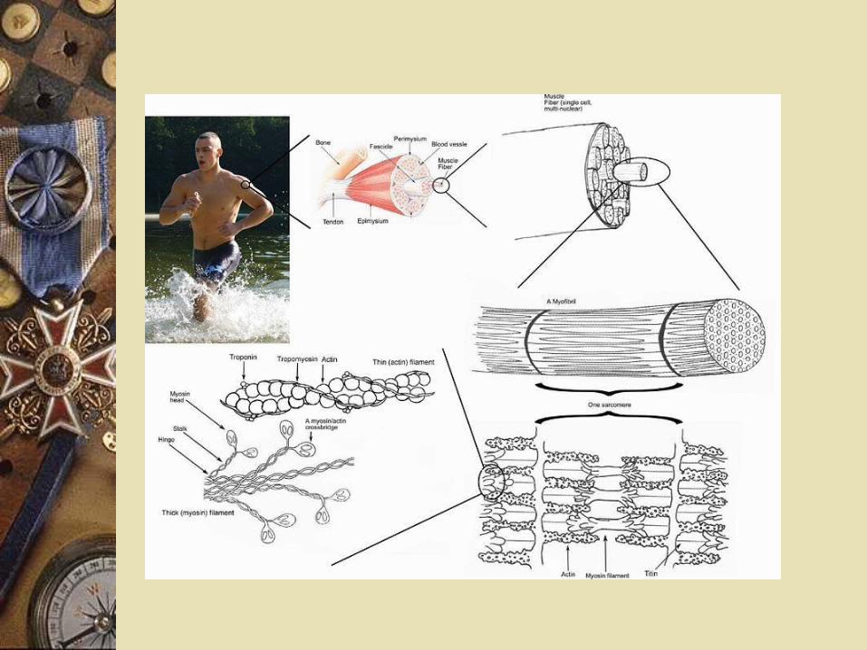
Muscle fibers Single cell with many nuclei Each fiber has a bundle of
myofibrils Each myofibril contains
myofilaments Thick(myosin) or thin (actin)

Muscle structure
Myosin (thick) proteins with a head region
Several actin (thin) form a double helix Cross-bridges Head region from myosin Extends to actin


Cross-bridgeCross-bridge
Cross-bridgeADP
P i
ATP
Actin
Myosin
Myosin binding sites

Muscle Muscle
Bundle ofmuscle fibers
Muscle
Single muscle fiber(cell)
Nuclei
Z lines
Plasma membrane
Myofibril
Sarcomere
Muscle structure
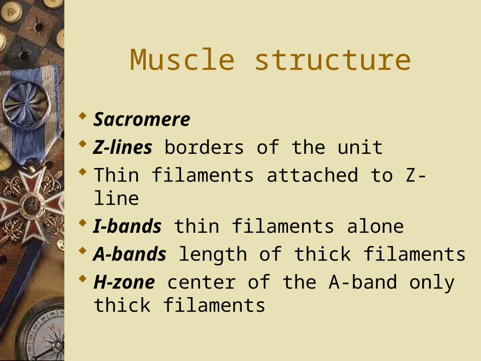
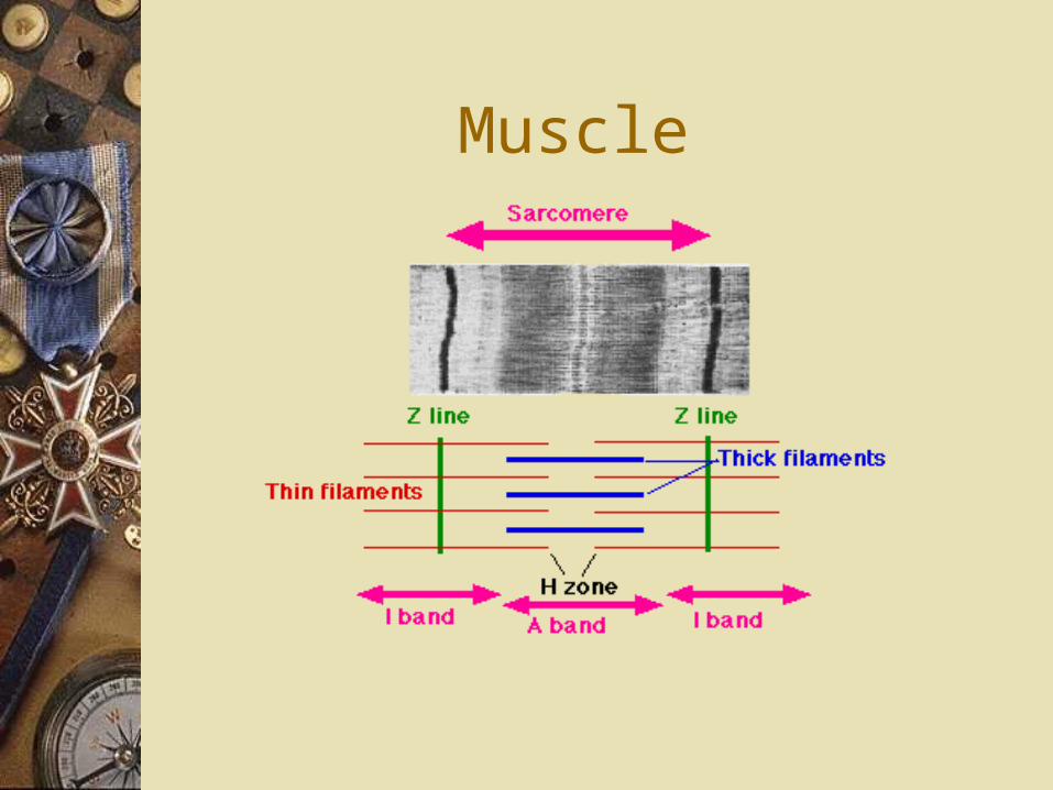
Sacromere Z-lines borders of the unit Thin filaments attached to Z-line I-bands thin filaments alone A-bands length of thick filaments H-zone center of the A-band only
thick filaments


Muscle
Muscle

Muscle

Muscle structure


Muscle contraction
Myosin head binds an ATP Spits ATP Forms cross-bridge Binds actin Pulls the actin Cross-bridge broken when binds new ATP Continues muscle is stimulated to contract Myofilaments move by sliding mechanism

Muscle structure

Control of contraction
Calcium Tropomyosin: Protein found on actin Troponin: Regulatory protein Transverse tubules (T tubules): Tube in the muscle fiber Sarcoplasmic reticulum (SR): Stores calcium in muscle

Mechanism of contraction
Relaxed muscle Myosin heads are ready (split ATP) Not attached to actin Cross-bridges cannot form Tropomyosin is on the actin Blocking binding site on actin

Mechanism of contraction
Contracting muscle Calcium binds the troponin Removes tropomysin off actin binding
sites Cross-bridges form Muscles contract

Contraction Contraction
Myosin-binding site
Tropomyosin
(a) Myosin-binding sites blocked
(b) Myosin-binding sites exposed
Ca2+
Ca2+-binding sites
Troponin complexActin

Mechanism of contraction
Relaxed muscle low calcium levels Contracting muscle high calcium levels Electric impulse (nerve) Stimulates calcium release from SR Travels down T tubules Binds troponin Contraction happens

Mechanism
Impulses stop Calcium is pumped back into SR Troponin no longer attached to
calcium Tropomysin returns to actin


Cardiac muscle
Shorter, branched cells Each with own nucleus Form a lattice Gap junctions electrical impulses
Smooth muscle
Internal organs Myosin/actin not organized into
sacromeres No sacroplasmic reticulum Need calcium to contract Capable of contracting when
stretched

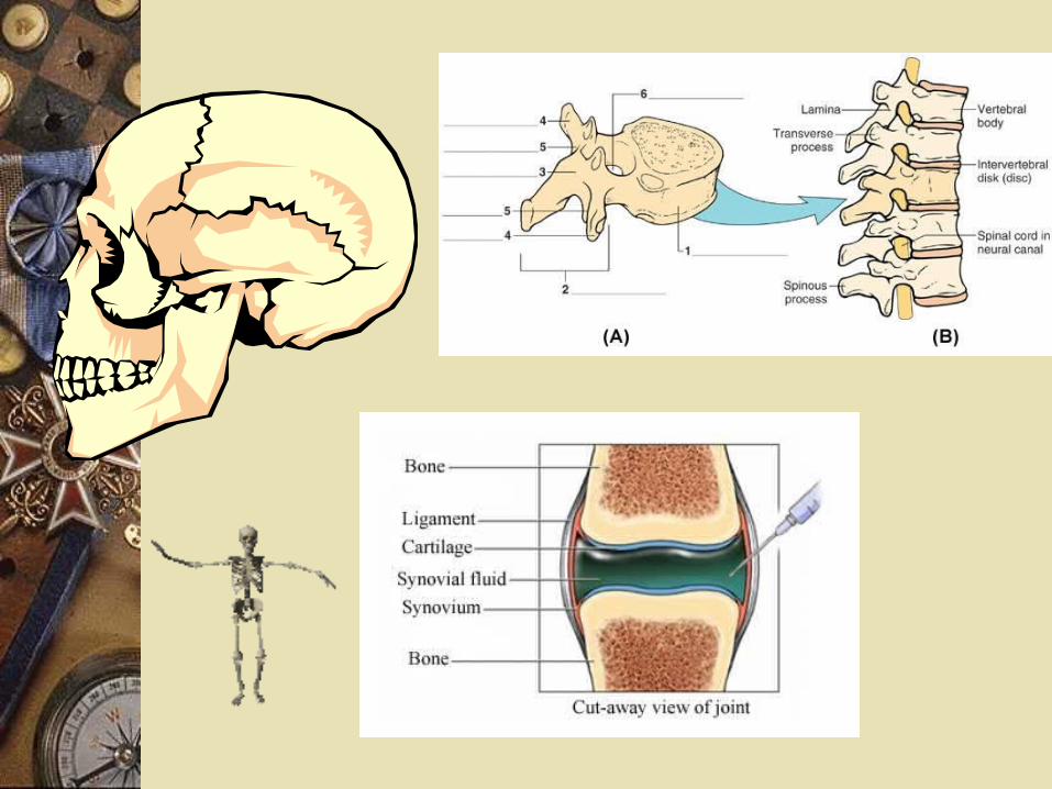
Skeleton
Axial skeleton Supports Protects

Skeleton
Appendicular skeleton
Attach to axial skeleton

Skeleton
Articulations Joints, where movement happens
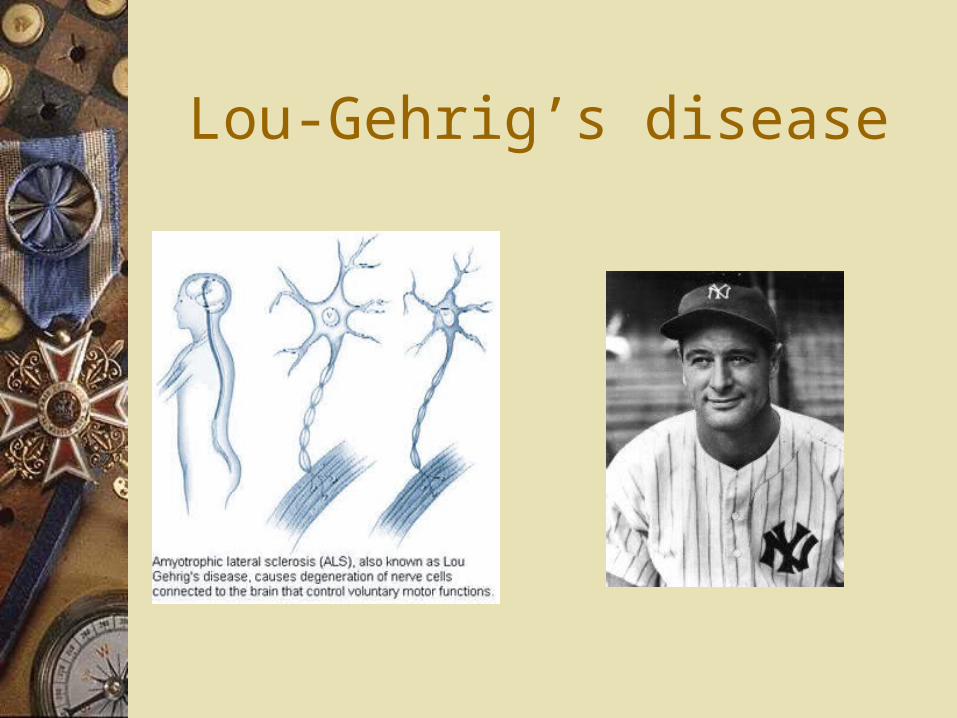
Lou-Gehrig’s disease