mutagenesis study reveals the rim of catalytic entry site of
TRANSCRIPT

RESEARCH ARTICLE
Mutagenesis Study Reveals the Rim ofCatalytic Entry Site of HDAC4 and -5 as theMajor Binding Surface of SMRT CorepressorGwang Sik Kim1, Ha-Eun Jung1, Jeong-Sun Kim2, Young Chul Lee1*
1 Hormone Research Center, School of Biological Sciences and Technology, ChonnamNational University,Gwangju, 500–757, Republic of Korea, 2 Department of Chemistry and Institute of Basic Sciences,ChonnamNational University, Gwangju, 500–757, Republic of Korea
AbstractHistone deacetylases (HDACs) play a pivotal role in eukaryotic gene expression by modu-
lating the levels of acetylation of chromatin and related transcription factors. In contrast to
class I HDACs (HDAC1, -2, -3 and -8), the class IIa HDACs (HDAC4, -5, -7 and -9) harbor
cryptic deacetylases activity and recruit the SMRT-HDAC3 complex to repress target genes
in vivo. In this regard, the specific interaction between the HDAC domain of class IIa HDACs
and the C-terminal region of SMRT repression domain 3 (SRD3c) is known to be critical, but
the molecular basis of this interaction has not yet been addressed. Here, we used an exten-
sive mutant screening system, named the “partitioned one- plus two-hybrid system”, to iso-
late SRD3c interaction-defective (SRID) mutants over the entire catalytic domains of
HDAC4 (HDAC4c) and -5. The surface presentation of the SRID mutations on the HDAC4c
structure revealed that most of the mutations were mapped to the rim surface of the catalytic
entry site, strongly suggesting this mutational hot-spot region as the major binding surface
of SRD3c. Notably, among the HDAC4c surface residues required for SRD3c binding,
some residues (C667, C669, C751, D759, T760 and F871) are present only in class IIa
HDACs, providing the molecular basis for the specific interactions between SRD3c and
class IIa enzymes. To investigate the functional consequence of SRID mutation, the in vitroHDAC activities of HDAC4 mutants immuno-purified from HEK293 cells were measured.
The levels of HDAC activity of the HDAC4c mutants were substantially decreased com-
pared to wild-type. Consistent with this, SRID mutations of HDAC4c prevented the associa-
tion of HDAC4c with the SMRT-HDAC3 complex in vivo. Our findings may provide structural
insight into the binding interface of HDAC4 and -5 with SRD3c, as a novel target to design
modulators specific to these enzymes.
PLOS ONE | DOI:10.1371/journal.pone.0132680 July 10, 2015 1 / 25
a11111
OPEN ACCESS
Citation: Kim GS, Jung H-E, Kim J-S, Lee YC (2015)Mutagenesis Study Reveals the Rim of CatalyticEntry Site of HDAC4 and -5 as the Major BindingSurface of SMRT Corepressor. PLoS ONE 10(7):e0132680. doi:10.1371/journal.pone.0132680
Editor: Luis Menéndez-Arias, Centro de BiologíaMolecular Severo Ochoa (CSIC-UAM), SPAIN
Received: March 25, 2015
Accepted: June 17, 2015
Published: July 10, 2015
Copyright: © 2015 Kim et al. This is an open accessarticle distributed under the terms of the CreativeCommons Attribution License, which permitsunrestricted use, distribution, and reproduction in anymedium, provided the original author and source arecredited.
Data Availability Statement: All relevant data arewithin the paper and its Supporting Information files.
Funding: This work was supported by a grant fromthe National Research Foundation of Korea (NRF),funded by the Korean Government (NRF 2011-0029484).
Competing Interests: The authors have declaredthat no competing interests exist.

IntroductionChromatin is the basic structure of the eukaryotic chromosomes, formed as an array of nucleo-somes composed of histone octamers wrapped with 146 bp of DNA [1]. The tight binding ofDNA with histones in condensed chromatin acts as an obstacle for DNA binding of variousproteins involved in DNAmetabolism, including transcription. In this regard, chromatinremodeling and histone tail modifications are major mechanisms to convert chromatin statusfrom closed conformation (inactive) to open conformation (active), or vice versa [2]. Epige-netic control of gene expression is achieved via the complicated interplay among DNAmethyl-ation, histone tail modifications, and chromatin remodeling. The consequential chromatinstatus is thought to play a major role in X-inactivation, heterochromatin formation and main-tenance, and homeotic gene expression during early development in animals [3]. Currently,epigenetic control is also regarded as a general mechanism for the gene-specific regulation ofeukaryotic transcription, as most epigenetic modifications were proven to be reversible and toarise in a promoter-specific manner [4,5]. Among these dynamic epigenetic markers, acetyla-tion and deacetylation occur at specific lysine residues within the N-terminal tail of nucleoso-mal histones through the opposite actions of two respective families of enzymes, the histoneacetyltransferases (HATs) and histone deacetylases (HDACs) [2]. In general, histone acetyla-tion by HATs acts as an active mark for gene transcription, whereas histone deacetylation byHDACs correlates with transcriptional repression. Therefore, the transcriptional activity of aspecific gene, or the compaction level of a local chromatic region is established through the bal-anced actions of HAT and HDAC enzymes [6]. HDACs play a central role in the regulation ofmany biological processes, such as the cell-cycle, cell differentiation and survival [6,7]. Geneticmouse models revealed HDACs to be essential in embryonic development, cardiovascularhealth and energy metabolism [8,9]. In this respect, the selective blocking of specific HDACfunction has a great therapeutic impact on many diseases, including cancer, cardiovascular,neurodegenerative, and metabolic disorders [10–13]. At present, a variety of HDAC inhibitorsare under clinical investigation, while two HDAC inhibitors, SAHA (vorinostat) and FK228(romidepsin), were already approved for the treatment of cutaneous T-cell lymphomas [14].
To date, 18 kinds of mammalian HDACs have been identified and classified into four classesbased on their sequence similarities and domain structure [15]. The class I HDACs (HDAC1,-2, -3, and -8) are mammalian homologues of the yeast Rpd3 corepressor, and contain only theHDAC domain with a size of about 350 amino acids [16]. They are nuclear proteins with ubiq-uitous expression in most cell types. The class II HDACs (HDAC4, -5, -6, -7, -9, and -10) sharehomology with yeast Hda1p, and display cell type-specific expression [17]. For example,HDAC4, -5 and -9 are known to be enriched in the heart, skeletal muscle and brain tissues.Class II enzymes can be further subdivided into class IIa (HDAC4, -5, -7, and -9) and IIb(HDAC6 and -10) according to their modular structure. In addition to the C-terminal HDACdomain, class IIa HDACs have a long-extending N-terminal adaptor domain, which is targetedby various transcription factors and regulatory signals [17]. Class IIa enzymes can shuttlebetween the cytoplasm and nucleus based on the presence or absence of modifications to spe-cific residues in the adaptor domain, induced by external stimuli. Class IIb enzymes (HDAC6and -10) have a characteristic long extension of the C-terminal tail domain, and are typicallyfound in the cytoplasm. In particular, HDAC6 is a microtubule-associated deacetylase withdual HDAC domains, compared with the single domain of HDAC10 [18]. Class IV includesonly HDAC11, which shows strong sequence similarity to the class I HDACs and is predomi-nantly located in the nucleus [19]. The class I, II and IV HDACs are zinc-dependent enzymes,harboring a common enzymatic mechanism focused on zinc-catalyzed hydrolysis of the acetyl-lysine amide bond [20]. In contrast, the class III HDACs (sirtuin family) require NAD+ as a
Interaction Surface of HDAC4 and -5 with SMRT
PLOSONE | DOI:10.1371/journal.pone.0132680 July 10, 2015 2 / 25

cofactor for catalytic function, and are not sensitive to the HDAC inhibitors effective on class Iand II enzymes [8].
HDACs are unable to bind to DNA by themselves. They exist as components in a variety ofmultiprotein complexes that are recruited to target promoters via interactions with manyDNA-binding factors, such as unliganded nuclear receptors, the E-box binding factors, and themethylcytosine binding protein [21,22]. For example, HDAC1 and HDAC2 are found in threedistinct corepressor complexes, called SIN3A, NURD/Mi2 and CoREST [23,24]. NCoR(nuclear receptor-corepressor) and SMRT (silencing mediator for retinoid and thyroid recep-tor) are ubiquitously expressed corepressor proteins containing three autonomous repressiondomains (RD1 to RD3) in their N-terminal regions [25,26]. Each of the repression domainsplays a non-redundant role in the platform for recruitment of various DNA-binding repressorsor corepressors, including HDACs [27]. HDAC3 is exclusively found in SMRT/NCoR com-plexes, with which association is achieved through the conserved deacetylase activating domainof NCoR/SMRT [28,29]. The activation of HDAC3 through this association was proven to beessential for transcriptional repression by certain nuclear receptors, including thyroid hormonereceptor and Rev-erbs [30,31]. A recent study from the Lazar group indicated that a deacety-lase-independent but NCoR-dependent function of HDAC3 is essential for the transcriptionalregulation of hepatic genes, highlighting the non-enzymatic roles of HDAC3 in liver metabo-lism [32]. As mentioned previously, class IIa HDACs have a modular structure composed ofN-terminal adaptor and C-terminal HDAC domains. The adaptor domain recruits many core-pressors including BCoR, CtBP and HP1 as effector molecules, as well as various repressorssuch as MEF2, FOXP3 and RUNX2 as target transcription factors [17]. Consistent with this,knockout mice models demonstrated that the loss of class IIa HDACs can lead to abnormalitiesduring skeletogenesis and heart development [11,33–35]. Notably, the C-terminal region ofSMRT RD3 (SRD3c) specifically interacts with the HDAC domain of class IIa HDACs, but notwith class I enzymes [27]. This observation strongly suggested that class IIa HDACs functionas a bridge between target repressor proteins and the SMRT/NCoR-HDAC3 complex via inde-pendent interactions occurring through the two domains [36].
Structural and functional analyses of inhibitor or substrate-bound HDACs have beenattempted. The crystal structure of HDAC8 bound with acetylated peptide substrate revealedthat the residues (His142, His143, Asp176, Asp183 and Tyr306) around the catalytic cavityinteract with one water molecule and zinc ion [37]. These interactions are conserved amongthe catalytic zinc-binding pockets of class I enzymes, and are important for substrate recogni-tion and zinc-catalyzed hydrolysis of the acetyl-lysine amide bond of the peptide substrate[37,38]. The crystal structures of the catalytic domain of HDAC7, as well as inhibitor boundforms of HDAC4 and -7 were also investigated [39,40]. These studies revealed that HDAC4and -7 harbor a second zinc-binding domain, adjacent to the zinc-containing catalytic domain.This class IIa HDAC-specific region is well conserved in other class IIa HDACs, and has beenimplicated to have regulatory and structural roles [39–41]. Another structural feature of classIIa HDACs is the unique topology of the active site in an enlarged active site pocket [39]. In theclass IIa enzymes, the catalytic active site contains a histidine residue (His976 of HDAC4,His1006 of HDAC5 and His843 of HDAC7) instead of the tyrosine residue conserved in class Ienzymes (Tyr298 of HDAC3 and Tyr306 of HDAC8), which functions as a transition-state sta-bilizer of the catalytic reaction. Interestingly, a His-976-Tyr mutation in HDAC4 dramaticallyincreases its enzymatic activity to the level of class I enzymes [42], explaining the reason whyclass IIa HDACs purified from bacteria harbor very low deacetylase activity toward acetylatedlysine as compared with class I enzymes. Recently, proteomic analysis revealed that the absenceof HDAC4 had no effect on the acetylation profile of the murine neonate brain, providing invivo evidence that HDAC4 may not function as a lysine deacetylase in this tissue [43].
Interaction Surface of HDAC4 and -5 with SMRT
PLOSONE | DOI:10.1371/journal.pone.0132680 July 10, 2015 3 / 25

Considering the cryptic activity of class IIa enzymes, the HDAC activity shown by the endoge-nous class IIa HDAC complex purified from mammalian cells is supplied by the SMRT/NCoR-HDAC3 complex via its association with the HDAC domain [36]. In this regard, thecatalytic domain of class IIa HDACs is a pseudo-HDAC domain that functions as a SMRT-recruiting module, independent of the deacetylase activity.
The structural differences between class I and IIa enzymes strongly suggest that the struc-tural zinc-binding domain, which is specific to class IIa HDACs, may participate in their spe-cific interactions with the SRD3c region. To test this possibility, we employed a ‘one- plus two-hybrid system’ (OPTHiS) to obtain SRD3c interaction-defective (SRID) mutants over theentire catalytic domains of HDAC4 (HDAC4c) and HDAC5 (HDAC5c). Surprisingly, the sur-face presentation of the SRID mutations on the HDAC4c structure revealed that most of themutations were mapped to the rim surface of the catalytic entry site, rather than the structuralzinc-binding domain, as the mutational hot-spot. Furthermore, some mutant residues (C667,C669, C751, D759, T760 and F871) were found to be present only in class IIa HDACs, provid-ing the molecular basis by which SRD3c specifically interacts with class IIa HDACs, but notwith class I enzymes.
Materials and Methods
PlasmidsTo construct the bait plasmid pRS325LexA-RD3c used in OPTHiS, the human SRD3c region(amino acids 1281 to 1504) was amplified by PCR and inserted into the BglII/NcoI sites of thepRS325LexA vector. The catalytic domains of human HDAC4 (amino acids 650 to 1055) and-5 (amino acids 680 to 1087) were obtained from pBJ5.1-Flag clones by PCR and cloned intothe EcoRI/BamHI sites of pRS324UBG to make pRS324UBG-HDAC4c and -5c, respectively.To isolate SRID alleles of HDAC4c and HDAC5c, G4N, G4T, G5N, and G5M plasmids wereconstructed and used as gapped plasmids for OPTHiS screening (Fig 1A). The G4N and G4Tplasmids were made by inserting PCR fragments of HDAC4cT (amino acids 862 to 1055) andHDAC4cN (amino acids 651 to 862) into the EcoRI/BamHI sites of pRS324UBG, respectively.In the case of the G5N plasmid, the PCR fragment of HDAC5cT (amino acids 801 to 1087)was digested by EcoRI/BamHI and inserted into the corresponding sites of pRS324UBG. Toconstruct the G5M plasmid, the HDAC5A (amino acids 680 to 829) and -5B (amino acids1001 to 1087) fragments of HDAC5c were obtained by PCR amplification and cloned into theEcoRI/BamHI sites of pRS324UBG by three-piece ligation. pcDNA3-HA-HDAC4c and-HDAC5c were made by inserting the respective PCR fragments of HDAC4c and HDAC5cinto the EcoRI/XbaI sites of the pcDNA3-HA vector. For bimolecular fluorescence comple-mentation (BiFC) assay, KGN-MC-SRD3c (amino acid 1281 to 1504) was constructed by sub-cloning the KpnI/XhoI fragment from pcDNA3-HA-SRD3c into the KGC-MC vector (MLBInternational Corporation). As the first step to making KGC-MC-HDAC4c, pBS-HDAC4cwas prepared by inserting the EcoRI/XbaI fragment of pcDNA3-HA-HDAC4c into the pBlue-Script vector (Stratagene). The KpnI/NotI fragment of pBS-HDAC4c and the KpnI fragmentof pcDNA3-HA-HDAC4c were then sequentially were inserted into the KpnI/NotI sites ofKGC-MC (MLB international corporation), resulting in KGC-MC-HDAC4c. To constructKGC-MC-HDAC5c, the KpnI/XhoI fragment was obtained from pcDNA3-HA-HDAC5c andsubcloned into the corresponding sites of KGC-MC. For GST-pull down assay, pGEX4T-SRD3cwas constructed by subcloning the EcoRI/XhoI fragment from pcDNA3-HA-SRD3c intopGEX4T-1 (Amersham Biosciences). The SRID mutants of HDAC4c and -5c isolated byOPTHiS were subcloned into pcDNA3-HA (for in vitro translation) or KGC-MC (for BiFC
Interaction Surface of HDAC4 and -5 with SMRT
PLOSONE | DOI:10.1371/journal.pone.0132680 July 10, 2015 4 / 25
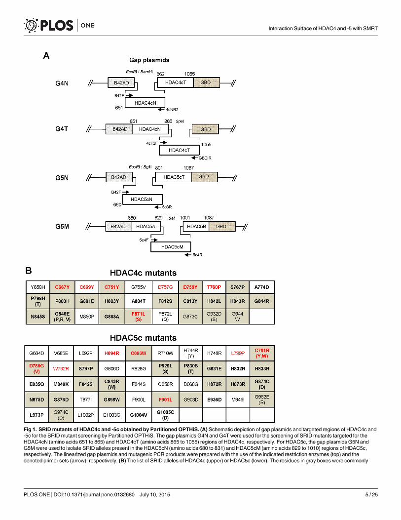
Fig 1. SRIDmutants of HDAC4c and -5c obtained by Partitioned OPTHiS. (A) Schematic depiction of gap plasmids and targeted regions of HDAC4c and-5c for the SRID mutant screening by Partitioned OPTHiS. The gap plasmids G4N and G4T were used for the screening of SRID mutants targeted for theHDAC4cN (amino acids 651 to 865) and HDAC4cT (amino acids 865 to 1055) regions of HDAC4c, respectively. For HDAC5c, the gap plasmids G5N andG5M were used to isolate SRID alleles present in the HDAC5cN (amino acids 680 to 831) and HDAC5cM (amino acids 829 to 1010) regions of HDAC5c,respectively. The linearized gap plasmids and mutagenic PCR products were prepared with the use of the indicated restriction enzymes (top) and thedenoted primer sets (arrow), respectively. (B) The list of SRID alleles of HDAC4c (upper) or HDAC5c (lower). The residues in gray boxes were commonly
Interaction Surface of HDAC4 and -5 with SMRT
PLOSONE | DOI:10.1371/journal.pone.0132680 July 10, 2015 5 / 25

assay) using the appropriate enzyme sites from the pRS324UBG version, respectively. All con-structs were confirmed by DNA sequencing.
Mutagenic PCR and Partitioned OPTHiS ScreeningRandom mutagenesis of HDAC fragments and OPTHiS screening were conducted as previ-ously described in Kim et al. [44,45]. Briefly, mutagenic PCR fragment containing theHDAC4cN or HDAC4cT regions were amplified in the presence of 0.1 mMMnCl2 usingpRS324UBG-HDAC4c as templates with oligomer pairs B42F/4cNR2 for HDAC4cN and4cT2F/GBDIR for HDAC4cT, respectively. In the case of random mutagenesis of theHDAC5cN and HDAC5cM fragments, PCR was conducted in the presence of 0.05 mMMnCl2using pRS324UBG-HDAC5c as templates with oligomer pairs B42F/5c3R for HDAC5cN and5c4F/5c4R for HDAC5cM, respectively. For OPTHiS screening to obtain SRID mutants, theyeast cell libraries containing HDAC mutants were constructed by a single step method via invivo gap repair [45]. The four kinds of mutagenic PCR products (1 μg) were co-transformedwith the corresponding linearized gap plasmids (250 ng) into yeast strain YOK400 (MATa,leu2, trp3, ura3, lexAop-LEU2, UASGAL-HIS3) carrying the pSH18-34 reporter as well as the baitplasmid pRS325LexA-SRD3c. The transformants were grown for 3 days at 30°C in syntheticglucose medium lacking histidine for the positive selection of intact HDAC fusions using theendogenous UASGAL-HIS3 reporter gene [44]. Among the surviving transformants, SRIDmutants were selected by isolating white colonies on X-gal plates using the episomal two-hybrid reporter (lexAop-LacZ). For the actual screening experiment, a large number of transfor-mants was obtained from several batches of standard-scale transformation. Subsequent verifi-cation of the HDAC mutants defective in SRD3c binding was carried out as describedpreviously [45].
Cell Culture and Transient Transfection AssayHEK293 cells were maintained in DMEM (Welgene) supplemented with 10% fetal bovineserum (Welgene) and antibiotics-antimycotic (Gibco). Cells were seeded in 24-well plates with4–8 X 10⁴ cells/well on the day prior to transfection. Transient transfections were performedusing the SuperFect (QIAGEN) or TurboFect (Fermentas) systems, as described in the manu-facturer’s instructions. After 24 h of transfection, cell lysates were prepared with RIPA buffer[50 mM Tris-HCl (pH 8.0), 5 mM EDTA, 150 mMNaCl, 1% NP-40, 1 mM PMSF] and usedfor luciferase and β-galactosidase assays. Luciferase activity was normalized to β-galactosidaseactivity for each sample.
BiFC AssayTo examine the protein interactions of SRD3c with HDAC4c or HDAC5c mutants in livingcells, bimolecular fluorescence complementation (BiFC) assays were carried out using a Fluo-Chase kit (Amalgaam), according to the manufacturer’s manual. Briefly, SRD3c and HDACproteins were fused to the N- or C-terminal portions of Kusabira Green protein, resulting inKGN-SRD3c and KGC-HDAC4c or -5c constructs, respectively. The KGN-SRD3c was coex-pressed with KGC-HDAC4c or -5c in HEK293 cells using the SuperFect system in a 96-wellblack plate (SPL life science). After 48 h of transfection, the fluorescent signals (excitationwavelength: 494 nm, emission wavelength: 538 nm) from the cell lysates were measured using
found between SRID mutants of HDAC4c and -5c based on sequence alignment. The residues existing on the surface of HDAC4c are presented in bold, andthe class IIa HDAC-specific residues are shown in red.
doi:10.1371/journal.pone.0132680.g001
Interaction Surface of HDAC4 and -5 with SMRT
PLOSONE | DOI:10.1371/journal.pone.0132680 July 10, 2015 6 / 25

a fluorescence spectrophotometer (Molecular Devices, Spectra max GEMINIXPS). The quanti-tation experiments were repeated two times for the triplicated samples.
Confocal Laser Scanning MicroscopyHEK293 cells were grown on 8-well slide plates (SPL Life science) and cotransfected withKGN-SRD3c and KGC-HDACmutants using the SuperFect system. After 48 h of transfection,cells were incubated in 4% paraformaldehyde for 10 m at room temperature for fixation. Thecells were then washed with 1X PBS, mounted onto micro cover-slides, and observed for greenfluorescence using a laser-scanning confocal microscope (Leica TCS SPE).
Immunoprecipitation, In Vitro HDAC Assay and Immunoblot AnalysisHuman HDAC3 was coexpressed with HA-tagged versions of the wild-type or HDAC4cmutants in HEK293 cells using the SuperFect system. After 48 h of transfection, the whole celllysates (400–600 μg) were prepared with RIPA buffer and mixed with 30 μl (50% slurry) of aga-rose beads coupled with anti-HA-monoclonal antibody (eBioscience). After overnight incuba-tion at 4°C, the beads were washed three times with RIPA buffer. For in vitroHDAC assay,immuoprecipitates (beads) were mixed with fluorescent-coupled Lys acetamide substrate (Bio-Vision. Cat. No. K330-100) and transferred into a 96- well black plate. HDAC activity assay wascarried out according to the manufacturer’s instructions, and the fluorescence signal (excitationwavelength: 380 nm, emission wavelength: 460 nm) was measured using a fluorescence platereader. For co-immunoprecipitation assay, the bound proteins were eluted from the immuno-precipitates using 0.1M glycine-acetate (pH 3.0), after which the eluents were precipitated usingtricholoroacetic acid. The precipitated proteins were resolved on SDS-PAGE gels and analyzedfor the presence of HA-HDAC4c and HDAC3 by immunoblot using anti-HAmouse (Cell Sig-naling, #2367; 1:3,000 dilution) and anti-HDAC3 rabbit (Abcam, Ab16047; 1:1,000 dilution)antibodies, respectively. Immunoblots were developed using the Optiblot ECL ultra detectionkit (Abcam, Ab133409), and images were captured using an HP imaging system.
Yeast Two-Hybrid and GST Pull-Down AssayYeast strain EGY48 containing pSH18-34 (8X LexAop-LacZ reporter plasmid) was co-trans-formed with the expression plasmids for LexA-SRD3c (pEG202-SRD3c as bait) and for wild-type or mutants of HDACs fused between B42AD and GBD (pRS324UBG-HDAC4c or -5c asa prey) by the lithium acetate method. Liquid assays for β-galactosidase activity were con-ducted at least three times, as described previously [44]. Detailed information for the expres-sion and purification of GST alone and GST-fused proteins were described in our previousreport [45]. The radiolabeled HDAC proteins were added to similar amounts of GST orGST-SRD3c proteins (2–3 μg) bound to glutathione-agarose beads pre-equilibrated with bufferA [150 mM Tris-HCl (pH 7.9), 5% glycerol, 1 mM EDTA, 1 mM dithiothreitol, 1x proteaseinhibitor, 0.01% NP-40, 150 mM KCl] in a final volume of 250 μl. The beads were washedthree times in the same buffer and the bound radiolabeled proteins were analyzed bySDS-PAGE followed by autoradiography.
Statistical AnalysisAll quantitation experiments were repeated two or three times for the triplicated samples. Thestudent’s t-test was used to measure statistically significant differences between wild-type andmutant groups in the corresponding graphs and their p-values were summarized in S3 Table.
Interaction Surface of HDAC4 and -5 with SMRT
PLOSONE | DOI:10.1371/journal.pone.0132680 July 10, 2015 7 / 25

Results
Partitioned OPTHiS for the Isolation of SRID Alleles over the EntireCatalytic Domain of HDAC4 and -5As mentioned, the SRD3c region specifically interacts with class IIa HDACs, but not with classI enzymes, strongly suggesting that a class IIa HDAC-specific region, such as the structuralzinc-binding domain, may be involved in this interaction. To gain an understanding of themolecular basis of the specific interactions between SRD3c and class IIa HDACs, we employedthe one- plus two-hybrid system (OPTHiS) to map the interaction surface of class IIa HDACswith SRD3c [44]. OPTHiS is a novel yeast genetic system designed to efficiently select for mis-sense mutant alleles which specifically disrupt a known protein-protein interaction. To operateOPTHiS, we first tried to define the minimal region of the catalytic domain of HDAC4(HDAC4c) essential for SRD3c binding. To accomplish this, serial truncation mutants ofHDAC4c were made and tested for SRD3c binding in the yeast two-hybrid system. We foundthat the entire region of HDAC4c (amino acids 651 to 1055) was necessary and sufficient foroptimal interaction with SRD3c (data not shown), indicating that the intactness of the SRD3c-binding surface of HDAC4 requires the three-dimensional structure, rather than a short motifof HDAC4c. We considered that the whole region of HDAC4c (about 400 amino acids) is toolong to maintain the optimal mutation rate by OPTHiS [44]. Therefore, the HDAC4c domainwas further divided into two regions (HDAC4cN, HDAC4cT) with a size of about 200 aminoacids, and each region was independently screened for SRID mutants by OPTHiS. As the firststep to isolate the full-length alleles of missense mutants, each mutagenic PCR fragment corre-sponding to the HDAC4cN or HDAC4cT region was co-transformed into yeast strainYOK400 with the linearized gap plasmids G4N and G4T, respectively. In this case, the gap plas-mids G4N and G4T were designed to contain HDAC4cT or HDAC4cN fragments, respec-tively, between the B42AD and GBD regions of the pRS324UBG plasmid (Fig 1A). After co-transformation into yeast, in vivo gap repair by the homologous recombination between PCRfragment and the linearized gap plasmid produced a full-length HDAC4c domain, insertedbetween B42AD and GBD, which harbored missense mutation(s) in the targeted region. Wenamed this strategy “Partitioned OPTHiS”, which enables the screening of interaction-defec-tive alleles targeted for the entire length of a relatively long protein (more than 300 aminoacids). We also employed the “Partitioned OPTHiS”method for the isolation of SRID mutantalleles over the whole region of HDAC5c (amino acids 680 to 1087). HDAC5c was partitionedinto HDAC5cN (amino acids 680 to 831) and HDAC5cM (amino acids 829 to 1010), and thenscreened for SRID mutants through independent operation of OPTHiS (Fig 1A). In this case,each gap plasmid, G5N and G5M, harbored the HDAC5c region(s) that combined with themutagenic PCR fragments (HDAC5cN and HDAC5cM) to generate the full-length HDAC5cdomain in prey fusion proteins (Fig 1A).
Isolation of SRID Mutants of HDAC4c and -5c Using “PartitionedOPTHiS”As described in the materials and methods, we adopted a PCR-mediated random mutagenesisand gap-repair recombination method to generate mutant cell libraries for the HDAC4cN andHDAC4cT regions [45]. As candidates of non-interactor, a total of 29 and 23 white colonieswere isolated on X-gal plates from the 2,700 and 2,200 transformants obtained, which con-tained missense mutations in the HDAC4cN and HDAC4cT regions, respectively (S1 Table).Through subsequent verification and sequencing analysis [45], a total of 36 SRID alleles for 29residues of HDAC4c were finally isolated. Fig 1B shows the mutational sites and the amino
Interaction Surface of HDAC4 and -5 with SMRT
PLOSONE | DOI:10.1371/journal.pone.0132680 July 10, 2015 8 / 25

acids changes in the isolated SRID mutants of HDAC4c (upper panel), from which it can beknown that the mutation residues were distributed over the 300 amino-acid region of the N-terminal HDAC4c domain (spanning from Y658 to G944 position). Some mutants were iso-lated multiple times, and more than one amino acid change was observed at some residues,such as G846 (to Glu, Pro, Arg or Val). Next, the SRID phenotype of the HDAC4c mutantswas confirmed in a quantitative yeast two-hybrid assay and in vitro GST-pull down analysis.After the pRS324UBG-HDAC4c mutants were transformed into the EGY48 strain bearingpRS325LexA-SRD3c, the binding strength between HDAC4c mutants and SRD3c was mea-sured in a liquid β-galactosidase assay. A total of 33 mutants showed severe defects in SRD3cbinding, whereas three mutants (C669Y, G755V, and G868A) displayed only a partial defect(S1A Fig). To confirm the SRID phenotype in vitro, [35S]-labeled HDAC4c mutant proteinswere prepared using a TNT in vitro translation kit and then subjected to binding reaction withGST-fused SRD3c protein. Consistent with the yeast two-hybrid data, almost all of the mutantslost the ability to interact with SRD3c with the exception of C669Y and G755V mutants show-ing partial defects (Fig 2A).
HDAC5 is the closest homologue of HDAC4 among the class IIa HDACs, based onsequence similarity. To determine the general feature of the SRID alleles of class IIa HDACs,we next tried to isolate SRID mutants of HDAC5c. Based on the SRID allele information ofHDAC4c, the 330 amino-acid region of the N-terminal of HDAC5c (amino acid 680 to 1084)was divided into two parts (HDAC5cN and HDAC5cM) and intensively screened for SRIDmutants using “Partitioned OPTHiS,” as in the screening of HDAC4c mutants (Fig 1A). We
Fig 2. Defective interactions of SRD3c with the isolated HDAC4c and -5c mutants in GST pull-down assays.GST-SRD3c protein was purified andtested for interactions with the wild-type (WT) and mutant versions of 35S-labeled HDAC4c (A) or -5c (B) proteins. GST protein was used as the negativecontrol. Input indicates 10% of the in vitro translated HDAC proteins used in the pull-down analysis.
doi:10.1371/journal.pone.0132680.g002
Interaction Surface of HDAC4 and -5 with SMRT
PLOSONE | DOI:10.1371/journal.pone.0132680 July 10, 2015 9 / 25
independently isolated 27 and 42 white colonies as SRID mutants of HDAC5c from the 2,510and 3,000 transformants harboring missense mutations in the HDAC5cN and HDAC5cMregions, respectively (S1 Table). After verification and sequencing of the mutant candidates, atotal of 57 kinds of SRID alleles for 46 residues of HDAC5c were obtained (Fig 1B, lowerpanel). The mutational sites were distributed over the 320 amino-acids of the N-terminalregion of the HDAC5c domain (spanning from G684 to G1005 position), similar to the resultsobserved for the HDAC4c mutants. Among the 57 mutants, 20 representative mutants wereselected, for which the SRID phenotypes were confirmed in the quantitative yeast two-hybridand GST-pull down assays, as described (S1B Fig and Fig 2B). As expected, none of themutants showed SRD3c-binding activity comparable to that of wild-type HDAC5c.
Defective Interactions between SRD3c and SRID Mutants in HEK293CellsTo confirm the SRID phenotype of the isolated HDAC mutants in mammalian cells, the bimo-lecular fluorescence complementation (BiFC) assay was performed, which is a novel protein/protein interaction assay system that is independent of transcription mechanism. Expressionplasmids for the N-terminal portion of the Kusabira Green protein (KGN) fused with SRD3c,as well as the C-terminal portion of the Kusabira Green protein (KGC) fused with HDACmutants were constructed, followed by transient transfection of the plasmids into HEK293cells. In this case, if the SRD3c and HDAC proteins associated with each other, green fluores-cent signals would be generated from the reconstituted Kusabira Green protein. WhenKGN-SRD3c and the wild-type version of KGC-HDACs were expressed as the positive controlof the BiFC assay, green fluorescent signals could be observed predominantly in the nucleus,with the shape of speckles via laser-scanning confocal microscopy (Fig 3A). This result is con-sistent with the previous observations that class IIa HDACs are colocalized with SMRT andHDAC3 in the nuclear matrix with a dot-like structure [36,46], confirming the physiologicalrelevance of the in vivo interaction of SRD3c with HDAC4c or -5c (Fig 3A). Next, we measuredthe levels of fluorescence signals generated by interactions between SRD3c and the isolatedSRID mutants via the BiFC assay using a fluorescence spectrophotometer (Fig 3B and 3C). Theassociation of SRD3c with HDAC4c or -5c increased the fluorescent signal by about two-foldcompared to the background signal generated by KGC empty vector (negative control). In con-trast, with the exception of HDAC4c G846R mutant, which showed partial defect, none of theSRID mutants of HDAC4c and -5c were able to interact with SRD3c in the quantitative BiFCassay (Fig 3B and 3C), consistent with the yeast two-hybrid and GST pull-down data. Statisticalanalysis for all compared groups between wild-type and mutant HDACs revealed the signifi-cant differences among them (see p-values in S3 Table). Finally, the SRID phenotypes of someselected HDAC4c and -5c mutants were examined in a BiFC assay with confocal microscopy(S2 Fig). As expected, the signals from the nuclei of HEK293 cells transfected with the mutantversions of KGC-HDACs were significantly weakened or disappeared compared with those ofthe positive control. Overall, we successfully isolated SRID mutants of HDAC4c and -5c viapartitioned operation of OPTHiS screening, targeted over the entire catalytic domains of theseproteins.
Positions and Features of SRID Mutations of HDAC4c and -5cThe positions of the SRID alleles of HDAC4c and -5c over the structure-based multiple-sequence alignment (MSA) of HDAC domains from class I, -IIa, -IIb and -IV HDACs are pre-sented in Fig 4. On the basis of MSA data, the positions of 20 SRID alleles were overlappedbetween 29 residues of HDAC4c mutations and 46 residues of HDAC5c mutations (gray boxes
Interaction Surface of HDAC4 and -5 with SMRT
PLOSONE | DOI:10.1371/journal.pone.0132680 July 10, 2015 10 / 25

Fig 3. Defective interactions of SRD3c with SRIDmutants of HDAC4c and -5c in HEK293 cells. (A)Confocal laser scanning microscope images illustrating the interactions between SRD3c and HDAC4c or -5cvia BiFC assay. Fluorescent signals were mainly localized in the nucleus, and observed in the shape ofspeckles.Magnification: 180 X. (B),(C)Quantitative measurement of fluorescence signals in the BiFC assay,generated by the association of SRD3c with the indicated SRID mutants of HDAC4c (B) or -5c (C). Theexpression constructs for KGN-SRD3c (1 μg) and the indicated KGC-HDAC4c or -5c mutants (1 μg) were
Interaction Surface of HDAC4 and -5 with SMRT
PLOSONE | DOI:10.1371/journal.pone.0132680 July 10, 2015 11 / 25

transiently cotransfected into HEK293 cells. After 48 hours of transfection, the fluorescent signals from wholecell lysates were measured with the use of a fluorescence spectrophotometer. E.V (empty vector) and N.T(no transfection) samples were used as negative controls. The p-values for all compared groups betweenwild-type and mutants are less than 0.01. RFU: relative fluorescence units.
doi:10.1371/journal.pone.0132680.g003
Fig 4. Positions of SRIDmutations over the MSA of HDAC domains. Structure-based MSA of the catalytic domains from HDAC1 to HDAC11 was builtusing the ESPript 3.0 program, and secondary structural elements are shown for the inhibitor-bound HDAC4c structure (PDB code 2VQJ). The residues inpink and blue shades represent the SRID alleles specifically found in HDAC4c and -5c mutants, respectively. The residues with yellow shade indicate theSRID alleles commonly found at the same positions between HDAC4c and -5c sequences on the basis of MSA. Red triangles: the residues of HDAC4cmutants located on the surface region of the HDAC4c structure. Blue Boxes indicate hot-spot regions for SRID mutations which are commonly found in allclasses of the HDAC family. Red boxes indicate hot-spot regions for SRID mutations which correspond to class IIa HDAC-specific regions.
doi:10.1371/journal.pone.0132680.g004
Interaction Surface of HDAC4 and -5 with SMRT
PLOSONE | DOI:10.1371/journal.pone.0132680 July 10, 2015 12 / 25

in Fig 1B and marked with yellow shade in Fig 4). These residues act as the general requirementfor the interaction of class IIa HDACs with SRD3c, suggesting that the molecular determinantsof HDAC4c interaction with SRD3c are similar to those of HDAC5c. Notably, some mutantresidues were found exclusively in HDAC4c or -5c (purple shade for HDAC4c and blue shadefor HDAC5c in Fig 4), suggesting these residues may act as the specific determinants requiredfor the isotype-dependent interaction of class IIa HDACs with SRD3c. This interesting issue isleft to be addressed in future work, and is currently under investigation.
As shown in Fig 4, most of the SRID mutants of HDAC4c and -5c were commonly found inthe six regions of HDAC domains, labeled as the mutational hot-spots. Among them, tworegions, corresponding to β5-α9 and β6-α10 loops, are conserved in all zinc-dependent HDACenzymes (blue boxes), which accounted for nearly half of the SRID mutations. The rest of theSRID mutations were found as the mutational hot-spots in four regions of the HDAC domains(α1-α2 loop, β3-β4, α11-α12 loop and β9-α15 loop) which are specific to class IIa HDACs (redboxes), according to the structure-based MSA. These results indicate that the interaction ofSRD3c with HDAC4c and -5c is largely mediated by the residues specific to class IIa HDACs,providing important clues to address the issue of how SRD3c specifically interacts with classIIa HDACs, but not with class I HDACs.
The Rim Surface of the Catalytic Entry Site Is the Major SRD3c-BindingSurface of—HDAC4c and -5cNext, we presented the SRID mutations on the surface of the HDAC4c structure because wereasoned that SRD3c associates with the surface region of the HDAC domains of class IIaenzymes. According to the crystal structure of inhibitor-bound HDAC4c (PDB code 2VQJ)[40], 21 residues (C667, C669, C751, D759, T760, S767, A774, P799, P800, G801, H803, A804,F812, C813, H842, H843, G844, N845, G846, G868 and F871) among the 29 identified residuesof the HDAC4c mutations are located at the surface region of HDAC4c (bold letters in Fig 1Band red triangles in Fig 4). These residues were represented on the corresponding positions ofthe three-dimensional structure of HDAC4c, nicely generating two adjacent hot-spot regionsat the surface of HDAC4c (orange in Fig 5B). Interestingly, one of the surface region, composedof four residues (C667 and C669 in α1-α2 loop, C751 in β3-β4 loop, and S767 in α8 region),was positioned at the structural zinc-binding domain known to exist only in class IIa enzymes(Fig 5A and 5B). In contrast, the remaining 17 residues generated a large surface region mainlycomposed of β5-α9 and β6-α10 loops on the rim of catalytic active site (Fig 5A and 5B). TheSRID mutations of HDAC5c were also presented on the surface of the HDAC4c structure, asstructural data for HDAC5c are not available. The positions of the SRID mutations ofHDAC5c were changed to those of HDAC4c according to the MSA data, and displayed at thecorresponding positions of the HDAC4c structure (orange in Fig 5C). In accordance with theHDAC4c data, one small region composed of four residues (H665/H694, C667/C696, C751/C781 and S767/S797 in HDAC4c/HDAC5c) appeared on the surface of the structural zinc-binding domain. Interestingly, 19 out of 25 surface residues of HDAC5c mutations were againmapped to the rim surface of the catalytic site, forming a major binding surface with a moreextended region than that of HDAC4c (Fig 5B and 5C).
Taken together, our findings consistently indicated that most of the surface mutations weremapped to the rim of the catalytic entry site, rather than to the structural zinc-binding domain,which strongly suggests that this mutational hot-spot region is the major binding surface ofSRD3c. This result is unexpected because the active site regions are evolutionally conservedamong all HDAC members, making it unable to explain the reason why SRD3c specificallyinteracts only with class IIa HDACs. However, detailed analysis of the SRID mutant residues
Interaction Surface of HDAC4 and -5 with SMRT
PLOSONE | DOI:10.1371/journal.pone.0132680 July 10, 2015 13 / 25

Fig 5. Surface presentation of SRIDmutations on HDAC4c structure. (A) Ribbon diagram of HDAC4cstructure (PDB code 2VQJ) [40]. The structural zinc-binding domain is shown by magenta. Two zinc ions andtheir chelating residues are drawn as spheres and sticks, respectively. (B) Positions of SRID mutations ofHDAC4c are presented on the surface of the HDAC4c structure (PDB code 2VQJ). Among SRID mutants ofHDAC4c, 21 residues located on the surface are indicated, and their surface positions are shown in orange.(C) Surface presentation of SRID mutations of HDAC5c at the corresponding positions of the HDAC4cstructure. The positions of the SRID mutations of HDAC5c were changed to those of HDAC4c according to
Interaction Surface of HDAC4 and -5 with SMRT
PLOSONE | DOI:10.1371/journal.pone.0132680 July 10, 2015 14 / 25

revealed that six residues (C667, C669, C751, D759, T760 and F871) of the HDAC4c surfacemutations and five residues (H694, C696, C781, D789 and F901) of the HDAC5c surface muta-tions could be identified as class IIa HDAC-specific residues on the basis of the structure-basedMSA (yellow letter in Fig 5). Among them, four residues (C696, C781, D789 and F901) ofHDAC5c had overlapped positions with those of HDAC4c (C667, C751, D759 and F871). Wesuggest that these residues act as the specific requirements for SRD3c interaction with the classIIa HDACs, providing the molecular basis of their specific interactions.
Functional Analyses of HDAC4c Mutants Containing SRID AlleleAs mentioned, the enzymatic activity associated with the endogenous class IIa HDAC complexis dependent on SMRT/NCoR-HDAC3, which is recruited through association of the HDACdomain [36]. To investigate the functional consequence of SRID mutations on HDAC4c, wenext measured the in vitroHDAC activities of HDAC4 mutants purified from mammaliancells. To accomplish this, HA-tagged versions of 25 SRID mutants of HDAC4c were con-structed and transiently expressed along with HDAC3 protein in HEK293 cells. The endoge-nous complex containing HDAC4c mutants was immuno-purified from the whole cell lysateusing anti-HA antibody and then subjected to measurement of the in vitroHDAC activitytoward Lys acetamide substrate, as described in the materials and methods. Intriguingly, thelevels of HDAC activity of the immuno-purified HDAC4c mutants were dramaticallydecreased by eight to ten-fold compared to the wild-type HDAC4c (Fig 6A). The significantdifferences between the in vitro HDAC activities of wild-type and mutants HDAC4c were con-firmed by statistical analysis (see p-values in S3 Table). We reasoned that the reduced HDACactivity of the immuno-purified HDAC4c mutants may be due to the compromised binding ofthe SMRT-HDAC3 complex to the HDAC4c mutants in vivo. To test this possibility, 10 surfacemutants of HDAC4c harboring mutations at the class IIa HDAC-specific regions (C667, C751,D759, S767, and F871) or at the conserved regions among class I and IIa HDACs (P800, H803,F812, H842, and G844) were selected. After transient expression of HDAC3 and HDAC4cmutants in HEK293 cells, the endogenous complex containing wild-type or mutant HDAC4cwas immuno-purified as described and subjected to immunoblot analysis to examine the pres-ence of HDAC3 protein in the immune complex. Although the expression levels of HDAC4cmutants were comparable to that of the wild-type (Fig 6B, upper panel), HDAC3 displayed sig-nificantly compromised association with the HDAC4 mutants in the cell lysate of HEK293cells (Fig 6B, lower panel), consistent with their low levels of in vitroHDAC activity (Fig 6A).From these observations, we concluded that the specific residues located at the rim surface ofthe catalytic entry site of HDAC4c are involved in the in vivo interactions with endogenousSMRT-HDAC3 complex.
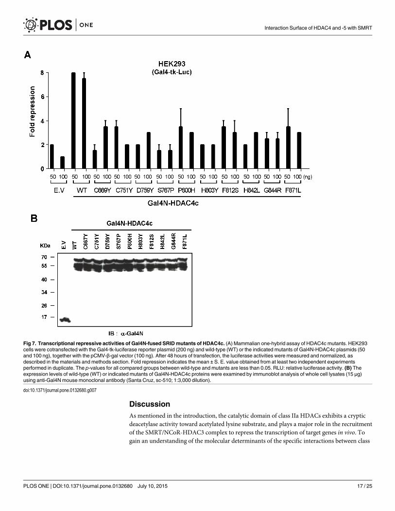
Finally, we investigated the functional consequences of SRID mutations on the transcrip-tional repressive activity of HDAC4c in a mammalian system. The Gal4N-fused version ofwild-type or selected mutants of HDAC4c were prepared and cotransfected with the Gal4N-driven luciferase reporter plasmid into HEK293 cells. As shown in Fig 7A, the Gal4N-mediatedrecruitment of wild-type HDAC4c showed a maximum of 7-fold repression of reporter geneexpression, suggesting the autonomous repressive activity of HDAC4c is provided by theendogenous SMRT-HDAC3 complex. In contrast, all HDAC4c mutants lost their autonomousrepressive activities, displaying only 2 to 3-fold repression of luciferase expression (Fig 7A).Statistical analysis by student’s t-test revealed the significance of the relevant differences
MSA data, and presented on the surface of HDAC4c structure in orange. The residues specific to class IIaHDACs are indicated in yellow color.
doi:10.1371/journal.pone.0132680.g005
Interaction Surface of HDAC4 and -5 with SMRT
PLOSONE | DOI:10.1371/journal.pone.0132680 July 10, 2015 15 / 25

between the autonomous repressive activities of wild-type and mutants HDAC4c (see p-valuesin S3 Table). All of the Gal4N-fused HDAC4c proteins showed similar expression levels, as evi-denced by immunoblotting using Gal4N antibody (Fig 7B). These results indicated that SRIDmutants of HDAC4c lost their repressive function due to the crippled interactions with theendogenous SMRT-HDAC3 complex in vivo, in accordance with the previous results obtainedby the co-immunoprecipitation assay (Fig 6B).
Fig 6. Loss of in vitroHDAC activity of immuno-purified HDAC4cmutants due to inability to associatewith endogenous SMRT-HDAC3 complex. (A) In vitro HDAC assay and (B) co-immunoprecipitation assayfor HDAC4c SRID mutant proteins immuno-purified from HEK293 cells. The HA-tagged versions of theindicated HDAC4c proteins were coexpressed with HDAC3 in HEK293 cells by transient transfection andpurified from cell lysates (300 μg) with the use of agarose beads coupled with anti-HA-antibody. (A) Theimmune complex was subsequently subjected to in vitro HDAC assay using fluorescent-coupled Lysacetamide as a substrate. HeLa nuclear extract (1 μg) was used as the positive control. The p-values for allcompared groups between wild-type and mutants are less than 0.0001. RFU: relative fluorescence units. (B)For co-immunoprecipitation assay, immunoprecipitates from 1 mg of whole cell lysate were prepared andanalyzed for the presence of HA-HDAC4c and HDAC3 by immunoblot analysis.
doi:10.1371/journal.pone.0132680.g006
Interaction Surface of HDAC4 and -5 with SMRT
PLOSONE | DOI:10.1371/journal.pone.0132680 July 10, 2015 16 / 25
DiscussionAs mentioned in the introduction, the catalytic domain of class IIa HDACs exhibits a crypticdeacetylase activity toward acetylated lysine substrate, and plays a major role in the recruitmentof the SMRT/NCoR-HDAC3 complex to repress the transcription of target genes in vivo. Togain an understanding of the molecular determinants of the specific interactions between class
Fig 7. Transcriptional repressive activities of Gal4N-fused SRIDmutants of HDAC4c. (A) Mammalian one-hybrid assay of HDAC4c mutants. HEK293cells were cotransfected with the Gal4-tk-luciferase reporter plasmid (200 ng) and wild-type (WT) or the indicated mutants of Gal4N-HDAC4c plasmids (50and 100 ng), together with the pCMV-β-gal vector (100 ng). After 48 hours of transfection, the luciferase activities were measured and normalized, asdescribed in the materials and methods section. Fold repression indicates the mean ± S. E. value obtained from at least two independent experimentsperformed in duplicate. The p-values for all compared groups between wild-type and mutants are less than 0.05. RLU: relative luciferase activity. (B) Theexpression levels of wild-type (WT) or indicated mutants of Gal4N-HDAC4c proteins were examined by immunoblot analysis of whole cell lysates (15 μg)using anti-Gal4Nmouse monoclonal antibody (Santa Cruz, sc-510; 1:3,000 dilution).
doi:10.1371/journal.pone.0132680.g007
Interaction Surface of HDAC4 and -5 with SMRT
PLOSONE | DOI:10.1371/journal.pone.0132680 July 10, 2015 17 / 25

IIa HDACs and SRD3c, we isolated various kinds of SRID alleles for HDAC4c and -5c throughthe operation of “Partitioned OPTHiS,” targeting the entire HDAC domains.
Partitioned OPTHiS for the Extensive Mutagenesis of the EntireCatalytic Domains of HDACsIn an effort to develop a method for the high-throughput analysis of protein interaction inter-faces, we devised a modified yeast two-hybrid system, termed “OPTHiS,” which efficientlyselects specific missense mutations that disrupt known protein-protein interactions [44]. Todate, we have utilized this system for analysis of the interfaces of many protein-protein interac-tions between various nuclear receptors and their transcriptional coregulators, such as SMRT,NCoR, SRC-1, ASC-2 and TRAP220 [47–53]. In all cases, we tried to select missense mutantalleles of the coregulator proteins defective in nuclear receptor binding, which resulted in theidentification of short motifs comprised of less than a dozen amino acids as the molecular ele-ments of coregulators required for their specific binding to nuclear receptors. As mentionedabove, our efforts to define the minimal region of the HDAC4c domain required for SRD3cbinding revealed that the entire domain of HDAC4c (amino acids 651 to 1055) is necessaryand sufficient for optimal binding to SRD3c. This result implicated that the intactness of theSRD3c-binding surface of HDAC4c requires the three-dimensional structure comprised of thewhole catalytic domain, and a short motif within HDAC4c is not involved in SRD3c binding.In OPTHiS, the prey protein for mutant screening is expressed as a triple fusion betweenB42AD and GBD. Therefore, HDAC4c protein, which requires proper three-dimensionalstructure for SRD3c binding, may not be a suitable target for mutant screening by OPTHiS[45]. Fortunately, three-piece fusion forms of HDAC4c or -5c (B42-HDACs-GBD as a prey)can interact well with the LexA-fused SRD3c protein (bait) in a yeast two-hybrid system,enabling the screening of SRID mutants of these HDACs using OPTHiS. However, there is arestriction on the length of the prey protein, owing to the difficulty of maintaining the optimalmutation rate by PCR-mediated random mutagenesis with increasing prey size. Thus far, wehave successfully analyzed prey proteins of up to 200 amino acids. We considered that the cata-lytic domain of class IIa HDACs (about 400 amino acids) is too long to allow isolation of theSRID mutants via the conventional OPTHiS screening method. Therefore, we solved this prob-lem by developing a novel strategy, named “Partitioned OPTHiS”, in which a relatively longprey protein (more than 300 amino acids) is divided into fragments of less than 200 aminoacids, and the interaction-defective alleles for each fragment are screened by the independentoperation of OPTHiS. In Partitioned OPTHiS, the design of the gap plasmid is critical, becausein vivo gap repair by the homologous recombination between the terminal regions of the PCRfragment (mutagenic target) and the linearized gap plasmid should produce the full-lengthprey protein, which can interact with the bait protein. For example, the gap plasmid G4Nemployed herein was designed to harbor the HDAC4cT region between the B42AD and GBDparts of the pRS324UBG plasmid (Fig 1A). During in vivo gap repair, the mutagenicHDAC4cN fragment was inserted between the B42AD and HDAC4cT regions of the linearizedG4N plasmid, resulting in the generation of the full-length HDAC4c domain harboring therandom mutation(s) at the HDAC4cN region. Using the “Partitioned OPTHiS”method, wewere able to successfully isolate various kinds of SRID alleles of HDAC4c and -5c via extensivemutagenesis of their entire catalytic domains. We propose that the “Partitioned OPTHiS”method can be generally applied for the characterization of protein-binding interfaces, inwhich the three-dimensional domain structure composed of more than 300 amino acids isinvolved, via the rapid and efficient isolation of interaction-defective missense mutants.
Interaction Surface of HDAC4 and -5 with SMRT
PLOSONE | DOI:10.1371/journal.pone.0132680 July 10, 2015 18 / 25

Interaction Surface of Class IIa HDACs with SMRT CorepressorAccording to the MSA data, most of the SRID mutants of HDAC4c and -5c were commonlyfound in six regions of the HDAC domains as mutational hot-spots (Fig 4). Two of the regionsare conserved in all HDAC members (β5-α9 and β6-α10 loops), while the remaining four arespecific to class IIa HDACs (α1-α2 loop, β3-β4 region, α11-α12 loop and β9-α15 loop). A pre-vious report showed that some catalytic core mutants of HDAC4 exhibited compromised bind-ing to the SMRT-HDAC3 complex in vivo, and consequential loss of in vitroHDAC activitywhen immuno-purified from HEK293 cells [36]. Among these mutants, three mutationslocated in the β5-α9 loop (H803Y, H842L and N845S) were isolated in our SRID mutantscreening, verifying that these residues, positioned in the evolutionally conserved catalyticpocket, are involved in SRD3c binding.
The surface display of SRID mutant residues on the corresponding positions of theHDAC4c structure allowed identification of two adjacent hot-spot regions as the SRD3c-bind-ing surface of class IIa HDACs: one minor surface located on the structural zinc-bindingdomain, and one major region corresponding to the rim surface of the catalytic pocket ofHDAC enzymes (Fig 5). As noted, the structural zinc-binding domain exists exclusively inclass IIa members, and is considered to participate in substrate recognition and modulation ofenzyme activity via protein interaction(s) with the regulatory proteins [39–41]. In addition, aprevious report indicated that mutation of the residues (e.g. C669A) coordinating the struc-tural zinc ion prevented its association with the endogenous SMRT-HDAC3 complex, pointingto a key role of the structural zinc-binding domain in the recruitment of binding partner pro-teins [40]. All of these results strongly suggested the structural zinc-binding domain as a goodcandidate for the specific interactions of class IIa HDACs with SRD3c. This notion left openthe possibility that both of the two adjacent surface regions mapped by our SRID mutantscreening are directly involved in SRD3c binding via simultaneous interactions with two inde-pendent parts of the SRD3c region.
However, our analysis of the SRID alleles consistently indicated that most of the surfacemutations (17 out of 21 residues of HDAC4c and 19 out of 25 residues of HDAC5c) weremapped to the rim surface of the catalytic pocket, rather than to the structural zinc-bindingdomain (Fig 5). This observation raises another intriguing possibility that SRD3c solely inter-acts with the major binding surface on the catalytic pocket, but not with the minor surfaceregion on the structural zinc-binding domain. According to this view, the SRID phenotypecaused by mutations at the structural zinc-binding domains can be explained by “indirecteffects," in which the conformational changes of the structural zinc-binding domain couldaffect the structural stability of the neighboring catalytic pocket region and indirectly lead tothe loss of SRD3c-binding ability observed in these mutants. The previous structural analysessupport this notion, that the two adjacent regions have an intimate structural relationship andthat the flexibility of the structural zinc-binding domain might be involved in this connection.For example, the binding of some active site inhibitors and/or substrates to the catalytic pocketof HDAC4c may affect the conformation of its structural zinc-binding domain [40]. This opin-ion is based on the observation that the apo-form of the active site mutant of HDAC4c(H976Y) showed considerable differences in the conformation of the structural zinc-bindingdomain (“closed” conformation) compared with the inhibitor-bound structures (“open” con-formation). This structure resembles the structure of the substrate-bound HDAC8 complex,regarded as the active conformation of the HDAC domain [40]. We propose that the confor-mational change of the structural zinc-binding domain may affect the intactness and SRD3c-binding capacity of the genuine interacting surface formed on the catalytic pocket region. Con-versely, it is also possible that the structural zinc-binding domain is the unique and genuine
Interaction Surface of HDAC4 and -5 with SMRT
PLOSONE | DOI:10.1371/journal.pone.0132680 July 10, 2015 19 / 25

binding partner of SRD3c, and that the mutational effects of the major surface mutants areindirect. We considered this case as unlikely for the following two reasons. Firstly, there was awidespread distribution of major surface mutations from the catalytic entry site to the bottomwall region of the catalytic pocket (β5-α9 loop and α11- α12 region), which is spatially distantfrom the structural zinc-binding domain (Figs 4 and 5). Secondly, there was a striking differ-ence between the numbers of mutations in the two SRD3c-binding surfaces, in which most ofthe residues of the surface mutations (17 out of 21 residues of HDAC4c and 19 out of 25 resi-dues of HDAC5c) formed the large SRD3c-binding surface on the catalytic pocket region.Taken together, we suggest the major mutational hot-spot region formed on the active sitepocket to be the SRD3c-binding surface of class IIa HDACs, though we cannot rule out thepossibility of direct involvement of the structural zinc-binding domain in this interaction.
Molecular Determinants of HDAC4c and -5c Required for Their SpecificBinding to SRD3cAmong the surface residues of HDACs required for SRD3c binding, three residues (C667,C669, and C751 in HDAC4c) located on the structural zinc-binding domain were identified asclass IIa HDAC-specific residues on the basis of the structure-based MSA. In the major bindingsurface on the catalytic pocket, three residues (D759, T760, F871) of HDAC4c and two ofHDAC5c (D789 and F901) were proven to be specific to class IIa HDACs. Considering thebinding mode and the structural basis typically observed in many protein-protein interactions,it is unlikely that all of the surface residues required for SRD3c binding are directly involved inthis interaction. Rather, we suppose that a few residues, including class IIa HDAC-specificones (e.g. D759, T760 or F871 of HDAC4c), actually participate in SRD3c-binding, while therest of the surface residues indirectly affect the intactness of the SRD3c-binding surface. Deter-mination of the molecular requirements of SRD3c for HDAC binding and detailed analysis ofthe binding interface by a crystallographic study will eventually clarify the binding mode ofSRD3c to class IIa HDACs and the structural basis of the specific interactions.
Class IIa HDACs have been implicated as potential therapeutic targets for many human dis-eases, including cancers, metabolic disorders, pathological cardiac hypertrophy, acute ischemicinjury, inflammatory or autoimmune conditions and central nervous system disorders[11,35,54–60]. Therefore, the development of small molecule activators or inhibitors selectivefor class IIa HDACs would have a great impact on the treatment of these diseases, with mini-mization of off-target effects. To date, most HDAC inhibitors share the common structuralcharacteristics of a pharmacophore model comprised of a metal binding moiety, linker regionand surface recognition domain [61]. Thus, all of these inhibitors can bind simultaneously tothe rim, channel and active site of the catalytic pocket, thereby inhibiting HDAC activity byblocking substrate access. Most inhibitors effective for class IIa HDACs also have the samepharmacophore structure, with direct binding to this catalytic site. The active site regionbound by these inhibitors is evolutionally conserved among the all zinc-dependent HDACenzymes [61], explaining why these inhibitors do not show isotype or class-specific effect.Using a mutagenesis approach, we identified the surface region of HDAC4c responsible for theinteraction with SRD3c, which acts as the binding module specific to class IIa HDACs. Ourresults strongly suggest that the surface region of the catalytic pocket of class IIa HDACs, ratherthan active site channel, could be a more plausible and reasonable target for the developmentof class IIa HDAC-specific inhibitors, because this region has structural features distinguish-able from those of class I enzymes.
Recently, novel inhibitors selective for class IIa HDACs were developed, and their therapeu-tic effects on Huntington’s disease were evaluated [62]. Interestingly, these HDAC inhibitors
Interaction Surface of HDAC4 and -5 with SMRT
PLOSONE | DOI:10.1371/journal.pone.0132680 July 10, 2015 20 / 25

were designed to exploit the lower pocket of the catalytic entry site that shows a characteristicof the class IIa HDACs, but not found in other HDAC classes. From the co-crystal structure ofHDAC4c bound by these inhibitors, we found that the targeted lower pocket region is over-lapped with the major SRD3c-binding surface mapped by our mutagenesis study. Furthermore,the inhibitors dock on the lower pocket region via key interactions with the HDAC4 residues,including D759, H803, F812, D840, H842, F871 and D943 [62]. Most of these residues (D759,H803, F812, H842 and F871) were found in the major SRD3c-binding surface revealed herein,raising the intriguing possibility that the surface region composed of these residues directlyparticipates in the binding of SRD3c by means of structural characteristics specific to class IIaHDACs.
In conclusion, our mutagenesis study provided the molecular basis for the specific interac-tions of SRD3c with HDAC4 and -5, as well as structural insights into the binding interface,which may be helpful for the design of modulators specific to class IIa HDACs.
Supporting InformationS1 Fig. Defective interactions of SRD3 with the isolated HDAC4c and -5c mutants in yeast-two hybrid assay. The expression constructs for LexA-fused SRD3c and B42AD-GBD-fusedHDAC4c (A) or -5c (B) mutants were co-transformed into EGY48 strains containing a LacZreporter plasmid, pSH18-34. Transformants were subjected to liquid β-galactosidase assay tomeasure the binding strength between SRD3c and HDAC mutants. β-galactosidase activity wasshown as the representative of three independent experiments. E.V (empty vector) sampleswere used as the negative control. The p-values for all compared groups between wild-type andmutants are less than 0.001(TIF)
S2 Fig. Confocal analysis of the images produced by the interactions between SRD3c andSRIDmutants of HDAC4c in BiFC assay. BiFC assay was performed to assess the interactionsbetween SRD3c and the indicated SRID mutants of HDAC4c (A) and -5c (B) in HEK293 cells.The expression constructs for KGN-SRD3c (500 ng) and KGC-HDAC4c or -5c mutants (500ng) were transiently cotransfected into HEK293 cells. After 48 hours of transfection, the cellswere fixed on micro cover-slides and the green fluorescence signals from the cells wereobserved using a laser-scanning confocal microscope.Magnification: 60 X.(TIF)
S1 Table. Transformation and screening of SRID mutants by OPTHiS. Each of the muta-genic PCR products of the HDAC domains was generated and co-transformed with the indi-cated gap plasmid into strain YOK400 carrying the pSH18-34 reporter as well as the baitplasmid, pRS325LexA-SRD3c. His+ transformants were obtained after a 3-day incubation at30°C on glucose media lacking histidine. Transformants were picked onto plate media contain-ing X-gal but lacking histidine, and the yeast colonies showing white color were isolated as can-didates of non-interactor.(DOCX)
S2 Table. List and sequences of oligonucleotides used in this study.(DOCX)
S3 Table. The p-values obtained by student’s t-test for the compared groups in graphs.(DOC)
Interaction Surface of HDAC4 and -5 with SMRT
PLOSONE | DOI:10.1371/journal.pone.0132680 July 10, 2015 21 / 25

AcknowledgmentsWe thank Prof. Hyun Kook (Chonnam National University, South Korea) for providinghuman HDAC4 and -5 clones. This work was supported by a grant from the National ResearchFoundation of Korea (NRF) funded by the Korea Government (MSIP) (NRF-2011-0029484).
Author ContributionsConceived and designed the experiments: YCL GSK. Performed the experiments: GSK HEJ.Analyzed the data: GSK HEJ JSK YCL. Wrote the paper: YCL GSK.
References1. Luger K, Mader AW, Richmond RK, Sargent DF, Richmond TJ (1997) Crystal structure of the nucleo-
some core particle at 2.8 A resolution. Nature 389: 251–260. PMID: 9305837
2. Berger SL (2007) The complex language of chromatin regulation during transcription. Nature 447:407–412. PMID: 17522673
3. Nightingale KP, O'Neill LP, Turner BM (2006) Histone modifications: signalling receptors and potentialelements of a heritable epigenetic code. Curr Opin Genet Dev 16: 125–136. PMID: 16503131
4. Lachner M, Jenuwein T (2002) The many faces of histone lysine methylation. Curr Opin Cell Biol 14:286–298. PMID: 12067650
5. Kooistra SM, Helin K (2012) Molecular mechanisms and potential functions of histone demethylases.Nat Rev Mol Cell Biol 13: 297–311. doi: 10.1038/nrm3327 PMID: 22473470
6. Di Gennaro E, Bruzzese F, Caraglia M, Abruzzese A, Budillon A (2004) Acetylation of proteins as noveltarget for antitumor therapy: Review article. Amino Acids 26: 435–441. PMID: 15290351
7. Bolden JE, Peart MJ, Johnstone RW (2006) Anticancer activities of histone deacetylase inhibitors. NatRev Drug Discov 5: 769–784. PMID: 16955068
8. Finkel T, Deng CX, Mostoslavsky R (2009) Recent progress in the biology and physiology of sirtuins.Nature 460: 587–591. doi: 10.1038/nature08197 PMID: 19641587
9. Haberland M, Johnson A, Mokalled MH, Montgomery RL, Olson EN (2009) Genetic dissection of his-tone deacetylase requirement in tumor cells. Proc Natl Acad Sci U S A 106: 7751–7755. doi: 10.1073/pnas.0903139106 PMID: 19416910
10. Minucci S, Pelicci PG (2006) Histone deacetylase inhibitors and the promise of epigenetic (and more)treatments for cancer. Nat Rev Cancer 6: 38–51. PMID: 16397526
11. EomGH, Kook H (2014) Posttranslational modifications of histone deacetylases: implications for car-diovascular diseases. Pharmacol Ther 143: 168–180. doi: 10.1016/j.pharmthera.2014.02.012 PMID:24594235
12. Kazantsev AG, Thompson LM (2008) Therapeutic application of histone deacetylase inhibitors for centralnervous system disorders. Nat Rev Drug Discov 7: 854–868. doi: 10.1038/nrd2681 PMID: 18827828
13. Wardell SE, Ilkayeva OR,Wieman HL, Frigo DE, Rathmell JC, Newgard CB, et al. (2009) Glucosemetabolism as a target of histone deacetylase inhibitors. Mol Endocrinol 23: 388–401. doi: 10.1210/me.2008-0179 PMID: 19106193
14. Slingerland M, Guchelaar HJ, Gelderblom H (2014) Histone deacetylase inhibitors: an overview of theclinical studies in solid tumors. Anticancer Drugs 25: 140–149. doi: 10.1097/CAD.0000000000000040PMID: 24185382
15. Verdin E, Dequiedt F, Kasler HG (2003) Class II histone deacetylases: versatile regulators. TrendsGenet 19: 286–293. PMID: 12711221
16. Rundlett SE, Carmen AA, Kobayashi R, Bavykin S, Turner BM, Grunstein M (1996) HDA1 and RPD3are members of distinct yeast histone deacetylase complexes that regulate silencing and transcription.Proc Natl Acad Sci U S A 93: 14503–14508. PMID: 8962081
17. Martin M, Kettmann R, Dequiedt F (2007) Class IIa histone deacetylases: regulating the regulators.Oncogene 26: 5450–5467. PMID: 17694086
18. Hubbert C, Guardiola A, Shao R, Kawaguchi Y, Ito A, Nixon A, et al. (2002) HDAC6 is a microtubule-associated deacetylase. Nature 417: 455–458. PMID: 12024216
19. Gao L, Cueto MA, Asselbergs F, Atadja P (2002) Cloning and functional characterization of HDAC11, anovel member of the human histone deacetylase family. J Biol Chem 277: 25748–25755. PMID:11948178
Interaction Surface of HDAC4 and -5 with SMRT
PLOSONE | DOI:10.1371/journal.pone.0132680 July 10, 2015 22 / 25

20. Finnin MS, Donigian JR, Cohen A, Richon VM, Rifkind RA, Marks PA, et al. (1999) Structures of a his-tone deacetylase homologue bound to the TSA and SAHA inhibitors. Nature 401: 188–193. PMID:10490031
21. Ng HH, Bird A (2000) Histone deacetylases: silencers for hire. Trends Biochem Sci 25: 121–126.PMID: 10694882
22. Xu L, Glass CK, Rosenfeld MG (1999) Coactivator and corepressor complexes in nuclear receptorfunction. Curr Opin Genet Dev 9: 140–147. PMID: 10322133
23. Zhang Y, Sun ZW, Iratni R, Erdjument-Bromage H, Tempst P, Hampsey M, et al. (1998) SAP30, anovel protein conserved between human and yeast, is a component of a histone deacetylase complex.Mol Cell 1: 1021–1031. PMID: 9651585
24. Zhang Y, LeRoy G, Seelig HP, LaneWS, Reinberg D (1998) The dermatomyositis-specific autoantigenMi2 is a component of a complex containing histone deacetylase and nucleosome remodeling activi-ties. Cell 95: 279–289. PMID: 9790534
25. Horlein AJ, Naar AM, Heinzel T, Torchia J, Gloss B, Kurokawa R, et al. (1995) Ligand-independentrepression by the thyroid hormone receptor mediated by a nuclear receptor co-repressor. Nature 377:397–404. PMID: 7566114
26. Park EJ, Schroen DJ, Yang M, Li H, Li L, Chen JD (1999) SMRTe, a silencing mediator for retinoid andthyroid hormone receptors-extended isoform that is more related to the nuclear receptor corepressor.Proc Natl Acad Sci U S A 96: 3519–3524. PMID: 10097068
27. Huang EY, Zhang J, Miska EA, Guenther MG, Kouzarides T, Lazar MA (2000) Nuclear receptor core-pressors partner with class II histone deacetylases in a Sin3-independent repression pathway. GenesDev 14: 45–54. PMID: 10640275
28. Yoon HG, Chan DW, Huang ZQ, Li J, Fondell JD, Qin J, et al. (2003) Purification and functional charac-terization of the human N-CoR complex: the roles of HDAC3, TBL1 and TBLR1. EMBO J 22: 1336–1346. PMID: 12628926
29. Oberoi J, Fairall L, Watson PJ, Yang JC, Czimmerer Z, Kampmann T, et al. (2011) Structural basis forthe assembly of the SMRT/NCoR core transcriptional repression machinery. Nat Struct Mol Biol 18:177–184. doi: 10.1038/nsmb.1983 PMID: 21240272
30. Ishizuka T, Lazar MA (2005) The nuclear receptor corepressor deacetylase activating domain is essen-tial for repression by thyroid hormone receptor. Mol Endocrinol 19: 1443–1451. PMID: 15695367
31. Yin L, Wu N, Lazar MA (2010) Nuclear receptor Rev-erbalpha: a heme receptor that coordinates circa-dian rhythm and metabolism. Nucl Recept Signal 8: e001. doi: 10.1621/nrs.08001 PMID: 20414452
32. Sun Z, Feng D, Fang B, Mullican SE, You SH, Lim HW, et al. (2013) Deacetylase-independent functionof HDAC3 in transcription and metabolism requires nuclear receptor corepressor. Mol Cell 52: 769–782. doi: 10.1016/j.molcel.2013.10.022 PMID: 24268577
33. Vega RB, Matsuda K, Oh J, Barbosa AC, Yang X, Meadows E, et al. (2004) Histone deacetylase 4 con-trols chondrocyte hypertrophy during skeletogenesis. Cell 119: 555–566. PMID: 15537544
34. Chang S, McKinsey TA, Zhang CL, Richardson JA, Hill JA, Olson EN (2004) Histone deacetylases 5and 9 govern responsiveness of the heart to a subset of stress signals and play redundant roles inheart development. Mol Cell Biol 24: 8467–8476. PMID: 15367668
35. Chang S, Young BD, Li S, Qi X, Richardson JA, Olson EN (2006) Histone deacetylase 7 maintains vas-cular integrity by repressing matrix metalloproteinase 10. Cell 126: 321–334. PMID: 16873063
36. Fischle W, Dequiedt F, Hendzel MJ, Guenther MG, Lazar MA, Voelter W, et al. (2002) Enzymatic activ-ity associated with class II HDACs is dependent on a multiprotein complex containing HDAC3 andSMRT/N-CoR. Mol Cell 9: 45–57. PMID: 11804585
37. Vannini A, Volpari C, Gallinari P, Jones P, Mattu M, Carfi A, et al. (2007) Substrate binding to histonedeacetylases as shown by the crystal structure of the HDAC8-substrate complex. EMBORep 8: 879–884. PMID: 17721440
38. Somoza JR, Skene RJ, Katz BA, Mol C, Ho JD, Jennings AJ, et al. (2004) Structural snapshots ofhuman HDAC8 provide insights into the class I histone deacetylases. Structure 12: 1325–1334. PMID:15242608
39. Schuetz A, Min J, Allali-Hassani A, Schapira M, Shuen M, Loppnau P, et al. (2008) Human HDAC7 har-bors a class IIa histone deacetylase-specific zinc binding motif and cryptic deacetylase activity. J BiolChem 283: 11355–11363. doi: 10.1074/jbc.M707362200 PMID: 18285338
40. Bottomley MJ, Lo Surdo P, Di Giovine P, Cirillo A, Scarpelli R, Ferrigno F, et al. (2008) Structural andfunctional analysis of the human HDAC4 catalytic domain reveals a regulatory structural zinc-bindingdomain. J Biol Chem 283: 26694–26704. doi: 10.1074/jbc.M803514200 PMID: 18614528
Interaction Surface of HDAC4 and -5 with SMRT
PLOSONE | DOI:10.1371/journal.pone.0132680 July 10, 2015 23 / 25

41. Ago T, Liu T, Zhai P, ChenW, Li H, Molkentin JD, et al. (2008) A redox-dependent pathway for regulat-ing class II HDACs and cardiac hypertrophy. Cell 133: 978–993. doi: 10.1016/j.cell.2008.04.041 PMID:18555775
42. Lahm A, Paolini C, Pallaoro M, Nardi MC, Jones P, Neddermann P, et al. (2007) Unraveling the hiddencatalytic activity of vertebrate class IIa histone deacetylases. Proc Natl Acad Sci U S A 104: 17335–17340. PMID: 17956988
43. Mielcarek M, Seredenina T, Stokes MP, Osborne GF, Landles C, Inuabasi L, et al. (2013) HDAC4 doesnot act as a protein deacetylase in the postnatal murine brain in vivo. PLoS One 8: e80849. doi: 10.1371/journal.pone.0080849 PMID: 24278330
44. Kim JY, Park OG, Lee JW, Lee YC (2007) One- plus two-hybrid system, a novel yeast genetic selectionfor specific missense mutations disrupting protein/protein interactions. Mol Cell Proteomics 6: 1727–1740. PMID: 17609197
45. Kim JY, Park OG, Lee YC (2012) One- plus two-hybrid system for the efficient selection of missensemutant alleles defective in protein-protein interactions. Methods Mol Biol 812: 209–223. doi: 10.1007/978-1-61779-455-1_12 PMID: 22218862
46. Downes M, Ordentlich P, Kao HY, Alvarez JG, Evans RM (2000) Identification of a nuclear domain withdeacetylase activity. Proc Natl Acad Sci U S A 97: 10330–10335. PMID: 10984530
47. Kim JY, Son YL, Kim JS, Lee YC (2010) Molecular determinants required for selective interactionsbetween the thyroid hormone receptor homodimer and the nuclear receptor corepressor N-CoR. J MolBiol 396: 747–760. doi: 10.1016/j.jmb.2009.12.008 PMID: 20006618
48. Kim JY, Son YL, Lee YC (2009) Involvement of SMRT corepressor in transcriptional repression by thevitamin D receptor. Mol Endocrinol 23: 251–264. doi: 10.1210/me.2008-0426 PMID: 19098224
49. Kim JY, Son YL, Lee YC (2009) A role of helix 12 of the vitamin D receptor in SMRT corepressor inter-action. Biochem Biophys Res Commun 379: 780–784. doi: 10.1016/j.bbrc.2008.12.155 PMID:19133230
50. Son YL, Lee YC (2010) Molecular determinants of the interactions between SRC-1 and LXR/RXR het-erodimers. FEBS Lett 584: 3862–3866. doi: 10.1016/j.febslet.2010.07.056 PMID: 20682316
51. Son YL, Lee YC (2009) Molecular determinants of the interactions between LXR/RXR heterodimersand TRAP220. Biochem Biophys Res Commun 384: 389–393. doi: 10.1016/j.bbrc.2009.04.131 PMID:19410560
52. Son YL, Park MJ, Lee YC (2012) General and specific determinants of the selective interactionsbetween SRC-1 NR box-2 and target nuclear receptors. Mol Biol Rep 39: 177–184. doi: 10.1007/s11033-011-0723-4 PMID: 21553227
53. Son YL, Park OG, Kim GS, Lee JW, Lee YC (2008) RXR heterodimerization allosterically activatesLXR binding to the second NR box of activating signal co-integrator-2. Biochem J 410: 319–330.PMID: 18031289
54. Clocchiatti A, Florean C, Brancolini C (2011) Class IIa HDACs: from important roles in differentiation topossible implications in tumourigenesis. J Cell Mol Med 15: 1833–1846. doi: 10.1111/j.1582-4934.2011.01321.x PMID: 21435179
55. Mihaylova MM, Vasquez DS, Ravnskjaer K, Denechaud PD, Yu RT, Alvarez JG, et al. (2011) Class IIahistone deacetylases are hormone-activated regulators of FOXO and mammalian glucose homeosta-sis. Cell 145: 607–621. doi: 10.1016/j.cell.2011.03.043 PMID: 21565617
56. Zhang CL, McKinsey TA, Chang S, Antos CL, Hill JA, Olson EN (2002) Class II histone deacetylasesact as signal-responsive repressors of cardiac hypertrophy. Cell 110: 479–488. PMID: 12202037
57. Granger A, Abdullah I, Huebner F, Stout A, Wang T, Huebner T, et al. (2008) Histone deacetylase inhi-bition reduces myocardial ischemia-reperfusion injury in mice. FASEB J 22: 3549–3560. doi: 10.1096/fj.08-108548 PMID: 18606865
58. HancockWW, Akimova T, Beier UH, Liu Y, Wang L (2012) HDAC inhibitor therapy in autoimmunity andtransplantation. Ann Rheum Dis 71 Suppl 2: i46–54. doi: 10.1136/annrheumdis-2011-200593 PMID:22460138
59. Mielcarek M, Benn CL, Franklin SA, Smith DL, Woodman B, Marks PA, et al. (2011) SAHA decreasesHDAC 2 and 4 levels in vivo and improves molecular phenotypes in the R6/2 mouse model of Hunting-ton's disease. PLoS One 6: e27746. doi: 10.1371/journal.pone.0027746 PMID: 22140466
60. Sung YM, Lee T, Yoon H, DiBattista AM, Song JM, Sohn Y, et al. (2013) Mercaptoacetamide-basedclass II HDAC inhibitor lowers Abeta levels and improves learning and memory in a mouse model of Alz-heimer's disease. Exp Neurol 239: 192–201. doi: 10.1016/j.expneurol.2012.10.005 PMID: 23063601
61. Miller TA, Witter DJ, Belvedere S (2003) Histone deacetylase inhibitors. J Med Chem 46: 5097–5116.PMID: 14613312
Interaction Surface of HDAC4 and -5 with SMRT
PLOSONE | DOI:10.1371/journal.pone.0132680 July 10, 2015 24 / 25

62. Burli RW, Luckhurst CA, Aziz O, Matthews KL, Yates D, Lyons KA, et al. (2013) Design, synthesis, andbiological evaluation of potent and selective class IIa histone deacetylase (HDAC) inhibitors as a poten-tial therapy for Huntington's disease. J Med Chem 56: 9934–9954. doi: 10.1021/jm4011884 PMID:24261862
Interaction Surface of HDAC4 and -5 with SMRT
PLOSONE | DOI:10.1371/journal.pone.0132680 July 10, 2015 25 / 25