mutation in a primate-conserved retrotransposon reveals … · mutation in a primate-conserved...
TRANSCRIPT

Mutation in a primate-conserved retrotransposonreveals a noncoding RNA as a mediatorof infantile encephalopathyFrançois Cartaulta,b,1, Patrick Muniera, Edgar Benkoc, Isabelle Desguerred, Sylvain Haneinb, Nathalie Boddaerte,Simonetta Bandierab, Jeanine Vellayoudoma, Pascale Krejbich-Trototf, Marc Bintnerg, Jean-Jacques Hoarauf,Muriel Girardb, Emmanuelle Géninh, Pascale de Lonlayb, Alain Fourmaintrauxa,i, Magali Navillej, Diana Rodriguezk,Josué Feingoldb, Michel Renouili, Arnold Munnichb,l, Eric Westhofm, Michael Fählingc,2, Stanislas Lyonnetb,l,2,and Alexandra Henrion-Caudeb,1
aDépartement de Génétique and fGroupe de Recherche Immunopathologies et Maladies Infectieuses, Université de La Réunion, Centre Hospitalier Régionalde La Réunion, 97405 Saint-Denis, La Réunion, France; bInstitut National de la Santé et de la Recherche Médicale (INSERM) U781 and Imagine Foundation,Hôpital Necker-Enfants Malades, Université Paris Descartes, 75015 Paris, France; cInstitut für Vegetative Physiologie, Charité-Universitätsmedizin Berlin,D-10115 Berlin, Germany; dDépartement de Neurologie Pédiatrique, eDépartement de Radio-Pédiatrie and INSERM Unité Mixte de Recherche (UMR)-1000,and lDépartement de Génétique, Hôpital Necker-Enfants Malades, Assistance Publique Hôpitaux de Paris, 75015 Paris, France; gDépartement deNeuroradiologie and iDépartement de Pédiatrie, Centre de Maladies Neuromusculaires et Neurologiques Rares, Centre Hospitalier Régional de La Réunion,97448 Saint-Pierre, La Réunion, France; hINSERM U946, Fondation Jean Dausset, Centre d’Etude du Polymorphisme Humain, 75010 Paris, France;jDynamique et Organisation des Génomes Group, Centre National de la Recherche Scientifique UMR8541, Ecole Normale Supérieure, 75005 Paris, France;kService de Neuropédiatrie, Hôpital Armand Trousseau, Université Pierre et Marie Curie-Paris 6, 75012 Paris, France; and mInstitut de Biologie Moléculaireet Cellulaire du Centre National de la Recherche Scientifique, Université de Strasbourg, 67084 Strasbourg, France
Edited by Mary-Claire King, University of Washington, Seattle, WA, and approved February 7, 2012 (received for review July 23, 2011)
The human genome is densely populated with transposons andtransposon-like repetitive elements. Although the impact of thesetransposons and elements on human genome evolution is recog-nized, the significance of subtle variations in their sequence re-mains mostly unexplored. Here we report homozygosity mappingof an infantile neurodegenerative disease locus in a genetic isolate.Complete DNA sequencing of the 400-kb linkage locus revealed apoint mutation in a primate-specific retrotransposon that was tran-scribed as part of a unique noncoding RNA, whichwas expressed inthe brain. In vitro knockdown of this RNA increased neuronal apo-ptosis, consistentwith the inappropriate dosage of this RNA in vivoand with the phenotype. Moreover, structural analysis of the se-quence revealed a small RNA-like hairpin that was consistent withthe putative gain of a functional site whenmutated.We show herethat a mutation in a unique transposable element-containing RNAis associated with lethal encephalopathy, and we suggest thatRNAs that harbor evolutionarily recent repetitive elements mayplay important roles in human brain development.
genetic disease | long noncoding RNA | long interspersed element 1 |medulla oblongata | pediatrics
Short and long interspersed elements (SINEs and LINEs, re-spectively) constitute the major retrotransposons of higher
vertebrate genomes (1). Despite the parallels between their abun-dance and the evolution of higher cognitive capacities, most ofthese repetitive elements are still regarded as “junk”DNA. Severalcopies of retrotransposons inserted into noncoding genomic se-quences have evolved either as new regulatory sequences (2, 3)or as a source of nonprotein coding RNAs (ncRNAs), includinghumanmicroRNAs (miRNAs) (4, 5) and other RNA species, suchas the neuronal BC1 (6). In addition, retrotransposons themselveshave emerged as specific and transient targets of regulation bysmall RNAs in both germ cells and somatic cells. Interestingly, thebrain is thought to be the major site of RNA expression (7–9).There is increasing evidence that retrotransposition can induce
genetic changes responsible for human diseases, as reviewed byDeininger and colleagues (10). Moreover, we and others haveshown that either mutations in or deletion of the noncoding partof the genome can cause disease (11–14). Overall, ncRNA-basedregulatory circuits appear central to all complex cellular, physio-logical, and neurological systems, and in particular, to specific ge-netic phenomena, including transcriptional and posttranscriptional
silencing. However, whether variation in ncRNA that contains arepetitive element can result in pathogenicity remains unknown.In this study, we report that a rare nucleotide variation in a uniquetransposable element-containing RNA is associated with infantileencephalopathy. This study provides further evidence of hownoncoding mutations may contribute to human diseases.
Results and DiscussionProgressive Encephalopathy with Severe Infantile Anorexia Segregatesin a Geographic Isolate. Because of historic, socioeconomic, andgeographic constraints (SI Methods), a Caucasian isolate that livesin the southern part of Reunion Island (Fig. 1A), located in theIndian Ocean, presents with a high prevalence of autosomal re-cessive disorders. Most patients specifically originate from a re-gion named Ravine. In this Ravine isolate, familial recurrence ofan extreme phenotype of infantile anorexia led us to suspect au-tosomal recessive inheritance of the disease (Fig. S1). Inclusioncriteria were: (i) infantile anorexia with irrepressible and repeatedvomiting during infancy (SI Methods and Table S1) (15), (ii) acutebrainstem dysfunction, (iii) severe failure to thrive, and (iv) spe-cific involvement of the posterior fossa upon MRI. Indeed, brainMRI of patients revealed progressive and severe vanishing of thecerebellar white matter and brainstem atrophy, as well as sus-tentorial periventricular white-matter hyperintensities associatedwith basal ganglia anomalies (Fig. 1B).
Author contributions: F.C. and A.H.-C. designed research; F.C., P.M., E.B., S.H., S.B., J.V.,P.K.-T., J.-J.H., M.G., E.G., M.N., E.W., and M.F. performed research; M.F. and A.H.-C.contributed new reagents/analytic tools; F.C., P.M., E.B., S.H., N.B., P.K.-T., J.-J.H., E.G.,J.F., A.M., E.W., M.F., S.L., and A.H.-C. analyzed data; F.C., P.d.L., A.F., and M.R. contrib-uted patients and defined the phenotype of patients; I.D., N.B., and D.R. helped in fineendophenotyping of patients; N.B., M.B., and A.H.-C. reviewed the radiological material;and F.C. and A.H.-C. wrote the paper.
The authors declare no conflict of interest.
This article is a PNAS Direct Submission.
Data deposition: The data reported in this paper have been deposited in the SingleNucleotide Polymorphism Database (dbSNP), www.ncbi.nlm.nih.gov/projects/SNP (acces-sion nos. rs76603550, rs77619061, rs80022018, rs79197328, rs76885599, and rs74720879).1To whom correspondence may be addressed. E-mail: [email protected] [email protected].
2M.F. and S.L. contributed equally to this work.
This article contains supporting information online at www.pnas.org/lookup/suppl/doi:10.1073/pnas.1111596109/-/DCSupplemental.
4980–4985 | PNAS | March 27, 2012 | vol. 109 | no. 13 www.pnas.org/cgi/doi/10.1073/pnas.1111596109

Autosomal Recessive Infantile Anorexia Locus Maps to Chromosome8p22. Genome-wide linkage analysis was performed in ninefamilies, comprising 15 patients and 17 unaffected siblings (SIMethods and Fig. S1) and showed linkage to chromosome 8p22in a 9.5-Mb region (Fig. 2). Homozygosity fine mapping defined
a small candidate region between the marker loci D8S1731 andD8S261, which reached maximum cumulative LOD scores of6.35 and 6.12, respectively, at θ = 0. This region is flanked byrs76603550 and rs13262614 (Figs. S1 and S2). An additional 17patients from the same population were also typed in this regionand were all homozygous for the haplotype (32 patients in total).This region spanned a 400-kb genetic interval, which containsfive coding genes (ZDHHC2, CNOT7, VPS37A, MTMR7, andSLC7A2) (Fig. 2). All five genes at the disease locus wereregarded as candidates because each was expressed in the brainwith discrete and similar patterns (Fig. S2). Sequencing of theexons of those genes was performed in two patients, but nomutations were identified.
Intronic Nucleotide Variation in a Transposable Element Leads to theIdentification of a Long Noncoding RNA. Complete sequencing ofthe 400-kb region was then performed on both strands in twopatients and two ethnically matched controls. Within the region,we identified 1,725 polymorphisms referenced in the SNP Da-tabase (dbSNP) build 132 and five nonreferenced (Dataset S1).The referenced polymorphisms included 1,699 SNPs, 24 indels,and 2 microsatellites. Within the two patients, none of the 1,725polymorphisms was different from the reference sequence or thecontrols (Dataset S1). Each of the five nonreferenced variationswas intronic SNPs (Dataset S1). One of the five was the same inthe homozygous state in the parents as in the patients and wasthus disregarded. Genotyping of the remaining four poly-morphisms in 1,000 Reunionese controls resulted in identifica-tion of IVS1-1713 A > G (position 17,358,053 assembly hg19) asthe only rare variation segregating with the disease. Indeed,heterozygosity for the mutant allele was detected in 2% of theReunionese ancestry-matched population, with no homozygotesin the 1,000 Reunionese chromosome controls. The diseasehaplotype frequency was 1% with a disease prevalence of 1 in10,000, which was in accordance with Hardy–Weinberg equilib-rium. These data support a founder effect in this Caucasian-admixtured community of Reunion Island.Themutation was located within the 5-kb first intron of SLC7A2
in a degenerated transposable element (Fig. 2). This sequence wascomposed of the 3′ end of a LINE-1 element, L1PA8, whichcomprised the mutation and was embedded in a SINE element,AluSz (Fig. 2). Regarding the disease phenotype, SLC7A2 appears
**
*
Reunion Island
RAVINE des CABRIS
B
A
Fig. 1. Progressive encephalopathy associated with vanishing posteriorfossa in the Ravine geographic isolate. (A) Distribution of a Caucasian isolatein the Ravine des Cabris region of Reunion Island in the Southern IndianOcean. Birth locations of patients are indicated by dots. (B) Brain MRI scansof a patient aged 23 y: sagittal T2 (Upper Left), coronal T2 (Upper Right),axial T1 (Lower Right), and axial FLAIR (Lower Left) images, revealing severeatrophy of the pons (indicated by the star), the medulla oblongata, and thespinal cord. Abnormalities of the entire cerebellum are indicated by T2hyperintensity and T1 hypointensity with a vanishing aspect (indicated bythe asterisk). Sus-tentorial periventricular white matter hyperintensities(indicated by the square) are associated with a basal ganglia anomaly (in-dicated by the solid circle).
3.322.321 .32 22
3.122.121.12 21
2.111.111.1112.1132.11
21 31
3.12
2.12
1.12
1.222.223.22 32
1.42
2.42
3.4222.11
p q
17.20 Mb 17.40 Mb
SLC7A2MTMR7
VPS37ACNOT7ZDHHC2
Exon 1 Intron
CT G A GC A/G A CCA C CA TA
TG CAG CAG AC A CC AT
T G C A G C A A C CA CCA TA
C
Parent
Patient
Control
Exon 2IVS1-1713 A>G
AluSz/L1PA8
rs76
6035
50
rs13
2626
14
Fig. 2. Mapping of an autosomal recessiveinfantile anorexia locus. A genome-wide scanusing microsatellites mapped the diseaselocus to chromosome 8p22, and then thecandidate region was finely mapped usingadditional microsatellite markers and SNPs.The minimal region of the disease locus wasmapped with selected genetic markers at lociD8S550 and D8S280. All genomic coordinatesare relative to the human hg19 assembly, andgenes located between markers rs76603550and rs13262614 are represented as solid lines.Full sequencing of the candidate interval ledto the identification of a single-nucleotidevariation (indicated by an arrow) in the firstintron of the SLC7A2 gene. IVS1-1713 A > Gmaps within a repeated element, L1PA8,embedded in an AluSz that were identifiedusing Repbase (35) (gray box). An electro-phoretogram indicated that the variation ishomozygous in patients (AA), heterozygousin parents (AG), and homozygous in ethni-cally matched controls (GG).
Cartault et al. PNAS | March 27, 2012 | vol. 109 | no. 13 | 4981
GEN
ETICS

to be a good candidate because of the discrete pattern of expres-sion of SLC7A2 in the brain tectal plaque that closely matches thepattern of aqueductal stenosis observed in some Ravine patients(Fig. 3A and Fig. S3A). In the mouse, the SLC7A2 locus is complexbecause of an overlapping transcript, the CTN-RNA, which understress conditions serves as a reservoir for SLC7A2 through post-transcriptional cleavage of its repeat elements (16). We thus
checked whether dysregulation of SLC7A2 expression or splicingbecause of the mutation could have occurred. Indeed, exonizationmediated by transposed elements is regarded as one of the un-derlying causes for the high frequency of alternative splicing inhuman protein-coding genes (17, 18). However, we did not ob-serve ab initio exons, splicing alterations, or a change in the level ofexpression of SLC7A2 in patients with a combination of in silicoanalyses, reverse transcription, and real-time quantitative PCR(qRT-PCR) experiments (Fig. S3 B and C, and Table S2). Wefurther determined whether transcriptional activity was associatedwith the repetitive elements and found a unique expressed se-quence tag (AV720089) that comprised the IVS1-1713 A > Gmutation. Using 5′- and 3′- RACE experiments, we cloned twounique transcripts, A and B, which were 1,536 bp and 962 bp inlength, respectively, and that were colinear to SLC7A2 (Fig. 3Band Fig. S4). Using RT-PCR, we found that transcripts A/B wereexpressed in several different brain structures (Fig. 3B). However,no homology was found with any known protein, motif, or regu-latory motif in either transcript (Table S2). In cross-species se-quence comparisons using University of California at Santa Cruzhg19 46-speciesMultiZ track, we observed the homology-spanningtranscript A sequence among all primates. However, the degen-erated transposable element spanning the mutation was absent inall placental mammals (Fig. S5). Megablast against the variousprimate genomes available indicated a homology range between87% and 97%, with conservation of the A nucleotide, which isthe wild-type nucleotide in Reunionese and in all primategenomes (Fig. S5). Thus, the A nucleotide is the ancestral allele.
Phenotypic Consequences of the Transposable Element-ContainingNoncoding Variant. According to the nomenclature for RNAs,SLC7A2 intronic transcripts A and B were considered to be longnoncoding RNA (lncRNA) and named SLC7A2-IT1A andSLC7A2-IT1B, respectively (19, 20). We found that the overallexpression of SLC7A2-IT1A/B was specifically reduced morethan eightfold in patient brain tissue compared with control, asassessed with qRT-PCR using GAPDH as the internal reference(Fig. 3C). Conversely, the expression of a brain-specific mRNAand the two genes of the mapped locus serving as controlsremained unchanged (Fig. 3C).To investigate the function of wild-type SLC7A2-IT1A/B and
to mimic the reduced levels in the disease, we performed tran-sient siRNA-based knockdown of SLC7A2-IT1A/B in humanneuronal cells (Fig. 4A). We used a set of four SLC7A2-IT1A/Btargeting siRNAs, individually as well as pooled, that are di-rected against different and nonoverlapping sites in the tran-scripts. Knockdown elicited a significant increase in apoptoticcells as assessed by detection of active Caspase3 (Figs. 4 B andC). A correlation study based on different SLC7A2-IT1A/Bknockdown conditions showed a significantly inverse correlationof SLC7A2-IT1A/B transcript levels and active Caspase-3 pro-tein levels (Fig. 4D). Similarly, knockdown of SLC7A2-IT1A/Bby antisense oligonucleotides resulted in a significant increase inapoptotic cells as assessed by dual staining of mitochondrial Baxand active Caspase 3 (Fig. S6).
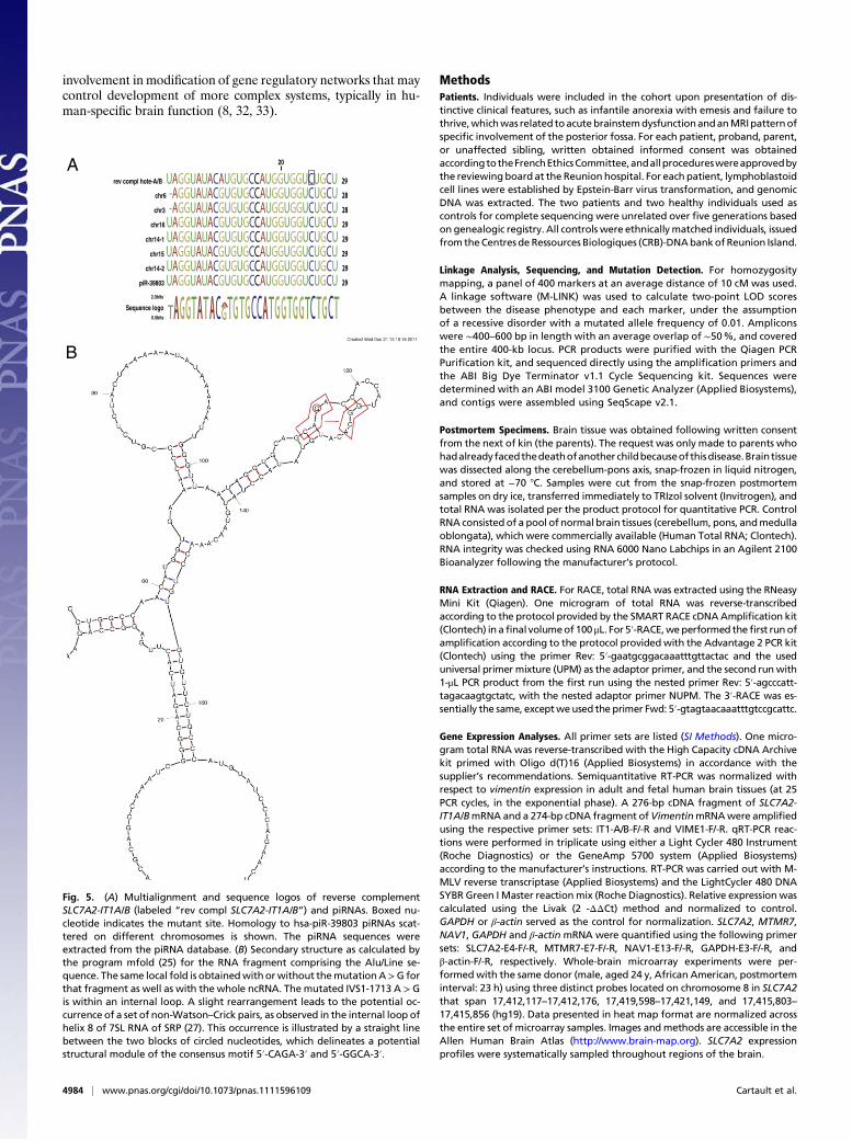
Potential Structural Effect of the Mutation.We blasted the SLC7A2-IT1A sequence against the Rfam database and found that thetranscript did not correspond to any of the 1,973 ncRNA families.Nonetheless, blasting the mutant sequence of SLC7A2-IT1Aagainst the small RNAs from the comprehensive ncRNA se-quence database fRNAdb (<50 nt) identified antisense homologyto a Piwi-interacting RNA (piRNA) at the level of the mutation(Fig. 5A). The mutation increased the probability for the mutantSLC7A2-IT1A sequence to de facto form the complementarysequence to a piRNA by 103-fold, reaching an E-value of 4 × 10⁻⁶
vs. 0.001 for the wild-type. Based on the threshold values, thehomology to the mutant but not the wild-type sequence was
A
B Exon 2Exon 1SLC7A2 transcript-A transcript-B
IVS1-1778A>G
L1PA8AluSz
C
0
0.5
1
C MC M C MC M
SLC7A2-IT1A/B SLC7A2 MTMR7 NAV1
Rel.
exp.
of t
rans
crip
ts (q
RT-P
CR) ***
Fig. 3. Identification of a noncoding RNA by analysis of intronic nucleotidemutation in a transposed element. (A) Expression of the SLC7A2 gene in brainof control and patient. In the control brain (Left), the 3D planar viewof SLC7A2 expression shows reproducible signals with three distinct probes incerebellum and midbrain. Probes were all located in SLC7A2 on chromosome8 and span from 17,412,117–17,412,176; 17,419,598–17,421,149; and17,415,803–17,415,856 (relatively to the human hg19 assembly) (see Methodsand the Allen Human Brain Atlas). In the patient brain, the spatial corre-spondence between images and T1-weighted MR volumes, obtained viaa series of assisted registration processes, are consistent with acqueductalstenosis, indicated by an arrow observed on a sagittal T2 plane MRI imagefrom a patient aged 10 y old. (B) Identification of a totally intronic ncRNA.RACE-PCR in human brain led to the identification of two overlappingtranscripts: A and B, of 1536-bp and 962-bp in length, respectively. The hg19coordinates are 17,356,587 and 17,357,161 for the transcription start sites ofA and B, respectively, and 17,358,123 for the end of both transcripts. Cloningand sequencing revealed that the rare single nucleotide variation (indicatedas an arrow) (hg19 coordinate: 17,358,053), which was found homozygous inpatients only, is included in both transcripts. Gray boxes schematize the re-petitive elements. Expression of transcripts A/B was evaluated by RT-PCR inadult and in fetal human brain tissues. Vimentin (NM_003380) served asa positive control. Negative controls were tested on each total RNA showingthe absence of genomic DNA. (C) Expression of SLC7A2-IT1A/B ncRNA in thebrain of patient versus control by qRT-PCR. Postmortem brain RNA extractionwas performed similarly in the patient as in the control. Control RNA con-sisted of commercially available pool of normal brain tissues (Human TotalRNA; Clontech). Patient RNA was from a Reunionese female individual, age23. RNA integrity was ensured using an on-chip capillary electrophoresis,reaching 8.1 and 7.6 for the patient and the control RNA, respectively. Ex-pression of SLC7A2-IT1A/B, SLC7A2, MTMR7, and NAV1 was measured byqRT-PCR, normalized to GAPDH, and shown as a ratio to control. Data aremean ± SD. ***P < 0.01 (n = 3).
4982 | www.pnas.org/cgi/doi/10.1073/pnas.1111596109 Cartault et al.

significant. Retrotransposon silencing can be mediated by piR-NAs (21) and may lead to silencing of the transcript (22). Thus,we hypothesize that the mutated segment within SLC7A2-IT1A/Bis a privileged target of piRNAs. According to the recent identi-fication of piRNAs in mammalian brain (23, 24), piRNAs mayinvade and bind to the complementary mutant sequence, whichultimately leads to the silencing of the transcript (22).We further questioned the significance of the mutation on the
secondary structure of the lncRNA. Using the mfold program(25), we calculated the 2D structure of the AluSZ/L1PA8 186-bpfragment of SLC7A2-IT1A (Fig. 5B), which was similar to thefull-length and mostly unaffected by the mutation. In fact, themutation occurs in an asymmetrical internal loop of one of thesmall hairpins of the second three-way junction. Interestingly,upon slight rearrangement of the pairs, the mutated internalloop presents a conserved motif of helix 8 in domain IV of the7SL RNA of the signal recognition particle (SRP) (i.e., 5′-CAGA-3′ and 5′-GGCA-3′) (Fig. 5B) (26). This latter motif isknown to form a tight structural module with very specificbinding to the highly conserved SRP protein SRP54 (27). Inparticular, alignments of 7SL RNAs showed that the third G ofthe 5′ strand is invariant (28). SRP54 is a key component forinteractions with the signal peptide, the SRP receptor, and thebinding to two ribosomal proteins, L23a and L35 (26). Thisobservation suggests the intriguing possibility that the recruit-ment of SRP54 induced by the mutation participates in the ob-served deleterious effects.Prompted by the finding that the mutation occurred in a small
hairpin, we further studied the structural modularity of SLC7A2-IT1A/B to assess the likelihood that a small RNA is processedfrom the lncRNA (29). Assessing the SLC7A2-IT1A/B sequenceas a source of miRNAs revealed an 82-bp sequence that gen-erated a good triplet-support vector machine candidate that
could only be retrieved using the mutant sequence and not thewild-type, consistent with other algorithms used for predictingmiRNA precursors (Table S2) (30).In sum, several hypotheses could explain the effect of the
mutation in SLC7A2-IT1A/B, including the possibility of alteredposttranscriptional regulation of SLC7A2 as observed with theCTN-RNA in stress conditions (16). From the current insights,we propose to view the A > G mutation as: (i) a mediator ofRNA:protein interactions, which may potentially be SRP54; (ii)or as a mediator of RNA:RNA interactions, acting either asa piRNA target or as a miRNA-like sequence. In either scenario,the mutation supports a putative gain of a functional site. Achallenge ahead will be to test whether any of these predictedeffects of the mutation occurs in the disease context of Ravineencephalopathy.
Concluding Remarks. In conclusion, the noncoding A > G muta-tion we identified in Ravine encephalopathy results in signifi-cantly decreased levels of expression of the SLC7A2-IT1A/BRNA, which is associated with an increase in cell-death markers.As shown with RNAi experiments, silencing this lncRNA resul-ted in increased apoptosis, which is consistent with the atrophyand vanishing white matter observed in patients’ brain MRIs.Similarly, the intriguing overlap between SLC7A2 expression andthe acqueductal stenosis that we observed may suggest that thespatial expression of SLC7A2-IT1A/B RNA shares regulatoryelements with SLC7A2, as has been documented for severalncRNAs and their host genes (31).Overall, our data unveil the contribution of a mutation in a
specific lncRNA in a progressive disorder of the posterior hind-brain involving the medulla oblongata and leading to anorexicbehavior. The existence of taxon- and species-specific ncRNAsencompassing repetitive elements suggests the possibility of their
Fig. 4. RNAi knockdown of SLC7A2-IT1A/B transcripts. Human neuroblastoma Kelly cells were transiently transfected with 10 nM of a nontargeting siRNA pool(control siRNA) or a 10 nM pool of four SLC7A2-IT1-targeting siRNAs (Pool). The targeting siRNAs were further tested individually at 10 nM each. In the figure“30” is A-specific, “589” is specific for A and B transcripts, “1326” is preAluSz-specific, and “1497” is L1PA8 specific. (A) Real-time PCR analysis to estimate SLC7A2-IT1A/B transcript levels. Values were normalized to relative β-actin levels. (B) Detection of active Caspase-3 protein levels by Western blotting. A representativeoriginal blot is shown. β-Actin served as loading control. (C) Statistical analysis ofWestern blotting results. Knockdown of SLC7A2-IT1A/B by∼40% leads to amorethan 2.5-fold elevated active Caspase-3 level. n = 3. (D) Correlation of SLC7A2-IT1A/B transcript levels and active Caspase-3 protein levels. Using differenttransfection conditions (10 nM up to 100 nM siRNA concentrations for 24 h up to 48 h) SLC7A2-IT1A/B transcript levels were assessed by real-time PCR andcorrelated to corresponding active Caspase-3 protein levels. PCC, Pearson correlation coefficient; n = 32. *P < 0.05, **P < 0.01, ***P < 0.001.
Cartault et al. PNAS | March 27, 2012 | vol. 109 | no. 13 | 4983
GEN
ETICS
involvement in modification of gene regulatory networks that maycontrol development of more complex systems, typically in hu-man-specific brain function (8, 32, 33).
MethodsPatients. Individuals were included in the cohort upon presentation of dis-tinctive clinical features, such as infantile anorexia with emesis and failure tothrive,whichwas related toacutebrainstemdysfunctionandanMRIpatternofspecific involvement of the posterior fossa. For each patient, proband, parent,or unaffected sibling, written obtained informed consent was obtainedaccordingtotheFrenchEthicsCommittee,andallprocedureswereapprovedbythe reviewingboard at the Reunion hospital. For each patient, lymphoblastoidcell lines were established by Epstein-Barr virus transformation, and genomicDNA was extracted. The two patients and two healthy individuals used ascontrols for complete sequencing were unrelated over five generations basedon genealogic registry. All controls were ethnicallymatched individuals, issuedfrom theCentres deRessources Biologiques (CRB)-DNAbankof Reunion Island.
Linkage Analysis, Sequencing, and Mutation Detection. For homozygositymapping, a panel of 400 markers at an average distance of 10 cM was used.A linkage software (M-LINK) was used to calculate two-point LOD scoresbetween the disease phenotype and each marker, under the assumptionof a recessive disorder with a mutated allele frequency of 0.01. Ampliconswere ∼400–600 bp in length with an average overlap of ∼50%, and coveredthe entire 400-kb locus. PCR products were purified with the Qiagen PCRPurification kit, and sequenced directly using the amplification primers andthe ABI Big Dye Terminator v1.1 Cycle Sequencing kit. Sequences weredetermined with an ABI model 3100 Genetic Analyzer (Applied Biosystems),and contigs were assembled using SeqScape v2.1.
Postmortem Specimens. Brain tissue was obtained following written consentfrom the next of kin (the parents). The request was only made to parents whohadalreadyfacedthedeathofanother childbecauseof thisdisease.Brain tissuewas dissected along the cerebellum-pons axis, snap-frozen in liquid nitrogen,and stored at −70 8C. Samples were cut from the snap-frozen postmortemsamples on dry ice, transferred immediately to TRIzol solvent (Invitrogen), andtotal RNA was isolated per the product protocol for quantitative PCR. ControlRNA consisted of a pool of normal brain tissues (cerebellum, pons, andmedullaoblongata), which were commercially available (Human Total RNA; Clontech).RNA integrity was checked using RNA 6000 Nano Labchips in an Agilent 2100Bioanalyzer following the manufacturer’s protocol.
RNA Extraction and RACE. For RACE, total RNA was extracted using the RNeasyMini Kit (Qiagen). One microgram of total RNA was reverse-transcribedaccording to the protocol provided by the SMART RACE cDNAAmplification kit(Clontech) in afinal volumeof100 μL. For 5′-RACE,weperformed thefirst runofamplification according to the protocol provided with the Advantage 2 PCR kit(Clontech) using the primer Rev: 5′-gaatgcggacaaatttgttactac and the useduniversal primer mixture (UPM) as the adaptor primer, and the second runwith1-μL PCR product from the first run using the nested primer Rev: 5′-agcccatt-tagacaagtgctatc, with the nested adaptor primer NUPM. The 3′-RACE was es-sentially the same, exceptweused the primer Fwd: 5′-gtagtaacaaatttgtccgcattc.
Gene Expression Analyses. All primer sets are listed (SI Methods). One micro-gram total RNA was reverse-transcribed with the High Capacity cDNA Archivekit primed with Oligo d(T)16 (Applied Biosystems) in accordance with thesupplier’s recommendations. Semiquantitative RT-PCR was normalized withrespect to vimentin expression in adult and fetal human brain tissues (at 25PCR cycles, in the exponential phase). A 276-bp cDNA fragment of SLC7A2-IT1A/BmRNA and a 274-bp cDNA fragment ofVimentinmRNAwere amplifiedusing the respective primer sets: IT1-A/B-F/-R and VIME1-F/-R. qRT-PCR reac-tions were performed in triplicate using either a Light Cycler 480 Instrument(Roche Diagnostics) or the GeneAmp 5700 system (Applied Biosystems)according to the manufacturer’s instructions. RT-PCR was carried out with M-MLV reverse transcriptase (Applied Biosystems) and the LightCycler 480 DNASYBR Green I Master reactionmix (Roche Diagnostics). Relative expression wascalculated using the Livak (2 -ΔΔCt) method and normalized to control.GAPDH or β-actin served as the control for normalization. SLC7A2, MTMR7,NAV1, GAPDH and β-actin mRNA were quantified using the following primersets: SLC7A2-E4-F/-R, MTMR7-E7-F/-R, NAV1-E13-F/-R, GAPDH-E3-F/-R, andβ-actin-F/-R, respectively. Whole-brain microarray experiments were per-formedwith the same donor (male, aged 24 y, African American, postmorteminterval: 23 h) using three distinct probes located on chromosome 8 in SLC7A2that span 17,412,117–17,412,176, 17,419,598–17,421,149, and 17,415,803–17,415,856 (hg19). Data presented in heat map format are normalized acrossthe entire set of microarray samples. Images andmethods are accessible in theAllen Human Brain Atlas (http://www.brain-map.org). SLC7A2 expressionprofiles were systematically sampled throughout regions of the brain.
piR-39803 UAGGUAUACGUGUGCCAUGGUGGUCUGCU 29 chr14-2 UAGGUAUACGUGUGCCAUGGUGGUCUGCU 29
chr15 UAGGUAUACGUGUGCCAUGGUGGUCUGCU 29 chr14-1 UAGGUAUACGUGUGCCAUGGUGGUCUGCU 29
chr16 UAGGUAUACGUGUGCCAUGGUGGUCUGCU 29chr3 -AGGUAUACGUGUGCCAUGGUGGUCUGCU 28
chr6 -AGGUAUACGUGUGCCAUGGUGGUCUGCU 28rev compl hote-A/B UAGGUAUACAUGUGCCAUGGUGGUCUGCU 29
Sequence logo0.0bits
2.0bits
20A
B
Fig. 5. (A) Multialignment and sequence logos of reverse complementSLC7A2-IT1A/B (labeled “rev compl SLC7A2-IT1A/B”) and piRNAs. Boxed nu-cleotide indicates the mutant site. Homology to hsa-piR-39803 piRNAs scat-tered on different chromosomes is shown. The piRNA sequences wereextracted from the piRNA database. (B) Secondary structure as calculated bythe program mfold (25) for the RNA fragment comprising the Alu/Line se-quence. The same local fold is obtainedwith orwithout themutationA>G forthat fragment as well as with the whole ncRNA. The mutated IVS1-1713 A > Gis within an internal loop. A slight rearrangement leads to the potential oc-currence of a set of non-Watson–Crick pairs, as observed in the internal loop ofhelix 8 of 7SL RNA of SRP (27). This occurrence is illustrated by a straight linebetween the two blocks of circled nucleotides, which delineates a potentialstructural module of the consensus motif 5′-CAGA-3′ and 5′-GGCA-3′.
4984 | www.pnas.org/cgi/doi/10.1073/pnas.1111596109 Cartault et al.

Transfection. SLC7A2-IT1A/B knockdown experiments were performed in Kellyneuroblastoma cells, which were purchased from the German DSMZ, and gen-otyped aswild type for IVS1-1713A>G (ACC 355). Stealth siRNAs (LifeTech)weredesigned to target four different sites of the transcript (SI Methods and Fig. S4).Kelly cellswere grown to∼70%confluency in six-well plates and transfectedwitheither 10–100 nM each siRNA or a pool of the four siRNAs (2.5 nM each) for 24 or48 h. A commercially available nontargeting siRNA pool (Dharmacon) served asa negative control. Cells were transfected using SilenceMag (SM10500; OZ Bio-science), according to themanufacturer’s protocol. Cells were harvested 24 or 48h after transfection for gene or protein expression analyses. Preparation ofprotein extracts, Western blotting, and immunodetection were performed aspreviously described (34). Active Caspase 3 was detected using a monoclonalantibody to Caspase3 (AM08377PU-N, Acris Antibodies; 1:500). Anti–β-actin an-tibody (MAB1501R; Chemicon) was applied after stripping of the membranes tocontrol for differences in protein loading. Primary antibodieswere detectedwithperoxidase-coupled secondary antibodies and visualized using the Chemi-Glow-West Detection kit (Alpha Innotech). Autoradiographic signals were scanned andquantified using Scion Image software (Scion). The Student’s paired t test wasapplied to reveal statistical significances. P values less than 0.05 were consideredsignificant. To correlate SLC7A2-IT1A/B knockdownwith apoptosis,we calculatedthe fold-change relative to control siRNA transfection for SLC7A2-IT1A/B tran-script levels and corresponding active Caspase-3 protein levels per tested condi-tion. The P value based on the Pearson correlation coefficient was calculated.
Sequence Analyses. Thehuman SLC7A2 sequencewasextracted fromtheHumanGenome Project Assembly (GRCh37/hg19) using the transcript ENST00000494857as a reference from Ensembl. Briefly, the human sequence was analyzed forSNPs using BioMart from Ensembl, repeat sequences (LINEs, SINEs, etc.) usingRepeatMasker, and species-specific collection of repeat sequences from thelatest Repbase-Update (35). A variety of complementary approaches wereused to search for exons and regulatory elements (36): GENSCAN and Exon-ScanWeb Server to identify ab initio exons; Expasy Translate tool to search forpotential ORFs checked for homology with BLASTP against Swiss-Prot andPDB-databases; BLASTN (37) against mRNA (expressed sequence tag or cDNA)sequences from GenBank (release 123); the Rfam database (v10.1) (http://rfam.sanger.ac.uk/); tRNAscan-SE (38); the fRNA database (v3.4) (39); andsupport vector machine-based algorithms to search for real miRNA hairpins(40, 41) (Table S2).
ACKNOWLEDGMENTS. We thank the DNA bank (Centres de RessourcesBiologiques–La Réunion), the families for participating in the study, andthe clinicians for the sample collection; J. M. Rozet, C. Huber, A. Rötig,H. Quesneville, H. Roest-Crollius, and S. Boissinot for their contributions;Chris Gordon for helpful proofreading; and F. Darcel, M. L. Jaquemont, andJ. F. Lesure for discussions, referring patients, and support. This work wassupported by the Institut National de la Santé et de la Recherche Médicale,an Agence Nationale de Recherche EvoDevoMut grant; and Deutsche For-schungsgemeinschaft Grant FA845/2-2 (to M.F.).
1. Huang CR, et al. (2010) Mobile interspersed repeats are major structural variants inthe human genome. Cell 141:1171–1182.
2. Levy A, Sela N, Ast G (2008) TranspoGene and microTranspoGene: Transposed ele-ments influence on the transcriptome of seven vertebrates and invertebrates. NucleicAcids Res 36(Database issue):D47–D52.
3. Whitelaw E, Martin DI (2001) Retrotransposons as epigenetic mediators of phenotypicvariation in mammals. Nat Genet 27:361–365.
4. Seitz H, et al. (2003) Imprinted microRNA genes transcribed antisense to a reciprocallyimprinted retrotransposon-like gene. Nat Genet 34:261–262.
5. Smalheiser NR, Torvik VI (2005) Mammalian microRNAs derived from genomic re-peats. Trends Genet 21:322–326.
6. Zhong J, et al. (2010) Regulatory BC1 RNA and the fragile X mental retardationprotein: Convergent functionality in brain. PLoS ONE 5:e15509.
7. Mercer TR, Dinger ME, Sunkin SM, Mehler MF, Mattick JS (2008) Specific expression oflong noncoding RNAs in the mouse brain. Proc Natl Acad Sci USA 105:716–721.
8. Cao X, Yeo G, Muotri AR, Kuwabara T, Gage FH (2006) Noncoding RNAs in themammalian central nervous system. Annu Rev Neurosci 29:77–103.
9. Satterlee JS, et al. (2007) Noncoding RNAs in the brain. J Neurosci 27:11856–11859.10. Belancio VP, Roy-Engel AM, Deininger P (2008) The impact of multiple splice sites in
human L1 elements. Gene 411:38–45.11. Hüttenhofer A, Schattner P, Polacek N (2005) Non-coding RNAs: Hope or hype?
Trends Genet 21:289–297.12. Taft RJ, Pang KC, Mercer TR, Dinger M, Mattick JS (2010) Non-coding RNAs: Regu-
lators of disease. J Pathol 220:126–139.13. Benko S, et al. (2009) Highly conserved non-coding elements on either side of SOX9
associated with Pierre Robin sequence. Nat Genet 41:359–364.14. de Pontual L, et al. (2011) Germline deletion of the miR-17∼92 cluster causes skeletal
and growth defects in humans. Nat Genet 43:1026–1030.15. Renouil M, et al. (1999) Severe anorexia in infants in Reunion: A new autosomal re-
cessive disease? (French). Arch Pediatr 6:725–734.16. Prasanth KV, et al. (2005) Regulating gene expression through RNA nuclear retention.
Cell 123:249–263.17. Krull M, Brosius J, Schmitz J (2005) Alu-SINE exonization: En route to protein-coding
function. Mol Biol Evol 22:1702–1711.18. Lev-Maor G, Sorek R, Shomron N, Ast G (2003) The birth of an alternatively spliced
exon: 3′ Splice-site selection in Alu exons. Science 300:1288–1291.19. Wright MW, Bruford EA (2011) Naming ‘junk’: Human non-protein coding RNA
(ncRNA) gene nomenclature. Hum Genomics 5:90–98.20. Vorechovsky I (2010) Transposable elements in disease-associated cryptic exons. Hum
Genet 127:135–154.21. Desset S, Buchon N, Meignin C, Coiffet M, Vaury C (2008) In Drosophila melanogaster
the COM locus directs the somatic silencing of two retrotransposons through bothPiwi-dependent and -independent pathways. PLoS One 3:e1526.
22. Khurana JS, Theurkauf W (2010) piRNAs, transposon silencing, and Drosophilagermline development. J Cell Biol 191:905–913.
23. Dharap A, Nakka VP, Vemuganti R (2011) Altered expression of PIWI RNA in the ratbrain after transient focal ischemia. Stroke 42:1105–1109.
24. Lee EJ, et al. (2011) Identification of piRNAs in the central nervous system. RNA 17:1090–1099.
25. Zuker M (2003) Mfold web server for nucleic acid folding and hybridization pre-diction. Nucleic Acids Res 31:3406–3415.
26. Sauer-Eriksson AE, Hainzl T (2003) S-domain assembly of the signal recognition par-ticle. Curr Opin Struct Biol 13:64–70.
27. Batey RT, Rambo RP, Lucast L, Rha B, Doudna JA (2000) Crystal structure of the ri-bonucleoprotein core of the signal recognition particle. Science 287:1232–1239.
28. Regalia M, Rosenblad MA, Samuelsson T (2002) Prediction of signal recognitionparticle RNA genes. Nucleic Acids Res 30:3368–3377.
29. Lee MT, Kim J (2008) Self containment, a property of modular RNA structures, dis-tinguishes microRNAs. PLOS Comput Biol 4:e1000150.
30. Malone CD, Hannon GJ (2009) Molecular evolution of piRNA and transposon controlpathways in Drosophila. Cold Spring Harb Symp Quant Biol 74:225–234.
31. Rodriguez A, Griffiths-Jones S, Ashurst JL, Bradley A (2004) Identification of mam-malian microRNA host genes and transcription units. Genome Res 14(10A):1902–1910.
32. Ramocki MB, Zoghbi HY (2008) Failure of neuronal homeostasis results in commonneuropsychiatric phenotypes. Nature 455:912–918.
33. Mehler MF, Mattick JS (2007) Noncoding RNAs and RNA editing in brain de-velopment, functional diversification, and neurological disease. Physiol Rev 87:799–823.
34. Fähling M, et al. (2009) Translational regulation of the human achaete-scute homo-logue-1 by fragile X mental retardation protein. J Biol Chem 284:4255–4266.
35. Kohany O, Gentles AJ, Hankus L, Jurka J (2006) Annotation, submission and screeningof repetitive elements in Repbase: RepbaseSubmitter and Censor. BMC Bioinformatics7:474.
36. Lomelin D, Jorgenson E, Risch N (2010) Human genetic variation recognizes functionalelements in noncoding sequence. Genome Res 20:311–319.
37. Altschul SF, et al. (1997) Gapped BLAST and PSI-BLAST: A new generation of proteindatabase search programs. Nucleic Acids Res 25:3389–3402.
38. Lowe TM, Eddy SR (1997) tRNAscan-SE: A program for improved detection of transferRNA genes in genomic sequence. Nucleic Acids Res 25:955–964.
39. Kin T, et al. (2007) fRNAdb: A platform for mining/annotating functional RNA can-didates from non-coding RNA sequences. Nucleic Acids Res 35(Database issue):D145–D148.
40. Hertel J, Stadler PF (2006) Hairpins in a haystack: Recognizing microRNA precursors incomparative genomics data. Bioinformatics 22:e197–e202.
41. Batuwita R, Palade V (2009) microPred: Effective classification of pre-miRNAs forhuman miRNA gene prediction. Bioinformatics 25:989–995.
Cartault et al. PNAS | March 27, 2012 | vol. 109 | no. 13 | 4985
GEN
ETICS