mutationin the cyp21b gene (ile-172->asn) steroid 21 ... · proc. nati. acad. sci. usa vol. 85,...
TRANSCRIPT
Proc. Nati. Acad. Sci. USAVol. 85, pp. 1600-1604, March 1988Genetics
Mutation in the CYP21B gene (Ile-172->Asn) causes steroid21-hydroxylase deficiency
(gene conversion/missense mutation/cytochrome P450/11LA complex/congenital adrenal hyperplasia)
MOUNIRA AMOR*, KEITH L. PARKERt, HADAS GLOBERMAN*, MARIA 1. NEW*, AND PERRIN C. WHITE*:*Division of Pediatric Endocrinology, Cornell University Medical College, New York, NY 10021; and tHoward Hughes Medical Institute andDepartment of Medicine, Duke University Medical Center, Durham, NC 27710
Communicated by Bernard L. Horecker, November 6, 1987 (received for review August 25, 1987)
ABSTRACT Steroid 21-hydroxylase deficiency is the mostcommon cause of congenital adrenal hyperplasia. It resultsfrom a deficiency in a specific cytochrome P450, P450c21(P45OXXIA). The gene encoding this protein (CYP21B) and aclosely linked pseudogene (CYP21A) are located in the HLAcomplex on chromosome 6p. Many mutant alleles are associ-ated with deletions of CYP21B; we report the cloning andcharacterization of a nondeleted mutant CYP21B gene. Thismutant gene is expressed on transfection into mouse Y1adrenal cells, producing mRNA levels similar to those seenafter transfection of the normal CYP21B gene. In codon 172 ofthe mutant gene, the normal codon ATC, encoding isoleucine,has been changed to AAC, encoding asparagine. This muta-tion is normally present in the CYP21A pseudogene, so that itmay have been transferred to the mutant CYP21B gene bygene conversion. Hybridization of oligonucleotide probes cor-responding to this and two other mutations normally presentin CYP21A demonstrated that 4 out of 20 patients carried thecodon 172 mutation; in one of these patients, the mutation waspresent as part of a larger gene conversion involving at leastexons 3-6. Gene conversion may be a frequent cause of21-hydroxylase deficiency alleles due to the presence of sixchi-like sequences (GCTGGGG) in the CYP21 genes and theclose proximity of the CYP21A pseudogene, which has severalpotentially deleterious mutations.
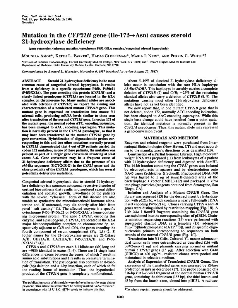
Congenital adrenal hyperplasia due to steroid 21-hydroxy-lase deficiency is a common autosomal recessive disorder ofcortisol biosynthesis that results in disordered sexual differ-entiation and somatic growth. Two-thirds of the patientswith the severe, "classical" form of this disease are alsounable to synthesize the mineralocorticoid hormone aldos-terone and, if untreated, may die shortly after birth fromrenal "salt wasting" (1). The affected enzyme is a specificcytochrome P450 (P450c21 or P45OXXIA), a heme-contain-ing microsomal protein. The gene CYP21B, encoding thisenzyme, and a pseudogene, C YP21A, are located in the HLAmajor histocompatibility complex on chromosome 6p, re-spectively adjacent to C4B and C4A, the genes encoding thefourth component of serum complement (Fig. 1A) (2, 3)[other names for the 21-hydroxylase genes are 21-OHaseA/B, OH21A/B, CA21HA/B, P450C21A/B, and P450-XXIA1/2 (4)].CYP21A and CYP2JB are each 3.1 kilobases (kb) long and
are -98% identical in exons (5, 6). There are 11 single-basedifferences in exons between the genes, of which 7 result inamino acid substitutions and 1 results in premature termina-tion of translation. The pseudogene also contains an 8-base-pair (bp) deletion and a 1-bp insertion, each of which shiftsthe reading frame of translation. Thus, the hypotheticalproduct of the CYP21A gene is completely nonfunctional.
About 5-10% of classical 21-hydroxylase deficiency al-leles occur in association with the rare HLA haplotypeA3;Bw47;DR7. This haplotype invariably carries a completedeletion of CYP21B (7) and C4B; -25% of the remainingclassical alleles also carry a deletion of CYP21B (8, 9). Themutations causing most other 21-hydroxylase deficiencyalleles have not as yet been identified.We now report that, in one mutant CYP21B gene that is
not deleted, codon 172, normally ATC encoding isoleucine,has been changed to AAC encoding asparagine. While thissingle-base change could have resulted from a point muta-tion, the identical mutation is normally present in theCYP21A pseudogene. Thus, this mutant allele may representa gene-conversion event.
MATERIALS AND METHODSEnzymes and related reagents were purchased from Inter-national Biotechnologies (New Haven, CT) and used accord-ing to the manufacturer's directions or as described (10).
Construction of a Partial Genomic Library. High molecularweight DNA was prepared (11) from leukocytes of a patientwith 21-hydroxylase deficiency and digested with BamHI.The 14-kb fraction containing the CYP21 genes was isolatedby electrophoresis in agarose and by electroelution ontoNA45 paper (Schleicher & Schuell). Fractionated DNA (400ng) was ligated to 1 Ag of BamHI-digested arms of thebacteriophage A vector EMBL3 (12) and packaged in vitrointo phage particles (reagents obtained from Stratagene, SanDiego, CA).
Isolation and Analysis of a Mutant CYP21B Gene. Thelibrary was screened (13) for the desired insert by hybridiza-tion with pC21/3c, which contains a nearly full-length cDNAinsert encoding P450c21 (6). Clones carrying CYP21A and -Bgenes were distinguished by restriction mapping (Fig. 1B). A7-kb Xho I-BamHI fragment containing the CYP21B genewas subcloned into the corresponding sites of pIBI24. Chain-termination sequencing reactions (14) were performed withsupercoiled plasmid DNA as a template (15), adenosine5'[a-35S]thiotriphosphate (dATP[3IS]), and 20 specific oligo-nucleotide primers corresponding to sequences on bothstrands of the normal CYP21B gene (Fig. 1C).
Transfection into Y1 Cells. Cultured Y1 mouse adrenocor-tical tumor cells were cotransfected as described (16) withpSV2-neo (2 ,ug) and plasmids carrying normal or mutanthuman CYP21B genes (15 ,ug). After selection with G418(GIBCO) at 400 ,ug/ml, resistant clones were pooled andmaintained in selective medium.
Analysis of Expression of Transfected CYP21B Genes. Theexpression of the transfected genes was assessed by RNaseprotection assays as described (17). The probe consisted of a316-bp Pst I-EcoRI fragment of the normal human CYP21Bgene, containing the third exon (155 bp), the third intron, and48 bp from the fourth exon, cloned into pIBI31. A radioac-
tTo whom reprint requests should be addressed.
1600
The publication costs of this article were defrayed in part by page chargepayment. This article must therefore be hereby marked "advertisement"in accordance with 18 U.S.C. §1734 solely to indicate this fact.
Proc. Natl. Acad. Sci. USA 85 (1988) 1601
C4B CYP21B
B X E B
I I
C1 2 3 4 5 6 7 8 9 10
>) ) I >
(--H i-I-- - - -
D149Arg Met Arg Ala Gln Pro Gly Thr Pro Val Ala IleCGC ATG AGA GCC CAG CCC GGC ACC CCT GTG GCC ATT
AsnGlu Glu Glu Phe Ser Leu Leu Thr Cys Ser Ile IleGAG GAG GM TTC TCT CTC CTC ACC TGC AGC ATC ATC
182 ACys Tyr Leu Thr Phe Gly Asp Lys Ile LysTGT TAC CTC ACC TTC GGA GAC AAG ATC AAG
FIG. 1. Cloning and sequencing of a CYP21 gene encodingP450c21. (A) HLA complex on the short arm of chromosome 6. Thecentromere is indicated by the circle at left. HLA class II, III, and Igenes are grouped. Class I and II genes encode transplantationantigens; there are more class I and II genes than are diagrammedhere. C2, Bf, C4A, and C4B encode serum complement components;21A and 21B are the CYP21 genes. (B) Strategy for cloning of theCYP21 genes. The relative sizes and locations of the C4 and CYP21genes are shown. The genes are all transcribed left to right. Relevantrestriction sites: B, BamHI; E, EcoRI; T, Taq I; X, Xho I. (C)Sequencing strategy for the CYP21B gene. Numbered bars representsequences expressed in mRNA, or exons, whereas the spacesbetween bars correspond to introns. The shorter bars at the ends ofthe diagram represent 5'- and 3'-untranslated regions. Arrows at thebottom of the diagram indicate positions and orientations of primersused for sequencing. (D) Sequences of the fourth exon from thenormal and mutant CYP21B genes. The sequences are identicalexcept for codon 172.
tively labeled antisense transcript was synthesized with 17polymerase (Boehringer Mannheim) and [32P]UTP. Totalcellular RNA prepared (18) from a human adrenal gland orfrom pools of transfectants was hybridized to the radiola-beled probe for 12 hr at 50°C under the described conditions.Single-stranded RNA was then digested with a mixture ofRNase A and RNase T1. The protected fragments wereresolved by electrophoresis on an 8 M urea/6% polyacryl-amide gel, and the dried gel was autoradiographed.
Oligonucleotide Hybridization. Each oligonucleotide corre-sponding to a mutation (250 ng) was end-labeled to a specificactivity of 109 dpm/,g with [32P]ATP and polynucleotidekinase, mixed with 5 ,ug of the corresponding unlabelednormal oligonucleotide, and purified by electrophoresis in a
polyacrylamide gel.Ten micrograms of DNA from each patient (Table 1) was
digested with Taq I and electrophoresed in 0.8% agarose.Dried gels were hybridized with each labeled mutant oligo-nucleotide and washed with tetramethylammonium chloridesolution as described (19, 20). The gels were autoradio-graphed between two intensifier screens at -70°C.
RESULTSCloning and Characterization of a Mutant CYP21B Gene.
Patients with 21-hydroxylase deficiency who carried hetero-
Table 1. Patients used in hybridization studies shown in Fig. 3
HLA
Patient Diagnosis B DR / B DR
A SW 47 7 57 7B SW 39 2 47 7C SW 52 2 52 2D SW 39 9 51 5E SW 22 4 39 9F SW 44 4 51 4G SV 5 5 12 1H* SW 35 5 47 7It SW 8 3 35 4J SW 44 7 47 7K SV 51 4 51 4L SW 7 10 7 10M SW 39 2 39 8N SW 15 5 35 50* SV 7 1 40 1P SW 7 2 47 7Q SW 35 5 40 7R SW 45 5 49 6S* SV 44 7 51 4T SW 18 1 18 5
Patients are salt-wasters (SW), with an inability to synthesizealdosterone normally, or are simple virilizers (SV), who are able tosynthesize aldosterone. HLA-B;DR haplotypes are shown.*Patients with a mutation in codon 172.tPatient has a larger gene conversion.
zygous deletions of CYP2JB were identified by Southernblot hybridization analysis of genomic DNA digested withvarious enzymes (Taq I digests are shown, see Fig. 3B) byusing a cDNA clone, pC21/3c, encoding human P450c21. Inthese individuals, any remaining 21-hydroxylase activitymust reside in the product of the single nondeleted CYP2JBgene [heterozygous carriers of CYP21B deletions who haveone normal CYP2JB gene do not have 21-hydroxylase defi-ciency, consistent with the known autosomal recessivemode of inheritance of this disorder (1)]. One such patient(patient H) was studied further. She has the HLA genotypeA3;Bw47;DR7/AJJ;B35;DRS; the first haplotype carries adeletion of CYP2JB.A partial genomic library was prepared from peripheral
blood leukocyte DNA of this patient. Six bacteriophage Aclones that contained CYP21 genes were isolated afterscreening 20,000 plaques by hybridization with pC21/3c, ofwhich two contained CYP2JB genes (Fig. 1B).One of these clones was subjected to DNA sequence
analysis. To rule out the possibility of artifacts arising fromthe use of specific sequencing primers, a characterized (6)normal CYP21B gene was analyzed in parallel with the sameprimers. [We have corrected several sequence errors in our
V
4 I
.1 .ADRENAL
Y1
NORMAL
MUTANT
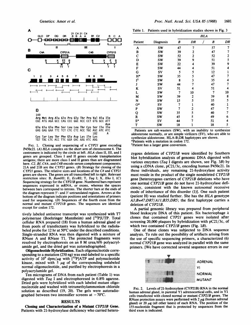
FIG. 2. Levels of 21-hydroxylase (CYP21B) RNA in the normalhuman adrenal gland, in parental Y1 adrenocortical cells, and in Y1cells transfected with cloned normal or mutant CYP21B genes. TheRNase protection assays were performed with 2 ,ug (human adrenalgland) or 20 ,ug (all other lanes) of each RNA. The position of the155-nucleotide fragment that is protected by sequences from thethird exon is indicated.
A21 C4 21 C4
GLO DP DQ OR B B A A Bf C2 B C A
I IIH i I
B C4A CYP21A
B X EE B
rr I
Genetics: Amor et al.

1602 Genetics: Amor et al. Proc. Natl. Acad. Sci. USA 85 (1988)
A1.169 175
B Cys Ser Ile Ile Cys Tyr LeuTGC AGC ATC ATC TGT TAC CTC
A Cys Ser Ile Asn Cys Tyr LeuTGC AGC ATC AAC TGT TAC CTC
2.104 113
B Tyr Pro Asp Leu Ser Leu Gly Asp Tyr SerTAC CCG GAC CTG TCC TTG GGA GAC TAC TCC
TGC AGC ATC ATC TGT TAC CTC
*TGC AGC ATC AAC TGT TAC CTC
TTG TCC CTG GGA GAC TAC TCC
A Tyr Pro Asp Leu Ser Leu V alTAC CCG GAC CTG TCG TTG G-- --- --- TCT *AC CCG GAC CTG TCG TTG GTCT
3.234 240
B Asp His Ile Val Glu Net GlnGAT CAC ATC GTG GAG ATG CAG
A Asp His Asn Glu Giu Lys GlnGAC CAC AAC GAG GAG AAG CAG
B I I
CTG CAT CTC CAC GAT GTG ATC
*CTG CTT CTC CTC GXT GTG GTC
4,4 4,
A R C D E F G H
I 4,
4 4I 4,
1 s ... #..I
*A i -.f
A:_ .^.
*o. .z-
A P. C D
F G H J K L M N O P 0 R S r
-3.7
-3.2
E F G H K M N O P 0 R S T
4,
3
A B C D E F G H J K L M N 0 P Q R S T
FIG. 3. Hybridization analysis of 20 patients with classical 21-hydroxylase deficiency. (A) To the left are sequences of the normal CYP2JBgene and CYP2IA pseudogene in codons 169-175 (probe 1), codons 104-113 (probe 2), and codons 234-240 (probe 3). To the right are thecorresponding oligonucleotides. Differences between each normal (probes B) and mutant (probes A) probe are underlined, and radioactiveend-labeling is indicated by an asterisk. (B) Hybridization to Taq I digests of patient DNA samples (letters under each blot identify the patients).Numbers to the right indicate fragment sizes in kb. The top row shows Southern blots with a cDNA probe, pC21/3c. Arrows indicate probableheterozygous deletions of the CYP21B gene. (Based on analysis with other restriction enzymes, patient G may have a gene conversion involvingthe polymorphic Taq I site rather than a deletion.) Numbers to the left of other rows correspond to the probes listed in A. All patients carrya 3.2-kb Taq I fragment that hybridizes with each probe corresponding to the CYP21A pseudogenes that normally carry these mutations. Fourpatients (arrows) carry a 3.7-kb Taq I fragment that also hybridizes with the probe corresponding to the codon 172 mutation, presumablysignifying a mutant CYP2IB gene (row 1). The mutant gene was first isolated from DNA of patient H. Patient I (arrow) carries a 3.7-kb TaqI fragment hybridizing with two additional probes (rows 2 and 3).
GenBank entry for the CYP21 genes§ based on new andreviewed data and comparison to other published sequencesof these genes (5, 6, 21).]Two single-base substitutions were found in the coding
sequences of the mutant CYP2IB gene. One (CTC to CTG incodon 248, encoding leucine) is silent, but the other (ATC to
AAC in codon 172, exon 4) changes an isoleucine residue toasparagine, a nonconservative substitution (Fig. 1D). Nochanges were detected affecting intron-exon junctions of themutant gene.
Expression of Human CYP21B Genes in Transfected Y1Cells. To establish that this mutant gene did not containadditional mutations in its promoter that altered transcrip-tion, plasmids carrying the normal and mutant CYP21Bgenes were transfected into Y1 mouse adrenocortical tumorcells, and steady-state mRNA levels were assessed by
- 3.7
-3 2
J K L M N O P 0 R S T
4,
-3.7
-3.2
-3.7
-3.2
§This sequence has been deposited in the EMBL/GenBank database (Bolt, Beranek, and Newman Laboratories, Cambridge, MA,and Eur. Mol. Biol. Lab., Heidelberg) (accession no. M13937).
E
i.
'f...t--F, ;.
Of. %O 40ftillw
-f

Proc. Nat!. Acad. Sci. USA 85 (1988) 1603
RNase protection assays. Pools of transfectant clones wereprepared to minimize the effect of clonal variation on 21-hydroxylase (CYP21B) expression. The results (Fig. 2) showthat Y1 cells transfected with either the normal or the mutantgene contain CYP21B transcripts, as documented by theirprotection of a 155-nucleotide fragment that is identical insize to that protected by authentic human adrenal mRNA.As noted (16), parental Y1 cells do not express their own21-hydroxylase genes. Although this technique is only semi-quantitative, the expression of 21-hydroxylase mRNA levelsin cells transfected with the codon 172 mutant is roughlyequal to that seen with transfection of the normal humanCYP21B gene. In addition to the protected fragment of 155nucleotides, these samples also contained a 48-nucleotidefragment protected by sequences contained in the fourthexon, which was poorly resolved under the gel conditionsused (data not shown).
Oligonucleotide Hybridization Studies of Patients. To deter-mine the frequency with which the mutation in codon 172causes 21-hydroxylase deficiency, two 21-mer oligonucleo-tides were synthesized (Fig. 3A) corresponding to the normaland mutant CYP21B genes from codon 169 to codon 175.DNA samples from 20 unrelated patients with classical21-hydroxylase deficiency (Table 1) were digested with TaqI and electrophoresed in agarose gels. The oligonucleotidecarrying the mutation was radioactively end-labeled, mixedwith a molar excess of the unlabeled normal oligonucleotide,and incubated with the dried gel. The gel was washed underconditions sufficiently stringent that only a perfectlymatched probe could yield a hybridization signal (Fig. 3B).The mutation in codon 172 (as well as the mutation in
codon 248) is normally found in the CYP21A pseudogene.Thus, the 3.2-kb Taq I fragment associated with the pseu-dogene is expected to hybridize with the mutant probe in allindividuals, whereas the 3.7-kb Taq I fragment in theCYP21B gene should not hybridize with the probe unless itcarries the codon 172 mutation.
In addition to patient H from whose DNA the mutant genewas isolated, DNA samples from three other patients (pa-tients I, 0, and S) contained 3.7-kb Taq I fragments thathybridized with the probe. Although 21-hydroxylase defi-ciency is frequently associated with particular HLA-B anti-gens, the four patients carrying this mutation had no HLA-Bantigen in common. Patients H and 0 carried heterozygousdeletions of CYP21B as determined by Southern blot analy-sis.
Patient H is a 7-year-old female who, when studied as aninpatient at 1 year of age, could conserve sodium at theexpense of a high plasma renin/urinary aldosterone ratio.Thus, her ability to synthesize aldosterone was decreasedbut not absent. Patients S and 0 are 12-year-old males whohave never demonstrated electrolyte disturbances or aldos-terone deficiency ("simple virilizing" 21-hydroxylase defi-ciency). Patient I is a 26-year-old female who, at birth, hadclear salt-wasting symptoms with an aldosterone deficiencythat continued until age 12. She subsequently discontinuedmedication and has been documented to conserve sodiumnormally.Because the codon 172 mutation is normally present in the
CYP21A pseudogene, it seemed possible that it had beentransferred to the CYP21B gene by a gene conversion event.To determine if any of the patients with this mutation carriedlarger gene conversions, DNA samples were hybridized withprobes corresponding to two regions in the CYP21A thatflank codon 172 (Fig. 3A). One probe corresponds to an areain exon 3 that contains an 8-bp deletion. The other iscomplementary to codons 234-240 in exon 6; this area in the
CYP21A gene contains four point mutations resulting inthree nonconservative amino acid substitutions.
A
B
1 s 1--MI -mm wmSu- mm
CONSERVATIVE CHANGE TERMINATION/FRAMESHIFT
C NON- OONSERVATIVE CHANGE
FIG. 4. Possible gene conversions causing 21-hydroxylase defi-ciency. The CYP21A and -B genes are drawn schematically as inFig. 1, with mutations in CYP21A indicated by various shadings ofexons. Xs in introns indicate the presence of chi-like sequences,which are postulated to be sites for recombination between homol-ogous regions of CYP21A and CYP21B. The three probes discussedin Fig. 2 are indicated by numbered bars. The solid line encompass-ing exon 4 and part of exon 7 represents the missense mutation incodon 172 and the silent mutation in codon 246 observed in thesequenced mutant CYP2IB gene. The dashed line represents theminimum boundaries of the large gene conversion observed in onepatient. The resulting mutant CYP21B genes are also shown.
One of the four patients (patient I) who carried the codon172 mutation also carried an CYP21B gene that hybridizedwith both of these additional probes (Fig. 3B). Hybridizationanalysis of her parents (data not shown) demonstrated thatthe CYP21B gene donated by her father carried both of theseadditional mutations, presumably representing a single-geneconversion encompassing at least exons 3-6 (Fig. 4).
DISCUSSIONThe mutant CYP21B gene was isolated from a patient withclassical 21-hydroxylase deficiency, suggesting that the mu-tation in codon 172 affects the synthesis, enzymatic activity,or both of the encoded P450c21 relatively severely. How-ever, as the patients with this mutation (two ofwhom have aheterozygous deletion of CYP21B) retain the ability to makesome aldosterone, the mutation probably does not com-pletely abolish enzymatic function. The lower degree ofcompromise of aldosterone synthesis, as compared to corti-sol synthesis, may relate to the much smaller relativeamounts of aldosterone normally secreted by the adrenalcortex. The differences in phenotype between patient H(who has decreased but not absent ability to synthesizealdosterone) and patients 0 and S (who apparently synthe-size aldosterone normally) may be due to individual varia-tion in levels of synthesis of the mutant enzyme or todiffering rates of catabolism or excretion of various steroidhormones. Similar variations in aldosterone biosyntheticcapacity have been documented (22) in HLA-identical sib-lings with 21-hydroxylase deficiency.
Ile-172 is conserved in the P450c21 (P45OXXI) enzymes ofmice (23) and cattle (24, 25) and in otherwise poorly homol-ogous enzymes, such as steroid 17-hydroxylase (P450XVII)(26) or P450s induced in the liver by phenobarbital (P450IIB)(27), dioxin (P450I) (28), or steroids (P450111) (29) (Fig. 5).This suggests that Ile-172 participates in a hydrophobicinteraction that is important for maintaining the correctconformation of the enzyme. Mutation at this position to apolar residue, such as asparagine, might disrupt such aninteraction.
It is unlikely that the mutation in codon 172 or anadditional undetected mutation in the promoter region af-
Genetics: Amor et al.

Proc. Natl. Acad. Sci. USA 85 (1988)
172
XXI-nl Leu Thr Cys Ser Ile Ile Cys Tyr Leu Thr Phe Gly-abn ASN
XXI-bov Leu Thr Cys Ser Ile Ile Cys Tyr Leu Thr Phe Gly
XXI-mur Leu Thr Cys Ser Ile Ile Ser Cys Leu Thr Phe Gly
XVII Val Ile Ser Leu Ile Cys Phe Asn Thr Ser Tyr
IIB Ile Thr Ala Asn Ile Ile Cys Ser Ile Val Phe Gly
I Val Thr Asn Val Ile Cys Ala Ile Cys Phe Gly
III Met Phe Pro Ile Ile Glu Gln Tyr Gly
FIG. 5. The predicted amino acid sequences in the region ofcodon 172 of normal (nl) and abnormal (abn) human P450c21 [XXI,referring to the gene family within the P450 superfamily (4)] arecompared with bovine (bov) and murine (mur) P450c21, with humansteroid 17-hydroxylase (XVII), and with cytochromes P450 that areinduced in rat liver by phenobarbital (IIB) or by steroids (III) or inhuman liver by dioxin (I). Functionally conserved residues areindicated by asterisks.
fects transcription of this mutant CYP21B gene, becausenormal levels ofmRNA are detected after transfection of themutant gene into Y1 cells.
It is notable that the missense mutation in codon 172 andthe silent mutation in codon 246 of the mutant CYP21B gene
are normally found in the CYP21A pseudogene. Althoughthey might be conventional point mutations, they could havearisen in gene conversion events (Fig. 4) (the silent mutation,of course, could be a polymorphism in the normal popula-tion). One patient apparently carries the codon 172 mutationas part of a larger rearrangement involving at least fourexons. This large gene conversion includes the 8-bp deletionin exon 3, and, because of the resulting frameshift, theencoded protein is predicted to be truncated and completelynonfunctional. The exact borders of the conversion have notbeen determined. Because this gene carries a 3.7-kb (not a
3.2-kb) Taq I fragment, the conversion cannot extend to theextra Taq I site near CYP21A, located 211 bp upstream of theinitial ATG. The conversion also does not include thenonsense mutation in codon 318 of CYP21A (unpublishedobservations).Gene conversion events may be facilitated by the presence
of chi-like sequences. Chi is a sequence, usually GCTGG-GG, involved in recombination in bacteriophage A (30).Similar sequences have been observed near recombinationalhot spots in immunoglobulin genes (31) and in human andmurine class I and class II genes in the major histocompat-ibility complex (32). Although a single 7-bp sequence isexpected to occur about once every 8000 bp in randomDNA, GCTGGGG occurs six times in CYP21B and fivetimes in CYP21A. One such site is present -50 bp away fromthe mutation in codon 172.These findings suggest that gene conversion may be a
frequent cause of 21-hydroxylase deficiency, a hypothesisthat could be tested by hybridizing DNA from patients withadditional oligonucleotide probes corresponding to eachmutation normally present in the CYP21A pseudogene. Infact, three of the 20 patients carry the nonsense mutationnormally found in codon 318 of the CYP21A gene (unpub-lished observations). If other gene conversions are equallycommon [however, simple point mutations will certainlyaccount for some mutant alleles (21)], it should be possible toprenatally diagnose 21-hydroxylase deficiency by chorionicvillus sampling by using a small number of oligonucleotideprobes. This would be of interest in light of the possibility ofprenatal therapy of this disorder to prevent abnormal sexualdifferentiation in female fetuses (33).
We thank Ms. Alena Vitek (Cornell University) and Ms. JeannaMeade (Duke University) for technical assistance, and Drs. R. B.Wallace, M. Adesnik, B. Schimmer, D. D. Chaplin, J. G. Seidman,and M. Carroll for helpful discussions. HLA genotyping of patientswas performed by Dr. B. Dupont. Oligonucleotides were synthe-sized by the Department of Microbiology, Cornell University Med-ical College, New York. M.A. is supported by a fellowship fromCentre National de la Recherche Scientifique (France); H.G. is therecipient of a Charles H. Revson Foundation fellowship, and herwork has been aided by a grant from the American PhilosophicalSociety. This work is supported by Grants DK37862 (P.C.W.),CA22507 (B. Dupont, Principal Investigator), and HD00072(M.I.N.) from the National Institutes of Health, and by the HoraceGoldsmith Foundation. P.C.W. is an Andrew W. Mellon Teacher-Scientist.
1. White, P. C., New, M. I. & Dupont, B. (1987) N. Engl. J. Med. 316,1519-1524, 1580-1586.
2. Carroll, M. C., Campbell, R. D. & Porter, R. R. (1985) Proc. Nat!.Acad. Sci. USA 82, 521-525.
3. White, P. C., Grossberger, D., Onufer, B. J., Chaplin, D. D., New,M. I., Dupont, B. & Strominger, J. L. (1985) Proc. Natl. Acad. Sci.USA 82, 1089-1093.
4. Nebert, D. W., Adesnik, M., Coon, M. J., Estabrook, R. W., Gonzalez,F. J., Guengerich, F. P., Gunsalus, I. C., Johnson, E. F., Kemper, B.,Levin, W., Phillips, I. R., Sato, R. & Waterman, M. R. (1987) DNA 6,1-11.
5. Higashi, Y., Yoshioka, H., Yamane, M., Gotoh, 0. & Fujii-Kuriyama,Y. (1986) Proc. Nat!. Acad. Sci. USA 83, 2841-2845.
6. White, P. C., New, M. I. & Dupont, B. (1986) Proc. Nat!. Acad. Sci.USA 83, 5111-5115.
7. White, P. C., New, M. I. & Dupont, B. (1984) Proc. Nat!. Acad. Sci.USA 81, 7505-7509.
8. Rumbsby, G., Carroll, M. C., Porter, R. R., Grant, D. B. & Hjelm, M.(1986) J. Med. Genet. 23, 204-209.
9. Werkmeister, J. W., New, M. I., Dupont, B. & White, P. C. (1986) Am.J. Hum. Genet. 39, 461-469.
10. Maniatis, T., Fritsch, E. F. & Sambrook, J. (1982) Molecular Cloning:ALaboratory Manual (Cold Spring Harbor Lab., Cold Spring Harbor,NY).
11. Wyman, A. R. & White, R. (1980) Proc. Nat!. Acad. Sci. USA 77,6754-6758.
12. Frischauf, A. M., Lehrach, H., Poustka, A. & Murray, N. (1983) J. Mol.Biol. 170, 827-842.
13. Benton, W. D. & Davis, R. W. (1977) Science 196, 180-182.14. Biggin, M. D., Gibson, T. J. & Hong, G. F. (1983) Proc. Nat!. Acad.
Sci. USA 80, 3963-3965.15. Chen, E. Y. & Seeburg, P. H. (1985) DNA 4, 165-170.16. Parker, K. L., Chaplin, D. D., Wong, M., Seidman, J. G., Smith, J. A.
& Schimmer, B. P. (1985) Proc. Natl. Acad. Sci. USA 82, 7860-7864.17. Krieg, P. A. & Melton, D. A. (1988) Methods Enzymol., in press.18. Chirgwin, J. M., Przybyla, A. E., MacDonald, R. J. & Rutter, W. J.
(1979) Biochemistry 18, 5294-5299.19. Wood, W. I., Gitschier, J., Lasky, L. A. & Lawn, R. M. (1985) Proc.
Nat!. Acad. Sci. USA 82, 1585-1588.20. DiLella, A. G. & Woo, S. L. C. (1987) Methods Enzymol. 152, 447-450.21. Rodrigues, N. R., Dunham, I., Yu, C. Y., Carroll, M. C., Porter, R. R.
& Campbell, R. D. (1987) EMBO J. 6, 1653-1661.22. Stoner, E., Dimartino-Nardi, J., Kuhnle, U., Levine, L. S., Oberfield,
S. E. & New, M. I. (1986) Clin. Endocrinol. 24, 9-20.23. Chaplin, D. D., Galbraith, L. J., Seidman, J. G., White, P. C. & Parker,
K. L. (1986) Proc. Nat!. Acad. Sci. USA 83, 9601-9605.24. White, P. C., New, M. I. & Dupont, B. (1984) Proc. Nat!. Acad. Sci.
USA 81, 1986-1990.25. Yoshioka, H., Morohashi, K., Sogawa, K., Yamane, M., Kominami, S.,
Takemori, S., Okada, Y., Omura, T. & Fujii-Kuriyama, Y. (1986) J.Biol. Chem. 261, 4106-4109.
26. Bradshaw, K. D., Waterman, M. R., Couch, R. T., Simpson, E. R. &Zuber, M. X. (1987) Mol. Endocrinol. 1, 348-354.
27. Mizukami, Y., Sogawa, K., Suwa, Y., Muramatsu, M. & Fujii-Kuri-yama, Y. (1983) Proc. Nat!. Acad. Sci. USA 80, 3958-3962.
28. Jaiswal, A. K., Gonzalez, F. J. & Nebert, D. W. (1985) Science 228,80-83.
29. Gonzalez, F. J., Nebert, D. W., Hardwick, J. P. & Kasper, C. B. (1985)J. Biol. Chem. 260, 7435-7441.
30. Smith, G. R., Kunes, S. M., Schultz, D. W., Taylor, A. & Triman,K. L. (1981) Cell 24, 429-436.
31. Krawinkel, U., Zoebelien, G., Bruggemann, M., Radbruch, A. &Ralewsky, K. (1983) Proc. Nat!. Acad. Sci. USA 80, 4997-5001.
32. Wu, S., Saunders, T. L. & Bach, F. H. (1986) Nature (London) 324,676-679.
33. Evans, M. I., Chrousos, G. P., Mann, D. W., Larsen, J. W., Green, I.,McCluskey, J., Loriaux, D. L., Fletcher, J. C., Koons, G., Overpeck, J.& Schulman, J. D. (1985) J. Am. Med. Assoc. 253, 1015-1020.
1604 Genetics: Amor et al.