mutations in the drosophila gene bullwinklecause the
TRANSCRIPT
3023Development 121, 3023-3033 (1995)Printed in Great Britain © The Company of Biologists Limited 1995
Mutations in the Drosophila gene bullwinkle cause the formation of abnormal
eggshell structures and bicaudal embryos
Kimberley R. Rittenhouse and Celeste A. Berg*
University of Washington, Department of Genetics, Box 357360, Seattle, WA 98195-7360, USA
*Author for correspondence (e-mail: [email protected])
Subcellular localization of gene products and cell migrationare both critical for pattern formation during development.The bullwinkle gene is required in Drosophila for disparateaspects of these processes. In females mutant at the bull-winkle locus, the follicle cells that synthesize the dorsaleggshell filaments do not migrate properly, creating short,broad structures. Mosaic analyses demonstrate that wild-type BULLWINKLE function is required in the germ linefor these migrations. Since the mRNA for gurken, theputative ligand that signals dorsal follicle cell fate, iscorrectly localized in bullwinkle mutants, we conclude thatour bullwinkle alleles do not affect the dorsoventralpolarity of the oocyte and thus must be affecting the folliclecell migrations in some other way. In addition, the embryosthat develop from bullwinkle mothers are bicaudal. AKINESIN:β-GALACTOSIDASE fusion protein iscorrectly localized to the posterior pole of bullwinkleoocytes during stage 9. Thus, the microtubule structure ofthe oocyte and general transport along it do not appear to
be disrupted prior to cytoplasmic streaming. Unlike otherbicaudal mutants, oskar mRNA is localized correctly to theposterior pole of the oocyte at stage 10. By early embryo-genesis, however, some oskar mRNA is mislocalized to theanterior pole. Consistent with the mislocalization of oskarmRNA, a fraction of the VASA protein and nanos mRNAare also mislocalized to the anterior pole of bullwinkleembryos. Mislocalization of nanos mRNA to the anterior isdependent on functional VASA protein. Although themirror-image segmentation defects appear to result fromthe action of the posterior group genes, germ cells are notformed at the anterior pole. The bicaudal phenotype is alsogerm-line dependent for bullwinkle. We suspect thatBULLWINKLE interacts with the cytoskeleton and extra-cellular matrix and is necessary for gene product localiza-tion and cell migration during oogenesis after stage 10a.
Key words: bicaudal, eggshell defects, maternal effect, RNAlocalization, cell migration, cytoskeleton, Drosophila, bullwinkle
SUMMARY
INTRODUCTION
The Drosophila egg chamber is composed of a cyst of sixteeninterconnected, germ-line cells surrounded by somaticallyderived follicle cells. Of the sixteen germ-line cells, onebecomes the oocyte and the remaining fifteen function as nursecells. The nurse cells provide the oocyte with RNA andproteins to be used by the embryo during its development,while the follicle cells synthesize various layers and structuresof the eggshell (reviewed by Spradling, 1993). A small numberof RNAs and proteins are specifically transported from thenurse cells to the oocyte early in development, while themajority are non-specifically deposited into the oocyte duringthe latter half of oogenesis (reviewed by Mahajan-Miklos andCooley, 1994).
Products deposited in the egg during oogenesis determineembryonic polarity. Genetic analyses of dorsoventralmutations indicate that a signal from the germ-line cells todorsal follicle cells establishes the dorsoventral axis of the egg(Schüpbach, 1987). This event regulates the subsequentmigration of the dorsal follicle cells and their synthesis of thedorsal eggshell appendages. In addition, reception of the germ-
line signal by dorsal follicle cells regulates the production ofa later signal that determines the ventral axis of the embryo(Schüpbach, 1987). Thus, mutations early in the pathway affectthe dorsoventral polarity of both the eggshell and the embryo,while downstream mutations affect either the eggshell or theembryo. cappuccino (capu), spire (spir), squid (sqd), orb andfs(1)K10 are necessary for the correct localization of gurken(grk) mRNA, which encodes the presumptive signal from theoocyte to the follicle cells (Neuman-Silberberg and Schüpbach,1993; Christerson and McKearin, 1994). TORPEDO (TOP),the Drosophila homologue of the mammalian EGF receptor, isthought to receive the ‘dorsal’ signal (Schüpbach, 1987; Priceet al., 1989; and Schejter and Shilo, 1989), which is then trans-duced through the Ras pathway to determine dorsal follicle cellfate (Brand and Perrimon, 1994; Berg and Schnorr, unpub-lished data).
Mutations that disrupt the dorsoventral polarity of the oocytealter the shape of the eggshell. The dorsal and ventral sides ofthe eggshell have two major morphological differences: theeggshell is shorter on the dorsal side, and the filaments arelocated dorsally just lateral to the midline (Fig. 1). These dif-ferences in eggshell structure result from differences in the
3024 K. R. Rittenhouse and C. A. Berg
Table 1. bullwinkle8482 excision linesClass Number of lines Phenotype
Fertile female 31 (65%) Both eggshell and embryophenotypes are wild type
Sterile female 12 (25%) Eggshells have bwk phenotypeAbnormal wings 1 (2%) Line is semi-lethal, wings have
extra veins, eggshells havebwk phenotype
Lethal 4 (8%) Either late larval lethal or latepupal lethal
pattern of migration of follicle cells on the dorsal and ventralsides of the eggs. At mid-oogenesis, the dorsal anterior folliclecells, which have received the GURKEN signal, migrate intotwo circles. These cells secrete chorion proteins centripetallyto form the base of the eggshell filaments. When the base isfinished, a layer of follicle cells migrates past the previous cellsand forms another circle of eggshell protein. The migrationcontinues in this fashion, forming a hollow cylinder of chorion,until the follicle cells spread out to form the flat paddle struc-tures at the end (King, 1970; King and Koch, 1963). Incontrast, the ventral follicle cells simply expand to maintain anepithelial sheet around the oocyte at the time that the nursecells deposit their contents into the oocyte. Dorsalizingmutations cause the ventral side to resemble the dorsal side,resulting in the formation of short eggs with an entire ring ofeggshell filaments. Conversely, ventralizing mutations causethe eggs to be much longer and have a single dorsal appendageor no dorsal appendages (Schüpbach, 1987; Manseau andSchüpbach, 1989; Kelley, 1993; Wieschaus et al., 1978; Chris-terson and McKearin, 1994).
Genes required maternally for anterior/posterior polarityhave also been identified. Two morphogens are important forproper anterior/posterior development: BICOID (BCD), theanterior morphogen, and NANOS (NOS), the posteriormorphogen. Both morphogens are localized as RNA via their3′UTRs to their respective poles during oogenesis (Macdonaldand Struhl, 1988; Wang and Lehmann, 1991; Gavis andLehmann, 1992). Localization of the bcd message to theanterior pole requires exuperantia, swallow and staufen (forreview see Driever, 1993). Localization of the nos message tothe posterior pole requires the products of the staufen, magonashi, oskar (osk), vasa, valois and tudor genes, which areknown collectively as the posterior group genes (reviewed bySt. Johnston, 1993). osk mRNA, VASA, VALOIS andTUDOR proteins, and nos mRNA are localized in a step-wisemanner to the posterior pole, and localization of each dependsupon the correct localization of all previous products (Ephrussiet al., 1991; Lehmann and Nüsslein-Volhard, 1991). oskmRNA is localized via its 3′ UTR. If the coding region of oskis fused to the bcd 3′ UTR, osk mRNA is localized to theanterior pole (Ephrussi and Lehmann, 1992). Upon translation,the mislocalized OSK protein then recruits other posteriorgroup gene products, including nos mRNA, and a bicaudalembryo is formed due to translational repression of bcd RNA(Wharton and Struhl, 1991). Mutations in several genes,including bicaudal, Bicaudal-C (Bic-C) and Bicaudal-D (Bic-D), also cause the formation of bicaudal embryos (Bull, 1966;Mohler and Wieschaus, 1986).
A few genes are required in both the dorsoventral and theanterior/posterior pathways. Mutations in capu and spireinduce premature cytoplasmic streaming in the oocyte(Theurkauf, 1994), preventing the localization of grk mRNAand the posterior group gene products and thereby causing theloss of ventral and posterior structures, respectively (Manseauand Schüpbach, 1989). These defects are general localizationdefects and do not cause the formation of bicaudal embryos.
bullwinkle (bwk) is unusual in that it affects several of theseprocesses. Mutations in bwk cause both the formation ofabnormal eggshell filaments and bicaudal embryos. We presentgenetic and cell biological analyses of bwk.
MATERIALS AND METHODS
Fly stocks Canton-S and mwh red flies were used as wild-type controls. Thebwk8482 and PZ5650 lines were created in a P[lacZ, ry+] mutagen-esis screen (described in Karpen and Spradling, 1992). Df(3R)DlBX12
spans 91F1-2 to 92D3-6 and was obtained from the Bloomingtonstock center. The bwk151 line was created by Rick Kelley (1993). Thechic7886 stock was provided by Lynn Cooley. The quaHM14, grkHG21
and vasaPD23 stocks were provided by Trudi Schüpbach. ThevasaLYG2 stock was created by Lin Yue (Yue, Berg and Spradling,unpublished data). KZ32 was created by Clark et al. (1994), and theP[ovoD1] insertion line was created by Chou et al. (1993). osk301 wasprovided by Ruth Lehmann.
Detecting β-GALACTOSIDASE expression in ovariesP[lacZ, ry+] linesOvaries were fixed and stained according to Cooley et al. (1992). Theovarioles and individual egg chambers were mounted in 65% glyceroland examined with a Nikon microphot FXA using differential inter-ference contrast optics.
KIN:β-GAL linesOvaries were fixed and stained according to Clark et al. (1994), exceptthat fixation was carried out in 0.5% glutaraldehyde (SIGMA, EMgrade) and the staining solution contained 0.75% X-gal (5-bromo-4-chloro-3-indolyl-β-D-galactopyranoside).
Excision screen ry506 Sb P[ry+, ∆2-3]/TM6 females (Robertson et al., 1988) werecrossed to ry506 bwk8482 males. Fifty ry506 bwk8482/ry506 Sb P[ry+, ∆2-3] males were selected from the progeny and crossed to ry506/TM3,ryRK Sb females in individual vials. In the next generation, one rosyeyed (from loss of ry+ in the PZ element) ry506 bwk8482*/TM3, ryRK
Sb male was chosen from each vial and crossed to females from theoriginal ry506 bwk8482/TM3, ryRK Sb stock to test fertility and toestablish lines.
cDNA in situ hybridization cDNA in situ hybridizations to ovaries were carried out essentiallyaccording to Ephrussi et al. (1991), and to embryos as per Tautz andPfeifle (1989). Digoxigenin probes were prepared using BoehringerMannheim DNA labeling and detection kit. We used a gurken cDNA(provided by Shira Neuman-Silberberg and Trudi Schüpbach), abicoid cDNA (provided by Markus Noll), an oskar cDNA (providedby Ruth Lehmann) and a nanos cDNA (provided by Paul Macdonald)to prepare our DNA probes.
Cuticle preparationsCuticle preparations were carried out according to Wieschaus andNüsslein-Volhard (1986), except the embryos were mounted in 4:1:1Hoyer’s : lactic acid : ddH2O and incubation times were adjustedaccordingly.

3025Eggshell defects and bicaudal embryos
bwk egg chambers are due to altered follicle cell migration. Anterior ispage, unless otherwise noted. (A) Wild-type stage 14 egg chamber,er from a bwk8482 mother. The dorsal filaments are shorter and broadereggs are shorter and rounder due to the slightly dumpless nature of
ber showing the PZ5650 enhancer trap pattern. The follicle cell. (D) Alterations in the PZ5650 enhancer trap pattern in a bwk151
do not migrate out as far over the nurse cells, creating short, broad
B
D
Embryo antibody stainingEmbryos were fixed and devitellinized according to Ashburner (1989)(protocol 96), except that after rinsing in methanol, the embryos wereplaced in 100% ethanol for storage at −20°C. After storage theembryos were rinsed in methanol. Endogenous peroxidase activitywas removed by treating with 0.3% H2O2 in methanol for 30 minutes.The embryos were rehydrated by washing in 75% methanol in PBTr(PBS + 0.1% Triton X-100), 50% methanol in PBTr, 25% methanolin PBTr, and then in PBTr. The blocking and antibody incubationswere carried out according to Ashburner (1989), except that theblocking solution was 5% normal goat serum in PBTr. The anti-VASA antibody, a gift from Paul Lasko, was diluted 1:1000. Anti-rabbit antibody from the Vector elite ABC kit was used as thesecondary antibody. Subsequent steps were carried out according tothe elite ABC kit (Vector). The embryos were mounted in 75%glycerol/25% PBS.
ovoD1 germ-line clonesmwh red control females, bwk8482/TM3 females, and bwk151/TM3females were crossed to P[ovoD1]/Ki males to generate larvae of thecorrect genotypes. The first instar larvae (24 to 48 hours old) wereirradiated with a Picker 805D X-ray machine at a constant dose of1000 rads (70Kv, 3mA, 0.5 mm aluminum filter for 2.2 minutes), asdescribed by Chou et al. (1993). Adult flies transheterozygous for thegene of interest and ovoD1 were selected by the presence of wild-typebristles. bwk/P[ovoD1] females were mated to bwk151 males to facili-tate progeny testing. The females were allowed to lay eggs for 6-7days. Any eggs laid were scored for eggshell phenotype and viableembryos. If any eggs were laid in the vial, all females within the vialwere dissected. Some females in vials where no eggs were laid werealso dissected.
Pole cell transplantation pp osk301 sbd bwk8482 / pp osk301
females were crossed to cv-csbd bwk151 males to generatehost embryos. Femaleshomozygous for osk301 wereused so that the host embryoswould lack pole cells (Lehmannand Nüsslein-Volhard, 1986).Donor embryos were Canton-S.All embryos were collected for1-2 hours at 25°C on apple juiceagar plates, dechorionated in50% bleach and rinsed in 0.5%NaCl/0.03% Triton X-100. Cel-lularized embryos were lined upon an agar block and transferredto a sticky coverslip. Host anddonor embryos were placed onseparate coverslips to allowdifferent dehydration times andto facilitate removal of thedonor embryos. (Sticky cover-slips were made by shaking ~1meter of Scotch 3M doublestick tape in 10 ml of heptanefor 1 hour, centrifuging anddipping coverslips into thesticky heptane.) The embryoswere dehydrated by placing in acontainer with drierite: hostembryos for 11-14 minutes,donor embryos for 4 minutes.After dehydration, Halocarbon
Fig. 1. Abnormal dorsal filaments in to the left, dorsal is facing out of the dorsal is up. (B) Stage 14 egg chambthan wild-type dorsal filaments. The bwk. (C) Wild-type stage 14 egg chamnuclei stain over the dorsal filamentsbackground. The stained follicle cellsdorsal filaments. Bar, 20 µm.
A
C
oil series 200 (Halocarbon Products, NJ) was used to cover theembryos. Pole cells were transferred using a Narishige NU-202 3 waycontrol micromanipulator and model IM-6 syringe. The injectiontubing was filled with Halocarbon oil series 27 (Halocarbon Products,NJ). After pole cell transplantation, host embryos were transferred ontheir coverslips to apple juice agar plates and allowed to hatch at 20°C.Larvae were transferred to fresh food and placed at 25°C. Survivingbwk151/bwk8482 females were crossed to males and allowed to layeggs. Any female that failed to lay eggs after 4-6 days was dissectedin Drosophila Ringer’s solution. Our pole cell transplantationtechnique is based on Van Deusen (1976), with modifications bySeigfried Roth, Trudi Schüpbach and ourselves.
RESULTS
We obtained bwk8482 (bullwinkle), a female sterile mutation at92D, from a large scale P[lacZ, ry+] (PZ) mutagenesis screencarried out in Allan Spradling’s laboratory (described inKarpen and Spradling, 1992). Mutations in bwk affect theproper formation of the Drosophila eggshell and embryo. Fliesthat carry bwk8482 in trans to a deficiency for the region(Df(3R) DlBX12) are also sterile and have similar eggshelldefects. In addition, these flies have blistered wings, indicat-ing that bwk is required for proper wing morphogenesis andthat bwk8482 is not a null allele.
To analyze these phenotypes more thoroughly, we generatednew alleles by transposase-induced excision (Table 1).Excision of the PZ element reverts both eggshell and embryophenotypes to wild type, indicating that the PZ insertion is
3026 K. R. Rittenhouse and C. A. Berg
A
B
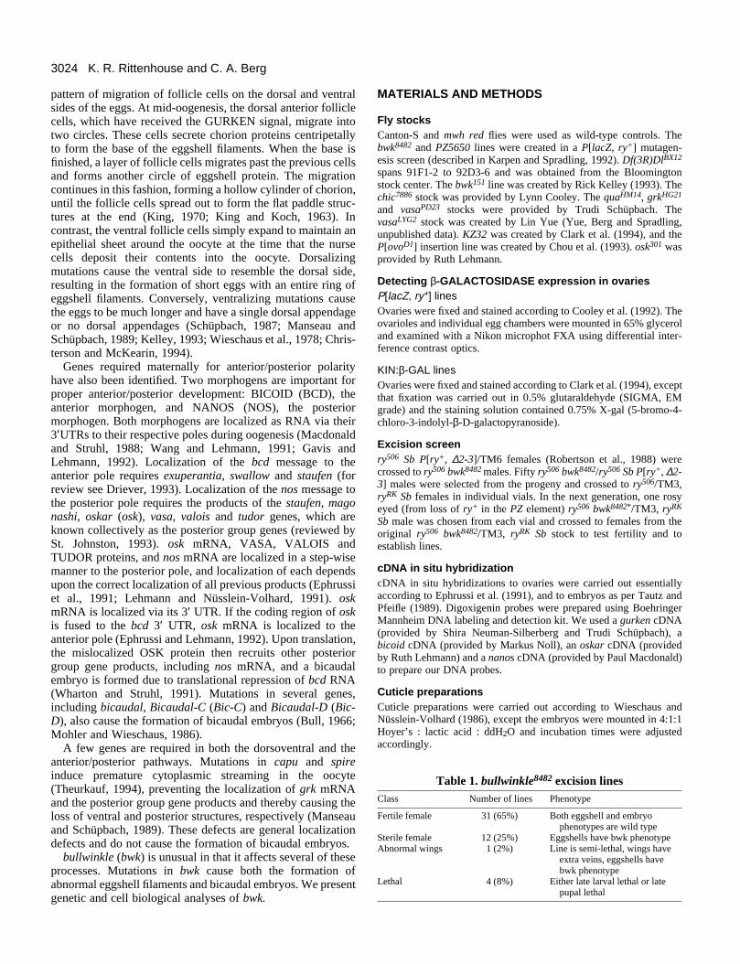
Fig. 2. Partially dumpless mutants have less severe dorsal filamentdefects than bwk eggshells. Anterior is to the left, dorsal is facing outof the page. (A) Stage 14 egg chamber from a chic7886 mother. Thedorsal filaments are formed on top of the remaining nurse cells. Notethat although the filaments are broader than wild-type filaments, theyare longer than bwk filaments. The chic7886 filaments are also fusedat the base. (B) Stage 14 egg chamber from a quaHM14 mother. Thedorsal filaments are short, but are not nearly as broad as bwkfilaments. Bar, 20 µm.
responsible for the bwk8482 phenotype. In addition to obtainingseveral lines with phenotypes similar to the starting allele, werecovered five lines with additional phenotypes (Table 1). Inone excision line, which retains the bwk8482 egg phenotype, thewings of homozygous flies curve down and have extra veins.Four lethal excision alleles were also obtained. In three of thelethal lines, homozygotes die as pharate adults. The mostsevere lethal mutation, a deletion of at least 10 kb, is a latelarval lethal with some escapers that die as pupae (data notshown). We also obtained a P[ry+] insertion allele of bwk,bwk151, which was generated by Rick Kelley. bwk151 haseggshell and embryo defects similar to bwk8482.
Eggshell defectsFemales homozygous for bwk8482 lay few eggs, which oftenappear deflated shortly after being laid. bwk eggs are shorterthan wild-type eggs, being slightly ‘dumpless’ due to theincomplete transfer of nurse cell contents into the oocyte atstage 11. In addition, bwk eggs have a number of anterioreggshell defects (Fig. 1B). The dorsal eggshell filaments areshort and broad and have ragged edges and thin chorion,sometimes resembling moose antlers, hence the name bull-winkle. The operculum is weak and yolk often streams from
the anterior of the egg upon dissection. Females homozygousfor bwk151 lay slightly more eggs, which have similar eggshelldefects.
The eggshell phenotype of bwk mutants suggests that thefollicle cells do not migrate properly as they form the dorsaleggshell filaments. In order to examine the follicle cell migra-tions more closely, we labeled these cells to observe theirmovements. We took advantage of line PZ5650, an enhancertrap line that marks the follicle cells that form the dorsalfilaments, providing information on their number andplacement (Gillespie and Berg, unpublished, and Kelley,1993). During stage 10, PZ5650 egg chambers express lacZ intwo patches of follicle cells just dorsal to the oocyte nucleus(data not shown). During stages 11-14, these follicle cellscontinue to express lacZ as they migrate anteriorly to form thedorsal filaments (Fig. 1C). Mutations that affect the dorsoven-tral axis of the egg also alter the staining pattern of PZ5650.For example, the dorsalizing mutation sqd causes ventral andlateral follicle cells to contribute to the formation of the‘dorsal’ filaments. This change in follicle cell fate in sqd eggchambers can be seen in a PZ5650 background: during stage10, the lacZ expression is expanded from the two dorsalpatches to a broad ring that encircles the oocyte (Kelley, 1993).Both the number and position of the follicle cells expressinglacZ change in a sqd background, reflecting the change in fate.
To examine the follicle cell migration in bwk egg chambers,we crossed bwk151 to PZ5650. During stage 10, egg chambersof PZ5650; bwk151 females express lacZ in two patches offollicle cells on the dorsal side of the oocyte, identical toPZ5650 alone (data not shown). In later stages, however, theposition of the staining follicle cells is altered in PZ5650;bwk151 egg chambers. The follicle cells expressing lacZ do notmigrate out as far, remaining over the short broad bwk dorsaleggshell filaments (Fig. 1D). These clearly marked folliclecells indicate that the migration that forms the dorsalappendages is abnormal in bwk151. While the migration patternof the follicle cells is altered in bwk egg chambers, the numberof follicle cells expressing lacZ appears to be the same in bwkand wild-type egg chambers, suggesting that their cell fate isnot altered.
bwk filament defects are not due to the dumplessphenotypeThe slightly dumpless nature of bwk egg chambers presents apossible mechanism to explain the disruption in follicle cellmigration. Mutations in ‘dumpless’ genes, such as chickadee(chic), singed (sn) and quail (qua) (reviewed in Mahajan-Miklos and Cooley, 1994), cause the formation of small eggswith abnormal dorsal filaments. Other investigators(Schüpbach and Wieschaus, 1991) have proposed that theresidual nurse cell material present in these mutants acts as abarrier that inhibits the normal follicle cell migration.
We examined eggshells from two weakly dumpless lines,chic7886 and quaHM14 to compare their dorsal filament defectsto those of bwk. Eggs from chic7886 females vary in the amountof cytoplasm that is dumped into the oocyte. The eggs rangefrom being more dumpless than bwk eggs to being equivalentlydumpless. Superficially, the filaments share some similarities;the chic7886 filaments are slightly shortened and broadened.The length of the filaments tends to correspond to egg length:the shorter the egg is, the shorter and broader the dorsal

3027Eggshell defects and bicaudal embryos
dorsal filaments independently of the dorsoventral polarity pathway.facing out of the page. (A) Stage 14 egg chamber from a grkHG21
med on the dorsal midline. (B) Stage 14 egg chamber from a grkHG21;ment is formed on the dorsal midline. (C,D) In situ hybridization to the; dorsal is up. (C) Wild type. (D) bwk151/bwk8482: grk mRNA
B
D
filaments are. In general, however, even chic7886 eggs that aremore dumpless than bwk have longer dorsal filaments (Fig.2A). In addition, the bases of chic7886 dorsal filaments arecloser together than wild-type filament bases, while bwkfilament bases are further apart. quaHM14 eggshells show thesame range of phenotypes as chic7886 eggshells (Fig. 2B), andcompare similarly to bwk eggshells: eggs that are equivalentlydumpless have much longer dorsal filaments that only slightlyresemble bwk filaments. Thus the slightly dumpless nature ofbwk eggs is insufficient to explain the dorsal filament defects.
bwk8482 does not affect the dorsoventral polaritywithin the oocyteAlthough our migration studies suggested that bwk affectsfollicle cell movement and not follicle cell fate, bwk eggs dohave characteristics of those that are slightly dorsalized: theeggs are short and round and the dorsal eggshell appendagesare broader than wild type. We therefore employed severalapproaches to determine if our bwk alleles affect the dorsoven-tral pathway. First, we constructed double mutants betweenbwk8482 and gurken (grkHG21), a ventralizing mutation. Second,we examined localized grk mRNA in a bwk8482/bwk151 back-ground. Finally, we examined the dorsoventral polarity ofembryos produced by bwk mothers.
grkHG21 is a ventralizing mutation: the eggs are longer andless rounded than wild type, and a single dorsal eggshellfilament is formed on the dorsal midline (Schüpbach, 1987)(Fig. 3A). Egg chambers from grkHG21; bwk8482 females alsohave single dorsal filaments, but they are short and ragged,resembling bwk filaments (Fig. 3B). The follicle cells initiatefilament formation according to the dorsoventral patterndictated by grkHG21, but continue synthesis in a bwk pattern.These studies suggest thatthe wild-type bwk geneproduct is needed to definethe shape of the dorsaleggshell filaments aftertheir placement has beendetermined by thedorsoventral pathway.
It is possible that subtlechanges in the dorsoven-tral polarity would havebeen missed by the doublemutant analysis. Wetherefore examined thelocalization pattern of grkmRNA to determinewhether there were anychanges in the D/V pat-terning within the oocyte.In wild-type egg chambers,grk mRNA is localized tothe dorsal anterior cornerof the oocyte during stage10 (Neuman-Silberbergand Schüpbach, 1993, seeFig. 3c). Strong dorsaliz-ing mutations such as K10(Wieschaus et al., 1978)cause grk mRNA to be
Fig. 3. bwk affects the structure of theAnterior is to the left. (A,B) Dorsal is mother. A single dorsal filament is forbwk8482 mother. A single bwk-like filagrk message in stage 10 egg chamberslocalization is normal. Bar, 20 µm.
A
C
extended ventrally along the entire anterior margin of theoocyte (Neuman-Silberberg and Schüpbach, 1993). If bwk8482
slightly dorsalizes the oocyte, we would expect the grk mRNAlocalization to extend ventrally. We performed cDNA in situhybridization to grk mRNA in wild-type and bwk8482/bwk151
backgrounds and found that the two patterns were indistin-guishable (Fig. 3D). This result supports our previous conclu-sion that our P insertion alleles of bwk do not slightly dorsalizethe oocyte. It remains possible, however, that wild-type BWKfunction is required for correct translation or localization of theGRK protein.
Finally, we examined the polarity of embryos from bwk8482
mothers. Previously characterized mutations that dorsalize theeggshell also dorsalize the embryo. Dorsalized embryos havereduced ventral dentical belts and are twisted (reviewed byChasan and Anderson, 1993). Cuticle preparations of embryosfrom both bwk8482 mothers and bwk151 mothers reveal nodorsoventral defects (Fig. 4B). Surprisingly, the embryos havesevere anterior/posterior defects; they are bicaudal (Table 2).
Nature of bwk anterior/posterior defectsThe number of eggs that develop from bwk8482/bwk151 mothersvaries from 5 to 35% of eggs laid. Of these, almost all formcuticle, 86% of which are bicaudal. These embryos lack headstructures, thoracic segments and most of the abdominalsegments: the posterior abdominal segments and the telson arereflected in mirror-image symmetry in the anterior (Fig. 4B).Most of the bicaudal embryos have two and a half abdominalsegments reflected across the midline (Table 2). The bicaudalphenotype suggests that bwk is important for determining ormaintaining anterior/posterior polarity during oogenesis.
Since anterior/posterior polarity is established by RNA

3028 K. R. Rittenhouse and C. A. Berg
localized via a microtubule network (Pokrywka and Stephen-son, 1991; Clark et al., 1994), we examined the microtubulesand general transport along them in bwk oocytes by using aKINESIN:β-GALACTOSIDASE fusion protein (KIN:β-GAL)reporter construct (Clark et al., 1994). The KINESIN HEAVYCHAIN is a plus end directed microtubule motor. Fusion to β-GALACTOSIDASE allows visualization of the KINESINlocalization with X-gal staining. In wild-type stage 8 and 9 egg
Fig. 4. bwk bicaudal embryos arecaused by a localization defect latein oogenesis. Anterior is to the left,dorsal is up. (A) Wild-type cuticle.(B) Cuticle of an embryo from abwk151/bwk8482 mother showing twoposterior ends in mirror-imagesymmetry. The head structures,thoracic segments and most of theabdominal segments have beenreplaced by additional posteriorabdominal segments and a secondtelson. Note that the ventral denticalbands are not reduced. (C-J) In situhybridization to wild-type andbwk151/bwk8482 egg chambers andembryos. (C) bcd is localized to theanterior pole in wild-type eggchambers at stage 11. (D) In bwkstage 11 egg chambers, bcd mRNAis present and is correctly localizedto the anterior pole. (E) By stage 10,osk is localized to the posterior polein wild-type egg chambers. (F) osk iscorrectly localized to the posteriorpole in bwk oocytes at stage 10.(G) The osk message remainslocalized to the posterior pole inearly embryogenesis in wild-typeembryos. (H) In bwk embryos, aportion of the osk message ismislocalized to the anterior pole.(I) The nos message is located at theposterior pole during earlyembryogenesis in wild type.(J) Some of the nos message ismislocalized to the anterior pole inbwk embryos. Bar, 20 µm.
A
C
E
G
I
chambers, KIN:β-GAL localizes to the posterior end of theoocyte (Fig. 5A). In later stages the localization is lost, pre-sumably due to the cytoplasmic streaming that begins at stage10b (Clark, et al., 1994; Theurkauf et al., 1992). We examinedKIN:β-GAL localization in bwk8482 egg chambers. Theenhancer trap pattern of bwk8482 is observed in the nurse cellsat stage 10 and the follicle cells that synthesize the dorsalfilaments at stages 10-13, and thus can be easily distinguished
B
F
H
J
D

3029Eggshell defects and bicaudal embryos
BA
Fig. 5. Localization of KIN:β-GAL. (A) Posterior localization of KIN:β-GAL in a wild-type stage 9 egg chamber. (B) KIN:β-GAL is correctlylocalized to the posterior pole in bwk8482 egg chambers at stage 9. Bar, 20 µm.
from the KIN:β-GAL pattern in the oocyte. In bwk8482 oocytes,KIN:β-GAL correctly localizes to the posterior of the oocyte,indicating that general transport along the microtubules is func-tioning properly at least until stage 10b (Fig. 5B).
We also examined the localization of specific moleculesnecessary for establishing anterior/posterior polarity in theembryo. Anterior/posterior polarity is determined by bicoid(bcd), the anterior morphogen and nanos (nos), the posteriormorphogen, which are localized initially as RNA to theirrespective poles. Posterior localization of nos depends onposterior localization of osk RNA, its subsequent translationinto protein and the localization of VASA protein (reviewed
Fig. 6. Role of VASA in bwkembryo formation. Anterior is to theleft. (A,B) α-VASA antibodystaining in cellularized embryos. (A) VASA protein is localized to thepole cells at the posterior pole inwild-type embryos. (B) Ιn embryosfrom bwk151/bwk8482 mothers, themajority of VASA is localized to theposterior in the pole cells. A smallamount of VASA protein is detectedat the anterior pole in approximately30% of bwk embryos, but no anteriorpole cells are formed. Inset: highermagnification of the anterior pole.(C,D) cDNA in situ hybridization tonos mRNA. (C) nos mRNA is notlocalized in embryos fromvasaPD23/vasaLYG2 mothers. (D) Avery small amount of nos mRNA isseen at one pole in embryos fromvasaPD23/vasaLYG2 ; bwk8482/bwk151
mothers. (E) Cuticle pattern showingthe loss of abdominal segments inembryos derived fromvasaPD23/vasaLYG2 females.(F) Cuticle pattern showing thefailure to generate bicaudalstructures in embryos fromvasaPD23/vasaLYG2; bwk8482/bwk151
females. Bar, 20 µm.
A
C
E
by St. Johnston, 1993). We performed cDNA in situ hybridiz-ation to bcd, osk and nos mRNA in egg chambers and/orembryos from bwk8482/bwk151 females in order to examine anychanges in their localization pattern. bcd is localized as a ringat the anterior of wild-type oocytes and appears to be similarlylocalized in bwk oocytes (Fig. 4C,D). At stages 10 and 11, oskis correctly localized to the posterior pole in bwk oocytes (Fig.4F). By early embryogenesis, however, some osk is mislocal-ized to the anterior pole in embryos from bwk mothers,although most of the osk mRNA is localized to the posteriorpole (Fig. 4H). The amount of osk at the anterior pole of bwkembryos varies, as indicated by faint to very strong staining.
B
D
F

3030 K. R. Rittenhouse and C. A. Berg
Table 3. Germline mosaic analysis: irradiated female fliesPhenotype
Genotype No. scored Laying eggs Wild-type eggs bwk eggs
mwh red/ovoD1 400 5 (1.2%) 100% 0%bwk8482/ovoD1 500 4 (0.8%) 0% 100%bwk151/ovoD1 290 5 (1.7%) 20%* 80%
*The germ line of this fly was heterozygous for bwk151 and thus these eggsmay be disregarded.
Table 2. bullwinkle embryo* defectsClass Percent† Phenotype
Number ofsegments duplicated:
0-2 2.5 3-4 uneven‡
Bicaudal 86.3% 11.7% 65.9% 2.8% 5.8%
Head skeleton
None Partial WholeAnterior defects§ 13.3% 6.0% 7.0% 0.2%
Hatching larvae 0.4% No segmental defects
*Embryos were collected from bwk151/bwk8482 mothers. 1946 eggs werelaid. 497 of these eggs developed and made cuticle.
†Percentages were calculated from the total number of embryos thatformed cuticle.
‡Uneven: embryos that were not perfect mirror images.§Embryos that contained some normal posterior segments, but had variable
anterior defects.
Unfortunately, the in situ hybridization technique is noteffective in stages 12 through 14 of oogenesis. Consistent withthe mislocalization of osk, some nos mRNA is also mislocal-ized to the anterior pole in bwk embryos (Fig. 4J). Presumablythe nos mRNA is translated into protein, which directsabdomen formation at the anterior pole.
VASA is also normally required during oogenesis for thelocalization of the nos message (Wang et al., 1994). We wishedto determine whether the normal stepwise process was func-tioning to localize nos mRNA at the anterior or whether therewas a general defect in RNA localization. We thereforeexamined VASA protein localization in embryos frombwk8482/bwk151 females. In cellularized embryos, some VASAwas detected at the anterior pole of approximately 30% of bwkembryos (Fig. 6B). Since the immunocytochemical analysis onVASA protein was inconclusive, we tested whether VASA isrequired for the formation of bwk bicaudal embryos byexamining embryos from vasaPD23/vasaLYG2; bwk8482/bwk151
mothers. vasa− females produce embryos that lack abdominalsegments (Fig. 6E) (Schüpbach and Wieschaus, 1986) due tothe absence of localized NOS protein. In situ hybridization toembryos from vasaPD23/vasaLYG2; bwk8482/bwk151 mothersrevealed that the amount of nos message at the anterior pole isgreatly reduced (Fig. 6D). These embryos have either auniform distribution of the nos message or a small amount ofnos at one pole, indicating that VASA is required for the mis-localization of nos mRNA to the anterior pole in bwk embryos.As expected from these RNA studies, cuticle preparations ofvasaPD23/vasaLYG2; bwk8482/bwk151 embryos resemble vasaembryos in that they form head structures and lack abdominalsegments (Fig. 6F). We occasionally observe head skeletaldefects, possibly due to the small amount of nos messagedetected in the anterior of these embryos. Our vasa; bwkstudies demonstrate that the mislocalization of the nos messagein bwk oocytes depends on a previously mislocalized posteriorgroup gene product.
Since the mechanism that establishes the posteriorabdominal segments also establishes the pole cells, or futuregerm cells, we examined embryos from bwk8482/bwk151
mothers to determine whether pole cells are also formed at theanterior end. Pole cells are easy to identify because they are
the first cells to form in the syncitial blastoderm, they have adistinctive round shape, and they are specifically labeled byantibodies to VASA. We examined pole cell formation in bwkembryos using α-VASA antibodies and found that pole cellsform only at the posterior end (Fig. 6B, inset) despite lowlevels of VASA detected at the anterior pole in approximately30% of the embryos. Lack of pole cell formation at the anteriorpole is not surprising because Bic-D mutants also fail to formpole cells at the anterior pole despite the presence of oskmRNA (Kim-Ha et al., 1991; Ephrussi et al., 1991). Overex-pression studies of osk demonstrate that a lower threshold ofosk is required for abdomen formation than is needed for polecell formation (Smith et al., 1992). Presumably enough osk ispresent at the anterior of bwk embryos to drive the formationof an abdomen, but not pole cells.
bullwinkle is required in the germ line, not in thefollicle cellsThe enhancer trap pattern and mutant phenotypes of bwk8482
suggest that the wild-type bwk gene product might be requiredin both the follicle cells and the germ line: in the follicle cells,for proper cell migration and, in the germ line, to localize oskand nos mRNA correctly. We examined the tissue requirementfor bwk function through two different types of mosaicanalysis.
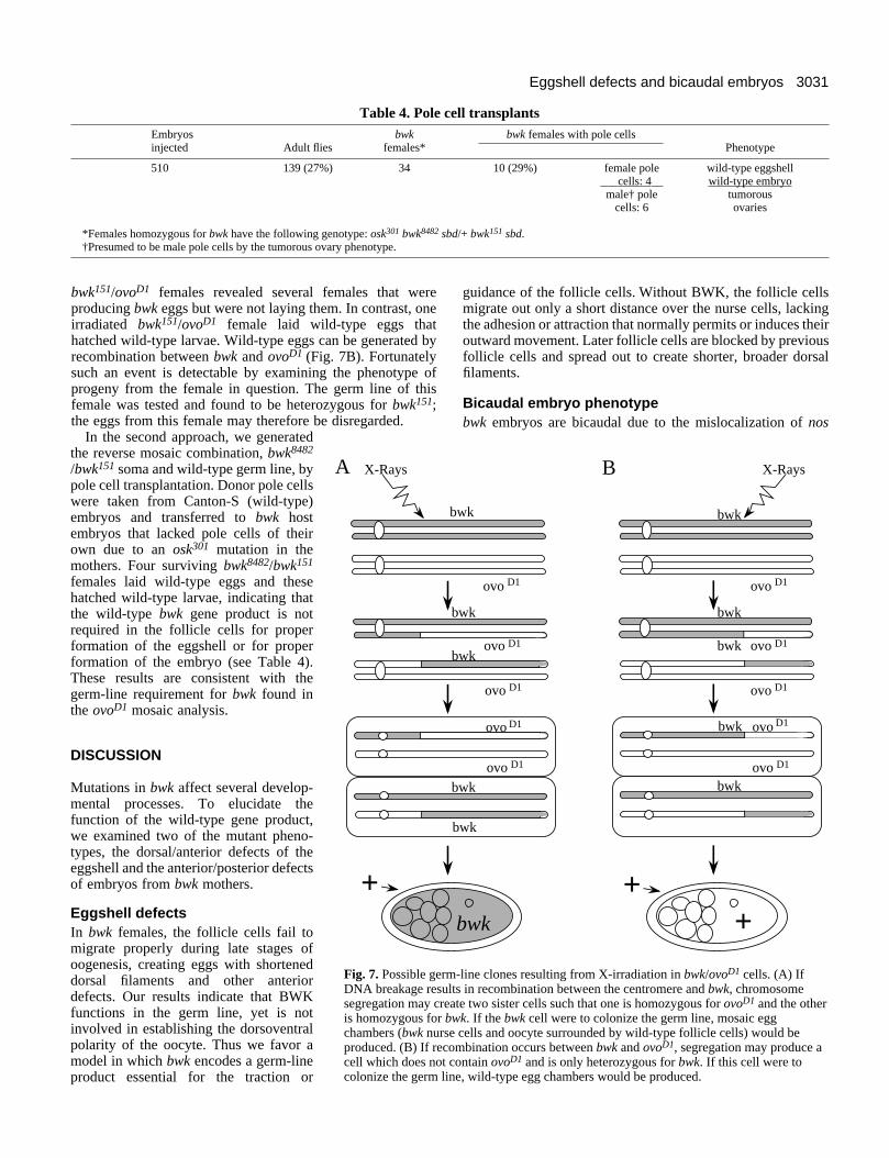
In the first approach, we used the ‘dominant female sterile’technique to generate germ-line clones (Chou et al., 1993).This method takes advantage of autosomal insertions ofP[ovoD1] that allow the formation of germ-line clones with X-ray irradiation. ovoD1 is a dominant female sterile mutation thatis germ-line specific and blocks the formation of late stage eggchambers. A late stage egg chamber can be formed only if agerm-line clone lacking ovoD1 is created. Mitotic recombina-tion in a female transheterozygous for ovoD1 and bwk cancreate two sister cells, such that one is homozygous for ovoD1
and the other is homozygous for bwk (Fig. 7A). In such afemale, the eggs produced would have bwk/+ follicle cells andbwk nurse cells and oocyte. Production of a wild-type eggwould indicate that the wild-type bwk gene product is notrequired in the germ cells. Conversely, production of a bwk eggwould indicate that the wild-type bwk gene product is requiredin the germ line.
Mitotic recombination was induced in bwk8482/ovoD1,bwk151/ovoD1 and control mwh red/ovoD1 females (see Table3). 1.2% of the mwh red transheterozygous females laid wild-type eggs, confirming that mitotic recombination in the germline was occurring at a measurable frequency. 0.8% of thebwk8482/ ovoD1 females and 1.4% of the bwk151/ovoD1 femaleslaid eggs with bwk eggshells, indicating that wild-type bwkfunction is required in the germ line. None of the bwk eggs laidby either line hatched. Dissection of many irradiated
3031Eggshell defects and bicaudal embryos
+
wk
ovo D1
wk
ovo
ovo
ovo
bwk
wk
bwk
D1
D1
D1
ovo
wk
D1
bwk
ovo D1
bwk
bwk
ovo
ovobwk
bwk D1
D1
ovo D1
ovo D1
+
X-RaysB
-line clones resulting from X-irradiation in bwk/ovoD1 cells. (A) Ifts in recombination between the centromere and bwk, chromosomete two sister cells such that one is homozygous for ovoD1 and the other
wk. If the bwk cell were to colonize the germ line, mosaic egg cells and oocyte surrounded by wild-type follicle cells) would bembination occurs between bwk and ovoD1, segregation may produce aontain ovoD1 and is only heterozygous for bwk. If this cell were toe, wild-type egg chambers would be produced.
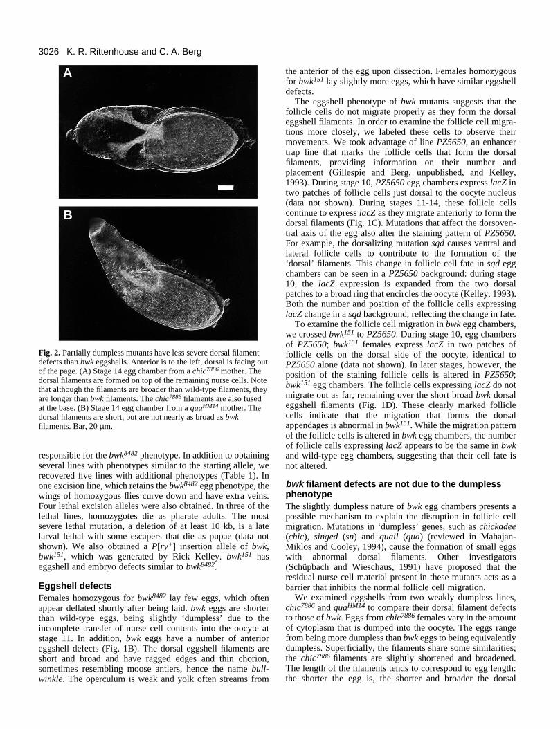
Table 4. Pole cell transplantsEmbryos bwk bwk females with pole cellsinjected Adult flies females* Phenotype
510 139 (27%) 34 10 (29%) female pole wild-type eggshell___cells: 4__ wild-type embryomale† pole tumorous
cells: 6 ovaries
*Females homozygous for bwk have the following genotype: osk301 bwk8482 sbd/+ bwk151 sbd.†Presumed to be male pole cells by the tumorous ovary phenotype.
bwk151/ovoD1 females revealed several females that wereproducing bwk eggs but were not laying them. In contrast, oneirradiated bwk151/ovoD1 female laid wild-type eggs thathatched wild-type larvae. Wild-type eggs can be generated byrecombination between bwk and ovoD1 (Fig. 7B). Fortunatelysuch an event is detectable by examining the phenotype ofprogeny from the female in question. The germ line of thisfemale was tested and found to be heterozygous for bwk151;the eggs from this female may therefore be disregarded.
In the second approach, we generatedthe reverse mosaic combination, bwk8482
/bwk151 soma and wild-type germ line, bypole cell transplantation. Donor pole cellswere taken from Canton-S (wild-type)embryos and transferred to bwk hostembryos that lacked pole cells of theirown due to an osk301 mutation in themothers. Four surviving bwk8482/bwk151
females laid wild-type eggs and thesehatched wild-type larvae, indicating thatthe wild-type bwk gene product is notrequired in the follicle cells for properformation of the eggshell or for properformation of the embryo (see Table 4).These results are consistent with thegerm-line requirement for bwk found inthe ovoD1 mosaic analysis.
DISCUSSION
Mutations in bwk affect several develop-mental processes. To elucidate thefunction of the wild-type gene product,we examined two of the mutant pheno-types, the dorsal/anterior defects of theeggshell and the anterior/posterior defectsof embryos from bwk mothers.
Eggshell defectsIn bwk females, the follicle cells fail tomigrate properly during late stages ofoogenesis, creating eggs with shorteneddorsal filaments and other anteriordefects. Our results indicate that BWKfunctions in the germ line, yet is notinvolved in establishing the dorsoventralpolarity of the oocyte. Thus we favor amodel in which bwk encodes a germ-lineproduct essential for the traction or
+
X-Rays
b
b
b
b
A
Fig. 7. Possible germDNA breakage resulsegregation may creais homozygous for bchambers (bwk nurseproduced. (B) If recocell which does not ccolonize the germ lin
guidance of the follicle cells. Without BWK, the follicle cellsmigrate out only a short distance over the nurse cells, lackingthe adhesion or attraction that normally permits or induces theiroutward movement. Later follicle cells are blocked by previousfollicle cells and spread out to create shorter, broader dorsalfilaments.
Bicaudal embryo phenotypebwk embryos are bicaudal due to the mislocalization of nos

3032 K. R. Rittenhouse and C. A. Berg
mRNA during oogenesis. The nos message is normallylocalized to the posterior pole through the stepwise action ofother posterior group gene products, which require a functionalmicrotubule network (Ephrussi et al., 1991; Lehmann andNüsslein-Volhard, 1991; Clark et al., 1994). Correct localiza-tion of KIN:β-GAL and osk mRNA to the posterior pole ofbwk oocytes during stages 9 and 10, respectively, implies thatthe structure of the microtubules and the transport along themis normal in bwk egg chambers during this earlier stage ofdevelopment (Clark et al., 1994). In this respect, bwk differsfrom other bicaudal mutations such as Bic-D, which cause oskmRNA to be mislocalized to the anterior pole of the oocyteduring stage 10 (Ephrussi et al., 1991; Kim-Ha et al., 1991).These results imply a fundamentally different defect in bwkoocytes.
The microtubule structure of the egg chamber changes dra-matically several times during oogenesis, most notably at stage10b after osk mRNA has been localized to the posterior pole.During stages 8 through 10a, the microtubules are localizedprimarily at the anterior cortex of the oocyte, with a corticalgradient of microtubules emanating from the anterior to theposterior pole (Theurkauf et al., 1992). During stage 10b, sub-cortical microtubules form within the oocyte and cytoplasmicstreaming begins, continuing through stage 12. In addition, thenurse cells begin transporting their contents into the oocyte atthe end of stage 10b. That bwk interferes with several processesthat begin after stage 10a (osk mRNA and other posterior groupgene product localization as well as the cytoplasmic transferfrom the nurse cells) suggests that BWK functions at this time.Preliminary studies show that cytoplasmic streaming occurscorrectly in bwk egg chambers, indicating that some aspects ofmicrotubule function are normal.
Localization of the nos mRNA in wild-type egg chambersoccurs after stage 12 (Ephrussi et al., 1991), when cytoplasmicstreaming has stopped, indicating that a new cytoskeletal archi-tecture must be organized in order to transport nos message tothe posterior pole. In bwk egg chambers, osk mRNA is notdetected at the anterior pole until after stage 12. bwk may benecessary for the proper formation of this post-streaming local-ization system, in which case, further analysis of the bwkphenotype will be invaluable in elucidating this later process.Such a localization system may be more similar to other RNAtransport systems since it is contained within a single cell.
Any models of BWK function must account for the defectsin follicle cell migrations, the partially dumpless phenotype,and the mislocalization of osk mRNA and other posterior groupgene products. The combination of these defects suggests thatbwk may encode a transmembrane protein whose extracellulardomain forms heterophilic interactions with a cell surfacecomponent on the surface of the follicle cells, and whose cyto-plasmic domain interacts with the actin cytoskeleton within thegerm cells. Alternatively, bwk could encode an extracellularmatrix (ECM) molecule or integral cytoskeleton proteinrequired for cell-cell interactions and adaptation of thecytoskeleton to the signaling process. The interaction of bwkneed not be direct; bwk could encode a transcription factorrequired to express critical components in these processes.During the last decade, a body of knowledge has accumulateddocumenting the interactions of cell surface receptors withcouterparts on other cells, with ECM molecules, and withmultiple components of the cytoskeleton (reviewed by Otey
and Burridge, 1990; Clark and Brugge, 1995). These moleculesare essential in such developmental processes as axon guidance(Whitington, 1993; Harrelson, 1992), neural crest cellmigration (Delannet et al., 1994; Bronner-Fraser et al., 1991),organ morphogenesis (Nelson et al., 1990) and yeast cellmating (Chenervet et al., 1994). In addition, changes in thesecell surface and cytoskeletal molecules occur during metasta-sis of cancer cells (Zetter, 1993), wound healing (Piersch-bacher et al., 1994) and aging (Yaar and Gilchrist, 1990). Mostof these studies employ in vitro techniques to examine celladhesion and the localization of products within the cell thatmediate these cell-cell interactions. Studies on the bwk geneprovide an excellent in vivo system for documenting the roleof these molecules in subcellular localization, cell migrationand pattern formation.
We are currently cloning the bwk gene to characterize it atthe molecular level. Preliminary studies suggest that the geneis large, encoding a 9 kb transcript (data not shown).
We thank Michael Gorsuch for work on the KIN:β-GAL studies.We are grateful to Trudi Schüpbach and Siegfried Roth for instruc-tion on and demonstration of pole cell transplantation, and to JonSchnorr for dark-room assistance. We are also grateful to DonaldMorisato and Kathryn Anderson for sharing information prior to pub-lication. We thank Hannele Ruohola-Baker, Paul Macdonald,members of our laboratory, and the reviewers for critical commentson the manuscript. We also thank Shira Neuman-Silberberg, TrudiSchüpbach, Markus Noll, Ruth Lehmann and Paul Macdonald forproviding cDNA clones and Rick Kelley, Trudi Schüpbach, HanneleRuohola-Baker, Norbert Perrimon, Lynn Cooley and Ruth Lehmannfor sending stocks. This work was supported by NIH grants RO1-GM45248 and T32-GM07735.
REFERENCES
Ashburner, M. (1989). Drosophila A Laboratory Manual. pp. 217-218. ColdSpring Harbor: Cold Spring Harbor Laboratory Press.
Brand, A. and Perrimon, N. (1994). Raf acts downstream of the EGF receptorto determine dorsoventral polarity during Drosophila oogenesis. Genes Dev.8, 629-639.
Bronner-Fraser, M., Stern, C. and Fraser, S. (1991). Analysis of neural crestcell lineage and migration. J. Craniofac. Genet. Dev. Biol. 11, 214-222.
Bull, A. (1966). Bicaudal, a genetic factor which affects the polarity of theembryo in Drosophila melanogaster. J. Exp. Zool. 161, 221-242.
Chasan, R. and Anderson, K. (1993). Maternal Control of Dorsal-VentralPolarity and Pattern in the Embryo. In The Development of Drosophilamelanogaster. (ed. M. Bate and A. Martinez-Arias), pp. 387-424. ColdSpring Harbor: Cold Spring Harbor Laboratory Press.
Chenervet, J., Valtz, N. and Herskowitz, I. (1994). Identification of genesrequired for normal pheromone-induced cell polarization in Saccharomycescerevisiae. Genetics 136, 1287-1296.
Chou, T.-B., Noll, E. and Perrimon, N. (1993). Autosomal P[ovoD1]dominant female-sterile insertions in Drosophila and their use in generatinggerm-line chimeras. Development 119, 1359-1369.
Christerson, L. and McKearin, D. (1994). orb is required for anteroposteriorand dorsoventral patterning during Drosophila oogenesis. Genes Dev. 8,614-628.
Clark, E. and Brugge, J. (1995). Integrins and signal transduction pathways:the road taken. Science 268, 233-239.
Clark, I., Giniger, E., Ruohola-Baker, H., Jan, L. Y.. and Jan, Y.N. (1994).Transient posterior localization of a kinesin fusion protein reflectsanteroposterior polarity of the Drosophila oocyte. Current Biology 4, 289-300.
Cooley, L., Verheyen, E. and Ayers, K. (1992). chickadee encodes a PROFILIN
required for intercellular cytoplasm transport during Drosophila oogenesis.Cell 69, 173-184.
Delannet, M., Martin, F., Bossy, B., Cheresh, D., Reichert, L. and Duband,
3033Eggshell defects and bicaudal embryos
J. (1994). Specific roles of the αVβ1, αVβ3 and αVβ5 INTEGRINS in avianneural crest cell adhesion and migration on vitronectin. Development 120,2687-2702.
Driever, W. (1993). Maternal Control of Anterior Development in theDrosophila embryo. In The Development of Drosophila melanogaster. (ed.M. Bate and A. Martinez-Arias), pp. 301-324. Cold Spring Harbor: ColdSpring Harbor Laboratory Press.
Ephrussi, A., Dickinson, L. and Lehmann, R. (1991). oskar organizes thegerm plasm and directs localization of the posterior determinant nanos. Cell66, 37-50.
Ephrussi, A. and Lehmann, R. (1992). Induction of germ cell formation byoskar. Nature 358, 387-392.
Gavis, E. and Lehmann, R. (1992). Localization of nanos RNA controlsembryonic polarity. Cell 71, 301-313.
Harrelson, A. (1992). Molecular mechanisms of axon guidance in thedeveloping insect nervous system. J. Exp. Zool. 261, 310-321.
Karpen, G. and Spradling, A. (1992) Analysis of subtelomericheterochromatin in the Drosophila minichromosome Dp1187 by single Pelement insertional mutagenesis. Genetics 132, 737-53.
Kelley, R. (1993). Initial organization of the Drosophila dorsoventral axisdepends on an RNA-binding protein encoded by the squid gene. Genes Dev.7, 948-960.
Kim-Ha, J., Smith, J. and Macdonald, P. (1991). oskar mRNA is localized tothe posterior pole of the Drosophila oocyte. Cell 66, 23-35.
King, R. (1970) Ovarian development in Drosophila melanogaster. New York:Academic Press.
King, R. and Koch, E. (1963). Studies on the ovarian follicle cells ofDrosophila. Quart. J. Micr. Sci. 104, 297-320.
Lehmann, R. and Nüsslein-Volhard, C. (1986). Abdominal segmentation,pole cell formation, and embryonic polarity require the localized activity ofoskar, a maternal gene in Drosophila. Cell 47, 141-152.
Lehmann, R. and Nüsslein-Volhard, C. (1991). The maternal gene nanos hasa central role in posterior pattern formation of the Drosophila embryo.Development 112, 679-691.
Macdonald, P. and Struhl, G. (1988). Cis-acting sequences responsible foranterior localization of bicoid mRNA in Drosophila embryos. Nature 336,595-598.
Mahajan-Miklos, S. and Cooley, L. (1994). Intercellular cytoplasm transportduring Drosophila oogenesis. Dev. Biol. 165, 336-351.
Manseau, L. and Schüpbach, T. (1989). cappuccino and spire: two uniquematernal-effect loci required for both the anteroposterior and dorsoventralpatterns of the Drosophila embryo. Genes Dev. 3, 1437-1452.
Mohler, J. and Wieschaus, E. (1986). Dominant maternal-effect mutations ofDrosophila melanogaster causing the production of double-abdomenembryos. Genetics 112, 803-822.
Neuman-Silberberg, F. and Schüpbach, T. (1993). The Drosophiladorsoventral patterning gene gurken produces a dorsally localized RNA andencodes a TGF α-like protein. Cell 75, 165-74.
Nelson, W. J., Hammerton, R., Wang, A. and Shore, E. (1990). Involvementof the membrane-cytoskeleton in development of epithelial cell polarity.Sem. Cell Biol. 1, 359-371.
Otey, C. and Burridge, K. (1990). Patterning of the membrane cytoskeletonby the extracellular matrix. Sem. Cell Biol. 1, 391-399.
Pierschbacher, M., Polarek, J., Craig, W., Tschopp, J., Sipes, N. andHarper, J. (1994). Manipulation of cellular interactions with biomaterialstoward a therapeutic outcome: a perspective. J. Cell. Biochem. 56, 150-154.
Pokrywka, N. and Stephenson, E. (1991). Microtubules mediate thelocalization of bicoid RNA during Drosophila oogenesis. Development 113,55-66.
Price, J., Clifford, R. and Schüpbach, T. (1989). The maternal ventralizing
locus torpedo is allelic to faint little ball, an embryonic lethal, and encodesthe Drosophila EGF receptor homolog. Cell 56, 1085-92.
Robertson, H., Preston, C., Phillis, R., Johnson-Schlitz, D., Benz, W. andEngels, W. (1988). A stable source of P-element transposase in Drosophilamelanogaster. Genetics 118, 461-470.
Schejter, E. and Shilo, B. (1989). The Drosophila EGF receptor homolog(DER) is allelic to faint little ball, a locus essential for embryonicdevelopment. Cell 56, 1093-1104.
Schüpbach, T. (1987). Germ line and soma cooperate during oogenesis toestablish the dorsoventral pattern of the egg shell and embryo in Drosophilamelanogaster. Cell 49, 699-707.
Schüpbach, T. and Wieschaus, E. (1986). Maternal-effect mutations alteringthe anterior-posterior pattern of the Drosophila embryo. Roux’s Arch. Dev.Biol. 195, 302-317.
Schüpbach, T. and Wieschaus, E. (1991). Female sterile mutations on thesecond chromosome of Drosophila melanogaster. II. Mutations blockingoogenesis or altering egg morphology. Genetics 129, 1119-1136.
Smith, J., Wilson, J. and Macdonald, P. (1992). Overexpression of oskardirects ectopic activation of nanos and presumptive pole cell formation inDrosophila embryos. Cell 70, 849-859.
Spradling, A. (1993). Developmental Genetics of Oogenesis. In TheDevelopment of Drosophila melanogaster. (ed. M. Bate and A. Martinez-Arias), pp. 1-69. Cold Spring Harbor: Cold Spring Harbor Laboratory Press.
St. Johnston, D. (1993). Pole Plasm and the Posterior Group Genes. In TheDevelopment of Drosophila melanogaster. (ed. M. Bate and A. Martinez-Arias), pp. 325-364. Cold Spring Harbor: Cold Spring Harbor LaboratoryPress.
Tautz, D. and Pfeifle, C. (1989). A non-radioactive in situ hybridizationmethod for the localization of specific RNAs in Drosophila embryos revealstranslational control of the segmentation gene hunchback. Chromosoma 92,81-85.
Theurkauf, W. (1994). Premature microtubule-dependent cytoplasmicstreaming in cappuccino and spire mutant oocytes. Science 265, 2093-2096.
Theurkauf, W., Smiley, S., Wong, M. and Alberts, B. (1992). Reorganizationof the cytoskeleton during Drosophila oogenesis: implications for axisspecification and intercellular transport. Development 115, 923-936.
Van Deusen, E. (1976). Sex determination in the germ line chimeras ofDrosophila melanogaster. J. Embryol. Exp. Morph. 37, 173-185.
Wang, C., Dickinson, L. and Lehmann, R. (1994). Genetics of nanoslocalization in Drosophila. Dev. Dynam. 199, 103-115.
Wang, C. and Lehmann, R. (1991). nanos is the localized posteriordeterminant in Drosophila. Cell 66, 637-647.
Wharton, R. and Struhl, G. (1991). RNA regulatory elements mediate controlof Drosophila body pattern by the posterior morphogen nanos. Cell 67, 955-967.
Whitington, P. (1993). Axon guidance factors in invertebrate development.Pharmacol. Ther. 58, 253-299.
Wieschaus, E., Marsh, J. and Gerhring, W. (1978). fs(1)K10, a germline-dependent female sterile mutation causing abnormal chorion morphology inDrosophila melanogaster. Roux’s Arch. Dev. Biol. 184, 75-82.
Wieschaus, E. and Nüsslein-Volhard, C. (1986). Looking at embryos. InDrosophila a practical approach (ed. D. B. Roberts), pp. 214-216. Oxford:Information Printing Ltd.
Yaar, M. and Gilchrest, B. (1990). Cellular and molecular mechanisms ofcutaneous aging. J. Dermatol. Surg. Oncol. 16, 915-922.
Zetter, B. (1993). Adhesion molecules in tumor metastasis. Sem. Cancer Biol.4, 219-229.
(Accepted 31 May 1995)