mycelial carton galleries of azteca brevis (formicidae)...
TRANSCRIPT

Proc. R. Soc. B (2009) 276, 3265–3273
on May 20, 2018http://rspb.royalsocietypublishing.org/Downloaded from
*Author
Electron1098/rsp
doi:10.1098/rspb.2009.0768
Published online 25 June 2009
ReceivedAccepted
Mycelial carton galleries of Azteca brevis(Formicidae) as a multi-species network
Veronika E. Mayer1,* and Hermann Voglmayr2
1Department of Palynology and Structural Botany, Faculty Centre of Biodiversity,
Faculty of Life Sciences, University of Vienna, Rennweg 14, A-1030 Wien, Austria2Department of Systematic and Evolutionary Botany, Faculty Centre of Biodiversity,
Faculty of Life Sciences, University of Vienna, Rennweg 14, A-1030 Wien, Austria
Apart from growing fungi for nutrition, as seen in the New World Attini, ants cultivate fungi for reinforce-
ment of the walls of their nests or tunnel-shaped runway galleries. These fungi are grown on organic
material such as bark, epiphylls or trichomes, and form stable ‘carton structures’. In this study, the
carton of the runway galleries built by Azteca brevis (Formicidae, Dolichoderinae) on branches of
Tetrathylacium macrophyllum (Flacourtiaceae) is investigated. For the first time, molecular tools are
used to address the biodiversity and phylogenetic affinities of fungi involved in tropical ant carton
architecture, a previously neglected ant–fungus mutualism.
The A. brevis carton involves a complex association of several fungi. All the isolated fungi were unequivo-
cally placed within the Chaetothyriales by DNA sequence data. Whereas five types of fungal hyphae were
morphologically distinguishable, our DNA data showed that more species are involved, applying a phyloge-
netic species concept based on DNA phylogenies and hyphal morphology. In contrast to the New World
Attini with their many-to-one (different ant species—one fungal cultivar) pattern, and temperate Lasius
with a one-to-two (one ant species—two mutualists) or many-to-one (different ant species share the same
mutualist) system, the A. brevis–fungi association is a one-to-many multi-species network. Vertical fungus
transmission has not yet been found, indicating that the A. brevis–fungi interaction is rather generalized.
Keywords: ant fungiculture; Azteca brevis; Chaetothyriales; multi-species network;
nest-wall fungi; Tetrathylacium macrophyllum
1. INTRODUCTIONAnt–fungus associations in the New World fungus-
growing ants (Myrmicinae: Attini) are well-studied examples
of mutualism between ants and fungi; the ants involved
are obligate agriculturists and cultivate basidiomycetes
of the mushroom families Lepiotaceae and Pterulaceae
for food (Mueller et al. 1998; Munkacsi et al. 2004). A
less well-studied mutualism is the cultivation of fungi
for carton constructions seen in Old World Lasius ants
(Formicinae) (Elliott 1915; Maschwitz & Holldobler
1970; Schlick-Steiner et al. 2008), arboricolous Asian
ants of the genera Camponotus, Crematogaster,
Dolichoderus, Monomorium and Technomyrmex (Weissflog
2001) and the Neotropical genus Allomerus (Dejean
et al. 2005). The stabilization of the carton construction
was first investigated in detail in nests of Lasius subgenera
Dendrolasius and Chthonolasius, and seems to work in that
the fungal mycelium ‘grows through the walls of the carton
and reinforces them in the same way that steel mesh or
rods reinforce the walls of a building’ (Holldobler &
Wilson 1990). The material used for the carton may be
soil and shredded wood particles as in Lasius (Elliott
1915; Maschwitz & Holldobler 1970), or cut trichomes
bound together ‘with a compound that they [the ants]
regurgitate’ as in Allomerus decemarticulatus (Myrmicinae)
for correspondence ([email protected]).
ic supplementary material is available at http://dx.doi.org/10.b.2009.0768 or via http://rspb.royalsocietypublishing.org.
5 May 20092 June 2009 3265
nesting obligately in the domatia of Hirtella physophora
(Chrysobalanaceae; Dejean et al. 2005).
In nearly all cases, the associated fungi are not further
investigated or classified. Dejean et al. (2005) stated that
the carton walls of the galleries of Allomerus decemarticulatus
are ‘reinforced by the mycelium of a complex of
sooty-mould species that has been manipulated by the ants’.
The taxonomic position of the carton wall fungi found
in Lasius subgenera Dendrolasius and Chthonolasius was
only recently clarified as ascomycetes belonging to the
Capnodiales, Chaetothyriales and Venturiaceae (Schlick-
Steiner et al. 2008). Interestingly, Schlick-Steiner et al.
(2008) found three species of Venturiaceae occurring
exclusively and invariably with their respective hosts,
and two other species of Chaetothyriales and Capnodiales
occurring only occasionally in nests of both subgenera.
There is some evidence that the latter two are not
mutualists (Schlick-Steiner et al. 2008).
In the present study, the previously unknown carton
structure of the runway galleries built by Azteca brevis
Forel inhabiting live stems of Tetrathylacium macrophyllum
(Flacourtiaceae) was examined. Apart from T. macrophyllum,
A. brevis is found on Grias (Lecythidaceae), Licania
(Chrysobalanaceae), Myriocarpa (Urticaceae), and Ocotea
nicaraguensis (Lauraceae) (Longino 2008). Ants of the
genus Azteca are known to subdivide pre-formed cavities
with carton constructions into functional units using small
platforms and baffles (Wheeler & Bequaert 1929; Longino
1996); A. brevis also builds extensive systems of galleries
made of arched tunnels of a black, very crusty carton with
This journal is q 2009 The Royal Society

3266 V. E. Mayer & H. Voglmayr Mycelial carton galleries of Azteca brevis
on May 20, 2018http://rspb.royalsocietypublishing.org/Downloaded from
small circular holes. The carton galleries and the holes, which
are large enough for the workers to pass through, enhance a
sophisticated hunting and defence strategy (Schmidt 2001).
We demonstrate experimentally that the materials used for
the construction of the galleries are chiefly particles found
on the branches of the host tree. SEM images show that the
particles are densely interwoven and stabilized by fungal
hyphae. With the aid of light microscopy and DNA analyses,
the phylogenetic affinities of the fungi involved in the carton
nest construction of A. brevis are revealed. We can demon-
strate for the first time for A. brevis that at least four different
fungi coexist in the same carton gallery, which indicates that
this mutualism involves more than one fungal partner per
host and is a multi-species mutualism in respect not only of
the host tree but also of the fungi used to stabilize the carton.
2. MATERIAL AND METHODS(a) Species and study site
Azteca brevis Forel (Formicidae, Dolichoderinae) is a species
known only from the Pacific side of Costa Rica, and mostly
from the wet forests of the southern Pacific lowlands. It
nests in live stems of trees in polydomous colonies (more
than one nest) with workers and brood found in many
branch tips. The nesting chambers inside the stems are usually
connected externally by an extensive system of runway gal-
leries made of a characteristic black, crusty carton. Inside
the stems and in the tunnels of the carton galleries A. brevis
maintained large populations of an as yet undescribed species
(P. Gullan 2004, personal communication) of the coccoid
hemipteran genus Cryptostigma. The carton galleries investi-
gated in this study were found on T. macrophyllum Poepp. &
Endl. (Flacourtiaceae), a tree of approximately 15–20 m
height occurring preferentially on steep slopes near rivers and
streams in primary forests (Janzen 1983). It can be colonized
by various ant species (Tennant 1989; Schmidt 2001), of
which A. brevis is the most frequent inhabitant.
In 2007 and 2008, samples from eight different
Tetrathylacium trees (four per year) were taken. Each tree
was separated from the others by a walking distance of at
least 10 min, therefore it is assumed that each tree was inhab-
ited by a different colony. Pieces of the carton approximately
0.5–1.0 cm long were removed, placed in a paper bag using
sterile forceps and dried at 308C. Carton specimens from
three different branches were sampled from each tree and
ant colony, and each sample kept in a separate paper bag.
Due to contamination of the samples with Aspergillus and
Penicillium, fungal isolates could only be grown successfully
from three of the eight trees. CR07/2 and CR07/3 were col-
lected in 2007 along the ‘waterfall trail’, while CR08/2 was
collected in 2008 along the ‘ocelot trail’, both of which are
within lowland primary rain forest near the Biological Research
Station La Gamba (0884204600 N, 8381209000 W, 70 m.a.s.l.) in
the vicinity of the Parque Nacional Piedras Blancas on the
southern Pacific slope of Costa Rica. Voucher specimens of
plants and ants were deposited at the Museo Nacional in San
Jose, Costa Rica and in the Herbarium of the University of
Vienna (WU Schaber 2860, Mayer 07-2, 07-3, 08-2).
(b) SEM and light microscopy (LM) investigation of
carton nest structure and nest material
Carton nests were sputter coated in high vacuum mode at
15 kV and the inner and outer side investigated in a Phillips
XL30 environmental scanning electron microscope. For
Proc. R. Soc. B (2009)
light microscopy, freshly collected specimens were placed
on a microscope slide, moistened with water and examined
and documented without staining in a Zeiss AxioImager
A1 compound microscope equipped with a Zeiss AxioCam
ICc3 digital camera.
(c) Field experiments
To test whether particles from the branches’ surfaces are used
for the construction of the carton, the following experiment
was conducted. Ten easily accessible trees occupied by
A. brevis were randomly selected. On two branches per
tree, the carton tunnel was locally destroyed and a bright
blue textile fibre adhesive tape (TESA, 19 mm width) was
mounted (n ¼ 20, total number of taped branches), placing
the sticky tape on the bark surface of the tree. One week
after mounting the tapes, the adhesive tapes were examined
to determine whether the ants had removed fibres from the
tape and incorporated the particles in their carton structure.
(d) Fungal cultures
Small parts of the dried carton tunnels were soaked in a drop
of sterile water on a sterile microscope slide for approxi-
mately 5 min, and carefully broken up after soaking using
fine forceps and preparation needles. The resulting mycelial
suspension was pipetted onto a 2 per cent malt extract agar
plate (MEA) containing antibiotics (0.5% penicillin and
0.5% streptomycin), and spread by gently rotating the
plates. The plates were incubated at room temperature and
visually checked at least daily under a dissecting microscope.
Owing to the rapid growth of contaminating fungi (mainly
Aspergillus and Penicillium spp.), especially during the initial
phase of incubation, fast-growing mycelia with hyaline
hyphae were immediately cut out from the plate. Upon ger-
mination, the hyphal fragments were examined under the
microscope to ensure they originated from hyphae building
the carton structure and transferred to a 2 per cent MEA
plate. Water mounts of the mycelium of the cultures were
examined with the light microscope to determine hyphal
morphology and the presence of dispersal units (conidia).
(e) DNA extraction, amplification
and sequencing
For DNA extraction, mycelium was grown in 1.5 ml reaction
tubes containing 1 ml 2 per cent malt extract solution at
room temperature for 14–25 days depending on growth
rate. During incubation, the tubes were gently shaken on a
horizontal shaker at 30 r.p.m. and regularly vortexed to
avoid agglutination of the mycelia. After incubation, the
tubes were centrifuged for 10 min at 13 000 r.p.m. to pellet
the mycelium, the supernatant was removed, the pellet
washed once with 3 � CTAB extraction buffer (75 mM
Tris pH 8, 0.17 M sorbitol, 2 M sodium chloride, 15 mM
EDTA, 3% CTAB, 4% PVP-40) and immediately processed
for DNA extraction or stored at 2208C.
The mycelia were ground using sterile quartz sand and a
micropestle fitting the tube. The DNA was extracted with the
modified CTAB method described in Riethmuller et al.
(2002), but using a high salt 3 � CTAB buffer (see above)
with 1 per cent mercaptoethanol and two chloroform–
isoamyl alcohol (24 : 1) extraction steps. After precipitation,
the pellet was washed twice with 70 per cent aqueous etha-
nol, dried in a vacuum centrifuge, and the pellet dissolved
in 50 ml 1 � TE buffer. One ml RNAse (5 mg/ml) was
added, followed by incubation at 378C for 60 min.

Mycelial carton galleries of Azteca brevis V. E. Mayer & H. Voglmayr 3267
on May 20, 2018http://rspb.royalsocietypublishing.org/Downloaded from
PCR was performed in 20 ml volumes: 1 ml of template
DNA, 18 ml of 1.1 � ReddyMix PCR Master Mix
(ABgene, Epsom, UK) and 0.25 mM forward and reverse
primers. A ca 1.1 kb fragment of the nuclear small subunit
ribosomal DNA was amplified using the primers NS1 and
NS4 (White et al. 1990) or nssu1088 (Kauff & Lutzoni
2002). A 1.5–1.9 kb fragment containing the 50 end of the
small subunit, the complete ITS1-5.8S-ITS2 and the vari-
able D1 and D2 domains of the large subunit of the nuclear
ribosomal DNA were amplified with primers V9G (de Hoog
& Gerrits van den Ende 1998) or ITS5 (White et al. 1990)
and LR5 (Vilgalys & Hester 1990). The PCR products
were purified using an enzymatic PCR cleanup (Werle et al.
1994): 20 ml PCR reactions were digested with 10 U
Exonuclease I (Fermentas, St. Leon-Rot, BRD) and 2 U
Calf Intestine Alkaline Phosphatase (Fermentas) for 30 min
at 378C, followed by an enzyme deactivation step at 858Cfor 15 min. DNA was sequenced using the ABI PRISM Big
Dye Terminator Cycle Sequencing Ready Reaction
Kit v. 3.1 (Applied Biosystems, Warrington) with the same
primers as for PCR; in addition, fITS5.8Sr
(TGCGTTCAAARATTCGATG), fITS5.8Sf (CAACAA
CGGATCTCTTGGYTC), ITS4 (White et al. 1990),
LR0R (Rehner & Samuels 1994) and LR3 (Vilgalys &
Hester 1990) were used as internal primers for the long
SSU-ITS-LSU fragment. Sequences were analysed with an
automated DNA sequencer (3130xl Genetic Analyzer,
Applied Biosystems).
(f) Analysis of sequence data
For the phylogenetic analyses of the nuSSU rDNA (SSU),
the nuLSU rDNA (LSU) and the complete ITS1-5.8S
rDNA-ITS2 (ITS) regions, a representative sample of most
similar sequences were selected from GenBank after a
BLAST search; representative sequences from other lineages
were also added. Sequence alignments were produced with
MUSCLE v. 3.6 (Edgar 2004). After exclusion of leading/
trailing gap regions and large insertions in some sequences,
938 characters remained across 35 sequences in the nuLSU
rDNA alignment. One thousand and sixty six characters
across 34 sequences of the nuSSU rDNA alignment and
765 characters across 95 sequences of the complete ITS
alignment were included in the subsequent phylogenetic ana-
lyses. The resulting alignments were checked and refined
using BIOEDIT v. 7.0.9.0 (Hall 1999).
Maximum parsimony (MP) analyses of the data matrices
were performed with PAUP* v. 4.0 b10 (Swofford 2002),
using 1000 replicates of heuristic search with random
addition of sequences and subsequent TBR branch swapping
(MULTREES option in effect, COLLAPSE¼MAXBRLEN,
steepest descent option not in effect, and limiting the
number of rearrangements per replicate to 500 000 in
the ITS dataset). All molecular characters were unordered
and given equal weight; analyses were performed with gaps
treated as missing data. Bootstrap analysis with 1000 repli-
cates was performed in the same way, but using 10 rounds
of random sequence addition and subsequent branch
swapping during each bootstrap replicate.
For Bayesian analyses, the program MRBAYES (v. 3.1.2;
Huelsenbeck & Ronquist 2001) was used. According to the
nucleotide substitution models suggested by MODELTEST 3.6
(Posada & Crandall 1998), the general time reversible
model was implemented for the three (LSU, SSU, ITS)
Proc. R. Soc. B (2009)
datasets, assuming a proportion of invariant sites with the
remaining sites having substitution rates drawn from a
gamma distribution (GTRþIþG). Three parallel runs of
four incrementally heated simultaneous Markov chains
were performed over 1 million generations from which
every 100th tree was sampled in each run. Trees saved
before the cold chain reached the stationary stage were dis-
carded as burn-in (300 for LSU, 500 for SSU, 1000 for
ITS). A 90 per cent majority rule consensus of the remaining
trees was computed, giving estimates for the probabilities
that groups are monophyletic given the sequence data
(posterior probabilities).
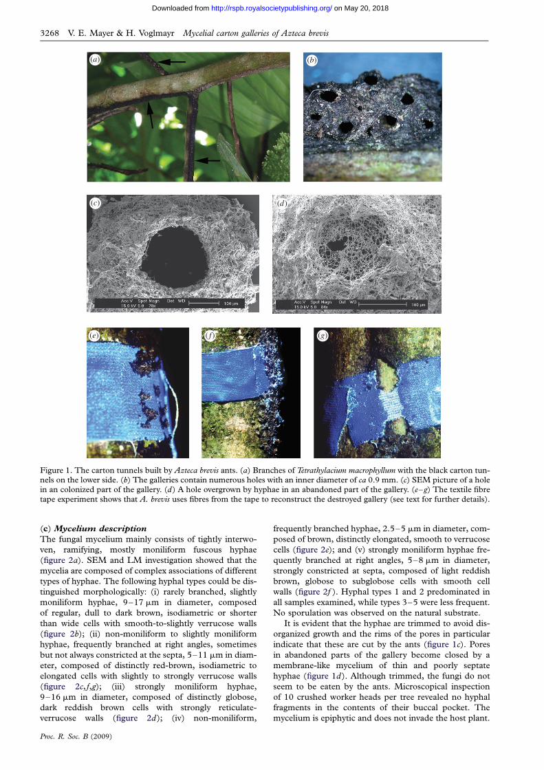
3. RESULTS(a) Carton structure and construction
Extensive carton galleries were seen to cover the natural
cavities of T. macrophyllum formed through partial pith
degeneration of the secondary branches. The tissue sur-
rounding the cavity was scarified on one side of the
branch, with a slit making the cavity easily accessible to
ants. A. brevis occupied all the domatium chambers avail-
able within its territory and excavated the remaining pith
inside the living stem to form domatia with a maximum
length of more than 1 m (Schmidt 2001). The carton gal-
leries take the form of arched tunnels, ca 3 mm broad and
2 mm high, which run along the lower sides of branches
(figure 1a). To construct the gallery, A. brevis workers
regurgitate a pulp to construct two rows of lateral pillars
spaced 1 mm apart along the lower surface of young
branches. Adjacent pillars are then connected by an
arch. The galleries contain holes, each surrounded by a
funnel-shaped ring with an inner diameter of 0.9
(+0.2) mm (figure 1b,c). Newly added construction
material was a light brown colour which, after a few
days, turned black and had a distinctive tar-like odour.
Even newly produced pillars were observed to be structu-
rally stable, since those located on the lateral surface of
the branch resisted heavy rains during the construction
period. SEM investigations of the carton structure
showed the fibre particles to be woven into a dense net
of fungal hyphae. Although it is difficult to quantify the
absolute ratio, detailed LM investigations revealed that,
upon fungal colonization, the carton structure consists
mainly of fungal biomass. Few Cryptostigma sp. coccids
were tended in the carton galleries, the majority being
maintained in the domatia of the hollow branches.
(b) Carton material
When a textile fibre tape was placed around branches with
a carton runway (n ¼ 20), A. brevis workers reconstructed
the destroyed carton gallery over the textile fibre tape
within one week (figure 1e, f ). After removing the
newly built carton, only a minority of the tapes was
intact or only slightly removed. In 85 per cent of the
cases, A. brevis removed much or all of the tape
(figure 1g). On all taped branches, the reconstructed
part of the carton gallery was spotted with bright blue
fibre particles, which seemed to be the main source of
construction material in this area (figure 1f ). On
branches where the tape was entirely removed, the fibres
could be found incorporated into parts of the gallery up
to 1 m away from where the tape was originally placed.
(a)
(c)
(e) (g)( f)
(d)
(b)
Figure 1. The carton tunnels built by Azteca brevis ants. (a) Branches of Tetrathylacium macrophyllum with the black carton tun-nels on the lower side. (b) The galleries contain numerous holes with an inner diameter of ca 0.9 mm. (c) SEM picture of a holein an colonized part of the gallery. (d) A hole overgrown by hyphae in an abandoned part of the gallery. (e–g) The textile fibre
tape experiment shows that A. brevis uses fibres from the tape to reconstruct the destroyed gallery (see text for further details).
3268 V. E. Mayer & H. Voglmayr Mycelial carton galleries of Azteca brevis
on May 20, 2018http://rspb.royalsocietypublishing.org/Downloaded from
(c) Mycelium description
The fungal mycelium mainly consists of tightly interwo-
ven, ramifying, mostly moniliform fuscous hyphae
(figure 2a). SEM and LM investigation showed that the
mycelia are composed of complex associations of different
types of hyphae. The following hyphal types could be dis-
tinguished morphologically: (i) rarely branched, slightly
moniliform hyphae, 9–17 mm in diameter, composed
of regular, dull to dark brown, isodiametric or shorter
than wide cells with smooth-to-slightly verrucose walls
(figure 2b); (ii) non-moniliform to slightly moniliform
hyphae, frequently branched at right angles, sometimes
but not always constricted at the septa, 5–11 mm in diam-
eter, composed of distinctly red-brown, isodiametric to
elongated cells with slightly to strongly verrucose walls
(figure 2c, f,g); (iii) strongly moniliform hyphae,
9–16 mm in diameter, composed of distinctly globose,
dark reddish brown cells with strongly reticulate-
verrucose walls (figure 2d); (iv) non-moniliform,
Proc. R. Soc. B (2009)
frequently branched hyphae, 2.5–5 mm in diameter, com-
posed of brown, distinctly elongated, smooth to verrucose
cells (figure 2e); and (v) strongly moniliform hyphae fre-
quently branched at right angles, 5–8 mm in diameter,
strongly constricted at septa, composed of light reddish
brown, globose to subglobose cells with smooth cell
walls (figure 2f ). Hyphal types 1 and 2 predominated in
all samples examined, while types 3–5 were less frequent.
No sporulation was observed on the natural substrate.
It is evident that the hyphae are trimmed to avoid dis-
organized growth and the rims of the pores in particular
indicate that these are cut by the ants (figure 1c). Pores
in abandoned parts of the gallery become closed by a
membrane-like mycelium of thin and poorly septate
hyphae (figure 1d). Although trimmed, the fungi do not
seem to be eaten by the ants. Microscopical inspection
of 10 crushed worker heads per tree revealed no hyphal
fragments in the contents of their buccal pocket. The
mycelium is epiphytic and does not invade the host plant.

(a)
(c)
(e)
(g)( f)
(d)
(b)
Figure 2. Light microscopy of the various morphological types of fungi colonizing the tunnels. (a) Overview of a squash mount,showing fungal hyphae of types 1, 2 and 5. (b) Hyphal type 1; note cells being as broad as or broader than long and the dull,dark brown, smooth cell walls. (c) Hyphal type 2; note the cells as long as or longer than broad and the distinctly reddish brown,
verrucose cell wall. (d) Hyphal type 3; note the dark reddish brown globose cells with reticulate-verrucose ornamentation.(e) Hyphal type 4; note the comparatively thin, brown, elongated cells. ( f ) Hyphal types 2 (centre) and 5 (above andbelow); note the strongly moniliform, subglobose cells and the brown, smooth cell wall of type 5. (g) Germinating hyphaeof type 2. Bars: a, G 50 mm, b– f, 20 mm.
Mycelial carton galleries of Azteca brevis V. E. Mayer & H. Voglmayr 3269
on May 20, 2018http://rspb.royalsocietypublishing.org/Downloaded from
(d) Isolation of fungi in pure culture
Pure cultures could be obtained from hyphal types 1, 2, 4
and 5, while type 3 failed to grow. Few cultures per
sample could be obtained from the mycelial suspensions.
This may be because of sub-optimal preservation of the
material after collecting in the field (high humidity and
temperature) as well as the slow germination and
Proc. R. Soc. B (2009)
growth of the fungi. In all attempts at isolation, fast-
growing fungal contaminants (mostly from the genera
Aspergillus and Penicillium) had to be continually removed
to avoid rapid overgrowth of the agar plate. All cultures
produced dark brown to blackish slow-growing mycelia
with abundant aerial mycelium. The hyphae were similar
to those observed on the tunnels, although there were

3270 V. E. Mayer & H. Voglmayr Mycelial carton galleries of Azteca brevis
on May 20, 2018http://rspb.royalsocietypublishing.org/Downloaded from
some differences in pigmentation, surface ornamentation
and hyphal diameter; such differences are commonly
observed in fungi grown on artificial media. With the
exception of culture CR07/2-2, the mycelia did not
produce conidia or fruiting bodies in pure culture.
(e) Phylogenetic affiliation and classification
of the fungi
Except for one isolate (CR07/2-2), which may be
assigned morphologically to the anamorph genus
Cladophialophora, the cultures did not sporulate in pure
culture and could not therefore be formally classified.
However, their phylogenetic affiliation could be clarified
unequivocally with DNA data.
The nuLSU, the nuSSU rDNA and the ITS1-5.8S
rDNA-ITS2 matrices contained 218, 116 and 354
parsimony-informative characters, respectively. The maxi-
mum parsimony analysis of the nuLSU rDNA revealed a
single most parsimonious tree of score 800, which is
shown in figure 3. Analysis of the nuSSU rDNA data
revealed four equally most parsimonious trees of score
351 (figure S1 in the electronic supplementary material).
Maximum parsimony analysis of the complete ITS1-5.8S
rDNA-ITS2 matrix resulted in 16 712 trees of score
1765, one of which is shown as a phylogram in figure S2
in the electronic supplementary material. Bayesian ana-
lyses revealed trees fully compatible with the MP trees.
The nuLSU, nuSSU and ITS1-5.8S rDNA-ITS2 data
matrices and the corresponding trees have been deposited
at TreeBASE (http://www.treebase.org/treebase/) as S2423.
The molecular investigations of both SSU and LSU
rDNA sequence alignments placed all isolates unequivo-
cally into the Chaetothyriales (Ascomycetes) with high
internal support (figure S3, figure S1 in the electronic
supplementary material). In both datasets, the fungal
isolates formed three distinct clades within the
Chaetothyriales, which corroborated the ITS rDNA tree
(figure S2 in the electronic supplementary material).
Applying a phylogenetic species concept (Donoghue
1985) to the DNA phylogenies and hyphal morphology,
the isolated cultures belonged to at least six species
(species 1: CR07/2-3 with hyphal type 4; species 2
(Cladophialophora sp.): CR07/2-2 with hyphal type 4;
species 3: CR07/3-3 and CR07/3-4 with hyphal type 5;
species 4: CR07/2-4 and CR07/3-1 with hyphal type 1;
species 5: CR07/2-1 with undefined hyphal type; species
6: CR07/3-2, CR08/2-1 and CR08/2-2 with hyphal type
2). Including the uncultured hyphal type 3, at least
seven species are therefore present on Azteca carton nests.
4. DISCUSSIONThis is the first investigation using molecular tools that
addresses the biodiversity as well as the phylogenetic affi-
nities of fungi involved in the construction of ant carton
tunnels in the tropics. While the microscopic investi-
gations of the tunnel material alone clearly showed that
the colonized carton material involves a complex associ-
ation of several morphological types of fungal hyphae
(figure 2), the isolation in pure culture and subsequent
molecular analyses of fungal cultures allowed a more
detailed evaluation of systematic affinities and biodiversity
of the fungi involved. Remarkably, despite showing quite
distinct morphological features in culture, all the isolated
Proc. R. Soc. B (2009)
fungi were placed unequivocally within the Chaetothyr-
iales in the molecular analyses (figure S3, figures S1 and
S2 in the electronic supplementary material). This
order currently contains two families (Chaetothyriaceae
and Herpotrichiellaceae) and is ecologically unusual. Its
members are primarily saprotrophic, and numerous
species are extremophiles colonizing nutrient-poor sub-
strates like rocks and living leaves; others are plant
parasites or important animal and human pathogens
causing chromoblastomycoses and phaeohyphomycoses
(Cannon & Kirk 2007). Apart from the important patho-
gens, however, little is known in detail about the overall
biodiversity and ecology, and the order is in need of
detailed taxonomic revision.
The DNA data for the different isolates indicate that
numerous related species are involved in the construction
of the carton tunnels. While five primary types of fungal
hyphae could be distinguished morphologically, the cultures
isolated during the present study belonged to at least 6
species using a phylogenetic species concept. Sequence
divergence of ITS (see branch lengths in figure S2 in the
electronic supplementary material) and cultural character-
istics indicate that an even higher species number (up to
9) may be involved, but this needs additional investigation
including more isolates and sequence data. Hyphal type 4
may comprise several species, as similar hyphae showing
little morphological differentiation are commonly observed
in various lineages of fungi. Only the cultures of hyphal type
1 (CR07/2-4, CR07/3-1), the most common hyphae on the
carton, clearly form a single species, since their ITS-LSU
sequences are identical.
The species isolated from different samples only over-
lapped to a minor extent; this may be largely owing to the
few samples investigated and the methodological difficul-
ties involved in obtaining pure cultures. From the current
data, we predict that the full spectrum of fungi present on
the ant tunnels has not yet been isolated. However, the
two most important hyphal types 1 and 2 were grown in
culture several times each.
The simultaneous presence of several species involved in
the construction of the carton tunnels suggests a widely
generalized relationship between Azteca and their fungal
symbionts. This is a marked difference between Azteca
and the leaf-cutting ants of the genera Atta and Acromyrmex
which cultivate a single biological species of fungus
(Mikheyev et al. 2006, 2007; Schultz & Brady 2008).
It is also in contrast to Lasius, which show a one-to-two
(one ant species with two fungal mutualists) or many-to-one
(different ant species share the same fungal mutualist)
pattern (Schlick-Steiner et al. 2008). The A. brevis–fungi
association is a one-to-many multi-species network.
There are also further differences between Azteca and
Attini. There is no indication that Azteca uses its fungal
symbionts for nutrition. Contrary to previous authors in
the early twentieth century (Emery 1899; Lagerheim
1900; Ferdinandsen & Winge 1908), Bailey (1920)
stated that there is no indication that ants are fungivorous,
other than the Attini, even though many other ants are
closely associated with fungi. Detailed observations con-
firmed that neither Lasius fuliginosus nor other ants with
fungal carton structures seem to eat the hyphae
(Maschwitz & Holldobler 1970; Weissflog 2001). Asian
Technomyrmex sp. colonies kept only with nest material
rich in fungi and water died from starvation, whereas

Phialophora americana AF050280
Cladophialophora carrionii AF050262
Cladophialophora chaetospira EU035406
CR07/2-3
CR07/3-4
CR07/3-3
CR07/2-2
Cladophialophora potulentorum EU035409
Rhinocladiella compacta AF050275
Exophiala pisciphila DQ823101
Fonsecaea pedrosoi AF050276
Rhinocladiella mackenziei AF050288
Capronia munkii AF050250
Exophiala dermatitidis AF050270
Capronia pilosella AF050254
CR07/3-1
CR07/2-4
CR07/2-1
CR08/2-2
CR08/2-1
CR07/3-2
Metulocladosporiella musae DQ008162
Cladosporium adianticola DQ008143
Glyphium elatum AF346420
Phaeococcomyces catenatus AF050277
Cladophialophora proteae EU035411
Ceramothyrium carniolicum AY004339
Cyphellophora laciniata EU035416
Capnodium coffeae DQ247800
Fumagospora capnodioides EU019269
Scorias spongiosa DQ678075
Mycosphaerella punctiformis DQ470968
Peziza vesiculosa DQ470948
Aleuria aurantia AY544654
10 changes
95/100
100/100
100/100
100/100
100/100
82/
99/100
87/100
79/100
84/100
81/100
96/100
94/
99/100
/93
88/100
99/99
95/100
/92
/100
Capnodiales
Pezizomycetes
Cha
etot
hyri
ales
hyphal type 1
hyphal type 2
hyphal type 5
hyphal type 4
hyphal type 4
Figure 3. Phylogram showing the single most parsimonious tree of 800 steps revealed by an MP analysis of 938 characters of thenuLSU rDNA alignment of representative sequences of Chaetothyriales, Capnodiales and Pezizomycetes (outgroup), demonstrat-ing the phylogenetic affinities of the fungi isolated from Azteca carton tunnels. MP bootstrap and Bayesian posterior probabilityvalues above 70 per cent and 90 per cent, respectively, are given above or below the branches. Numbers following taxon namesdenote GenBank accession numbers. If known, the corresponding hyphal types are given for the isolates from Azteca tunnels.
Mycelial carton galleries of Azteca brevis V. E. Mayer & H. Voglmayr 3271
on May 20, 2018http://rspb.royalsocietypublishing.org/Downloaded from
those kept with a honey solution survived (Weissflog
2001). The unsuitability of the fungi growing in and on
the carton tunnels for consumption is evident from the
morphology of the hyphae, which are thick-walled,
darkly pigmented and therefore difficult to digest. It is,
however, clear that A. brevis workers constantly groom
the hyphae to prevent the carton tunnels and entrance
holes from being overgrown, as happens in abandoned
Proc. R. Soc. B (2009)
or infrequently visited parts (figure 1c,d). The main role
of the fungi appears to be, therefore, to increase the stab-
ility of the building material used for the construction of
the carton tunnels rather than to provide nutrition.
Such reinforcement is important for superficial structures
exposed to heavy rain in tropical climates.
Azteca brevis ants were not particular in their choice of
material used for the carton construction, as our

3272 V. E. Mayer & H. Voglmayr Mycelial carton galleries of Azteca brevis
on May 20, 2018http://rspb.royalsocietypublishing.org/Downloaded from
experiments with adhesive tape showed. It can include
bark from the host tree, shredded epiphylls, small par-
ticles of epiphytes and even shredded fibres of adhesive
tape. It is not yet known whether the fungi are nourished
with carbohydrates (e.g. of the exudates of Cryptostigma
coccids found in the tunnels), as in Lasius fuliginosus
(Maschwitz & Holldobler 1970), or with the ants’
faeces, as in tropical Technomyrmex sp. and Crematogaster
sp. ants (Weissflog 2001). However, the microscopic
investigations provide evidence that their main source of
carbon is the organic particles of the carton structure
itself, which is subsequently replaced by the fungal
hyphae. Though the substrate seems to be suitable for
many fungi, the manner of management and maintenance
by the ants is likely to determine the fungal composition.
The use of fungi to stabilize carton nest structures has
long been known for some temperate species of Lasius
(Lagerheim 1900; Elliott 1915; Maschwitz & Holldobler
1970), and has recently been investigated in detail by
Schlick-Steiner et al. (2008). For palaeotropic arborico-
lous ants, Weissflog (2001) found that 70 per cent of
old Technomyrmex nests and 50 per cent of Monomorium
nests were almost entirely built from fungi, but unfortu-
nately the fungi were not identified. Similarly, in some
Crematogaster, Camponotus and Dolichoderus species,
fungal hyphae seem to be used for nest stabilization
(Weissflog 2001), thus indicating that this is a characteristic
that has evolved more than once independently.
For a long time, it was thought that the carton nest
fungus cultivated by Lasius belonged to a single species
(for ‘pure cultures’, see Elliott 1915), with the two sub-
genera culturing one each (Elliott 1915; Maschwitz &
Holldobler 1970). Schlick-Steiner et al. (2008) recently iso-
lated several species from the carton structures and found a
different pattern of ant-to-fungus specificity. In subgenus
Dendrolasius, a one-to-two specifity was observed, whereas
Chthonolasius displayed a many-to-one specifity, as found
in the Attini (Mikheyev et al. 2006). In addition, non-
mutualistic fungal species are present at lower frequencies
that are apparently controlled by the ants to protect their
mutualists. The occurrence of a one-to-many system occur-
ring on the same carton material, as in A. brevis, has not
previously been proved. A remarkable difference between
the mutualistic fungi of Lasius and Azteca concerns, how-
ever, their systematic affiliation. Whereas the mutualistic
fungi grown by Lasius belong to or near the Venturiaceae,
all fungi isolated from the Azteca carton tunnels cluster
within the Chaetothyriales. Among others, important habi-
tats for Chaetothyriales are plant leaf surfaces, where they
apparently grow saprotrophically, a niche which is
especially prominent in the tropics. The preparation of
the infrabuccal pocket, a filtering structure within the oral
cavity, did not reveal conidia or hyphal fragments. How-
ever, as only 10 workers per tree were investigated, this
evidence has to be considered cautiously. In future studies,
a higher proportion of the colony, including young queens
before their nuptial flight, will need to be investigated to
determine whether they take hyphal fragments from their
home nest to new nest sites. We assume that the fungi
from the Azteca tunnels originate from the surface myco-
biota of the leaves or bark that provides the construction
material. Epifoliar fungi were found on approximately 29
per cent of the plant species in the canopy of a Panamanian
rainforest (Gilbert et al. 2007) and are an abundant
Proc. R. Soc. B (2009)
component of tropical forest communities. Presumably
most—if not all—of the carton wall fungi were taken into
culture by the ants, and the transmission of the carton
fungi may be horizontal. In the Attini, vertical transmission
of the hyphae was found (von Ihering 1898; Huber 1905),
with frequent horizontal transmission between ant species
and recombination between cultivars in different nests
(Bot et al. 2001; Green et al. 2002; Mikheyev et al. 2006,
2007). In Lasius, the transmission is also vertical and
seems sometimes to be augmented by horizontal trans-
mission (Schlick-Steiner et al. 2008). However, as little is
known about the biodiversity and ecology of plant surface
mycobiota in general, and from the current study area in
particular, additional investigations are needed to test
whether the fungi occur without the ants or whether the
ant–fungus association is more specific, with at least
some of the fungi being confined to the carton tunnels.
The stabilization of the carton walls can be provided by
several fungal species, and a multi-species system may
have the advantage of increased stability under variable
environmental conditions.
Multi-species networks often occur in mutualisms
between free-living organisms and there is strong evidence
that the degree of specificity tends to be strikingly asymme-
trical (Bascompte et al. 2003, 2006; Guimaraes et al. 2006).
In the A. brevis–fungi association, we do not yet know
which side of the interaction is the more specialized.
We thank J. Longino and P. Gullan for their identificationof A. brevis and the Cryptostigma coccids inhabitingT. macrophyllum, respectively. M. R. Schmidt is thanked forperforming the experiments with the Tesa tape in the fieldand the photos of figure 1e–g. We also thank C. J. Dixonfor improving the English of our manuscript.
REFERENCESBailey, I. W. 1920 Some relations between ants and fungi.
Ecology 1, 174–189. (doi:10.2307/1929134)Bascompte, J., Jordano, P., Melian, C. J. & Olesen, J. M.
2003 The nested assembly of plant–animal mutualisticnetworks. Proc. Natl Acad. Sci. USA 100, 9383–9387.(doi:10.1073/pnas.1633576100)
Bascompte, J., Jordano, P. & Olesen, J. M. 2006 Asymmetriccoevolutionary networks facilitate biodiversity maintenance.Science 312, 431–433. (doi:10.1126/science.1123412)
Bot, A. N. M., Rehner, S. A. & Boomsma, J. J. 2001 Partialincompatibility between ants and symbiotic fungi in two sym-
patric species of Acromyrmex leaf-cutter ants. Evolution 55,1980–1991. (doi:10.1111/j.0014-3820.2001.tb01315.x)
Cannon, P. F. & Kirk, P. M. 2007 Fungal families of the world.Wallingford, UK: CABI International.
de Hoog, G. S. & Gerrits van den Ende, A. H. G. 1998
Molecular diagnostics of clinical strains of filamentousBasidiomycetes. Mycoses 41, 183–189. (doi:10.1111/j.1439-0507.1998.tb00321.x)
Dejean, A., Solano, P. J., Ayroles, J., Corbara, B. & Orivel, J.
2005 Insect behaviour: arboreal ants build traps tocapture prey. Nature 434, 973. (doi:10.1038/434973a)
Donoghue, M. J. 1985 A critique of the biological speciesconcept and recommendations for a phylogenetic alterna-tive. Bryologist 88, 172–181. (doi:10.2307/3243026)
Edgar, R. C. 2004 MUSCLE: multiple sequence alignmentwith high accuracy and high throughput. Nucl. AcidsRes. 32, 1792–1797. (doi:10.1093/nar/gkh340)
Elliott, J. S. B. 1915 Fungi in the nests of ants. Trans. Brit.Mycol. Soc. 5, 138–142.

Mycelial carton galleries of Azteca brevis V. E. Mayer & H. Voglmayr 3273
on May 20, 2018http://rspb.royalsocietypublishing.org/Downloaded from
Emery, M. C. 1899 Vegetarianisme chez les fourmis. Arch.Sci. Phys. Nat. 8, 488–490.
Ferdinandsen, C. & Winge, O. 1908 Fungi from the
Danish West Indies collected 1905–1906. Bot. Tidskr.29, 1–25.
Gilbert, G. S., Reynolds, D. R. & Bethancourt, A. 2007 Thepatchiness of epifoliar fungi in tropical forests: host range,host abundance, and environment. Ecology 88, 575–581.
(doi:10.1890/05-1170)Green, A. M., Mueller, U. G. & Adams, M. M. 2002 Exten-
sive exchange of fungal cultivars between sympatricspecies of fungus-growing ants. Mol. Ecol. 11, 191–195.
(doi:10.1046/j.1365-294X.2002.01433.x)Guimaraes, P. R., Rico-Gray, V., Furtado dos Reis, S. &
Thompson, J. N. 2006 Asymmetries in specialization inant–plant networks. Proc. R. Soc. B 273, 2041–2047.(doi:10.1098/rspb.2006.3548)
Hall, T. A. 1999 BioEdit: a user-friendly biological sequencealignment editor and analysis program for Windows 95/98/NT. Nucl. Acids Symp. Ser. 41, 95–98.
Holldobler, B. & Wilson, E. O. 1990 The fungus growers.Ant–fungus symbioses outside the Attini. In The Ants,p. 607. Cambridge, MA: Belknap University Press.
Huber, J. 1905 Uber die Koloniegrundung bei Atta sexdens.Biol. Centbl. 25, 606–619, 625–635.
Huelsenbeck, J. P. & Ronquist, F. 2001 MRBAYES: Bayesianinference of phylogenetic trees. Bioinformatics 17,
754–755. (doi:10.1093/bioinformatics/17.8.754)Janzen, D. H. (ed.) 1983 Costa Rican natural history. Chicago,
IL: The University of Chicago Press.Kauff, F. & Lutzoni, F. 2002 Phylogeny of the Gyalectales
and Ostropales (Ascomycota, Fungi): among and withinorder relationships based on nuclear ribosomal RNAsmall and large subunits. Mol. Phyl. Evol. 25, 138–156.(doi:10.1016/S1055-7903(02)00214-2)
Lagerheim, G. 1900 Uber Lasius fuliginosus und seine
Pilzzucht. Entomol. Tidskr. 21, 17–29.Longino, J. T. 1996 Taxonomic characterization of some live-
stem inhabiting Azteca (Hymenoptera: Formicidae) inCosta Rica, with special reference to the ants of Cordia(Boraginaceae) and Triplaris (Polygonaceae). J. Hym.Res. 5, 131–156.
Longino, J. T. 2008 The ants of Costa Rica. http://academic.evergreen.edu/projects/ants/AntsofCostaRica.html.
Maschwitz, U. & Holldobler, B. 1970 Der Kartonnestbau beiLasius fuliginosus Latr. (Hym. Formicidae). Z. vergl.Physiol. 66, 176–189. (doi:10.1007/BF00297777)
Mikheyev, A. S., Mueller, U. G. & Abbot, P. 2006 Crypticsex and many-to-one coevolution in the fungus-growingant symbiosis. Proc. Natl Acad. Sci. USA 103,
10 702–10 706. (doi:10.1073/pnas.0601441103)Mikheyev, A. S., Mueller, U. G. & Boomsma, J. J. 2007 Popu-
lation genetic signatures of diffuse co-evolution betweenleaf-cutting ants and their cultivar fungi. Mol. Ecol. 16,209–216. (doi:10.1111/j.1365-294X.2006.03134.x)
Mueller, U. G., Rehner, S. A. & Schultz, T. R. 1998The evolution of agriculture in ants. Science 281,2034–2038. (doi:10.1126/science.281.5385.2034)
Proc. R. Soc. B (2009)
Munkacsi, A. B., Pan, J. J., Villesen, P., Mueller, U. G.,Blackwell, M. & McLaughlin, D. J. 2004 Convergentcoevolution in the domestication of coral mushrooms
by fungus-growing ants. Proc. R. Soc. Lond. B 271,1777–1782. (doi:10.1098/rspb2004.2759)
Posada, D. & Crandall, K. A. 1998 Modeltest: testing themodel of DNA substitution. Bioinformatics 14, 817–818.(doi:10.1093/bioinformatics/14.9.817)
Rehner, S. A. & Samuels, G. J. 1994 Taxonomy and phylo-geny of Gliocladium analysed from nuclear large subunitribosomal DNA sequences. Mycol. Res. 98, 625–634.(doi:10.1016/S0953-7562(09)80409-7)
Riethmuller, A., Voglmayr, H., Goker, M., Weiß, M. &Oberwinkler, F. 2002 Phylogenetic relationships of thedowny mildews (Peronosporales) and related groupsbased on nuclear large subunit ribosomal DNAsequences. Mycologia 94, 834–849. (doi:10.2307/
3761698)Schlick-Steiner, B. C., Steiner, F. M., Konrad, H., Seifert,
B., Christian, E., Moder, K., Stauffer, C. & Crozier,R. H. 2008 Specificity and transmission mosaic of antnest wall fungi. Proc. Natl Acad. Sci. USA 105,
941–944. (doi:10.1073/pnas.0708320105)Schmidt, M. R. 2001 Interactions between Tetrathylacium
macrophyllum (Flacourtiaceae) and its live-stem inhabitingants. Austria: University of Vienna.
Schultz, T. R. & Brady, S. G. 2008 Major evolutionary tran-
sitions in ant agriculture. Proc. Natl Acad. Sci. USA 105,5435–5440. (doi:10.1073/pnas.0711024105)
Swofford, D. L. 2002 PAUP*: phylogenetic analysis usingparsimony (*and other methods), Version 4.0b10.
Sunderland, MA: Sinauer Associates.Tennant (Alonso), L. E. 1989 A new ant–plant, Tetra-
thylacium costaricense. Symposium: interactions between antsand plants, p. 27. Oxford.
Vilgalys, R. & Hester, M. 1990 Rapid genetic identification
and mapping of enzymatically amplified ribosomal DNAfrom several Cryptococcus species. J. Bacteriol. 172,4238–4246.
von Ihering, H. 1898 Die Anlagen neuer Colonien undPilzgarten bei Atta sexdens. Zool. Anz 21, 238–245.
White, T. J., Bruns, T., Lee, S. & Taylor, J. 1990 Amplifica-tion and direct sequencing of fungal ribosomal RNAgenes for phylogenetics. In PCR protocols: a guide tomethods and applications (eds M. A. Innis, D. H. Gelfand,J. J. Sninsky & T. J. White), pp. 315–322. San Diego,
CA: Academic Press.Weissflog, A. 2001 Freinestbau von Ameisen (Hymenoptera,
Formicidae) in der Kronenregion feuchttropischer WalderSudostasiens. Bestandsaufnahme und Phanologie, Ethookolo-gie und funktionelle Analyse des Nestbaus. Main, Germany:J. W. Goethe University Frankfurt am Main.
Werle, E., Schneider, C., Renner, M., Volker, M. & Fiehn,W. 1994 Convenient single-step, one tube purification ofPCR products for direct sequencing. Nucl. Acids Res. 22,
4354–4355. (doi:10.1093/nar/22.20.4354)Wheeler, W. M. & Bequaert, J. C. 1929 Amazonian
myrmecophytes and their ants. Zool. Anz. 82, 10–39.