n -acetylcysteine abolishes the protective effect of losartan against left ventricular...
TRANSCRIPT
ANTIOXIDANTS & REDOX SIGNALINGVolume 10, Number 12, 2008© Mary Ann Liebert, Inc.DOI: 10.1089/ars.2008.2069
Original Research Communication
N-Acetylcysteine Abolishes the Protective Effect of Losartan Against Left Ventricular Remodeling in
Cardiomyopathy Hamster
Seiji Matsuhisa, Hajime Otani, Toru Okazaki, Koji Yamashita, Yuzo Akita, Daisuke Sato, Akira Moriguchi, and Toshiji Iwasaka
Abstract
Oxidative stress mediated by activation of angiotensin II type-1 receptor (AT1R) plays a crucial role in the pro-gression of heart failure. We investigated the effect of N-acetylcysteine (NAC) and an AT1R blocker on oxidativestress and left ventricular (LV) remodeling in BIO14.6 cardiomyopathy hamsters. The cardiomyopathy hamsterswere treated with NAC or the AT1R blocker losartan for 20 weeks. Although NAC and losartan inhibited oxida-tive stress and upregulation of iNOS in the cardiomyopathy hamster heart, only losartan inhibited LV chamber di-lation, myocardial fibrosis, and LV dysfunction in the cardiomyopathy hamster. Co-treatment with NAC abolishedthe protective effect of losartan against LV remodeling associated with inhibition of phosphatidylinositol 3-kinase(PI3K)/Akt and eNOS activation. An iNOS inhibitor 1400W or a nonselective NOS inhibitor N�-nitro-L-argininemethyl ester (L-NAME) exacerbated LV remodeling in the cardiomyopathy hamster. However, L-NAME but not1400W abrogated losartan-mediated inhibition of LV remodeling. These results suggest that redox-sensitive up-regulation of iNOS plays a crucial role in preventing LV remodeling in the BIO14.6 cardiomyopathy hamster. Losar-tan inhibits LV remodeling by switching the cardioprotective mechanism from iNOS- to eNOS-dependence, butNAC abolishes the protective effect of losartan by inhibiting redox-sensitive activation of PI3K/Akt and eNOS inthe cardiomyopathy hamster. Antioxid. Redox Signal. 11, 1999–2008.
1999
Introduction
OXIDATIVE STRESS MEDIATED BY ACTIVATION of angiotensinII type-1 receptor (AT1R) plays a crucial role in the pro-
gression of heart failure (3). However, the efficacy of anti-oxidant therapy in heart failure remains controversial (7, 33).The limited efficacy of antioxidant therapy for heart failuremay at least in part be due to the fact that oxidative stress isa double-edged sword that mediates myocardial injury butsimultaneously confers cardioprotection through the activa-tion of redox-sensitive survival pathways (17, 18).
Inducible nitric oxide synthase (iNOS) is upregulated inthe heart predisposed to oxidative stress under the variouspathological conditions by activation of the redox-sensitivetranscriptional factors such as NF-�B (31). We have previ-ously demonstrated that cardiomyopathy heart is protectedagainst ischemia/reperfusion injury by exposure to oxida-tive/nitrosative stress via upregulation of iNOS and the use of a genuine antioxidant, 2-mercaptopropionylglycine,
inhibited upregulation of iNOS, and abrogated tolerance toischemia/reperfusion injury in these hearts (13). It is, there-fore, anticipated that indiscriminate elimination of reactiveoxygen species (ROS) compromises the cardioprotective sig-nal transduction, offsets a beneficial antioxidative effect, and exaggerates left ventricular (LV) remodeling and heartfailure.
In contrast to genuine antioxidants that eliminate both in-jurious and protective ROS, AT1R blockers can inhibit onlythe source of ROS, particularly NADPH oxidase, in the heart(24). Thus, these agents may represent causal antioxidantsthat eliminate only injurious oxidative stress without inter-rupting cardioprotective redox-signal transduction medi-ated by other ROS sources such as mitochondrial KATP chan-nels (22). In addition to its antioxidant effect, it has beendemonstrated that AT1R blockers can promote activation ofendothelial nitric oxide synthase (eNOS) which is also redox-sensitive (1) and plays a cardioprotective role in hyperten-sion and heart failure (14, 28). Therefore, we tested the hy-
The Second Department of Internal Medicine, Division of Cardiology, Kansai Medical University, Moriguchi City, Japan.
pothesis that a genuine antioxidant and an AT1R blocker pro-voke a different effect on the progression of LV remodelingand dysfunction in the cardiomyopathic heart despite simi-lar inhibition of oxidative stress and that a genuine antioxi-dant abrogates the cardioprotective effect of an AT1R blockerby inhibiting redox-sensitive activation of eNOS.
Materials and Methods
Animals
Male BIO14.6 hamsters which are devoid of ä-sarcoglycangene (16) and the control BIOFIB hamsters at 5 weeks of agewere obtained from BIO Breeders (Fitchburg, MA). All ex-periments were conducted in accordance with the Guidelinesfor the Care and Use of Laboratory Animals (NIH publica-tion No. 85-23, revised 1996) and approved by the institu-tional Committee of Animal Care and Use in Kansai Med-ical University (Moriguchi, Japan).
Experimental protocol
To evaluate the effect of chronic treatment with an anti-oxidant and an AT1R blocker, N-acetylcysteine (NAC, 1g/kg/day, p.o.) or losartan (30 mg/kg/day, p.o.) or bothwas administered for 20 weeks starting from 6 weeks of age.The dose of NAC is relatively higher than reported in thework in which NAC given at 0.5 g/kg/day did not com-pletely prevent hyperglycemia-induced oxidative stress (9),but was lower than that (1.5 g/kg/day) used to prevent L-NAME-induced hypertension in rats (25). The dose of losar-tan was chosen on the basis of our previous study demon-strating that losartan had no significant cardioprotectiveeffect against ischemia/reperfusion injury in the control ratbut inhibited oxidative stress and conferred cardioprotectionin the hypertensive rat (15). The role of nitric oxide synthase(NOS) in the progression of LV remodeling and its modula-tion by losartan were studied using 1400W, which is highlyselective for iNOS possessing an IC50 value of 2.0 �M andis 50-fold more potent to iNOS compared to eNOS (10), at adose 10 mg/kg/day, p.o. or a nonselective NO synthase in-hibitor N�-nitro-L-arginine methyl ester (L-NAME; 100mg/kg/day, p.o). The doses of 1400W and L-NAME werechosen to achieve expected maximum serum concentrationsabove the IC50 value. These agents were administered aloneor co-administered with losartan for 20 weeks, starting from6 weeks of age. Each treatment group had 10 animals; fiveanimals were assigned for biochemical experiments, and fiveanimals underwent echocardiography followed by histolog-ical analysis.
Measurements of myocardial GSH and GSSG
We determined myocardial GSH/GSSG, because thechange in GSH/GSSG in the tissue is known to reflect mag-nitude of oxidative stress and a global change in all of theintracellular antioxidant redox systems (12). The hamsterswere euthanized and the hearts were removed as describedabove. The hearts were snap-frozen in liquid nitrogen andstored at -80°C until use. Myocardial glutathione (GSH) andglutathione disulfide (GSSG) were measured using aBIOXYTECH GSH/GSSG-412™ colorimetric assay kit fromOxis Research (Portland, OR). At an indicated time, micewere anesthetized by overdose sodium pentobarbital, and
the heart was rapidly excised and snap-frozen in liquid ni-trogen. Frozen myocardial tissue samples were homoge-nized (g/10 ml) in 5% metaphosphoric acid. The procedurewas followed as per manufacturer’s instructions and the lev-els were quantified as micromolar GSH or GSSG based onstandard supplied along with the kit.
Determination of myocardial MDA�HNE
We also measured myocardial MDA�HNE as an index oflipid peroxidation (21). Frozen heart samples were groundin a small volume of ice-cold 20 mM Tris-HCl buffer pH 7.4,in a 1:10 wt/vol ratio, and homogenized using a Teflon pes-tle. After centrifugation at 3,000 g for 10 min at 4°C, the su-pernatant was used for the determination of malondialde-hyde (MDA) plus 4-hydroxy-noneal (HNE), using an assaykit (BIOXYTECH LPO-586, Oxis Research, Foster, CA). Theprotein concentration was determined with a Bio-Rad pro-tein assay kit (Bio-Rad Laboratories, Hercules, CA).
Immunoblot analysis
Frozen heart samples were ground with lysis buffer con-taining 30 mM Tris, pH 7.4, 150 mM NaCl, 1% NP-40, 0.25%sodium deoxycholate, 1 mM EDTA, 0.1 mM phenylmethyl-sulfonyl fluoride, and a protease inhibitor cocktail Complete(Roche Diagnostics, Mannheim, Germany). The protein con-centration was determined as described before. Equalamount of protein (50 �g) from tissue homogenates was sep-arated on a 7.5% SDS-PAGE and the separated proteins weretransferred to a polyvinylidene-difluoride membrane with atransfer buffer containing 25 mM Tris, 192 mM glycine, and10% methanol. The membranes were blocked with 5%skimmed milk and incubated with rabbit polyclonal primaryantibodies specific for iNOS (Santa Cruz Biotechnology,Santa Cruz, CA), eNOS (Cell Signaling Technology, Beverly,MA), phospho-eNOS (Ser-1177) (Cell Signaling Technology),Akt or phosphorylated Akt (Ser-473) (Cell Signaling Tech-nology) at a 1:1,000 dilution. These membranes were subse-quently incubated with peroxidase-conjugated anti-rabbitsecondary antibodies at a 1:1,000 dilution and developed us-ing an enhanced chemiluminescence detection system(Amersham Biosciences, Tokyo, Japan) according to themanufacturer’s instructions. The immunolabeling was quan-tified with a densitometric analysis using an image analyz-ing software Win Roof (Mitani Co., Fukui, Japan). Consis-tency in the data analysis was ensured by normalization ofeach immunoblot signal to the corresponding CoomassieBlue stain signal as described previously (23). Phospho-Akt/total Akt and phospho-eNOS/total eNOS were calcu-lated as an indicator for activation of phosphatidylinositol 3-kinase (PI3K) (29) and eNOS (6), respectively.
Echocardiography
The hamster was anesthetized with a mixture of ketamine,xylazine, and acepromazine as described previously (26).Echocardiography was performed using a SONOS-7500(Philips Medical Systems, Andover, MA) equipped with a 6-15 MHz transducer (Model 21390A, Philips). M-mode mea-surements of LV internal diameter were made from morethan three beats and averaged. Measurements of the LV end-diastolic diameter (LVEDD) were taken at the time of the ap-
MATSUHISA ET AL.2000
parent maximal LV diastolic dimension, while measure-ments of the LV end-systolic diameter (LVESD) were takenat the time of the most anterior systolic excursion of the pos-terior wall. LV ejection fraction (LVEF) was calculated ac-cording to the cubic method as described previously (26).The echocardiographer was blinded to the groups.
Morphological and histological analysis
At the end of echocardiography, animals were anesthe-tized with overdose sodium pentobarbital (100 mg/kg), andthe hearts were removed. The hearts were dissected into thefree wall of the right ventricle and the left ventricle includ-ing the septum and both atria and weighed. Then, the heartswere fixed with a 10% solution of formalin in phosphate-buffered saline at 4°C for 24 h, cut perpendicularly to theapex, embedded in paraffin, and sectioned at 6 �m thick-ness. The section was stained with hematoxylin-eosin, andgross morphology of the heart was viewed under a lowpower field (�0.5). LV wall thickness and LV diameter weremeasured at the mid ventricular level. The area of fibrosis
was identified by Masson trichrome staining, viewed undera low power field (X2), and quantified using Win Roof.
Statistical analysis
All numerical data are expressed as mean � SE. Statisticalanalysis was performed by Student t-test to analyze the dif-ference between two groups and one-way ANOVA followedby the Bonferroni post hoc test to compare the differencewithin the groups. The differences were considered signifi-cant at a p value of �0.05.
Results
Effect of NAC and losartan on oxidative stress
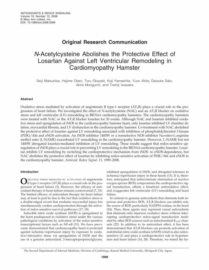
GSH/GSSG was significantly increased by treatment withNAC but not with losartan in the BIOFIB hamster heart (Fig.1A). GSH/GSSG was significantly lower in the BIO14.6 ham-ster heart compared to the BIOFIB hamster heart. Treatmentwith NAC or losartan significantly inhibited the decrease ofGSH/GSSG in the cardiomyopathy heart. In addition, the in-crease in MDA�HNE was also inhibited by treatment withNAC or losartan in the cardiomyopathy heart (Fig. 1B).
Effect of NAC and losartan on expression of iNOS andactivation of eNOS
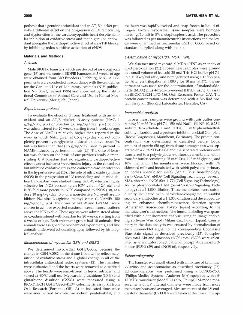
iNOS expression was increased in the BIO14.6 hamsterheart compared to the BIOFIB hamster heart at 26 weeks ofage (Fig. 2). Treatment with NAC or losartan had no effecton iNOS expression in the BIOFIB hamster heart, but inhib-ited it in the cardiomyopathy hamster heart.
N-ACETYLCYSTEINE AND LOSARTAN IN HEART FAILURE 2001
FIG. 1. Actions of N-acetylcysteine and losartan. (A) Theeffect of N-acetylcysteine (NAC) and losartan on myocardialglutathione (GSH) and glutathione disulfide (GSSG). (B) Theeffect of NAC and losartan on myocardial malondialdehyde(MDA) plus 4-hydroxy-noneal (HNE) content. Each bar rep-resents mean � SE of five experiments. *p � 0.05 comparedto the nontreated BIOFIB hamster (FIB-control); #p � 0.05compared to the nontreated BIO14.6 hamster (14.6-control).
FIG. 2. The effect of N-acetylcysteine (NAC) and losartanon iNOS expression in the heart. Upper panels are the rep-resentative immunohistochemistry images for iNOS. Lowerpanels are the representative immunoblot images for iNOS.Bars represent the data of quantitative analysis for iNOS.Each bar represents mean � SE of five experiments. *p � 0.05compared to the nontreated BIOFIB hamster (FIB-control),#p � 0.05 compared to the nontreated BIO14.6 hamster (14.6-control). (For interpretation of the references to color in thisfigure legend, the reader is referred to the web version ofthis article at www.liebertonline.com/ars).
Effect of NAC and losartan on activation ofphosphatidylinositol 3-kinase and eNOS
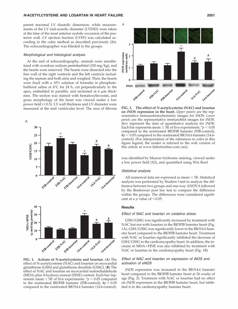
Treatment with NAC had no significant effect on the ex-pression of total Akt and phospho-Akt and phospho-Akt/to-tal Akt in the BIOFIB and the BIO14.6 cardiomyopathy ham-ster heart (Fig. 3A). However, losartan increased theexpression of phospho-Akt and phospho-Akt/total Akt inthe BIOFIB and the cardiomyopathy hearts, although themagnitude of an increase in phospho-Akt/total Akt wasgreater in the cardiomyopathy heart. NAC inhibited the in-crease in phospho-Akt/total Akt induced by losartan in theBIOFIB and the BIO14.6 hamster hearts.
Treatment with NAC or losartan had no significant effecton the expression of total eNOS and phospho-eNOS andphospho-eNOS/total eNOS in the BIOFIB heart (Fig. 3B). Al-though there was no difference in total eNOS expression be-tween the NAC-treated and the losartan-treated cardiomy-opathy heart, losartan but not NAC increased the expressionof phospho-eNOS and phospho-eNOS/total eNOS in thecardiomyopathy heart. However, NAC inhibited the in-
crease in phospho-eNOS/total eNOS induced by losartan inthe BIO14.6 hamster heart.
Effects of NAC and losartan on gross morphology of theheart, heart weight/body weight, and myocardial fibrosis
Gross morphology of the BIO14.6 hamster heart at 26weeks of age showed dilation of the LV chamber and thin-ning of the LV wall compared with BIOF1B hamster heart atthe same age associated with an increase in heartweight/body weight (Fig. 4A–C). Treatment of the BIOFIBhamster with NAC or losartan had no significant effect ongross morphology of the heart and heart weight/bodyweight. Treatment with NAC had no significant effect on di-lation of the LV chamber, thinning of the LV wall, and heartweight/body weight in the cardiomyopathy hamster heart.In contrast, treatment with losartan ameliorated dilation ofthe LV chamber and thinning of the LV wall and decreasedheart weight/body weight in the cardiomyopathy hamsterheart. Co-treatment with NAC and losartan abrogated losar-tan-induced amelioration of LV chamber dilation and LV
MATSUHISA ET AL.2002
FIG. 3. (A) The effect of N-acetylcysteine(NAC) and losartan on phosphorylation of Akt.The lower panel shows representative immunoblotimages for phospho-Akt (p-Akt) and total Akt.Upper bars represent the data of quantitative anal-ysis for p-Akt/total Akt. (B) The effect of NACand losartan on phosphorylation of eNOS. Thelower panel shows representative immunoblot im-ages for phospho-eNOS (p-eNOS) and totaleNOS. Upper bars represent the data of quantita-tive analysis for p-eNOS/total eNOS. Each barrepresents mean � SE of five experiments. *p �0.05 compared to the nontreated BIOFIB hamster(FIB-control); #p � 0.05 compared to the non-treated BIO14.6 hamster (14.6-control); §p � 0.05compared to the losartan-treated BIOFIB hamster;¶p � 0.05 compared to the losartan-treatedBIO14.6 hamster.
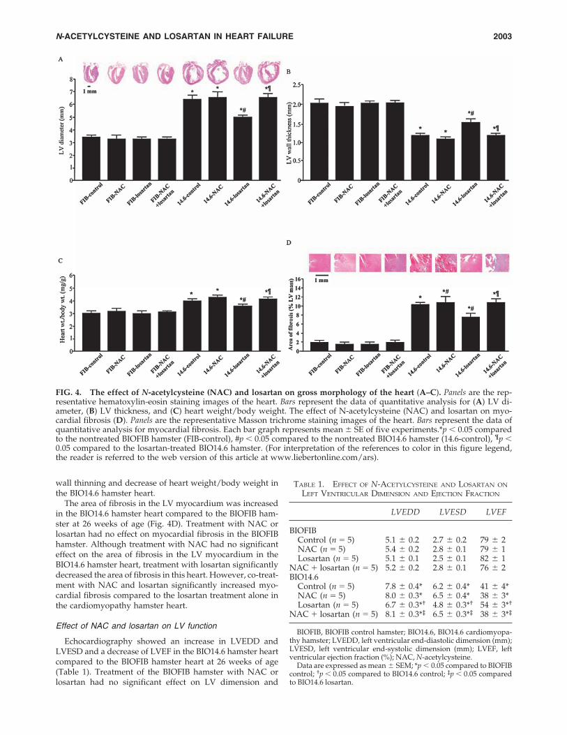
wall thinning and decrease of heart weight/body weight inthe BIO14.6 hamster heart.
The area of fibrosis in the LV myocardium was increasedin the BIO14.6 hamster heart compared to the BIOFIB ham-ster at 26 weeks of age (Fig. 4D). Treatment with NAC orlosartan had no effect on myocardial fibrosis in the BIOFIBhamster. Although treatment with NAC had no significanteffect on the area of fibrosis in the LV myocardium in theBIO14.6 hamster heart, treatment with losartan significantlydecreased the area of fibrosis in this heart. However, co-treat-ment with NAC and losartan significantly increased myo-cardial fibrosis compared to the losartan treatment alone inthe cardiomyopathy hamster heart.
Effect of NAC and losartan on LV function
Echocardiography showed an increase in LVEDD andLVESD and a decrease of LVEF in the BIO14.6 hamster heartcompared to the BIOFIB hamster heart at 26 weeks of age(Table 1). Treatment of the BIOFIB hamster with NAC orlosartan had no significant effect on LV dimension and
N-ACETYLCYSTEINE AND LOSARTAN IN HEART FAILURE 2003
FIG. 4. The effect of N-acetylcysteine (NAC) and losartan on gross morphology of the heart (A–C). Panels are the rep-resentative hematoxylin-eosin staining images of the heart. Bars represent the data of quantitative analysis for (A) LV di-ameter, (B) LV thickness, and (C) heart weight/body weight. The effect of N-acetylcysteine (NAC) and losartan on myo-cardial fibrosis (D). Panels are the representative Masson trichrome staining images of the heart. Bars represent the data ofquantitative analysis for myocardial fibrosis. Each bar graph represents mean � SE of five experiments.*p � 0.05 comparedto the nontreated BIOFIB hamster (FIB-control), #p � 0.05 compared to the nontreated BIO14.6 hamster (14.6-control), ¶p �0.05 compared to the losartan-treated BIO14.6 hamster. (For interpretation of the references to color in this figure legend,the reader is referred to the web version of this article at www.liebertonline.com/ars).
TABLE 1. EFFECT OF N-ACETYLCYSTEINE AND LOSARTAN ON
LEFT VENTRICULAR DIMENSION AND EJECTION FRACTION
LVEDD LVESD LVEF
BIOFIBControl (n � 5) 5.1 � 0.2 2.7 � 0.2 79 � 2NAC (n � 5) 5.4 � 0.2 2.8 � 0.1 79 � 1Losartan (n � 5) 5.1 � 0.1 2.5 � 0.1 82 � 1
NAC � losartan (n � 5) 5.2 � 0.2 2.8 � 0.1 76 � 2BIO14.6
Control (n � 5) 7.8 � 0.4* 6.2 � 0.4* 41 � 4*NAC (n � 5) 8.0 � 0.3* 6.5 � 0.4* 38 � 3*Losartan (n � 5) 6.7 � 0.3*† 4.8 � 0.3*† 54 � 3*†
NAC � losartan (n � 5) 8.1 � 0.3*‡ 6.5 � 0.3*‡ 38 � 3*‡
BIOFIB, BIOFIB control hamster; BIO14.6, BIO14.6 cardiomyopa-thy hamster; LVEDD, left ventricular end-diastolic dimension (mm);LVESD, left ventricular end-systolic dimension (mm); LVEF, leftventricular ejection fraction (%); NAC, N-acetylcysteine.
Data are expressed as mean � SEM; *p � 0.05 compared to BIOFIBcontrol; †p � 0.05 compared to BIO14.6 control; ‡p � 0.05 comparedto BIO14.6 losartan.

LVEF. Although treatment with NAC had no significant ef-fect on LV dimension and LVEF, treatment with losartan sig-nificantly decreased LVEDD and LVESD and increasedLVEF in the cardiomyopathy hamster heart. However, co-treatment with NAC and losartan significantly decreasedLVEF compared to the losartan treatment alone in the car-diomyopathy hamster heart.
Effects of 1400W and L-NAME on losartan-inducedreduction of LV chamber dilation, heart weight/bodyweight, and myocardial fibrosis
Treatment with 1400W had no significant effect on LV di-ameter, LV wall thickness, and heart weight/body weight inthe BIOFIB hamster, while the same treatment aggravatedLV chamber dilation and LV wall thinning and increasedheart weight/body weight in the BIO14.6 cardiomyopathyhamster (Figs. 5A–5C). Treatment with L-NAME tended to
increase LV wall thickness and heart weight/body weightin the BIOFIB hamster heart, but significantly decreased LVwall thickness and increased LV diameter and heartweight/body weight in the cardiomyopathy hamster. Treat-ment with 1400W or L-NAME aggravated LV chamber dilation and LV wall thinning and increased heartweight/body weight in the BIO14.6 hamster. However, co-treatment with 1400W did not prevent losartan-inducedamelioration of LV chamber dilation and LV wall thinningand decrease of heart weight/body weight in the cardiomy-opathy hamster. In contrast, co-treatment with L-NAME reversed losartan-induced amelioration of LV chamber dila-tion and LV wall thinning and decrease of heart weight/body weight in the cardiomyopathy hamster.
Treatment with 1400W had no significant effect on myo-cardial fibrosis in the BIOFIB hamster heart, while the sametreatment significantly increased myocardial fibrosis in theBIO14.6 cardiomyopathy hamster heart (Fig. 5D). Treatment
MATSUHISA ET AL.2004
FIG. 5. The effect of 1400W and N�-nitro-L-arginine methyl ester (NAME) on losartan-mediated modulation of grossmorphology of the heart (A–C). Panels are the representative hematoxylin-eosin staining images of the heart. Bars repre-sent the data of quantitative analysis for (A) LV diameter, (B) LV thickness, and (C) heart weight/body weight. The effectof 1400W and N�-nitro-L-arginine methyl ester (NAME) on losartan-mediated modulation of myocardial fibrosis (D). Pan-els are the representative Masson trichrome staining images of the heart. Bars represent the data of quantitative analysis formyocardial fibrosis. Each bar represents mean � SE of 5 experiments.*p � 0.05 compared to the nontreated BIOFIB ham-ster (FIB-control), #p � 0.05 compared to the nontreated BIO14.6 hamster (14.6-control), ¶p � 0.05 compared to the losartan-treated BIO14.6 hamster. (For interpretation of the references to color in this figure legend, the reader is referred to the webversion of this article at www.liebertonline.com/ars).
with L-NAME tended to increase myocardial fibrosis in theBIOFIB hamster heart and significantly increased myocar-dial fibrosis in the cardiomyopathy hamster heart. Co-treat-ment with 1400W did not prevent losartan-induced inhibi-tion of myocardial fibrosis in the cardiomyopathy hamsterheart. In contrast, co-treatment with L-NAME and losartansignificantly increased myocardial fibrosis compared to thelosartan treatment alone in the cardiomyopathy hamsterheart.
Effect of 1400W and L-NAME on losartan-inducedimprovement of LV function
Treatment with 1400W had no significant effect on LV di-mension and LVEF in the BIOFIB hamster, while 1400W sig-nificantly increased LVESD and decreased LVEF in theBIO14.6 cardiomyopathy hamster heart (Table 2). Treatmentwith L-NAME tended to increase LVEDD and LVESD anddecrease LVEF in the BIOFIB hamster and significantly in-creased LVEDD and LVESD and decreased LVEF in theBIO14.6 hamster. Co-treatment with 1400W did not preventlosartan-induced reduction of LVEDD and LVESD and im-provement of LVEF in the cardiomyopathy hamster. In con-trast, co-treatment with L-NAME and losartan significantlyincreased LVEDD and LVESD and decreased LVEF com-pared to the losartan treatment alone in the cardiomyopathyhamster.
Discussion
The salient findings of the present study were: (a) oxida-tive stress was increased in the BIO14.6 cardiomyopathyhamster heart associated with enhanced expression of iNOS;(b) chronic treatment with NAC and losartan equally pre-vented oxidative stress and expression of iNOS but onlylosartan conferred protection against LV remodeling in thecardiomyopathy hamster heart; (c) activation of eNOS wasinduced by treatment with losartan but not with NAC, andNAC inhibited losartan-induced activation of eNOS and pro-
tection against LV remodeling in the cardiomyopathy ham-ster heart; (d) 1400W and L-NAME aggravated LV remod-eling in the cardiomyopathy hamster heart; (e) L-NAME butnot 1400W abrogated losartan-induced inhibition of LV re-modeling in the cardiomyopathy hamster heart. These re-sults are consistent with the hypothesis that oxidative stresspromotes upregulation of iNOS that plays a crucial role ininhibiting LV remodeling in the BIO14.6 cardiomyopathyhamster heart. Therefore, it is conceivable that no protectiveeffect of NAC against LV remodeling is a result of inhibitionof iNOS upregulation despite a possible beneficial effect onLV remodeling via inhibition of oxidative stress. On the con-trary, it is suggested that similar inhibition of oxidative stressand upregulation of iNOS by losartan confers protectionagainst LV remodeling by switching the cardioprotectivemechanism from iNOS- to eNOS-dependent. Moreover, it isnoteworthy that the antioxidant may abrogate the AT1Rblocker-mediated cardioprotection presumably through in-hibition of eNOS activation.
AT1R-mediated oxidative stress is known to play a detri-mental role in the progression of heart failure. Whaley–Con-nell and associates (32) demonstrated that the AT1R blockervalsartan was capable of inhibiting oxidative stress in theheart associated with amelioration of cardiac remodeling andLV dysfunction in a rodent model of chronically elevated tis-sue levels of angiotensin II. It has also been demonstratedthat angiotensin II-mediated oxidative stress and inflamma-tion mediate the age-dependent cardiomyopathy in an-giotensin converting enzyme 2 null mice (19). Accordingly,prevention of AT1R-mediated oxidative stress protects a va-riety of cellular constituents from degeneration and inacti-vation and promotes the cell survival under a variety ofpathological environments. On the other hand, Das and associates (4) demonstrated that pre-ischemic treatment withangiotensin II exerts cardioprotection against ischemia/reperfusion injury in the isolated and perfused rat heart andsuch a cardioprotective effect was reversed by co-treatmentwith a NADPH oxidase inhibitor apocynin or NAC, indi-
N-ACETYLCYSTEINE AND LOSARTAN IN HEART FAILURE 2005
TABLE 2. EFFECT OF 1400W AND L-NAME ON LOSARTAN-MEDIATED MODULATION
OF LEFT VENTRICULAR DIMENSION AND EJECTION FRACTION
LVEDD LVESD LVEF
BIOFIBControl (n � 5) 5.1 � 0.2 2.7 � 0.2 79 � 21400W (n � 5) 5.2 � 0.1 2.5 � 0.1 81 � 3NAME (n � 5) 5.5 � 0.1 3.2 � 0.1 73 � 2
NAC � losartan (n � 5)BIO14.6
Control (n � 5) 7.8 � 0.4* 6.2 � 0.4* 41 � 4*1400W (n � 5) 8.8 � 0.4* 7.6 � 0.4*† 27 � 2*†
NAME (n � 5) 9.2 � 0.4*† 8.2 � 0.3*† 23 � 3*†
Losartan (n � 5) 6.7 � 0.4*† 4.8 � 0.3*† 54 � 3*†
Losartan � 1400W (n � 5) 6.6 � 0.4*† 4.7 � 0.4*† 54 � 4*†
Losartan � NAME (n � 5) 8.8 � 0.4*‡ 7.8 � 0.5*†‡ 24 � 3*†‡
BIOFIB, BIOFIB control hamster; BIO14.6, BIO14.6 cardiomyopathy hamster; LVEDD, left ven-tricular end-diastolic dimension (mm); LVESD, left ventricular end-systolic dimension (mm);LVEF, left ventricular ejection fraction (%); NAC, N-acetylcysteine.
Data are expressed as mean � SEM; *p � 0.05 compared to BIOFIB control; †p � 0.05 compared to BIO14.6 control; ‡p � 0.05 compared to BIO14.6 losartan.
cating that NADPH oxidase-derived ROS mediates an-giotensin II-induced cardioprotection against ischemia/reperfusion injury. The finding that AT1R-mediated oxida-tive stress confers cardioprotection is consistent with the hy-pothesis that ROS and oxidants can function as intracellularsignaling molecules that convert a death signal into a sur-vival signal (5). Such redox signaling acutely promotes acti-vation of survival kinases known as reperfusion injury sal-vage kinase [i.e., PI3K/Akt and p42/p44 extracellularsignal-regulated kinase cascades (11)]. The same oxidativestress can chronically confer cardioprotection through acti-vation of redox-sensitive transcriptional factors such as nu-clear factor-kappa B, thereby promoting upregulation ofiNOS which has consistently been implicated in the mecha-nism of late cardioprotection by ischemic preconditioning(2). Indeed, our previous study demonstrated that theBIO14.6 cardiomyopathy hamster hearts were markedly tol-erant to ischemia/reperfusion injury through the redox-sen-sitive activation of iNOS (13). The present study demon-strating that treatment with 1400W or L-NAME aggravatedLV remodeling in the cardiomyopathy hamster also pointsto the conclusion that iNOS is a mediator of protectionagainst LV remodeling in the cardiomyopathy hamster. Inaddition, the fact that losartan inhibited oxidative stress andupregulation of iNOS but improved LV remodeling in thecardiomyopathy hamster heart suggests that generally ob-served amelioration of LV remodeling and heart failure byAT1R blockers can not simply be explained by inhibition ofoxidative stress but is also attributed to additional mecha-nisms.
The results of the present study suggest that the salu-tary effect of losartan on LV remodeling in the BIO14.6 car-diomyopathy hamster heart is mediated by activation ofeNOS. AT1R blockers can exert cardioprotection throughmultiple mechanisms. It decreases blood pressure, therebymitigating ventricular wall stress. Contribution of lower-ing blood pressure by treatment with losartan to the pre-vention of heart failure is unlikely, because it has been reported that treatment with losartan or NAC equally decreased blood pressure in the cardiomyopathy ham-ster (8). On the other hand, bradykinin-dependent and –independent mechanisms have been implicated in AT1Rblocker-mediated cardioprotection against ischemia/reperfusion injury (27). Activation of eNOS has been sug-gested as a downstream event of these mechanisms. Ourstudy demonstrating that activation of eNOS and preven-tion of LV remodeling by treatment with losartan wasblocked by co-treatment with L-NAME but not with 1400Wsuggests that eNOS is a mediator of cardioprotection bylosartan in the cardiomyopathy hamster heart. However,because we did not study the role of nNOS which is knownto exist in the heart and is inhibited by L-NAME, the con-tribution of nNOS to losartan-mediated amelioration ofheart failure can not be ignored.
The finding that NAC reverses the redox-sensitive acti-vation of eNOS and the cardioprotective effect of losartanunderscores the need for site-specific antioxidant therapy.The redox-sensitive nature of eNOS activation has beendemonstrated by Sun and associates (30) who showed thatexposure of bovine aortic endothelial cells to 2,4,6-trini-trotoluene was capable of phosphorylating and activatingeNOS through the generation of ROS. A major source of
ROS in endothelial cells is the NADPH oxidase enzymecomplex that is activated in specific membrane rafts specif-ically in caveolae in response to a variety of humoral fac-tors (34). NADPH-derived ROS are thought to be gener-ated in the proximity of the PI3K/Akt signaling complex,which is responsible for activation of eNOS (20, 30). ThePI3K/Akt axis is activated not only by various growth fac-tors and cytokines but also by G-protein-coupled receptoragonists such as bradykinin via transactivation of receptortyrosine kinase or activation of non-receptor tyrosine ki-nase such as Src in a ROS-dependent manner (17). Indeed,the present study demonstrated that losartan activatedPI3K in a NAC-sensitive manner, suggesting that losartanpromotes redox-sensitive activation of the PI3K/Akt axis.Although the source of ROS generated by treatment withlosartan remains to be investigated, maintaining ROS gen-eration in a specific cellular compartment would be es-sential to preserve the ability of losartan to activate PI3Kand eNOS. Therefore, indiscriminate elimination of ROSby treatment with NAC may abolish this redox-sensitivesignaling for losartan-induced activation of PI3K andeNOS. In line with this notion, antioxidant medicine mustbe site-specific, being targeted to a specific ROS or cellu-lar compartment, without a deleterious effect on other re-dox-sensitive signaling pathways necessary for cardiopro-tection.
In conclusions, redox-sensitive upregulation of iNOSplays a crucial role in preventing LV remodeling and heartfailure in the BIO14.6 cardiomyopathy hamster heart. Losar-tan inhibits LV remodeling by switching the cardioprotec-tive mechanism from iNOS- to eNOS-dependent but NACabolishes the protective effect of losartan by inhibiting re-dox-sensitive activation of PI3K and eNOS in the cardiomy-opathy hamster.
Acknowledgments
This work was supported in part by Research Grant16591420 from the Ministry of Education, Science, and Cul-ture of Japan and Promotion and Mutual Aid Corporationfor Private Schools of Japan.
Abbreviations
AT1R, angiotensin II type-1 receptor; eNOS, endothelialnitric oxide synthase; GSH, glutathione; GSSG, glutathionedisulfide; HNE, 4-hydroxy-noneal; iNOS, inducible nitric ox-ide synthase; L-NAME, N�-nitro-L-arginine methyl ester;LV, left ventricular; LVEDD, LV end-diastolic diameter;LVEF, LV ejection fraction; LVESD, LV end-systolic diame-ter; MDA, malondialdehyde; NAC, N-acetylcysteine; NOS,nitric oxide synthase; PI3K, phosphatidylinositol 3-kinase;ROS, reactive oxygen species.
References
1. Anselm E, Chataigneau M, Ndiaye M, Chataigneau T, Schini–Kerth VB. Grape juice causes endothelium-de-pendent relaxation via a redox-sensitive Src- and Akt-de-pendent activation of eNOS. Cardiovasc Res 73: 404–413,2007.
2. Bolli R. Cardioprotective function of inducible nitric oxidesynthase and role of nitric oxide in myocardial ischemia and
MATSUHISA ET AL.2006
preconditioning: an overview of a decade of research. J MolCell Cardiol 33: 1897–1918, 2001.
3. Colucci WS. Molecular and cellular mechanisms of myocar-dial failure. Am J Cardiol 80: 15L–25L, 1997.
4. Das M, Das S, and Das DK. Caveolin and MAP kinase in-teraction in angiotensin II preconditioning of the my-ocardium. J Cell Mol Med 11: 788–797, 2007.
5. Das DK, Maulik N, and Engelman RM. Redox regulation ofangiotensin II signaling in the heart. J Cell Mol Med 8:144–152, 2004.
6. Dimmeler S, Fleming I, Fisslthaler B, Hermann C, Busse R,and Zeiher AM. Activation of nitric oxide synthase in en-dothelial cells by Akt-dependent phosphorylation. Nature399: 601–605, 1999.
7. Dresdale AR, Barr LH, Bonow RO, Mathisen DJ, Myers CE,Schwartz DE, d’Angelo T, and Rosenberg SA. Prospectiverandomized study of the role of N-acetyl cysteine in re-versing doxorubicin-induced cardiomyopathy. Am J ClinOncol 5: 657–663, 1982.
8. Escobales N and Crespo MJ. Angiotensin II-dependent vas-cular alterations in young cardiomyopathic hamsters: rolefor oxidative stress. Vascul Pharmacol 44: 22–28. 2006.
9. Fiordaliso F, Bianchi R, Staszewsky L, Cuccovillo I, DoniM, Laragione T, Salio M, Savino C, Melucci S, SantangeloF, Scanziani E, Masson S, Ghezzi P, and Latini R. Antiox-idant treatment attenuates hyperglycemia-induced car-diomyocyte death in rats. J Mol Cell Cardiol 37:959–968,2004.
10. Garvey EP, Oplinger JA, Furfine ES, Kiff RJ, Laszlo F,Whittle BJ, and Knowles RG. 1400W is a slow, tight bind-ing, and highly selective inhibitor of inducible nitric-ox-ide synthase in vitro and in vivo. J Biol Chem 272:4959–4963, 1997.
11. Hausenloy DJ and Yellon DM. New directions for protect-ing the heart against ischaemia-reperfusion injury: targetingthe Reperfusion Injury Salvage Kinase (RISK)-pathway. Car-diovasc Res 61: 448–460, 2004.
12. Kehrer JP and Lund LG. Cellular reducing equivalents andoxidative stress. Free Radic Biol Med 17: 65–75, 1994.
13. Kyoi S, Otani H, Matsuhisa S, Akita Y, Enoki C, Tatsumi K,Hattori R, Imamura H, Kamihata H, and Iwasaka T. Role ofoxidative/nitrosative stress in the tolerance to ischemia/reperfusion injury in cardiomyopathic hamster heart. An-tioxid Redox Signal 8: 1351–1361, 2006.
14. Massion PB and Balligand JL. Relevance of nitric oxide formyocardial remodeling. Curr Heart Fail Rep 4: 18–25, 2007.
15. Matsuhisa S, Otani H, Okazaki T, Yamashita K, Akita Y, SatoD, Moriguchi A, Imamura H, and Iwasaka T. AngiotensinII type-1 receptor blocker preserves tolerance to isch-emia/reperfusion injury in Dahl salt-sensitive rat heart. AmJ Physiol Heart Circ Physiol, in press.
16. Nigro V, Okazaki Y, Belsito A, Piluso G, Matsuda Y, Poli-tano L, Nigro G, Ventura C, Abbondanza C, Molinari AM,Acampora D, Nishimura M, Hayashizaki Y, and Puca GA.Identification of the Syrian hamster cardiomyopathy gene.Hum Mol Genet 6: 601–607, 1997.
17. Otani H. Reactive oxygen species as mediators of signaltransduction in ischemic preconditioning. Antioxid RedoxSignal 6: 449–469, 2004.
18. Otani H. Ischemic preconditioning: from molecular mecha-nisms to therapeutic opportunities. Antioxid Redox Signal 10:207–48, 2008.
19. Oudit GY, Kassiri Z, Patel MP, Chappell M, Butany J, BackxPH, Tsushima RG, Scholey JW, Khokha R, and PenningerJM. Angiotensin II-mediated oxidative stress and inflam-
mation mediate the age-dependent cardiomyopathy inACE2 null mice. Cardiovasc Res 75: 29–39, 2007.
20. Paravicini TM, Miller AA, Drummond GR, and Sobey CG.Flow-induced cerebral vasodilatation in vivo involves acti-vation of phosphatidylinositol-3 kinase, NADPH-oxidase,and nitric oxide synthase. J Cereb Blood Flow Metab 26:836–845, 2006.
21. Parola M, Bellomo G, Robino G, Barrera G, and DianzaniMU. 4-Hydroxynonenal as a biological signal: molecular ba-sis and pathophysiological implications. Antioxid Redox Sig-nal 1: 255–284, 1999.
22. Penna C, Mancardi D, Rastaldo R, Losano G, and PagliaroP. Intermittent activation of bradykinin B2 receptors and mitochondrial KATP channels trigger cardiac post-conditioning through redox signaling. Cardiovasc Res 75:168–177, 2007.
23. Ping P, Zhang J, Qiu Y, Tang XL, Manchikalapudi S, Cao X,and Bolli R. Ischemic preconditioning induces selectivetranslocation of protein kinase C isoforms epsilon and etain the heart of conscious rabbits without subcellular redis-tribution of total protein kinase C activity. Circ Res 81:404–414, 1997.
24. Privratsky JR, Wold LE, Sowers JR, Quinn MT, and Ren J.AT1 blockade prevents glucose-induced cardiac dysfunctionin ventricular myocytes: role of the AT1 receptor andNADPH oxidase. Hypertension 42: 206–212, 2003.
25. Rauchová H, Pechánová O, Kunes J, Vokurková M,Dobesová Z, and Zicha J. Chronic N-acetylcysteine ad-ministration prevents development of hypertension inN(omega)-nitro-L-arginine methyl ester-treated rats: therole of reactive oxygen species. Hypertens Res 28:475–482,2005.
26. Sadoshima J, Montagne O, Wang Q, Wang G, Warden J, LiuJ, Takagi G, Karoor V, Hong C, Johnson GL, Vatner DE, andVatner SF. The MEKK1-JNK pathway plays a protective rolein pressure overload but does not mediate cardiac hyper-trophy. J Clin Invest 110: 271–279, 2002.
27. Sato M, Engelman RM, Otani H, Maulik N, Rousou JA, FlackJE 3rd, Deaton DW, and Das DK. Myocardial protection bypreconditioning of heart with losartan, an angiotensin IItype 1-receptor blocker: implication of bradykinin-depen-dent and bradykinin-independent mechanisms. Circulation102: III346–III351, 2000.
28. Savoia C, Ebrahimian T, He Y, Gratton JP, Schiffrin EL, andTouyz RM. Angiotensin II/AT2 receptor-induced vasodila-tion in stroke-prone spontaneously hypertensive rats in-volves nitric oxide and cGMP-dependent protein kinase. JHypertens 24: 2417–2422, 2006.
29. Stephens L, Anderson K, Stokoe D, Erdjument-Bromage H,Painter GF, Holmes AB, Gaffney PR, Reese CB, McCormickF, Tempst P, Coadwell J, and Hawkins PT. Protein kinase Bkinases that mediate phosphatidylinositol 3,4,5-trisphos-phate-dependent activation of protein kinase B. Science 279:710–714, 1998.
30. Sun Y, Sumi D, and Kumagai Y. Serine 1179 phosphoryla-tion of endothelial nitric oxide synthase caused by 2,4,6-trini-trotoluene through PI3K/Akt signaling in endothelial cells.Toxicol Appl Pharmacol 214: 55–60, 2006.
31. Theuer J, Dechend R, Muller DN, Park JK, Fiebeler A,Barta P, Ganten D, Haller H, Dietz R, and Luft FC. An-giotensin II induced inflammation in the kidney and in theheart of double transgenic rats. BMC Cardiovasc Disord 2:1–12, 2002.
32. Whaley–Connell A, Govindarajan G, Habibi J, HaydenMR, Cooper SA, Wei Y, Ma L, Qazi M, Link D, Karuparthi
N-ACETYLCYSTEINE AND LOSARTAN IN HEART FAILURE 2007
PR, Stump C, Ferrario C, and Sowers JR. Angiotensin II-mediated oxidative stress promotes myocardial tissueremodeling in the transgenic (mRen2) 27 Ren2 rat. Am J Physiol Endocrinol Metab 2007 293: E355–E363, 2007.
33. Williams IA and Allen DG. The role of reactive oxygenspecies in the hearts of dystrophin-deficient mdx mice. AmJ Physiol Heart Circ Physiol 293: H1969–H1977, 2007.
34. Yang B and Rizzo V. TNF-alpha potentiates protein-tyrosinenitration through activation of NADPH oxidase and eNOSlocalized in membrane rafts and caveolae of bovine aorticendothelial cells. Am J Physiol Heart Circ Physiol 292:H954–H962, 2007.
Address reprint requests to:Hajime Otani, M.D.
The Second Department of Internal MedicineDivision of Cardiology, Kansai Medical University
10-15 Fumizono-choMoriguchi City
570-8507, Japan
E-mail: [email protected]
Date of first submission to ARS Central, March 6, 2008; dateof final revised submission, April 15, 2008; date of accep-tance, May 13, 2008.
MATSUHISA ET AL.2008