nad + metabolism and oxidative stress: the golden nucleotide on a crown...
TRANSCRIPT

Review article
NAD+ metabolism and oxidative stress: thegolden nucleotide on a crown of thornsHassina Massudi1,2, Ross Grant1,2, Gilles J Guillemin1,3, Nady Braidy1,4
1Department of Pharmacology, School of Medical Sciences, University of New South Wales, Faculty ofMedicine, Sydney, Australia, 2Australasian Research Institute, Sydney Adventist Hospital, Sydney, Australia,3St Vincent’s Centre for Applied Medical Research, Sydney, Australia, 4School of Psychiatry, University of NewSouth Wales, Faculty of Medicine, Sydney, Australia
In the twentieth century, NAD+ research generated multiple discoveries. Identification of the important role ofNAD+ as a cofactor in cellular respiration and energy production was followed by discoveries of numerousNAD+ biosynthesis pathways. In recent years, NAD+ has been shown to play a unique role in DNA repairand protein deacetylation. As discussed in this review, there are close interactions between oxidativestress and immune activation, energy metabolism, and cell viability in neurodegenerative disorders andageing. Profound interactions with regard to oxidative stress and NAD+ have been highlighted in thepresent work. This review emphasizes the pivotal role of NAD+ in the regulation of DNA repair, stressresistance, and cell death, suggesting that NAD+ synthesis through the kynurenine pathway and/orsalvage pathway is an attractive target for therapeutic intervention in age-associated degenerativedisorders. NAD+ precursors have been shown to slow down ageing and extend lifespan in yeasts, andprotect severed axons from degeneration in animal models neurodegenerative diseases.
Keywords: Sirtuins, Oxidative stress, NAD+, PARP, Ageing
IntroductionCurrent thinking regarding the importance of NAD(including NAD+ and NADH) metabolism in healthand disease stems from the original discovery thatniacins were efficacious for the treatment of pellagra.1
In 1937, Elvehjem2 showed that nicotinic acid (NA)and nicotinamide (NM) (the breakdown product ofNAD+) were effective agents for both the treatmentand prevention of black tongue in dogs, and thehuman equivalent, pellagra. While the disappearanceof pellagra in the developed world reduced interest inthis life-threatening deficiency disease, a recent obser-vation that several disorders mimic pellagra in its rangeof symptoms (e.g. dermatitis, diarrhoea, dementia, anddeath) has re-ignited considerable interest on the mech-anism of action of NAD+ and its key metabolites.3–7
Intriguingly, with the understanding of the pharma-cological role of niacins was the almost simultaneousdiscovery of the structure and function of NAD+ byfour Nobel laureates.8 In 1904, Sir Arthur Harden sep-arated Buchner’s yeast juice into a high- and low-mol-ecular-weight fraction, neither of which could undergo
fermentation individually. However, recombination ofboth fractions allowed fermentation to take place.Harden inferred the existence of high-molecular-weight ferment (enzyme) and a low-molecular-weightcoferment or ‘cozymase’.8
The involvement of cozymase in fermentation, res-piration, and glycolysis was identified in a variety oforganisms in subsequent years. However, due to thelow-molecular-weight nature of cozymase, it was diffi-cult to isolate.8 It was Hans von Euler-Chelpin9 whoeventually succeeded in isolating cozymase fromyeast extracts in the late 1920s. He determined thedinucleotide structure of NAD+ and two other mono-nucleotides, adenosine monophosphate (AMP) andnicotinamide mononucleotide (NMN).9 The centralredox function of NAD+ was determined by OttoWarburg and Christian in the mid-1930s.10 He ident-ified the capability of NAD+ to transfer hydrogenfrom one molecule to another.10 Since then, ongoinginvestigations have been directed towards the identifi-cation of the enzymatic pathways involved in its syn-thesis and metabolism.11 In 1954, Arthur Kornberg12
discovered the NAD phosphorylase reaction, thecrucial step of NAD+ synthesis. He detected the enzy-matic activity in yeast extracts which catalysed the
Correspondence to: Nady Braidy, School of Psychiatry, Faculty ofMedicine, University of New South Wales, Sydney 2052, Australia.Email: [email protected]
© W.S. Maney & Son Ltd 2012DOI 10.1179/1351000212Y.0000000001 Redox Report 2012 VOL. 17 NO. 128

condensation of ATP with NMN to form NAD.12
While this reaction is catalysed by nicotinamidemononucleotide adenylyltransferase (NMNAT) [EC2.7.7.1], it took another 55 years for the primary struc-ture of this enzyme to be discovered.13
Another important milestone was the discovery ofthe NAD+-consuming activity associated with thetransfer of ADP-ribosyl moieties to protein acceptorswhich occurred in the 1960s.14 In the same period,the term ‘pyridine nucleotide cycle’ was introducedto define several enzymatic reactions involved in thebiosynthesis and catabolism of NAD+, although thesignificance of NAD+ metabolism remainedunclear.15 Rechsteiner and Catanzarite (1974)16
showed that NAD+ turnover was strongly suppressedin enucleated yeast cells, suggesting that the nucleusis the major compartment responsible for the anabo-lism and breakdown of NAD+. Since then, studieson the enzymology of mammalian NAD+ metabolismhave increased in line with those investigating thepotential involvement of NAD-related metabolites incellular physiology.11,17,18 The involvement of NAD+
in several key cellular processes has suggested tosome the possibility that NAD+ metabolism may bean attractive therapeutic target.19,20
Changes in NAD+metabolism have been associatedwith several pathologies, including neurodegenerativediseases, cancer, cardiovascular disease, and normalageing. A number of comprehensive papers haveappeared in the literature, which provide a generalizedoverview of the information in this field.11,17,18,21
Beginning with an overview of relevant backgroundresearch by others, this review provides additionalinsight into the involvement of NAD+ metabolism inneurodegenerative disorders and ageing.
NAD+ biosynthesis and salvage pathwaysThe kynurenine pathway and de novo NAD+
synthesisIn mammals, the kynurenine pathway (KP) representsthe de novo synthesis of NAD+ from dietary trypto-phan (TRP) (Fig. 1). The KP begins with the oxidativecleavage of TRP to N-formylkynurenine by the twoenzymes tryptophan 2,3-dioxygenase (TDO) [EC1.13.1.2] and indoleamine 2,3-dioxygenase (IDO)[EC 1.13.11.17].22,23 Both IDO and TDO are haem-requiring enzymes and are considered rate limitingfor this pathway. TDO has been localized predomi-nantly in the liver, but has also been found in thebrain, and is induced by a number of factors includingTRP, glucocoticoids, hydrocortisone, and fasting.22,23
IDO, on the other hand, has been identified inseveral extrahepatic tissues including the brain, andis up-regulated by various cytokines and inflammatorymolecules such as lipopolysaccharides,24 amyloidpeptides,25 human immunodeficiency virus (HIV)
proteins,26 and tumour cells.27 TDO uses L-TRP exclu-sively as the substrate, whereas IDO can metabolizeL- and D-TRP as well as serotonin and other relatedindoleamines.The catabolite of TRP, formylkynurenine is then
hydrolysed to form kynurenine (KYN) by the actionof kynurenine formamidase (KFase) [EC 3.5.1.9].28
KYN can be transformed to kynurenic acid(KYNA) by the action of kynurenine-oxoglutaratetransaminase [EC 2.6.1.7].29 The remaining enzymesof the pathway include kynurenine 3-hydroxylase(K3H) [EC 1.14.13.9];29 and kynureninase [EC3.7.1.3].30 K3H can convert KYN to 3-hydroxykynur-enine (3-HK) by the hydroxylation of the aromaticring. Kynureninase produces anthranilic acid (AA)by the cleavage of the alanine side chain from KYN.AA can also undergo hydroxylation to 5- and 3-hydro-xyanthranilic acid (3-HAA) via non-specific micro-somal hydroxylating enzymes. Kynureninase is alsoinvolved in the formation of 3-HAA from 3-HK.Furthermore, 3-hydroxyanthranilic acid 3,4-dioxygen-ase [EC 1.13.11.6] converts 3-HAA to the unstableintermediate, α-amino-β-carboxymuconic-∑-semial-dehyde which cyclizes spontaneously to form quinoli-nic acid (QUIN). 3-HAA can also be converted topicolinic acid (PIC) by aminocarboxymuconate-semi-aldehyde decarboxylase [EC 4.1.1.45].31,32 Quinolinicacid phosphoribosyltransferase (QPRT) [EC 2.4.2.19]catalyses the formation of nicotinic acid mononucleo-tide (NAMN) using phosphoribosyl pyrophosphate(PRPP), which is then transformed spontaneously toNAD+.33
In recent years, the KP has generated considerableinterest following the observation that some intermedi-ates are endowed with neuroactive properties. Amongthem is the selective N-methyl-D-aspartate (NMDA)receptor agonist and free radical generator, QUIN;34
KYNA, a non-selective antagonist at both theNMDA site and the glycine strychnine-resistant siteof the same receptor, which can antagonize the cyto-toxic effects of QUIN;35,36 PIC, an endogenousmetal chelator;37 3-HK and 3-HAA, which can gener-ate free radicals.38,39 Alterations in the endogenouslevels of these metabolites has been implicated in thepathogenesis of several inflammatory brain diseases,including AD,40–42 AIDS dementia complex,43,44 cer-ebral malaria,45 amyotrophic lateral sclerosis,46
MS,47 Huntington’s disease,48,49 schizophrenia,50 andParkinson’s disease (PD).51 The inflammatory statecan induce IDO, mainly by interferon gamma(IFN-γ).52 IDO induction in immune-activated astro-cytes and microglia appears to be a primary site forTRP catabolism via the KP, leading to increased pro-duction of the neurotoxic TRP metabolites, whoseaccumulation in the brain has been implicated in thepathogenesis of the aforementioned disorders.51
Massudi et al. NAD+ metabolism and oxidative stress
Redox Report 2012 VOL. 17 NO. 1 29

Our group has identified a direct involvement of theKP in NAD+ synthesis in human brain cells, and thatreduced intracellular NAD+ synthesis leads to reducedactivity of NAD+-dependent histone deacetylase func-tion. We have shown that the KP metabolite andNMDA receptor agonist, QUIN is a substrate ofNAD+ synthesis in nanomolar concentrations inboth astrocytes and neurons but is cytotoxic athigher amounts in both cell types. These results alsoshow a clear neuroprotective effect of NMDA receptorantagonism and nitric oxide (NO) synthase inhibitionagainst QUIN-mediated neuronal and glial cytoxicity,strongly suggesting that QUIN-induced excitotoxicityin astrocytes and neurons is mediated by overactiva-tion of the NMDA receptor. Recently, we showedthat natural polyphenolic compounds can attenuateQUIN-mediated neurotoxicity by a number of differ-ent mechanisms.Also, the KP represents a defence mechanism fol-
lowing IDO induction during an immune-mediatedresponse. Up-regulation of IDO expression in infected
tissue can deprive the infected area of the essentialamino acid, TRP, and together with the cytotoxiceffects of the released KP intermediates, can exert anti-microbial activity.53 Enhanced IDO expression hasbeen reported in rats during endotoxin shock,54 orduring malarial infection,55 suggesting that IDO maybe involved in the host response following systemicinfections. Moreover, increased IDO expression indendritic cells and macrophages can deplete TRPlevels from the surrounding microenvironment,which can selectively affect surrounding T-cells andinhibit their replication or induce apoptosis. It is wellestablished that IDO induction is associated with thedevelopment of T-cell-mediated immune tolerance.53
Indeed, increased IDO expression is involved in theinhibition of T-cell-mediated rejection of allogenic foe-tuses,56 allografted pancreas islets in mice,57 as well assuppression of T-cell-mediated experimental asthma.58
IDO induction in tumour cells can also suppress T-cellimmunity in the tumour microenvironment leading totumoural immune resistance.59,60 The KP therefore
Figure 1 NAD+ metabolism in higher eukaryotes. The de novo pathway represents the catabolism of the amino acidL-tryptophan to quinolinic acid through the KP. Quinolinic acid is then converted to nicotinic acid mononucleotide (NaMN)which connects to the salvage pathway. The three different salvage pathways start either from nicotinamide (Nam), nicotinicacid (Na), or nicotinamide riboside (NR). Nicotinamide is a by-product of NAD metabolism which gets converted intonicotinamide mononucleotide (NMN by nicotinamide phosphoribosyl transferase (NamPRT)) and then into NAD by the actionof nicotinamide mononucleotide adenylyl transferases (Na/NMNAT1, 2, and 3).
Massudi et al. NAD+ metabolism and oxidative stress
Redox Report 2012 VOL. 17 NO. 130

represents a potential target for the development ofnovel therapies in several disorders associated withimmune function.53 However, the involvement of theKP in maintaining cellular NAD+ homeostasis invarious tissues, and its potential role in neurodegen-erative disease and ageing, remains unclear.Since 2007, another enzyme with IDO-1like activity,
IDO-like-protein 1 (INDOL1, IDO2), has beendescribed in both mice and humans. In an exploratoryapplication, Maiwald et al. (2011) substantial differ-ences exist between the expression of IDO1 andIDO2 by professional antigen-presenting cells andMSCs (mesenchymal stromal cells) under the influ-ence of IFN-γ and T lymphocyte media (TCM),although the exact mechanisms remain unclear.Further research is needed to delineate the functionaldifferences between IDO1 and IDO2 in degenerativediseases and ageing.Some studies have observed that IDO activity is
directly linked to the maintenance of NAD+ levels indiverse cell types. IDO induction due to IFN-γresulted in a significant increase in de novoNAD+ syn-thesis in a mouse macrophage/monocyte cell lineRAW 264.7.61 This suggests that KP activation mayplay a key role in maintaining NAD+ levels duringthe immune response, mainly due to the activation ofpoly(ADP-ribose) polymerase (PARP), which con-sumes a majority of intracellular NAD+.62 Similarly,IDO induction has been shown to boost NAD+ con-centrations and reduce cell death following PARP acti-vation in murine astrocytes treated with hydrogenperoxide.62 Conversely, inhibition of IDO has beenshown to significantly reduce NAD+ levels inprimary human astrocytes.63 Together, these resultssuggest that IDO and KP metabolites are an impor-tant source of NAD+ synthesis particularly under con-ditions of increased NAD+ turnover. Therefore, theuse of KP inhibitors as potential therapeutic targetsneeds to be reconsidered since they can profoundlyreduce cellular NAD+ levels. Another study reporteda decline in intracellular NAD+ levels in primarymurine macrophages following IDO induction viatumour necrosis factor.64 This study shows that acti-vation of primary murine macrophages increasesNAD+ turnover due to increased oxidative stress.
NAD+ salvage pathwayApart from the de novo biosynthesis pathway, NAD+
can also be synthesized by the NAD+ salvagepathway (Fig. 1). In this pathway, NAD+ can be syn-thesized by one of two routes from NM, the break-down product of NAD+.18,65 Firstly, the enzymenicotinamide phosphoribosyl transferase (NMPRT)[EC 2.4.4.12] converts NM to NMN using PRPP asa cosubstrate, and subsequently to NAD+ by theaction of NMNAT.66 Alternatively, the enzyme
nicotinamide deamidase (NM deamidase) [EC3.5.19] can convert NM to NA, and then to NAMNby the enzyme nicotinic acid phosphoribosyl transfer-ase [EC 2.4.2.11].67 NAMN can also be directly con-verted to nicotinic acid adenine dinucleotide viaNMNAT. A further amidation reaction catalysed byNAD synthetase [EC 6.3.5.1] is necessary to achievethe effective NAD+ form using glutamine as the nitro-gen donor. Genomic analysis suggests that these twopathways are often exclusive: many organismscontain either NMPRT or NM deamidase.65
Nicotinamide riboside (NR) represents another pre-cursor for NAD+ synthesis. The phosphorylation ofNR to NMN is catalysed by nicotinamide ribosidekinase (NRK) [EC 2.7.1.22].68 In mammalian cells,NR is formed from NMN in a reaction catalysed byNMN 5′-nucleotidase [EC 3.1.3.5].68 Apart from itsrole in NAD+ synthesis, NRK is also involved in thephosphorylation of the compounds tiazofurin and,benzamide riboside, which is necessary to enabletheir further conversion into NAD+ analogues (TADand BAD) by NMNAT.68 These analogues havebeen previously shown to inhibit the action ofinosine mononucleotide dehydrogenase (IMPDH),the rate-limiting enzyme involved in guanine nucleo-tide biosynthesis. Up-regulation of IMPDH activityhas been reported in cancer and tiazofurin has beenapproved as an orphan drug for the treatment ofchronic myelogenous leukaemia.69 However, sincethe active forms of these compounds are NAD+ ana-logues, administration of these drugs is associatedwith some amount of toxicity in the clinic.66
It is important to note that both NM and NAappear to be involved in numerous physiological pro-cesses. NM can enhance energy metabolism by inhibit-ing PARP and a new class of NAD+-dependenthistone deacetylase enzymes known as sirtuins.70
Numerous studies have highlighted the potential neu-roprotective role of NM in multiple diseases such ascerebral ischaemia.70 NA has been shown to signifi-cantly affect brain function by inducing glutamaterelease.71 Additionally, NR has been shown toextend the replicative lifespan of yeast.72
Oxidative stress in cellular degenerationAgeing is defined as the time-dependent progressivedecline of biochemical and physiological functions,and is associated with increased risk of mortality andmorbidity.73 There is a growing awareness that oxi-dative stress plays a key role not only in the ageingprocess but also in various other clinical conditionsincluding neurodegenerative diseases such asAlzheimer’s disease and PD, cancer, diabetes,chronic inflammation and ischaemia reperfusioninjury.74 Harman (1956)75 was the first to proposethe free-radical theory which suggested that
Massudi et al. NAD+ metabolism and oxidative stress
Redox Report 2012 VOL. 17 NO. 1 31

age-related biochemical and physiological decline isassociated with an accumulation of reactive oxygenspecies (ROS) that is generated as a result of normalcellular metabolic processes. These free radicals arethought to have deleterious side effects on the cellularconstituents and play an essential role in ageing.76 Inthe last 50 years, Harman’s hypothesis has been modi-fied to include other forms of activated oxygen such asaldehydes and peroxides which also contribute to cel-lular damage. The contemporary version of this tenetis now known as the oxidative stress theory ofageing. While there are several hypotheses thatexplain how ageing occurs, the oxidative stresstheory of ageing is considered a key player duringthe ageing process and cell senescence.The oxidative stress theory of ageing suggests that
there is an imbalance between the amount of ROS(such as singlet oxygen, superoxide anion, hydrogenperoxide, and NO) that is generated during normaloxidative metabolism and the complex system ofendogenous antioxidants (i.e. glutathione, pyridinenucleotides, and retinoic acid), and damage repairmechanisms which counter-act the deleterious effectsgenerated by the antioxidants.77 These antioxidantsare thus essential to maintain the cells in theirbalanced state of redox. The gradual shift in the netdisparity between the ROS generation and antioxidantsystem is thought be an important driving force behindthe ageing process.77,78
Although current information from the studies per-formed on selected in-vitro model organisms are insupport of the oxidative stress theory of ageing, itsexact role in some unicellular organisms is still debata-ble.79 Previous studies in clonal ageing usingSaccharomyces cerevisiae (a unicellular fungus) foundno significant evidence to support the limiting role oxi-dative stress in reducing lifespan.80 However, a unicel-lular organism would seem a poor choice as a modelsystem for ageing cell division leaves two identicaldaughter cells, which would result in either population
immortality or extinction. Oxidative stress also appearsto be unambiguously limited in hyphae senescence inP. Anserine.81 Considering the strong dose-responsebetween lifespan and oxidative stress in Drosophillamelanogaster, a provisional case can be made that oxi-dative stress appears to play a major role in influencinglifespan, even in wild-type flies under normal physio-logical conditions. Further studies are necessary toevaluate the role of oxidative stress and ROSproduction in an ageing human population.
Sources of ROSCells are vulnerable to ROS insult from a large varietyof both exogenous and endogenous sources.82 Thecentral nervous system (CNS) is extremely vulnerableto free radical-mediated destruction due to significantoxidative metabolic activity, lower levels of antioxi-dants and protective enzymes and an abundance ofpolyunsaturated fatty membranes.82 Since mostneurons do not proliferate, the CNS neural networkcan be readily disrupted.83 Importantly, the agedhave an increased susceptibility to oxidative stressdue to generally negative changes in dietary patterns,absorption or utilization of nutrients, or underlyinginfections, which can disrupt the normal antioxidantdefence system.83
It is generally accepted that the mitochondrial elec-tron transport chain (ETC) is the major site for thegeneration of ROS.77 The ETC is continuouslyinvolved in reducing molecular oxygen to water in afour electron reduction process.77 However, only asmall percentage of the oxygen consumed escapesthe ETC as superoxide anion (O2
−), which can gener-ate other endogenous ROS, therefore posing a greatthreat to aerobic organisms, and their intracellularconstituents.77 The main forms of ROS are summar-ized in Table 1.
Superoxide is relatively labile which means it israpidly dismutated to H2O2 in the mitochondrialmembrane by superoxide dismutase. H2O2 is
Table 1 Main ROS and their effects
SpeciesChemicalstructure Description Occurrence Action
Superoxideradical
O2− Most potent radical in the induction of
cellular damageAlmost all aerobic cells Majority of reactions
as a reducingagent
Hydroxylradical
OH• O2− acid conjugate, highly reactive Formed through water radiolysis DNA, proteins,carbohydrates andlipids
Hydrogenperoxide
H2O2 It is not a free radical because did notsubmit electrons paired in the lastlayer
Reactions for the productionof OH•
Proteins and lipids
Singletoxygen
1O2 Excited form of molecular oxygen. Itis not a free radical, because didnot submit electrons paired in thelast layer
Generated by phagocytes,luminous induction andcatalysed reactions byperoxidases
DNA changes
Adapted from Oliveira MC et al. (2010).78
Massudi et al. NAD+ metabolism and oxidative stress
Redox Report 2012 VOL. 17 NO. 132

detoxified by catalases to H2O and O2. Alternatively,transition metals such as ferrous and cuprous ionscan reduce H2O2 into hydroxyl anion (OH).84 O2
− isunable to cross the mitochondrial membranewhereas H2O2 can easily diffuse across the membrane,and is thought to have important effects on intracellu-lar signalling pathways in the cytosol, which can affectredox balance, cellular responses, and energymetabolism.84
Over the last decade, new insights on the potentialrole of mitochondrion H2O2 have been uncovered. Inparticular, these studies have shown that H2O2 mayregulate mitochondrial ROS production through acti-vation of mild mitochondrial uncoupling.85
Furthermore, O2−, NO, and other ROS may play a
role as important mediators of cell signalling processesat moderate concentrations.84 For instance, O2
− can beinvolved in oxidative stress responses and maintenanceof redox homeostasis. These studies support the notionthat low levels of ROS are vital to the maintenance ofvarious cell functions. However, detrimental effects oncells are likely if ROS are in excess.86
A major exogenous source of ROS in living organ-isms is exposure to ionizing and non-ionizingirradiation. Exposure of tissue and cells toγ-irradiation results in the formation of several freeradical species following the ionization of intracellularwater.87 ROS such as OH, O2
−·, and H2O2 can also beproduced following exposure to non-ionizingirradiation such as ultraviolet B (UV-B).88
Haemolytic cleavage of H2O2 by UV radiation pro-duces the ·OH radical. Air pollutants such as cigarettesmoke, car exhaust, and industrial waste containingNO derivatives are another major source of ROScapable of damaging the organism by either inhalationinto the lungs or direct interaction with the skin.88
Certain drugs may also produce ROS.89,90 Forinstance, the mechanism of action of the antibioticbleomycin is mediated by ROS production.90 Severalxenobiotics, such as pesticides and herbicides, mayalso form ROS as by-products of their metabolism invivo.91 Additionally, pathogenic organisms may alsoinduce free radical production either by direct releasefrom the pathogen or as an immune response by pha-gocytes and neutrophils.92
Oxidative damage targets and typesAcute and chronic exposure to ROS fromexogenous and endogenous sources can lead to theaccumulation of oxidative damage to cellular com-ponents, and a significant impairment in normal cellu-lar function. Among the biological targets vulnerableto oxidative damage are proteins, lipid membranes,and DNA.86
The phospholipid bilayer of all cellular membranesis extremely vulnerable to oxidation due to their higher
concentrations of polyunsaturated fatty acids.93 Thegeneral process of oxidative damage to lipids consistsof three stages: initiation, propagation, and termin-ation. The first phase involves hydrogen ion abstrac-tion. Several ROS species are capable of abstractinga hydrogen atom from the methylene group in thelipid. Following hydrogen abstraction, the remainingfatty acid radical retains a single electron and is stabil-ized following the rearrangement of the molecularstructure of the newly formed conjugate diene. Thefatty acid can react to form the peroxyl radical(ROO−) in the presence of oxygen in the surroundingmicroenvironment during the propagation phase. TheROO− can become a lipid hydroperoxide that canfurther breakdown to form reactive aldehyde products,including malondialdehyde, 4-hydroxy-2-nonenal,4-hydroxy-2-hexenal, and acrolein in the presence ofreduced metals or ascorbate (Fig. 2).92 Lipid peroxi-dation can disrupt the normal assembly of the cellularmembrane leading to alterations in fluidity and per-meability, disregulation of ion transport, and inhi-bition of normal metabolic processes. Lipidperoxidation is one of the major outcomes of ROS-mediated damage to cells and tissues.86,92,93
Proteins, which also represent major components ofthe cellular membrane, are also potential targets forROS-mediated damage. H2O2 appears to have aweak effect on proteins.86,94,95 However, proteins con-taining the sulfhydryl (-SH) group can undergo oxi-dation following interaction with H2O2. Proteinoxidation products can include aldehydes, ketonecompounds, and carbonyls.86,94,95 A major adductthat can be easily detected as a marker for protein oxi-dation is 3-nitrotyrosine (3-NT). 3-NT is formed fol-lowing the interaction of the peroxynitrite (ONOO−)and other reactive nitrogen species with the amino
Figure 2 Formation of lipid hydroperoxides. A great varietyof compounds are formed during lipid peroxidation ofmembrane phospholipids.
Massudi et al. NAD+ metabolism and oxidative stress
Redox Report 2012 VOL. 17 NO. 1 33

acid tyrosine.96 Proteins can undergo direct and indir-ect damage following exposure to ROS, includingperoxidation, damage to amino-acid residues,denaturation of the tertiary protein structure, and frag-mentation, leading to loss of function.86,94,95
A major factor associated with age-related diseasesis the increase in oxidative DNA damage. ROS caninteract with DNA leading to the modification ofDNA bases, loss of purines, and single- and double-strand DNA breaks. The main ROS capable ofcausing damage to DNA is the ·OH radical.86,97,98
Following exposure of DNA to OH, several adducts,such as the oxidation product, 8-hydroxydeoxyguano-sine are formed at the C-8 position.99 ·NO and O2
−
can also lead to the formation of ONOO−, whichcan also cause DNA damage similar to the ·OHradical. More recently, ROS induced DNA double-stranded breaks have been shown to induce histoneH2AX phosphorylation on serine 139 (Fig. 3).100,101
DNA damage may affect the expression of a varietyof genes involved in the regulation of cell proliferationor inhibit expression of other genes associated withDNA repair. Many cell types do not show time-related degeneration due to rapid turnover. However,some mammalian cells, particularly neurons changeconsiderably with age.102 Elevated levels of ·OHmediated DNA and RNA damage are consistentlyobserved in mutations, carcinogenesis, degenerativeand other diseases, inflammation, ageing and duringdevelopment.86,97,98
Importance of maintaining intracellularNAD+ levelsNAD+ has now been identified as a ubiquitous mol-ecule which plays a critical role in several biologicalprocesses, including cellular respiration, DNA repair,and transcriptional regulation.103 NAD+ and itsreduced form NADH have enormous importance incell biology, and are generally considered core com-ponents in redox reactions.104 NAD+ plays a criticalrole in an increasingly diverse range of cellular pro-cesses, including signal transduction, DNA repair,and post-translational protein modifications andapoptosis.104 NAD+ and NADH are used during cel-lular respiration during the process of oxidative phos-phorylation and ATP production. Therefore, ATPsynthesis and redox potential is directly proportionalto intracellular NAD+ concentrations.104 In additionto its role in cellular metabolism, NAD+ is also impor-tant for a number of ADP-ribosylation reactionsassociated with cell regulation and repair mechanismswhich are discussed later in this paper.
NAD+ in energy metabolismAs mentioned previously, NAD+ and NADH serve askey regulators of glycolysis by acting as importantcofactors for GAPDH in the cytosol.105 CytosolicNAD+ and NADH also mediate other energy metab-olism-related reactions in the cytosol, including thelactate dehydrogenase-catalysed lactate–pyruvate con-versions and the PDHC, which converts pyruvate toacetyl-CoenzymeA, a substrate for the tricarboxyliccycle (TCA).106 Moreover, cytosolic NADH mayalso affect oxidative phosphorylation, since the redu-cing equivalents of NADH can enter the mitochondriathrough NADH shuttles.107
NAD+ and NADH also play pivotal roles in theTCA and mitochondrial electron transport chain(NAD+ and NADH serve as cofactors for at leastthree rate-limiting enzymes in the TCA).108 NADHis also one of the major electron donors in the electrontransport chain, mediated by cytochome c and mito-chondrial cytochrome oxidase.109 Cytosolic NADHis transferred to cytochrome c by the NADH-cyto-chrome b5 oxido-reductase complex on the externalmitochondrial membrane, and the cytochrome c trans-fers the electron to mitochondrial complex IV (cyto-chrome oxidase) at the respiratory site. Subsequently,molecular oxygen is reduced with generation of theelectrochemical membrane potential. This processhas been shown to occur in the early stage of apopto-sis, and may also occur under physiological con-ditions, as constitutive release of mitochondrialcytochrome c to the cytosol does occur.109,110 This bio-logical process may act not only to enhance removal ofexcessiveNADHbut also to promote cell survivalwhenthe first three respiratory complexes are impaired.
Figure 3 Phosphorylation of H2AX at Serine 139 followingROS damage to DNA. Under mild-to-moderate oxidativedamage, phosphorylation of H2AX can lead to temporarycessation of cellular function and DNA repair. However, underextreme conditions, hyperphosphorylation of H2AX can leadto cell death via an apoptotic mechanism.
Massudi et al. NAD+ metabolism and oxidative stress
Redox Report 2012 VOL. 17 NO. 134

Poly(ADP-ribose) polymerase and NAD+
depletionIn genomic DNA repair, NAD+ is the sole substratefor the DNA nick sensor, PARP. The PARP familyof enzymes is DNA-binding enzymes activated byROS-initiated breaks to the DNA and is critical tothe base excision repair (BER) process.111 Whilemany proteins are involved in repairing DNAdamage, the majority of lesions are repaired byBER.111 For many years, PARP1 was thought to bethe only enzyme catalysing poly-ADP-ribosylation.PARPs now constitute a new class of protein, consist-ing of six members in humans.112
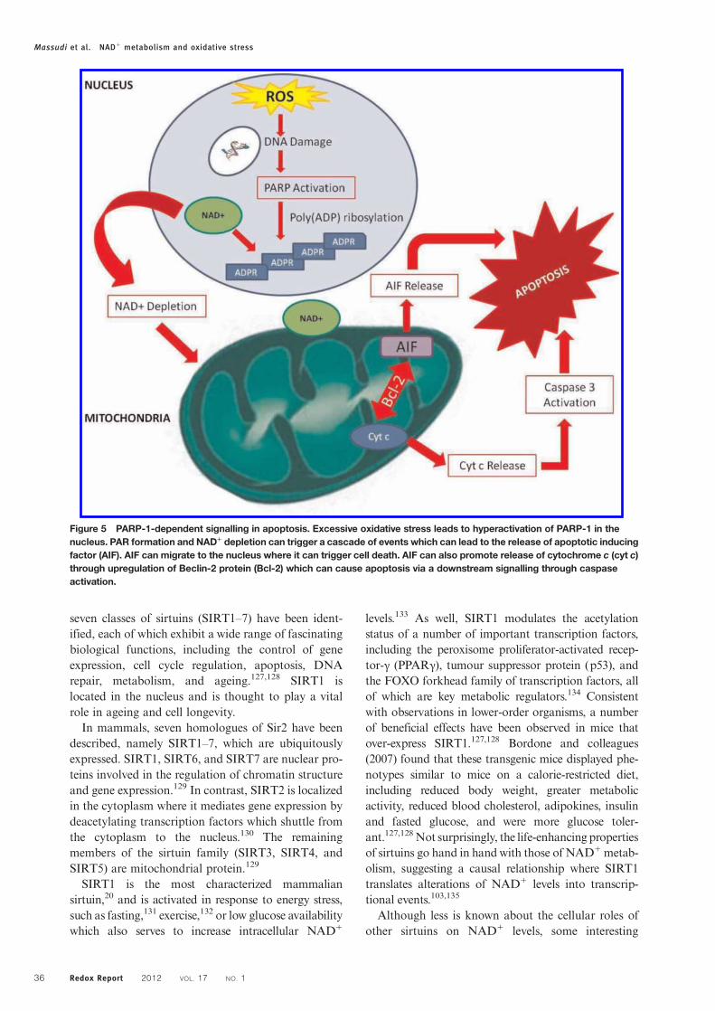
Several reports indicate that PARP activity rep-resents the main NAD+ catabolic process in the cell,thereby forcing the cell to continuously synthesizeNAD+ from the de novo or salvage pathway to main-tain cellular viability following oxidativedamage.113–115 The fast recycling is consistent withthe short half-life of NAD+, which is estimated to bearound 1–2 hours.116 Activation of PARP catalysesthe cleavage of NAD+ into adenosine 5′-diphosphori-bose (ADPR) and nicotinamide, and the covalentattachment of polymers of ADP-ribose to histonesand other nuclear proteins, including PARP itself(Fig. 4).117 PARP1, which accounts for the majorityof the ADPR synthesized, is found at the highest con-centrations in the nucleus.PARP1 activation leads to DNA repair and recov-
ery of normal cellular function. Experimental studieshave shown that PARP1 is activated in response to
free radical-mediated injury to DNA after brainischaemia and reperfusion.118 Hyperactivation ofPARP1 following DNA strand breaks can rapidlyconsume intracellular NAD+ pools, resulting in aloss of ability to synthesize ATP, and the cessation ofall energy-dependent functions and consequent celldeath (Fig. 5).112,119,120
PARP-mediated NAD+ depletion has been impli-cated in the pathogenesis of AD, with one studyshowing that poly(ADP-ribose) (PAR) polymersaccumulate at higher concentrations in the temporaland frontal cortex in the brains of AD patients com-pared with control brains.121 This indicates thatPARP1 is over expressed in the AD brain andimplies an excessive NAD+ turnover in susceptibleneuronal cells.121 Over-activation of PARP1 has alsobeen reported in diabetes, 1-methyl-4-phenyl-1,2,3,6-tetrahydropyridine-induced parkinsonism, traumaticbrain injury, hypoglycaemic brain disease, andshock.73,112,122–124
Sirtuin activityIn addition to its role in PARP activity, another essen-tial factor that is greatly altered by changes in intra-cellular NAD+ levels are the sirtuins, or the silentinformation regulators of gene transcription.125
Sirtuins are a highly conserved family of class III dea-cetylase proteins which catalyse a unique reaction inwhich NAD+ is used to remove an acetyl groupfrom the lysine residue, releasing nicotinamide andacetyl-ribose as end products (Fig. 6).126 At least
Figure 4 Schematic representation of poly(ADP-ribose) synthesis. Poly(ADP-ribose) polymerases breaks the bond betweennicotinamide and ribose in NAD+ leading to the formation of ADP-ribosyl moiety. Repeated reaction triggers the formation of PARchains.
Massudi et al. NAD+ metabolism and oxidative stress
Redox Report 2012 VOL. 17 NO. 1 35
seven classes of sirtuins (SIRT1–7) have been ident-ified, each of which exhibit a wide range of fascinatingbiological functions, including the control of geneexpression, cell cycle regulation, apoptosis, DNArepair, metabolism, and ageing.127,128 SIRT1 islocated in the nucleus and is thought to play a vitalrole in ageing and cell longevity.In mammals, seven homologues of Sir2 have been
described, namely SIRT1–7, which are ubiquitouslyexpressed. SIRT1, SIRT6, and SIRT7 are nuclear pro-teins involved in the regulation of chromatin structureand gene expression.129 In contrast, SIRT2 is localizedin the cytoplasm where it mediates gene expression bydeacetylating transcription factors which shuttle fromthe cytoplasm to the nucleus.130 The remainingmembers of the sirtuin family (SIRT3, SIRT4, andSIRT5) are mitochondrial protein.129
SIRT1 is the most characterized mammaliansirtuin,20 and is activated in response to energy stress,such as fasting,131 exercise,132 or low glucose availabilitywhich also serves to increase intracellular NAD+
levels.133 As well, SIRT1 modulates the acetylationstatus of a number of important transcription factors,including the peroxisome proliferator-activated recep-tor-γ (PPARγ), tumour suppressor protein (p53), andthe FOXO forkhead family of transcription factors, allof which are key metabolic regulators.134 Consistentwith observations in lower-order organisms, a numberof beneficial effects have been observed in mice thatover-express SIRT1.127,128 Bordone and colleagues(2007) found that these transgenic mice displayed phe-notypes similar to mice on a calorie-restricted diet,including reduced body weight, greater metabolicactivity, reduced blood cholesterol, adipokines, insulinand fasted glucose, and were more glucose toler-ant.127,128Not surprisingly, the life-enhancing propertiesof sirtuins go hand in hand with those of NAD+metab-olism, suggesting a causal relationship where SIRT1translates alterations of NAD+ levels into transcrip-tional events.103,135
Although less is known about the cellular roles ofother sirtuins on NAD+ levels, some interesting
Figure 5 PARP-1-dependent signalling in apoptosis. Excessive oxidative stress leads to hyperactivation of PARP-1 in thenucleus. PAR formation and NAD+ depletion can trigger a cascade of events which can lead to the release of apoptotic inducingfactor (AIF). AIF can migrate to the nucleus where it can trigger cell death. AIF can also promote release of cytochrome c (cyt c)through upregulation of Beclin-2 protein (Bcl-2) which can cause apoptosis via a downstream signalling through caspaseactivation.
Massudi et al. NAD+ metabolism and oxidative stress
Redox Report 2012 VOL. 17 NO. 136

functions have been associated with these sirtuinswhich are worth mentioning. SIRT2 is a tubulin deace-tylase which is down-regulated in gliomas.136 Ectopicexpression of SIRT2 in glioma cell lines has beenshown to decrease colony formation, suggesting thatSIRT2 may have a tumour suppressor role.136
Furthermore, SIRT2 also acts as a mitotic checkpointwhich maintains chromosomal stability in the earlymetaphase.137 Recently, SIRT2 has been shown topromote myelination in oligodendrocytes.138
SIRT2 and SIRT1 have been thoroughly investigatedin regard to ageing. Activation can mimic the effect ofcalorie restriction (CR) and extend the lifespan of yeast,worms, and flies. Moreover, overexpression of SIRT2has been shown to extend lifespan in Caenorhabditiselegans.139 Recently, Pearson et al. (2008)140 showedthat resveratrol (a putative SIRT1 activator) improvedmetabolism and significantly enhanced the health andsurvival of mice on high-calorie diet. In addition, bothoverexpression of SIRT1 and administration resveratrolhave been shown to have neuroprotective effects.140–142
Collectively, these studies raise the possibility that acti-vation of human sirtuins may slow down the ageingprocess. However, additional studies in the human popu-lation are required in order to elucidate the involvementof sirtuins in human population health.Impaired SIRT1 activity due to PARP-1-mediated
NAD+ depletion stimulates the activity of several
apoptotic effectors such as p53,143 therefore, sensitiz-ing cells to apoptosis. Both human and mouseSIRT1 are thought to promote cell survival by deace-tylating and thus deactivating p53 tumour suppressorgene hence enhancing p53 degradation.20,144
Adequate NAD+ levels are therefore critical to main-taining SIRT1 activity which can delay apoptosis andprovide vulnerable cells with additional time to repaireven after the repeated oxidative stress insult.SIRT3 has been linked to adaptive thermogenesis,145
mitochondrial function,145 energy homeostasis,146 andcellular viability following genotoxic insult.147 On theother hand, despite the presence of a conserved sirtuindomain, SIRT4 does not appear to exhibit deacetylaseactivity in vitro. Instead, SIRT4 appears to targetprotein activities through ADP-ribosylation.148 LikeSIRT3 and SIRT4, SIRT5 is a mitochondrial sirtuinwith deacetylase activity, although the exact role inmaintaining cellular homeostasis remains unknown.149
Upcoming research will shed some light in our under-standing of SIRT5 biology.While SIRT6 was initially suggested to possess
ADP-ribosylation activity only,150 it recently hasbeen shown to deacetylate histones and DNA poly-merase β, a DNA repair enzyme.151 Several lines ofevidence suggest that SIRT6 regulates genomic stab-ility and DNA repair. SIRT6-deficient mice die pre-maturely, and exhibit severe defects, such as
Figure 6 Sirtuin enzymatic activities. Sirtuins display two different NAD+ consuming activities, both of which render NM as aproduct.
Massudi et al. NAD+ metabolism and oxidative stress
Redox Report 2012 VOL. 17 NO. 1 37

lymphopenia, decreased bone mineral density, andimpaired glucose homeostasis. This phenotypemimics multiple pathologies observed in elderlyhumans, suggesting that SIRT6 could play an essentialrole in maintaining organ integrity during ageing anddevelopment.150,151
Finally, SIRT7 is localized in the nucleolus where itpositively regulates transcription of ribosomal DNAduring elongation, which can account for up to 60%of total transcription in metabolically activestates.152,153 Over-expression of SIRT7 increasesRNA polymerase transcription in an NAD-dependentmanner, whereas down-regulation of SIRT7 canreduce cell proliferation and trigger apoptosis.152,153
In addition, the tumorigenic potential of several celllines inversely correlates with SIRT7 expression.154
Interactions between PARPs and sirtuinsThere is now emerging evidence indicating an associ-ation between the PARP-1 and SIRT1 pathways,which have been previously independently studied.As previously described, overactivation of PARP-1
following extensive DNA damage can lead to celldeath, since prolonged PARP-1 activation candeplete the essential substrate NAD+, leading to asubsequent increase in the byproduct, nicotinamide.The decline in NAD+ and the rise in nicotinamidemay downregulate the SIRT1 deacetylase activity.155
It is conceivable, therefore, that poly(ADP)-ribosyla-tion by PARP-1, as induced by DNA damage, canmodulate SIRT1 protein deacetylation via theNAD+/nicotinamide connection (Fig. 7).
Subcellular distribution of NAD+ and itsmetabolismCompartmentation of NAD+
A major complication associated with the assessmentof intracellular NAD+ levels is the subcellular com-partmentation of NAD+ in mammalian cells. Thereare two major independent NAD+ pools: cytosolicand mitochondrial.119 It is estimated that the relativenumber of mitochondria within a tissue correspondsto the share of mitochondrial NAD+. The mitochon-drial fraction of NAD+ is therefore significantly
Figure 7 NAD+/nicotinamide levels may serve as converging points for interaction of PARP-1 and SIRT1 pathways. It isconceivable that poly(ADP-ribose) metabolism can downregulate SIRT1 through NAD+ depletion and nicotinamide productionduring oxidative stress. Reciprocally, regulation by SIRT1 deacetylation for expressing genes related to apoptosis or longevitymay depend on PARP-1 activity. PARP-1 inhibitors maintain NAD+ levels and suppress the nicotinamide surge, and thereforemay indirectly serve as SIRT1 enhancers. The interactions of PARP-1/SIRT1 pathways provide a network for multicellulareukaryotes to effectively deal with nutritional supply and oxidative stress.
Massudi et al. NAD+ metabolism and oxidative stress
Redox Report 2012 VOL. 17 NO. 138

higher in the liver and other mitochondria-rich tissuesuch as the brain and heart. The cytosolic pool ofNAD+ appears to be freely exchangeable with thenucleus. As no physiological means of exchange havebeen identified between the mitochondria and cytoso-lic NAD+ compartments, these two pools are con-sidered independent of each other.119,156,157 di Lisaand Ziegler (2001),119 however, suggested that mito-chondrial permeability transition (MPT) poreopening can lead to mitochondrial NAD+ releasethat is metabolized by NAD-dependent dehydrogen-ases in the heart. However, it is unclear as towhether MPT-dependent NAD+ release also takesplace in neurons and glial cells.
The NAD+:NADH ratioThe NAD+:NADH ratio plays a pivotal role in regu-lating the intracellular redox state, and is hence con-sidered to be a measure of the metabolic state.144 Aspreviously mentioned, several enzymes appear to beregulated by the NAD+:NADH ratio, such asGAPDH and dehydrogenase reactions.158 Severalstudies have shown that the NAD+:NADH ratio fluc-tuates in response to a change in metabolism.159–163
If NAD+ is a metabolic regulator, then the ratio ofthe intracellular NAD+ to NADH is close to1. Therefore, the NAD+:NADH is regulated bysmall changes in the NAD+ concentration.161 Forinstance, if the ratio is very high (e.g. 600), then onewould assume that the NAD+:NADH ratio will bemore sensitive to a change in the NADH concen-tration, and not NAD+.144 Table 2 shows the reportedratios of total intracellular NAD+:NADH. Thesevalues suggest that NAD+ is a key metabolic regulatorof the NAD+:NADH ratio in a variety of tissues.
Compartmentation of NAD+ metabolism inmammalian cellsInterestingly, the key enzymes involved in the biosyn-thesis of NAD+ are localized within the nucleus.164
In humans, the predominant isoform of NMNAT(NMNAT1) is located within the nucleus (Fig. 1.17).The recent discovery of nuclear NAD+-dependent sig-nalling pathways such as poly-ADP-ribosylation andsirtuin-mediated histone deacetylation suggests that
nuclear NAD+ production is necessary to compensatefor the high rate of NAD+ turnover. Indeed, theinhibitory effect of NMNAT1 on PARP activity hasbeen previously described to counteract NAD+ andATP depletion following hyperactivation of PARP inresponse to oxidative stress.165
Recent studies have indicated the presence of twoadditional isoforms of human NMNATs –
NMNAT2 and NMNAT3, which are located in theGolgi complex and mitochondria, respectively.166,167
These findings, together, with discovery of NAD-dependent tankyrases in Golgi complex and NAD+-consuming enzymes in the mitochondria, providefurther evidence to support the existence of indepen-dent NAD+ synthesizing machinery in the nucleus,Golgi complex, and mitochondria.168
Therapeutic potential of NAD+ metabolismNAD+ in neurodegenerationConsidering the importance of NAD+ in energymetabolism, DNA repair and transcriptional regu-lation, maintaining intracellular NAD+ reservesemerges as a major therapeutic target for the treatmentof several age-related degenerative diseases, includingAD.169 In particular, increased nuclear NAD+ biosyn-thesis and consequent activation of SIRT1 has beenshown to protect mouse neurons from mechanicaland chemical injury.170 Restoring cellular NAD+
levels have been shown to protect against axonaldegeneration in the experimental autoimmune ence-phalomyelitis animal model for MS in humans.19
Axonopathy is a critical feature of several neurode-generative diseases and often precedes the death ofneuronal bodies in AD, PD, and MS.171 As axonaldeficits are central to the patient’s neurological disabil-ity, therapies that prevent axonal degradation are ofgreat therapeutic importance.172 Increased NMNAT1activity has also been shown to protect againstaxonal degeneration in Wallerian degeneration slow(Wlds) mice. Exogenous administration of NAD+
prior to axotomy also delayed axonal degeneration,but to a lesser extent in NMNAT1 expressed mice,further indicating the importance of maintainingintracellular NAD+ pools as a preventive measureagainst axonal degradation. In the absence of exogen-ous NAD+, PARP inhibition increased the survival ofdorsal root ganglion cultures following mechanicalinjury. No protective effect onWlds mice was observedfollowing PARP inhibition in the presence of exogen-ous NAD+.170,172 This suggests that maintaining ade-quate intracellular NAD+ levels can promote neuronalsurvival.Another link between neurodegeneration and
NAD+ metabolism lies in the fact that many neurode-generative disorders, including AD, PD, and MS, are
Table 2 Baseline ratio of cytosolic NAD+:NADH ratio invarious species
Species Tissue type NAD+:NADH ratio References
Mice Liver 4 160Mice Kidney 2.6 160Mice Brain 0.26 159Mice Blood 0.03 159Mice Pancreas 0.1 163Swine Liver 0.07 162
Massudi et al. NAD+ metabolism and oxidative stress
Redox Report 2012 VOL. 17 NO. 1 39

associated with mitochondrial dysfunction due toextensive oxidative damage.173 Therefore, it is possiblethat activation of PARP1 can mediate the neuronalcell death observed in these pathological states.73
Indeed, increased PARP expression has been reportedin the brains and peripheral cells in post-mortempatients with AD, PD, and MS.121,174,175 Furtherwork is necessary to identify the mechanism(s) under-lying the major differences in PARP expression inthese neurodegenerative diseases.As previously mentioned, the KP represents the
major route of TRP catabolism leading to the syn-thesis of a number of neuroreactive compounds.176
Among those, QUIN appears to be involved in thepathogenesis of several neuroinflammatory dis-orders.177 Therefore, KP inhibitors have been devel-oped to reduce QUIN levels in the CNS.178 Itremains unresolved whether KP inhibition will alsoresult in decreased NAD+ biosynthesis in the brain.
NAD+ in ageingAs chronic oxidative stress and associated nucleardamage can promote NAD+ catabolism, the NAD+
metabolic pathway has been implicated as a potentialtherapeutic target to promote longevity.113,114,179
Owing to the importance of oxidative stress inageing, it is highly likely that PARPs may playmajor roles in ageing by promoting NAD+
depletion.73,123 Grube and Burkle (1992)124 showedthat PARP-1 activity in mononuclear blood cellsincreases with ageing in at least thirteen mammalianspecies, and this is likely to be associated with itsimportant DNA repair function. Consistent with itsrole as the major player in the OS theory of ageing,the mitochondrial dysfunction which can occur inthe ageing process is associated with changes inNAD+ levels.180 Although a number of studies havedemonstrated elevated levels of oxidative stress-mediated damage in aged tissue, to our knowledge,no study has yet reported on changes in NAD+
levels during the human ageing process. Braidy et al.(2011)181 recently reported a parallel increase in p53acetylation with age, closely correlating with increasedlevels of lipid peroxidation, protein cross-linking, andDNA damage in the heart, lung, liver, and kidney,or aged female wistar rats. However, the potentialfor increased NAD+ catabolism appears to beinvolved in ageing, the impact of this process and itscontribution on NAD+ in extending lifespan needsto be clarified.It has been well established that CR is one of the
best strategies to improve lifespan in several species.The hypothesis that increased mitochondrial functionupon CR is associated with the beneficial effects onlifespan has been recently extended to humans, inwhich a general increase in metabolism occurred
during CR.20 The beneficial effects of CR appear tobe NAD+ dependent and partly mediated bySIRT1/Sir2 activity.182 In C. elegans, extra copies ofthe SIRT2 orthologue sir-2.1 extended lifespan.139
Both human and mouse SIRT2 have been shown tofunction as NAD-dependent p53 deacetylases, anddeacetylation of p53 by SIRT2 can promote cell survi-val under stress.20,144 Recently, Pearson et al. (2008)140
showed that activation of SIRT1 using resveratrol inmice fed a high-calorie diet improved metabolismand significantly enhanced the lifespan of theseanimals. Previous work from our laboratory showedfor the first time that resveratrol does have a novelfunction in specifically upregulating NAD+ synthesis.Additionally, SIRT3, SIRT6, and SIRT7 have beenlinked with a longer lifespan in mice,151,154,183 whileSIRT1 gene polymorphisms can affect lifespan by sti-mulating energy release.184 Additional human studiesare required to clarify the involvement of sirtuins inhuman population health.
However, Anderson et al. (2003)147 suggested analternative mechanism by which CR and Sir2 maymediate longevity in yeast. Pyrazinamide/nicotinami-dase (PNC1) encodes an enzyme which converts NMto NA, thereby promoting Sir2 activity and depletingthe endogenous Sir2 inhibitor, NM.147 PNC1appears necessary for the lifespan extension mediatedby CR. Within the NAD+ biosynthesis pathwayNMPRT has been shown to regulate the ageingprocess.185 Reduced NMPRT expression resulted inpremature senescence in ageing human smoothmuscle cells, whereas over-expression of NMPRTdelayed senescence while promoting enhanced antiox-idant defence.185
Due to the important role of oxidative stress inageing, it is conceivable that PARPs may play signifi-cant roles in ageing by increasing demand for its sub-strate, NAD+.73,123 PARP1 activities in mononuclearblood cells strongly correlate with longevity in atleast 13 mammalian species, which may be associatedwith its important DNA repair function.124 Moreover,PARP1 has been shown to inhibit the catalytic activi-ties of the protein of Werner syndrome, a humandisease of premature ageing.186 While the NAD+
metabolic pathway appears to be involved in ageing,the exact role of NAD+ in extending lifespan is notso clear-cut. Further research in this field is necessaryto determine whether NAD+ is an effective target toincrease lifespan.
ConclusionPellegra, a syndrome caused by a diet deficient ineither NA or TRP can lead to psychotic symptomsleading to presenile dementia likely due to upregula-tion of IDO, which can deplete neurons of the essentialamino acid, TRP causing neurodegeneration.6
Massudi et al. NAD+ metabolism and oxidative stress
Redox Report 2012 VOL. 17 NO. 140

Administration of the NAD+ precursors, NA or NMpreviously improved the neurological state of dementiapatients in the 1930s.6 Pharmacological doses of eitherNA and NM have also provided dramatic therapeuticbenefits for other diseases, including rheumatoidarthritis, type I diabetes, colitis, MS, and schizo-phrenia in animal models and in the clinical setting.4
Among these precursors, NA appears to specificallyactivate the G-protein coupled receptor, GPR109,leading to the release of the prostaglandins, PGE2
and PGD2.187 These prostaglandins exert potent
anti-inflammatory effects through endogenous signal-ling mechanisms involving PPARγ.187 While NMcan prevent MS in animal models, it is also an inhibi-tor of sirtuins, and may therefore prove detrimental onlong-term cell survival and longevity.147,188
There is growing evidence suggesting that NAD+
administration may also reduce cellular injury in mul-tiple diseases.115 NAD+ treatment has been shown toreduce PARP1-induced astrocyte death.189 It hasbeen also shown to prevent PARP1-mediated NAD+
depletion in cardiac myocytes in the presence ofH2O2.
190 PARP1 has been implicated in the pathogen-esis of several diseases including diabetes, AD and PD(Ying, 2006, 2008).115 Since supplementation withNAD+ can protect against PARP1 mediated celldeath, NAD+ administration may improve cell viabi-lity in these diseases by at least partially amelioratingPARP1 toxicity. In vitro studies have shown thatNAD+ remains protective even when administered at3–4 hours following PARP1 activation, suggestingthat NAD+ administration has a long windowperiod for reducing cellular injury.189 In addition,NAD+ may also improve cell viability by enhancingsirtuin activities and/or improving energy metabolism(Ying, 2006, 2008).115
Resveratrol is a polyphenol with major healthbenefits that is thought to operate through direct acti-vation of the ‘antiageing’ enzyme SIRT1.191 However,recent reports have challenged this ‘direct-activation’hypothesis,192 suggesting that the mechanism bywhich resveratrol increases SIRT1 function is stillunknown. Previous work from our group has shownfor the first time that resveratrol induces a dose-depen-dent increase in activity of the NAD+ syntheticenzyme nicotinamide mononucleotide adenyl transfer-ase (NMNAT1) (unpublished data). As SIRT1requires NAD+ as a substrate to perform its gene-silencing function, higher NAD+ levels will enhanceSIRT1 activity.172 This finding suggests that resvera-trol may promote SIRT1 function by enhancingNAD+ synthesis in whole cell systems without requir-ing direct activation.Our observation that resveratrol increases NAD+
levels in primary human brain cells by acting onNMNAT, together with the neuroprotective effects
of green tea polyphenols against QUIN-mediated exci-totoxicity, supports the view that polyphenols haveconsiderable therapeutic potential, particularly forthe treatment of neurodegenerative diseases.193 AsNMNAT can accelerate NAD+ synthesis from allthree substrates, QUIN, NA, and NM (Berger et al.,2005),17,167 NMNAT activation by resveratrol rep-resents an ideal natural therapeutic to replenishNAD+ levels. Maintenance of higher cellular NAD+
will enhance SIRT1 activity and other NAD+-depen-dent pathways, impacting positively on cell viabilityand longevity. This work has therefore formed thebasis of relevant patent applications.While the potential involvement of NAD+ meta-
bolic pathways in energy metabolism and mitochon-drial function have been known for quite some time,suggestions of the involvement of NAD+ in DNArepair and longevity have grown at a rapid rate inthe last decade.8 Characterization of the NAD+ syn-thetic pathways has not only made these advance-ments possible, but also contributed extensively tothe understanding of the diverse roles of pyridinenucleotides in cellular biology.8 Despite this, infor-mation regarding the fundamental roles of NAD+ inneurodegeneration and ageing remains limited.Further investigations are necessary in this increas-ingly interesting field. Research in the followingareas may be of particular interest.Firstly, maintaining intracellular NAD+ levels in
human brain cells such as astrocytes and neurons iscrucial for the retention of cellular viability duringconditions of chronic oxidative stress and immuneactivity through the promotion of oxidative phos-phorylation (ATP production), DNA repair (PARPactivity), and gene expression (sirtuin activity).Therefore, characterization of the effect of varyingdegrees of immune activation on de novo NAD+ syn-thesis in selected brain cells is necessary to validatethe role of NAD+ metabolism as a primary contribu-tor to neuronal dysfunction and cell death during neu-roinflammatory disorders.Secondly, as NAD+ is an essential molecule for all
living organisms, it is not surprising that numerouscell types may possess a number of different strategiesto generate NAD+, particularly under conditions ofacute and chronic oxidative stress and NAD+
depletion.194 For instance, post-mitotic cells such asneurons may rely on different pathways than thoseactively in dividing cells such as astrocytes.194
Further studies may therefore need to be aimed atidentifying the preferred substrate for NAD+ synthesisin neuronal cells under various conditions.Thirdly, additional NAD+ regulates diverse path-
ways which may control lifespan. The importance ofNAD+ is further underscored by recent work provid-ing genetic evidence for the existence of several
Massudi et al. NAD+ metabolism and oxidative stress
Redox Report 2012 VOL. 17 NO. 1 41

pathways necessary for NAD+ synthesis. For example,Belenky et al. (2007)72 recently demonstrated that anewly identified NAD+ precursor, NR, can contributeto NAD+ synthesis by at least two unique pathways inthe yeast S. cerevisiae. Both pathways require the nico-tinamide ring for entry into the previously establishedpathways for NAD+ synthesis.72 Future studies arerequired to address the importance of NR in humanhealth and disease, and whether it can be effectivelyused to replenish lowered NAD+ levels in age-relateddiseases, such as AD.Fourthly, the essential cofactor PRPP is an impor-
tant regulator for the de novo NAD+ syntheticenzyme, QPRT, which catalyses the conversion of theexcitotoxin QUIN to NAMN11,18,166. PRPP concen-tration has been positively correlated with cytosolicNAD+ and ATP levels in whole animals.195
Therefore, the availability of PRPP for QPRT activitymay be compromised during increased NAD+ turn-over. This may occur in neurodegenerative disordersand ageing due to ROS-mediated DNA damage.196
Increased QUIN secretion into the CSF may be dueto increased flux through the KP parallel to reducedQPRT activity associated with an increased demandfor PRPP for NAD+ synthesis in damaged cells.Additional studies are required to investigate theeffect of PRPP on de novo NAD+ synthesis duringneuroinflammation and ageing. Fifthly, the roles ofdifferent sirtuins in brain function need to be furtherinvestigated under different physiological and patho-logical conditions. Numerous studies have highlightedthe importance of sirtuins as key regulators inageing.197 However, the precise roles of differentsirtuin isoforms (SIRT1-7) and their response tovarying NAD+ concentrations in brain functionremain unclear. Future work may be aimed at estab-lishing the roles of sirtuins in brain function as a thera-peutic target for the treatment of a variety of braindisorders and increasing lifespan.Sixthly, NAD+ plays a key role in regulating intra-
cellular calcium homeostasis by acting as the substratefor NAD-dependent glycohydrolase (NADase) (seeSection 1.4.5). While PARP1 is a major NAD-con-suming enzyme in the cell, recent studies have raisedthe possibility that NADase may play a key role inNAD+ metabolism under physiological conditions.8
For instance, increased NAD+ levels have beenreported in the brain, lung, and kidney in NADase-deficient mice.198 Moreover, NADase activity wasabsent in the plasma membranes, mitochondria, sarco-plasmic reticulum, and nuclei in NADase-deficientmice.199 These studies suggest that NADase is a keyregulator of cellular NAD+ levels under physiologicalconditions, while PARP1 is a key factor determiningintracellular NAD+ levels when significant oxidativestress and DNA damage occurs.115 Owing to the
critical roles of NADase and calcium in cellular func-tion, it is warranted to further examine the roles ofNAD+-dependent changes in calcium homeostasisnot only in normal brain function but also in brainageing and neurological disorders, in general.
Seventhly, while the current investigations reportedherein focussed on PARP1 in cellular degeneration,the role of other PARPs such as tankyrases in cellularfunction remains largely unknown. Since NAD-dependent tankyrases are main mediators of telomer-ase activity, it is highly likely that NAD+ may alsoaffect the ageing process through regulation of tankyr-ase activity.200 It would therefore be intriguing tostudy the effects of tankyrases and telomerases oncertain biological functions, including neurogenesis,which might affect the ageing brain.
AcknowledgementsHassina Massudi is a reciepient of the AustralianPostgraduate Award (APA) at the University of NewSouth Wales. Nady Braidy is the recipient of anAlzheimer's Australia Viertel Foundation PostdoctoralResearch Fellowship at the University of New SouthWales.
References1 Elvehjem C, Madden R, Strong F, Woolley D. Relation of nic-otinic acid and nicotinic acid amide to canine black tongue.J Am Chem Soc 1937;59:1767–8.
2 Elvehjem C. Pellagra – a deficiency disease. Proc Am Philos Soc1949;93:335–9.
3 Broer S, JA C, Rasko J. Neutral amino acid transport in epi-thelial cells and its malfunction in Hartnup disorder. BiochemSoc Trans 2005;33:233–6.
4 Monteiro J, da Cunha D, Filho D, SilvaVergara M, dos SantosV, da Costas J, Jr, et al. Niacin metabolite excretion in alcholicpellagra and AIDS patients with and without diarrhea.Nutrition 2004;20:778–82.
5 Comaish J, Felix R, McGrath H. Topically applied niacinamidein ioniazid-induced pellagra. Arch Dermatol 1976;112:70–2.
6 Penberthy WT. Pharmacological targeting of IDO-mediatedtolerance for treating autoimmune disease. Curr Drug Metab2007;8:245–66.
7 Brown RR, Ozaki Y, Datta SP, Borden EC, Sondel PM,Malone DG. Implications of interferon-induced tryptophancatabolism in cancer, auto-immune diseases and AIDS. AdvExp Med Biol 1991;294:425–35.
8 Berger F, Ramirez-Hernandez MH, Ziegler M. The new life ofa centenarian: signalling functions of NAD(P). Trends BiochemSci 2004;29:111–8.
9 Schlenk F, von Euler H. Cozymase. Naturwissenschaften 1936;24:794–5.
10 Warburg O, Christian W. Pyridin, der wasserstoffubertragendeBestandteil von Garungsfermenten (Pyridin-Nucleotide).Biochem Z 1936;287:291–328.
11 Magni G, Orsomando G, Raffelli N, Ruggieri S. Enzymologyof mammalian NAD metabolism in health and disease. FrontBiosci 2008;13:6135–54.
12 Kornberg A. The participation of inorganic pyrophosphate inthe reversible enzymatic synthesis of diphosphopyridine nucleo-tide. J Biol Chem 1948;176:1475–6.
13 Rizzi M, Schindelin H. Structural biology of enzymes involvedin NAD and molybdenum cofactor biosynthesis. Curr OpinStruct Biol 2002;12:709–20.
14 Chambon P, Weill J, Mandel P. Nicotinamide mononucleotideactivation of new DNA-dependent polyadenylic acid synthesis-ing nuclear enzyme. Biochem Biophys Res Commun 1963;11:39–43.
Massudi et al. NAD+ metabolism and oxidative stress
Redox Report 2012 VOL. 17 NO. 142

15 Gholson RK. The pyridine nucleotide cycle. Nature 1966;212:933–4.
16 Rechsteiner M, Catanzarite V. The biosynthesis and turnover ofnicotinamide adenine dinucleotide in enucleated culture cells.J Cell Physiol 1974;84:409–22.
17 Magni G, Amici A, Emanuelli M, Orsomando G, Raffaelli N,Ruggieri S. Structure and function of nicotinamide mononu-cleotide adenylyltransferase. Curr Med Chem 2004;11:873–85.
18 Magni G, Amici A, Emanuelli M, Raffaelli N, Ruggieri S.Enzymology of NAD+ synthesis. Adv Enzymol Relat AreasMol Biol 1999;73:135–82, xi.
19 Khan JA, Forouhar F, Tao X, Tong L. Nicotinamide adeninedinucleotide metabolism as an attractive target for drug discov-ery. Expert Opin Ther Targets 2007;11:695–705.
20 Houtkooper R, Canto C, Wanders R, Auwerx J. The secret lifeof NAD+: an old metabolite controlling new metabolic signal-ing pathways. Endocr Rev 2010;31:194–223.
21 Braidy N, Guillemin G, Grant R. Promotion of cellular NAD+anabolism: therapeutic potential for oxidative stress in ageingand Alzheimer’s disease. Neurotox Res 2008;13:173–84.
22 Mackay GM, Forrest CM, Stoy N, Christofides J, Egerton M,Stone TW, et al. Tryptophan metabolism and oxidative stress inpatients with chronic brain injury. Eur J Neurol 2006;13:30–42.
23 Ruddick JP, Evans AK, Nutt DJ, Lightman SL, Rook GA,Lowry CA. Tryptophan metabolism in the central nervoussystem: medical implications. Expert Rev Mol Med 2006;8:1–27.
24 Fujigaki S, Saito K, Sekikawa K, Tone S, Takikawa O, Fujii H,et al. Lipopolysaccharide induction of indoleamine 2,3-dioxy-genase is mediated dominantly by an IFN-gamma-independentmechanism. Eur J Immunol 2001;31:2313–8.
25 Guillemin GJ, Smith DG, Williams K, Smythe GA, DormontD, Brew BJ. β amyloid peptide 1–42 induces human macro-phages to produce the neurotoxin quinolinic acid. JNeuroimmunol 2001;118:112, A:336.
26 Boasso A, Herbeuval JP, Hardy AW, Anderson SA, Dolan MJ,Fuchs D, et al. HIV-1 inhibits CD4+ T cell proliferation byinducing indoleamine 2,3-dioxygenase in plasmacytoid dendri-tic cells. Blood 2006;109:3351–9.
27 Frumento G, Piazza T, Di Carlo E, Ferrini S. Targeting tumor-related immunosuppression for cancer immunotherapy. EndocrMetab Immune Disord Drug Targets 2006;6:233–7.
28 De Castro F, Price J, Brown R. Reduced triphophopyridine-nucleotide requirement for the enzymatic formation of 3-hydro-xykynurenine from L-kynurenine. J Am Chem Soc 1956;78:2904–5.
29 Mason M. The kynurenine transaminase of rat kidney. J BiolChem 1954;211:839–44.
30 Wiss O, Fuchs H. Uber den Abbau von Kynurenin,Oxykynurenin und verwandten Substanzen durchRattenleberenzym. Experientia (Basel) 1950;6:472–3.
31 Bokman AH, Schweigert BS. 3-Hydroxyanthranilic acidmetabolism. IV. Spectrophotometric evidence for the formationof an intermediate. Arch Biochem Biophys 1951;33:270–6.
32 Mehler A, May F. Studies with carboxyl-labelled 3-hydroxyan-thranilic acid and picolinic acid in vivo and in vitro. J BiolChem 1956;223:449–55.
33 Cao H, Pietrak BL, Grubmeyer C. Quinolinate phosphoribo-syltransferase: kinetic mechanism for a type II PRTase.Biochemistry 2002;41:3520–8.
34 Connick JH, Stone TW. Quinolinic acid effects on amino acidrelease from the rat cerebral cortex in vitro and in vivo. Br JPharmacol 1988;93:868–76.
35 Bergeron R, Meyer TM, Coyle JT, Greene RW. Modulation ofN-methyl-D-aspartate receptor function by glycine transport.Proc Natl Acad Sci USA 1998;95:15730–4.
36 Boegman RJ, el-Defrawy SR, Jhamandas K, Beninger RJ,Ludwin SK. Quinolinic acid neurotoxicity in the nucleusbasalis antagonized by kynurenic acid. Neurobiol Aging1985;6:331–6.
37 Coggan S, Smythe G, Bilgin A, Grant R. Age and circadianinfluences on picolinic acid concentrations in human cerebrosp-inal fluid. J Neurochem 2009;108:1220–5.
38 Guidetti P, Schwarcz R. 3-Hydroxykynurenine and quinolinate:pathogenic synergism in early grade Huntington’s disease? AdvExp Med Biol 2003;527:137–45.
39 Morita T, Saito K, Takemura M,Maekawa N, Fujigaki S, FujiiH, et al. 3-Hydroxyanthranilic acid, an L-tryptophan metab-olite, induces apoptosis in monocyte-derived cells stimulatedby interferon-gamma. Ann Clin Biochem 2001;38:242–51.
40 Guillemin GJ, Brew BJ, Noonan CE, Knight TG, Smythe GA,Cullen KM. Mass spectrometric detection of quinolinic acid inmicrodissected Alzheimer’s disease plaques. In: Takai K, (ed.)International Congress Series. 2007. Elsevier B.V., Amsterdam.p. 404–8.
41 Guillemin GJ, Brew BJ, Noonan CE, Takikawa O, Cullen KM.Indoleamine 2,3 dioxygenase and quinolinic acid immunoreac-tivity in Alzheimer’s disease hippocampus. Neuropathol ApplNeurobiol 2005;31:395–404.
42 Guillemin GJ, Brew BJ. Implications of the kynureninepathway and quinolinic acid in Alzheimer’s disease. RedoxRep 2002;7:199–206.
43 Guillemin GJ, Brew BJ. Chronic HIV infection leads to anAlzheimer’s disease like illness. Involvement of the kynureninepathway. In: Takai K, (ed.) International Congress Series.2007. Elsevier B.V., Amsterdam. p. 324–34.
44 Guillemin GJ, Kerr SJ, Brew BJ. Involvement of quinolinic acidin AIDS dementia complex. Neurotox Res 2005;7:103–23.
45 Clark CJ, Mackay GM, Smythe GA, Bustamante S, Stone TW,Phillips RS. Prolonged survival of a murine model of cerebralmalaria by kynurenine pathway inhibition. Infect Immun2005;73:5249–51.
46 Guillemin G, Meininger V, Brew B. Implications for the kynur-enine pathway and quinolinic acid in amyotrophic lateral scler-osis. Neurodegener Dis 2006;2:166–76.
47 Chiarugi A, Cozzi A, Ballerini C, Massacesi L, Moroni F.Kynurenine 3-mono-oxygenase activity and neurotoxic kynure-ninemetabolites increase in the spinal cordof ratswith experimen-tal allergic encephalomyelitis. Neuroscience 2001;102:687–95.
48 Beal MF, Ferrante RJ, Swartz KJ, Kowall NW. Chronic quino-linic acid lesions in rats closely resemble Huntington’s disease.J Neurosci 1991;11:1649–59.
49 Beal MF, Matson WR, Storey E, Milbury P, Ryan EA, OgawaT, et al. Kynurenic acid concentrations are reduced inHuntington’s disease cerebral cortex. J Neurol Sci 1992;108:80–7.
50 Chess AC, Simoni MK, Alling TE, Bucci DJ. Elevations ofendogenous kynurenic acid produce spatial working memorydeficits. Schizophrenia Bull 2007;33:797–804.
51 Schwarcz R. The kynurenine pathway of tryptophan degra-dation as a drug target. Curr Opin Pharmacol 2004;4:12–7.
52 Grant RS, Naif H, Thuruthyil SJ, Nasr N, Littlejohn T,Takikawa O, et al. Induction of indolamine 2,3-dioxygenasein primary human macrophages by human immunodeficiencyvirus type 1 is strain dependent. J Virol 2000;74:4110–5.
53 Moffett JR, Namboodiri MA. Tryptophan and the immuneresponse. Immunol Cell Biol 2003;81:247–65.
54 Takikawa O, Yoshida R, Kido R, Hayaishi O. Tryptophandegredation in mice initiated by indoleamine 2,3-dioxygenase.J Biol Chem 1986;261:3648–53.
55 Hansen AM, Ball HJ, Mitchell AJ, Miu J, Takikawa O, HuntNH. Increased expression of indoleamine 2,3-dioxygenase inmurine malaria infection is predominantly localised to the vas-cular endothelium. Int J Parasitol 2004;34:1309–19.
56 Munn DH, Zhou M, Attwood JT, Bondarev I, Conway SJ,Marshall B, et al. Prevention of allogeneic fetal rejection bytryptophan catabolism. Science 1998;281:1191–3.
57 Alexander A, Crawford M, Bertera S, Rudert W, Takikawa O,Robbin P, et al. Indoleamine 2,3-dioxygenase expression intransplanted NOD Islets prolongs graft surviva; after adoptivetransfer of diabetogenic splenocytes. Diabetes 2002;51:356–65.
58 Hayashi T, Beck L, Rossetto C, Gong X, Takikawa O,Takabayashi K, et al. Inhibition of experimental asthma byindoleamine 2,3-dioxygenase. J Clin Invest 2004;114:270–9.
59 Uyttenhove C, Pilotte L, Theate I, Stroobant V, Colau D,Parmentier N, et al. Evidence for a tumoral immune resistancemechanism based on tryptophan degradation by indoleamine2,3-dioxygenase. Nat Med 2003;9:1269–74.
60 Munn DH, Mellor AL. IDO and tolerance to tumors. TrendsMol Med 2004;10:15–8.
61 Grant RS, Passey R, Matanovic G, Smythe G, Kapoor V.Evidence for increased de novo synthesis of NAD in immune-activated RAW264.7 macrophages: a self-protective mechan-ism? Arch Biochem Biophys 1999;372:1–7.
62 Grant RS, Naif H, Espinosa M, Kapoor V. IDO induction inIFN-gamma activated astroglia: a role in improving cell viabi-lity during oxidative stress. Redox Rep 2000;5:101–4.
63 Grant R, Kapoor V. Inhibition of indoleamine 2,3-dioxygenaseactivity in IFN-gamma stimulated astroglioma cells decreasesintracellular NAD levels. Biochem Pharmacol 2003;66:1033–6.
Massudi et al. NAD+ metabolism and oxidative stress
Redox Report 2012 VOL. 17 NO. 1 43
64 Iqbal J, Zaidi M. TNF regulates cellular NAD+ metabolism inprimary macrophages. Biochem Biophys Res Commun 2006;342:1312–8.
65 Rongvaux A, Andris F, Van Gool F, Leo O. Reconstructingeukaryotic NAD metabolism. Bioessays 2003;25:683–90.
66 Khan JA, Tao X, Tong L. Molecular basis for inhibition ofhuman NMPRTase, a novel target for anticancer agents. NatStruct Mol Biol 2006;13:582–8.
67 Gross J, Rajavel M, Grubmeyer C. Kinetic mechanism of nic-otinic acid phosphoribosyltransferase: implications for energycoupling. Biochem 1998;37:4189–99.
68 Bieganowski P, Brenner C. Discoveries of nicotinamide ribosideas a nutrient and conserved NRK genes establish a Priess-Handler independent route to NAD+ in fungi and humans.Cell 2004;117:495–502.
69 Grifantini M. Tiazofurine ICN Pharmaceuticals. Curr OpinInvest Drugs 2000;1:257–62.
70 Klaidman L, Morales M, Kem S, Yang J, Chang M, Adams J,Jr. Nicotinamide offers multiple protective mechanisms instroke as a precursor for NAD+, as a PARP inhibitor and bypartial restoration of mitochondrial function. Pharmacology2003;69:150–7.
71 Wang B, Liao W, Chang C, Wang S. Facilitation of glutamaterelease by nicotine involves the activation of Ca2+/calmodulinsignaling pathway in rat prefrontal cortex nerve terminals.Synapse 2006;59:491–501.
72 Belenky P, Racette F, Bogan KL, McClure J, Smith J, BrennerC. Nicotinamide riboside promotes Sir2 silencing and extendslifespan via Nrk and Urh1/Pnp1/Meu1 pathways to NAD+.Cell 2005;129:473–84.
73 Burkle A, Beneke S, Muiras ML. Poly(ADP-ribosyl)ation andaging. Exp Gerontol 2004;39:1599–1601.
74 Barnham KJ, Masters CL, Bush AI. Neurodegenerative dis-eases and oxidative stress. Nat Rev Drug Discov 2004;3:205–214.
75 Harman D. Aging: a theory based on free radical and radiationchemistry. J Gerontol 1956;11:298–300.
76 Bechman K, Ames B. The free radical theory of aging matures.Physiol Rev 1998;78:547–81.
77 Shi Y, Buffenstein R, PulliamD, Van R. Comparative studies ofoxidative stress and mitochondrial function in aging. IntegrComp Biol 2010;50:869–79.
78 Oliveira M, Schoffen J. Oxidative stress action in cellular aging.Braz Arch Biol Tech 2010;53:1333–42.
79 Buffenstein R, Edrey Y, Yang T, Mele J. The oxidative stresstheory of aging:embattled or invincible? Insights from non-tra-ditional model organisms. AGE 2008;30:99–109.
80 Koc A, Gasch A, Rutherford J, Kim H, Gladyshev V.Methionine sulfoxide reductase regulation of yeast lifespanreveals reactive oxygen species-dependent and independentcomponents of aging. Proc Natl Acad Sci 2004;101:7999–8004.
81 Osiewacz H. Aging in fungi: role of mitochondria in Podosporaanserina. Mech Ageing Dev 2002;123:755–64.
82 Mates JM. Effects of antioxidant enzymes in molecular controlof reactive oxygen species toxicology. Toxicology 2000;153:83–104.
83 Martin R, Chan C, Veurink G, Laws S, Croft K, DharmarajanA. β-Amyloid and oxidative stress in the pathogenesis ofAlzheimer’s disease. In: Basu T, Temple N, Garg M, (eds.)Antioxidants in human health and disease. Oxford: CABIPublishing; 1999.
84 Droge W. Free radicals in the physiological control of cell func-tion. Physiol Rev 2002;82:47–95.
85 Murphy M. How mitochondria produce reactive oxygenspecies. Biochem J 2009;417:1–13.
86 Halliwell B, Cutteridge J. Free radicals in biology and disease.Oxford: Oxford Science Pub; 1999.
87 Pentland A. Active oxygen mechanisms of UV inflammation.Adv Exp Med Biol 1994;366:87–97.
88 Koren H. Association between criteria air pollutants andasthma. Environ Health Perspect 1995;103:235–42.
89 Naito Y, Yoshikawa M, Yoshida A, Kondo M. Role of oxygenradical and lipid peroxidation in indomethacin-induced gastricmucosal injury. Dig Dis Sci 1998;43: 30S–4S.
90 Rav R, Mehrotra S, Shanker U, Babu G, Joshi P, Hanss R.Evaluation of UV-induced superoxide radical generation poten-tial of some common antibiotics. Drug Chem Toxicol 2001;24:191–200.
91 Obata T, Yamanaka Y, Kinemuchi H, Oreland L. Release ofdopamine by perfusion with 1-methyl-4-phenylpyridinium ion
(MPP(+)) into the striatum is associated with hydroxyl freeradical production generation. Brain Res 2001;906:170–5.
92 Kohen R, Nyska A. Oxidation of biological systems: oxidativestress phenomena, antioxidants, redox reactions, and methodsfor their quantification. Toxicol Pathol 2001;30:620–50.
93 Meral A, Tuncel P, Surmen-Gur E, Ozbek R, Ozturk E, GunayU. Lipid peroxidation and antioxidant status in beta-thalasse-mia. Pediatr Hematol Oncol 2000;17:687–93.
94 Davis K. Protein damage and degradation by oxygen radicals.J Biol Chem 1987;262:9895–901.
95 Grune T, Reinheckei T, Davies K. Degradation of oxidised pro-teins in mammalian cells. FASEB J 1997;11:526–34.
96 Ryberg H, Soderling AS, Davidsson P, Blennow K, Caidahl K,Persson LI. Cerebrospinal fluid levels of free 3-nitrotyrosine arenot elevated in the majority of patients with amyotrophic lateralsclerosis or Alzheimer’s disease. Neurochem Int 2004;45:57–62.
97 Halliwell B. Oxygen radicals as key mediators in neurologicaldisease: Fact or fiction? Ann Neurol 1992;32:S10–15.
98 Halliwell B. Role of free radicals in the neurodegenerative dis-eases: therapeutic implications for antioxidant treatment.Drugs Aging 2001;18:685–716.
99 Iida T, Furuta A, Kawashima M, Nishida J, Nakabeppu Y,Iwaki T. Accumulation of 8-oxo-2′-deoxyguanosine andincreased expression of hMTH1 protein in brain tumors.Neuro-oncol 2001;3:73–81.
100 Huang X, Tanaka T, Kurose A, Traganos F, Darzynkiewicz Z.Constitutive histone H2AX phosphorylation on Ser-139 in cellsuntreated by genotoxic agents is cell-cycle phase specific andattenuated by scavenging reactive oxygen species. Int J Oncol2006;29:495–501.
101 Tanaka T, Huang X, Halicka H, Zhao H, Traganos F, AlbinoA, et al. Cytometry of ATM activation and histone H2AXphosphorylation to estimate extent of DNA damage inducedby exogenous agents. Cytometry A 2007;71:648–61.
102 Miquel M. The rate of DNA damage and ageing. In: Emerit I,Button C, (eds.) Free radicals in ageing. Basel, Switzerland:Birhauser Verlag; 1992.
103 Anderson RM, Bitterman KJ, Wood JG. Manipulation ofnuclear NAD+ salvage pathway delays aging without alteringsteady-state NAD+ levels. J Biol Chem 2002;277:18881–90.
104 Hassa P, Haenni S, Elser M, Hottiger M. Nuclear ADP-ribosy-lation reactions in mammalian cells: Where are we today andwhere are we going? Microbiol Mol Biol Rev 2006;70:789–829.
105 Kletzein R, Harris P, Foellmi L. Glucose-6-phosphate dehydro-genase: a ‘housekeeping’ enzyme subject to tissue-specific regu-lation by hormones, nutrients, and oxidant stress. FASEB J1994;8:174–81.
106 Berg JM, Tymockzo JL, Stryer L. Biochemistry. 6th ed.New York: Freeman; 2007.
107 Tzagoloff A. Mitochondria. New York: Plenum Press; 1982.108 McCormack J, Denton R. The role of of Ca2+ in the regulation
of intramitochondrial energy production in heart. BiomedBiochim Acta 1987;46:S487–92.
109 Marzulli D, La Piana G, Fransvea E, Lofrumento N.Modulation of cytochrome c-mediated extramitochondrialNADH oxidation by contact site density. Biochem BiophysRes Commun 1999;259:325–30.
110 La Piana G, Fransvea E, Marzulli D, Lofrumento N.Mitochondrial membrane potential supported by exogenouscytochrome c oxidation mimics the early stages of apoptosis.Biochem Biophys Res Commun 1998;246:556–61.
111 Bouchard V, Rouleau M, Poirier GG. PARP-1, a determinantof cell survival in response to DNA damage. Exp Hematol2003;31:446–54.
112 Meyer R, Meyer-Ficca M, Jacobsen E, Jacobsen M. Enzymesin poly(ADP-Ribose) metabolism. In: Burkle A, (ed.)Poly(ADP-Ribosyl)ation. New York: Springer-LandesBioscience; 2006.
113 Grant RS, Kapoor V. Murine glial cells regenerate NAD, afterperoxide-induced depletion, using either nicotinic acid, nicoti-namide, or quinolinic acid as substrates. J Neurochem 1998;70:1759–63.
114 Furukawa A, Tada-Oikawa S, Kawanishi S. H2O2 acceleratescellular senescence by accumulation of acetylated p53 viadecrease in the function of SIRT1 by NAD+ depletion. CellPhysiol Biochem 2007;20:45–54.
115 Ying W. NAD+ and NADH in brain functions, brain diseasesand brain aging. Front Biosci 2007;12:1863–88.
116 Wielckens K, Schmidt A, George E, Bredehorst R, Hilz H.DNA fragmentation and NAD depletion. Their relation to
Massudi et al. NAD+ metabolism and oxidative stress
Redox Report 2012 VOL. 17 NO. 144

the turnover of endogenous mono(ADP-ribosyl) andpoly(ADP-ribosyl) proteins. J Biol Chem 1982;257:12872–77.
117 de Murcia JM, Niedergang C, Trucco C, Ricoul M, DutrillauxB, Mark M, et al. Requirement of poly(ADP-ribose) polymer-ase in recovery from DNA damage in mice and in cells. ProcNat Acad Sci USA 1997;94:7303–7.
118 Zhang J, Dawson VL, Dawson TM, Snyder SH. Nitric oxideactivation of poly (ADP-ribose) synthetase in neurotoxicity.Science 1994;263:687–9.
119 di Lisa F, ZieglerM. Pathophysiological relevance of mitochon-dria in NAD+ metabolism. FEBS Lett 2001;492:4–8.
120 Wang H, Schimoji M, Yu SW, Dawson TM, Dawson VL.Apoptosis inducing factor and PARP mediated injury in theMPTP mouse model of Parkinson’s disease. Ann NY AcadSci 2003;991:132–9.
121 Love S, Barber R, Wilcock GK. Increased poly(ADP-ribosyl)a-tion of nuclear proteins in Azheimer’s Disease. Brain 1999;122:247–53.
122 Beneke S, Diefenbach J, Burkle A. Poly(ADP-ribosyl)ationinhibitors: promising drug candidates for a wide variety ofpathophysiologic conditions. Int J Cancer 2004;111:813–8.
123 Burkle A. Poly(ADP-ribose). The most elaborate metabolite ofNAD+. Febs J 2005;272:4576–89.
124 Grube K, Burkle A. Poly(ADP-ribose) polymerase activity inmononuclear cell lines of 13 mammalian species correlateswith species specific lifespan. Proc Nat Acad Sci USA 1992;89:11759–63.
125 Milne J, Denu JM. The Sirtuin family: therapeutic targets totreat diseases of aging. Curr Pharm Des 2008;12:11–7.
126 Anastasiou D, Krek W. SIRT1: linking adaptive cellularresponses to aging-associated changes in organismal physi-ology. Physiology (Bethesda) 2006;21:404–10.
127 Bordone L, Cohen D, Robinson A, Motta MC, van Veen E,Czopik A, et al. SIRT1 trangenic mice show phenotypes resem-bling calorie restriction. Aging Cell 2007;6:759–67.
128 Bordone L, Motta MC, Picard F. Sirt1 regulates insulinsecretion by repressing UCP2 in pancreatic beta cells. PLoSBiol 2006;4:e31.
129 Michishita E, Park J, Burneskis J, Barrett J, Horikawa I.Evolutionary conserved and nonconserved cellular localisationsand functions of human SIRT proteins. Mol Biol Chem 2005;16:4623–35.
130 Jing E, Gesta S, Kahn C. SIRT2 regulates adipocyte differen-tiation through FoxO1 acetylation/deacetylation. Cell Metab2007;6:105–14.
131 Rodgers JT, Lerin C, Haas W, Gygi SP, Spiegelman BM,Puigserver P. Nutrient control of glucose homeostasisthrough a complex of PGC-1alpha and SIRT1. Nature 2005;434:113–8.
132 Canto C, Gerhart-Hines Z, Feige J, Lagouge M, Noriega L,Milne J, et al.AMPK regulates energy expenditure by modulat-ing NAD+ metabolism and SIRT1 activity. Nature 2009;458:1056–60.
133 Fuclo M, Cen Y, Zhao P, Hoffman E, McBurney M, Sauve A,et al. Glucose restriction inhibits skeletal myoblast differen-tiation by activating SIRT1 through AMPK-mediated regu-lation of Nampt. Dev Cell 2008;14:661–73.
134 Fiege J, Auwerx J. Transcriptional targets of sirtuins in thecoordination of mammalian physiology. Curr Opin Cell Biol2008;20:303–9.
135 Porcu M, Chiarugi A. The emerging therapeutic potential ofsirtuin-interacting drugs: from cell death to lifespan extension.Trends Pharmacol Sci 2005;26:94–103.
136 Hiratsuka M, Inoue T, Toda T, Kimura N, Shirayoshi Y,Kamitani H, et al. Proteomics-based identification of differen-tially expressed genes in human gliomas: down-regulation ofSIRT2 gene. Biochem Biophys Res Commun 2003;309:558–66.
137 Inoue T, Hiratsuka M, Osaki M, Yamada H, Kishimoto I,Yamaguchi S, et al. SIRT2, a tubulin deacetylase, acts toblock the entry to chromosome condensation in response tomitotic stress. Oncogene 2007;26:945–57.
138 Li W, Zhang B, Tang J, Cao Q, Wu Y, Wu C, et al. Sirtuin2, amammalian homolog of yeast silent information regulator-2longevity regulator, is an oligodendroglial protein that decele-rates cell differentiation through deacetylating α-tubulin. JNeurosci 2007;27:2606–16.
139 Blander G, Guarente L. The Sir2 family of protein deacetylases.Annu Rev Biochem 2004;73:417–35.
140 Pearson K, Baur J, Lewis K, Peshkin L, Price N, Labinskyy N,et al. Resveratrol delays age-related deterioration and mimics
transcriptional aspects of dietary restriction without extendinglifespan. Cell Metab 2008;8:157–68.
141 Kim D, Nguyen MD, Dobbin MM, Fischer A, Sananbenesi F,Rodgers JT, et al. SIRT1 deacetylase protects against neurode-generation in models for Alzheimer’s disease and amyotrophiclateral sclerosis. Embo J 2007;26:3169–79.
142 Sinclair D. Toward a unified theory of calorie restriction andlongevity regulation. Mech Ageing Dev 2005;126:987–1002.
143 Sablina A, Budanov A, Iilinskaya J, Agapova L, KravchenkoE, Chumakov M. The antioxidant function of the p53tumour suppressor. Nat Med 2005;11:1306–13.
144 Lin SJ, Guarente L. Nicotinamide adenine dinucleotide, ametabolic regulator of transcription, longevity and disease.Curr Opin Cell Biol 2003;15:241–6.
145 Shi T,Wang F, Stieren E, TongQ. SIRT3, amitochondrial sirtuindeacetylase, regulates mitochondrial function and thermogenesisin brown adipocytes. J Biol Chem 2005;280:13560–67.
146 Ahn B, Kim H, Song S, Lee I, Liu J, Vassilopoulos A, et al. Arole for the mitochondrial deacetylase Sirt3 in regulating energyhomeostasis. PNAS 2008;105:14447–52.
147 Anderson RM, Bitterman KJ, Wood JG. Nicotinamide andPNC1 govern lifespan extension by calorie restriction inSaccharomyces cerevisiae. Nature 2003;423:181–5.
148 Haigis M, Mostoslavsky R, Haigis K, Fahie K, ChristodoulouD, Murphy A, et al. SIRT4 inhibits glutamate dehydrogenaseand opposes the effects of calorie restriction in pancreatic βcells. Cell 2006;126:941–54.
149 Nakagawa T, Lomb D, Haigis M, Guarente L. SIRT5Deacetylates carbamoyl phosphate synthetase 1 and regulatesthe urea cycle. Cell 2009;137:560–70.
150 Liszt G, Ford E, Kurtev M, Guarente L. Mouse Sir2 homologSIRT6 is a nuclear ADP-ribosyltransferase. J Biol Chem 2005;280:21313–20.
151 Mostoslavsky R, Chua K, Lombard D, Pang W, Fischer M,Gellon L, et al. Genomic instability and aging-like pheno-type in the absence of mammalian SIRT6. Cell 2006;124:315–29.
152 Ford E, Voit R, Liszt G, Magin C, Grummt I, Guarente L.Mammalian Sirt2 homolog SIRT7 is an activator of RNApoly-merase I transcription. Genes Dev 2006;20:1075–80.
153 Grummt I, Pikaard C. Epigenetic silencing of RNA polymeraseI transcription. Nat Rev Mol Cell Biol 2003;4:641–9.
154 Vakhrusheva O, Braeuer D, Liu Z, Braun T, Bober E. Sirt7-dependent inhibition of cell growth and proliferation mightbe instrumental to mediate tissue integrity during aging.J Physiol Pharmacol 2008;59:201–12.
155 Zhang J. Are poly(ADP-ribosyl)ation by PARP-1 and deacety-lation by Sir2 linked? Bioessays 2003;25:808–14.
156 de Lisa F, Ziegler M. Pathophysiological relevance of mito-chondria in NAD+ metabolism. FEBS Lett 2001;492:4–8.
157 Douce R, Neuburger M. The uniqueness of plant mitochon-dria. Ann Rev Plant Physiol Plant Mol Biol 1989;40:371–414.
158 Mathew C, Van Holde K, Ahern K. Biochemistry. 3rd ed.Boston, MA: Addison-Wesley; 2000.
159 Sanni LA, Rae C, Maitland A, Stocker R, Hunt N. Is ischemiainvolved in the pathogenesis of cerebral malaria? Am J Pathol2001;159:1105–12.
160 Gaikwad A, Long DI, Stringer J, Jaiswal A. In vivo role ofNAD(P)H: quinone oxidoreductase 1 (NQO1) in the regulationof intracellular redox state and accumulation of abdominaladipose tissue. J Biol Chem 2001;276:22559–64.
161 Ramasamy R, Trueblood N, Schaefer S. Metabolic effects ofaldose reductase inhibition during low-flow ischemia and reper-fusion. Am J Physiol 1998;275:H195–203.
162 Mongan P, Capacchione J, West S, Karaian J, Dubois D,Keneally R, et al. Pyruvate improves redox status anddecreases indicators of hepatic apoptosis during hemorrhagicshock in swine. Am J Physiol Heart Circ Physiol 2002;384:143–53.
163 MacDonald M, Marshall L. Mouse lacking NAD+ linked gly-cerol phosphate dehydrogenase has normal pancreatic beta cellfunction but abnormal metabolite pattern in skeletal muscle.Arch Biochem Biophys 2000;384:143–53.
164 Schweiger M, Hennig K, Lerner F, Niere M, Hirsch-Kauffmann M, Specht T, et al. Characterization of recombi-nant human nicotinamide mononucleotide adenylyl transferase(NMNAT), a nuclear enzyme essential for NAD synthesis.FEBS Lett 2001;492:95–100.
165 Berger F, Lau C, Ziegler M. Regulation of poly(ADP-ribose)polymerase 1 activity by the phosphorylation state of the
Massudi et al. NAD+ metabolism and oxidative stress
Redox Report 2012 VOL. 17 NO. 1 45

nuclear NAD biosynthetic enzyme NMN adenylyl transferase1. Proc Natl Acad Sci USA 2007;104:3765–70.
166 Magni G, Amici A, Emanuelli M, Orsomando G, Raffaelli N,Ruggieri A. Enzymology of NAD+ homeostasis in man. CellMol Life Sci 1999;61:19–34.
167 Raffaelli N, Sorci L, Amici A, Emanuelli M,Mazzola F, MagniG. Identification of a novel human nicotinamide mononucleo-tide adenylyltransferase. Biochem Biophys Res Commun 2002;297:835–40.
168 Ziegler M. NAD: Metabolism and Regulatory Functions. In:Burkle A, (ed.) Poly(ADP-ribosyl)ation. Georgetown, TX:Landes Bioscience; 2006.
169 Belenky P, Bogan KL, Brenner C. NAD+metabolism in healthand disease. Trends Biochem Sci 2007;32:12–9.
170 Bedalov A, Simon DA. NAD to the rescue. Sci Mag 2004;305:954–8.
171 Raff MC, Whitemore AV, Finn JT. Axonal self-destruction andneurodegeneration. Science 2002;296:868–71.
172 Arraki T, Sasaki A, Milbrandt J. Increased nuclear NAD bio-synthesis and SIRT1 activation prevent axonal degeneration.Science 2004;305:1010–3.
173 Lin M, Beal MF. Mitochondrial dysfunction and oxidativestress in neurodegenerative diseases. Nature 2006;443:787–95.
174 Kauppinen TM, Suh SW, Genain C, Swanson RA. Poly(ADP-ribose) polymerase-1 activation in a primate model of multiplesclerosis. J Neurosci Res 2005;81:190–8.
175 Cosi C, Colpaert F, Koek W, Degryse A, Marien M. Poly(ADP-ribose) polymerase inhibitors protect against MPTP-induced depletions of striatal dopamine and cortical noradrena-line in C57B1/6 mice. Brain Res 1996;729:264–9.
176 Stone TW, Behan WM, Jones PA, Darlington LG, Smith RA.The role of kynurenines in the production of neuronal death,and the neuroprotective effect of purines. J Alzheimers Dis2001;3:355–66.
177 Stone TW.Kynurenines in the CNS: from endogenous obscurityto therapeutic importance. Prog Neurobiol 2001;64:185–218.
178 Stone TW, Darlington LG. Endogenous kynurenines as targetsfor drug discovery and development. Nat Rev Drug Discov2002;1:609–20.
179 YingW, Sevigny MB, Chen Y, Swanson RA. Poly(ADP-ribose)glycohydrolase mediates oxidative and excitotoxic neuronaldeath. Proc Natl Acad Sci USA 2001;98:12227–32.
180 Berger SJ, Sudar DC, Berger NA. Metabolic consequences ofDNA damage: DNA damage induces alterations in glucosemetabolism by activation of Poly(ADP-ribose) polymerase.Biochem Biophys Res Commun 1986;134:227–32.
181 Braidy N, Guillemin G, Mansour H, Chan-Ling T, Poljak A,Grant R. Age related changes in NAD+ metabolism, oxidativestress and Sirt1 activity in Wistar Rats. PLOSONE 2011;6:e19194.
182 Bordone L, Guarente L. Calorie restriction, SIRT1 and metab-olism: understanding longevity. Nat Rev Mol Cell Biol 2005;6:298–305.
183 Bellizzi D, Rose G, Cavalcante P, Covello G, Dato S, De RangoF, et al. A novel VTNR enhancer within the SIRT3 gene, ahuman homologue of SIR2 is associated with survival atoldest ages. Genomics 2005;85:258–63.
184 Lagouge M, Argmann C, Gerhart-Hines Z, Meziane H, LerinC, Daussin F, et al. Resveratrol improves mitochondrial func-tion and protects against metabolic disease by activatingSIRT1 and PGC-1alpha. Cell 2006;127:1109–22.
185 van der Veer E, Ho A, O’Neil C, Barbosa N, Scott R, Cregan S,et al. Extension of human lifespan by nicotinamide phosphor-ibosyltransferase. Am Soc Biochem Mol Biol. 2007;282:10841–5.
186 von Kobbe C, Harrigan J, Schreiber V, Stiegler P, Piotrowski J,Dawut L, et al. Poly(ADP-ribose) polymerase 1 regulates boththe exonuclease and helicase activities of the Werner syndromeprotein. Nucleic Acids Res 2004;32:4003–14.
187 Gille A, Bodor E, Ahmad K, Offermanns S. Nicotinic acid:pharmacological effects and mechanisms of action. Annu RevPharmacol Toxicol 2008;48:79–106.
188 Penberthy WT, Tsunoda I. The importance of NAD in multiplesclerosis. Curr Pharm Des 2009;15:64–99.
189 Alano CC, Ying W, Swanson RA. Poly(ADP-ribose) polymer-ase-1 mediated cell death in astrocytes required NAD+
depletion and mitochondrial permeability transition. J BiolChem 2004;279:18895–902.
190 Pillai JB, Isbatan A, Imai SI, Gupta MP. Poly(ADP-ribose)polymerase-1-dependent cardiac myocyte cell death duringheart failure is mediated by NAD+ depletion and reducedSirt2 deacetylase activity. J Biol Chem 2005;280:43121–30.
191 Borra MT, Smith BC, Denu JM. Mechanism of human SIRT1activation by resveratrol. J Biol Chem 2005;280:17187–95.
192 Dasgupta B, Milbrandt J. Resveratrol stimulates AMP kinaseactivity in neurons. Proc Natl Acad Sci USA 2007;104:7217–22.
193 Braidy N, Grant R, Adams S, Guillemin G. Neuroprotectiveeffects of naturally occurring polyphenols on quinolinic-acidinduced excitotoxicity in human neurons. FEBS J 2010;277:368–82.
194 Denu JM. Vitamins and aging: pathways to NAD+ synthesis.Cell 2007;129:453–4.
195 Chikenji T, Asai T, Tatibana M. Protein-diet-induced elevationof 5-phosphoribosyl 1-disphosphate concentrations in mouseliver associated with increased syntheses of various nucleotidesand the possible involvement of glucagon. Biochim BiophysActa 1984;802:274–81.
196 Adlebiassette H, Bell J, Creange A, Sazdovitch V, Authier F,Gray F, et al. DNA breaks detected by in situ end-labelling indorsal root ganglia of patients with AIDS. Neuropathol ApplNeurobiol 1998;24:373–80.
197 Dali-Youcef N, Lagouge M, Froelich S, Koehl C, SchoonjansK, Auwerx J. Sirtuins: the ‘magnificent seven’, function, metab-olism and longevity. Ann Med 2007;39:335–45.
198 Young G, Choleris E, Lund F, Kirkland J. Decreased cADPRand increased NAD+ in the CD38−/− mouse. BiochemBiophys Res Commun 2006;346:188–92.
199 Aksoy P, White T, Thompson M, Chini E. Regulation of intra-cellular levels of NAD: a novel role for CD38. BiochemBiophys Res Commun 2006;345:1386–92.
200 Seimiya H, Muramatsu Y, Ohishi T, Tsuruo T. Tankyrase 1 as atarget for telomere-directed molecular cancer therapeutics.Cancer Cell 2005;7:25–37.
Massudi et al. NAD+ metabolism and oxidative stress
Redox Report 2012 VOL. 17 NO. 146