naval medical research institute - apps.dtic.mil · produce anttiggi, arti-lgg2. sold anti.isg3...
TRANSCRIPT

NAVAL MEDICAL RESEARCH INSTITUTEBETHESDA, MARYLAND
90
CaMcyilable W D'tC ft0"
82-50
POLYCLONAL ACTIVATION OF THE MURINEIMMUNE SYSTEM BY AN ANTIBODY TO IGD,II. GENERATION OF POLYCLONAL ANTIBODYPRODUCTION AND CELLS WITH SURFACE IGG.
F.DFINKELMANi I.SCHER, J,JoMOND,S.KESSLER, J.T.KUNG, AND E,S.METCALF
J. Vorosmarti, CAPT, MC, USN E1 t3
Commending Ofrwer
Naval Medical Retewrch Institute c A
UWNAVAL° MEDICAl. RESEARH(:Il AND IEVEI.01MENT CI)MMANI)
83 02,00 0,~8a5
DISCLAIMER NOTICE
THIS DOCUMENT IS BEST QUALITYPRACTICABLE. THE COPY FURNISHEDTO DTIC CONTAINED A SIGNIFICANTNUMBER OF PAGES WHICH DO NOTREPRODUCE LEGIBLY.
______________ ____
0022-1767/62/1292-0631112.000THE JOuanal. oi ha~m,,.coav Val. 1"5. Noe. 2. August 1982Copyright, 0 1982 by The Ameuican Assoclari of irturlogists Prined in U.S.A
POLYCLONAL ACTIVATION OF THE MURINE IMMUNE SYSTEM BY AN ANTIBODY TOIgD
II. Generation of Polyclonal Antibody Production and Cells with Surface IgG1
FRED D. FINKELMAN, IRWIN SCHER, JAMES J. MOND. STEVEN KESSLER. JOHN T. KUNG, ANDELEANOR S. METCALF
From the Departments of Medicine and Microbiology, Uniformed Services University of the Health Sciences, the Division of Immunology NavalMedical Research Institute, Bethesda, MD 20814, and the Laboratory of Immunology. National Institute of Allergy and Infectious Diseases.
National Institutes of Health, Bethesda. MD 20205*1A In the accompanying paper we showed that the Injec- cells. The injection of mice with heat-aggregated goattion of BALB/c mice with 800 jg of GaM6 induced a IgG or a rat antibody to ThB failed to estimate a switch indirect polyclonal increase In B cell proliferation as well surface Ig isotype or the differentiation of B cells into Ig-as an Indirect increase in T cell proliferation. This led us secreting cells. Thus, the anti-B.induced events probablyto investigate whether the same treatment might lead to involve specific triggering of B cells by a surface Ig4igandpolyclonal Ig secretion. We found that 6 to 7 days after interaction rather than simply the binding of ligand to theGaM8 injection one-quarter to one-half of splenic B lym- B cell. After the catabolism of injected GaMS there is a* phocytes lost detectable surface IgM and acquired sur- rapid loss of surface IgG and Ig-secreting cells thatface IgG. Surface labeling experiments established that appears to be a consequence of cell death and thatmuch of this IgG had the electrophoretic characteristics causes the spleen to return to a condition approximatingof membrane rather than serum IgG, and was, therefore its prestimulated state.intrinsic rather than cytophilic. GaM6 also induced astriking Increase in Ig secretion 6 to 7 days after injection,as Indicated by 1) an approximately eightfold increase in In vitro studies have demonstrated that anti-immunoglobulinserum IgG1 levels and smaller increases in serum IgM (Ig) antibodies can directly stimulate murine B lymphocyteand IgG2a levels; 2) greater than fivefold and 50-fold proliferation (1-9). Moreover, in the presence of supernatants
A increases in the In vitro Incorporation of 3H-leucine into of lectin-stimulated T cells and macrophages. anti-Ig antibodiesIgM and IgG, respectively, by spleen cells from GaM&- can induce the differentiation of B lymphocytes into antibody-Injected mice; and 3) three to 10-fold and 20 to 50-fold secreting cells (10-12). In the absence of such stimulated
* increases in the percentages of spleen cells with large macrophage/T cell supernatants, however, anti-I and anti-Samounts of intracytoplasmic IgM and IgG, respectively, antibodies fail to stimulate the in vitro generation of antibody-The great majority of the Increase in cell surface and secreting cells, and, in fact, inhibit the in vitro antigenic stimu-secreted IgG was accounted for by an increase in the lation of the differentiation of B cells into cells secreting specificIgG1 subclass. Both absorption and plaquing studies antibodies (12-18). In the accompanying paper, we estab-indicated that the Increase in Ig secretion was polyclonal lished that anti-B antibodies can induce B cell proliferation inrather than a specific Immune response to goat Ig. The vivo under physiolgic conditions. In addition, we found hatInjection of anti-8 antibodies failed to induce B cells from viv und iduc coditins In io wemfou htcongenitally athymic mice or mice that were tolerant to this antibody induced two distinct waves of in viva lymphocytethe injcted anti-B antibody to undergo a switch in cell proliferation: an Initial wave that involved B but not T cells andsurface isotype or to differentiate Into antibody-secreting was neither carrier-dependent nor T-dependent. and a second
4 cells. We interpret these data and data presented In the wave that involved both B and T cells and was both T-depend-companion paper as suggesting the binding to and cross- ent and carrier-dependent. The T-dependent nature of the
A linking of B cell surface IgD by ligand leads to 1) direct second wave of lymphocyte proliferation suggested that the in* activation of B lymphocytes that can include prolifera- vivo injection of anti-S antibodies might, by Indirectly inducing
tion; 2) an Indirect activation of T.lymphocytes that can polyclonal T lymphocyte activation, stimulate the polyclonalrecognize the ligand as foreign; and 3) stimulation of differentiation of B lymphocytes into antibody-secreting cells.
a activated B lymphocytes by T cell and/or accessory cell The studies reported here demonstrate that 6 to 7 days afterproduced helper factors to undergo a switch in surface goat anti-mouse-8 (GaMB)-injection, large increases in the19 Isotype and to differentiate into antibody-secreting percentage of spleen cells with surface (s) IgG and intracyto-
plasmic (c) IgG are observed, as well as many fold increasesin serum IgGi levels and in the polyclonal secretion of IgG.ReCeived for publication March 8. 1982. These effects are both T-dependent and carrier-dependent.Accepted for publication May 5. 1052.
The costs of publication of this article were defrayed m part by the payment 'Abbreviations used in this Poper: GCF,. (BALB/c x Csat/aF, mice; C.' of Page charges. This article must therefore be hereby marked avertisement In Intracytoplasmic: FACS. fluorobcence-activated cel oler; FiGc. fluoresceinaccordance with 18 U.s.C. Section 1734 solely to Indicate this fact. isothlocyanate; OaF and Ga'4M. goat antibodies to lerriln and mouse Ig%'This work was supported by Uniformed Services University of the Health GPaRIg, guinea pig ant-rab.t ig antibody: N 0 Ig. normal 9oat Ig4; RaKLH.Sciences Research Protocols RO8308. ROS315, C0'305. and C08328. and RaMS. RaM Ra.y.fRaMyz. RaMV3. RaMIg. Ral4. tbil antibodies to keyholeNaval Medical Research and Development Command Research Taslk.M095. limpet hOmocyanin and to mouse 190. 19O (not subclass specific). ftI.19W2.
-". 1The opinions and assertions contained herein are the private 1g03. 1g (not class specific). and 12M.; RaThS, rat entibody t i mouse The cellviewlvs of the o ori and re not to be construed as official or as reflecting the surface antigen a. cell surface. Staph A. PICei A beating Staphyfococcuse" o the Department of Defense and/or the Department of the Navy. Army. aureus. 4.22aM.5. a monoclonal mouse alo-a0.4nome 190 antbofleclrtedot Air Force by clone 10-4.22; ELISA. enzyme-linked Immuncoorbent aasa.
... 638 eiiIhih bII hllC.l imiiammkd
MATRILS AN I STIMULATES POLYCLONAL ANTIBODY PRODUCTION 639MTRASADMETHODS 10") were cultured overnight of 37*C in 5 ml of le~cire-free medium to
Wit th folowng xcetios. he ateial an mel~os ~in his which 0.1 mCi/mi of 3t4-ieucine (Schwarz-Mann. Orangeburg. NY) hadWit th falowr-9excptins.thematrias ad mttos ued n tis eenadded. Media and culture condtions were as described (27) exceptstudy are described in the companion paper. that cells were cultured In Costar cluster dishes (Costar. Cambridge. MA)
Antiodis. OPC-1c Ig~r. 1-4.2 (gG~a). LOP-2t IgGr). instead of culture tubes. Cells were removied from £...pernatants by centrif-MOPC-195s 0IgO2bc). J606 (igG34. and M0PC-21a gGloo) were purified ugation. after which supernatants were dialyzed aganrst 0.,1 M Tris. PH 8.3.from hybridoma supernatant or ascitic fluid by fractional elution from protein Two millifler aliquols of dialyzed SL-;ernalanl were ircubaled with RaMigA-Sepharose columns with PH 6.. pH 4.5. and PiH 3.5 0. 1 M citrate butters plus protein A-bearing Staphjvloccccus aureus (Staph A) (28). The 311-(19). Rabbits were immunized tW~ce at monthly intervals with 100 jig of labeled Ig-RaMig-Staph A complexes we-re then washed and treated for 5MOPC-31 c. 10-4.22. or FLOPC-21 in com~plete Freund~s adjuvant (CFA) to min in Laemmli sample buffer plus 2-n'erca:,toethanol to elute and reduceproduce anttIgGI, arti-lgG2. Sold anti.ISG3 antisera. respectively. Before the 21--labeled Ig. The eluted. reduced 19 was analVzed by sodium dodecylabsorption and affinity purification 50- saturated (NH.)zSO. cuts of each sultatepolyacrytamide gel el,,,clropl'c.-esis (SOS-PAGE) (29). The gels wereserum were dialyzed ag;ainst 0.1 M scidirra acetate. pH 4.5. and digested then fractionated with a Gilson gel mnincer (Gilson Medical Electronics.with pepsin (I mlg/50 mg protein) for 18 hr at 37'C. Peptic digests were Middleton. VI). and the counts per rn;rute of 1H activiy were determined by 1* dialyzed against 0.1 M~ Tris. pH 8.3. and then were centrifuged for 1 hor at scintillation spectroscopy. Total prctein-associated radioactivity in 25-l100.000 x G. The pePtic digest of anti-IgGI antiserum was first absorbed aliquots of cell supernatants was determined by trict *croacetic acid precip-twice with TEPC-1 83- (IgMK) Sepharose. MOPC-195s-Sepharose. and itation and scintillation speotroscopy as previously described.J606-Sepharose and then was affinity-purified by absorption to and 3.5 I'l Araa~ysis of sig. Lactoperoxidase-catalizei "' s~rf ace membrane-label-MgCI5 elution from MOPC-21 a-Sepharose. The eluate. Ratty, was dialyzed ing of normal spleen cells. NP-40 Cetergent cell extraction, and immuno-against 0. 1 M Tris. pH 8.3. and was absorbed with protein A-Sepharose precipitation of antigens with antibocd es and Staph A have been described(Pharmacia Fine Chemicals. Piscataway. NJ) to remove Fc fragments and (28). as have the conditions for sab SOS-PAGE. the staining of gels toundigested antibodies. F(ab')5? fragments of affinity-purified anti-m ouse -12 reveal the positions of standards end unlabeled proteins. and autoradi-antibody (RaM-12) were similarly prepared fromn anti-lgG2 antiserum by orapic detection of 12514abeled proteins (30).absorption with Sepharose-bound TEPC-I 83. MOPC-21 a, and J606. fol-lowed by affinity purification with MOPC-1 95s-Sepherose and protein A- RESULTSSepliarose absorption. F(ab%) fragments of affinity-purified anti-mouse -11antibody (RaMy,3) were prepared from antli-lgG3 antiserum by absorption.With Sepharose-bound TEPC-1 83. MOPC-21 a, and MOPC-195s. followed Efcso niSatbde ncl ufc oeue.A
baffinity purification with J606-Sepharose and protein A-Sepharose at)- shown in the accompanying paper, injection of GaMS rapidlysorption. The antibodies prepared were stibclass-specific by both Ouchier- removed almost all slgD from splenic B lymphocytes whiletony and ELISA2 anal jeis. The F(ab')2 fragment of affinity-purified guinea having little immediate effect on the percentage of slgM *spleenpig anti-rabbit Ig (GPanlg) was prepared as previously described (20) andwas absorbed by passage over a column of normal mouse serum bound to cells (Table 1). The percentage of slgD* spleen cells and theCNBr-activated Sepharose (Pharrnacia Fine Chemicals). RaM-/%. RaM-12. fluorescence intensity of posit vely stained cells remained lowGPaRIg. and undigested normal goat lgG (NI G IgG) were labeled with until injected GaMS was no longer detectable in mouse serumfluorescein isothiocyanale (FITC) to molar F:P ratios of frm 0.9 to 1 *4Three antisera that boo..id la specificities on 8 lymphocytes of BALB/c mice (day 10 after injection). Also, as previously noted (31). thewere used. A.TH anti-A.TL was prepared as previously described (21'). The fluorescence intensity of sla- cells stained for this surfacemonoclonal IgG2a poduced by hybridorna 25-9.17 (a gift of Drs. David marker was greatly increased. These changes were accom-Sachs and Keiko Ozato. National Cancer Institute. Natonal Institutes of panied by an increase in the percentage of sla* spleen cellsHealth, which has specificity for determinant 8 on I-A of the b and d
* haplotypes (22). and iht-e monoctonal IgG2 produced by hybridoma M'KD6 and a concomitant decrease in the percentage of Thy-i .2*(a gift of Drs. John Kappler and Ph-illipa Marracc. National Jewish Hospital. spleen cells 2 to 3 days after GaMS injection, and are presum- -
Denver, CO) that binds to an I-A determinant of the d haptotye (23). were ebly, Pehe result of the early sfimula lion of B cell but not T cellprepared as originally described and were FITC-iabeted for direct fluores- prlfato by aM.B 5dysfer aM inctnthcence staining.prlfrto byGM.B 5dasatrGM ineinth
Immunofluorescer-ce staining for surface antigens. Spleen cell suspen- percentages of sla* and sThy-1 .2* spleen cells from the GaMS-sions were stained directly for surface markers with FITC-labeled antibodies treated and control mice were similar, but 6 to 7 days afteras Previously described (24). In some experiments cells were stained GaMS injection spleens from anti-S-treated mice again showedindirectly With 2.5i~g/mlI of the F(ab') 2 fragment of RaM-% (100 pl/2 x 10'cells; 4'C; 30 min) followed after washing with 10 #g/mI of FITC-GPaRlg increased percentages of sla- and decreased percentages of r(same conditions) or with a 1/100 dilution of ultracentrifuged A.TH- anti- sThy-1 .2*' cells (Table 1). Similar results vwere found in threeA.TL antiserumt (same conditions) followed by FITC-RaM-1 as previously separate experiments. These findings are compatible withdescribed. RaKLH' was used assa control for staining witl' RaMv3: ultracen- ete nIces nTcltrifuged normal mouse serum was the con~trol for A.TH anti-A.TL. Stained eihra nraeI elproiferation or an- increase in B cellcell suspensions were analyzed or sorted with an FACS 11 fluorescence- death at day 5, and a further increase in protiferation of B cellsactivated cell sorter (Becton Dickinson. Mountain View. CA) (24-26). above that of T cells at day 7 after GaMS iniection.
Fluorescence staining for clg. Aliquots (0.5 ml) of a spleen cell suspen- By 6 to?7 days after GaMS inj;ection an increased percentagesion (5 x W0 cells/mi) in Hanks' balarced sal solution containing 10%newborn calf serum and 0.2% NaNs were centrifuged onto glass slides with of Sla- IgM- cells is Seen that is accompanied by a strikinga Shandon Southern Cytospin cytocenlrifuge (Shandon Southern fnstru- Increase in the percentage of cells with slgG (Table 1). Thements. Sewickly. PA) for S min at 800 rpm. Slides were sir-dried, fixed for appearance of Increased numbers of slgG- spleen cells was L30 min in absolute mnethanol at 4-C. dried again, and then stained for 30.rrolin at room temperature in a humid chamber with FITC-fl G lgG or FITC- dependent on the presence of T lymphocytes and on thelabeled affinity-purified F(ab')2 fragments of RaMp, RaMy. RaM-1 , or RaMi', recognition of the anti-S mo'ecule as foreign. Congenitally,(all at 100 pg/mI or with the unfluoresceinated Flab%) fragment of affinity- athymic (nu/nu) mice had cnly a slight increase in the per-purified Ra~la (25 $d/nwI) followed after washing by FITC-GPaRIg (100 #p0/ etgofsG+plnclsinnexerm t(Tbe1admll. Stained slides were washed three times for 5 min each with PBS and cnaeo lG sle el noeeprmn TbeI none time with detionized water. Slides ware then a,' dried, mounted under had no Increase In a second experiment (data not shown).glyceroil, and examined by fluorescence microscopy using a Leltz Ortholux Normal mice made tolerant to goat lgG before injection ofphase contraa*/fluoreiscance microscope With 1 2.5X eyepiecesl and a 63X GaMS also failed to have an increase in splenic slgG' cells 71.4 NA oil Immersion objective for enumeration of calia with bright Intracy-toplasmic fluorescerce. At least 500 cells on each slide were examined. In days after GaM8 injection (Table I). In addition, whereaspreliminary experiments cytocentnifuge preparationsi were made of IgGI. BALB/c mice, which have slgD and slgG of the a allotype,10028. IgG2b, lgG3. 19M. and IgA-secreting plasomacyoma cells anid Showed an increase in the percentage of sIgo' spleen cells 7hybrldoMa Cells (gifts of Dr. Michael Potter and Dr. Phillip Fox. NIH) and days after Injection of 800 i'o ci 4.22aMSa (an lgG2a of the bwere s11tined for clg. Positive Staining was seen only when the appropriateFITO-labded antibody was used, and lgO2a and lgG21b-secreting calls were allotype that binds lgD of the a allotype). Injection of 4.22aMVstaled equlrelently by the FITC-RaMyl reagenrt. Into (BALB/c x C57BL/6)F, (BCF1 ) mice, which have equal
Ouenatien of stramf lg levels. Levels of 1gM. IgGI. 1g02a. 1902b. and numbers of B cells with sIgO of the a or b atlotypoe and serum09A In mocuse sera were analyzed by the radial immutyknodiffuslon technique gofbtteaanbaltys.aidtondcanIraewllh Munchil plates purchased from Meloy (Springfield. VA). g ofbtthaanb ltye.aidtoIucanklse
OuWifatM, of 1g?4, IgO, and total prlotein secretion. Sp~e cells (2 x in the percent of slgG spleen cells (Table 1).

ANTI4 STIMULATES POLYCLONAL ANTIOOY PRODUCTION
TABLE IE(fif of Gabl on spleen call surtsce anavers*
Perceu: UeAad Fkwuesr.e lersity) of soome Cos with Surface19Q is Thy-1.2
BALS/c I N G IOG 55.3(000) 55.8 3.8 5.1(71)WASM <2.0 47.1 5.9 47.50(25)I3 MoI 6 g 0.01176) 52.5 3.6 49.0483) 33.6
Gams &.3(32) 64.3 2.8 63.50156) 21.8
S M 1gB 52.3(113) 55.7 3.9 49.688a) 33.1
0916118 2.30C0) 55.9 6.2 52.8(207) 31.2
a NU OW 37.5(8) 37.9 2.0 40.7 (63)- 36.4GaI3.7045) 46.11 12.3 C5.9 (236)P 24.7.17 MGWg 47.8(99) 47.9 4.6 49.2(80), 37.3
GeMW 21.6014) 42.S 23.8 71.2 (150) 20.5
1"10 M G OgB 47.4(62) V2.7 5.0 60.2 (78)' 26.4caGms 30.9(42) 45.6 29.2 72.4(87)- 20.8
13 NIGIgO 64.4(72) 56.7 4.1 31.5Gams 37.5(62) 40.0 13.9 38.1
14 MGIgOG 44.11028) 43.4 6.5 51.4(007) 40.5Gams 31.30135) 31.6 7.9 42.10(113) 48.5
BAL/C nu/nu 2 MGIgG 66.0(87) 71.8 8.6 79.9GamS 25.40(4) 66.8 6.1 68.5
7 N1 GIgG 79.6(81) 78.9 6.6 52S0(16)Gams 18.10(3) 52.2 11.2 44.8(221)
SALB/c tolerized to NI G IgG 2 NI G IgG 59.0(80) S9.9 49.4(69) 28.6Gams 6.40(3) 56.2 62.50(45) 30.5
7 NIB 1g B 47.20(09) 41A 5.6 41.3GaMA 7.6(10) 25.1 5.7 54.8
SALD/c 2 CBPC-101 42.0 47.7 3&7 (123) 36.24.22aMP .0.0 37.1 31.8(249) 46.0
8CF1 2 CBPC-101 44.4 53.4 45.50(1) 32.04.22aMP 24.5 S0'S 41.90193) 33.3
BALB/c 7 CBPC-101 53.0(023) 56.4 5.2 38.24.22aMr .. 3.7(22) 34.1 12.9 60.0
SOP, 7 CBPC-101 55.20(115) 56.3 3.6 48.94.228MV 36.4(95) 49.3 4.0 48.1
Suspensions of spleen cells pooled from at least three mice injected I to 14 days earlier with 600 pg of ant"- of control antibody ware stained with FITG.4abeledantibodies specific for 1gO, 1gM. 19G. or Thy-i .2. or with A.TH anti-A.TL. followed by FITC-RaMy. These suspensions were analyzed for the percent of specificallyStained Cells and for Ite median fluorescence Intensity (average brightness) of specifically stained ce' s (shown In parer theses) with an FACS 11 fluorescence-activated
3 Cell sorter.Stained with FrrC-labete hybridomea MKD8.*Stained with FITC-labeled hybrldoma 25-9.17.
The sIgO found on increased numbers of spleen cells 6 to 7 TABLE 11days after GaM8 injection could represent intrinsic membrane Surface 1gFA and surface IgB - spleen calls are predominantly SeparateIgG and/or cytophillo lgG. Indeed, the possibility that the sIgG populatrions,was entirely cytophitic Initially seemed very likely, because Da Airibdyln. Percent SPw'1 COS .,% SUMf
GaM8 Injected mice are producing anti-goat Ig antibodies 7PC g to OorV Th-..4days after injection, and the resulting mouse anti-goat ig-GaMs 7 toNI gB 48.0 3.9 50.9 33.3
GeMS 42.5 21.7 59.3 24.2complexes might be expected to bind to spleen cell Fc recep- 13 NIB 1GBI 56.7 4.1 59.1 29.8lts and slgD. Staining of spleen cells from these mice with an GeMW 40.0 13.9 53.2 38.1FITC-Iabeled rabbit anti-goat Ig antibody (a gift of Dr. Ellen *Suspensions of pooled spleen cells from at leest three mice Injected?7 or 13Vitotta), however. failed to reveal the presence of more than daysieorier weiftfo 80 , 19M at GsM4 .2 or B1Bwire sainedur o w ith 9 FCan~dtrceamuns f ot gGonth srfc o tes cll. n antibodie apil fs. and, we r Thay-zed or wth c cair of aniag ntrceamutsofgotlg o tesufae fthsecel. n nu-gl atioie. adwreanlye fr&h percent of spectically stainedaddition. when spleen cells from mice injected 7 or 13 days 0ell with an FACS.earlier with NM G IgG or GaMS were stained with FITC-IabeledI RaMIS, RaMp, or a mixture of both antibodies, the percentagesof cells that were bound by the mixture of antibodies were by an increase in the percentage of ala' cells and a fall In thenearly the sums of the percentages that were stained by either percentage of sThy-1 .2* cells (Tables l and 11). If the presenceantibody alone (Table 11). Thus, slgM* and sIqgO spleen cells of slgO on B lymphocytes resulted from the binding of Igo-were predominantly separate populations. The slgG* spleen containing Immune complexes to IgG Fc receptors, s1gM* BCellS were most likely B rather than T lymphocytes, because cells, which bear Such receptors. would be expected to alsothe Increase In the percentage of sigG* cells was accomrrpanied bear stgG. Tho finding that almost &N algh' 8 calls were stgG-
ANTI-. STIMULATES POLYCLONAL ANTIBODY PRODUCTION 641
therefore suggested that the sig on the slgG' 8 cell population chain with the mobility of surface --chain from an NP-40 extractmight be Intrinsic rather than cytophilic. of mesenteric lymph node cells that had been absorbed with
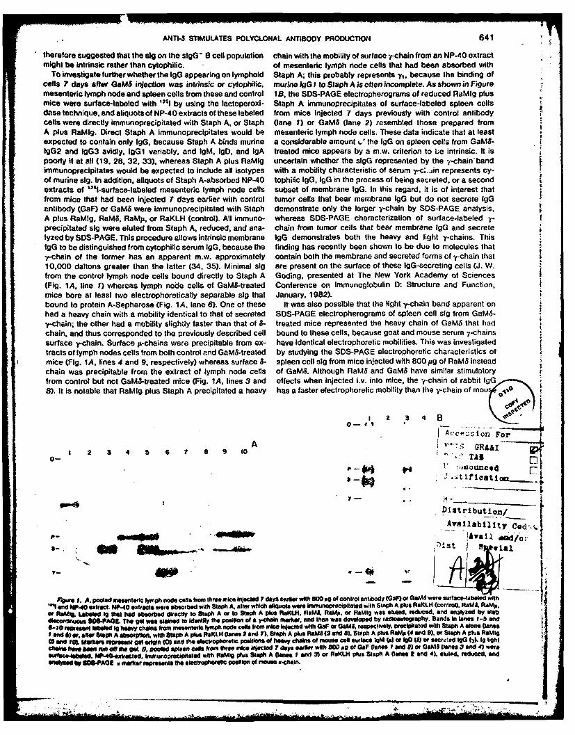
To investigate further whether the IgG appearing on lymphoid Staph A; this probably represents y,. because the binding ofcells 7 days after GaM6 injection was intrinsic or cytophilic, murine EgG I to Staph A is often incomplete. As shown in Figuremesenteric lymph node and spleen cells from these and control 1 B, the SDS-PAGE electropherograms of reduced RaMIg plusmice were surface-labeled with 'I by using the lactoperoxi- Staph A immunoprecipitates of surface-labeled spleen cellsdase technique, and aliquots of NP-40 extracts of these labeled from mice injected 7 days previously with control antibodycells were directly immunoprecipitated with Staph A. or Staph (lane 1) or GaMS (lane 2) resembled those prepared fromA plus RaMIg. Direct Staph A immunoprecipitates would be mesenteric lymph node cells. These data indicate that at leastexpected to contain only IgG, because Staph A binds murine a considerable amount .' the IgG on spleen cells from GaM6-IgG2 and IgG3 avidly. IgGi variably, and IgM. IgD. and IgA treated mice appears by a m.w. criterion to be intrinsic. It ispoorly if at all (19. 28. 32. 33). whereas Staph A plus RaMig uncertain whether the slgG represented by the -f-chain-bandimmunoprecipitates would be expected to include all isotypes with a mobility characteristic of serum y-c..in represents cy-of murine slg. In addition. aliquots of Staph A-absorbed NP-40 tophilic IgG. lgG in the process of being secreted, or a second I
extracts of 1251-surface-labeled mesenteric lymph node cells subset of membrane IgG. In this regard, it is of interest tnatfrom mice that had been injected 7 days earlier with control tumor cells that bear membrane IgG but do not secrete IgGantibody (GaF) or GaMS were immunoprecipitated with Staph demonstrate only the larger '--chain by SDS-PAGE analysis,A plus RaMIg. RaM8, RaMA, or RaKLH (control). All immuno- whereas SDS-PAGE characterization of surface-labeled -
precipitated sig were eluted from Staph A. reduced, and ana- chain from tumor cells that bear membrane IgG and secretelyzed by SDS-PAGE. This procedure allows intrinsic membrane lgG demonstrates both the heavy and light y-chains. ThislgG to be distinguished from cytophilic serum lgG. because the finding has recently been shown to be due to molecules thaty-chain of the former has an apparent m.w. approximately contain both the membrane and secreted forms of y-chain that10.000 daltons greater than the latter (34. 35). Minimal slg are present on the surface of these IgG-secreting cells (J. W.from the control lymph node cells bound directly to Staph A Goding. presented at The New York Academy of Sciences(Fig. 1A, line 1) whereas lymph node cells of GaMS-treated Conference on Immunoglobulin D: Structure and Function,mice bore at least two electrophoretically separable slg that January, 1982).bound to protein A-Sepharose (Fig. 1A, lane 6). One of these It was also possible that the light y-chain band apparent onhad a heavy chain with a mobility identical to that of secreted SOS-PAGE electropherograms of spleen cell slg from GaM6-y-chain; the other had a mobility slightly faster than that of 8- treated mice represented the heavy chain of GaMs that hadchain, and thus corresponded to the previously described cell bound to these cells, because goat and mouse serum -chainssurface y-chain. Surface A-chains were precipitable from ex- have identical electrophoretic mobilities. This was investigatedtracts of lymph nodes cells from both control and GeMS-treated by studying the SDS-PAGE electrophoretic characteristics ofmice (Fig. 1A, lines 4 and 9. respectively) whereas surface 8- spleen cell slg from mice injected with 800 pg of RaMS insteadchain was precipitable from the extract of lymph node cells of GaMS. Although RaMS and GaMS have similar stimulatoryfrom control but not GaMS-treated mice (Fig. 1A. lines 3 and effects when injected iv. into mice, the -y-chain of rabbit IgG ,8). It is notable that RaMig plus Staph A precipitated a heavy has a faster electrophoretic mobility than the v-chain of mou~A~
O - f t -
AcePL;sion ForA "" GRAAI
I 2 3 4 5 6 7 a 9 10 rr0--" " TAB
a J t Ificatioui.. t
Ditributon/
Availabllity Ced,.l
;)ist19sa
7- A1
dUPXsWe 1. A. e mesentea/s lymph node coels from three mice Injected 7 days earlier with 800 po of control antibody (GaF or GWM were surface-labeled with
"4t and NP-40 extract. NP-40 extracts were absorbed with Staph A. aflter which eliquots were immunoprecipitated with Staph A plus RaKLH (control). RaMS. RaMp.
Or RaMI. Labeled le that had absorbed direcily to Staph A or to Stao A plus RaKLH. RaMS, RaM. or RaMig was euted. reduced, end analyzed by slabdiscontuots 1S-PAGI. The gel was stalined to identify the postion Of a ihain marlik. and then was developed by radlcWftaphy. Bands In Ines 1-3 and
6- 1O sent labeled Ig heavy chains from mesenteatc lymph node ceis from mice injocled with GaF or GME, respectively. precipitated with Staph A alone (lanes& d 6) e, aftfr Saph A absorption, with ftph A plus AKLH (lanes 2 and 7). Staph A plus RaMS (3 and 8). Staph A plus RaM p (4 and 9). or Slph A plus RaMig
15 end 10). Ma rkor rewee gel origin (0) and the electropho eti positions of heavy cha of mouse cell Surface IgM ) of 1o (6) or secre ted IgO ( T). Ig lightshi n ht r b ea e 6 uW no of gee 0 pooled apeen ca i FrOm * ee mice Iietod 7 days earloer with 800opg of OAF 00e 1 and ) or O WS anes 3 and4) wereurface.Ieabeled. NP-40.extateled. immunopreoipitoed with RMI9 plus Staph A Danes I and 3) or RsKLH plus Staph A (lanes 2 and 4). eutled, reduced, and
Ieeo by /SOS.PA aII. marker represents the electrophorfetc position of om xr.halin.
.. ..
i N h . > jv

842 ANTI-8 STIMULATES POLYCLONAL ANTIBODY PRODUCTION
TABLE IllStimulati'on of immunogiobuin secretion by 00e46
Percent @9 5lesel eii thi wee- GPM @9 -L~cie kInC~oralgd into Silaieted"MieDy___lid r~c~ clitlasmlc_____________ _______
NUM 590 P Totw Proten
BALD/c 5 NI 0 I;G 0.9 (1.45)Y <0.2GeMW 2.2(0.53) 0.4(2.03)
6 NI G I;G 0.3(2.07) 0.5(1.60)Gems 4.92(1.04) 3.110.57)
7 NI G 0.4(1.09) 0.5(2.14) 643 (2.56111 1.480(1.17) 216.000(1.54)GateS 2.5(1.67) 15.7(0.28) 3.680(1.57) 70.300(1.29) B69.000(1.47)
10 tJItQGO 0.9(1.14) 1.301.12)GateS 0.5 (1.23) 4.9 (1 .97)
13 N G IgG 0.501.23) 0.3(2.19) 3.10(1.81) 722 (2.54)GaMb 0.8(1.34) 1.0(2.30) 2.9110(11.98)1 2.630 (1.71)
BALD/c lu/lnu 7 NI G IZG 2.6(1.29) 0.8(2.90) 7.100 2.000 83.800Gems 1.7(2.56) 1.2(1.45) 23.000 4.060 174.000
BALD/c tolerized to NI G IgG 7 NI G IgG 0.5(1.24) 0.6(1.47) 860 (1.19) 95501.53) 1 61.00C (1. 12)GaMb 1.2(1.21) 1.5 (1.42) 44a8(1.28) 926(1.53) 159.000(0.50)
BALB/c 7 CBPC-101 1.1 (2.93) 0.30(1.51) 7.610 2.2204.22a.Ve 5.00(.73) 6.40(.96) 48.800 65.400
SCIF. 7 CBPc-1li 0.90(.20) 0.3(1.78) 12.140 2.0004.22al'o 0.7 (2.73) 0.4 (2.08) 3.720 2.200
Cytocentufuge preparations of spleen cells from mice in~ected 5 to 13 days earlier with anti-B or control antibody were fixed. stained with FITC-Ramp, or IFITC-* RaMy. and ware examined with a Leitz Ortholux phaso/fluorescence microscope fot percentage of cells with bright intracytoplasnic fluorescence. At leas! 500 cells
were examined on each slit.6Spleen cells from mice Injected 5 to 13 days earlier with anti-S or control antibody were Incubated overnight in Ieucine-tree RPNII 1640. supplemented: with 'fl-
leucine (0. 1 mQ/mI; 4 X 100 spleen cells/mQ for 18 hr. Supernatants of cell cultures were dialyzed anid aliquote were immunoprecipitated with Rateig ar.:;body plus* Staph A. Washed immunoprecipitates were eluted. reduced, and analyzed by SOS-PAGE. SDS-PAGE Vals were fractionated with a Gilson gel mincer; fracl;ons were
suspended in Aquasol and counted by scintillation apectroscopy to delermine 31l-cpm in p- and -chain peaks. Aquots of dialyzed supernatanhs were elvo precip.:atedwith cold 10% trichloroacetic acid, flttered onto cellulose dscs. suspended in Aquasol. and counted ty scint,!ation spectroscopy. All cpm are normalized to I-mt
* volumes of dialyzed supernatant.c Geometric mean (geomnetrc standard deviation) of determinations made with spleen cells from tbrae to fire mice. Where rno figures in parentheses are shown.
results were determined with pooled spleen cells from three to five mice.
serum lgG and thus can be differentiated by SOS-PAGE. SOS- TABLE IVPAGE electropherograms of reduced 1251-labeed spleen cell Effect of Ga.tISl on serum imiruniogobdmn levels"Sig from mice injected 7 days previously with RaMS demon- M"a Awbody Serum 'a Conicentration tmige:;'
strated clear bands characteristic of heavy and light mouse yr- he-letd 1WM 10 %GC2achain, but no bands characteristic of rabbit r-chain (data not BALB/c NIGCIgG 38.1 (1.02) 174 (1.54) 178 (1.63)shown); thus little or no RamS bound to the surface of such Gams 68.9(l.41) 1.220 (1.12) 344 (1.55)
spencel.BALB/c tolerized N G 19G 38.3 (1.33) 337 (1.64) 223 (1.38)spleencells.Gay's 235(0.27) 218a(2.04) 195(1.02)Effects of GaMS on Ig secretion. The observation that GaMS S eta were obtained from normal BALD/c mice as well also BALD/c mice
Induced the differentiation of slgM* lymphocytes into slgG* tolerized to goat IgG 7 days after injection of 800 Ito of NI G 1gG or 433MJ. anidcells suggested the possibility that the same antibody also were analyzed for 1gM. Wgt. and lgG2a concentration by the radial immunodif-
fusion technique.setiulated differentiation of resting B cells Into Ig-secreting *Geometric mean (georetric: atandard deviation) of sara from three mice.cells. This was investigated by staining spleen cells for clgMand clgG. by measuring the incorporation of 3H-leucine into of clgM* and clgG* spleen cells in response to 800 lug of1gM, lgG. and protein by cultured spleen cells, and by measur- 4.22aMS8, BCF1 mice showed no such increase (Table 1ll).ing serum Ig levels of mice injected with GaM8 or control goat Because it was possible that the intracytoplasmic staining1g. As shown in Table IV. a slight increase In the percentage of technique was detecting cells that were Synthesizing but notspleen cells with clgM was noted 5 days after GaMS injection, secreting Ig, the incorporation of 3H-leucmne into 1gM and lgGbut no Increase In the percentage of ctgG* cells was seen at In cell culture supernatants was studied as a direct means ofthis time. By 6 days after GeMVS Injection, however, consider- quantitating secreted Ig and comparing It to total protein secre-able increases In the percentages of both clgM* and cIgO' tion. As shown in Table Ill, spleen cells cultured 7 days afterCeONS Were noted, and by day 7. the percentage of clgG* cells GaMS injection incorporated six tirns more 3H-teucine intohad increased to over 30 times the control value. At this time, secreted jt-chain. 48 times more 3H-leUCIne Into secreted y-greater than 20% of the splenic B cell population was clgG4'. chain, and four times more "'If-leucmne Into total secreted pro-Trhe majority of cIgO * cells appeared microscopically to be tein than did an equal number of spleen cells from control mice.blasts (large cells with a central nucleus and a high nuclear to Thus, the GeM 6stimulated Increase In lgG secretion wasCytoplasmic ratio) rather than mature plasma cells. The In- greatly in excess of the Increase In total protein secretion.crease In the percentage of c~lg' cells was T-dependent end Considerable Increases In Ig secretion were also noted withcarrier-dependent. Neither congenitally athymic mice nor mice spleen cells from DAIS/c mice Injected with .4.22aM&.d" how-tole0rized to goat 9Ga generated Increased percentages of clg* ever. neither spleen cells from goat IgO-toterant mice injectedspleen cells In response to GeMS Inje~ction (Tab.le Ill). In addi- wititOaMS nor BCF% mice injected with 4.22aM6" demonstratedtDoor while BALD/c mice generated c , iiderable percentages Increased 19 secretion (Table 11l). Although spleen ce'ls from

ANTI-I STIMULATES POLYCLONAL ANTIBOOPRODUCTION 643.
Congenitally athymic mice injected 7 days previously with GaM6 cretion within 7 days of injection, because secreted anti-goatdid show increased incorporation of 3H-!eucine into secreted Ig might have been absorbed in vivo by injected GaMS. To1g. the increase was proportionate to an ircrease in total protein investigate this possibility 2-mi aliquots of supernatants ofsecretion. and probably represents something other than the spleen cells cultured in 3 H-leucine-containing medium were Xappearance of increased numbers of antibody-secreting cells, allowed to adsorb for 30 min to a 1 x 9 cm column of goat
Measurement of serum Ig levels from,, GaM6-injected and serum globulin-Sepharose, which had approximately 10 mg of -
control mice gave results consistent with the intracyoplasmic a 50% saturated (NH 4)2SO. cut of goat serum bound per 7
staining and internal labeling studies. As shown in Table IV. milliliter of Sepharose. This column was then washed with twosera drawn from BALB/c mice 7 days afier GaMS injection had column volumes of 0.1 M Tris, pH 8.3, (absorbed fraction) and i-IgG I levels that were sevenfold increased over control values, was eluted with 3.5 M MgCI 2 (eluted fraction). Ovalbumin was A.
as well as slightly increased IgG2a and IgM levels. Serum added to the eluted fraction to a concentration of I mg/ml.IgG2b and IgA levels were not increaseci by GaMS injection after which it was dialyzed against 0.1 M Tris, pH 8.3. Both(data not shown). As expected, mice zolerized to goat IgG absorbed and eluted fractions were then concentrated to 2-mlbefore GaM6 injection did not show increased serum IgG or volumes with an Amicon Minicon concentrator. RaMIg plusIgM levels. Staph A immunoprecipitates were prepared from bound and
The GaM8-induced increase in clg secretion is polyclonal. eluted fractions and were analyzed for cpm of g- and y-chains ,The above data show that the in vivo injection of GaMS induces as described earlier. As shown in Table V. approximately one- ,a marked increase in IgG secretion that appears to involve the third of the total cpm of p- and -f-chains were in the eluteddifferentiation of a substantial percentage of splenic B lympho- fraction. These results do not necessarily mean that one-thirdcytes into antibody-secreting cells. AltJhough the large per- of the Ig secreted by spleen cells from GaMS-stimulated micecentage of clg" cells found strongly sug;ested that this was a was goat serum globulin-specific; although the high antigen topolyclonal process, we felt it necessary to investigate the antibody ratio, long absorption period, and limited wash of thepossibility that the increase in Ig secretion was an antigen- column allowed binding of low affinity antibody to the goatspecific response directed entirely to the immunogen (goat Ig) serum globulin column, it also maximized nonspecific binding.because previous anti-S stimulation stud' cs with monkeys, rats, To estimate the extent to which nonspecific binding did occur,and mice had demonstrated this finding (18, 36-41). As a first a similar experiment was performed with the 3H-labeled super-step, cytocentrifuge preparations of sp~een cells from mice natant of cultured spleen cells from BALB/c mice injected 7 4injected 7 days earlier with GaM8 were stained with FITC- days before sacrifice with 4.22aMS. Because this antibody islabeled NI G IgG and were examined for the presence of cells a murine Ig. it is unlikely that cells stimulated by it wouldwith intracytoplasmic fluorescence. Wh:!e 10 to 20% of the secrete an appreciable amount of antibody specific for goatcells in these preparations demonstrated intracytoplasmic flu- serum gobulins; however, approximately 15% of the IgM andorescence after staining with FITC-RaM- and FITC-RaMu. less 10% of the IgG in this supernatant bound to goat serumthan 0.2% of cells in any preparation sho'ved intracytoplasmic globulin-Sepharose under the conditions used (Table V). Thus,fluorescence after staining with FITC-NI G IgG. This result was it may be estimated that as much as 20% (33% minus 13%) ofnot definitive, however, because it is uncertain whether cells the Ig secreted by GaMS-stimulated mice represents a specificsynthesizing anti-goat Ig antibodies of low affinity would be response to goat serum globulins, while approximately 80%detected by this technique. For this reascn, the ability of goat represents a true polyclonal response.Ig to absorb Ig from the sera of GaMS-incculated mice was also To investigate further whether the GaMS-stimulated antibodyexamined. Sera from each of two mice injected 8 days previ- response was in large measure polyclonal. the in vivo effectsously with 800 /g of NI G igG or GaMS were incubated overnight of GaMS on the generation of cells secreting antibody to anwith equal volumes of either whole goat serum, heat-insolubi- antigen unrelated to goat serum globulins was studied by i,lized goat IgG. or saline, centrifuged to remove insoluble ma- comparing the number of direct anti-TNP plaque-forming cellsterial, and then were analyzed by radial immunodiffusion for (PFC) in a modified Jerne plaque assay (42, 43) with the totalIgG1 content. None of these absorptic-is decreased serum number of IgM-secreting cells in a protein A reverse plaqueIgG1 levels by more than 5% (data nct shown). This result assay (44). As shown in Table VI, 7 days after its injection,strongly suggested that GaMS induced polyclonal IgG1 secre- GaMS enhanced the numbers of both kinds of PFC by similartion. It did not, however, eliminate the possibility that GaM8 factors (seven to ninefold), whereas 14 days after GaM8 injec-also stimulated considerable specific anti-goat Ig antibody se- tion neither the frequency of IgM-secreting cells nor that of IgM
anti-TNP-secreting cells was increased. Similar results wereTABLE V seen in three additional experiments. This demonstration thatAesopion of intrnll laibeled irmmuogebuin screfted by sleen cells from GaMS stimulates the differentiation of cells secreting antibody
'-mew- .C O to an unrelated antigen provides further evidence for the poly-
clonal nature of the GaMS-induced antibody response.Ar :l>'.y ni~le Acawead to Goat $eM,- N id ,..gdl to Goat Serum- -seataroae stoawi
P 7 p TABLE VIGalMd indaces similar increases in the frequencies of total 1gM-secreting cellsGms 14.000 129.000 30.000 255,000 nd IgM anti-TriP-secreting cells'
4.22aMP 3.600 5.090 23.0G7 51.100• . lltyle. Haecle otrmll lllell"Jcutueaulenaans roI NI G 1gG 36,5 si: 25* 7 * Ilrd .l nternay label supernatants fmGaM 2.546 * 911 97 * 16
grsps f three mice ti'at had been injected 7 days evrlier wilh 800 #g of GeMS 14 NI 0 Ig 54S * 145 <Sor 4.2aMr were prepared as described in Table IV Labeltd supernatants were SaMs 605 2-125 1Sallowed to adsorb to a column of the 50% saturated (:H.) SO. cut of goat serumbomnd .0 C0ter-ictivaed Sepharose. after which rJWTns were briefly washed "Spleen cells from BALB/c mice injected 7 days earlier with 900 pg of Ni Gand then elut d with 35 M MgCI a. The eluted adt n.:bd fraction was dialyzed G or GaMV were plaqued against protein A-coated sheep erythrocytes in theaiainst 0.1i M Trig. 041S 3. aftir which both it and :he "raction that did not adsorb presence of RaMp antibody or agalmst TNP-hiapltenated sheep erythrocytes tot.0 the goat aerum-Selgrarose were concentrated el Amicon MVnicon concenlra- determ;ne the total numbers of tgM-secreting cels Or igM anl-TNP.secretingWrS and We IfmftnopreCipliated with ReMIg plus S:aph A. CPM of p- and y. cells per 10e apleen cells plaqued.
chains in both fractons were determined as descr,.,,.1 in Table IV. * Arithmetic mean I sandard deviation of PFC from three mice.
;B ... ..............................................................

644 ANTI-S STIMULATES POLYCLONAL ANTIBODY PRODUCTION
I TABLE. V1IgG subclass distribution of spleen celts with surface or intracytopasmic IgG 7 days after anti" kjection"
Pvcent Sp t" Cells with Surface Poert.rl Splee. Cels .'.*1, acylo -icCaifx9 AntodyID Ig G2 IgG3 IQG Igol 19G2 t2G3
I NI G IgG 4.1 1.2 1.5 1.5 0.5(2.14)"
Sams 27.2 18.3 7.4 2.8 15.7(1.28) 15.0 (. 15) 0.5(1.42) <0.2
2 NI G lgG 3.9 1.1 1.0 1.0GaMs 21.7 12.3 1.0 1.0j 3 NI G IgG 2.3 2.8 0.9(2.05) 0.4(3.65)GaMs 18.3 3.9 9.(1.111) 1.4(t.62)
4 RaKLH 4.6 2.6 1.2 2.1RaMs 26.1 25.0 3.4 2.3
5 CBPC-1Ot 5.2 1.7 1.5 <1.0 0.3(.51)4.22aMt" 12.9 6.1 5.3 <1.0 6.4 (1.96) 5.1 (1.96) 0.5 (2.27)
* Spleen cells from mice injected 7 days before sacrifice with 800 pg of anti-B or control antibody were analzed as described in Tables II and IV for surface orIntracytoplasmic IgGI. IgG2. or IgG3. by using affinity-purified F(ab'), fragments of rabbit antibodies specific for these isotypes. Surface Ig staining was performedwith spleen cells from individual mice.
* Geometric mean (geometric standard deviation) of values obtained from three mice.
The polyclonal antibody response induced by GaMe is pri- injection; however, as previously reported, injection of thismarily of the IgGI isolype. Experiments were performed to antibody failed to raise B cell sla levels. In contrast to threedetermine the subclass distribution of sIgG* and clgG* spleen antibodies with 6-chain specificity (4.22aMS'. RaMS, andcells from GaMS-treated mice, as well as the possible relation- GaM8) this antibody failed to induce an increase in the percentship between slgG and clgG+ cells. As shown in Table VII. a of spleen cells with slgG or clg (data not shown).large majority of the slgG+ cells, and almost all clgG cells Collapse of the activated immune state. Spleen cells fromwere of the IgG1 isotype. This was true regardless of whether mice injected more than 7 days previously with GaMS wereGaMS, RaMS, or 4.22aMSO was the anti-S antibody used. This evaluated for the presence of slg and clg. The percentage ofcorresponded to the assay of serum Ig levels, mentioned clgG+ cells was considerably decreased by 10 days afterabove, which indicated that GaMS induced a greater increase GaM8 administration and had almost returned to baseline at 14in IgGI levels than in IgG2a or IgG2b levels. days after GaMS administration (Tab:e II). Investigation of bone
To investigate whether there was a relationship between marrow cells 14 days after GaMS inlection also failed to showcells that bore slgG and cells that contained cIgG, spleen cells an Increased amount of Ig secretion, determined by internal
from mice injected 7 days previously with GaMS were stained labeling and SDS-PAGE studies (da'a not shown). The increasewith FITC-RaMS and were sorted with an FACS into slgG+ and in the percentage of slgG cells in spleen outlasted the in-slgG- populations. Reanalysis of the sorted populations indi- crease in clg * cells, a~though it too was greatly decreased 14cated that only 6% of the "sIgG-" population bore slgG, days after GaMS injection (Table I). In addition, slgG' spleenwhereas slgG could be detected on 72% of the cells In the cells detected 13 days after GaMS in,*ection were predominantly"slgG +'" population. Cells from both sorted populations were small lymphocytes rather than the slgG blasts detected 7
cytocentrifuged onto glass slides, fixed, and stained for clg days after GaMS injection. The percentages of B cells (slawith FITC-RaMy and FITC-RaM/. The percentages of clgG+ cells) 13 and 14 days after GaMS injection were decreasedcells In the sgG + and slgG- populations were 16.2 and 0.^% below the baseline value; this was particularly apparent for the
respectively; the percentages of cIgM cells In the sIgG and sIgM + slgD B cell subset (Table I). An increase in the percentslgG- populations were, respectively, 3.2 and 10.8%. Thus, of Thy-i.2 spleen cells 14 days after GaMS injection corre-the majority of cIgG cells 7 days after GaMS injection also sponded to the decrease in the percent of splenic B cells.bear sIgG. whereas the clgM cells are mostly sgG-. This DISCUSSIONassociation between slg and clg of one isotype and the sug-gested mutual exclusivity of the clgG + and clgM populations The data presented here confirms our previous report that it,corresponds to the apparent lack of overlap between the slgM + vivo injection of anti-S antibody induces an increase in B celland sIgG+ populations mentioned above. sla (31), and provides the initial demonstration that the same
Specificity of GaMS-induced polyclonal activation. Because treatment induces a polyclonal IgM to IgG switch in B cellGaMS can form immune complexes with serum and cell surface surface isotype as well as the simultaneous appearance ofIgD and 400 jg of heat-aggregated rabbit IgG has been large numbers of Ig-secreting cells. Although the increase in Breported to induce a polyclonal antibody response when in- cell sla appears to be a direct effect of the slgD-anti-8 interac-jected in vivo (45), we immunized mice i.v. with 400 pg of heat- tion, the slg Isotype switch and the differentiation of B lympho-aggregated goat IgG and examined their spleen cells 5 and 7 cytes into antibody-secreting cells both Involve T lymphocytesdays later for the presence of slgG cells and incorporation of and require recognition of the anti-S molecule as foreign. The3H-leucine into IgM and IgG. Little or no Increase was found, observation that CWMS predominantly stimulates IgGi secre-although GaM8 stimulated a strong polyclonal response (data tion Is consistent with a T-dependent model of anti-6 activationnot shown). The ability of a monoclonal rat antibody with of antibody secretion, because soluble, T-dependent antigensspecificity for the ThB determinant (present on almost all B stimulate a predominantly IgG1 response, whereas T-inde-lymphocytes and approximately 50% of thymocytes but not on pendent antigens stimulate significant IgG2 and IgG3 antibodymature T cells) (46) to stimulate polyclonal B lymphocyte responses (47, 48).activation was also Investigated. Intravenous Injection of 800 The data presented in this and the accompanying paperpg of this antibody substantially diminished the amount of the suggest that antigen-induced B cell activation proceeds in atThB determinant on spleen cells both 2 and 7 days after least two stages. In the first stage. increases In B cell a. B
0aa

ANTI-6 STIMULATES POLYCLONAL ANTIBODY PRODCT ION 645
-ceN size. and the rate of B cell DNA synthesis can be seen. As activation. In vitro studies have established trt anti-it anti-described In the companion paper, these changes lead to bodies, like anti-8 antibodies, can stimulate B y,, phocytes toactivation of T cells specific for determinants on the anti-4 increase their sla (31). to proliferate (2-6. 8. 11). and whenmolecule. The second stage results in further stimulation of the further stimulated, to differentiate into antibody-secreting cellsactivated B lymphocyte by thb activated T lymphocyte or its (10). Furthermore, antigens injected into mice that lack slgDjproducts. The increase in B cell sla may facilitate both antigen B lymphocytes as a consequence of injection from birth withpresentation to T lymphocytes and B lymphocyte acceptance rabbit anti-3 antibodies still make good antibody responses toof T cell help. At least two models of B cell activation that are sheep erythrocytes (SRBC). TNP-SRBC, TNP-KLH. and TNP-consistent with our data offer mechanisms for the specificity of Ficoll (52. 53), although they fail to respond to injected GaMSantibody responses to T-dependent antigens. In one, carrier- (E. S. Metcalf et al., manuscript in preparation). Thus, it seemsspecific T helper factors would bind 'o antigen (or anti-S) probable that ligand-slgM and ligand-slgO interactions canmolecules that had bound to slg and stimulate differentiation have similar B cell-activating effects. We have previously sum-towards antibody secretion (49). In a second model hapten marized evidence that 'aads us to believe that an importantspecificity would be restricted to the initial stages of B cell functional difference between slgM-ligand and slgD-ligand in-activation (i.e.. helper factors would be generated that are not teractions is that slgM-ligand interactions have a greater directcarrier-specific, but only those B lymphocytes initially activated inhibitory effect on the differentiation of B cells into antibody-by antigen or anti-Ig cross-linking of slg could respond to these secreting cells than do sigD-ligand interactions (18).factors by generating clones of antibody-secreting cells) (50). The stimulatory effects of anti-Ig antibodies on B cell acti- -Because experimental evidence supports the existence of both vation do not result simply from the binding of ligand to anycarrier-specific and nonspecific helper factors, there is no cell surface molecule. The injection of 400 pg of heat-aggre-reason not to believe that both mechanisms of T-dependent B gated goat IgG or 800 pg of a monoclonal rat antibody to the tcell activation play a role in the phenomena we have described. ThB determinant failed to increase B cell sla or induce B cells
The extent of B cell activation induced by GaM, injection to proliferate or differentiate into antibody-secreting cells. The.strongly suggested that the immune response was polyclonal. lack of effect of the latter antibody was not a consequence of
Our data indicate that although most control mice have less its monoclonality because 4.22aMS . a monoclonal antibodythan 10 antibody-secreting cells out of approximately 108 total with relatively low affinity for IgD, was capable of inducingspleen cells, mice injected 7 days earlier with GaMS often have polyclonal B lymphocyte activation.5 X 10' antibody-secreting cells in a spleen containing 3 to 4 The rapid collapse of the activated immune system after theX 106 nucleated cells. The polyclonal nature of the GaMS- catabolism of injected GaMS suggests, as discussed in ourinduced antibody response was confirmed by studies that previous paper, that polyclonal activation may stimulate poly-showed most of the Ig produced by stimulated mice lacked clonal suppression. Indeed, spleen cells taken from mice 7specificity for goat serum globulins, and that the increases in days after GaMS injection suppress a number of in vitro profit-the frequencies of total IgM-secreting spleen cells and IgM erative responses of spleen cells from normal mice (J. Mond et
j anti-TNP-secreting spleen cells were nearly identical. The de- aL, unpublished data). While the putative suppressor mecha-termination that the GaMS-stimulated Ig response was poly- nisms decrease numbers of both sIgG and clgG spleen cellsclonal rather than specific for goat Ig indicates that a ligand- substantially after day 7. the decrease in the number of clgG*slgD interaction, in the absence of a ligand-slgM interaction, cells occurs more rapidly. Because the persistent slgG* cellscan both induce B cells to proliferate and initiate a series of are small clgG- lymphocytes rather than the clgG' blasts seenevents that leads B lymphocytes to differentiate into antibody- 7 days after GaM8 injection, the former slgG* cell populationsecreting cells. This result suggests that the correct interpro- may consist of memory B cells that are somewhat less suscep-
tation of previous experiments in which anti-# antibodies tible to suppression than are antibody-secreting cells. Thus,blocked a wide range of in vitro antibody responses (13-18) is the injection of mice with large doses of GaMS antibody pro-that the anti-p-slgM interaction blocked antibody production vides a polyclonal model of at least one mechanism of B cellthrough a direct inhibitory effect on B cell differentiation rather activation by a T-dependent antigen that generates F -;Is thatthan by the prevention of a required antigen-slgM interaction, have undergone surface isotype switching and differentiation
Our results differ from previous in vitro studies that indicated into antibody-secreting cells and possibly memory cells as wellanti-S antibodies could induce B cells to differentiate Into as activated T helper and possibly T suppressor cells in num-antibody-secreting cells only when they were further stimulated bers that have previously been difficult to obtain. Our model,by T-dependent helper factors (10-12), treated with proteolytic therefore, Is useful both in elucidating B cell activation and inenzymes, or thoroughly washed (51); these additional treat- providing activated cell populations for further study. Ourments proved unnecessary for in vivo activation of antibody model, however, most likely illustrates only one of several T-production. These differences may result from anti-8 having dependent mechanisms that can lead to B lymphocyte activa-less of a direct Inhibitory effect on B cell differentiation in vivo tion. It is unlikely, for example, that the Injection of very smallthan in vitro, and/or a greater ability of anti-8 to stimulate quantities of antigen in adjuvant could directly induce an in-activation of helper T cells under in vivo conditions. In addition, crease in sla or an increase in the proliferation rate of anthere may be qualitative differences between the in vitro and in antigen-specific B cell population, yet such stimulation canvivo mechanisms of anti-S-stimulated B cell activation; anti-Ig generate strong antibody and memory responses. The factor-plus mitogen-stimulated helper factor fail to induce spleen cells generating and antigen-presenting roles of macrophages mayfrom mica with the CBA/N Immune defect to proliferate or be greatly enhanced by the presence of adjuvant in such andifferentiate Into antibody-secreting cells in vitro (10), whereas Immune response, and the triggering of B lymphocyte prolif-GaMS injected Into these mice induces both (L. Muul et at., eration and differentiation may be fundamentally different frommanuscript In preparation), that studied in our system (i.e., when antigen Is injected with
Our data do not establish that a igand-sIgO interaction has adjuvant an antigen --# macrophage --o T cell - B cell stimu-
unique B cel-activating effects or Is required for B lymphocyte latory pathway may be followed, rather than the antigen B
L , , i i I IilllI i

8 46 ANT14 STIMULATES POLYCLONAL ANTIBODY PRODUCTION
cell -- T cell --1 B cell stimulatory pathway that we have 23. Kappler. J W.. B Skidmore. J. White. and P. Marrack. 1981. Antigen-postulated from our results). Studies In which relatively small inducible. H.2-restiied. literleukifl.2.producing T conl hybridomas. Lack
of independent anigen and H4-2 recognition. J. Exp. Med. 153:1198.doses of anti-I antibodies are injected in adjuvant are currently 24. Finkelman. F. U.. and 1. Scher. 1979. Rhesus monkey 8 lymphocyte surfacebeing performed to investigate whether the B cell activation immurtoglcbut'n: analysis with a fluorescence-activated cell sorter. 1_ Bin-
mechanisms involved can be manipulated to generate a poly- 2.munol. 122.1 757.25token. PA1. R . and L. A. Iterzenberg. 1975. Analysis of cell populations with
clonal response. a fluoirescence-activated cell sorter. Ann. N.Y. Acad. Sri. 254.163.26. Horan. P. K.. ard 1. L. Wheetess. Jr. 1977. Quantitative single cell analysis
Acknowledgments. We thank Drs. P. Fox. J. Kappler. P. and sorting. Science 193:149.* Marrack. K. Ozato. M. Potter. D. Sachs. and E. Vitetta for their 27. Finketrean. F. D.. and P. E. Lipsky. 1978. lmmunoglobulin secretion by
Drs.M. & ~, L A.Herznbeg. . Jcobon. human spleric lymchocytels in vitro: the effects of antibodies to 1gM and IgD.*gifts of reagents; DsM.t;zL.A ezn rgE.Jcbo, J. lInmurol. 1 2:1465.L. Muut. W. Paul, B. Perris. and J. Thorbecke for their helpful 28. Kessler. S. VI. 197%. Rapid isoation of antigens from cells with a slaphylo-discussions, and Mrs. J. Smith. Mr. E. Daco, and Mr. P. Acala coccat pro-ein A-antittody adsorbent parameters of the interaction of anti-
body-arti;en compleyes with protein A. J. limmunol. 115:1617.for their expert technical assistance. 29. Laceirni. U. K. 1970. Cleavage of structural proteins during the assembly of
the heed of bacterio;,hage T4. Nature 227:660..1REFERENCES 30. Kessler. S. W.. V.1L. Woods. F. 0. Finketman. and 1. Scher. 1979. Membraneorientation and locati. - of multiple and distinct allotypic determinants oft
I. Parker. D. C. 1975. Stimulation of mouse lymphocytes by insoluble anti- mouse maerrtrane lglD. J Immunoll. 123:2772.mouse immunoglobulin. Nature 258:361. 31. Mond. J. J.. E 5of-~sl, J. Kung. and F. 0. Finkelman. 1981. Increased
2. Weiner. H. L.. J. W. Moorhead, K. Yamaga. and R. T. Kubo. 1976. Anti- expression of I region--associeted anti;en CIal on B cells after cross-linkingIimmunoglobulin stimulation of murine lymphocytes [I. Identification of coll f surface imm-riogfcbulmn. J Immunot. 127:881.surface target molecules and requirements for cross linkage. J. Immunol. 32. Kronvall. C.. H. P.. Grey, and R. C. Witlams. Jr. 1970. Protein A reactivity117:1527. with mouse it munoglobulins. Structural relationship between some mouse
*3. Sieckmann. D. G.. R. Osofsky. D. E. Mosier. 1. M. Zitron. and W. E. Paul. and humnan imrnunc-globulins. J. Immunol. 105:1116.1976. Activation of mouse lymphocytes by anti-immunoglobutin. 1. Pararn- 33. Grey. H. M.1. J. V1. MaLst. and fal. Cohn. 1971. A new mouse immunoglobulin:eters ofthe proliferative response. J. Exp. Med. 147:814. lgG3. J. Exp. Med. 133:283.
4. Sieckmann. 0.0G.. 1. Scher, R. Asofsky, D. E. Mosier, arid W. E. Paul. 1978. 34. Oi. V. T.. V. 1.. Bryan. L. A. Herzenbcrg. and L. A. Herzenberg. 1sa0.Activation of mouse lymphocytes by anti-immunogtobuin. 11. A thymus- Lymphocite memnbrane IgG and secreted 19G are structurally and allolypi-independent response by a mature subset of B lymphocytes. J. Exp. Med. cally d.stincl. J. Esp. Med. 151: 1 20.148:1628. 35. Word. C. J.. and W. M. Kuehl. 1981. Expression of surface and secreted
S . Isakson. P. C.. K. A. Krolick. J. W. Uhr. and E. S. vitetta. 1980. The effect lgG2a by a mujrine 13--ymphoma before and alter hybridization to mnyelomaof anti-immunoglobulin antibodies on the in vitro proliferation and differen- cells. M.ol. Imnrunol. 13:311.Niation of normal and neoplaslic murine B cetlls. J. Immunol. 125:886. 36. Pernis. B. 1977. Lymphocyte membrane IgD. Immunol. Rev. 37;210.
6. Pure. E.. and E. Vitetta. 1980. Induction of murina 8 cell protiferation by 37. Cuchers. M. A.. L. N. Martin. and G. A. Leslie. 1978. The effects of anti-lgDIInsolubilized anti-fmmunogfobulins. J. Immunol. 125:1240. on serum imrnunogfob-Ains. an~ibody production, and immunoglobtin-bear-7. Zitron.1. M.. and B. L.Clevinger. 1980. Regulation of murine Bcells through . ng cetisin adult rats J. lmrnenol. 121:2257.
surface immunoglobulin. 1. Monoclonal anti-h antibody that induces aflotype- 38. Martin. L. M.. and G. A. Leslie. 1979. In vivo effects of antiserum to IgD onspecific proliferation. J. Exp. Med. 152:1135. .surface immunogtobutins. serum immurogiobutins. and lymphocyte blasto-
8. Sieckmann. D. G. 1981. The use of anti-immunoglobultns to induce a signal genesis in rhesu-s monkeys. 1-munolcgy 37:253.for cell division In B lymphocytes via their membrane IgM and IgO. Iminunot. 39. Finketman. F 0.. 1. Sch-er. E. S. Viteti, R. Ifabbersett. W. C. Vt-lcon. and J.Rev. 52: 18 1. W. Unr. 197q. Etioclts of in vivo injecltion of rabbit anti-human lgD in th-e
9. Sidman. C. L.. and E. R. Unanue. 1979. Requirements for mitogjenic stimu- rhesus nachey. In B Lymphocytes in the Immune Response. M. Cooper. D.*1 ton of murine B cells by soluble anti-1gM antibodies. J. Immunol. 122:406. Mosier. 1. Sch-er. and E. Vitetta. eds.. Elsevier/ North Holland. P. 1 59.10. Parker. D. C.. J. J. Fothergill. and D. C. Wadsworth. 1979. 8 lymphocyte 40. Finkelman. F. D.. end P. E. Lipsky. 1979. The rote of cell mnembraneactivation by Insoluble anti-iraimunoglobutin: induction of irninunoglobufin by irnmunoglotulfn in primate B lymphocyte differentiation. Immunot. Rev.
a T cell-dependent soluble factor. .!. Immunol. 123:931. 45: 117.11. Parker. D. C. 1980. InduL,ion and suppression of polycloral antibody 41. Finkelma.n. F. 0.. V. L. Wlood.. S. B. Wibturn, ce f 1930. Augmentation o!
responses by anli-Ig reagents and anligen-nonspecific helper factors: a in vivo humcra! irmmune responses in the mouse by an antibody to JOE) J_comparison of the effects of anti-Fab. anti-19M. and anti-IgO on murine B Esp. Med. 152:493.cefls. Immunof. Rev. 52:115S. 42. Jeme. N. K.. and A. A. Nord.n. 1963. Plaque formation In agar by single
12. Pure. E.. P. C. lsakson. K. Takatsu. of at. 1981. Induction of B cell differ- antibody-producing cet;s. Science 140:405.*1entiation byT7 cell factors. 1. Stimulation of 1gM secretion by products of a T 43. Ri'tenberg. M. B.. and C. Pratt. 1969. Anri-trinitrophenyl (TNP);,!aque assay.
cell hybridoma and a T cell line. J. Immunol. 127:1953. Primar; resporse of BALB/c mice to soluble and particulate irnmunogcn..413. Pierce. C. W.. S. M. Sotliday, and R. Asofaky. 1972. Immune responses in Proc. Soc. Exp. Diol. Med. 132.575.vitro. IV. Suppression of primary IM, yG and VA plaque-forming cell ro- 44. Gronowicz. E.. A. Coutinho. and F. Matchers. 1976. A plaque assay for allsponses in mouse spleen cell cultures by class-specific antibody to mouse cells secreting Ig of a given type or class. Eur. J. Immunot. 6:588.krnmunogfobulins. J. Exp. Med. 135:675. 45. Rosenberg. Y. J.. and J. M. Chiller. 1979. Ability of antigen-specific helper.414. Anderson. J.. W. W. Bullock, and F. Metchers. 1974. Inhibition of mitogenic cells to et'ec! a ciass-rastricted increase in total Ig-secreling cells in spleensstimulation of mouse lymphocytes by anti-mouse immunoglobulin antibodies. after immunization wirti the antligen. J. Exp. Med. 150:517.11 . Mode of actions. Eur. J. Immunoil. 4:71t5. 4. Yutoku. M, A. L Grossberg. and 0. Pressman. 1974. A cell surface
15. Zitron. 1. M., D. E. Mosier. and W. E. Paul. 1977. The role of surface Igo In antigenic determinant present on mouse Plasmacytes and only about halt cifthe response to thymic-independent antigens. J. Exp. Med. 145:1707. mouse thyrn.cytes. J. Inumunol. 112:1774.
16. Cambier. J. C.. F. S. Ligler. J. W. Uhr. J. R. Ketlrnan. and E. S. Vitetta. 47. Rosenberg. Y J. 19E.1. The atility of non.specific T-cell stimulators to induce1978. Blocking of primary in vitro antibody responses to thymus-independ. helper-call-de-rendernt increases in either otyclonal or isoltype-restricted Iga nt antigens with antiserum specific for 1gM or IgO. Proc. Nall. Acaed. Sci. production i~n Yvvo. Cell. Inuimuraf. 61:416.USA 75:432. 48. Perlmutter. Ft. M... M. Nahm. K. E. Stein. at at. 1979. Ittmunoglobulin4.17. Pure. E.. and E. S. Vitetla. 1980. The murine B cell response to TNP- subcllass-specific immunodeficiency in mice with an X-litiked B-lymphocytepotlyacrytamide beads: the relationship between the epitope density of the defect. J. Exp. M~ed. 149:993.antigen and the requirements for T cell help end surface 19D. J. Immunol. 49. 'Apte, R. 1!.. 1. Lcy. P. DeBastactier. and E. Mozes. 1931. Establishment-1125.420. and ctharacferizaiori or continuous helper T cell fines specific to poly-CL-Tyr.18. Flnkelman. F. D.. J. J. Mond. V. L. Woods. of at. 1980. Effects of anti- L-Glu)o;ly(o-.-Aa)-P~OIy(L-Lys). J. Immuncil. 127:25..4immunoglobutin antibodies on murine B lymphocytes and humoral immune 50. Andersson. J.. M. H. Schreier. and F. Melchers. 1980. T-cell-dependent B-response$. Immunoil. Rev. 52:38. cell stimulation is 1-2-restricted and an-tigen-depenident only at the resting
19. Ey. P. L.. S. J. Prowse. and C. R. Jenkin. 1978. Isolation of pure IgGI. B-cell level. Proc. flatr Acad. Sdi. UiSA 771612.IgG2a. and lgG2b immunoigicbulins from mouse serum using protein A- 51. Priml. D.. O.KX. tewls. arrdJ. Wi. Goodman. 1980, The role of immunogibglinSepharoso. Immunochemistry 15;429. receptorsarid T cell mvediators in B lymphocyte activation. 1. ft celactivation
20. Hunter. K. W.. F. D. Finkelman. G. T. Strickland. P. C. Sayles. and 1. Scher. by anti- irmunogtebulin and anli-ldiotype reagents. J. trnmunol. 125:12136.1980. Murine malaria: Analysis of erythrocyte surface-bound immunoglob- 52. Metcalf. E. S. L. Scher. J. J. &land. S. Wilburn. K. Chipman. and F. 0.dU'n b'y flow microfluorlmetry. J. tInmunoll. 125: 169. Finketman. 19,31. Effects of neonatal Onti-IgO treatment on the murine
21. Sachs.D. N .and J. L. Cone. 1973. A mouse -8 "cellalloantligen dtermined lymphoid system. In 8 Lymphocytes in the Immune 1esponse: Functional,dby geone(s) linked to the major histocompatibility, complex. J, Exp. Med. Developmental. and, Intoractive Properties. N. Klinman. D. F.. Mosier. 1.
I138:1289. Scher. and E.. S. Vitet'a. eds. Elsevier/North-H-olland. P. 211.-i22. Ozato. K.. and 0. H. Sachs. 1981. Monoclonal antibodies to mouse MHC 53. Jacobson. E B.. Y Bainke, Y.-W. ChOn. et 80. 1981. Physiulogy of 1gB. 1.antigens. Ill. Hybridoma antibodies rea~cting to antigens ofltho14.2hapoype compensatory t t er omena in B lymphocyte activation In mice treated withreveal genetic control of isofype expression. j. Immunol. 126:31 7. anli-IgO~ 2nlt.,_Ices J. Exp. Med. 154:3 18.