neonatal resuscitation for ems · pdf fileneonatal resuscitation for ems providers alix...
TRANSCRIPT

Neonatal Resuscitation for EMS Providers
Alix Paget-Brown, MD FAAP
Associate Professor, Division of Neonatology Associate Medical Director, UVA Medical
Transport Network, University of Virginia Charlottesville, VA

Goals
1. Identify the first steps of neonatal resuscitation 2. Identify aspects of field management which
complicate neonatal resuscitation 3. Identify airway differences between the neonate
and other age groups 4. Describe methods of basic neonatal airway
management 5. Identify several neonatal airway abnormalities,
airway emergencies and respiratory diseases 6. Describe management techniques for neonatal
airway emergencies and respiratory diseases

Outline
The Basics of Neonatal Resuscitation
Field Considerations The Neonatal Airway
Basic Neonatal Airway Management
Airway Abnormalities
Prehospital Management of Airway Emergencies
and Respiratory Diseases

Outline
The Basics of Neonatal Resuscitation
Field Considerations The Neonatal Airway
Basic Neonatal Airway Management
Airway Abnormalities
Prehospital Management of Airway Emergencies
and Respiratory Diseases

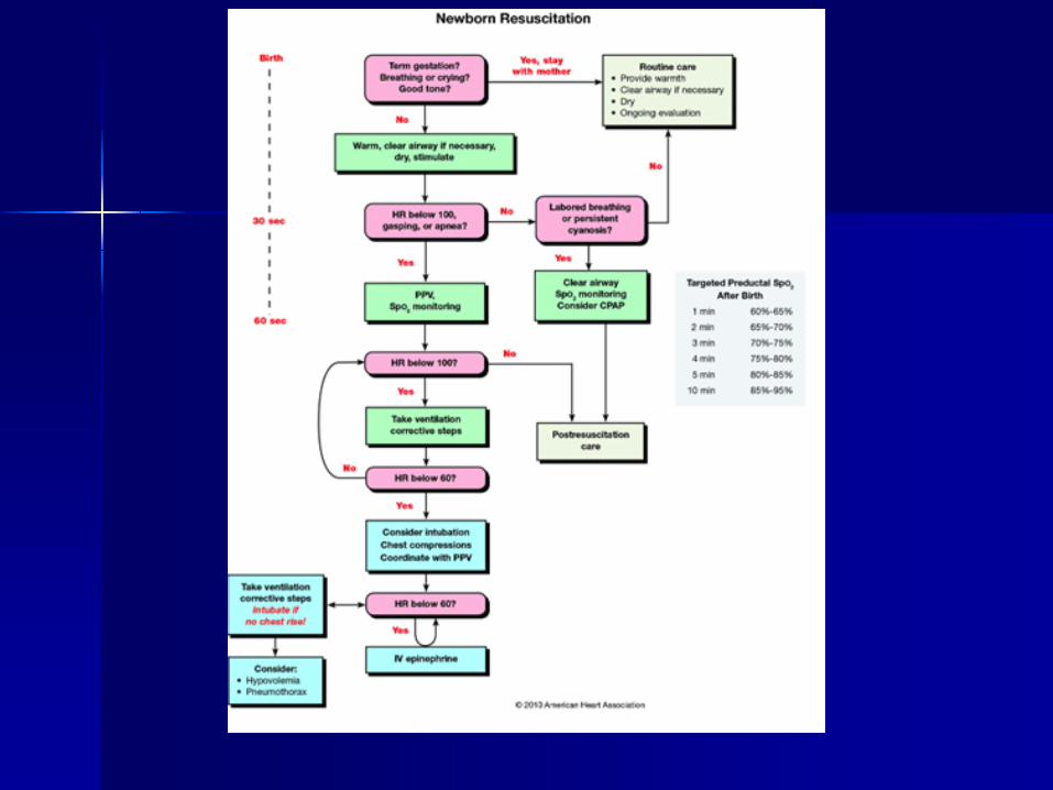
Basic Neonatal Resuscitation Clamp cord
Goals: – Assess – Airway Breathing Circulation
– Address findings – Warm, dry, stimulate
Do NOT place infant on Mom’s chest if it is not vigorous

Cord Clamping and Care
ACOG recommendation of 30-60 seconds of delayed cord clamping o Baby level with mom
Inappropriate position during cord clamping: – Hypovolemia
Delayed clamping, baby in high position
– Polycythemia Delayed clamping, baby held below mother

Cord Accidents
Tearing (avulsions) of the umbilical cord – Slicing through cord by using dental floss – Cord or clamp catches and tears – Tearing of cord after delivery
IMMEDIATELY – Pinch cord – Apply STRONG pressure above and below
umbilicus

Pressure Application
UA UV

Assessment (1)
Term Gestation? – Peeling/cracking skin – Vernix caseosum – Scant lanugo – Well defined palmar/plantar creases – Well defined genitalia

Breathing/Crying Color
– Acrocyanosis is normal – Color should be pink centrally within 15
minutes of birth – Mottling should not be present
Muscle tone
Assessment (2)

Color???

Full Term Newborn

32 week gestation

25-27 week infant

Size comparisons

Vital Signs
Breathing – Present? – Regular? – Labored? (grunting, intercostal
retractions)

Risks Specific to Premature Deliveries Immature lungs
– Respiratory Distress Syndrome Surfactant deficiency Signs
– Grunting – Retractions – Nasal flaring – Supplemental oxygen requirement – Difficulty ventilating
Complications – Pneumothorax – Chronic lung disease of prematurity (long term)

http://www.youtube.com/watch?v=J2R8MOoQtd8
Retractions


Vital Signs
Heart rate – >100 – normal – 60-100 – intervene with oxygen/PPV if
poor breathing – <60 – Supplemental oxygen/PPV and
chest compressions until heart rate >60
Perfusion – Color, mottled, capillary refill time

Poor Perfusion
Mottled appearance (above)
Delayed capillary refill time (below)
Indications of poor perfusion
Centralizing of perfusion

Risks Specific to Premature Deliveries Skin immaturity
– ↑Insensible water losses – ↑Heat loss – ↑Bruising – ↑Sloughing
Brain and vascular immaturity – Brain bleeds – Blood pressure instability

Surface Area

Surface Area
Small weight, but large surface area – Proportionally larger head – Larger torso
Increased effect of temperature Risk of hypothermia Insensible water loss
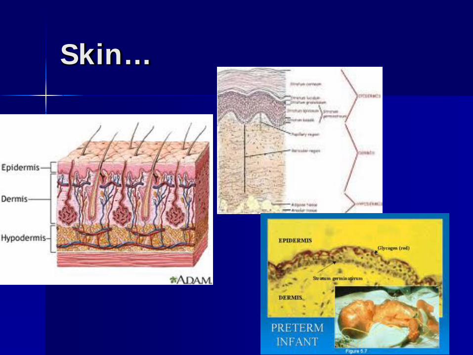
Skin…

Skin
Largest immune organ in the body Noticeably immature/thin in premature
infants – ↑ insensible water loss – ↑ heat loss – ↑ risk for infection – ↑ risk for trauma

Skin…

Medications
Epinephrine for persistent bradycardia despite successful other resuscitative measures
Naloxone – CAUTION – NOT recommended by AAP/NRP – 0.1mg/kg IO/ET – Half-life shorter than most opioids!!

Epinephrine
Recommended dose is 0.01-0.03mg/kg i.v. or IO
If no i.v. access available, 0.05-0.1mg/kg may be used endotracheally
Epinephrine is acidic Needs a “flush” 0.5ml NS Uncertain dose administered Interferes with CO2 detector use

Intra-Osseus Access
A good choice in infants if IV access unavailable
Different kinds – Jamshidi – type – EZ IO – babies >5kg – Cook type
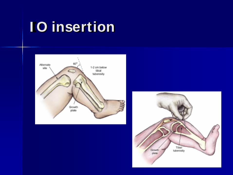
IO insertion

Outline
The Basics of Neonatal Resuscitation
Field Considerations The Neonatal Airway
Basic Neonatal Airway Management
Airway Abnormalities
Prehospital Management of Airway Emergencies
and Respiratory Diseases

Field Considerations (1)
Temperature control – Hypothermia common, causes include:
Birth significantly prior to EMS arrival Birth on transport (private car, ambulance) Birth in area without heating (unheated home) Baby remains in wet/exposed environment
– Remedies: Dry infant Place on mother’s chest/abdomen (direct skin contact) Heated blankets

Blood loss due to delayed clamping – Signs include:
Pallor, mottled skin, delayed capillary refill time (>4secs), tachycardia, hypotension
Frequently accompanied by hypothermia Hypothermia signs may mask signs of hypovolemia
– Remedy: Volume expansion (normal saline, 10ml/kg) Emergent low-lying umbilical catheter Insert umbilical catheter to 3-4cm (term) or UNTIL
BLOOD RETURN, no further IO needle
Field Considerations (2)

Infection risk – Primarily affected by location of delivery
Baby born in toilet etc.
– Not usually immediately life-threatening if acquired post-natally
– May be prompting pre-term birth May present as hypotension and shock Requires immediate hospital attention
Field Considerations (3)

Field Considerations (4)
Remember sugar! – Infants have decreased reserves and/or
increased glucose utilization – More so in cases of small for gestational
age or large for gestational age infants, or in particularly stressed neonates
– Start D10W at 80ml/kg/day (roughly 4ml/kg/hr) after initial fluid resuscitation

Outline
The Basics of Neonatal Resuscitation
Field Considerations The Neonatal Airway
Basic Neonatal Airway Management
Airway Abnormalities
Prehospital Management of Airway Emergencies
and Respiratory Diseases

The Neonatal Airway
More anterior and superior than pediatric or adult
Higher in the neck at birth – Birth laryngeal level C1-C4 – Laryngeal level age 6-adult C4-C7
Narrowest part is the area of the cricoid cartilage in 93%
Vocal cords are likely to be mucosal in color or light pink, NOT white
Long epiglottis
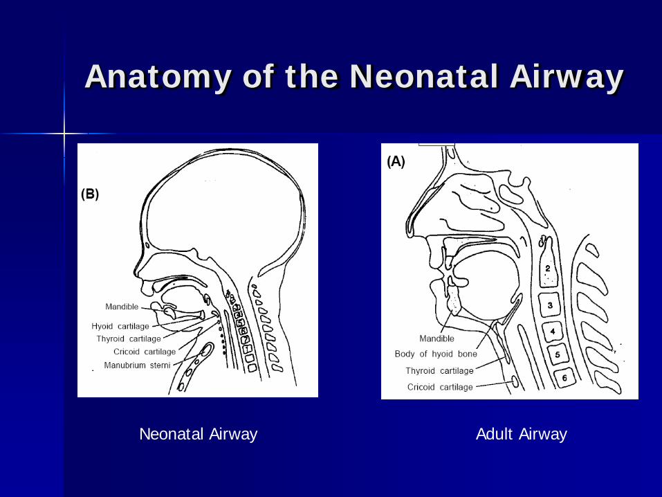
Anatomy of the Neonatal Airway
Neonatal Airway Adult Airway

Laryngoscopic View

Outline
The Basics of Neonatal Resuscitation
Field Considerations The Neonatal Airway
Basic Neonatal Airway Management
Airway Abnormalities
Prehospital Management of Airway Emergencies
and Respiratory Diseases

Newborns and the airway
Majority of infants’ ‘arrests’ are respiratory Focus on airway and breathing
– Infant positioning – Tactile stimulation – Positive pressure ventilation
Neonatal Rescusitation Program (NRP) guideline: – No CPR until >30 secs EFFECTIVE PPV – MR SOPA

MR SOPA (or MRS OPA)
Mask – verify size Reposition mask and/or baby Suction Open mouth Pressure – increase if no chest rise Adjunct airway – consider ETT/LMA if
all fails

Bag-Mask Ventilation
Choosing the appropriate size mask Infant positioning Ventilation pressures

Mask Choice
The correct size is determined by facial/infant size and fit
The mask should cover the nose and mouth Do NOT apply pressure in the following
places: – over the eyes (mask too large) – will prompt
vagally mediated bradycardia – nose (mask too small) – will obstruct nose,
decreased ability to ventilate – overlap beyond the chin (mask too large) –
inability to obtain a good seal, persistent leak, inability to ventilate

Appropriate Mask Positioning

Vagal Tone
Highly sensitive to vagal stimuli Avoid pressure over eyes, rectal
temperatures, deep suction Rapid decompensation with vagal
stimulation – Bradycardia – Desaturation
Rapid resolution if stimulus ceases

Equipment
Self-inflating (a.k.a. Ambu Bag): – Use room air or O2 – Use reservoir for O2 – Must add manometer – PEEP valve available – Different sizes available
Neonate (250ml) Pediatric (500ml) Adult 2000ml

To Tube or Not to Tube…
Data not supporting routine intubation – Traumatic intubation – Delay in transport – Accidental extubations – Complications of endotracheal intubation
Right mainstem
Ability to support many infants with BVM

Endotracheal Intubation
Equipment: – Mask, bag, suction, stethoscope – Appropriately sized Miller straight blade
laryngoscope – Appropriately sized infant (uncuffed)
endotracheal tube – End-tidal CO2 detector (if available)

Endotracheal Intubation
When? – Apnea, respiratory failure, hypoxia not
responsive to PPV – Prolonged (>5min) need for CPR – Airway abnormalities requiring intubation
(CDH, PPHN, vocal cord paralysis…)

Endotracheal Intubation
Equipment – Bag-valve-mask
– Laryngoscope
– Endotracheal tube
– Suction
– Stethoscope
– Extra hands
– Drugs

Laryngoscope Choice
Miller 0 blade: – <34 weeks – <2 kg
Miller 1 blade: – >34 weeks – >2 kg

Endotracheal Tubes
Neonatal ETT: – Constant diameter – Uncuffed
Pediatric/Adult ETT: – Can be tapered – Cuffed

Infant positioning

Endotracheal Tube Depth
Vocal cord marker at the vocal cords 1, 2, 3… 7, 8, 9
– 1, 2, or 3 kg, 7, 8 or 9 cm at the lip (or fraction thereof)
Symmetrical breath sounds

Endotracheal Intubation
2.5Fr <1kg 3.0Fr 1-2kg 3.5Fr >2kg
Vocal cord marker at the
vocal cords
1, 2, 3… 7, 8, 9!!! Check placement!
– Auscultate, CO2 detector, improvement, condensation

Evaluation of Successful Airway Management Condensate in the ETT Symmetrical breath sounds heard over
the lung fields No breath sounds/distention over the
abdomen End-tidal CO2 detector color change
The infant improves!

Endotracheal Tube Evaluation Tools
Vocal Cord Marker End-tidal CO2 detector

Ventilation Pressures
Starting pressures – PEEP 4-6cm H2O (if a PEEP valve is available) – PIP enough for SUBTLE chest rise (attempt to
keep <20cm H2O) and clinical improvement – NOT high pressures – increased risk for air
leak/pneumothorax Adjust if…
– Clinical deterioration, no chest rise… – Check ETT placement, continue to worry about
pneumothorax

Pneumothorax
Missing lung markings at periphery
May be accompanied by acute desaturations, difficulty ventilating, vascular instability
May transilluminate Resolves after needle
aspiration/chest tube placement

In Practice…
Asymmetrical chest rise and breath sounds
Narrowed pulse pressure
Shifted PMI

Needle Aspiration of Pneumothorax (new NRP)
4th intercostal space Anterior axillary line Avoid neuromuscular
bundle May use angiocath-
stopcock method for ongoing evacuation

Outline
The Basics of Neonatal Resuscitation
Field Considerations The Neonatal Airway
Basic Neonatal Airway Management
Airway Abnormalities
Prehospital Management of Airway Emergencies
and Respiratory Diseases

Airway Abnormalities
Craniofacial abnormalities – Micrognathia, retrognathia – Cleft palate – Choanal atresia
Laryngeal abnormalities – Webs and clefts – Vocal cord paralysis
Tracheal abnormalities – Complete tracheal ring – Tracheo-esophageal fistula

Craniofacial abnormalities
Micrognathia, retrognathia – Can be associated with
syndromes such as Pierre-Robin sequence, Trisomies, Smith-Lemli-Opitz
– Results in airway obstruction due to large tongue compared to mandible/mouth
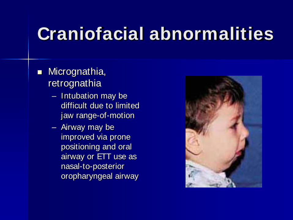
Craniofacial abnormalities
Micrognathia, retrognathia – Intubation may be
difficult due to limited jaw range-of-motion
– Airway may be improved via prone positioning and oral airway or ETT use as nasal-to-posterior oropharyngeal airway

Craniofacial abnormalities
Cleft palate – Should not require increased airway support based
purely on anatomy – Usually able to intubate fairly easily – Difficulties frequently encountered due to ‘optical
illusions’

Craniofacial abnormalities
Choanal atresia – Normal appearing
infant – Blue at rest, but
pinks with crying/screaming
– May be lethal if not recognized
– Treatment via oral airway

Laryngeal Abnormalities
Laryngeal web – Stridor/respiratory
distress – May result in difficult
intubation – Smaller airway diameter
than anticipated Vocal cord paralysis
– Idiopathic or the result of traumatic delivery
– Respiratory failure at birth

Tracheal Abnormalities
Complete tracheal ring – May show respiratory failure while extubated,
but exceedingly low vent settings and no evidence of parenchymal disease
Tracheo-esophageal fistula – Increased secretions, signs of aspiration – Failure to pass oro- or naso-gastric tube – Usually requires constant suction in pouch,
not respiratory support

Outline
The Basics of Neonatal Resuscitation
Field Considerations The Neonatal Airway
Basic Neonatal Airway Management
Airway Abnormalities
Prehospital Management of Airway
Emergencies and Respiratory Diseases

Airway Emergencies
Bilateral choanal atresia – Cyanotic at rest, gasping infant – Pinks with screaming – Requires oral airway
Bilateral vocal cord paralysis – Requires intubation – May require tracheostomy

Airway Emergencies
C-spine transection/contusion – Think of in cases of severe respiratory
failure/apnea in infant following difficult/traumatic/instrumented delivery
– Poor prognosis – Requires PPV and immediate intubation

Meconium Aspiration Syndrome
Meconium inactivates surfactant Ball-valve effect and patchy ventilation May lead to persistent pulmonary
hypertension

Thank you for all you do!

Aren’t you glad you are not medics here???
http://www.youtube.com/watch?v=5SORYzaBLTI
PS – all the comments said how cool this was and that they were trying to replicate this here!