neural dysfunction in postpartum depression: an … · neural dysfunction in postpartum depression:...
TRANSCRIPT
Neural Dysfunction in Postpartum Depression: An fMRI Pilot Study
By Michael E. Silverman, PhD, Holly Loudon, MD, Michal Safier, BA, Xenia Protopopescu, PhD, Gila Leiter, MD, Xun Liu, PhD, and Martin Goldstein, MD
853CNS Spectr 12:11 © MBL Communications November 2007
Original Research
Needs AssessmentGreater than 50% of the 760,000 women who suffer from a clinically significant postpartum psychiatric illness each year go unrecognized. Postpartum illnesses account for the largest cause of maternal death, with suicide rates of up to 5% and infanticide rates of nearly 4%. Because untreated mood disorders place the mother at risk for recur-rent disease and maternal depression is associated with diminished enrichment behavior, which is known to result in long-term cognitive, emotional, and behavioral problems in the child, characterizing the behavioral and neurobiological features of postpartum depression is important for early diagnosis and intervention. This study addresses a critical gap in the mechanistic understanding of postpartum depression by probing its systems-level neuropathophysiology, in the context of a specific neurobiological model of fronto-limbic-striatal function.
Learning ObjectivesAt the end of this activity, the participant should be able to: • List the various mechanisms hypothesized to be responsible for post-
partum depression to date.• Understand the application of functional neuroimaging toward
informing clinical and cognitive disorders associated with affective dysregulation.
• Comprehend key components of the fronto-limbic-striatal network associated with the neuropathophysiology of emotional dysregula-tion in postpartum depression.
Target Audience: Neurologists and psychiatrists
CME Accreditation StatementThis activity has been planned and implemented in accordance with the Essentials and Standards of the Accreditation Council for Continuing Medical Education (ACCME) through the joint sponsorship of the Mount Sinai School of Medicine and MBL Communications, Inc. The Mount Sinai School of Medicine is accredited by the ACCME to provide con-tinuing medical education for physicians.
Credit DesignationThe Mount Sinai School of Medicine designates this educational activ-ity for a maximum of 3 AMA PRA Category 1 Credit(s)TM. Physicians should only claim credit commensurate with the extent of their partici-pation in the activity.
This activity has been peer-reviewed and approved by Eric Hollander, MD, chair at the Mount Sinai School of Medicine. Review date: October 15, 2007. Dr. Hollander does not have an affiliation with or financial interest in any organization that might pose a conflict of interest.
To Receive Credit for This ActivityRead this article and the two CME-designated accompanying articles, reflect on the information presented, and then complete the CME post-test and evaluation found on page 864. To obtain credits, you should score 70% or better. Early submission of this posttest is encouraged: please submit this posttest by November 1, 2009, to be eligible for credit. Release date: November 1, 2007. Termination date: November 30, 2009. The estimated time to complete all three articles and the posttest is 3 hours.
ABSTRACTIntroduction: With ~4 million births each
year in the United States, an estimated 760,000
women annually suffer from a clinically signifi-
cant postpartum depressive illness. Yet even
though the relationship between psychiatric
disorders and the postpartum period has been
documented since the time of Hippocrates, fewer
than half of all these cases are recognized.
Objective: Because postpartum depression
(PPD), the most common complication of child-
bearing, remains poorly characterized, and its eti-
ology remains unclear, we attempted to address
a critical gap in the mechanistic understanding
of PPD by probing its systems-level neuropatho-
physiology, in the context of a specific neurobio-
logical model of fronto-limbic-striatal function.
Methods: Using emotionally valenced word
probes, with linguistic semantic specificity within
an integrated functional magnetic resonance
imaging (fMRI) protocol, we investigated emo-
tional processing, behavioral regulation, and their
interaction (functions of clinical relevance to PPD),
in the context of fronto-limbic-striatal function.
Results: We observed attenuated activity in
posterior orbitofrontal cortex for negative versus
neutral stimuli with greater PPD symptomatology,
increased amygdala activity in response to nega-
tive words in those without PPD symptomotology,
3CME
Affilations and Disclosures: Please see page 862 for biographies and disclosure information.
854
and attenuated striatum activation to positive word
conditions with greater PPD symptomotology.
Conclusion: Identifying the functional neuro-
anatomical profile of brain systems involved in
the regulation of emotion and behavior in the
postpartum period will not only assist in deter-
mining whether the Diagnostic and Statistical
Manual of Mental Disorders, Fourth Edition
psychiatric diagnostic specifier of PPD has an
associated, unique, functional neuroanatomical
profile, but a neurobiological characterization
in relation to asymptomatic (postpartum non-
depressed) control subjects, will also increase
our understanding of the affective disorder
spectrum, shed additional light on the possible
mechanism(s) responsible for PPD and provide
a necessary foundation for the development of
more targeted, biologically based diagnostic and
therapeutic strategies for PPD.
CNS Spectr. 2007;12(11):853-862
INTRODUCTIONConsiderable evidence exists suggesting
that mood disorders are twice as prevalent in women compared to men.1 While the dissocia-tion between gender and affective disorders can be first observed at menarche (prior to puberty prevalence is equal among males and females), the preponderance of mood disorders occurs in women during the childbearing years,2 with peak lifetime prevalence for psychiatric disorders and hospital admissions for women occurring in the first 3 months after childbirth.3
Postpartum depression (PPD), the most com-mon complication of childbearing,4 is a prevalent disorder in the spectrum of affective illness asso-ciated with significant morbidity. Traditionally viewed as a time of emotional well-being, the weeks that follow childbirth are, in fact, more often a time of heightened psychic vulnerability. Indeed, mood and behavioral symptoms dur-ing the puerperal period reportedly affect up to 85% of all new mothers.4 With an onset usually between 3 and 14 days (although possibly up to 1 year5,6) postpartum symptoms of anxiety, exhaustion, alternating mood, and an inability
to concentrate are usually short-lived. However, up to 20% of all postpartum women will go on to develop a more severe mood disorder that meets criteria for Diagnostic and Statistical Manual of Mental Disorders, Fourth Edition7 depression characterized by impairment in functioning with symptoms, including neurovegetative dysregula-tion and ideation of harm to the self or the baby.3 While these episodes account for a seven-fold increase in psychiatric hospital admissions com-pared with pre-pregnancy,8 <50% of all of these cases will be recognized.
PPD is a cross-cultural health concern with significant public health consequences. Postnatal psychiatric illness is not only the largest cause of maternal death in the United Kingdom (statistics are difficult to obtain in the United States due to current documenting standards),9,10 it is associ-ated with nearly 60% of all infanticides occur-ring in the first 3 month postpartum.11 Despite its suspected prevalence, PPD remains under-diag-nosed and under-treated, possibly because con-troversy still exists about how to characterize the depression that occurs in the postpartum period. For example, while the DSM-IV utilizes “postpar-tum onset” as a modifier defined as an episode of depression within the first 4 weeks of child-birth, the American College of Obstetricians and Gynecologists12 defines the postpartum period as extending for 1 year.
Even though numerous theories have been advanced to explain the well-documented and frequent co-occurrence of depression and child-birth, the etiology of PPD remains unclear. For example, recent research has suggested causal mechanisms, such as postpartum estrogen and progesterone shifts13-15 thyroid disease and thy-roid antibodies,16 inflammatory responses,17 situational triggers, such as traumatic obstetric experiences,18 low socioeconomic status,19 infant health20 and psychological stress.21 Yet, because the hormonal changes observed in childbirth are unprecedented among all other reproductive cycle events,22 the most prevalent theories of PPD genesis relate to the effects of sex hormones on brain regions mediating mood and cognition.
Sex hormones have been shown to influence the central nervous system in a large number of varied ways, including effects on neurogenesis, gliagenesis, cell survival, ion-channel modula-tion, neurochemical modulation, transcription, neural excitation, and neural inhibition.23-31 Unfortunately, the neuroedocrinology of PPD
Original Research
CNS Spectr 12:11 © MBL Communications November 2007
855
remains poorly understood and studies explor-ing PPD symptomotology22,23 do not seem to cor-relate well with absolute differences in hormone levels between affected and unaffected women with multiple and contradictory findings. Rather, it is quite possible that the observed symptoms may correspond to differences in the way the central nervous system responds to various (and possibly interactive) hormonal and immu-nologic fluctuations. Nevertheless, although brain response to sex hormones in PPD patients appears central to a neurobiological understand-ing of PPD’s psychopathology, to date, a specific hormonal mechanism has remained elusive.
Because the clinical characterization and neu-robiologic mechanisms of this evolving condi-tion remain inadequately defined, the aim of this work was to probe and begin identifying the sys-tems-level neuropathophysiology of PPD, in the context of a specific neurobiological model of fronto-limbic-striatal function. Using functional magnetic resonance imaging (fMRI) methods with specific neuropsychological probes of emotional processing and behavioral regula-tion (functions of clinical relevance to the symp-tomatology of PPD), and their interaction, in well-characterized patient samples, we tested mechanistic hypotheses concerning fronto-lim-bic-striatal circuit dysfunction in PPD in compari-son to asymptomatic postpartum female control subjects. Such a neurobiological characterization in relation to non-depressed postpartum control subjects is hoped to increase our understand-ing of the affective disorder spectrum and shed additional light on the mechanism(s) respon-sible for PPD. Given the increased prevalence of mood and anxiety disorders in females, it is also hoped that this research will also provide a deeper foundation for the development of more targeted, biologically based diagnostic and ther-apeutic strategies for PPD.
METHODS
SubjectsParticipants consisted of eight postpartum
women (mean age: 28 years). Subjects gave informed consent before study participation (part of a Mount Sinai Medical Center Institutional Review Board-approved protocol). All subjects were right-handed, native English speakers, with a history free of psychiatric difficulty (including antepartum depression), head trauma, neuro-
psychiatric complication, illicit substance abuse, or chemical/alcohol dependence. No subjects were actively taking birth control or psychoactive medication at the time of screening or scanning. The Structured Clinical Interview for DSM-IV Axis I Disorders33 was used to ensure that compari-son subjects did not have any Axis I psychiatric diagnoses and that depressed participants were free of any Axis I comorbidity. The Hamilton Depression Inventory34 was used to identify spe-cific symptoms of depression as delineated by the DSM-IV. The Edinburgh Postnatal Depression Scale (EPDS),35 a 10-item 4-point inventory with a maximum score of 30, was used to determine eligibility. Because a multinational review of the EPDS36 demonstrated that scores of 8.5–12 points had a specificity of 49% to 100% and sensitiv-ity of 65% to 100%, subject groups were based on the EPDS as follows: those scoring >12 were included in the depressed group (n=4), whereas those subjects scoring <6 were included in the normal comparison group (n=4). The EPDS was re-administered immediately prior to entering the MRI and an average of the two scores was taken (depressed group mean: 15.33; range: 12–19; normal comparison mean: 1.33; range: 0–4). The change of the EPDS score between administra-tions never varied >2 points. All scans occurred between weeks 7 and 8 postpartum.
Neuropsychological Activation Paradigm The fMRI activation paradigm consisted of an
emotional word probe, with linguistic-semantic specificity, allowing for a complementary higher-level examination of the hypothesized fronto-limbic-striatal circuitry. This paradigm employs stimuli whose emotional qualities are incidental relative to the explicit nature of the word/non-word determination behavioral task demand (2-alternative forced choice method [2AFC]). By using this technique, the evocation of potentially confounding cognitive processes (eg, semantic categorization) are hoped to be minimized.
Stimuli consisted of positive, negative (both threat and non-threat), and neutral words (adjec-tives, nouns, and verbs) balanced across catego-ries for frequency, length, and part of speech, with the exception that, within the neutral list, verbs were substituted for adjectives. This was done because adjectives, comprising an impor-tant component of the valence categories, are by nature generally not free of valence. Verbs were substituted, rather than nouns, as their image-
Original Research
CNS Spectr 12:11 © MBL Communications November 2007
856CNS Spectr 12:11 © MBL Communications November 2007
Original Research
ability is more similar to that of adjectives. Stimuli were rated for suitability as defined by Bradley and Lang.37 Examples include positive-success, admired, praise; negative-worthless, murder, burn; neutral-transfer, trunks, fasten.
Behavioral responses were based on word/non-word judgment cues, such that subjects were instructed to perform a right index finger button-press immediately upon presentation of a word (eg, MURDER) and to perform a right middle fin-ger button press upon presentation of a random letter string (eg, DSKDFA). Corresponding button presses were counterbalanced across subjects. Subjects were not pre-informed of the emotional nature of the stimulus words.
The task was presented in a block design. Presentation was counterbalanced to control for order and time effects. Each block was comprised of 10 stimuli words/non-words (trials) of the same valence; there 100 trials per condition, 400 total tri-als per complete study session. Blocks included 90%, 80%, or 70% words (compared to random let-ter-strings). Each stimulus appeared for 1.5 seconds, followed by a jittered interstimulus interval averag-ing 1,900 milliseconds, for a total block duration of 34 seconds (not including intertrial interval rest). Each block was followed by 12 seconds of rest. Each run was preceded and followed by an additional 36 second rest periods. During rest periods, subjects were instructed to look at a cross at the center of the screen, with their minds either blank or floating freely. Stimulus presentation and response collec-tion were performed within the E-Prime environ-ment (Psychology Software Tools, Inc., Pittsburgh, Penn.). Stimuli were presented in white against a black background subtending an average visual angle of ~2 degrees in height by 6 degrees in width.
We designed a factorial paradigm with a block (rather than event-related) design for sev-eral reasons: to maximize operationalization of sustained emotional tone; to facilitate factorial comparison of various permutations of emotion and response conditions; to exploit the imag-ing sensitivity bestowed by block design; and to minimize potentially confounding extraneous cognitive-behavioral functions.
There were two objectives associated with the neuropsychological tasks. First, by giving subjects a task, we ensure that they were focusing on the stimuli presented to them. In turn, this enhances the likelihood that blood-oxygen level depen-dent (BOLD) response changes are related to the relevant stimuli. Thus, the first objective was to
present subjects a “probe” that would activate relevant brain circuitry. The second objective was to measure differences in motor response perfor-mance by condition. It is well documented that emotion-inducing stimuli can generate cognitive and/or behavioral task processing interference.38,39
Immediately after imaging, subjects were removed from the scanner and presented via computer with a list of words consisting of the stimuli seen during scanning (targets) randomly interspersed with an equal number of new words (distractors) divided equally into each stimulus category, and balanced for the same qualities as the targets. Subjects were asked to indicate which words they believe were presented during the scan-ning session using a 2AFC button press. Accuracy was measured for later analysis. Following the completion of this task, subjects were asked to rate a similarly counter-balanced subset of target words presented on a touch-screen monitor along a Likert-like scale using the subjective-assessment man-nequin37 according to emotional valence (strongly positive, neutral, or strongly negative, ranging in value from +3 to –3, respectively).
Image AcquisitionImaging data were acquired with a research-
dedicated Seimens Allegra Magnetron 3 Tesla head-dedicated MRI scanner). T1-weighted spoiled gradient (MP-RAGE) MRI whole brain anatomical scans (208 slices; 8 mm in-plane resolution, 0.8 mm slice thickness, contiguous slices) were acquired followed by T2-weighted turbo spin echo axial whole-brain images (3 mm slice thickness) to explore potential pathology. Finally, gradient echo planar imaging–blood-oxygen level dependent (EPI-BOLD) fMRI were acquired (repetition time: 2,000 milliseconds, time to echo: 30, 32 slices; 3 mm thickness; 1 mm gap) as an index of neuronal activity during the neuropsychological activation paradigm.
Image Processing and Data AnalysisPrior to statistical analysis, the first two vol-
umes of each run were discarded to allow the magnetic resonance signal to reach steady state. The remaining images in each partici-pant’s time series were motion corrected using the Motion Correction using the FMRIB Linear Image Registration Tool (MCFLIRT) module of the Functional Magnetic Resonance Imaging of the Brain (FMRIB) Center’s Software Library, v 3.3) package (available at www.fmrib.ox.ac.uk/
857
fsl). Images in the data series were then spatially smoothed with a three-dimensional Gaussian kernel (full width at half maximum: x 8 x 8 mm3), and temporally filtered using a high-pass filter (320 seconds). The FMRIB Expert Analysis Tool (FEAT) module of the FMRIB Software Library package was used for these steps and later sta-tistical analysis.
Customized square waveforms were generated for each individual. These waveforms were con-volved with a double γ-hemodynamic response function. For each participant, we used FMRIB’s Improved Linear Model (FILM), with local autocor-relation correction, to estimate the hemodynamic parameters for four explanatory variables (neutral, positive, negative, and threat) and generate statis-tical contrast maps of interest. The six movement parameters (ie, translation and rotation of x, y, and z axes) were modeled as covariates.
Each of the five runs for each participant was analyzed separately and the average of these five runs for each individual was obtained through a higher-level analysis using the FMRIB’s Local Analysis of Mixed Effects (FLAME) module (stage 1 only). Contrast maps were warped into common stereotaxic space before mixed-effects group analyses were performed. The normaliza-tion procedure involved registering the aver-age EPI image to the MP-RAGE image from the same participant, and then to the International Consortium for Brain Mapping 152 T1 template,40 using the FMRIB’s Linear Image Registration Tool (FLIRT) module.
To identify the regions of brain activation, we defined the regions of interest (ROI) by clusters of >30 contiguous voxels41 in which there was signif-icant difference in brain activity across conditions (Z>2.81, P<.005 two-tailed). Using the Mintun peak algorithm,42 we further located the local peaks (maximal activation) within each ROI. Additional ROI analyses were performed using the average signals extracted from these clusters.
RESULTS
Word Valence RatingsAnalysis of the post-scan ratings of all stimu-
lus words (positive, negative threat/non-threat, and neutral) confirmed our assignment of word stimuli to negative, neutral, or positive catego-ries. Subjects rated negative, neutral, and posi-tive words as significantly negative, neutral, and positive, respectively. Therefore, it is fair
to assume that the emotional word categories employed in this study were reasonable probes of emotional linguistic-stimulus processing within the participating subject population.
Neuropsychiatric Activation Paradigm Findings (Reaction Time)
A two-way repeated measures analysis of variance of reaction times of the 2AFC word/non-word judgment task performed during scanning revealed significant valence by diag-nosis interactions (F=4.61, P<.01). Further anal-ysis of these differences revealed that while affective stimuli were associated with enhanced responsivity (positive word vs neutral words; P<.01, and negative word vs neutral word; P<.01) to the word/non-word judgment task in the non-depressed control subjects (no differ-ence was found between the two affective word conditions; positive vs negative; not significant), those with PPD tended to take significantly lon-ger to make word/non-word judgments dur-ing the positive word condition compared with negative (P<.03) and neutral (P<.01) word con-ditions (differences observed between nega-tive and neutral word conditions were not significant). Therefore, not only was enhanced processing of negative stimuli not observed in our PPD subjects, as is regularly reported in the depression literature,43,44 reaction times were increased in the positive word condition as compared to neutral word condition. That is, positive words seemingly had the effect of inhibiting responsivity in those with PPD.
Functional Imaging Findings of Hypothesized Regions
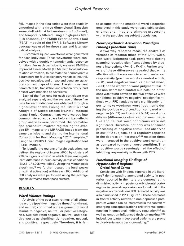
Orbital Frontal Cortex Consistent with findings reported in the litera-
ture45 demonstrating attenuated activity in pos-terior reported in the literature demonstrating diminished activity in posterior orbitofrontal cortex regions in general depression, we found that in the negative word conditions BOLD-related activity was also diminished in PPD (Figure 1). These decreases in frontal activity relative to non-depressed post-partum women can be interpreted in the context of emerging conceptualizations orbitofrontal cortex function in emotional inhibitory regulation46-49 as well as emotion-influenced decision-making.50-52 Indeed, postpartum depressed patients are prone to disadvantageous decision-making.22
CNS Spectr 12:11 © MBL Communications November 2007
Original Research
858
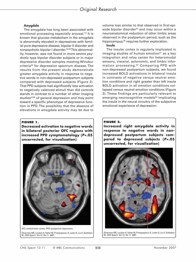
AmygdalaThe amygdala has long been associated with
emotional processing especially arousal.53 It is known that glucose metabolism in the amygdala is abnormally elevated in depressives with famil-ial pure depressive disease, bipolar II disorder and nonpsychotic bipolar I disorder.54,55 This abnormal-ity, however, was not found in more severe psy-chotic type bipolar disorder subjects or in major depressive disorder samples meeting Winokur criteria56 for depression spectrum disease. The results from the present study demonstrate greater amygdala activity in response to nega-tive words in non-depressed postpartum subjects compared with depressed subjects (Figure 2). That PPD subjects had significantly less activation to negatively valenced stimuli than did controls stands in contrast to a number of other imaging studies57,58 of general depression and may point toward a specific phenotype of depressive func-tion in PPD. The possibility that the absence of elevations in amygdala activity may be due to
volume loss similar to that observed in first-epi-sode bipolar disorder59 and may occur within a neuroanatomical reduction of other limbic areas observed in the postpartum period, such as the hippocampus,60 requires further exploration.
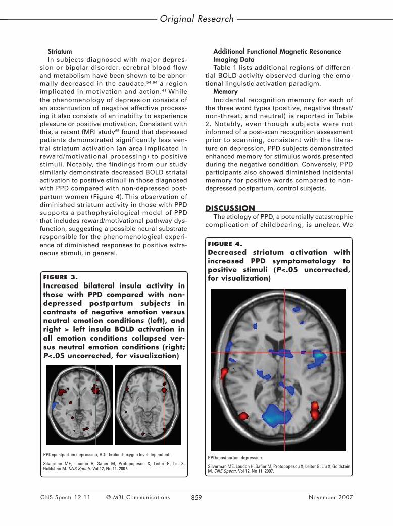
InsulaThe insular cortex is regularly implicated in
imaging studies of human emotion61 as a key integration and relay center for heteromodal sensory, visceral, autonomic, and limbic infor-mation processing.62 Comparing PPD with non-depressed postpartum subjects, we found increased BOLD activations in bilateral insula in contrasts of negative versus neutral emo-tion conditions and right greater than left insula BOLD activation in all emotion conditions col-lapsed versus neutral emotion conditions (Figure 3). These findings are particularly relevant to emerging neurocognitive models63 implicating the insula in the neural circuitry of the subjective emotional experience of depression.
CNS Spectr 12:11 © MBL Communications November 2007
Original Research
FIGURE 2.Increased right amygdala activity in response to negative words in non-depressed postpartum subjects com-pared to depressed subjects (P<.05 uncorrected, for visualization)
Silverman ME, Loudon H, Safier M, Protopopescu X, Leiter G, Liu X, Goldstein M. CNS Spectr. Vol 12, No 11. 2007.
FIGURE 1.Decreased activation to negative words in bilateral posterior OFC regions with increased PPD symptomatology (P<.05 uncorrected, for visualization)
OFC=orbitofrontal cortex; PPD=postpartum depression.
Silverman ME, Loudon H, Safier M, Protopopescu X, Leiter G, Liu X, Goldstein M. CNS Spectr. Vol 12, No 11. 2007.
859
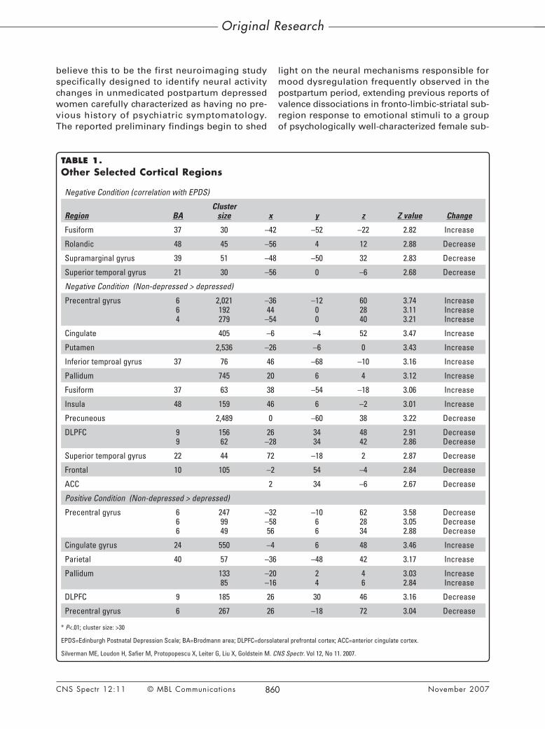
StriatumIn subjects diagnosed with major depres-
sion or bipolar disorder, cerebral blood flow and metabolism have been shown to be abnor-mally decreased in the caudate,54,64 a region implicated in motivation and action.41 While the phenomenology of depression consists of an accentuation of negative affective process-ing it also consists of an inability to experience pleasure or positive motivation. Consistent with this, a recent fMRI study65 found that depressed patients demonstrated significantly less ven-tral striatum activation (an area implicated in reward/motivational processing) to positive stimuli. Notably, the findings from our study similarly demonstrate decreased BOLD striatal activation to positive stimuli in those diagnosed with PPD compared with non-depressed post-partum women (Figure 4). This observation of diminished striatum activity in those with PPD supports a pathophysiological model of PPD that includes reward/motivational pathway dys-function, suggesting a possible neural substrate responsible for the phenomenological experi-ence of diminished responses to positive extra-neous stimuli, in general.
Additional Functional Magnetic Resonance Imaging DataTable 1 lists additional regions of differen-
tial BOLD activity observed during the emo-tional linguistic activation paradigm.
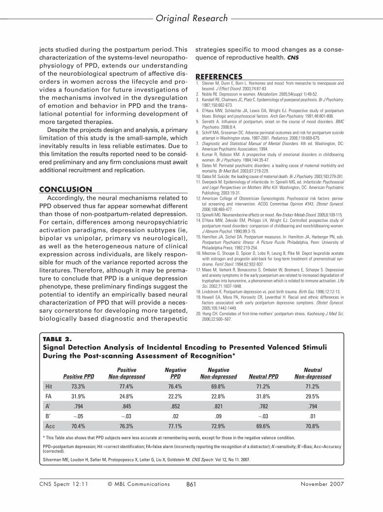
MemoryIncidental recognition memory for each of
the three word types (positive, negative threat/non-threat, and neutral) is reported in Table 2. Notably, even though subjects were not informed of a post-scan recognition assessment prior to scanning, consistent with the litera-ture on depression, PPD subjects demonstrated enhanced memory for stimulus words presented during the negative condition. Conversely, PPD participants also showed diminished incidental memory for positive words compared to non-depressed postpartum, control subjects.
DISCUSSIONThe etiology of PPD, a potentially catastrophic
complication of childbearing, is unclear. We
CNS Spectr 12:11 © MBL Communications November 2007
Original Research
FIGURE 3.Increased bilateral insula activity in those with PPD compared with non-depressed postpartum subjects in contrasts of negative emotion versus neutral emotion conditions (left), and right > left insula BOLD activation in all emotion conditions collapsed ver-sus neutral emotion conditions (right; P<.05 uncorrected, for visualization)
PPD=postpartum depression; BOLD=blood-oxygen level dependent.
Silverman ME, Loudon H, Safier M, Protopopescu X, Leiter G, Liu X, Goldstein M. CNS Spectr. Vol 12, No 11. 2007.
FIGURE 4.Decreased striatum activation with increased PPD symptomatology to positive stimuli (P<.05 uncorrected, for visualization)
PPD=postpartum depression.
Silverman ME, Loudon H, Safier M, Protopopescu X, Leiter G, Liu X, Goldstein M. CNS Spectr. Vol 12, No 11. 2007.
860CNS Spectr 12:11 © MBL Communications November 2007
Original Research
believe this to be the first neuroimaging study specifically designed to identify neural activity changes in unmedicated postpartum depressed women carefully characterized as having no pre-vious history of psychiatric symptomatology. The reported preliminary findings begin to shed
light on the neural mechanisms responsible for mood dysregulation frequently observed in the postpartum period, extending previous reports of valence dissociations in fronto-limbic-striatal sub-region response to emotional stimuli to a group of psychologically well-characterized female sub-
TABLE 1.Other Selected Cortical Regions
Negative Condition (correlation with EPDS)
Region BACluster
size x y z Z value Change
Fusiform 37 30 –42 –52 –22 2.82 Increase
Rolandic 48 45 –56 4 12 2.88 Decrease
Supramarginal gyrus 39 51 –48 –50 32 2.83 Decrease
Superior temporal gyrus 21 30 –56 0 –6 2.68 Decrease
Negative Condition (Non-depressed > depressed)
Precentral gyrus 6 6 4
2,021 192 279
–36 44
–54
–12 0 0
60 28 40
3.74 3.11 3.21
Increase Increase Increase
Cingulate 405 –6 –4 52 3.47 Increase
Putamen 2,536 –26 –6 0 3.43 Increase
Inferior temproal gyrus 37 76 46 –68 –10 3.16 Increase
Pallidum 745 20 6 4 3.12 Increase
Fusiform 37 63 38 –54 –18 3.06 Increase
Insula 48 159 46 6 –2 3.01 Increase
Precuneous 2,489 0 –60 38 3.22 Decrease
DLPFC 9 9
156 62
26 –28
34 34
48 42
2.91 2.86
Decrease Decrease
Superior temporal gyrus 22 44 72 –18 2 2.87 Decrease
Frontal 10 105 –2 54 –4 2.84 Decrease
ACC 2 34 –6 2.67 Decrease
Positive Condition (Non-depressed > depressed)
Precentral gyrus 6 6 6
247 99 49
–32 –58 56
–10 6 6
62 28 34
3.58 3.05 2.88
Decrease Decrease Decrease
Cingulate gyrus 24 550 –4 6 48 3.46 Increase
Parietal 40 57 –36 –48 42 3.17 Increase
Pallidum 133 85
–20 –16
2 4
4 6
3.03 2.84
Increase Increase
DLPFC 9 185 26 30 46 3.16 Decrease
Precentral gyrus 6 267 26 –18 72 3.04 Decrease
* P<.01; cluster size: >30
EPDS=Edinburgh Postnatal Depression Scale; BA=Brodmann area; DLPFC=dorsolateral prefrontal cortex; ACC=anterior cingulate cortex.
Silverman ME, Loudon H, Safier M, Protopopescu X, Leiter G, Liu X, Goldstein M. CNS Spectr. Vol 12, No 11. 2007.
861CNS Spectr 12:11 © MBL Communications November 2007
Original Research
jects studied during the postpartum period. This characterization of the systems-level neuropatho-physiology of PPD, extends our understanding of the neurobiological spectrum of affective dis-orders in women across the lifecycle and pro-vides a foundation for future investigations of the mechanisms involved in the dysregulation of emotion and behavior in PPD and the trans-lational potential for informing development of more targeted therapies.
Despite the projects design and analysis, a primary limitation of this study is the small-sample, which inevitably results in less reliable estimates. Due to this limitation the results reported need to be consid-ered preliminary and any firm conclusions must await additional recruitment and replication.
CONCLUSION Accordingly, the neural mechanisms related to
PPD observed thus far appear somewhat different than those of non-postpartum-related depression. For certain, differences among neuropsychiatric activation paradigms, depression subtypes (ie, bipolar vs unipolar, primary vs neurological), as well as the heterogeneous nature of clinical expression across individuals, are likely respon-sible for much of the variance reported across the literatures. Therefore, although it may be prema-ture to conclude that PPD is a unique depression phenotype, these preliminary findings suggest the potential to identify an empirically based neural characterization of PPD that will provide a neces-sary cornerstone for developing more targeted, biologically based diagnostic and therapeutic
strategies specific to mood changes as a conse-quence of reproductive health. CNS
REFERENCES1. Steiner M, Dunn E, Born L. Hormones and mood: from menarche to menopause and
beyond. J Effect Disord. 2003;74:67-832. Noble RE. Depression in women. Metabolism. 2005;54(suppl 1):49-52.3. Kendell RE, Chalmers JC, Platz C. Epidemiology of puerperal psychosis. Br J Psychiatry.
1987;150:662-673.4. O’Hara MW, Schlechte JA, Lewis DA, Wright EJ. Prospective study of postpartum
blues. Biologic and psychosocial factors. Arch Gen Psychiatry. 1991;48:801-806.5. Serretti A. Influence of postpartum, onset on the course of mood disorders. BMC
Psychiatry. 2006;6:4.6. Schiff MA, Grossman DC. Adverse perinatal outcomes and risk for postpartum suicide
attempt in Washington state, 1987-2001. Pediatrics. 2006;118:669-675.7. Diagnostic and Statistical Manual of Mental Disorders. 4th ed. Washington, DC:
American Psychiatric Association; 1994.8. Kumar R, Robson KM. A prospective study of emotional disorders in childbearing
women. Br J Psychiatry. 1984;144:35-47.9. Oates M. Perinatal psychiatric disorders: a leading cause of maternal morbidity and
mortality. Br Med Bull. 2003;67:219-229.10. Oates M. Suicide: the leading cause of maternal death. Br J Psychiatry. 2003;183:279-281.11. Overpeck M. Epidemiology of infanticide. In: Spinelli MG, ed. Infanticide: Psychosocial
and Legal Perspectives on Mothers Who Kill. Washington, DC: American Psychiatric Publishing; 2003:19-31.
12. American College of Obstetrician Gynecologists. Psychosocial risk factors: perina-tal screening and intervention. ACOG Committee Opinion #343. Obstet Gynecol. 2006;108:469-477.
13. Spinelli MG. Neuroendocrine effects on mood. Rev Endocr Metab Disord. 2005;6:109-115.14. O’Hara MW, Zekoski EM, Philipps LH, Wright EJ. Controlled prospective study of
postpartum mood disorders: comparison of childbearing and nonchildbearing women. J Abnorm Psychol. 1990;99:3-15.
15. Hamilton JA, Sichel DA. Postpartum measures. In: Hamilton JA, Harberger PN, eds. Postpartum Psychiatric Illness: A Picture Puzzle. Philadelphia, Penn: University of Philadelphia Press; 1992:219-254.
16. Mezrow G, Shoupe D, Spicer D, Lobo R, Leung B, Pike M. Depot leuprolide acetate with estrogen and progestin add-back for long-term treatment of premenstrual syn-drome. Fertil Steril. 1994;62:932-937.
17. Maes M, Verkerk R, Bonaccorso S, Ombelet W, Bosmans E, Scharpe S. Depressive and anxiety symptoms in the early puerperium are related to increased degradation of tryptophan into kynurenine, a phenomenon which is related to immune activation. Life Sci. 2002;71:1837-1848.
18. Lindstrom K. Postpartum depression vs. post birth trauma. Birth Gaz. 1996;12:12-13.19. Howell EA, Mora PA, Horowitz CR, Leventhal H. Racial and ethnic differences in
factors associated with early postpartum depressive symptoms. Obstet Gynecol. 2005;105:1442-1449.
20. Hung CH. Correlates of first-time mothers’ postpartum stress. Kaohsiung J Med Sci. 2006;22:500–507.
TABLE 2.Signal Detection Analysis of Incidental Encoding to Presented Valenced Stimuli During the Post-scanning Assessment of Recognition*
Positive PPDPositive
Non-depressedNegative
PPDNegative
Non-depressed Neutral PPDNeutral
Non-depressed
Hit 73.3% 77.4% 76.4% 69.8% 71.2% 71.2%
FA 31.9% 24.8% 22.2% 22.8% 31.8% 29.5%
A’ .794 .845 .852 .821 .782 .794
B’ –.05 –.03 .02 .09 –.03 .01
Acc 70.4% 76.3% 77.1% 72.9% 69.6% 70.8%
* This Table also shows that PPD subjects were less accurate at remembering words, except for those in the negative valence condition.
PPD=postpartum depression; Hit =correct identification; FA=false alarm (incorrectly reporting the recognition of a distractor); A’=sensitivity; B’=Bias; Acc=Accuracy (corrected).
Silverman ME, Loudon H, Safier M, Protopopescu X, Leiter G, Liu X, Goldstein M. CNS Spectr. Vol 12, No 11. 2007.
862CNS Spectr 12:11 © MBL Communications November 2007
Original Research
21. Hung CH. Psychosocial features at different periods after childbirth. Kaohsiung J Med Sci. 2007;23:71-79.
22. Buckwalter JG, Stanczyk FZ, McCleary CA, et al. Pregnancy, the postpartum, and steroid hormones: effects on cognition and mood. Psychoneuroendocrinology. 1999;24:69-84.
23. Mortola JF. Issues in the diagnosis and research of premenstrual syndrome. Clin Obstet Gynecol. 1992;35:587-598.
24. McEwen BS. Clinical review 108: The molecular and neuroanatomical basis for estrogen effects in the central nervous system. J Clin Endocrinol Metab. 1999;84:1790-1797.
25. McEwen BS, Alves SE. Estrogen actions in the central nervous system. Endocr Rev. 1999;20:279-307.
26. van Amelsvoort T, Compton J, Murphy D. In vivo assessment of the effects of estrogen on the human brain. Trends Endocrinol Metab. 2001;12:273-276.
27. Travis M, Mulligan O, Mulligan RS, et al. Preliminary investigation of the effect of oestradiol treatment on cortical 5-HT2A receptor binding - a single photon emission tomography (SPET) study using 123I-5-I-R91150 [abstract]. Neuroimage. 1999;9:S672.
28. Moses EL. Effects of estradiol and progesterone administration on human serotonin 2A receptor binding: a PET study. Biol Psychiatry. 2000;48:854-860.
29. Berman KF, Schmidt PJ, Rubinow DR. Modulation of cognition-specific cortical activity by gonadal steroids: a positron-emission tomography study in women. Proc Natl Acad Sci U S A. 1997;94:8836-8841.
30. Maki PM, Resnick SM. Longitudinal effects of estrogen replacement therapy on PET cerebral blood flow and cognition. Neurobiol Aging. 2000;21:373-383.
31. Neele SJ, Rombouts SA, Bierlaagh MA, Barkhof F, Scheltens P, Netelenbos JC. Raloxifene affects brain activation patterns in postmenopausal women during visual encoding. J Clin Endocrinol Metab. 2001;86:1422-1424.
32. Klier CM, Muzik M, Dervic K, et al. The role of estrogen and progesterone in depres-sion after birth. J Psychiatr Res. 2007;41:273-279.
33. First MB, Spitzer RL, Gibbon M, et al. Structured Clinical Interview for DSM-IV Axis I Disorders–Patient Edition (SCID-I/P, Version 2.0). New York, NY: New York State Psychiatric Institute Biometrics Research Department; 1996.
34. Hamilton M. The assessment of anxiety states by rating. Br J Med Psychol. 1959;32:50-55.35. Cox JL, Holden JM, Sagovsky R. Detection of postnatal depression. Development of the
10-item Edinburgh Postnatal Depression Scale. Br J Psychiatry. 1991;150:782-786.36. Eberehard-Gran M, Eskild A, Tambs K, Opjordsmoen S, Samuelsen SO. Review of
validation studies of the Edinburgh Postnatal Depression Scale. Acta Psychiatr Scand. 2001;104:243-249.
37. Bradley MM, Lang PJ. Affective Norms for English Words (ANEW): Stimuli, Instruction Manual and Affective Ratings. Technical Report C-1. Gainesville, Fla: The Center for Research in Psychophysiology, University of Florida; 1999.
38. Whalen PJ, Bush G, McNally RJ, et al. The emotional counting Stroop paradigm: a functional magnetic resonance imaging probe of the anterior cingulate affective divi-sion. Biol Psychiatry. 1998;44:1219-1228.
39. Goldstein M, Brendel G, Tuescher O, et al. Neural substrates of the interaction of emotional stimulus processing and motor inhibitory control: an emotional linguistic go/no-go fMRI study. Neuroimage. 2007;36:1026-1040.
40. Mazziotta J, Toga A, Evans A, et al. A probabilistic atlas and reference system for the human brain: International Consortium for Brain Mapping (ICBM). Philos Trans R Soc Lond B Biol Sci. 2001;356:1293-1322.
41. Xiong J, Gao JH, Lancaster JL, Fox PT. Clustered pixels analysis for functional MRI activation studies of the human brain. Hum Brain Mapp. 1995;3:287-301.
42. Mintun MA, Fox PT, Raichle ME. A highly accurate method of localizing regions of neuronal activation in the human brain with positron emission tomography. J Cereb Blood Flow Metab. 1989;9:96-103.
43. Damasio AR, Tranel D, Damasio H. Individuals with sociopathic behavior caused by frontal damage fail to respond autonomically to social stimuli. Behav Brain Res. 1990;41:81-94.
44. Erickson K, Drevets WC, Clark L, et al. Mood-congruent bias in affective go/no-go per-formance of unmedicated patients with major depressive disorder. Am J Psychiatry. 2005;162:2171-2173.
45. Drevets WC. Orbitofrontal cortex function and structure in depression. Ann N Y Acad Sci. 2007:Sep 13 [Epub ahead of print].
46. Rubinsztein JS, Michael A, Paykel ES, Sahakian BJ. Cognitive impairment in remission in bipolar affective disorder. Psychol Med. 2000;30:1025-1036.
47. Dolan RJ. On the neurology of morals. Nat Neurosci. 1999;2:927-929.48. Mitchell DG, Colledge E, Leonard A, Blair RJ. Risky decisions and response reversal:
is there evidence of orbitofrontal cortex dysfunction in psychopathic individuals? Neuropsychologia. 2002;40:2013-2022.
49. Price BH, Daffner KR, Stowe RM, Mesulam MM. The comportmental learning disabili-ties of early frontal lobe damage. Brain. 1990;113:1383-1393.
50. Roberts AC, Wallis JD. Inhibitory control and affective processing in the prefrontal cortex. Cereb Cortex. 2000;10:252-262.
51. Elliott R, Dolan RJ, Frith CD. Dissociable functions in the medial and lateral orbitofrontal cortex: evidence from human neuroimaging studies. Cereb Cortex. 2000;10:308-317.
52. Rolls ET. The orbitofrontal cortex and reward. Cereb Cortex. 2000;10:284-294.53. Phelps EA, Ledoux JE. Contributions of the amygdala to emotion processing: from
animal models to human behavior. Neuron. 2005;48:175-187.54. Drevets WC, Videen TO, Price JL, et al. A functional anatomical study of unipolar
depression. J Neurosci. 1992;12:3628-3641.55. Wu JC, Gillin JC, Buchsbaum MS. Effect of sleep deprivation on brain metabolism of
depressed patients. Am J Psychiatry. 1992;149:538-543.56. Drevets WC. Neuroimaging studies of mood disorders. Biol Psychiatry. 2000;48:813-829.57. Sheline YI, Barch DM, Donnelly JM, Ollinger JM, Snyder AZ, Mintun MA. Increased
amygdala response to masked emotional faces in depressed subjects resolves with antidepressant treatment: an fMRI study. Biol Psychiatry. 2001;50:651-658.
58. Siegle GJ, Steinhauer SR, Thase ME, Stenger VA, Carter CS. Can’t shake that feeling: event-related fMRI assessment of sustained amygdala activity in response to emo-tional information in depressed individuals. Biol Psychiatry. 2002;51:693-707.
59. Rosso IM, Killgore WD, Cintron CM, Gruber SA, Tohen M, Yurgelun-Todd DA. Reduced amygdala volumes in first-episode bipolar disorder and correlation with cerebral white matter. Biol Psychiatry. 2007;61:743-749.
60. Leuner B, Mirescu C, Noiman L, Gould E. Maternal experience inhibits the production of immature neurons in the hippocampus during the postpartum period through eleva-tions in adrenal steroids. Hippocampus. 2007;17:434-442.
61. Phan KL, Wager T, Taylor SF, Liberzon I. Functional neuroanatomy of emotion: a meta-analysis of emotion activation studies in PET and fMRI. Neuroimage. 2002;16:331-348.
62. Mesulam MM. Behavioral neuroanatomy. In: Mesulam MM, ed. Principles of Behavioral and Cognitive Neurology. New York, NY: Oxford University Press; 2000:1-120.
63. Mayberg HS. Positron emission tomography imaging in depression: a neural systems perspective. Neuroimaging Clin N Am. 2003;13:805-815.
64. Baxter LR Jr, Phelps ME, Mazziotta JC, et al. Cerebral metabolic rates for glucose in mood disorders. Studies with positron emission tomography and fluorodeoxyglucose F 18. Arch Gen Psychiatry. 1985;42:441-447.
65. Epstein J, Pan H, Kocsis J, et al. Lack of ventral striatal response to positive stimuli in depressed versus normal subjects. Am J Psychiatry. 2006;163:1784-1790.
Dr. Silverman is assistant professor in the Department of Psychiatry and co-director of the Division of Cognitive and Behavioral Neurology at the Mount Sinai School of Medicine (MSSM) in New York City. Dr. Loudon is assistant professor and medical director of the Ob/Gyn Diagnostic and Treatment Center in the Department of Obstetrics, Gynecology and Reproductive Sciences at MSSM. Ms. Safier is doctoral candidate at the Ferkauf Graduate School of Psychology of Yeshiva University in New York City. Dr. Protopopescu is an MD/PhD candidate at the Weill Medical College of Cornell University in New York City. Dr. Leiter is assistant clinical professor in the Department of Obstetrics, Gynecology and Reproductive Sciences at MSSM. Dr. Liu is assistant professor in the Department of Psychiatry at MSSM. Dr. Goldstein is assistant professor in the Department of Neurology and co-director of the Division of Cognitive and Behavioral Neurology at MSSM.
Disclosures: The authors do not have an affiliation with or financial interest in any organization that might pose a conflict of interest.
Funding/Support: This study was funded by grant MO1-RR-00071 from the National Center for Research Resources.
Acknowledgment: We would like to thank the Mount Sinai Hospital OB/GYN Department of Social Work, Rhoda Sperling, MD, Alan Schlechter, MD, Catherine Daniels-Brady, MD, Ahron Friedberg, MD, and Mariel Gallego, MS, for their clinical assistance, and Frank Macaluso and Hanna Oltarzewska for their technical assistance.
Submitted for publication: July 23, 2007; Accepted for publication: October 15, 2007.
Please direct all correspondence to: Michael E. Silverman, PhD, Mount Sinai School of Medicine, Department of Psychiatry, One Gustave L. Levy Place, Box 1230, New York, NY 10029; Tel: 212.659.8813; E-mail: [email protected].
BIOGRAPHIES AND DISCLOSURE INFORMATION