neuraxial anesthesia: spinal epidural caudal rebecca johnson, ca 3 november 29, 2012
TRANSCRIPT

NEURAXIAL ANESTHESIA:
SPINAL
EPIDURAL
CAUDAL
Rebecca Johnson, CA3November 29, 2012

Outline Anatomy Mechanism of Action Systemic Manifestations Indications/Contrandications Anticoagulants/Antiplatelets Anatomic Approaches Spinal Anesthesia Epidural Anesthesia Caudal Anesthesia Complications

All of the following are true EXCEPT:
A. The interspinous ligament attaches to the ligamentum flavum.
B. The ligamentum nuchae continues inferiorly as the supraspinous ligament.
C. The ligamentum flavum is thickest in the midline and elastin is the primary component.
D. The epidural space terminates cranially at C1.
E. The epidural space is bounded inferiorly by the sacrococcygeal ligament.

Answer:
Boundaries of Epidural Space: Posterior:
ligamentum flavum/vertebral laminae Anterior:
posterior longitudinal ligament Lateral:
vertebral pedicles/intervertebral foramina Inferior:
sacrococcygeal ligament covering sacral hiatus Superior:
foramen magnum
D.


Vertebral Column
7 cervical vertebrae 12 thoracic vertebrae 5 lumbar vertebrae 5 fused sacral vertebrae Rudimentary coccygeal vertebrae Paired spinal nerves exit at each level, C1 to S5 At cervical level
nerves arise above respective vertebrae Starting at T1
nerves exit below their vertebrae As a result…
8 cervical nerve roots but only 7 cervical vertebrae

Spinal Canal
Contains: Spinal cord Meninges (3 layers)
Pia Mater Arachnoid Mater Dura Mater
Fatty tissue Venous plexus CSF
Subdural space Poorly demarcated, potential space that exists between the dura
and arachnoid membranes

Anatomic features pertinent to the performance of neuraxial blockade include all
EXCEPT:
A. In adults, the spinal cord ends at L1-L2.
B. The angulation of the spinous process of the thoracic vertebrae makes a paramedian approach preferable.
C. In adults the dural sac ends at S2.
D. The largest interspace in the vertebral column is L4-L5.
E. Midline insertion of an epidural needle is least likely to result in unintended meningeal puncture.

Answer
D.
The largest interspace is L5-S1. The ligamentum flavum is farthest from the
spinal meninges in the midline, measuring 4-6mm at L2-L3 interspace.

Anatomy
The spinal cord extends from the foramen magnum to the level of L1 in adults
In infants, the spinal cord ends at L3 and moves up as they grow older
Lower nerve roots course some distance before exiting the intervertebral foramina Forms the cauda equina Pushing vs piercing the cord
The dural sac, subarachnoid and subdural spaces usually extend to S2 in adults Often to S3 in children

Blood Supply
Anterior 2/3 of cord Anterior spinal artery
vertebral artery
Posterior 1/3 of cord Two posterior spinal arteries
posterior inferior cerebellar arteries
Radicular arteries intercostal arteries in the thorax lumbar arteries in the abdomen
The artery of Adamkiewicz Aorta Typically unilateral and on the ___ side?
Left Major blood supply to the anterior, lower 2/3 of the spinal cord Injury to this artery can result in …?
Anterior spinal artery syndrome


Outline Anatomy Mechanism of Action Systemic Manifestations Indications/Contrandications Anticoagulants/Antiplatelets Anatomic Approaches Spinal Anesthesia Epidural Anesthesia Caudal Anesthesia Complications
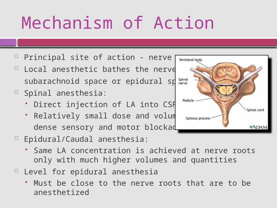
Mechanism of Action
Principal site of action - nerve root Local anesthetic bathes the nerve root in the
subarachnoid space or epidural space Spinal anesthesia:
Direct injection of LA into CSF Relatively small dose and volume to achieve
dense sensory and motor blockade Epidural/Caudal anesthesia:
Same LA concentration is achieved at nerve roots only with much higher volumes and quantities
Level for epidural anesthesia Must be close to the nerve roots that are to be anesthetized

Somatic Blockade
Sensory blockade interrupts both somatic and visceral painful stimuli
Motor blockade produces skeletal muscle relaxation Provides excellent OR conditions
LA effect on nerve fibers varies according to many factors: Size of the nerve fiber Myelination Concentration achieved Duration of contact
Smaller and myelinated fibers are more easily blocked

Somatic Blockade
Spinal nerve roots contain varying mixtures of these fiber types and they vary in their sensitivity to the LA blockade
This results in a differential block Which nerve fibers are blocked by the lowest sensitivity to LA?
A. pain B. motor C. sympathetic D. touch
Order of sensitivity: Sympathetic > pain > touch > motor
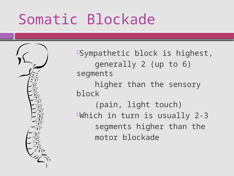
Somatic Blockade
Sympathetic block is highest, generally 2 (up to 6) segments higher than the sensory block (pain, light touch)Which in turn is usually 2-3 segments higher than the motor blockade

Autonomic Blockade
Block of efferent autonomic transmission sympathetic and some parasympathetic blockade
Sympathetic outflow from the spinal cord Thoracolumbar Sympathetic preganglionic nerve fibers
exit the spinal cord with the spinal nerves from T1 to the L2 level and may course many levels along sympathetic chain before synapsing with a
postganglionic cell in a sympathetic ganglia Parasympathetic outflow
Craniosacral Parasympathetic preganglionic fibers exit the spinal cord with the cranial and
sacral nerves Neuraxial anesthesia does not block the vagus nerve
decreased sympathetic tone and/or unopposed parasympathetic tone

Outline Anatomy Mechanism of Action Systemic Manifestations Indications/Contrandications Anticoagulants/Antiplatelets Anatomic Approaches Spinal Anesthesia Epidural Anesthesia Caudal Anesthesia Complications

A pt receives a spinal anesthetic with a sensory level of T5. Which of the following is likely to
occur?
A. The small bowel will be dilated and relaxed.
B. Glomerular filtration will be decreased by one third.
C. Tidal volume will be reduced by one third.
D. The cardioaccelerator nerves will be unaffected.
E. Blood pressure will lower predominantly by decreasing venous return.

Answer
E
Level of sympathetic block can be 2-6 levels higher than sensory block.

Cardiovascular Manifestations Variable decreases in blood pressure
+/- decrease in heart rate and cardiac contractility Generally proportional to degree of the sympathectomy Arterial and venous smooth muscle vasomotor tone:
Innervated by sympathetic fibers from T5 to L1 Blocking these nerves causes:
vasodilation of the venous capacitance vessels pooling of blood decreased venous return to the heart
Arterial vasodilation may also decrease SVR May be minimized by compensatory vasoconstriction above the level
of the block

Cardiovascular Manifestations
A high sympathetic block prevents compensatory vasoconstriction blocks the sympathetic cardiac accelerator fibers
that arise at …? T1–T4
Profound hypotension may occur Vasodilation combined with bradycardia and decreased contractility Exaggerated if venous return is further compromised
head-up position or gravid uterus Sudden cardiac arrest sometimes seen with spinal anesthesia
Unopposed vagal tone

Cardiovascular Manifestations
Steps to minimize the degree of hypotension: Volume loading with 10–20 mL/kg of IVF
partially compensates for the venous pooling LUD in the third trimester of pregnancy
minimizes obstruction to venous return Hypotension may still occur
Increase IVFs Autotransfusion - head-down position Vasopressors (phenylephrine/ephedrine)
Excessive or symptomatic bradycardia Atropine
If profound hypotension and/or bradycardia persist Epinephrine (5–10 mcg)

Pulmonary Manifestations
Usually are minimal diaphragm innervated by the phrenic nerve
with fibers originating from C3–C5 Even with high thoracic levels…
tidal volume is unchanged only a small decrease in vital capacity
from loss of abdominal muscles' contribution to forced expiration Phrenic nerve block may not occur even with total spinal
anesthesia apnea often resolves with hemodynamic resuscitation suggests that brain stem hypoperfusion is responsible

Pulmonary Manifestations
Severe chronic lung disease patients Rely upon accessory muscles of respiration Coughing and clearing of secretions require these muscles High levels of neural blockade impair these muscles
Use caution in patients with limited respiratory reserve Must weigh against the advantages of avoiding airway
instrumentation and PPV Surgery above the umbilicus
Pure regional technique may not be the best choice

Pulmonary Manifestations
Thoracic or upper abdominal surgery Decreased diaphragmatic function postop Decreased FRC Atelectasis and hypoxia via V/P mismatch
Postop thoracic epidural analgesia may improve pulmonary outcome decrease the incidence of
pneumonia and respiratory failure improve oxygenation decrease duration of vent
support

GI Manifestations
Sympathetic outflow originates at T5–L1 Sympathectomy - vagal tone dominance
small, contracted gut with active peristalsis Excellent operative conditions for lap procedures when used as
an adjunct to GENA Postoperative epidural analgesia has been shown to hasten
return of GI function Hepatic blood flow will decrease with reductions
in MAP from any anesthetic technique Intraabdominal surgery - decrease in hepatic
perfusion related more to surgical manipulation than to anesthetic technique.

Urinary Tract Manifestations
Renal blood flow – maintained through autoregulation little clinical effect upon renal function
Neuraxial anesthesia at the lumbar and sacral levels blocks both sympathetic and parasympathetic control of bladder function
Loss of autonomic bladder control results in urinary retention until the block wears off
If no urinary catheter is anticipated perioperatively: use the shortest acting and smallest amount of
LA necessary for the procedure limit the amount of IVF as much as possible
Monitored pt for urinary retention to avoid bladder distention following neuraxial anesthesia
Metabolic & Endocrine Manifestations
Surgical trauma produces a neuroendocrine response localized inflammatory response activation of somatic and visceral afferent nerve fibers increases in ACTH, cortisol, epinephrine, NE, and vasopressin activation of the renin–angiotensin–aldosterone system
Clinical manifestations: HTN, tachycardia, hyperglycemia, protein catabolism, suppressed immune
responses, and altered renal function Neuraxial blockade can partially suppress (during major invasive
surgery) or totally block (during lower extremity surgery) this stress response
Reduction in catecholamine release may decrease perioperative arrhythmias and reduce the incidence of
ischemia Neuraxial block should precede incision and extend postop

Outline Anatomy Mechanism of Action Systemic Manifestations Indications/Contrandications Anticoagulants/Antiplatelets Anatomic Approaches Spinal Anesthesia Epidural Anesthesia Caudal Anesthesia Complications

Indications for Neuraxial
Used alone or in conjunction with GENA for most procedures below the neck
Most useful for: lower abdominal inguinal urogenital rectal lower extremity surgery
Lumbar spinal surgery may also be performed under spinal anesthesia
Upper abdominal procedures difficult to achieve a sensory level adequate for patient comfort yet avoid the
complications of a high block Spinal anesthesia for neonatal surgery

Contrandications
Patient refusal Infection at the site of
injection Coagulopathy or
other bleeding diathesis
Severe hypovolemia Increased intracranial
pressure Severe aortic stenosis Severe mitral stenosis
Preexisting neurological deficits
Sepsis Uncooperative patient Demyelinating lesions Stenotic valvular heart
lesions Severe spinal
deformity
Absolute Relative Controversial
Inability to communicate with pt
Prior back surgery at site of injection
Complicated surgery Prolonged operation Major blood loss Maneuvers that
compromise respiration
Preexisting neurological deficitsPatient refusalInfection at the site of injectionInability to communicate with ptSepsisCoagulopathy or other bleeding diathesis

Outline Anatomy Mechanism of Action Systemic Manifestations Indications/Contrandications Anticoagulants/Antiplatelets Anatomic Approaches Spinal Anesthesia Epidural Anesthesia Caudal Anesthesia Complications

Oral Anticoagulants
Long-term warfarin therapy Must be stopped Need PT/INR to be normalized
Perioperative thromboembolic prophylaxis If initial dose given > 24 h prior to the block or if more than one
dose was given PT and INR need to be checked
If only one dose given within 24 h Safe
Removing an epidural catheter from patients receiving low-dose warfarin (5 mg/d) Safe

Antiplatelets
Aspirin and NSAIDs Alone don’t appear to increase risk of spinal hematoma
More potent agents Ticlopidine (Ticlid)
14 days Clopidogrel (Plavix)
7 days Abciximab (Rheopro)
48 h Eptifibatide (Integrilin)
8 h

Unfractionated Heparin
Minidose subQ prophylaxis OK to proceed
Patients to receive heparin intraoperatively 1 h or more before heparin administration A bloody epidural or spinal does not necessarily require cancellation of surgery
discussion of the risks with the surgeon careful postoperative monitoring needed
Removal of an epidural catheter 1 h prior to dosing or 4 h following dosing
Patients on therapeutic doses of heparin (elevated PTT) Avoid neuraxial
The risk of spinal hematoma is undetermined in the setting of full anticoagulation for cardiac surgery
LMWH (Enoxaparin, Dalteparin, -parin)
Intro of Lovenox in the US in 1993 Reports of spinal hematomas associated
with neuraxial anesthesia Many involved intraop or early postop use,
and several also taking antiplatelets If bloody needle or catheter placement occurs
Delay until 24 hrs postop Postop LMWH thromboprophylaxis if epidural catheter in place
Remove 2 hrs prior to the first dose Or 10 hrs after last dose and subsequent dosing should not
occur for another 2 hrs

Fibrinolytic or Thrombolytic Tx Best to avoid neuraxial.
Please note…
Drugs/regimens not considered to put pts at increased risk of neuraxial bleeding when used alone (minidose subQ heparin, NSAIDS) may in fact increase the risk when combined.

Outline Anatomy Mechanism of Action Systemic Manifestations Indications/Contrandications Anticoagulants/Antiplatelets Anatomic Approaches Spinal Anesthesia Epidural Anesthesia Caudal Anesthesia Complications

Which of the following statements regarding spinal needle insertion is TRUE?
A. The first significant resistance encountered when advancing a needle using the paramedian approach is the interspinous ligament.
B. If bone is repeatedly encountered at the same depth when the needle is advanced, the needle is likely walking down the inferior spinous process.
C. The midline approach is preferred in patients with heavily calicified interspinous ligaments.
D. Free flow of CSF after resolution of a paresthesia usually indicates that the needle is in a good position.
E. Penetration of the dura mater is more easily detected with a beveled needle.

Answer
D. If a paresthesia occurs you should
immediately stop advancing the needle and check for CSF.
Obtaining CSF after resolution of a paresthesia indicates the needle encountered a cauda equina nerve root in the subarachnoid space and the needle tip is in a good position.
DO NOT inject LA in presence of a persistent paresthesia!

Anatomic Approaches
Spinous processes Cervical and lumbar spine – horizontal
Needle directed with only a slight cephalad angle
Thoracic spine – slant in a caudal direction and can overlap Needle angled significantly more cephalad
First palpable cervical spinous process is C2 Most prominent is…?
C7
Inferior tip of the scapula at level of …? Spinous process of T7
Highest points of both iliac crests (Tuffier's line) ? Body of L4 or the L4–L5 interspace
Posterior superior iliac spine S2 posterior foramina
Sacral hiatus Depression just above or between the gluteal clefts and above the coccyx

Midline Approach
Body positioned with the plane of the back perpendicular to the floor Palpate for depression between the spinous processes of the vertebra above and
below the level to be used Subcutaneous tissues offer little feeling of resistance Supraspinous and interspinous ligaments felt as an increase in tissue density If bone contacted superficially
needle is likely hitting..? the lower spinous process
If bone contacted at a deeper depth and needle is in the midline it is likely hitting…?
the upper spinous process or if it is lateral to the midline it is likely hitting…?
a lamina Ligamentum flavum - obvious increase in resistance At this point, spinal and epidural anesthesia differ

Paramedian Approach
May be useful in certain patients severe arthritis kyphoscoliosis prior lumbar spine surgery
2 cm lateral to the inferior aspect of superior spinous process
Penetrates the paraspinous muscles lateral to the interspinous ligaments needle may encounter little resistance
initially and may not seem to be in firm tissue
Needle advanced at a 10–25° angle toward the midline
LOR is often more subtle than with the midline approach

Bone encountered deep lateral part of the
lower lamina redirected only
slightly upward, more toward the midline
Bone at a shallow depth medial part of the
lower lamina redirect mostly
upward and slightly more lateral
Outline Anatomy Mechanism of Action Systemic Manifestations Indications/Contrandications Anticoagulants/Antiplatelets Anatomic Approaches Spinal Anesthesia Epidural Anesthesia Caudal Anesthesia Complications

Spinal Needles
Available in an array of sizes (16–30 gauge), lengths, and bevel and tip designs
Tightly fitting removable stylet avoids tracking epithelial cells into the subarachnoid space
2 broad groups1. Sharp (cutting)-tipped
Quincke needle is a cutting needle with end injection
2. Blunt tip (pencil-point) needles Whitacre – rounded point with side injection Sprotte – rounded point with long side opening markedly decreased the incidence of PDPH
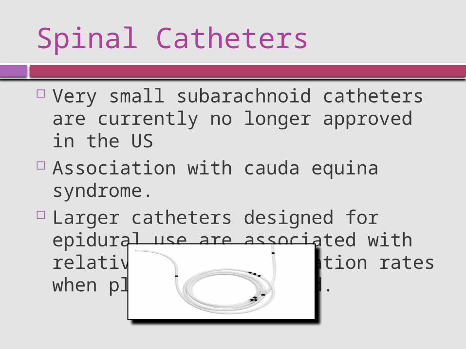
Spinal Catheters
Very small subarachnoid catheters are currently no longer approved in the US
Association with cauda equina syndrome. Larger catheters designed for epidural use are
associated with relatively high complication rates when placed subarachnoid.

Spinal Anesthesia
Midline, paramedian, or prone approach
Two "pops" are felt: ligamentum flavum dura–arachnoid membrane
Successful dural puncture confirmed by free flow of CSF
Persistent paresthesia or pain upon injection withdraw and redirect
Aspiration of CSF may be necessary in certain cases: presence of low CSF
pressure (dehydrated patient)
prone position
Which of the following statements is FASLE?
A. A patient in the sitting position will have a higher block if the solution is hypobaric and the patient remains erect.
B. A patient placed supine and in the Trendelenburg position is at high risk for developing a total spinal block after injection of an isobaric solution.
C. A patient in the prone jackknife position should not have a hyperbaric solution injected.
D. The normal lumbar lordosis limits the spread of hyperbaric solution is a supine patient.

Answer
B.
An isobaric solution should not ascend to cause a total spinal regardless of the patient’s position.

Factors Affecting the Level of Spinal Anesthesia
Baricity Position of the
patient During and
immediately after injection
Dosage Site of injection
Age CSF Curvature of the spine Drug volume Intraabdominal
pressure Needle direction Patient height Pregnancy
Most Important Factors Other Factors

Baricity 101
A hyperbaric solution of local anesthetic is denser (heavier) than CSF Addition of glucose
Hypobaric solution is less dense (lighter) than CSF Addition of sterile water
Head-down position Hyperbaric solution - spreads cephalad Hypobaric anesthetic solution - moves caudad
A head-up position Hyperbaric solution - settles caudad Hypobaric solution - ascends cephalad
Lateral position Hyperbaric spinal solution - greater effect on dependent (down) side Hypobaric solution - higher level on nondependent (up) side
Isobaric solution tends to remain at the level of injection

Baricity 101
Hyperbaric solutions tend to move to the most dependent area of the spine T4–T8 in the supine position
Apex of the thoracolumbar curvature is T4 In the supine position, this should limit a hyperbaric solution to
produce a level of anesthesia at or below T4 Abnormal curvatures of the spine, such as scoliosis and kyphoscoliosis,
have multiple effects on spinal anesthesia Difficult landmarks Decreased CSF

Baricity 101
CSF has a specific gravity of 1.003–1.008 at 37°CAgent Specific Gravity
Bupivacaine
0.5% in 8.25% dextrose 1.0227–1.0278
0.5% plain 0.9990–1.0058
Lidocaine
2% plain 1.0004–1.0066
5% in 7.5% dextrose 1.0262–1.0333
Procaine
10% plain 1.0104
2.5% in water 0.9983
Tetracaine
0.5% in water 0.9977–0.9997
0.5% in D5W 1.0133–1.0203

Spinal Anesthesia
CSF volume inversely correlates with level of anesthesia Increased intraabdominal pressure or conditions that cause
engorgement of the epidural veins, thus decreasing CSF volume, are associated with higher blocks Pregnancy Ascites Large abdominal tumors
Conflicting opinion exists as to whetherincreased CSF pressure caused by coughing or straining, or turbulence on injection has any effect on the spread of LA

Spinal Agents
Only preservative-free solutions used Addition of vasoconstrictors (epi or neo) and opioids may enhance the
quality and/or prolong the duration of spinal anesthesia
Drug Preparation DOA (plain) DOA (w/epi)
Procaine 10% solution 45 60
Bupivacaine 0.75% in 8.25%dextrose 90-120 100-150
Tetracaine 1% solution in 10%glucose 90-120 120-240
Lidocaine 5% in 7.5%glucose(dilute to 2.5% or less)
60-75 60-90
Ropivacaine 0.2-1%solution(Off-label use)
90-120 90-120

Spinal Agents
Hyperbaric bupivacaine and tetracaine are two of the most commonly used agents for spinal Relatively slow in onset (5–10 min) Prolonged duration (90–120 min) Similar sensory levels Tetracaine more motor blockade Addition of epi to bupivacaine prolongs its duration only modestly In contrast, epi to tetracaine prolongs by more than 50% Phenylephrine also prolongs tetracaine anesthesia but has no effect on
bupivacaine Ropivacaine
Experience with spinals is more limited A 12-mg intrathecal dose of ropivacaine is roughly equivalent to 8 mg of
bupivacaine, but it appears to have no particular advantages for spinal anesthesia

Spinal Agents
Lidocaine and procaine rapid onset (3–5 min) and short duration of action (60–90 min) modest if any prolonged effect with epi
Lidocaine associated with transient neurological symptoms (TNS) and cauda equina syndrome TNS: back pain radiating to the legs without sensory or motor
deficits after resolution of spinal resolves spontaneously within several days
Some experts suggest that lidocaine can be safely used as a spinal anesthetic if the total dose is limited to 60 mg and diluted to 2.5% or less

Outline Anatomy Mechanism of Action Systemic Manifestations Indications/Contrandications Anticoagulants/Antiplatelets Anatomic Approaches Spinal Anesthesia Epidural Anesthesia Caudal Anesthesia Complications
Epidural Anesthesia
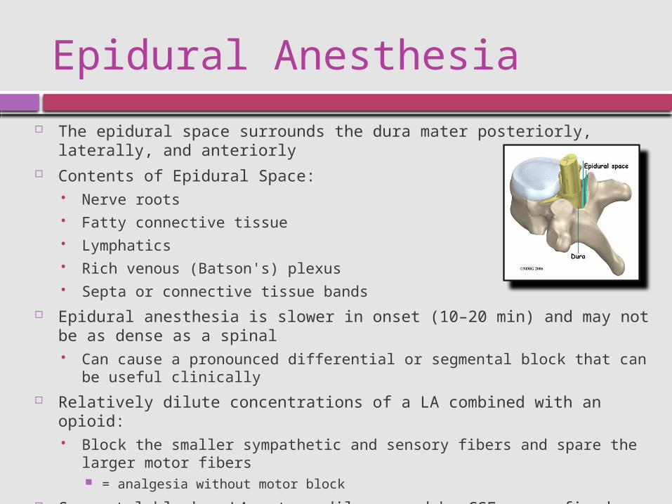
The epidural space surrounds the dura mater posteriorly, laterally, and anteriorly Contents of Epidural Space:
Nerve roots Fatty connective tissue Lymphatics Rich venous (Batson's) plexus Septa or connective tissue bands
Epidural anesthesia is slower in onset (10–20 min) and may not be as dense as a spinal Can cause a pronounced differential or segmental block that can be useful clinically
Relatively dilute concentrations of a LA combined with an opioid: Block the smaller sympathetic and sensory fibers and spare the larger motor fibers
= analgesia without motor block
Segmental block – LA not readily spread by CSF so confined close to level it was injected Characterized by a well-defined band of anesthesia at certain nerve roots Nerve roots above and below are not blocked Ex. thoracic epidural

Epidural Needles
Typically 17–18 gauge 9cm to hub Tuohy needle most commonly used
Blunt bevel with a gentle curve of
15–30° at the tip Pushes away the dura after passing
through the ligamentum flavum instead of penetrating it
Straight needles without a curved tip (Crawford needles) may have a higher incidence of dural puncture but facilitate passage of an epidural catheter.
Needle modifications include winged tips and introducer devices set into the hub designed for guiding catheter placement.

Epidural Catheters
Continuous infusion or intermittent boluses May allow a lower total dose of anesthetic to be used Intraop and/or postop analgesia 19- or 20-gauge catheter is introduced through a 17- or 18-gauge epidural needle Bevel opening directed either cephalad or caudad, and catheter advanced 2–6 cm The shorter the distance advanced:
more likely it is to become dislodged The further the catheter is advanced:
greater the chance of a unilateral block exiting the epidural space via an intervertebral foramen coursing into the anterolateral recesses
Single port at the distal end or multiple side ports close to a closed tip Some have a stylet for easier insertion Spiral wire-reinforced catheters are very resistant to kinking The spiral or spring tip is associated with fewer, less intense paresthesias and may
be associated with a lower incidence of inadvertent intravascular insertion

Epidural Techniques
LOR technique most commonly used Needle advanced through subQ tissues with the stylet in place Once interspinous ligament entered (increase in tissue resistance), stylet
removed Glass syringe filled with approximately 2 mL of fluid or air is attached If tip of needle is within the ligament, gentle attempts at injection are met with
resistance Needle slowly advanced, millimeter by millimeter, with either continuous or
rapidly repeating attempts As tip enters the epidural space there is a sudden LOR and injection is easy
Hanging Drop Techniquehttp://www.youtube.com/watch?v=7kDi47vqBis
Variation of Hanging Drop Techniquehttp://www.youtube.com/watch?v=TvCBDamF4jQ&feature=related
Activating an Epidural
Quantity LA for epidural anesthesia is very large compared to spinals Significant toxicity can occur if injected intrathecally or intravascularly Safeguards against this: epidural test dose and incremental dosing Test dose detects both subarachnoid and IV injection Classic test dose: 3mL of 1.5% lidocaine with 1:200,000 epinephrine (5mcg/mL)
45mg of lidocaine injected intrathecally – rapidly apparent spinal anesthesia 15 mcg of epinephrine injected intravascularly – noticeable increase in heart rate (20%
or more) with or without hypertension False positives (uterine contraction causing pain or an increase in heart rate
coincident to test dosing) False negatives (patients taking beta blockers) 25% or more increase in T-wave amplitude on EKG may be more reliable sign of IV
injection Both fentanyl and larger doses of local anesthetic without epinephrine have been
advocated as intravenous injection test doses Simply aspirating prior to injection – insufficient to avoid inadvertent IV injection

Activating an Epidural
Incremental dosing is a very effective method of avoiding serious complications Fraction of the total intended LA dose, typically 5 mL Should be large enough for mild symptoms of IV injection to occur but
small enough to avoid seizure or cardiovascular compromise. If a clinician uses an initial test dose, is diligent about
aspirating prior to each injection, and always uses incremental dosing, significant systemic toxicity or inadvertent intrathecal injections are rare.

Outline Anatomy Mechanism of Action Systemic Manifestations Indications/Contrandications Anticoagulants/Antiplatelets Anatomic Approaches Spinal Anesthesia Epidural Anesthesia Caudal Anesthesia Complications

When using a caudal approach to the epidural space, which of the following is TRUE?
A. The patient must be prone. B. An inadvertent subarachnoid block is much
less likely than when using the lumbar approach.
C. The technique becomes relatively more contraindicated as the patient’s age decreases.
D. Small volumes of agent are needed since the volume of the canal is only 8-12ml.
E. The needle enters through the sacral hiatus.

Answer
E.
Canal is of low volume but there is leakage through the foramina requiring injection of a larger volume compared to the lumbar approach.
Pt can be prone or lateral decubitus. Inadvertent dural puncture is very possible. Caudal approach is technically easier than
lumbar approach in babies, and is becoming increasingly more popular in pediatric anesthesia.

Caudal Anatomy
Caudal space is considered the sacral portion of the epidural space Sacral vertebrae fuse into one large bone – the sacrum Each one retains discrete anterior and posterior intervertebral foramina Laminae of S5 and all or part of S4 normally do not fuse, leaving a caudal
opening to the spinal canal, the sacral hiatus Sacrococcygeal ligament covers the sacral hiatus
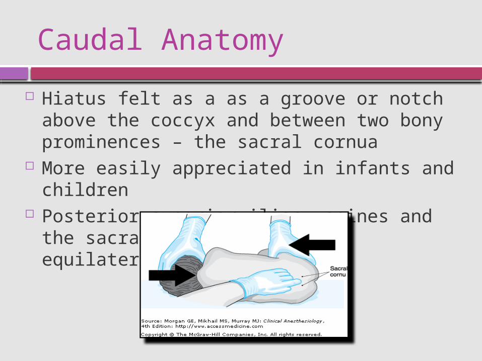
Caudal Anatomy
Hiatus felt as a as a groove or notch above the coccyx and between two bony prominences – the sacral cornua
More easily appreciated in infants and children Posterior superior iliac spines and the sacral hiatus
define an equilateral triangle

Caudal Epidural Anesthesia
One of the most commonly used regional techniques in pediatric patients
Used in anorectal surgery in adults 2nd stage of labor In children - typically combined with GENA
for intraop supplementation and postop analgesia Performed after induction Commonly used for procedures below the diaphragm
urogenital, rectal, inguinal, and lower extremity Within the sacral canal, the dural sac extends to…what level?
S2 in adults S3 in infants
Makes inadvertent intrathecal injection much more common in infants

Caudal Epidural Technique
Position lateral or prone with one or both hips flexed
Palpate sacral hiatus Sterile skin prep Needle advanced at a 45° angle
cephalad until a pop is felt (sacrococcygeal ligament)
Angle flattened and advanced Aspirate for blood and CSF If negative, proceed with injection Test dose vs incremental dosing
with frequent aspiration

Caudal Anesthesia
Complication rate for "kiddie caudals" is very low Total spinal and IV injection causing seizure or cardiac
arrest Intraosseous injection has also been reported to cause
systemic toxicity Calcification of the sacrococcygeal ligament may make
caudal anesthesia difficult or impossible in older adults

Pediatric Caudal Anesthesia
Dose: 0.5–1.0 mL/kg of 0.125–0.25% bupivacaine (or ropivacaine) +/- epi
Opioids may be added (ex 50–70 mcg/kg of morphine) not recommended for outpatients - delayed respiratory depression
Duration can extend for hours into the postop period Ok to d/c home even with mild residual motor block or
without urinating most children will urinate within 8 h
Higher epidural levels can be accomplished with catheters threaded cephalad into the lumbar or even thoracic epidural space

Caudal in Adults
Dense sacral sensory blockade with limited cephalad spread for anorectal procedures
Prone jackknife position Dose 15–20 mL of 1.5–2.0% lidocaine +/- epi Fentanyl 50–100 mcg may also be added

Outline Anatomy Mechanism of Action Systemic Manifestations Indications/Contrandications Anticoagulants/Antiplatelets Anatomic Approaches Spinal Anesthesia Epidural Anesthesia Caudal Anesthesia Complications

All of the following statements regarding complications associated with epidural and spinal anesthesia are true
EXCEPT:
A. Use of fluid instead of air for LOR during epidural anesthesia reduces the risk of headache upon accidental dural puncture.
B. An epidural blood patch immediately relieves PDPH symptoms in 99% of pts.
C. Transient reduction in hearing acuity after spinal anesthesia is more common in female than in male patients.
D. Back pain is more common after epidural anesthesia than after spinal anesthesia.
E. Neurologic injury occurs in about 0.03% to 0.1% of all central neuraxial blocks.

Answer
B
90% not 99%

All of the following statements regarding spinal or epidural anesthesia and spinal hematoma are true
EXCEPT:
A. Pts taking NSAIDS and receiving mini dose heparin are not at increased risk.
B. Pts treated with enoxaparin are at increased risk.
C. Pts most commonly present with numbness or lower extremity weakness.
D. Spinal hematoma occurs at an estimated incidence of less than 1:150,000.
E. The removal of an epidural or an intrathecal catheter presents nearly as great a risk for spinal hematoma as its insertion.

Answer
A
Combination may put patients at increased risk.

Adverse or exaggerated physiological responses
Urinary retention High block Total spinal anesthesia Cardiac arrest Anterior spinal artery syndrome Horner's syndrome
Complications related to needle/catheter
placement Trauma Backache Dural puncture/leak Postdural puncture headache Diplopia Tinnitus Neural injury Nerve root damage Spinal cord damage Cauda equina syndrome Bleeding Intraspinal/epidural hematoma Misplacement No effect/inadequate anesthesia Subdural block Inadvertent subarachnoid block1
Inadvertent intravascular injection Catheter shearing/retention Inflammation Arachnoiditis Infection Meningitis Epidural abscess
Drug toxicity Systemic local anesthetic toxicity Transient neurological symptoms Cauda equina syndrome

THE END