neurofibroma in neurofibromatosis type 1 · develop plexiform neurofibroma, a schwann cell tumor...
TRANSCRIPT

Robust surgical approach for cutaneousneurofibroma in neurofibromatosis type 1
Bahir H. Chamseddin, … , Juan Vega, Lu Q. Le
JCI Insight. 2019. https://doi.org/10.1172/jci.insight.128881.
In-Press Preview
BACKGROUND. Cutaneous neurofibromas (cNF) are physically disfiguring, painful, andcause extensive psychologic harm in patients with neurofibromatosis type 1 (NF1). There iscurrently no effective medical treatment and surgical procedures are inaccessible to mostNF1 patients globally.
OBJECTIVE. While research is underway to find an effective medical treatment for cNF,there is an urgent need to develop surgical approach that is accessible to all NF1 patients inthe world with the skill set and equipment found in most general medical office settings.Here, we present a robust surgical approach to remove cNF that does not require sterilesurgical field, utilizes accessible clinical equipment, and can be performed by any healthcare providers including family practitioners, and physician assistants.
METHODS. In a prospective case-series, patients with NF1 underwent this surgicalprocedure which removes multiple cutaneous neurofibromas. The Dermatology Life QualityIndex was given to subjects before and after the procedure as surrogate for patientsatisfaction.
RESULTS. 83 tumors were removed throughout the body from twelve individuals.Examination at follow-up visits revealed well-healed scars without infection or adverseevents including aberrant scarring. Patient satisfaction with the procedure was high withsignificant improvements […]
Clinical Medicine Dermatology Neuroscience
Find the latest version:
http://jci.me/128881/pdf
1
Robust Surgical Approach for Cutaneous Neurofibroma in Neurofibromatosis Type 1
Bahir H. Chamseddin1, La’Nette Hernandez1, Dezehree Solorzano1, Juan Vega1, Lu Q. Le1,2,3
1Department of Dermatology, UT Southwestern Medical Center, Dallas, TX 75390 2Comprehensive Neurofibromatosis Clinic, UT Southwestern Medical Center, Dallas, TX 75390 3Simmons Comprehensive Cancer Center, UT Southwestern Medical Center, Dallas, TX 75390
Corresponding Author: Lu Q. Le, M.D., Ph.D. Associate Professor Department of Dermatology 5323 Harry Hines Blvd., Dallas, TX. 75390-9069 Phone number: 214-648-5781 E-mail: [email protected] Conflicts of Interest: The authors have declared that no conflict of interest exists. Keywords: Neurofibromatosis Type 1, NF1, neurofibroma, dermal neurofibroma, cutaneous neurofibroma, treatment, elective surgery, patient satisfaction Funding sources: None

2
Abstract
Background: Cutaneous neurofibromas (cNF) are physically disfiguring, painful, and cause
extensive psychologic harm in patients with neurofibromatosis type 1 (NF1). There is currently
no effective medical treatment and surgical procedures are inaccessible to most NF1 patients
globally.
Objective: While research is underway to find an effective medical treatment for cNF, there is
an urgent need to develop surgical approach that is accessible to all NF1 patients in the world
with the skill set and equipment found in most general medical office settings. Here, we present
a robust surgical approach to remove cNF that does not require sterile surgical field, utilizes
accessible clinical equipment, and can be performed by any health care providers including
family practitioners, and physician assistants.
Methods: In a prospective case-series, patients with NF1 underwent this surgical procedure
which removes multiple cutaneous neurofibromas. The Dermatology Life Quality Index was
given to subjects before and after the procedure as surrogate for patient satisfaction.
Results: 83 tumors were removed throughout the body from twelve individuals. Examination at
follow-up visits revealed well-healed scars without infection or adverse events including aberrant
scarring. Patient satisfaction with the procedure was high with significant improvements in
symptoms, daily activities, leisure, personal relationships, and treatment experience (p=0.00062).
Conclusion: This study demonstrates a robust surgical approach to management cutaneous
neurofibromas which can be accessed world-wide to individuals with NF1 and performed by a
wide-variety of medical specialists with high clinical efficacy and patient satisfaction.

3
Introduction
Neurofibromatosis Type 1 (NF1) is one of the most common autosomal dominant genetic
disorders affecting 1 in 3000 live births and is found worldwide independent of gender, race, or
geographic location (1). It results from loss of the NF1 tumor suppressor gene in Schwann cell
lineage leading to extensive tumor formation throughout the body and a hallmark of the disorder
is multiple dermal or cutaneous neurofibromas (cNF) (2). About 30% of NF1 individuals also
develop plexiform neurofibroma, a Schwann cell tumor along the internal nerve plexuses that has
around 10% lifetime risk for malignant transformation (1). On the other hand, the cNF are
present in virtually all patients with NF1. They are exclusively located in the cutaneous dermis
layer and are not prone to malignancy. Despite their benign physiology, patients with NF1
attribute the cNF as their greatest medical burden due to their physical disfigurement (3). NF1
patients can develop thousands of cNF that are 2mm-3cm in size, soft, skin-colored nodules
covering the face, trunk, and extremities. cNF are the primary source of chronic physical
symptoms such as pain and itching and emotional distress given a greater burden of cNF strongly
correlates to negative quality of life and self-image (4).
The cNF composes diverse cellular components including Schwann cells, fibroblasts,
macrophages, mast cells, and blood vessels in a collagenous matrix within the dermis (5-7).
Although their pathophysiology is unknown, cNF are affected by genetic variation of NF1, the
skin microenvironment, and hormones and may grow in evolving stages (Figure 1) (8, 9). They
are classified by stage according to appearance: the nascent stage detected only through
ultrasound or other forms of imaging, the flat stage depicted by thinning or hyperpigmentation at
the surface of the skin, the sessile stage with a raised papule, the globular stage with a 20-30mm

4
base with comparable height, and the pedunculated stage which extrudes its dermal contents
through a visible stalk connecting the portions above and below the skin (9, 10). In all stages,
considerable mass is located within the deeper dermis layer whereas typical shaving or
electrodessication treatments which target only the visible projection may lead to tumor regrowth
and collagen scar formation (Figure 1) (11, 12). Currently, there is no available medical
treatment for cNF. Physical removal remains the mainstay of treatment, primarily focused
around surgical excision with primary closure by dermatologists or general and plastic surgeons
(13, 14). Unfortunately, this method may be inaccessible to a majority of the global population
due to the requirement of surgical expertise, sterile field, and general anesthesia. CO2 lasers
have been developed since the 1980’s to remove hundreds of tumors at one time but contain high
risk of hypopigmented or hypertrophic scarring (Figure 1) and continues to remain largely
inaccessible to the NF1 population due to lack of specialized training and equipment (15, 16).
Due to the dearth of accessibility to treatment for patients with NF1 worldwide, we offer
a robust surgical approach for management of cutaneous neurofibromas that can be performed in
an outpatient setting with equipment available to most general medical clinics and can be
performed by almost all medical providers including family practitioners, physician assistants,
and nurse practitioners. In this study, we recognize all adverse events associated with the
procedure and quantify patient satisfaction.
Results
Twelve patients with NF1 were consented and underwent the surgical procedure (Figure
2). The average age of the subjects was 46.6 (standard deviation (SD), 17), 10 (80%) of the

5
subjects were female, and 11 (92%) were Caucasian with one African American. In the entire
study, 83 cNF were removed and were an average size of 1cm (SD, 0.35). An average of 6.9
(range: 1-10) cNF were removed each operation. Thirty-one (37.3%) of cNF removed were
located on the upper extremity, 29 (35%) from the trunk, 11 (13.3%) from the head & neck, 10
(12%) from the abdomen, and 2 (2.4%) from the lower extremity. This surgical technique is
based on the biology and anatomy of the cutaneous neurofibroma where a large portion of the
tumor is in the dermis. Therefore, a critical component to this procedure is further removal of
the mass within the deeper dermis. This is accomplished after shaving off the outer projection of
the neurofibroma by grasping the remaining mass with forceps, lifting the mass outwards, and
using the dermablade (or razor blade) to remove the pale, collagenous tumor. The open lesion is
then closed by one or two interrupted stitches depending on its size. The whole procedure can be
done in less than 2 minutes per tumor. There were no complications during the operations
including excessive pain or problems with local anesthesia. Initial follow-up was 14 days and
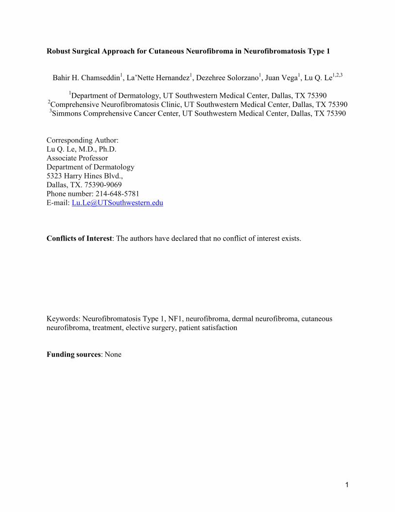
extended follow-up averaged 5 months. Representative photographs of the tumors before and
after the procedure are depicted in Figure 3. Examination at the extended follow-up revealed one
site with hypertrophic scar formation (1.2%) (Supplemental Figure 1) and 10 sites with post-
inflammatory hyperpigmentation (12%) (Supplemental Figure 2), but no other complications
were noted including skin infection, tumor regrowth, hypopigmentation, or keloid formation
(Table 1).
The Dermatology Life Quality Index (17) was administered to all twelve patients prior to
the surgical procedure and after the extended follow-up visit. Every question was answered to
completion and the ten-question survey took a maximum of five minutes to complete for each

6
person. The initial DLQI average and total scores were 9.83 and 118, respectively, which
improved significantly at the extended follow-up visit to 1.83 and 22, respectively (p=0.00062).
The individual DLQI dimensions prior to and after the surgical procedure are displayed in Table
2 and Supplemental Figure 3. The index of symptoms and feelings dropped from 3.1 (SD, 1.1)
to 1.1 (SD, 0.9) (p<0.0001), daily activities from 2.8 (SD, 1.2) to 0.5 (SD, 0.4) (p<0.0001),
leisure activities from 1.7 (SD, 1.1) to 0.3 (SD, 0.3) (p<0.0001). Personal relationships and
treatment efficacy dropped from average scores of 1.1 (SD, 0.8) and 0.6 (SD,08) to zero,
respectively. Although work and school functioning improved from 0.6 (SD, 1.2) to zero, the
results were not significant.
Discussion
The goal of the present report is to propose a novel technique of removing cutaneous
neurofibromas that is accessible to the global NF1 population. The procedure described herein is
considered accessible by using inexpensive medical equipment present in most outpatient
general clinical settings, employing a non-sterile technique, and following a low-risk procedure
which can be performed by most health care providers. The procedure yields favorable cosmetic
results and improves quality of life in NF1 patients.
Definitive treatment for cNF is a major obstacle for NF1 patients. NF1 patients are often
affected with other medical problems including bony deformities, malignant neoplasms, and
learning disability which could impact their access to treatment (18). Additionally, the type and
size of cNF, location of the tumor, and patient demographics must be considered for different
treatment modalities. Excisional operations performed by general and plastic surgeons have the

7
capability of removing extremely large cNF or hundreds of cNF in one operation with
exceptional reconstructive results (13, 14, 19). Unfortunately, this method mandates trained
surgical specialists, requires a sterile surgical field, runs a high-risk side effect profile with use of
general anesthesia, higher costs, and potential requirement of post-operative hospitalization (13,
14). Dermatologists typically will conduct excisional removal using local anesthesia to manage
cNF in an outpatient setting. It has a lower risk profile and cost to the NF1 patients than
operative surgery, yet the necessity for a longer excision to remove the cNF limits post-
operational cosmetic outcome (20). In addition, this traditional excision can only remove a few
tumors per session due to the time constraints. The procedure herein targets cNF anatomy by
selectively removing the dermal component which will prevent tumor regrowth and ultimately
yields more favorable post-procedural cosmetic outcomes for the patients by shortening the
excision length. Local anesthesia with Lidocaine 1% with epinephrine 1:100,000 is the agent of
choice for skin surgery and is generally safe to use with maximum dose of 40cc (mL) per session
or 7mg/kg total (21, 22). Cautionary use is advised when using epinephrine to the fingers, toes,
nose, or penis to avoid a theoretical complication of vasoconstriction and digital gangrene
despite evidence largely in support of its safety in the digits (23, 24). Rare, systemic toxicity
from lidocaine may include hypotension, bradycardia, or systemic allergic reaction (22). For
wound closure, the use of surgical glue or staples could replace sutures to expedite the removal
process of cNF and in addition to minimize the resultant skin tension from sutures after removal
of clustered cNF. Staples could be considered for larger neurofibroma in areas of less cosmetic
concern (i.e. non exposed skin) with benefits of quicker operational time and lesser infection
risks but are prone to greater post-operative pain than traditional sutures (25). Surgical glue
should only be implemented on small lesions under no tension post-operatively. This modality

8
may offer less scarring but additionally risk of allergic reaction to adhesives, and should not be
used in high moisture areas such as the axillae and perineum or highly mobile areas including
hands, feet, and joints (26).
The CO2 laser is commonly employed tool for treating cNF by rapidly heating and
vaporizing the intra-cellular water which leads to destruction of tissue (15). The advantage of
this technique includes the ability to treat hundreds of tumors at close proximity to one another
and can be performed in both the operating room or outpatient clinical setting with high patient
satisfaction (15, 27, 28). The CO2 laser technology is, however, unavailable to most global
clinical settings and specialized training is required to operate the machinery. Local infection
rate has been reported up to 15% and scarring is a frequently observed side effect (15, 27-29).
Minimally invasive photocoagulation including Er:YAG or Nd:YAG lasers have been
successfully implicated as recent treatment options for small to medium sized cNF with
complications such as post-inflammatory hyperpigmentation occurring in 4% of tumors yet the
access is limited to large academic centers (30, 31). Other destructive methods including
electrodessication and radiofrequency ablation have shown high patient satisfaction outcomes six
months post-procedure but should be considered as second-line therapies due to the high risks of
aberrant scarring and hypopigmentation associated with the techniques (10, 31, 32).
This study provides a robust, accessible method to remove multiple cNFs per visit that
can have a strongly positive impact on quality of life for NF1 patients. The DLQI survey was
specifically chosen due its ability to assess cNF removal impact directly on physical and
psychosocial factors related to the skin, while the Impact of NF1 on Quality of Life survey, an

9
important tool to asses quality of life for NF1 patients, assesses non-dermatologic features of the
disease including vision problem, gait abnormalities, and mental status changes that will not
reflect the outcomes of this procedure (4, 33). Symptoms of itchiness, soreness, or pain and
feelings of self-consciousness were among the highest reasons for poor quality of life pre-
operatively; however, the described procedure significantly diminished the magnitude of these
dimensions and most others based on the DLQI. Removal of cNF with this technique could be
implemented as a low-risk therapy for both symptomatic and aesthetically troublesome
neurofibromas in patients with a limited number of cNF. It is important to assess the potential
risks of complications including hypertrophic scarring, keloid formation and post-inflammatory
pigmentation. Keloids, abnormal collagen scarring formation, may occur in higher frequencies
in African-American patients or patients with a history of keloid formation (34). Thus clinicians
should warn such individuals about these risks when performing any surgical procedure in this
context. Post-inflammatory hyperpigmentation (PIH) more commonly occurs in people with
Fitzpatrick Skin Type IV-VI and Asian descent due to activation of melanocytes after trauma and
dermal procedures (35). Patients with PIH should be reassured for the benign nature of the
conditions and spontaneous resolution occurs after months, or potentially years (35). If PIH is an
issue, it can be managed post-operatively with UV protection, topical steroids, or retinoids and
lightening cream hydroquinone (36). Future studies should stratify treatment strategies based on
these risks, skin types, and location of cNF (31).
Physicians of different subspecialty training background and advanced practitioners can
utilize this simple technique to remove cNFs for their NF1 patients, but caution should be used to
ensure safety. cNF must be accurately identified from other potential differentiating soft tissue

10
masses including plexiform neurofibromas, dermatofibroma protuberans, or dermatofibromas.
Due to the superficial nature of this operation and low-risk for complications, the procedure is
deemed “low-risk” thus may be performed by practitioners licensed in the United States with
credentialing for performing these operations. We provide the Supplemental Movie showing
step by step of the whole procedure for additional training. Patients on anticoagulation or at risk
of bleeding should continue to undergo the procedure given the low-risk of the operation.
Additionally, cost of the operation would be comparable to a tangential biopsy (Billing Code:
11102 for the first lesion and 11103 for additional lesions). There is a need for more accurate
billing codes for cNF resection as a priority for future work given the physical disfigurement and
psychological harm of the lesions.
There are a several of limitations to this study to address. The procedure was performed
at a single-institution with relatively small sample size thus impacts generalizability. Selection
bias may have influenced the quality of life improvements given the individuals who were most
unhappy with their skin elected to undergo the surgery. The majority of subjects were of
Caucasian descent and post-procedural outcomes including post-inflammatory
hyperpigmentation may not accurately reflect outcomes for the entire population affected by
NF1.
Materials and Methods
Patients over the age of 18 with a diagnosis of NF1 established by the Neurofibromatosis
NIH Guidelines were recruited at the Neurofibromatosis Clinic at UT Southwestern Medical
Center, Dallas, Texas between February 01, 2018 to June 30, 2018 in a prospective clinical case

11
series (18). The procedure, risks and benefits, and importance of post-operative care were
described to the patients prior to consent. Preoperative assessment included a skin examination
and the collection of sociodemographic characteristics.
Materials
The surgical procedure requires isopropyl alcohol (70%) pads, local anesthetic (1%
Lidocaine Hydrochloride & Epinephrine 1:100,000), dermablade or razor blade, forceps, sutures,
needle driver, curved/straight Mayo scissors, non-sterile gloves, band aid and petrolatum
ointment. Required materials are depicted in Figure 4. Surgical glue or stapler may replace
suturing supplies for an expedient technique on smaller cNF.
Procedure
First, we disinfected the cNF with alcohol wipes and applied local anesthesia with 1%
Lidocaine/Epinephrine 1/100,000 to the neurofibroma and surrounding skin. After several
minutes to allow effects of anesthesia, a dermablade (or razor blade) was used to shave off the
outer projection of the neurofibroma level to the surrounding skin. Of note, the tumor within the
deeper dermis may naturally project out due to surrounding tension from the skin. A critical
component to this procedure is further excision or removal of the mass within the deeper dermis.
This is accomplished by grasping the remaining mass with forceps, lifting the mass outwards,
and using the dermablade (or razor blade) to remove the pale, collagenous tumor. The open
lesion is then closed by one or two interrupted stitches depending on its size. The closed wounds
are covered with white petroleum and a band aid. The subjects are educated to continue
application of petroleum ointment every day until the two-week follow-up for suture removal.

12
No antibiotics, oral nor topical, were given prior or after the procedure and clean, non-sterile
gloves were used throughout the procedure. The procedure can be viewed in Supplemental
Movie 1 and it takes less than two minutes to remove each tumor.
Demographic features of the subjects such as age, gender, ethnicity and the tumor
properties including size and location were recorded on the day of the procedure. The patients
had up to ten cNF removed per session with follow up in the clinic for suture removal in 2 weeks
and wound check. The subjects returned for an extended follow-up visit of at least 4 months
from the procedure date to analyze the following outcomes; infections, scar assessment, keloid
formation, hypo/hyperpigmentation, and other adverse events.
The Dermatology Life Quality Index (DLQI) (17), a survey which assesses patient satisfaction
with their skin, was administered to each patient before the procedure and at the extended
follow-up. The DLQI is a questionnaire containing ten questions, each with a response of “not at
all”, “a little”, “a lot”, or “very much” with corresponding scores of 0, 1, 2, and 3, respectively
(33). Higher scores are indicative of worsening quality of life. Question 1 and 2 are
representative of the patient’s symptoms and feelings, question 3 and 4 examines daily activities,
question 5 and 6 indicate impact on leisure activities, question 7 analyzes problems with work
and school, question 8 and 9 indicate personal relationships, and question 10 examines treatment
efficacy.
Statistics

13
Statistical analysis includes continuous data presented as means with standard deviations and
categorical data as counts with percentages. A two-tailed paired T-Test was used to compare the
outcomes of the DLQI. P≤.05 was considered significant.
Study Approval
The UT Southwestern Institutional Review Board approved this study and all patients
provided signed IRB consent and HIPPA privacy form for inclusion into the study. Written
informed consent was provided for pictures appearing in the manuscript.
Author Contributions
BHC: Acquiring data, analyzing data, drafting the manuscript
LH: acquiring data
DS: acquiring data
JV: acquiring data
LQL: Designing research study, acquiring data, analyzing data, draft and review manuscript
Acknowledgments
LQL holds a Career Award for Medical Scientists from the Burroughs Wellcome Fund, the
Thomas L. Shield, M.D. Professorship in Dermatology and supported by funding from the
National Cancer Institute of the National Institutes of Health (grant number R01 CA166593), the
Neurofibromatosis Therapeutic Acceleration Program (NTAP), the NF1 Research Consortium
Fund and the Giorgio Foundation.

14
References
1. Williams VC, Lucas J, Babcock MA, Gutmann DH, Korf B, Maria BL. Neurofibromatosis type 1 revisited. Pediatrics. 2009;123(1):124-33. 2. Le LQ, Shipman T, Burns DK, Parada LF. Cell of origin and microenvironment contribution for NF1-associated dermal neurofibromas. Cell Stem Cell. 2009;4(5):453-63. 3. Granstrom S, Langenbruch A, Augustin M, Mautner VF. Psychological burden in adult neurofibromatosis type 1 patients: impact of disease visibility on body image. Dermatology. 2012;224(2):160-7. 4. Ferner RE, et al. Evaluation of quality of life in adults with neurofibromatosis 1 (NF1) using the Impact of NF1 on Quality Of Life (INF1-QOL) questionnaire. Health Qual Life Outcomes. 2017;15(1):34. 5. Jouhilahti EM, et al. The development of cutaneous neurofibromas. Am J Pathol. 2011;178(2):500-5. 6. Ortiz-Hidalgo C, Weller R. Peripheral nervous system. In Histology for Pathologists, S. 1997. 7. Le L, Kesterson JP, Robert & H. Guttmann D. Defining the Research Landscape for Dermal Neurofbromas. Oncology Times. 2016;38.:14-5. 8. Allaway RJ, et al. Cutaneous neurofibromas in the genomics era: current understanding and open questions. Br J Cancer. 2018;118(12):1539-48. 9. Riccardi VM. Translational genetics and genomics: the fundamental nature of NF1 neurofibromas. Transl Genet Genom. 2007;1:3-14. 10. Ortonne N, et al. Cutaneous neurofibromas: Current clinical and pathologic issues. Neurology. 2018;91(2 Supplement 1):S5-S13. 11. Kim SH, Roh SG, Lee NH, Yang KM. Radiofrequency ablation and excision of multiple cutaneous lesions in neurofibromatosis type 1. Arch Plast Surg. 2013;40(1):57-61. 12. Roberts AH, Crockett DJ. An operation for the treatment of cutaneous neurofibromatosis. Br J Plast Surg. 1985;38(2):292-3. 13. Bromley GS, Sherman JE, Goulian D, Jr. Neurofibromatosis-distribution of lesions and surgical treatment. Ann Plast Surg. 1982;8(4):272-6. 14. Yuan SM, et al. Surgical management of giant neurofibroma in soft tissue: a single-center retrospective analysis. Int J Clin Exp Med. 2015;8(4):5245-53.

15
15. Becker DW, Jr. Use of the carbon dioxide laser in treating multiple cutaneous neurofibromas. Ann Plast Surg. 1991;26(6):582-6. 16. Roenigk RK, Ratz JL. CO2 laser treatment of cutaneous neurofibromas. J Dermatol Surg Oncol. 1987;13(2):187-90. 17. Finlay AY, Khan GK. Dermatology Life Quality Index (DLQI)--a simple practical measure for routine clinical use. Clin Exp Dermatol. 1994;19(3):210-6. 18. Ferner RE, et al. Guidelines for the diagnosis and management of individuals with neurofibromatosis 1. J Med Genet. 2007;44(2):81-8. 19. Pailheret JP. [Plastic surgery in benign cutaneous manifestations of von Recklinghausen's disease]. Chirurgie. 1990;116(4-5):368-72. 20. Onesti MG, Carella S, Spinelli G, Scuderi N. The megasession technique for excision of multiple neurofibromas. Dermatol Surg. 2010;36(9):1488-90. 21. Lawrence C. Drug management in skin surgery. Drugs. 1996;52(6):805-17. 22. Becker DE, Reed KL. Local anesthetics: review of pharmacological considerations. Anesth Prog. 2012;59(2):90-101; quiz 2-3. 23. Denkler K. A comprehensive review of epinephrine in the finger: to do or not to do. Plast Reconstr Surg. 2001;108(1):114-24. 24. Krunic AL, Wang LC, Soltani K, Weitzul S, Taylor RS. Digital anesthesia with epinephrine: an old myth revisited. J Am Acad Dermatol. 2004;51(5):755-9. 25. Iavazzo C, Gkegkes ID, Vouloumanou EK, Mamais I, Peppas G, Falagas ME. Sutures versus staples for the management of surgical wounds: a meta-analysis of randomized controlled trials. Am Surg. 2011;77(9):1206-21. 26. Bruns TB, Worthington JM. Using tissue adhesive for wound repair: a practical guide to dermabond. Am Fam Physician. 2000;61(5):1383-8. 27. Ostertag JU, Theunissen CC, Neumann HA. Hypertrophic scars after therapy with CO2 laser for treatment of multiple cutaneous neurofibromas. Dermatol Surg. 2002;28(3):296-8. 28. Meni C, et al. Treatment of neurofibromas with a carbon dioxide laser: a retrospective cross-sectional study of 106 patients. Dermatology. 2015;230(3):263-8. 29. Chiang YZ, Al-Niaimi F, Ferguson J, August PJ, Madan V. Carbon dioxide laser treatment of cutaneous neurofibromas. Dermatol Ther (Heidelb). 2012;2(1):7.

16
30. Kriechbaumer LK, Susani M, Kircher SG, Distelmaier K, Happak W. Comparative study of CO2- and Er:YAG laser ablation of multiple cutaneous neurofibromas in von Recklinghausen's disease. Lasers Med Sci. 2014;29(3):1083-91. 31. Verma SK, et al. Considerations for development of therapies for cutaneous neurofibroma. Neurology. 2018;91(2 Supplement 1):S21-S30. 32. Levine SM, Levine E, Taub PJ, Weinberg H. Electrosurgical excision technique for the treatment of multiple cutaneous lesions in neurofibromatosis type I. J Plast Reconstr Aesthet Surg. 2008;61(8):958-62. 33. Lewis V, Finlay AY. 10 years experience of the Dermatology Life Quality Index (DLQI). J Investig Dermatol Symp Proc. 2004;9(2):169-80. 34. Mofikoya BO, Adeyemo WL, Abdus-salam AA. Keloid and hypertrophic scars: a review of recent developments in pathogenesis and management. Nig Q J Hosp Med. 2007;17(4):134-9. 35. Davis EC, Callender VD. Postinflammatory hyperpigmentation: a review of the epidemiology, clinical features, and treatment options in skin of color. J Clin Aesthet Dermatol. 2010;3(7):20-31. 36. Sofen B, Prado G, Emer J. Melasma and Post Inflammatory Hyperpigmentation: Management Update and Expert Opinion. Skin Therapy Lett. 2016;21(1):1-7.

17
Tables
Table 1. Demographics and Operation Outcomes of Patients who underwent Cutaneous Neurofibroma Removal
Age Patient Sex Ethnicity Location No. of Tumors Tumor Type Complications
20-29 1 M AA Trunk 1 Globular Hypertrophic Scar
Upper Extremity 2 Globular (2) None 30-39 2 F C Trunk 6 Globular (5), Pedunculated None
Upper Extremity 1 Globular None
3 F C Abdomen 1 Sessile None Upper Extremity 5 Globular (2), Sessile (3) None 40-49 4 M C Lower Extremity 1 Sessile None 5 F C Head & Neck 3 Globular (2), Pedunculated None Trunk 1 Globular None Upper Extremity 3 Sessile (3) None 6 F C Head & Neck 1 Pedunculated None
Trunk 6 Globular (5), Sessile None
Upper Extremity 3 Pedunculated, Sessile (2) None 7 F C Abdomen 5 Globular (3), Sessile (2) None Trunk 2 Globular, Pedunculated None Lower Extremity 1 Sessile None 8 F C Abdomen 4 Globular (3), Pedunculated None
Trunk 3 Globular (3) None
Upper Extremity 2 Globular (2) None 50-59 9 F C Trunk 4 Globular (2), Pedunculated (2) None Upper Extremity 4 Globular (4) None 60-69 10 F C Head & Neck 4 Globular (4) None
Trunk 4 Globular (1), Pedunculated (3) None
Upper Extremity 2 Globular (2) None 11 F C Upper Extremity 8 Globular (4), Sessile (4) None 70-79 12 F C Head & Neck 3 Globular (2), Pedunculated None
Trunk 2 Globular, Pedunculated None Upper Extremity 1 Globular None
Abbreviations: M, male; F, female; AA, African American; C, Caucasian.

18
Table 2: Dermatology Life Quality Index averages among patients who removed cutaneous neurofibromas
Dimension Pre-Op
(mean, SD) Post-Op
(mean, SD) P Value Symptoms & Feelings (Maximum: 6) 3.1 (1.1) 1.1 (0.9) <0.0001 Daily Activities (Maximum: 6) 2.8 (1.2) 0.5 (0.4) <0.0001 Leisure (Maximum: 6) 1.7 (1.1) 0.3 (0.3) <0.0001 Personal Relationships (Maximum: 6) 1.1 (0.8) 0 (0) <0.0001 Work & School (Maximum: 3) 0.6 (1.2) 0 (0) 0.0967 Treatment (Maximum: 3) 0.6 (0.8) 0 (0) 0.0183 Abbreviations: SD, standard deviation. n = 12 patients Statistical Test: Two-tailed, paired T Test


Figure 1: Clinical Stages/Evolution of Cutaneous Neurofibromas and Treatment
A) Nascent, a dormant stage undetectable without instrumentation. B) Flat, thinning or
hyperpigmentation at the surface of the skin. C) Sessile, tumor raised with an apex. D) Globular,
20-30mm base with comparable height and globular shape. E) Peduncular, a stalk connects the
portions above and below the skin. F) Scars from CO2 laser treatment in its final stage
(permanent).


Figure 2: Flow Diagram for Operation

Figure 3. Cosmetic Outcomes of Cutaneous Neurofibromas Removal Using Described
Procedure at 5 months
Representative photos of before (left panel) and after (right panel) tumor removal at the A) Right
shoulder, B) Chest, C) Left nipple, D) Posterior auricular, E) Posterior neck, and F) Back.


Figure 4: Materials and Visual Procedure Sequences
A) Isopropyl Alcohol (70%) pads. B) Dermablade or Razor Blade C) Suture. D) Local
Anesthetic, (1% Lidocaine Hydrochloride & Epinephrine 1:100,000). E) Forceps. F) Needle
Driver. G) Curved/Straight Mayo Scissors H) Petrolatum. I) Bandage J) Non-Sterile Gloves.
K) Cutaneous Neurofibroma, sessile. L) Tumor removed at skin level with dermablade or razor
blade. M) Forceps grasp and expose dermal component. N) Removal of dermal component with
dermablade or razor blade. O) Site with completely removed cNF P) Skin sutured.