neuroimaging of language processes: fmri of silent and
TRANSCRIPT
Neuroradiology / Neuroradiologie
Neuroimaging of Language Processes:
fMRI of Silent and Overt Lexical Processing and
the Promise of Multiple Process Imaging in Single
Brain Studies
Ron Borowsky, PhD; William J Owen, PhD; Tammy L Wile, MA;
Chris Kelland Friesen, PhD; Jennifer Lynn Martin, BA; Gordon E Sarty, PhD
Abstract
Objective: To implement and evaluate a multiple-process functional magnetic resonance
imaging (fMRI) paradigm designed to effectively and efficiently activate several
language-related regions for use with neurosurgical patients. Both overt and covert
response conditions were examined.
Methods: The fMRI experiments compared the traditional silent word-generation condition
versus an overt one as they engage frontal language regions (Experiment 1) and silent
versus overt semantic association conditions as they engage multiple language
processing regions (Experiment 2).
Results: In Experiment 1, the overt condition yielded greater magnitude of activation, but
not volume of activation, in the left inferior frontal and insular cortices than did the silent
condition for most, but not all, participants. Experiment 2 demonstrated that the
activation of multiple established language processing regions (ie, orthographic,
phonological, and semantic) can be achieved in a significant number of participants,
particularly under overt semantic association conditions and that such activation varies
in predictable ways.
Conclusion: The traditional silent response condition cannot be considered as equivalent
to the overt response condition during word generation or semantic association. The
multiple-process imaging method introduced here was sensitive to processing robust
orthographic, phonological, and semantic regions, particularly under the overt response
condition.
Abrégé
Objectif : Appliquer et évaluer un paradigme aux processus multiples de l’imagerie par
résonance magnétique fonctionnelle (IRMF) destiné à activer de façon efficace et
efficiente plusieurs régions liées au langage, pour les patients de la neurochirurgie. Des
conditions de réponse ouvertes et fermées ont été examinées.
Méthodes : Les expériences d’IRMF comparaient la condition silencieuse classique de la
production de mots avec une condition ouverte, toutes deux faisant appel aux régions
du langage frontales (expérience 1), et une condition silencieuse avec une association
sémantique ouverte mobilisant les multiples régions de traitement du langage
(expérience 2).
Résultats : Dans l’expérience 1, comparativement à la condition silencieuse, la condition
ouverte donnait une plus grande ampleur d’activation, mais pas de volume
d’activation,dans le lobe frontal inférieur gauche et le lobe insulaire, pour la plupart des
participants, mais pas tous. L’expérience 2 a démontré que l’activation de multiples
régions établies du traitement du langage (c.-à-d., orthographique, phonologique et
sémantique) peut se faire chez un nombre significatif de participants, surtout dans des
conditions ouvertes d’association sémantique, et que cette activation varie de façon
prévisible.
204 JACR vol 56, no
4, octobre 2005
Borowsky, Sarty — Department of
Psychology, University of
Saskatchewan, Saskatoon, SK
Owen — Department of Psychology,
University of Northern British
Columbia, Prince George, BC
Wile—Nova Scotia Rehabilitation
Centre, Halifax, NS
Friesen — Department of Psychology,
North Dakota State University, Fargo,
N.D.
Martin — Faculty of Rehabilitation
Medicine, University of Alberta,
Edmonton, AB
Address for correspondence:
Dr Gordon Sarty, Dept of Psychology,
University of Saskatchewan,
Saskatoon, SK S7N 5A5; fax 306
966-6630; [email protected]
Submitted Apr 1, 2005
Accepted June 1, 2005
©2005 Canadian Association of
Radiologists
Can Assoc Radiol J 2005;56(4):204–213.
Conclusion : La condition de réponse silencieuse classique ne peut
être considérée comme étant équivalente à la condition de
réponse ouverte, durant la production de mots ou l’association
sémantique. La méthode d’imagerie aux processus multiples
introduite ici était sensible au traitement dans les régions
orthographique, phonologique et sémantique robustes, en
particulier dans une condition de réponse ouverte
Of all the cognitive tasks that we engage in on a regular
basis, one of the most frequent and important is generating
words (or in other words, using language), be it overtly or
silently. Indeed, when brain surgery is required (eg, to remove
epileptogenic tissue), great care is taken to avoid removal of
so-called eloquent cortex. In so doing, rather invasive methods
have typically been used to ascertain where language-related
cortex resides. For example, Wada’s test involves the injection
of sodium amobarbital to anesthetize one hemisphere of the
brain at a time (thus paralyzing the contralateral side of the
body), so as to evaluate errors in speech and memory and thus
determine the dominant hemisphere for language processing.
Prior to the removal of brain tissue, neurosurgeons typically
use electrodes to directly stimulate cortex while the patient is
conscious and responding to linguistic tasks, so as to determine
a safe boundary for tissue removal. The functional magnetic
resonance imaging (fMRI) method described in this paper
provides a noninvasive alternative for localizing language
function.
Over the past decade fMRI has developed into a method for
measuring blood oxygenation level–dependent (BOLD)
changes, thereby identifying regions of brain activation, specif-
ically excitatory local field potentials reflecting input and
intracortical processing.1 The utility and safety of fMRI in the
context of localizing language processes in the brain is less
invasive relative to the other commonly used methods
described above. However, access to magnetic resonance
imaging (MRI) equipment is often limited, and fMRI experi-
ments are often time-consuming (usually involving several
separate tasks to permit task subtraction) and are conducted on
a costly device at an expensive hourly rate. One major goal of
the present research is to evaluate the effectiveness and
efficiency of a new technique that attempts to activate many
language-related processing regions simultaneously and in a
very brief time (ie, less than 10 minutes). Such a technique may
prove to be useful not only for theoretical research (particularly
in its potential for effectively activating multiple language
functions without the need for subtracting multiple, hierarchi-
cal tasks), but also for its potential application as an efficient
pre- and postsurgical technique for language localization.
To experimentally study tasks like word generation with some
confidence that the participant is properly engaged in the task,
it is desirable to have a condition where they are able to respond
aloud. Responding aloud is a challenge in the MRI environ-
ment, where any motion of the head and magnetic susceptibil-
ity changes in the sinus cavities due to air volume changes can
result in artifactual activation. Therefore, silent responding has
traditionally been preferred by fMRI researchers. Nonetheless,
the development of tasks that allow the participant to generate
words to completion is critical for veridical and comprehensive
localization of language function in the brain.
Although head motion has been a source of problematic arti-
facts in fMRI, there have been some laudable efforts to develop
techniques that permit the participant to speak aloud.2–6
Recently, Rosen et al conducted a small-sample fMRI experi-
ment in which the participants (n = 5) engaged in both silent
and aloud word generation to 3-letter visual cues (eg, generat-
ing “steak” for “STE”).7 The authors concluded that there were
no average differences in activation magnitude between silent
and aloud conditions in the language-related cortical regions
that they examined, including a region that has long been impli-
cated in verbal language processing, which includes the left
frontal operculum and inferior frontal gyrus (IFG), often asso-
ciated with Broca’s area (see reviews by Binder and Price,8
Demb et al,9 and Grodzinsky10). Rosen et al’s conclusion is at
odds with some of the neuroimaging literature (eg, see review
by Demb et al9), given that the anterior limb of the IFG appears
to be more active during an overt response in semantic genera-
tion and semantic decision tasks compared with a covert
response,4,11 implicating articulation-related differences (or
attentional differences) between silent and overt conditions in
generation and decision tasks.
A region neighboring Broca’s area that may also be involved in
differences between silent and overt word generation is the
insular cortex. Raichle et al have suggested that the insular cor-
tex responds to familiarity with word naming tasks,12 so we
chose to explore activation in this region in case it was also sen-
sitive to silent versus overt response conditions. In Experiment
1, we conducted a detailed comparison of activation in these
left frontal language regions (ie, inferior frontal regions includ-
ing the frontal operculum and IFG, plus the insular cortex) as a
function of silent and overt word generation in order to deter-
mine whether these conditions differentially engage language
processing systems. The results of this experiment had impor-
tant implications for the design of Experiment 2, in that they
were expected to reveal whether one condition (silent or overt)
was better than the other at activating language-related regions,
and whether there were any language processing differences
for silent and overt conditions in individuals.
Material and Methods
These experiments were performed in compliance with the rel-
evant laws and institutional guidelines and were approved by
the University of Saskatchewan Behavioral Sciences Ethics
Committee. All imaging was conducted using a 1.5T
Magnetom Symphony (Siemens, Erlangen, Germany) imager.
Both experiments used TR = 1600 ms, TE = 55 ms, a 64 � 64
acquisition matrix, and a 128 � 128 reconstruction matrix.
CARJ Vol 56, No 4, October 2005 205
Neuroimaging of Language Processes
Echo planar imaging (EPI) slice thickness was 8 mm, with a
2 mm gap between slices. The first 10 volumes were used to
achieve a steady state of image contrast and were discarded
prior to analysis. To capture a full cortex volume of images for
each participant, either the third or fourth inferior-most slice
was centred on the posterior commissure, depending on dis-
tance between the posterior commisure and the top of the brain
for each participant. T1-weighted high-resolution spin-echo
anatomical images (TR = 525 ms, TE = 15ms, 192 � 256 acqui-
sition matrix) were acquired in axial, sagittal, and coronal
planes for the purpose of overlaying the activation maps. The
position and thickness of the T1 axial images matched the echo
planar images. A microcomputer running Micro Experimental
Laboratory (MEL) software (Psychology Software Tools, Inc,
Pittsburgh, PA) was used to trigger each image acquisition to
keep the stimulus presentations synchronized with the images.
Auditory instructions and stimuli were presented using the
standard pneumatic headphones and intercom system supplied
with the imager. Visual stimuli were presented using a Sharp
Notevision 3 data projector controlled by a personal computer
using MEL software, which also triggered each slice imaging
sequence. All participants were right-handed, spoke English as
a first language, and had university-level education.
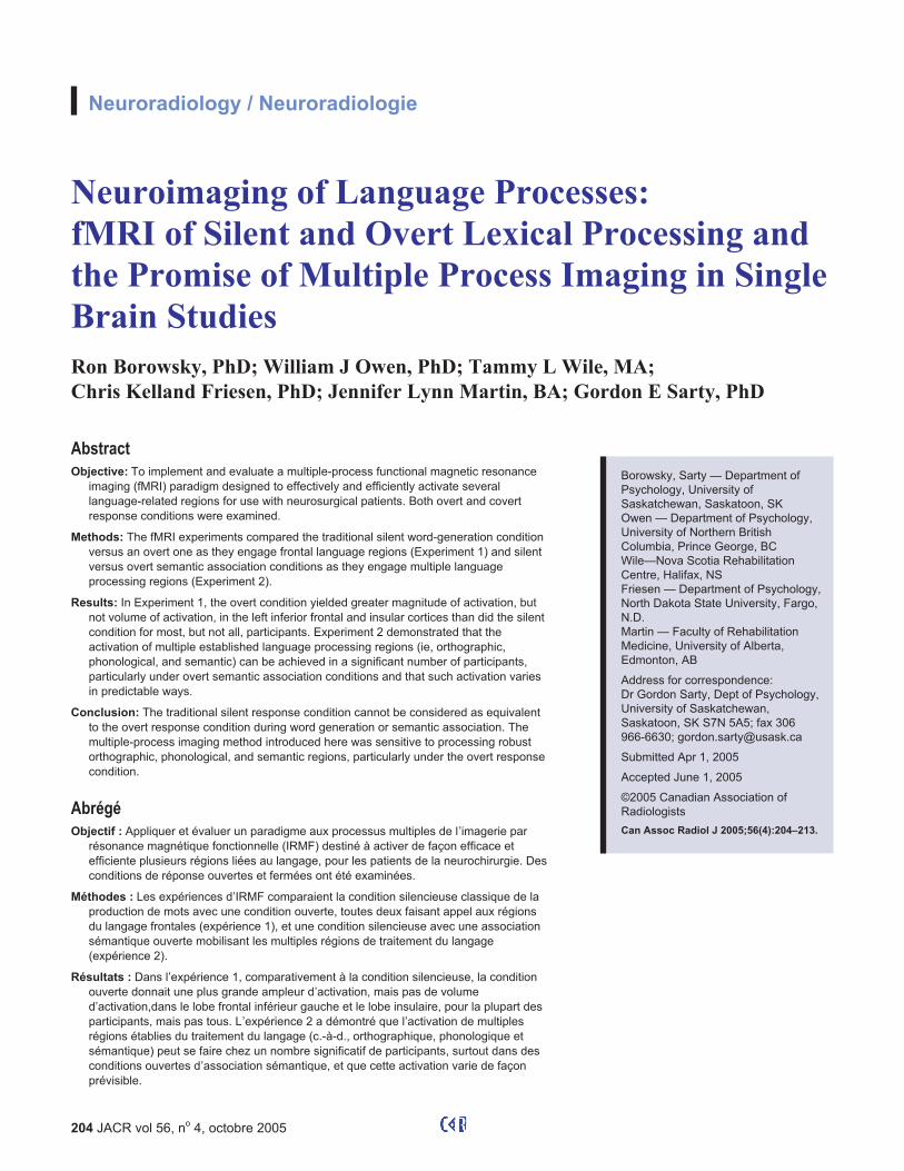
Experiment 1
Eight participants (6 men, 2 women) performed the word-
generation tasks, one-half with the aloud condition prior to the
silent condition and one-half with the silent condition prior to
the aloud condition. This experiment was a periodically
blocked, event-related design (Figure 1), a more sensitive
design than randomly presented single events for detecting the
effects of subtle BOLD functions13 that are correlated with the
task. For each condition (silent and aloud), 180 volumes of
10-slice axial single-shot echo planar images were obtained.
Total imaging time per condition was 288 seconds. Volumes
were organized into 5 blocks of 30 volumes (48 seconds per
block) with 5 volumes (8 seconds) between blocks that were
discarded in the subsequent analysis. The extra 5 volumes
between blocks were inserted to allow the hemodynamic
response to completely recover before the next block. The
rationale for the duration of relax time (see also Wise et al11)
was to ensure that the BOLD response had adequate time to
fully return to baseline (as determined from a preliminary pilot
study in which the relax time was varied). Each block consisted
of 10 volumes (16 seconds) of response followed by 20
volumes (32 seconds) of relax. The critical trials consisted of
5 alternating blocks of word generation to an aurally presented
cue (eg, “E as in echo,” with the stimulus letters occuring
equally often in silent and aloud conditions across partici-
pants), to which participants were instructed to generate as
many words as they could for 16 seconds that begin with the
stimulus letter and then to relax and concentrate on their breath-
ing for 40 seconds (including the between-block time) after the
experimenter said “stop.” Responses were monitored over the
MRI intercom during the overt condition.
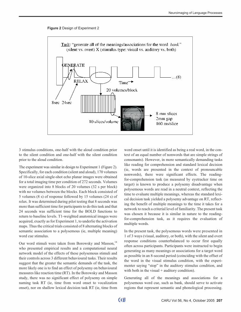
Experiment 2
Seven participants performed a semantic association task with
visual stimuli, 6 participants performed the same task but with
auditory stimuli, and 7 participants performed the task with
simultaneous visual and auditory stimuli. There were 9 male
and 11 female participants, distributed similarly across the
206 JACR vol 56, no
4, octobre 2005
NEURORADIOLOGY / NEURORADIOLOGIE
Figure 1 Design of Experiment 1
3 stimulus conditions, one-half with the aloud condition prior
to the silent condition and one-half with the silent condition
prior to the aloud condition.
The experiment was similar in design to Experiment 1 (Figure 2).
Specifically, for each condition (silent and aloud), 170 volumes
of 10-slice axial single-shot echo planar images were obtained
for a total imaging time per condition of 272 seconds. Volumes
were organized into 8 blocks of 20 volumes (32 s per block)
with no volumes between the blocks. Each block consisted of
5 volumes (8 s) of response followed by 15 volumes (24 s) of
relax. It was determined during pilot testing that 8 seconds was
more than sufficient time for participants to do this task and that
24 seconds was sufficient time for the BOLD functions to
return to baseline levels. T1-weighted anatomical images were
acquired, exactly as for Experiment 1, to underlie the activation
maps. Thus the critical trials consisted of 8 alternating blocks of
semantic association to a polysemous (ie, multiple meaning)
word cue stimulus.
Our word stimuli were taken from Borowsky and Masson,14
who presented empirical results and a computational neural
network model of the effects of these polysemous stimuli and
their controls across 3 different behavioural tasks. Their results
suggest that the greater the semantic demands of the task, the
more likely one is to find an effect of polysemy on behavioural
measures like reaction time (RT). In the Borowsky and Masson
study, there was no significant effect of polysemy on simple
naming task RT (ie, time from word onset to vocalization
onset), nor on shallow lexical decision task RT (ie, time from
word onset until it is identified as being a real word, in the con-
text of an equal number of nonwords that are simple strings of
consonants). However, in more semantically demanding tasks
like reading for comprehension and standard lexical decision
(ie, words are presented in the context of pronouncable
nonwords), there were significant effects. The reading-
for-comprehension task (as measured by eyetracker time on
target) is known to produce a polysemy disadvantage when
polysemous words are read in a neutral context, reflecting the
time to evaluate multiple meanings, whereas the standard lexi-
cal decision task yielded a polysemy advantage on RT, reflect-
ing the benefit of multiple meanings to the time it takes for a
network to reach a criterial level of familiarity. The present task
was chosen it because it is similar in nature to the reading-
for-comprehension task, as it requires the evaluation of
multiple words.
In the present task, the polysemous words were presented in
1 of 3 ways (visual, auditory, or both), with the silent and overt
response conditions counterbalanced to occur first equally
often across participants. Participants were instructed to begin
generating as many meanings or associations for a target word
as possible in an 8-second period (coinciding with the offset of
the word in the visual stimulus condition, with the experi-
menter saying “stop” in the auditory stimulus condition, and
with both in the visual + auditory condition).
Generating all of the meanings and associations for a
polysemous word cue, such as bank, should serve to activate
regions that represent semantic and phonological processing.
CARJ Vol 56, No 4, October 2005 207
Neuroimaging of Language Processes
Figure 2 Design of Experiment 2

By manipulating whether the stimulus cue is visual or auditory,
we can make further predictions. Participants should be more
likely to show activation in orthographic processing
regions—lateral temporal-occipital (LTO), lateral occipital
(LO), and medial extrastriate (ME) cortex—when a visual
orthographic cue is available, whereas phonological processing
should be more evident when an auditory stimulus is present,
such as during the overt response condition or when an audi-
tory cue is given. Finally, a visual-plus-auditory cue condition
was also included to evaluate whether the 2 modalities could be
combined to assess lexical processing more efficiently.
We also examined some brain regions that have been included
in other recent studies of word generation. Specifically,
Raichle et al have suggested that the insular cortex represents
familiarity with word naming,12 and Rosen et al have suggested
that the thalamus and putamen respond to increases in motor
demands of word generation.7 However, it would be premature
to generate strong predictions about these regions.
Image analysis
The BOLDfold method of analysis requires that sufficient time
elapse between active task conditions for the hemodynamic
response (or the BOLD function) to fully return to its baseline
level.15 After correction for linear baseline drift, the mean
BOLD function for each voxel, collapsing across the repeti-
tions of task and baseline, was empirically determined as the
average response of the repeated blocks and then repeated and
correlated to the actual data as a measure of consistency across
repetitions. Activation maps, which were displayed with AFNI
software16 were constructed using a criterial correlation (�) of
0.63 for Experiment 1 and 0.60 for Experiment 2.
Activation maps from Experiment 1 were masked to examine
volume (number of voxels) and magnitude of activation (maxi-
mum minus minimum of the average BOLD function) in the
left inferior frontal cortex, including the frontal operculum, and
the anterior portion of the insular cortex. These regions were
identified within 2 axial slices: the inferior slice was the first
slice above the eye sockets (approximately z = 0, the plane run-
ning through the posterior commissure), and the superior slice
was the next slice up (approximately z = 10). We adopted this
approach to identifying the regions of interest as opposed to
choosing a fixed z plane because the z plane is typically defined
relative to the anterior commissure–posterior commissure
line,17 which varies in angle between individuals. Student
t-tests were used to compare volumes and magnitudes between
the silent and aloud conditions in data averaged across partici-
pants.
Nonparametric sign tests were used on Experiment 2 data to
compare individual activations. The number of participants
showing activation in a particular region was considered signif-
icant at � = 0.05 by the sign test variant of chi-square if at least
4 out of 4 participants show an effect ( �2(1) = 4.0, p = 0.0455),
whereas 6 of 7 participants showing an effect would be mar-
ginal ( �2(1) = 3.571, p = 0.0588).
To identify a robust set of language processing regions, we
considered a recent review of the neuroimaging of language
processing. Demb et al have suggested that there is some con-
sensus in the literature with respect to regions that represent
orthographic (ie, visual spelling), phonological (ie, speech
sound), and semantic (ie, meaning) processing.9 Specifically,
phonological processing tends to activate superior temporal
(ST) (including primary auditory cortex) and posterior tempo-
ral (PT) (including Wernicke’s area) regions, semantic and
phonological processing tend to overlap in activating lateral
prefrontal (LPr) and inferior frontal (including Broca’s area)
regions, and orthographic processing tends to activate LTO,
LO (including primary visual cortex), and ME cortex.
Results
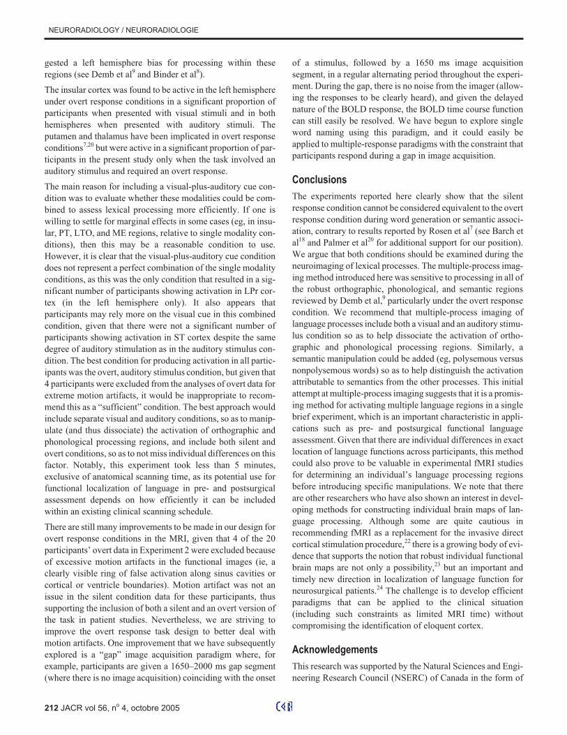
Experiment 1
For this word-generation task, the aloud condition resulted in
greater activation magnitude than did the silent condition in the
left frontal language regions, with the exception that the differ-
ence was only marginally significant in the inferior perspective
of the insular cortex (Table 1). Six of the 8 participants showed
greater activation in the regions of interest in the overt condi-
tion, whereas 2 of the participants showed greater activation in
the silent condition (Figure 3). The most similar slice to our
superior slice perspective in the Rosen et al study is z = 8 mm,
and they did not include any analyses of the next inferior slice.7
There were no significant differences when activation volume
(ie, number of active voxels) was used as the dependent vari-
able, although the superior perspective of the insular cortex
showed a trend toward greater activation volume for the aloud
condition.
Experiment 2
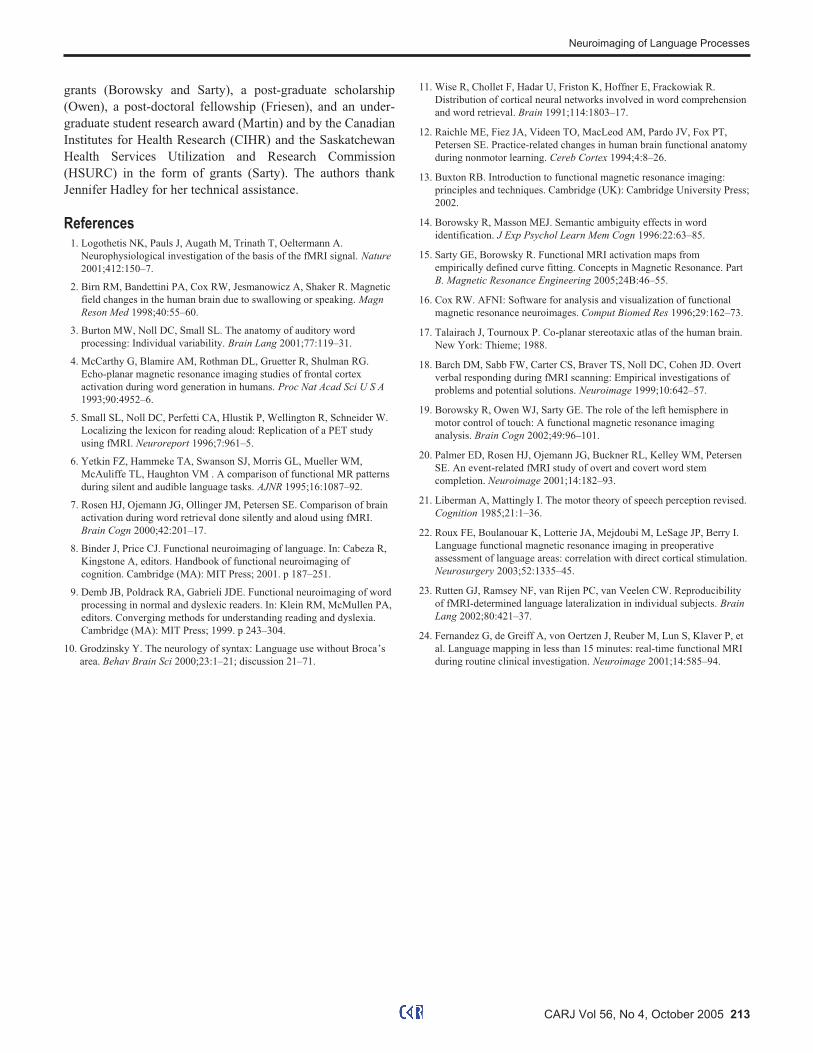
Activation maps for semantic association were constructed for
each participant using a criterial � = 0.60 (r(158) = 0.60, p =
9.245 × 10⁻¹⁷) (Figure 4, shown in red to yellow). For compari-
son purposes, and to ensure that our consistency criterion was
appropriate, activation at the level of � � 0.50 was also mapped
(shown in green). Activation in 11 regions of interest across the
axial slices shown in Figure 4 was examined for each partici-
pant and recorded as present or absent at the level of � � 0.60.
Following Barch et al,18 we examined the number of partici-
pants showing activation in each of these regions of interest as a
function of stimulus type, hemisphere, and task condition
(silent versus overt) (Table 2).
208 JACR vol 56, no
4, octobre 2005
NEURORADIOLOGY / NEURORADIOLOGIE
Discussion
Experiment 1
Using a motion-robust method of image analysis that is particu-
larly well suited for studying overt word generation,19 the fMRI
word-generation task presented here allowed us to quantify
activation volume and magnitude in left frontal language
regions for both silent and aloud conditions. The results of
Experiment 1 demonstrated that we can examine overt speech
in the MRI environment and that overt word generation results
in greater activation in frontal language regions than does the
more traditional silent condition. Specifically, fMRI activation
magnitude in silent versus overt word generation showed that
the left inferior frontal and insular cortical regions are more
active during the overt condition. This finding is inconsistent
with Rosen et al’s recent study.7 Given the smaller sample size
used by Rosen et al (n = 5), it is likely that their study lacked
sufficient power to reject the null hypothesis in these regions of
interest (they were able to demonstrate greater activation for
the aloud condition only in the motor cortex, auditory cortex,
thalamus, and putamen). A related possibility is that even a sin-
gle participant showing a strong reverse pattern compared with
the remainder of participants could be sufficient to eliminate
any effect. This latter possibility underscores the importance of
including an analysis at the level of the individual participant
(see Burton et al3). In the present study, most participants (ie, 6
of 8) support the pattern of differences obtained with the para-
metric analyses. Finally, there are differences in the exact
nature of the word-generation tasks used in the 2 studies. Rosen
et al’s word-generation task involved stem completion,
whereas our task involved generating words to a first letter cue.
It is possible that our word-generation task required greater
cognitive effort, and thus it may have recruited a higher degree
of phonological lexical processing, particularly in the aloud
condition where phonological lexical output is required. How-
ever, other tasks have elicited activation in inferior frontal lan-
guage regions during overt conditions, including Stroop color
naming and verb generation (eg, Barch et al18), which suggests
that activation in these regions is not particular to the variant of
word-generation task that we used.
The present findings are congruent, however, with Demb et al’s
review9 of the literature regarding greater activation in the
anterior limb of the left inferior frontal cortex for overt
word-generation and decision tasks than for silent versions of
these tasks.4,11 Perhaps more importantly, our findings are sup-
ported by the results of a study by Palmer et al20 that includes
the data from Rosen et al study7 with an additional 5 partici-
pants. Further, if we are to conceptualize the left inferior frontal
cortex as a region that serves to translate lexical entities into
their articulatory routines (ie, a phonological lexical output sys-
tem; see Binder et al’s review8), it seems quite sensible that
greater activation should occur here in the overt condition for
most of the participants.
The results of Experiment 1 support the notion that overt word
generation is different from the more common silent version of
the task, and the presence of individual differences in silent and
overt word generation suggest that both conditions should be
included when frontal regions are evaluated for language func-
tion. Thus in exploring a new technique for engaging multiple
language processing regions in Experiment 2, we chose to use
both silent and overt conditions.
Experiment 2
One critical region of interest is the speech tract region of motor
cortex, which provides a means to evaluate data quality. We
manipulated silent and overt response conditions in Experi-
ment 2, with the prediction that motor cortex should be acti-
vated to a greater extent in the overt condition across the
participants. Given the results of Experiment 1, we predicted
that the overt response condition should also evoke greater
CARJ Vol 56, No 4, October 2005 209
Neuroimaging of Language Processes
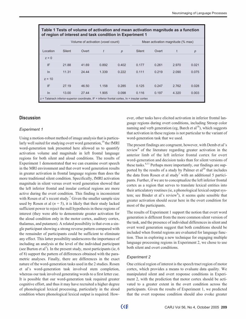
Table 1 Tests of volume of activation and mean activation magnitude as a functionof region of interest and task condition in Experiment 1
Volume of activation (voxel count) Mean activation magnitude (% max)
Location Silent Overt t p Silent Overt t p
z = 0
IF 21.88 41.69 0.892 0.402 0.177 0.261 2.970 0.021
In 11.31 24.44 1.339 0.222 0.111 0.219 2.090 0.075
z = 10
IF 27.19 46.50 1.158 0.285 0.125 0.247 2.762 0.028
In 13.00 27.44 1.905 0.098 0.116 0.197 4.320 0.003
z = Talairach inferior-superior coordinate, IF = inferior frontal cortex, In = insular cortex

210 JACR vol 56, no
4, octobre 2005
NEURORADIOLOGY / NEURORADIOLOGIE
Figure 3 Activation magnitude maps from representative individuals for silent and overt word generation (Experiment 1)exemplifying (a) a pattern where frontal language regions (inferior frontal [IF] and insular [In] cortex) are more active for silentthan overt conditions, which occurred in 2 of the 8 participants and (b) a pattern where these regions are more active forovert than silent conditions, which occurred in 6 of the 8 participants. Intensity values reflect activation intensity, thresholded
at � = 0.63, in greyscale units divided by 100.
Figure 4 Activation consistency maps from representative individuals for the visual stimulus condition of the semantic
association task (Experiment 2) under (a) overt and (b) silent conditions. Regions of interest (and the language processes
that they have been argued to reflect) include motor (M) cortex (phonological output), lateral prefrontal (LPr) cortex
(semantic), inferior frontal (IF) cortex (phonological), insular (In) cortex (phonological), superior temporal (ST) cortex
(phonological input), putatmen (Pu, phonological output), thalamus (Th, phonological output), posterior temporal (PT) cortex
(phonological and semantic), lateral temporal-occipital (LTO) cortex (orthographic), lateral occipital (LO) cortex
(orthographic), and medial extrastriate (ME) cortex (orthographic). Eta values represent the correlation between each
voxel’s blood oxygenation level dependent (BOLD) function timecourse and that voxel’s mean BOLD function, thus serving
as a measure of activation consistency.
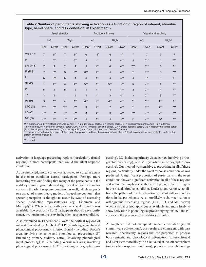
activation in language processing regions (particularly frontal
regions) in more participants than would the silent response
condition.
As we predicted, motor cortex was activated to a greater extent
in the overt condition across participants. Perhaps more
interesting was our finding that many of the participants in the
auditory stimulus group showed significant activation in motor
cortex in the silent response condition as well, which supports
one aspect of motor theory models of speech perception—that
speech perception is thought to occur by way of accessing
speech production representations (eg, Liberman and
Mattingly21). Whenever an orthographic visual stimulus was
available, however, only 1 or 2 participants showed any signifi-
cant activation in motor cortex in the silent response condition.
Also examined in Experiment 2 were the cortical regions of
interest described by Demb et al9: LPr (involving semantic and
phonological processing), inferior frontal (including Broca’s
area, involving semantic and phonological processing), ST
(including primary auditory cortex, involving phonological
input processing), PT (including Wernicke’s area, involving
phonological processing), LTO (involving orthographic pro-
cessing), LO (including primary visual cortex, involving ortho-
graphic processing), and ME (involved in orthographic pro-
cessing). Our method was sensitive to processing in all of these
regions, particularly under the overt response condition, as was
predicted. A significant proportion of participants in the overt
conditions showed significant activation in all of these regions
and in both hemispheres, with the exception of the LPr region
in the visual stimulus condition. Under silent response condi-
tions, the pattern of results was also consistent with our predic-
tions, in that participants were more likely to show activation in
orthographic processing regions (LTO, LO, and ME cortex)
when a visual orthographic cue is available and more likely to
show activation in phonological processing regions (ST and PT
cortex) in the presence of an auditory stimulus.
Although we did not manipulate semantic variables (ie, all
stimuli were polysemous), our results are congruent with past
research. Specifically, regions that are purported to process
both semantic and phonological information (inferior frontal
and LPr) were more likely to be activated in the left hemisphere
(under silent response conditions); previous research has sug-
CARJ Vol 56, No 4, October 2005 211
Neuroimaging of Language Processes
Table 2 Number of participants showing activation as a function of region of interest, stimulustype, hemisphere, and task condition, in Experiment 2
Visual stimulus Auditory stimulus Visual and auditory
Left Right Left Right Left Right
Silent Overt Silent Overt Silent Overt Silent Overt Silent Overt Silent Overt
Valid n = 7 5a
7 5a
6 4a
6 4a
7 7 7 7
M 1 5** 1 5** 5 4** 5 4** 2 7** 1 7**
LPr (P,S) 6* 4 2 4 5 4** 4 4** 7** 7** 5 6*
IF (P,S) 6* 5** 3 5** 6** 4** 5 4** 6* 7** 5 7**
In 5 5** 5 4 4 4** 4 4** 4 6* 3 6*
ST (P) 4 5** 3 5** 6** 4** 6** 4** 5 7** 5 7**
Pu 5 4 5 4 4 4** 4 4** 3 7** 4 7**
Th 3 4 1 4 4 4** 3 4** 3 7** 3 7**
PT (P) 5 5** 4 5** 6** 4** 6** 4** 6* 7** 7** 6*
LTO (O) 7** 5** 7** 5** 3 4** 2 4** 6* 7** 7** 7**
LO (O) 7** 5** 7** 5** 3 4** 3 4** 7** 7** 7** 7**
ME (O) 7** 5** 7** 5** 4 4** 4 4** 6* 7** 6* 7**
M = motor cortex, LPr = lateral prefrontal cortex, IF = inferior frontal cortex, In = insular cortex, ST = superior temporal cortex, Pu = putamen,Th = thalamus, PT = posterior temporal cortex, LTO = lateral temporal-occipital cortex, LO = lateral occipital cortex, ME = medial extrastriate cortex(P) = phonological, (S) = semantic, (O) = orthographic, from Demb, Poldrack and Gabrieli’s
9review
aThere were 2 participants in each of the visual stimulus and auditory stimulus conditions whose “aloud” data were not interpretable due to motion
artifact and thus excluded.* p < .06.
** p < .05.
gested a left hemisphere bias for processing within these
regions (see Demb et al9 and Binder et al8).
The insular cortex was found to be active in the left hemisphere
under overt response conditions in a significant proportion of
participants when presented with visual stimuli and in both
hemispheres when presented with auditory stimuli. The
putamen and thalamus have been implicated in overt response
conditions7,20 but were active in a significant proportion of par-
ticipants in the present study only when the task involved an
auditory stimulus and required an overt response.
The main reason for including a visual-plus-auditory cue con-
dition was to evaluate whether these modalities could be com-
bined to assess lexical processing more efficiently. If one is
willing to settle for marginal effects in some cases (eg, in insu-
lar, PT, LTO, and ME regions, relative to single modality con-
ditions), then this may be a reasonable condition to use.
However, it is clear that the visual-plus-auditory cue condition
does not represent a perfect combination of the single modality
conditions, as this was the only condition that resulted in a sig-
nificant number of participants showing activation in LPr cor-
tex (in the left hemisphere only). It also appears that
participants may rely more on the visual cue in this combined
condition, given that there were not a significant number of
participants showing activation in ST cortex despite the same
degree of auditory stimulation as in the auditory stimulus con-
dition. The best condition for producing activation in all partic-
ipants was the overt, auditory stimulus condition, but given that
4 participants were excluded from the analyses of overt data for
extreme motion artifacts, it would be inappropriate to recom-
mend this as a “sufficient” condition. The best approach would
include separate visual and auditory conditions, so as to manip-
ulate (and thus dissociate) the activation of orthographic and
phonological processing regions, and include both silent and
overt conditions, so as to not miss individual differences on this
factor. Notably, this experiment took less than 5 minutes,
exclusive of anatomical scanning time, as its potential use for
functional localization of language in pre- and postsurgical
assessment depends on how efficiently it can be included
within an existing clinical scanning schedule.
There are still many improvements to be made in our design for
overt response conditions in the MRI, given that 4 of the 20
participants’ overt data in Experiment 2 were excluded because
of excessive motion artifacts in the functional images (ie, a
clearly visible ring of false activation along sinus cavities or
cortical or ventricle boundaries). Motion artifact was not an
issue in the silent condition data for these participants, thus
supporting the inclusion of both a silent and an overt version of
the task in patient studies. Nevertheless, we are striving to
improve the overt response task design to better deal with
motion artifacts. One improvement that we have subsequently
explored is a “gap” image acquisition paradigm where, for
example, participants are given a 1650–2000 ms gap segment
(where there is no image acquisition) coinciding with the onset
of a stimulus, followed by a 1650 ms image acquisition
segment, in a regular alternating period throughout the experi-
ment. During the gap, there is no noise from the imager (allow-
ing the responses to be clearly heard), and given the delayed
nature of the BOLD response, the BOLD time course function
can still easily be resolved. We have begun to explore single
word naming using this paradigm, and it could easily be
applied to multiple-response paradigms with the constraint that
participants respond during a gap in image acquisition.
Conclusions
The experiments reported here clearly show that the silent
response condition cannot be considered equivalent to the overt
response condition during word generation or semantic associ-
ation, contrary to results reported by Rosen et al7 (see Barch et
al18 and Palmer et al20 for additional support for our position).
We argue that both conditions should be examined during the
neuroimaging of lexical processes. The multiple-process imag-
ing method introduced here was sensitive to processing in all of
the robust orthographic, phonological, and semantic regions
reviewed by Demb et al,9 particularly under the overt response
condition. We recommend that multiple-process imaging of
language processes include both a visual and an auditory stimu-
lus condition so as to help dissociate the activation of ortho-
graphic and phonological processing regions. Similarly, a
semantic manipulation could be added (eg, polysemous versus
nonpolysemous words) so as to help distinguish the activation
attributable to semantics from the other processes. This initial
attempt at multiple-process imaging suggests that it is a promis-
ing method for activating multiple language regions in a single
brief experiment, which is an important characteristic in appli-
cations such as pre- and postsurgical functional language
assessment. Given that there are individual differences in exact
location of language functions across participants, this method
could also prove to be valuable in experimental fMRI studies
for determining an individual’s language processing regions
before introducing specific manipulations. We note that there
are other researchers who have also shown an interest in devel-
oping methods for constructing individual brain maps of lan-
guage processing. Although some are quite cautious in
recommending fMRI as a replacement for the invasive direct
cortical stimulation procedure,22 there is a growing body of evi-
dence that supports the notion that robust individual functional
brain maps are not only a possibility,23 but an important and
timely new direction in localization of language function for
neurosurgical patients.24 The challenge is to develop efficient
paradigms that can be applied to the clinical situation
(including such constraints as limited MRI time) without
compromising the identification of eloquent cortex.
Acknowledgements
This research was supported by the Natural Sciences and Engi-
neering Research Council (NSERC) of Canada in the form of
212 JACR vol 56, no
4, octobre 2005
NEURORADIOLOGY / NEURORADIOLOGIE
grants (Borowsky and Sarty), a post-graduate scholarship
(Owen), a post-doctoral fellowship (Friesen), and an under-
graduate student research award (Martin) and by the Canadian
Institutes for Health Research (CIHR) and the Saskatchewan
Health Services Utilization and Research Commission
(HSURC) in the form of grants (Sarty). The authors thank
Jennifer Hadley for her technical assistance.
References1. Logothetis NK, Pauls J, Augath M, Trinath T, Oeltermann A.
Neurophysiological investigation of the basis of the fMRI signal. Nature
2001;412:150–7.
2. Birn RM, Bandettini PA, Cox RW, Jesmanowicz A, Shaker R. Magnetic
field changes in the human brain due to swallowing or speaking. Magn
Reson Med 1998;40:55–60.
3. Burton MW, Noll DC, Small SL. The anatomy of auditory word
processing: Individual variability. Brain Lang 2001;77:119–31.
4. McCarthy G, Blamire AM, Rothman DL, Gruetter R, Shulman RG.
Echo-planar magnetic resonance imaging studies of frontal cortex
activation during word generation in humans. Proc Nat Acad Sci U S A
1993;90:4952–6.
5. Small SL, Noll DC, Perfetti CA, Hlustik P, Wellington R, Schneider W.
Localizing the lexicon for reading aloud: Replication of a PET study
using fMRI. Neuroreport 1996;7:961–5.
6. Yetkin FZ, Hammeke TA, Swanson SJ, Morris GL, Mueller WM,
McAuliffe TL, Haughton VM . A comparison of functional MR patterns
during silent and audible language tasks. AJNR 1995;16:1087–92.
7. Rosen HJ, Ojemann JG, Ollinger JM, Petersen SE. Comparison of brain
activation during word retrieval done silently and aloud using fMRI.
Brain Cogn 2000;42:201–17.
8. Binder J, Price CJ. Functional neuroimaging of language. In: Cabeza R,
Kingstone A, editors. Handbook of functional neuroimaging of
cognition. Cambridge (MA): MIT Press; 2001. p 187–251.
9. Demb JB, Poldrack RA, Gabrieli JDE. Functional neuroimaging of word
processing in normal and dyslexic readers. In: Klein RM, McMullen PA,
editors. Converging methods for understanding reading and dyslexia.
Cambridge (MA): MIT Press; 1999. p 243–304.
10. Grodzinsky Y. The neurology of syntax: Language use without Broca’s
area. Behav Brain Sci 2000;23:1–21; discussion 21–71.
11. Wise R, Chollet F, Hadar U, Friston K, Hoffner E, Frackowiak R.
Distribution of cortical neural networks involved in word comprehension
and word retrieval. Brain 1991;114:1803–17.
12. Raichle ME, Fiez JA, Videen TO, MacLeod AM, Pardo JV, Fox PT,
Petersen SE. Practice-related changes in human brain functional anatomy
during nonmotor learning. Cereb Cortex 1994;4:8–26.
13. Buxton RB. Introduction to functional magnetic resonance imaging:
principles and techniques. Cambridge (UK): Cambridge University Press;
2002.
14. Borowsky R, Masson MEJ. Semantic ambiguity effects in word
identification. J Exp Psychol Learn Mem Cogn 1996:22:63–85.
15. Sarty GE, Borowsky R. Functional MRI activation maps from
empirically defined curve fitting. Concepts in Magnetic Resonance. Part
B. Magnetic Resonance Engineering 2005;24B:46–55.
16. Cox RW. AFNI: Software for analysis and visualization of functional
magnetic resonance neuroimages. Comput Biomed Res 1996;29:162–73.
17. Talairach J, Tournoux P. Co-planar stereotaxic atlas of the human brain.
New York: Thieme; 1988.
18. Barch DM, Sabb FW, Carter CS, Braver TS, Noll DC, Cohen JD. Overt
verbal responding during fMRI scanning: Empirical investigations of
problems and potential solutions. Neuroimage 1999;10:642–57.
19. Borowsky R, Owen WJ, Sarty GE. The role of the left hemisphere in
motor control of touch: A functional magnetic resonance imaging
analysis. Brain Cogn 2002;49:96–101.
20. Palmer ED, Rosen HJ, Ojemann JG, Buckner RL, Kelley WM, Petersen
SE. An event-related fMRI study of overt and covert word stem
completion. Neuroimage 2001;14:182–93.
21. Liberman A, Mattingly I. The motor theory of speech perception revised.
Cognition 1985;21:1–36.
22. Roux FE, Boulanouar K, Lotterie JA, Mejdoubi M, LeSage JP, Berry I.
Language functional magnetic resonance imaging in preoperative
assessment of language areas: correlation with direct cortical stimulation.
Neurosurgery 2003;52:1335–45.
23. Rutten GJ, Ramsey NF, van Rijen PC, van Veelen CW. Reproducibility
of fMRI-determined language lateralization in individual subjects. Brain
Lang 2002;80:421–37.
24. Fernandez G, de Greiff A, von Oertzen J, Reuber M, Lun S, Klaver P, et
al. Language mapping in less than 15 minutes: real-time functional MRI
during routine clinical investigation. Neuroimage 2001;14:585–94.
CARJ Vol 56, No 4, October 2005 213
Neuroimaging of Language Processes