neuronal migration and lamination in the vertebrate retina · amini et al. neuronal migration and...
TRANSCRIPT

REVIEWpublished: 09 January 2018
doi: 10.3389/fnins.2017.00742
Frontiers in Neuroscience | www.frontiersin.org 1 January 2018 | Volume 11 | Article 742
Edited by:
Vilaiwan M. Fernandes,
New York University, United States
Reviewed by:
Kazunori Nakajima,
Keio University, Japan
Olga Minkina,
New York University, United States
*Correspondence:
Caren Norden
†These authors have contributed
equally to this work.
Specialty section:
This article was submitted to
Neurogenesis,
a section of the journal
Frontiers in Neuroscience
Received: 25 October 2017
Accepted: 20 December 2017
Published: 09 January 2018
Citation:
Amini R, Rocha-Martins M and
Norden C (2018) Neuronal Migration
and Lamination in the Vertebrate
Retina. Front. Neurosci. 11:742.
doi: 10.3389/fnins.2017.00742
Neuronal Migration and Laminationin the Vertebrate RetinaRana Amini †, Mauricio Rocha-Martins † and Caren Norden*
Max Planck Institute of Molecular Cell Biology and Genetics, Dresden, Germany
In the retina, like in most other brain regions, developing neurons are arranged into distinct
layers giving the mature tissue its stratified appearance. This process needs to be highly
controlled and orchestrated, as neuronal layering defects lead to impaired retinal function.
To achieve successful neuronal layering and lamination in the retina and beyond, three
main developmental steps need to be executed: First, the correct type of neuron has to
be generated at a precise developmental time. Second, as most retinal neurons are born
away from the position at which they later function, newborn neurons have to move to
their final layer within the developing tissue, a process also termed neuronal lamination.
Third, these neurons need to connect to their correct synaptic partners. Here, we discuss
neuronal migration and lamination in the vertebrate retina and summarize our knowledge
on these aspects of retinal development. We give an overview of how lamination emerges
and discuss the different modes of neuronal translocation that occur during retinogenesis
and what we know about the cell biological machineries driving them. In addition, retinal
mosaics and their importance for correct retinal function are examined. We close by
stating the open questions and future directions in this exciting field.
Keywords: retina, lamination, neuronal migration, mosaics, connectivity
INTRODUCTION
The vertebrate retina is the part of the central nervous system (CNS) responsible for detecting,preprocessing, and sending visual information to the brain (Dowling, 1987). In this sense, it worksas a processor that extracts relevant information from rich visual scenes (He et al., 2003; Wässle,2004; Nassi and Callaway, 2009). To fulfill this function the retina has to be highly organized atthe cellular and tissue level to allow the visual information to be sufficiently compressed, leadingto fast transmission to the brain via the optic nerve (Nassi and Callaway, 2009; Sterling andLaughlin, 2015). In vertebrates, the retina is inverted, and light has to pass through the whole tissuebefore being collected by the photoreceptors. As a result, it was speculated that retinal architectureminimizes the number of neurons and wires that light has to pass through to avoid light scattering(Vos and Bouman, 1964; Hammer et al., 1995; Sterling and Laughlin, 2015). To solve such challengein a limited volume, vertebrates established their retinal neural circuits by arranging the neuronalcell bodies into distinct layers in respect with their function (Figure 1B). This feature is calledretinal lamination (Dowling, 1987; Hoon et al., 2014). This neuronal layer arrangement supportsprocessing of the sensory signals (e.g., color, depth, andmotion) in parallel. These are then analyzedin the different centers for visual perception in the cortex (Wässle, 2004; Werner and Chalupa,2014).
Neuronal lamination is a hallmark of retinas across vertebrate species (Ramón y Cajal, 1893;Dowling, 1987) and disorganization of retinal lamination often leads to impaired overall organ

Amini et al. Neuronal Migration and Lamination in the Vertebrate Retina
function (Lahav et al., 1975; Duncan et al., 2011; Hoon et al.,2014). Despite the functional relevance of retinal lamination, weare only beginning to understand how this cellular organizationis generated during development. We here provide an overviewof how retinal lamination emerges across diverse species. Wediscuss the sequence of events necessary for the generation ofa laminated retina and how these events are orchestrated. Sofar, migration of retinal neurons has been studied mainly in thezebrafish, chick and mouse models. However, since the birthorder of retinal neurons is widely conserved between species,findings in one model organism can often be translated andcompared to other species. In turn, differences between speciesprovide the opportunity to learn about robustness and minimalparameters that are required to build a functioning visual system.
In this review, we first summarize the genesis of the differentretinal cell types and discuss our current knowledge of howdistinct neurons are placed in specific lamina via neuronalmigration. In addition, the development of retinal mosaics isexamined in light of their importance for the formation offunctional neuronal units. We close by outlining open questionsand future directions of retinal lamination at the cellular andtissue level. We concentrate on vertebrate systems as excellentliterature on pattern formation in invertebrate retina alreadyexists (Wernet and Desplan, 2004; Carthew, 2007; Cagan, 2009).
BASIC PRINCIPLES OF NEURONALLAMINATION
Neuronal lamination is a common feature of the nervoussystem. Most areas of the CNS, including for example theneocortex and cerebellum, use layered neuronal arrangementsas a strategy for information processing (Meunier et al., 2010;Guy and Staiger, 2017). The generation of a laminated brainstructure can be achieved by different means which requiredifferent control mechanisms. During development, laminationof the CNS starts when neuroepithelial cells (NECs) enterdifferentiation programs. NECs follow a stereotypic sequence ofcell cycle exit, cell-type determination, migration, and terminaldifferentiation. To generate a laminated and functional neuronalnetwork, NECs need to give birth to the different neuronal typesin the correct proportions at precise developmental stages. Thesecell types can be generated from a single pool (e.g., cortex;Malatesta et al., 2000; Anthony et al., 2004) or multiple poolsof NECs (e.g., cerebellum; Miale and Sidman, 1961; Alder et al.,1996; Hoshino et al., 2005). The generation of different neuronaltypes often follows a stereotypic birth order (Donovan and Dyer,2005; Kohwi and Doe, 2013). This birth order is implementedby either modulating competence and timing of differentiationor via cell intrinsic clocks (Stolt et al., 2003; Elliott et al., 2008;Alsiö et al., 2013; La Torre et al., 2013; Saurat et al., 2013) and/orexposure to extrinsic signals (Zhang and Yang, 2001; Rodriguezet al., 2012; Kohwi and Doe, 2013). The lamination timing canreflect the birth order of the neurons. For example, the six layersof the vertebrate cerebral neocortex are generated in a birth-date-dependent inside-out (apical to basal) manner, where early borncells occupy the inside layers and late-born neurons populate
the more superficial layers (Cooper, 2008; Greig et al., 2013).In addition to this widely accepted view, recent studies suggestthat the ultimate laminar fate of some neurons in the mouseneocortex is not completely determined at the time of birth but itis rather specified by the surrounding environment according tothe positioning of the neurons within specific layers (Oishi et al.,2016).
Given that neurons are frequently born away from theposition at which they later function, they have to move to theirfinal layer through the developing tissue. Newborn neurons cantake on complex migration routes to their final destination usingdifferent cytoskeleton components and modes of cell migrationto navigate the developing tissue (Kriegstein and Noctor, 2004;Cooper, 2013; Icha and Norden, 2014). In addition, due to thefact that cell proliferation, migration and differentiation oftenoccur in parallel and can thus influence each other, they needto be precisely orchestrated (Ge et al., 2006; Nguyen et al., 2006;Mairet-Coello et al., 2012; Rodriguez et al., 2012). For example,premature cell cycle exit during the early stages of developmentmay disrupt laminar organization of neurons by increasing therelative number of early-born neurons at the expense of theneurons that constitute the layers formed at later developmentalstages (Wang et al., 2011; Harrison et al., 2012). What weknow about how these processes are regulated during retinaldevelopment will be discussed in the following sections.
BUILDING BLOCKS OF THE RETINA
Early Retinal Development from OpticVesicle to NeurogenesisThe retina originates from neural tube cells specified to form theeye field that is divided into two lateral domains that evaginate toform the optic vesicles (Fuhrmann, 2010). Each optic vesicle theninvaginates to form a tissue of characteristic hemispheric shape,the optic cup (Fuhrmann, 2010; Kwan et al., 2012; Sidhaye andNorden, 2017). The external epithelial cell layer forms the retinalpigment epithelium, whereas the internal layer gives rise to thepseudostratified retinal neuroepithelium. Before differentiationonset, the neuroepithelium is composed exclusively of NECsthat undergo symmetric proliferative divisions to expand thetissue. As development progresses, progenitor cells start toenter differentiation programs and produce postmitotic retinalneurons (Agathocleous and Harris, 2009).
Retinal Neuronal Cell Types, Their Position,and FunctionAs the NECs leave the cell cycle and differentiate, the developingretina transforms into a stratified structure, containing six majortypes of differentiated neurons: retinal ganglion cells, the coneand rod photoreceptors, bipolar cells, amacrine, and horizontalcells (Figures 1A,B). Most of these cell types feature multiplesubtypes that exert different functions and can be distinguishedbased on their morphology and transcription profile (Masland,2012a; Macosko et al., 2015).
The cell bodies of the different cell types are found in specificnuclear layers and segregated from their axonal and dendritic
Frontiers in Neuroscience | www.frontiersin.org 2 January 2018 | Volume 11 | Article 742
Amini et al. Neuronal Migration and Lamination in the Vertebrate Retina
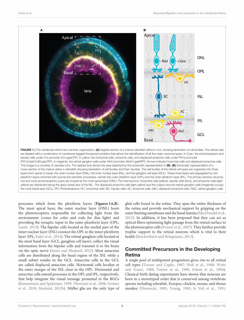
FIGURE 1 | The vertebrate retina has a laminar organization. (A) Sagittal section of a mature zebrafish retina in vivo, showing lamination of cell bodies. The retinal cells
are labeled with a combination of membrane-tagged fluorescent proteins that allows the identification of all the major neuronal types. In Cyan, the photoreceptors and
bipolar cells under Crx promoter (Crx:gapCFP). In yellow, the horizontal cells, amacrine cells, and displaced amacrine cells under Ptf1a promoter
(Ptf1a:Gal4/UAS:gapYFP). In magenta, the retinal ganglion cells under Ath5 promoter (Atoh7:gapRFP). Arrows indicate horizontal cells and displaced-amacrine cells.
This image is a courtesy of Jaroslav Icha. The dashed box shows the area depicted by the schematic representation in (B). (B) Schematic representation of a
cross-section of the mature retina in zebrafish showing lamination of cell bodies and their neurites. The cell bodies of the retinal cell types are organized into three
layers from apical to basal; the outer nuclear layer (ONL), the inner nuclear layer (INL), and the ganglion cell layer (GCL). These three layers are segregated by two
plexiform layers enriched with axonal and dendritic processes, namely the outer plexiform layer (OPL) and the inner plexiform layer (IPL). The primary sensory neurons;
rod and cone photoreceptors (cyan) are located at the most apical layer (ONL). The interneurons; horizontal cells (yellow), bipolar cells (blue), and amacrine cells (light
yellow) are distributed along the apico-basal axis of the INL. The displaced amacrine cells (light yellow) and the output neurons retinal ganglion cells (magenta) occupy
the most basal layer (GCL). PR, Photoreceptors; HC, horizontal cells; BC, bipolar cells; AC, amacrine cells; dAC, displaced amacrine cells; RGC, retinal ganglion cells.
processes which form the plexiform layers (Figures 1A,B).The most apical layer, the outer nuclear layer (ONL) hoststhe photoreceptors responsible for collecting light from theenvironment (cones for color and rods for dim light) andproviding the synaptic input to the outer plexiform layer (OPL;Lamb, 2013). The bipolar cells located at the medial part of theinner nuclear layer (INL) connect the OPL to the inner plexiformlayer (IPL; Euler et al., 2014). The retinal ganglion cells located atthe most basal layer (GCL, ganglion cell layer), collect the visualinformation from the bipolar cells and transmit it to the brainvia the optic nerve (Sanes and Masland, 2015). Most amacrinecells are distributed along the basal region of the INL while asmall subset resides in the GCL. Amacrine cells in the GCLare called displaced amacrine cells. Horizontal cells localize atthe outer margin of the INL close to the OPL. Horizontal andamacrine cells extend processes in the OPL and IPL, respectively,that help integrate the visual message presented to the RGCs(Kamermans and Spekreijse, 1999; Thoreson et al., 2008; Grimeset al., 2010; Masland, 2012b). Müller glia are the only type of
glial cells found in the retina. They span the entire thickness ofthe retina and provide mechanical support by gripping on theouter limitingmembrane and the basal lamina (MacDonald et al.,2015). In addition, it has been proposed that they can act asoptical fibers optimizing light passage from the retinal surface tothe photoreceptor cells (Franze et al., 2007). They further providetrophic support to the retinal neurons which is vital to theirhealth (Reichenbach and Bringmann, 2013).
Committed Precursors in the DevelopingRetinaA single pool of multipotent progenitors gives rise to all retinalcell types (Turner and Cepko, 1987; Holt et al., 1988; Wettsand Fraser, 1988; Turner et al., 1990; Fekete et al., 1994).Classical birth dating experiments have shown that neurons areborn in a stereotyped order that is conserved among vertebratespecies including zebrafish, Xenopus, chicken, mouse, and rhesusmonkey (Nawrocki, 1985; Young, 1985; la Vail et al., 1991;
Frontiers in Neuroscience | www.frontiersin.org 3 January 2018 | Volume 11 | Article 742

Amini et al. Neuronal Migration and Lamination in the Vertebrate Retina
Belecky-Adams et al., 1996; Rapaport et al., 2004; Wong andRapaport, 2009). The first neurons born are the retinal ganglioncells. Next, cone photoreceptors and horizontal cells emerge,followed by amacrine cells, rod photoreceptors, and bipolarcells (Figure 2A). Similar to cortical development, the timingof lamination in the retina reflects the neuronal birth order.However, in contrast to what has been shown in the cerebralneocortex, no strict relationship between the final laminarposition and time of birth is observed in the retina.
Until recently, it was widely believed that, as retinaldevelopment progresses, NECs go through a deterministic seriesof competence states, similar to Drosophila neuroblasts (Cepkoet al., 1996; Chen et al., 2012). This competence model waschallenged recently however, by lineage analysis in rat and fishthat suggest that stochastic mechanisms also play a role in thespecification of NECs (Gomes et al., 2011; He et al., 2012; Boijeet al., 2015). It was proposed that NECs commit to specific fatesin a stochastic manner after their last apical division. However,terminal and penultimate divisions were biased toward particularfates, which cannot purely be explained by the stochastic model(He et al., 2012; Boije et al., 2015). One possible interpretationis that these divisions correspond to symmetric divisions ofcommitted precursor cells. In agreement with this hypothesis,recent studies showed that a significant population of retinalneurons is generated by committed precursors, at least inzebrafish, chick, and mouse (Godinho et al., 2007; Rompani andCepko, 2008; Hafler et al., 2012; Emerson et al., 2013; Suzuki et al.,2013; Cepko, 2014; Weber et al., 2014; Engerer et al., 2017). Theycan be distinguished from NECs by morphology, expression offate determinants and/or mitotic position.
In zebrafish for example, it was shown that only the early bornneurons, retinal ganglion cells, and amacrine cells, are exclusivelygenerated by divisions of multipotent progenitors at the apicalsurface at early stages of retinogenesis. Later in development,cone photoreceptors, horizontal, and bipolar cells are born fromsymmetric divisions of committed precursors (Godinho et al.,2007; Suzuki et al., 2013; Weber et al., 2014; Figure 2B). Conephotoreceptor precursors show columnar epithelial morphologyand divide within the developing photoreceptor layer (Figure 2B;Suzuki et al., 2013; Weber et al., 2014). Horizontal cell precursorsare multipolar and divide either in the future INL or close to thefuture OPL (Godinho et al., 2007; Weber et al., 2014), whereasbipolar cell precursors show bipolar morphology and can divideat apical or subapical positions (Figure 2B; Weber et al., 2014;Engerer et al., 2017). So far, we are only beginning to decipherthe origin and behaviors of committed precursors. Learningmoreabout these particular progenitor types and how their emergencecontributes and potentially facilitates retinal lamination will beinteresting entry points for future studies.
NEURONAL TRANSLOCATION ANDLAMINATION DURING RETINALDEVELOPMENT
After the genesis of different neuronal cell types, the precisepositioning of these neurons along the apico-basal (radial) axis
of the retina is key for constructing the laminar architectureand subsequently functional neuronal circuits within the visualsystem. As such, neuronal migration is crucial for correct retinallayering. Given this, understanding how neurons migrate duringretinogenesis is important to understand lamination and circuitformation.
Cell Biology of Neuronal Migration: Modesand Subcellular Force GeneratorsNeuronal migration has been most extensively studied in exvivo cultures and organotypic slices of the cerebral neocortexand the cerebellum of rodents. The phenomenon of neuronalmigration in the cerebral neocortex has been reviewed in depthelsewhere (Nadarajah and Parnavelas, 2002; Cooper, 2013; Ichaand Norden, 2014; Hatanaka et al., 2016). Thus, here we onlysummarize key features of neuronal migration in the cerebralneocortex but focus on retinal neuronal migration and how it aidsthe generation of retinal wiring.
Traditionally, neuronal migration has been classified into twomain modes: (1) radial migration and (2) tangential migration(Figures 3A,B). This categorization is based on the relativeorientation of trajectories taken by the migrating neurons in thedeveloping tissue. Radial neuronal migration means migrationin parallel to the apico-basal axis of the tissue, while tangentialmigration is defined as neurons following a path perpendicularto the apico-basal axis of the tissue.
In the cerebral neocortex, radial migration is subdividedinto two distinct modes: (a) somal translocation and (b) glial-guided migration (Figure 3A). At earlier stages of corticaldevelopment, neurons radially migrate within the tissue throughsomal translocation (Morest, 1970a; Nadarajah et al., 2001;Nadarajah and Parnavelas, 2002). Neurons migrating via thismode, exhibit either a unipolar or bipolar morphology anduse their basal attachment to translocate their nucleus andother organelles (Nadarajah and Parnavelas, 2002; Cooper, 2013;Icha and Norden, 2014). As the cerebral neocortex thickensduring development, some neurons attach to the radially orientedprogenitor cells also known as radial glial cells. These neuronsassume a bipolarmorphology, with a leading (basal) and a trailing(apical) process and migrate along the glial cells to their targetlocation (Rakic, 1971, 1972; Sidman and Rakic, 1973; Nadarajahand Parnavelas, 2002; Marín et al., 2010; Hatanaka et al., 2016).
Some neurons that later build the cerebral cortex move awayfrom their point of origin via non-radial routes to reach theirtarget position using a tangential migration mode (O’Rourkeet al., 1992; Tan et al., 1995, 1998). Evidence for this modeof migration came from retroviral cell lineage tracing in ratand mouse, showing that clonally related neurons within thedeveloping cerebral neocortex can be tangentially dispersedfrom their birth site (Price and Thurlow, 1988; Austin andCepko, 1990; Walsh and Cepko, 1992; Reid et al., 1995). Mosttangentially migrating neurons switch to radial trajectories andmodes during their migration cycle to reach their final position(Figure 3B). Unlike the radially migrating neurons that have asingle leading process, tangentially migrating neurons typicallydisplay both single and branched leading processes (Martini et al.,
Frontiers in Neuroscience | www.frontiersin.org 4 January 2018 | Volume 11 | Article 742

Amini et al. Neuronal Migration and Lamination in the Vertebrate Retina
FIGURE 2 | Genesis of the neuronal types of the vertebrate retina. (A) Chronological order of neuron birth in the vertebrate retina is depicted based on classical birth
dating studies done across many vertebrate species. The first neurons born are the retinal ganglion cells, followed by cone photoreceptors, horizontal cells, amacrine
cells, rod photoreceptors, and bipolar cells. Note that birth orders are overlapping and that we did not depict Muller cells in the schematic but they are the latest born
cell-type. (B) Model of retinogenesis in the zebrafish embryo. Neuroepithelial progenitors (gray) divide asymmetrically at the apical side and give birth to one neuron
either a retinal ganglion cell (magenta) or amacrine cell (light yellow) and one neuronal precursor committed to cone photoreceptor (cyan), horizontal (yellow), or bipolar
(blue) cell fate. The committed precursors feature distinct morphology, expression of fate determinants and/or mitotic position. Cone photoreceptor precursors (cyan)
show columnar epithelial morphology and divide within the developing photoreceptor layer at the apical surface of the retina. Horizontal cell precursors (yellow) are
nonpolar and divide along the apico-basal axis of the INL, whereas bipolar cell precursors (blue) show bipolar morphology and can divide at apical or subapical
positions. PR, Photoreceptors; HC, horizontal cells; BC, bipolar cells; AC, amacrine cells; dAC, displaced amacrine cells; RGC, retinal ganglion cells.
2009). Selective stabilization of one of these branches determinesthe directionality of the tangential migration (Martini et al.,2009). However, it is important to note that some tangentiallymigrating neurons (e.g., precerebellar neurons) exhibit a singleleading process during their entire migratory cycle (Bourrat andSotelo, 1990).
As described above, during both radial and tangentialmigration in the cerebral neocortex neurons are polarized andexhibit leading and/or trailing processes and thereby take oneither a unipolar or bipolar shape. However, some neuronsextend multiple dynamic protrusions into different directions,adopting a multipolar morphology. These neurons undergo athird mode of migration known as multipolar migration or “freemigration.” Multipolar migration is characterized by frequentchanges in direction and rate of migration as neurons movetoward their final laminar position along either the radial ortangential axis of the tissue (Tabata and Nakajima, 2003).
In addition to cell morphology and orientation of migration,other parameters including speed, directional persistence, and
cell biological machineries driving the migration define theneuronal migration modes. Moreover, it was shown that neuronswithin the CNS can dynamically change patterns of migration enroute, depending on their type and location within the lamina(Tabata and Nakajima, 2003; Noctor et al., 2004; Marín et al.,2010; Faux et al., 2012).
The role of neuronal migration in building the distinguishinglaminar feature of the retina has been acknowledged sinceseminal studies by Ramón y Cajal (1893). However, the precisecellular andmolecular mechanisms of neuronal migration duringretinogenesis remain elusive. This in part is due to lack ofhigh-resolution time-lapse imaging of the developing retina. Assuch, so far, a rather descriptive understanding of retinal neuronmigration was acquired. More recent studies are only beginningto get insights into the cellular machineries driving differentmigration modes. Some disparities exist between neuronalmigration in the retina and the cortex. For example, in contrastto the cerebral neocortex, there is currently no evidence forradial glial guided-like migration in the retina. Considering that
Frontiers in Neuroscience | www.frontiersin.org 5 January 2018 | Volume 11 | Article 742

Amini et al. Neuronal Migration and Lamination in the Vertebrate Retina
FIGURE 3 | Modes of neuronal migration in the CNS. Two main modes of migration have been described in different parts of the nervous system: (A) Radial migration
and (B) tangential migration. (A) A prevalent type of radial migration in the cortex is the glial-guided migration. It can be schematically summarized in three steps: (1)
neurons born at the apical surface (2) lose their attachments to both the apical and basal surfaces of the tissue, (3) they attach to the radially oriented glial cells, (4)
move along them to their target location perpendicular to the surfaces of the tissue and (5) undergo differentiation. (B) Some neurons combine radial migration with
tangential migration to reach their target position. Tangentially migrating cells are not attached to the edges of the tissue and most of them form branched leading
processes (arrowheads). As an example, tangential migration of interneurons is shown in three steps: (1) migration toward the cortex and move in parallel to the apical
and basal surfaces, (2) interneurons subsequently associate with glial cells, (3) undergo radial movement along the glial-cells perpendicular to the apical surface and
(4) reach their position and (5) undergo differentiation.
most retinal neurons migrate before the differentiation of Müllercells, glial-guided migration in the retina is probably unlikely.To fully exclude this possibility however, in-depth investigationsof later developmental stages are needed. What is clear alreadyis that different retinal cell types use different trajectories andmigration modes to reach their destination within the retina.Below, we discuss the current state of knowledge of retinalneuron migration modes and their kinetics. We also summarizethe intracellular components involved in these phenomena invertebrates.
Retinal Ganglion Cell TranslocationRetinal ganglion cells are the first born retinal neurons. Theyundergo direct neurogenesis upon their apical division withoutan intermediate committed precursor step. Retinal ganglion cellsthen migrate away from their apical birth place to the mostbasal retinal layer adjacent to the lens (Figures 1A,B; Ramóny Cajal, 1893; Sidman, 1960; Nawrocki, 1985; Poggi et al.,2005; Zolessi et al., 2006). Retinal ganglion cell morphologyand migration patterns have been extensively studied alreadydecades ago using Golgi staining in the rat retina (Morest, 1970b)and by serial section electron microscopy in the mouse retina(Hinds and Hinds, 1974). Based on overall morphology of thesefixed samples at different developmental time points, it wasspeculated that retinal ganglion cells move via bipolar somaltranslocation. Later studies, using live confocal fluorescencemicroscopy confirmed this migratory mode and showed thatretinal ganglion cells translocate by moving their soma basallywhile remaining in contact with both apical and basal surfaces
(Poggi et al., 2005; Zolessi et al., 2006). Recently, light-sheetimaging of the intact embryonic zebrafish retina provided moredetailed insights into the retinal ganglion cell migration modes,kinetics, and mechanisms (Icha et al., 2016). The authors showedthat the canonical mode of retinal ganglion cell migration isbipolar somal translocation. It was further uncovered that thissomal translocation of retinal ganglion cells is accomplished intwo phases: (1) a rapid directional phase, during which the retinalganglion cell soma reaches the basal side of the retina and (2)a slower fine positioning phase within the retinal ganglion celllayer, which coincides with the loss of apical attachment that leadsto random movements of the retinal ganglion cells within theirlayer (Figure 4A) most likely important for the exact positioningof cells. The fine positioning phase usually ends when axons ofretinal ganglion cells start to grow (Icha et al., 2016). In addition,the authors unexpectedly found that retinal ganglion cells canalso move by multipolar migration which is a much less frequentand less efficient mode of migration than the somal translocation(Figure 4B).
To understand force generation of retinal ganglion celltranslocation, classic studies focused on the location of cellularorganelles. Centrosomes, the Golgi apparatus, the primarycilium, and microtubules all reside in the apical process behindthe nucleus of migrating retinal ganglion cells (Hinds and Hinds,1974; Zolessi et al., 2006; Icha et al., 2016; Lepanto et al.,2016), making centrosome-based pulling an unlikely mechanism.Instead, the first phase of retinal ganglion cell migration in thezebrafish retina depends on an apically stabilized microtubulecytoskeleton and the inheritance of the basal process attachment
Frontiers in Neuroscience | www.frontiersin.org 6 January 2018 | Volume 11 | Article 742

Amini et al. Neuronal Migration and Lamination in the Vertebrate Retina
FIGURE 4 | Modes of neuronal migration in the retina. (A,B) Scheme of different retinal ganglion cells (green) translocation modes: (A) somal translocation with basal
process and (B) multipolar migration. (A) Retinal ganglion cells inheriting the basal process in zebrafish translocate basally faster than the sister cell. Basal
translocation is followed by a period of fine positioning, during which cells lose their apical processes and project axons toward the optic nerve (depicted by arrows).
(B) In rare cases in zebrafish, retinal ganglion cells lose their basal process, subsequently detach their apical process, increase their protrusive activity, and move
basally in a multipolar migratory mode. The model shown is adapted from Icha et al. (2016) study. (C) Representation of retinal inhibitory neurons migration. Amacrine
cells (gray) and the committed horizontal cell precursors (green) migrate to their laminar position via a combination of bipolar somal translocation and multipolar
migration. Upon birth, they move away from the apical side using somal translocation. Later, they switch to a multipolar mode of migration and translocate their soma
deeper into the INL. Amacrine cells remain at the basal INL positions, while horizontal cells revert their trajectory and migrate back toward the most-apical region of
the INL, beneath the photoreceptor layer. On their way to the apical side, the committed horizontal cell precursors undergo mitosis with no positional preference along
the INL. This model takes into account results from previous studies in the zebrafish retina (Weber et al., 2014; Chow et al., 2015; Icha et al., 2016).
(Icha et al., 2016). When either basal process attachment ormicrotubule integrity is impaired, retinal ganglion cells canswitch to the multipolar migration mode that seems to lendrobustness to the system (Figure 4B; Icha et al., 2016). Disturbingboth migration modes results in a failure of retinal ganglion celltranslocation and subsequently misplaced retinal ganglion cells(Icha et al., 2016). This in turn, disturbs future lamination eventsand retinal development as other later-born neuronal cell typesare often ectopically placed within the retina. This implies thattranslocation of retinal ganglion cells sets the stage for all furtherretinal lamination programs in the zebrafish embryo (Icha et al.,2016). Overall morphological features of retinal ganglion cellsin fixed retinal samples of mouse and rat suggest that retinalganglion cells in other species also move by somal translocation(Morest, 1970b; Hinds andHinds, 1974). However, these findingsstill need to be confirmed with live-imaging methods.
Photoreceptor Cell TranslocationPhotoreceptors are born at apical positions and also later resideat the most apical side of the retina (Figures 1A,B). In zebrafish,it has been shown that these cells go through a committedprecursor stage and divide one more time apically, showingcolumnar epithelial morphology (Suzuki et al., 2013; Weberet al., 2014). As the birth-site and final residence of thesecells are both at the most apical positions, it is possible thatphotoreceptors do not need to translocate much after their apicalbirth. However, studies using 3D human retina derived fromembryonic stem cells reported that photoreceptors can be seenalong the radial axis of the tissue (Kaewkhaw et al., 2015). In thisstudy, live-cell imaging demonstrated that photoreceptors canslowly translocate their soma from the basal to the apical sideduring early stages of differentiation in 3D retinal cultures (day42–44). Similar observations were made in the zebrafish retina
Frontiers in Neuroscience | www.frontiersin.org 7 January 2018 | Volume 11 | Article 742

Amini et al. Neuronal Migration and Lamination in the Vertebrate Retina
wherein the photoreceptor precursors were detected at morebasal locations before being seen apically before division (Suzukiet al., 2013).Whether suchmigration phenomena are common tophotoreceptor precursors across species is not yet explored. In thefuture, it will be important to confirm this migratory mode andexplore how it arises and how it contributes to retinal lamination.
Horizontal Cell and Amacrine CellTranslocationHorizontal cells and amacrine cells are inhibitory neuronsoccupying the INL. Horizontal cells lie at the most-apical(beneath the photoreceptor layer), while amacrine cells resideat the most-basal (just above the retinal ganglion cell layer)regions of the INL (Figures 1A,B). A body of classic studiesusing a combination of Golgi staining and electron microscopydemonstrated that migrating horizontal cells (Hinds and Hinds,1979, 1983) and amacrine cells (Prada et al., 1987) can be seenwith bipolar and multipolar morphologies. This has led to thesuggestion that these cells could move via two different migrationmodes during retinal lamination. Amacrine cells have to moveto basal INL positions, thus a behavior similar to that of retinalganglion cells could be expected. While for horizontal cells, usingthe same rationale as for the photoreceptors, one would expectthat they migrate only a short distance from their apical birth-siteto their predestined position just slightly more basal.
In contrast to these assumptions, several studies over thepast two decades provided evidence that horizontal cells, similarto amacrine cells, migrate substantial basal distances beforereturning to apical location where they later reside. First, analysisof the spatiotemporal pattern of horizontal cells showed that theyare scattered along the radial axis of the developing retina inchicken (Prada et al., 1987), mouse (Liu et al., 2000), rabbit, cat,agouti, capybara (Silveira et al., 1989), andmacaque (Wässle et al.,2000). This indicated that horizontal cell precursors undergosubstantial migration after genesis and that this migrationpattern may be evolutionarily conserved. Subsequently, live-imaging approaches in chicken (Edqvist and Hallböök, 2004) andzebrafish (Chow et al., 2015; Icha et al., 2016) confirmed thathorizontal cells and amacrine cells both undergo basal migrationand stop once they reach the prospective amacrine cell layer.Amacrine cells then remain in this layer, while horizontal cellsrevert their trajectory and migrate apically toward the futurehorizontal cell layer adjacent to the OPL (Edqvist and Hallböök,2004; Godinho et al., 2007; Poche et al., 2007; Weber et al.,2014; Chow et al., 2015). These observations indicated thatacross species horizontal cells indeed undergo a bi-directionalmigration.
A detailed live-imaging study of the developing zebrafishretina focused on how horizontal cell precursors and amacrinecells reach the prospective amacrine cell layer. This work showedthat horizontal cell precursors and amacrine cells reach theirlaminar positions using a combination of different migratorymodes (Chow et al., 2015): first, they move from their apicalbirth-site to the INL while displaying bipolar morphology anddirectional persistence. Next, both cell types lose their apicalattachment, switch to a less directionally persistent multipolar
phase and translocate their soma deeper into the INL towardthe prospective amacrine cell layer (Figure 4C). During the thirdphase, both horizontal cell precursors and amacrine cells undergofine positioning via cell-type specific tangential migration.Amacrine cells move short distances and translocate deeper intothe INL (Chow et al., 2015; Icha et al., 2016). Horizontal cellson the other hand start to demix from the amacrine cells andmove toward the prospective horizontal cell layer and on the wayundergo a final committed division before reaching their finalposition (Edqvist andHallböök, 2004; Godinho et al., 2007; Pocheet al., 2007; Shirazi Fard et al., 2013; Weber et al., 2014).
Overall, we do not yet understand how and by whatintracellular mechanisms horizontal cells and amacrine cellsreach their final location. Thus far, it was shown that fine-positioning of amacrine cells is influenced by the atypicalcadherin Fat3 that cell autonomously influences the cytoskeletonand thereby the precise positioning of the dendritic arbor and thesoma of developing amacrine cells in the mouse retina (Deanset al., 2011; Krol et al., 2016). In the case of horizontal cells, itwas shown that demixing depends on the transcription factorLim1 (Poche et al., 2007). In conditional lim1 knockout mice,horizontal cells fail to sort out from the amacrine cells and insteadremain within the amacrine cell layer in the basal INL (Pocheet al., 2007). However, how demixing and differential laminationof horizontal and amacrine cells is triggered remains unexplored.In the future, it will be necessary to carefully monitor horizontalcell and amacrine cell migration kinetics and trajectories anddetermine whether and how these two cell types influence eachother during their migration. It will be also important to screenfor intracellular and extracellular factors that might influencetheir migration and lamination.
Bipolar Cell TranslocationDespite some insights on the regulation of bipolar cell fatespecification (Livesey and Cepko, 2001), there is currently littleinformation about the regulation of bipolar cell migrationand laminar positioning. As mentioned previously, bipolar cellprogenitors can divide apically or subapically depending ondevelopmental stage of the retina (Figure 2B). Notably, thedivision plane of bipolar cell progenitors shows no preferencefor a particular orientation or position along the INL (Weberet al., 2014; Engerer et al., 2017). Recent studies also proposedthat nuclei of bipolar cell precursors undergo interkineticnuclear migration-like movement toward the apical surface ofthe developing zebrafish retina (Weber et al., 2014; Engereret al., 2017). However, so far, we lack further information onhow exactly bipolar cells that divide at different locations arepositioned within the INL and how this positioning contributesto their neuronal function and circuitry.
Future Perspectives for UnderstandingRetinal Neuron TranslocationOverall, while some progress was made in recent years onneuronal translocation in the retina, this phenomenon and itsunderlying force generators are far from being understood.This needs further effort in future studies as only if themodes and kinetics of the different neurons as well as their
Frontiers in Neuroscience | www.frontiersin.org 8 January 2018 | Volume 11 | Article 742

Amini et al. Neuronal Migration and Lamination in the Vertebrate Retina
interplay are understood can we begin to grasp how retinallamination is achieved. As strong indications exist that differentmodes of migration are executed by different neuronal celltypes and that some cells can switch migratory modes (Chowet al., 2015; Icha et al., 2016), we further need to understandthe molecular mechanisms responsible for these changes.Accumulating evidence suggests that some modes of neuronalmigrations outlined above are conserved across vertebrate species(Hinds and Hinds, 1974, 1979, 1983; Prada et al., 1987; Silveiraet al., 1989; Liu et al., 2000; Edqvist and Hallböök, 2004; Godinhoet al., 2007; Poche et al., 2007; Suzuki et al., 2013; Weber et al.,2014; Chow et al., 2015; Kaewkhaw et al., 2015; Icha et al., 2016;Engerer et al., 2017). Whether similar modes of migration alsooccur during human retinal development remains unclear. Therecently developed human retinal organoids have the potentialto become valuable model system to investigate these questions(Nakano et al., 2012).
EXTRACELLULAR PLAYERS IN RETINALLAMINATION
Neuronal migration plays a central role in building the laminararchitecture of the visual system and it is thus important tounderstand not only the parameters that initiate but also theones that appropriately terminate the migration of neurons.Many aspects of neuronal migration are still unexplored and it isunclear: (a) what guides distinct neurons that are born at similarlocations and developmental stages, toward their differing layersand (b) how do neurons knowwhen and where to stopmigration.
It is clear that the gene expression profile of each neuronalcell type influences the cell’s journey to its final destination.In addition, we have some insights on factors secreted bydifferent cell types that could be involved in different aspectsof retinogenesis from axon pathfinding (Li, 2005) to lamination(Fu et al., 2006). However, currently our knowledge is notsufficient to explain how retinal lamination is achieved. In thedeveloping retina, neurons migrate in the context of neighboringcells and in an increasingly crowded environment, hence it islikely that they encounter contact-dependent cues from theirsurroundings. In response to such cues, neurons might modulatetheir physical features and adapt to those of their environment.This in turn might enable them to properly navigate withinthe tissue and reach their laminar destination. It is conceivablethat these cues could either attract neuron types into theirspecific layers or repel them from integrating into inappropriatelayers. Contact-dependent cues might be a source of positionalinformation especially for neurons that undergo multipolarmigration, particularly because they do not feature apicallyand/or basally attached processes that could provide them withdirectional information.
Contact-dependent cues could come from either cell-cellinteraction or the interactions between cells and the extracellularmatrix (ECM). The ECM has been shown to influence migratoryphenomena in different contexts within the developing nervoussystem (Franco andMüller, 2011; Sidhaye and Norden, 2017). To
test whether ECM also guides neurons in the developing retina, afirst step would be to find whether analogous ECM componentsare involved in retinal neuronal migration as was shown in thecerebral neocortex (e.g., laminins, tenascins, and proteoglycans;Franco and Müller, 2011).
So far, only a limited number of studies have attemptedto study extracellular influence on retinal neuronal migrationand lamination. One of these studies showed that knockingdown Laminin α1, a core component of the basal lamina,using morpholino approaches, impairs the basal translocationof retinal ganglion cells and results in their ectopic localizationand subsequently a tissue-wide lamination defect in the zebrafishembryo (Icha et al., 2016). In addition, it was shown thatthe laminin receptor β1-Integrin is required in a neuron-autonomous manner for precise lamination of the ganglioncells in the mouse retina (Riccomagno et al., 2014). In theabsence of β1-Integrin, the migrating ganglion cells do not stopupon reaching the laminin-rich inner limiting membrane andthereby form an ectopic retinal ganglion cell layer, phenocopyingthe retinal ganglion cell lamination defects observed in diverselaminin mutants (Edwards et al., 2010; Pinzón-Duarte et al.,2010).
In addition to ECM components, interactions betweendifferent cell types may also influence migratory behavior ofneurons. For example, because of the sequential birth order inthe retina one could speculate that the correct lamination ofearlier born neurons could influence the subsequent layeringevents. However, this does not seem to be true in all cases. In thezebrafish lakritz mutant (Kay et al., 2001) and the mouse atonalhomolog Atoh7 knockout (Brown et al., 2001), emergence ofretinal ganglion cells is suppressed. Nevertheless, the remainingcell types are able to laminate correctly (Brown et al., 2001; Kayet al., 2001). In addition, INL and ONL still form even whenretinal ganglion cell, amacrine cell, and horizontal cell fates aresuppressed (Almeida et al., 2014). These studies suggest thatneurons in the retina do not solely rely on the preceding neuronaltypes to migrate to their appropriate layer. However, how thisis achieved and what guides different neuronal types to theircorrect layer, despite the lack of previously born neurons, remainselusive. In addition, it is important to note that several studiesprovide evidence that attractive or repulsive transmembraneguidance cues regulate correct stratification of neurites intoIPL or OPL, across vertebrate species (Yamagata and Sanes,2008; Matsuoka et al., 2011). Whether there is a mechanisticlink between correct neurite stratification and termination ofneuron migration and consequently lamination, remains to beinvestigated.
One re-emerging approach to study how retinal neuronsinteract with their neighboring cells and extracellularcomponents are retinal reaggregate cultures. In early studies,reaggregate cultures were often used to understand how differenttissues such as the chick retina are formed (Moscona andMoscona, 1952; Moscona, 1961; Layer and Willbold, 1993;Rothermel et al., 1997). In line with this, it was recently shownthat the dissociated cells from the embryonic zebrafish retina,acquire a layered structure in a minimal culture condition,
Frontiers in Neuroscience | www.frontiersin.org 9 January 2018 | Volume 11 | Article 742

Amini et al. Neuronal Migration and Lamination in the Vertebrate Retina
suggesting that they possess some intrinsic self-organizing abilityto laminate (Eldred et al., 2017a,b). In the future, such in vivoreaggregate systems could provide a platform to investigate thenature of cell-cell and cell-ECM interactions required for retinallamination. Moreover, manipulating the components of suchin vivo aggregate systems could enable the simpler dissection ofmolecular and cellular mechanisms governing neuron migration,neurite stratification, and lamination within the retina.
LINKING LAMINATION TO RETINALFUNCTION
Visual perception is achieved through a series of processingsteps that enable the retina to assess different aspects ofthe visual information. So far, we discussed how genesis ofretinal cell types and their correct positioning contribute to thelamination of the retina. While these steps are prerequisites forproper retina development, building functional circuits is thenext fundamental step for processing visual information andthus retinal function (Galli-Resta, 2000, 2001). The featuredlaminar design of the retina establishes various “functionalunits” across the radial axis of the tissue. Anatomically, eachunit, depending on its function, is composed of differentsubsets of photoreceptors, bipolar cells, horizontal cells, amacrinecells, and retinal ganglion cells. However, each of these unitsdetects and processes only a limited portion of the visualscene. To generate a holistic picture of the visual environment,the retina implements a third building rule: neurons ofthe same type are spaced in regular patterns within theirrespective layer, a phenomenon also known as retinal mosaics(Wässle and Riemann, 1978). Such retinal mosaics allow thecomplete sampling and a uniform coverage of the visualscene. In this section, we review our current knowledge onprinciples of assembly and maintenance of retinal mosaics invertebrates.
Emergence of Retinal MosaicsThe highly ordered mosaic architecture in the vertebrate retinaensures that cell bodies of the same neuron-types, also calledhomotypic neurons are distributed in non-random arrays withintheir respective layer and at any given location in the retina(Figure 5A; Reese and Keeley, 2015). For example, the seminalstudy by Wässle and Riemann provided evidence that conephotoreceptors, retinal ganglion cells and horizontal cells in catsand monkeys are all arranged in a way that the distance betweencell bodies of neighboring homotypic neurons is uniform andsignificantly different than that of random points (Wässle andRiemann, 1978). In addition, the arrangement of dendritic arborsof the homotypic neighboring neurons within an array are alsospatially regulated so that they show little or no overlap, aphenomenon known as tiling (Figure 5B; Reese and Galli-Resta,2002; Reese and Keeley, 2015). Although the retinal mosaicpatterns are known to be fundamental to the processing ofvisual information in the retina, when and how neuronal mosaicsemerge during development is not yet completely understood.One reason for this gap of knowledge is the fact that most existing
markers are not expressed in neurons prior to their entry intotheir arrays during development.
Studies in different species, ranging from fish to primates, havesuggested that retinal mosaics emerge early during development,at times when neurogenesis, migration and lamination are stillin course. In fact, cone photoreceptors, horizontal cells, andcholinergic amacrine cells across species form regular arrays evenwhen layering and polarization is not yet complete (Larisonand Bremiller, 1990; Raymond et al., 1995; Scheibe et al., 1995;Galli-Resta et al., 1997; Novelli et al., 2005). Yet surprisingly,the geometry of the mosaic remains unaltered throughout thetime new neurons enter the previously assembled networks. Thisimplies that (a) regular spatial arrangement is a cell intrinsicproperty of the retinal neurons and that (b) their mosaicregularity is maintained during development.
Initially, it was assumed that the position of retinal neuronsis fixed along their radial axis from their birth site. Thisled to the hypothesis that the spatial regularity of the retinalmosaics is due to fate determination mechanisms that actaround the time of neuronal birth (Reese and Galli-Resta,2002). Interestingly however, several lines of evidence, mainlyusing lineage tracing techniques demonstrated that particulartypes of differentiating neurons (e.g., retinal ganglion cells,horizontal cell, cone photoreceptors, and some type of amacrinecells) are often laterally displaced from their columnar cloneof origin in the developing retina (Reese et al., 1994). Thekey observation confirming tangential dispersion came from aclonal territory analysis in X-chromosome inactivated transgenicmice (only half of the retinal progenitors are marked). There,it was shown that certain differentiating neurons includingcone photoreceptors, amacrine cells, horizontal cells, and retinalganglion cells tangentially move away from one another possiblyto generate spatial order within mosaic arrays (Reese et al.,1995). Furthermore, using chimeric mice, it was revealed thatthe amount of this tangential dispersion varies depending on celltype (Reese et al., 1995, 1999). While the existence of tangentialdispersion does not necessitate its direct contribution in formingmosaic arrays, following the cholinergic amacrine cells from theirbirth showed that theirmosaics are assembled andmaintained viatangential dispersion in mouse retina (Galli-Resta et al., 1997).Further, combining experiments with mathematical modelingshowed that the spatial regularity of cholinergic amacrine cellsis dynamically preserved throughout the period of new amacrinecell addition to a previously assembled array (Galli-Resta et al.,1997). This implies that amacrine cells within a mosaic arrayactively disperse to accommodate the new amacrine cells insertedinto their mosaics. In the current view this means that tangentialmigration is a local phenomenon that contributes to building themosaics of a particular subset of neurons (Cook and Chalupa,2000; Galli-Resta, 2002; Reese and Keeley, 2015).
Some effort has been made to understand what drivestangential dispersion during mosaic assembly and maintenance.One proposed mechanism is that either diffusible signalsor contact-mediated interactions between homotypic neuronsaccount for tangential dispersion of neurons and subsequentlymosaic regularity of neurons (Reese and Galli-Resta, 2002).This would suggest that a local spacing rule keeps a minimum
Frontiers in Neuroscience | www.frontiersin.org 10 January 2018 | Volume 11 | Article 742

Amini et al. Neuronal Migration and Lamination in the Vertebrate Retina
FIGURE 5 | Mosaic assembly of horizontal cells in the vertebrate retina. (A) Schematic representation of a cross-section of the vertebrate retina showing that
individual horizontal cells (yellow) are evenly spaced across the retina, a pattern known as retinal mosaics. (B) Schematic view from the surface of the retina in (A)
showing that cell bodies and dendrites of horizontal cells of the same type are non-randomly distributed within the horizontal cell layer. Dendritic territories of the
homotypic neighboring neurons show little or no overlap. This phenomenon is also referred to as dendritic tiling. The dashed box shows the area depicted in (C).
(C) Mosaic of horizontal cells arises by contact-mediated repulsion among these neurons. Horizontal cells of the same type express the same cell-surface molecules
(depicted in turquoise). This allows the homotypic neurons to recognize each other and generate contact-dependent repulsion between their dendrites (depicted by
red arrows), thereby creating mosaic spacing of horizontal cell soma. This figure takes into account results from previous studies in the vertebrate retina (Wässle and
Riemann, 1978; Scheibe et al., 1995; Huckfeldt et al., 2009; Kay et al., 2012).
distance between immediate homotypic neighbors in mosaicarrays. Consistent with this, mathematical modeling andsimulations showed that a simple minimal spacing rule termed“exclusion zone model” ensures that neighboring homotypiccone photoreceptors in the macaque retina are distributedwith a fixed minimal distance to each other and that theydo not reside closer than that distance (Shapiro et al.,1985; Eglen and Willshaw, 2002). Similar conclusions weredrawn from other computational studies seeking to simulatethe role of the minimal spacing rule in controlling mosaicformation of other retinal neuron types across different species(Cook and Chalupa, 2000; Galli-Resta, 2002). Biologically,the minimum spacing rule implies that homotypic neuronsrepel each other and take appropriate distances from oneanother to keep their orderly array. Early evidence showedthat emergence of horizontal cell dendritic neurites coincideswith their tangential dispersion in mouse retina (Reese et al.,1999). This observation led to the hypothesis that dendriticinteractions of homotypic horizontal cells might trigger theirtangential dispersion and consequently mosaic formation. Usingmodeling techniques, this hypothesis was further investigated(Eglen and Willshaw, 2002), but a biological evidence for therole of dendrites on mosaic formation came from studyingmosaic arrays of cholinergic retinal ganglion cells in the mouse(Galli-Resta et al., 2002) This work showed that disruption ofmicrotubules in dendrites of the cholinergic retinal ganglioncells results in a reversible disruption of their mosaic regularity(Galli-Resta et al., 2002). Subsequent studies showed thathorizontal cells achieve their mosaic arrangements via homotypicshort-ranged interactions of their developing dendritic arborsin the mouse retina. These interactions are repulsive andenable homotypic neighboring horizontal cells to spatiallyarrange their soma (Huckfeldt et al., 2009), suggesting thata molecular cue exists that allows for a homotypic cell-type-specific recognition which in turn orchestrates the cell-cell contact-dependent repulsion of homotypic neurons and
their mosaic patterning (Figure 5C). Direct evidence for theexistence of such a cue came from a study in the developingmouse retina. Using a combination of microarray and geneknockout analysis, the authors found that signals initiated bytwo transmembrane proteins, MEGF10 and MEGF11 mediatehomotypic interactions among horizontal cells and starburstamacrine cells and reposition their soma (Figure 5C; Kay et al.,2012).
Despite some progress, the exact mechanisms by whichthe homotypic neurons that form mosaic arrays reliablyrecognize each other while migrating in the pool of diverseneuron types in the vertebrate developing retina remainelusive. Besides tangential migration, programmed cell deathand cell-fate lateral inhibition have been also proposed tocontribute to building mosaic arrays within the developingretina (Reese and Keeley, 2015). Nevertheless, to what extentthey interplay with tangential dispersion and promote retinalmosaic formation across vertebrate species is not yet understood.Finally, other parts of the CNS also exhibit regular arrangedneuronal arrays (Cook and Chalupa, 2000; Budry et al., 2011).Therefore, in the future it will be interesting to examinesimilarities and disparities of spatial organization of neuronsin different regions of the CNS and study whether and howsuch spatial regularities contribute to CNS development andfunction.
CONCLUSIONS
Neuronal lamination requires the regulation of a well-orchestrated chain of events involving the generation of theright types and number of cells, positioning them at the correctplaces through migration and finally integrating them intotheir functional circuitries. Defects in the proper execution inany of these steps may result in neuronal layering defects andconsequently impaired retinal function. Despite recent progress,
Frontiers in Neuroscience | www.frontiersin.org 11 January 2018 | Volume 11 | Article 742

Amini et al. Neuronal Migration and Lamination in the Vertebrate Retina
we still lack an overarching picture of how different aspects oflamination from neuronal birth to final positioning occur at thecellular and tissue scales. This is partly due to the fact that moststudies investigating neuronal lamination in the retina reliedon fixed samples and conventional confocal microscopy thatimplies high phototoxicity leading to short observation times(Icha et al., 2017). Recently, light sheet microscopy enabledscientists to discover an unforeseen mode of migration for retinalganglion cells that would not have been discovered with moreconventional techniques (Icha et al., 2016). Thus, employingthe rapidly advancing imaging tools to understand the behaviorof retinal neurons from birth to differentiation will help togenerate a more-detailed understanding of lamination in thevertebrate retina. The zebrafish retina due to its fast development,translucency and availability of genetic and molecular tools,has been a prominent model for retinal development. As someimportant features of retinal lamination are conserved acrossvertebrate species this understanding is most likely widelyapplicable.
It is known that the process of lamination is regulatedby a combination of intrinsic and extrinsic influences duringdevelopment. We highlighted the potential role of ECM inneuronal migration, mosaic formation and lamination whichneeds to get into the spotlight in future studies. Anotherimportant question is to what extent and how cell intrinsicand extrinsic cues interact and how they contribute to differentaspects of lamination, from genesis to migration and mosaicformation, in the developing retina. As discussed in thisreview, communication between neurons could contribute toconstructing the retinal layers and functional units, and thenature of such complex interactions in vivo needs to be
investigated. In addition, the development of in vivo culturesystems such as human organoids, is an exciting entry routeto extract the conservation of diverse features of lamination.Remarkably, lamination is an organizational hallmark of manyregions of the brain. As such, the retina as an outgrowth of thebrain could provide a platform for a mechanistic understandingof neuronal migration and lamination in other laminated brainregions.
AUTHOR CONTRIBUTIONS
All authors conceived and planned the review outline. RA andMR-M: wrote the initial manuscript. All authors read and editedthe text and approved it for publication.
FUNDING
CN was supported by the Max Planck Society and the GermanResearch Foundation (DFG; grant SFB 655, A25). RA wassupported by the NSERC Program number 504201.
ACKNOWLEDGMENTS
We would like to thank J. Icha and J. Sidhaye for criticalreading and comments on the manuscript. We are also gratefulto all members of the Norden laboratory for helpful discussions,F. Friedrich of the Max Planck Institute of Molecular CellBiology and Genetics for graphic works. RA is a research fellowof the Natural Sciences and Engineering Research Council ofCanada (NSERC). CN was supported by the German ResearchFoundation (DFG) [SFB 655, A25].
REFERENCES
Agathocleous, M., and Harris, W. A. (2009). From progenitors to differentiated
cells in the vertebrate retina. Annu. Rev. Cell Dev. Biol. 25, 45–69.
doi: 10.1146/annurev.cellbio.042308.113259
Alder, J., Cho, N. K., and Hatten, M. E. (1996). Embryonic precursor cells from the
rhombic lip are specified to a cerebellar granule neuron identity. Neuron 17,
389–399. doi: 10.1016/S0896-6273(00)80172-5
Almeida, A. D., Boije, H., Chow, R. W., He, J., Tham, J., Suzuki, S. C., et al. (2014).
Spectrum of fates: a new approach to the study of the developing zebrafish
retina. Development 141, 2912–2912. doi: 10.1242/dev.114108
Alsiö, J. M., Tarchini, B., Cayouette, M., Livesey, F. J., Alsio, J. M., Tarchini,
B., et al. (2013). Ikaros promotes early-born neuronal fates in the cerebral
cortex. Proc. Natl. Acad. Sci.U.S.A. 110, E716–E725. doi: 10.1073/pnas.1215
707110
Anthony, T. E., Klein, C., Fishell, G., and Heintz, N. (2004). Radial glia serve as
neuronal progenitors in all regions of the central nervous system. Neuron 41,
881–890. doi: 10.1016/S0896-6273(04)00140-0
Austin, C. P., and Cepko, C. L. (1990). Cellular migration patterns in the
developing mouse cerebral cortex. Development 110, 713–732.
Belecky-Adams, T., Cook, B., and Adler, R. (1996). Correlations between terminal
mitosis and differentiated fate of retinal precursor cells in vivo and in vivo:
analysis with the “window-labeling” technique. Dev. Biol. 178, 304–315.
Boije, H., Rulands, S., Simons, B. D., William, A., Boije, H., Rulands, S.,
et al. (2015). The independent probabilistic firing of transcription factors : a
paradigm for clonal variability in the zebrafish retina. Dev. Cell 34, 532–543.
doi: 10.1016/j.devcel.2015.08.011
Bourrat, F., and Sotelo, C. (1990). Migratory pathways and selective aggregation of
the lateral reticular neurons in the rat embryo: a horseradish peroxidase in vivo
study with special reference to migration patterns of the precerebellar nuclei. J.
Comp. Neurol. 294, 1–13. doi: 10.1002/cne.902940102
Brown, N. L., Patel, S., Brzezinski, J., and Glaser, T. (2001). Math5 is required for
retinal ganglion cell and optic nerve formation. Development 128, 2497–2508.
Budry, L., Lafont, C., El Yandouzi, T., Chauvet, N., Conéjero, G., Drouin,
J., et al. (2011). Related pituitary cell lineages develop into interdigitated
3D cell networks. Proc. Natl. Acad. Sci. U.S.A. 108, 12515–12520.
doi: 10.1073/pnas.1105929108
Cagan, R. (2009). Principles of drosophila eye differentiation. Curr. Top. Dev. Biol.
89, 115–135. doi: 10.1016/S0070-2153(09)89005-4
Carthew, R.W. (2007). Pattern formation in the Drosophila eye.Curr. Opin. Genet.
Dev. 17, 309–313. doi: 10.1016/j.gde.2007.05.001
Cepko, C. (2014). Intrinsically different retinal progenitor cells produce specific
types of progeny. Nat. Rev. Neurosci. 15, 615–627. doi: 10.1038/nrn3767
Cepko, C. L., Austin, C. P., Yang, X., Alexiades, M., and Ezzeddine, D. (1996).
Cell fate determination in the vertebrate retina. Proc. Natl. Acad. Sci. U.S.A.
93, 589–595. doi: 10.1073/pnas.93.2.589
Chen, Z., Li, X., and Desplan, C. (2012). Deterministic or stochastic
choices in retinal neuron specification. Neuron 75, 739–742.
doi: 10.1016/j.neuron.2012.08.008
Chow, R.W.Y., Almeida, A. D., Randlett, O., Norden, C., and Harris, W. A. (2015).
Inhibitory neuron migration and IPL formation in the developing zebrafish
retina. Development 142, 2665–2677. doi: 10.1242/dev.122473
Cook, J. E., and Chalupa, L. M. (2000). Retinal mosaics: new insights into an old
concept. Trends Neurosci. 23, 26–34. doi: 10.1016/S0166-2236(99)01487-3
Cooper, J. A. (2008). A mechanism for inside-out lamination in the neocortex.
Trends Neurosci. 31, 113–119. doi: 10.1016/j.tins.2007.12.003
Cooper, J. A. (2013). Mechanisms of cell migration in the nervous system. J. Cell
Biol. 202, 725–734. doi: 10.1083/jcb.201305021
Frontiers in Neuroscience | www.frontiersin.org 12 January 2018 | Volume 11 | Article 742

Amini et al. Neuronal Migration and Lamination in the Vertebrate Retina
Deans, M. R., Krol, A., Abraira, V. E., Copley, C. O., Tucker, A. F., and Goodrich,
L. V. (2011). Control of neuronal morphology by the atypical cadherin fat3.
Neuron 71, 820–832. doi: 10.1016/j.neuron.2011.06.026
Donovan, S. L., and Dyer, M. A. (2005). Regulation of proliferation during
central nervous system development. Semin. Cell Dev. Biol. 16, 407–421.
doi: 10.1016/j.semcdb.2005.02.012
Duncan, J. L., Ratnam, K., Birch, D. G., Sundquist, S. M., Lucero, A. S., Zhang,
Y., et al. (2011). Abnormal cone structure in foveal schisis cavities in X-linked
retinoschisis from mutations in exon 6 of the RS1 gene. Investig. Ophthalmol.
Vis. Sci. 52, 9614–9623. doi: 10.1167/iovs.11-8600
Dowling, J. E. (1987). The Retina: An Approachable Part of the Brain? Cambridge:
Harvard University Press.
Edqvist, P.H. D., and Hallböök, F. (2004). Newborn horizontal cells migrate
bi-directionally across the neuroepithelium during retinal development.
Development 131, 1343–1351. doi: 10.1242/dev.01018
Edwards, M. M., Mammadova-Bach, E., Alpy, F., Klein, A., Hicks, W. L., Roux,
M., et al. (2010). Mutations in lama1 disrupt retinal vascular development
and inner limiting membrane formation. J. Biol. Chem. 285, 7697–7711.
doi: 10.1074/jbc.M109.069575
Eglen, S. J., and Willshaw, D. J. (2002). Influence of cell fate mechanisms upon
retinal mosaic formation: a modelling study. Development 129, 5399–5408.
doi: 10.1242/dev.00118
Eldred, M. K., Charlton-Perkins, M., Muresan, L., and Harris, W. A. (2017a). Self-
organising aggregates of zebrafish retinal cells for investigating mechanisms of
neural lamination. Development 144, 1097–1106. doi: 10.1242/dev.142760
Eldred, M. K., Muresan, L., and Harris, W. A. (2017b). Disaggregation and
reaggregation of zebrafish retinal cells for the analysis of neuronal layering.
Methods Mol. Biol. doi: 10.1007/7651_2017_46. [Epub ahead of print].
Elliott, J., Jolicoeur, C., Ramamurthy, V., and Cayouette, M. (2008). Ikaros confers
early temporal competence tomouse retinal progenitor cells.Neuron 60, 26–39.
doi: 10.1016/j.neuron.2008.08.008
Emerson, M., Surzenko, N., Goetz, J., Trimarchi, J., and Cepko, C. (2013).
Otx2 and onecut1 promote the fates of cone photoreceptors and
horizontal cells and repress rod photoreceptors. Dev. Cell 26, 59–72.
doi: 10.1016/j.devcel.2013.06.005
Engerer, P., Suzuki, S. C., Yoshimatsu, T., Chapouton, P., Obeng, N., Odermatt,
B., et al. (2017). Uncoupling of neurogenesis and differentiation during retinal
development. EMBO J. 36, 1134–1146. doi: 10.15252/embj.201694230
Euler, T., Haverkamp, S., Schubert, T., and Baden, T. (2014). Retinal bipolar
cells: elementary building blocks of vision. Nat. Rev. Neurosci. 15, 507–519.
doi: 10.1038/nrn3783
Faux, C., Rakic, S., Andrews, W., and Britto, J. M. (2012). Neurons on the move:
migration and lamination of cortical interneurons. Neurosignals 20, 168–189.
doi: 10.1159/000334489
Fekete, D. M., Perez-Miguelsanz, J., Ryder, E. F., and Cepko, C. L. (1994). Clonal
analysis in the chicken retina reveals tangential dispersion of clonally related
cells. Dev. Biol. 166, 666–682. doi: 10.1006/dbio.1994.1346
Franco, S. J., andMüller, U. (2011). Extracellular matrix functions during neuronal
migration and lamination in the mammalian central nervous system. Dev.
Neurobiol. 71, 889–900. doi: 10.1002/dneu.20946
Franze, K., Grosche, J., Skatchkov, S. N., Schinkinger, S., Foja, C., Schild, D., et al.
(2007). Muller cells are living optical fibers in the vertebrate retina. Proc. Natl.
Acad. Sci. U.S.A. 104, 8287–8292. doi: 10.1073/pnas.0611180104
Fu, X., Sun, H., Klein, W. H., and Mu, X. (2006). β-catenin is essential for
lamination but not neurogenesis in mouse retinal development. Dev. Biol. 299,
424–437. doi: 10.1016/j.ydbio.2006.08.015
Fuhrmann, S. (2010). Eye morphogenesis and patterning of the optic vesicle. Curr.
Top. Dev. Biol. 93, 61–84. doi: 10.1016/B978-0-12-385044-7.00003-5
Galli-Resta, L. (2000). Local, possibly contact-mediated signalling restricted to
homotypic neurons controls the regular spacing of cells within the cholinergic
arrays in the developing rodent retina. Development 127, 1509–1516.
Galli-Resta, L. (2001). Assembling the vertebrate retina: global patterning
from short-range cellular interactions. Neuroreport 12, A103–A106.
doi: 10.1097/00001756-200112040-00003
Galli-Resta, L. (2002). Putting neurons in the right places: local interactions
in the genesis of retinal architecture. Trends Neurosci. 25, 638–643.
doi: 10.1016/S0166-2236(02)02279-8
Galli-Resta, L., Novelli, E., and Viegi, A. (2002). Dynamic microtubule-dependent
interactions position homotypic neurones in regular monolayered arrays
during retinal development. Development 129, 3803–3814.
Galli-Resta, L., Resta, G., Tan, S. S., and Reese, B. E. (1997). Mosaics of islet-
1-expressing amacrine cells assembled by short-range cellular interactions. J.
Neurosci. 17, 7831–7838.
Ge, W., He, F., Kim, K. J., Blanchi, B., Coskun, V., Nguyen, L., et al. (2006).
Coupling of cell migration with neurogenesis by proneural bHLH factors. Proc.
Natl. Acad. Sci. U.S.A. 103, 1319–1324. doi: 10.1073/pnas.0510419103
Godinho, L., Williams, P. R., Claassen, Y., Provost, E., Leach, S. D., Kamermans,
M., et al. (2007). Nonapical symmetric divisions underlie horizontal cell
layer formation in the developing retina in vivo. Neuron 56, 597–603.
doi: 10.1016/j.neuron.2007.09.036
Gomes, F. L., Zhang, G., Carbonell, F., Correa, J. A., Harris, W. A., Simons, B.
D., et al. (2011). Reconstruction of rat retinal progenitor cell lineages in vivo
reveals a surprising degree of stochasticity in cell fate decisions. Development
138, 227–235. doi: 10.1242/dev.059683
Greig, L. C., Woodworth, M. B., Galazo, M. J., Padmanabhan, H., and
Macklis, J. D. (2013). Molecular logic of neocortical projection neuron
specification, development and diversity. Nat. Rev. Neurosci. 14, 755–769.
doi: 10.1038/nrn3586
Grimes, W. N., Zhang, J., Graydon, C. W., Kachar, B., and Diamond,
J. S. (2010). Retinal parallel processors: more than 100 independent
microcircuits operate within a single interneuron. Neuron 65, 873–885.
doi: 10.1016/j.neuron.2010.02.028
Guy, J., and Staiger, J. F. (2017). The functioning of a cortex without layers. Front.
Neuroanat. 11:54. doi: 10.3389/fnana.2017.00054
Hafler, B. P., Surzenko, N., Beier, K. T., Punzo, C., Trimarchi, J. M., Kong, J.
H., et al. (2012). Transcription factor Olig2 defines subpopulations of retinal
progenitor cells biased toward specific cell fates. Proc. Natl. Acad. Sci. U.S.A.
109, 7882–7887. doi: 10.1073/pnas.1203138109
Hammer, M., Roggan, A., Schweitzer, D., and Muller, G. (1995). Optical properties
of ocular fundus tissues-an in vivo study using the double-integrating-sphere
technique and inverse monte carlo simulation. Phys. Med. Biol. 40, 963–978.
doi: 10.1088/0031-9155/40/6/001
Harrison, S. J., Nishinakamura, R., Jones, K. R., and Monaghan, A. P. (2012).
Sall1 regulates cortical neurogenesis and laminar fate specification in mice:
implications for neural abnormalities in townes-brocks syndrome. Dis. Model.
Mech. 5, 351–365. doi: 10.1242/dmm.002873
Hatanaka, Y., Zhu, Y., Torigoe, M., Kita, Y., and Murakami, F. (2016). From
migration to settlement: the pathways, migration modes and dynamics of
neurons in the developing brain. Proc. Jpn. Acad. Ser. B Phys. Biol. Sci. 92, 1–19.
doi: 10.2183/pjab.92.1
He, J., Zhang, G., Almeida, A. D., Cayouette, M., Simons, B. D., and Harris, W.
A. (2012). How variable clones build an invariant retina. Neuron 75, 786–798.
doi: 10.1016/j.neuron.2012.06.033
He, S., Dong, W., Deng, Q., Weng, S., and Sun, W. (2003). Seeing more clearly:
recent advances in understanding retinal circuitry. Science 302, 408–411.
doi: 10.1126/science.1085457
Hinds, J. W., and Hinds, P. L. (1974). Early ganglion electron cell differentiation
analysis in the mouse utilizing retina : an microscopic serial sections. Dev. Biol.
416, 381–416. doi: 10.1016/0012-1606(74)90156-0
Hinds, J. W., and Hinds, P. L. (1979). Differentiation of photoreceptors and
horizontal cells in the embryonic mouse retina: an electron microscopic, serial
section analysis. J. Comp. Neurol. 187, 495–511. doi: 10.1002/cne.901870303
Hinds, J. W., and Hinds, P. L. (1983). Development of retinal amacrine cells in
the mouse embryo: evidence for two modes of formation. J. Comp. Neurol. 213,
1–23. doi: 10.1002/cne.902130102
Holt, C. E., Bertsch, T. W., Ellis, H. M., and Harris, W. A. (1988). Cellular
determination in the xenopus retina is independent of lineage and birth date.
Neuron 1, 15–26. doi: 10.1016/0896-6273(88)90205-X
Hoon, M., Okawa, H., Della Santina, L., and Wong, R. O. L. (2014). Functional
architecture of the retina: development and disease. Prog. Retin. Eye Res. 42,
44–84. doi: 10.1016/j.preteyeres.2014.06.003
Hoshino, M., Nakamura, S., Mori, K., Kawauchi, T., Terao, M., Nishimura, Y. V.,
et al. (2005). Ptf1a, a bHLH transcriptional gene, defines GABAergic neuronal
fates in cerebellum. Neuron 47, 201–213. doi: 10.1016/j.neuron.2005.06.007
Frontiers in Neuroscience | www.frontiersin.org 13 January 2018 | Volume 11 | Article 742

Amini et al. Neuronal Migration and Lamination in the Vertebrate Retina
Huckfeldt, R. M., Schubert, T., Morgan, J. L., Godinho, L., Di Cristo, G.,
Huang, Z. J., et al. (2009). Transient neurites of retinal horizontal cells
exhibit columnar tiling via homotypic interactions. Nat. Neurosci. 12, 35–43.
doi: 10.1038/nn.2236
Icha, J., Kunath, C., Rocha-Martins, M., and Norden, C. (2016). Independent
modes of ganglion cell translocation ensure correct lamination of the zebrafish
retina. J. Cell Biol. 215, 259–275. doi: 10.1083/jcb.201604095
Icha, J., and Norden, C. (2014). “Neuronal migration: an overview of modes,
molecular mechanisms and model systems,” in eLS (Chichester: John Wiley &
Sons Ltd). doi: 10.1002/9780470015902.0000796.pub2
Icha, J., Weber, M., Waters, J. C., and Norden, C. (2017). Phototoxicity in
live fluorescence microscopy, and how to avoid it. Bioessays 39:1700003.
doi: 10.1002/bies.201700003
Kaewkhaw, R., Kaya, K. D., Brooks, M., Homma, K., Zou, J., Chaitankar, V.,
et al. (2015). Transcriptome dynamics of developing photoreceptors in three-
dimensional retina cultures recapitulates temporal sequence of human cone
and rod differentiation revealing cell surface markers and gene networks. Stem
Cells 33, 3504–3518. doi: 10.1002/stem.2122
Kamermans, M., and Spekreijse, H. (1999). The feedback pathway from horizontal
cells to cones a mini review with a look ahead. Vision Res. 39, 2449–2468.
doi: 10.1016/S0042-6989(99)00043-7
Kay, J. N., Chu, M. W., and Sanes, J. R. (2012). MEGF10 and MEGF11 mediate
homotypic interactions required for mosaic spacing of retinal neurons. Nature
483, 465–469. doi: 10.1038/nature10877
Kay, J. N., Finger-Baier, K. C., Roeser, T., Staub, W., and Baier, H. (2001). Retinal
ganglion cell genesis requires lakritz, a zebrafish atonal homolog. Neuron 30,
725–736. doi: 10.1016/S0896-6273(01)00312-9
Kohwi, M., and Doe, C. Q. (2013). Temporal fate specification and neural
progenitor competence during development. Nat. Rev. Neurosci. 14, 823–838.
doi: 10.1038/nrn3618
Kriegstein, A. R., and Noctor, S. C. (2004). Patterns of neuronal migration in the
embryonic cortex.Trends Neurosci. 27, 392–399. doi: 10.1016/j.tins.2004.05.001
Krol, A., Henle, S. J., and Goodrich, L., V (2016). Fat3 and Ena/VASP proteins
influence the emergence of asymmetric cell morphology in the developing
retina. Development 143, 2172–2182. doi: 10.1242/dev.133678
Kwan, K. M., Otsuna, H., Kidokoro, H., Carney, K. R., Saijoh, Y., and Chien, C.B.
(2012). A complex choreography of cell movements shapes the vertebrate eye.
Development 139, 359–372. doi: 10.1242/dev.071407
Lahav, M., Albert, D. M., and Craft, J. L. (1975). Light and electron microscopic
study of dysplastic rosette-like structures occurring in the disorganized
mature retina. Albrecht Von Graefes Arch. Klin. Exp. Ophthalmol. 195, 57–68.
doi: 10.1007/BF02390031
Lamb, T. D. (2013). Evolution of phototransduction, vertebrate
photoreceptors and retina. Prog. Retin. Eye Res. 36, 52–119.
doi: 10.1016/j.preteyeres.2013.06.001
Larison, K. D., and Bremiller, R. (1990). Early onset of phenotype and cell
patterning in the embryonic zebrafish retina. Development 109, 567–576.
La Torre, A., Georgi, S., and Reh, T. A. (2013). Conserved microRNA pathway
regulates developmental timing of retinal neurogenesis. Proc. Natl. Acad. Sci.
U.S.A. 110, E2362–E2370. doi: 10.1073/pnas.1301837110
la Vail, M. M., Rapaport, D. H., and Rakic, P. (1991). Cytogenesis in the monkey
retina. J. Comp. Neurol. 309, 86–114. doi: 10.1002/cne.903090107
Layer, P. G., and Willbold, E. (1993). Histogenesis of the avian retina in
reaggregation culture: from dissociated cells to laminar neuronal networks. Int.
Rev. Cytol. 146, 1–47. doi: 10.1016/S0074-7696(08)60378-2
Lepanto, P., Davison, C., Casanova, G., Badano, J. L., and Zolessi, F. R. (2016).
Characterization of primary cilia during the differentiation of retinal ganglion
cells in the zebrafish. Neural Dev. 11:10. doi: 10.1186/s13064-016-0064-z
Li, Q. (2005). Chemokine signaling guides AXONS within the retina in zebrafish.
J. Neurosci. 25, 1711–1717. doi: 10.1523/JNEUROSCI.4393-04.2005
Liu, W., Wang, J. H., and Xiang, M. (2000). Specific expression of
the LIM/homeodomain protein Lim-1 in horizontal cells during
retinogenesis. Dev. Dyn. 217, 320–325. doi: 10.1002/(SICI)1097-
0177(200003)217:3<320::AID-DVDY10>3.0.CO;2-F
Livesey, F. J., and Cepko, C. L. (2001). Vertebrate neural cell-fate determination:
lessons from the retina. Nat. Rev. Neurosci. 2, 109–118. doi: 10.1038/35053522
MacDonald, R. B., Randlett, O., Oswald, J., Yoshimatsu, T., Franze, K., and
Harris, W. A. (2015). Müller glia provide essential tensile strength to
the developing retina. J. Cell Biol. 210, 1075–1083. doi: 10.1083/jcb.201
503115
Macosko, E. Z., Basu, A., Satija, R., Nemesh, J., Shekhar, K., Goldman, M., et al.
(2015). Highly parallel genome-wide expression profiling of individual cells
using nanoliter droplets. Cell 161, 1202–1214. doi: 10.1016/j.cell.2015.05.002
Mairet-Coello, G., Tury, A., Van Buskirk, E., Robinson, K., Genestine, M., and
DiCicco-Bloom, E. (2012). P57kip2 regulates radial glia and intermediate
precursor cell cycle dynamics and lower layer neurogenesis in developing
cerebral cortex. Development 139, 475–487. doi: 10.1242/dev.067314
Malatesta, P., Hartfuss, E., and Götz, M. (2000). Isolation of radial glial cells by
fluorescent-activated cell sorting reveals a neuronal lineage. Development 127,
5253–5263.
Marín, O., Valiente, M., Ge, X., and Tsai, L. H. (2010). Guiding
neuronal cell migrations. Cold Spring Harb. Perspect. Biol. 2:a001834.
doi: 10.1101/cshperspect.a001834
Martini, F. J., Valiente, M., Lopez Bendito, G., Szabo, G., Moya, F., Valdeolmillos,
M., et al. (2009). Biased selection of leading process branches mediates
chemotaxis during tangential neuronal migration. Development 136, 41–50.
doi: 10.1242/dev.025502
Masland, R. H. (2012a). The neuronal organization of the retina. Neuron 76,
266–280. doi: 10.1016/j.neuron.2012.10.002
Masland, R. H. (2012b). The tasks of amacrine cells. Vis. Neurosci. 29, 3–9.
doi: 10.1017/S0952523811000344
Matsuoka, R. L., Nguyen-Ba-Charvet, K. T., Parray, A., Badea, T. C., Chédotal,
A., and Kolodkin, A. L. (2011). Transmembrane semaphorin signalling
controls laminar stratification in the mammalian retina. Nature 470, 259–263.
doi: 10.1038/nature09675
Meunier, D., Lambiotte, R., and Bullmore, E. T. (2010). Modular and
hierarchically modular organization of brain networks. Front. Neurosci. 4:200.
doi: 10.3389/fnins.2010.00200
Miale, I. L., and Sidman, R. L. (1961). An autoradiographic analysis
of histogenesis in the mouse cerebellum. Exp. Neurol. 4, 277–296.
doi: 10.1016/0014-4886(61)90055-3
Morest, D. K. (1970a). A study of neurogenesis in the forebrain of opossum pouch
young. Z. Anat. Entwicklungsgesch. 130, 265–305. doi: 10.1007/BF00520999
Morest, D. K. (1970b). The pattern of neurogenesis in the retina of the rat. Z. Anat.
Entwicklungsgesch. 131, 45–67. doi: 10.1007/BF00518815
Moscona, A. (1961). Rotation-mediated histogenetic aggregation of dissociated
cells. A quantifiable approach to cell interactions in vitro. Exp. Cell Res. 22,
455–475. doi: 10.1016/0014-4827(61)90122-7
Moscona, A., and Moscona, H. (1952). The dissociation and aggregation of cells
from organ rudiments of the early chick embryo. J. Anat. 86, 287–301.
Nadarajah, B., Brunstrom, J. E., Grutzendler, J., Wong, R. O., and Pearlman, a L.
(2001). Two modes of radial migration in early development of the cerebral
cortex. Nat. Neurosci. 4, 143–150. doi: 10.1038/83967
Nadarajah, B., and Parnavelas, J. G. (2002). Modes of neuronal migration in the
developing cerebral cortex.Nat. Rev. Neurosci. 3, 423–432. doi: 10.1038/nrn845
Nakano, T., Ando, S., Takata, N., Kawada, M., Muguruma, K., Sekiguchi, K., et al.
(2012). Self-formation of optic cups and storable stratified neural retina from
human ESCs. Cell Stem Cell 10, 771–785. doi: 10.1016/j.stem.2012.05.009
Nassi, J. J., and Callaway, E. M. (2009). Parallel processing strategies of the primate
visual system. Nat. Rev. Neurosci. 10, 360–372. doi: 10.1038/nrn2619
Nawrocki, L. W. (1985). Development of the Neural Retina in the Zebrafish,
Brachydanio Rerio. Ph.D. thesis, University of Oregon, Eugene, OR.
Nguyen, L., Besson, A., Roberts, J. M., and Guillemot, F. (2006). Coupling cell cycle
exit, neuronal differentiation and migration in cortical neurogenesis. Cell Cycle
5, 2314–2318. doi: 10.4161/cc.5.20.3381
Noctor, S. C., Martínez-Cerdeño, V., Ivic, L., and Kriegstein, A. R. (2004). Cortical
neurons arise in symmetric and asymmetric division zones and migrate
through specific phases. Nat. Neurosci. 7, 136–144. doi: 10.1038/nn1172
Novelli, E., Resta, V., and Galli-Resta, L. (2005). Mechanisms controlling
the formation of retinal mosaics. Prog. Brain Res. 147, 141–153.
doi: 10.1016/S0079-6123(04)47011-3
O’Rourke, N. A., Dailey, M. E., Smith, S. J., and McConnell, S. K. (1992). Diverse
migratory pathways in the developing cerebral cortex. Science 258, 299–302.
Oishi, K., Nakagawa, N., Tachikawa, K., Sasaki, S., Aramaki, M., Hirano, S., et al.
(2016). Identity of neocortical layer 4 neurons is specified through correct
positioning into the cortex. Elife 5:e10907. doi: 10.7554/eLife.10907
Frontiers in Neuroscience | www.frontiersin.org 14 January 2018 | Volume 11 | Article 742

Amini et al. Neuronal Migration and Lamination in the Vertebrate Retina
Pinzón-Duarte, G., Daly, G., Li, Y. N., Koch, M., and Brunken, W. J. (2010).
Defective formation of the inner limiting membrane in laminin β2- and γ3-null
mice produces retinal dysplasia. Investig. Ophthalmol. Vis. Sci. 51, 1773–1782.
doi: 10.1167/iovs.09-4645
Poche, R. A., Kwan, K. M., Raven, M. A., Furuta, Y., Reese, B. E.,
and Behringer, R. R. (2007). Lim1 is essential for the correct laminar
positioning of retinal horizontal cells. J. Neurosci. 27, 14099–14107.
doi: 10.1523/JNEUROSCI.4046-07.2007
Poggi, L., Vitorino, M., Masai, I., and Harris, W. A. (2005). Influences on neural
lineage and mode of division in the zebrafish retina in vivo. J. Cell Biol. 171,
991–999. doi: 10.1083/jcb.200509098
Prada, C., Puelles, L., Genis-Gálvez, J. M., and Ramírez, G. (1987). Two modes of
free migration of amacrine cell neuroblasts in the chick retina. Anat. Embryol.
175, 281–287. doi: 10.1007/BF00309842
Price, J., and Thurlow, L. (1988). Cell lineage in the rat cerebral cortex: a study
using retroviral-mediated gene transfer. Development 104, 473–482.
Rakic, P. (1971). Neuron-glia relationship during granule cell migration in
developing cerebellar cortex. A Golgi and electonmicroscopic study inMacacus
rhesus. J. Comp. Neurol. 141, 283–312. doi: 10.1002/cne.901410303
Rakic, P. (1972). Mode of cell migration to the superficial layers of fetal monkey
neocortex. J. Comp. Neurol. 145, 61–83. doi: 10.1002/cne.901450105
Ramón y Cajal, S. (1893). La rétine des vertébrés. Cellule 9, 119–257.
Rapaport, D. H.,Wong, L. L.,Wood, E. D., Yasumura, D., and Lavail, M.M. (2004).
Timing and topography of cell genesis in the rat retina. J. Comp. Neurol. 474,
304–324. doi: 10.1002/cne.20134
Raymond, P. A., Barthel, L. K., and Curran, G. A. (1995). Developmental
patterning of rod and cone photoreceptors in embryonic zebrafish. J. Comp.
Neurol. 359, 537–550. doi: 10.1002/cne.903590403
Reese, B. E., and Galli-Resta, L. (2002). The role of tangential dispersion
in retinal mosaic formation. Prog. Retin. Eye Res. 21, 153–168.
doi: 10.1016/S1350-9462(01)00024-6
Reese, B. E., and Keeley, P. W. (2015). Design principles and developmental
mechanisms underlying retinal mosaics. Biol. Rev. 90, 854–876.
doi: 10.1111/brv.12139
Reese, B. E., Necessary, B. D., Tam, P. P. L., Faulkner-Jones, B., and Tan, S. S. (1999).
Clonal expansion and cell dispersion in the developing mouse retina. Eur. J.
Neurosci. 11, 2965–2978. doi: 10.1046/j.1460-9568.1999.00712.x
Reese, B. E., Harvey, a, R., and Tan, S. S. (1995). Radial and tangential dispersion
patterns in the mouse retina are cell-class specific. Proc. Natl. Acad. Sci. U.S.A.
92, 2494–2498. doi: 10.1073/pnas.92.7.2494
Reese, B. E., Thompson, W. F., and Peduzzi, J. D. (1994). Birthdates of neurons
in the retinal ganglion cell layer of the ferret. J. Comp. Neurol. 341, 464–475.
doi: 10.1002/cne.903410404
Reichenbach, A., and Bringmann, A. (2013). New functions of muller cells. Glia
61, 651–678. doi: 10.1002/glia.22477
Reid, C. B., Llang, I., and Walsh, C. (1995). Systematic widespread
clonal organization in cerebral cortex. Neuron 15, 299–310.
doi: 10.1016/0896-6273(95)90035-7
Riccomagno, M. M., Sun, L. O., Brady, C. M., Alexandropoulos, K., Seo,
S., Kurokawa, M., et al. (2014). Cas adaptor proteins organize the retinal
ganglion cell layer downstream of integrin signaling. Neuron 81, 779–786.
doi: 10.1016/j.neuron.2014.01.036
Rodriguez, M., Choi, J., Park, S., and Sockanathan, S. (2012). Gde2 regulates
cortical neuronal identity by controlling the timing of cortical progenitor
differentiation. Development 139, 3870–3879. doi: 10.1242/dev.081083
Rompani, S. B., and Cepko, C. L. (2008). Retinal progenitor cells can produce
restricted subsets of horizontal cells. Proc. Natl. Acad. Sci. U.S.A. 105, 192–197.
doi: 10.1073/pnas.0709979104
Rothermel, A., Willbold, E., Degrip, W. J., and Layer, P. G. (1997). Pigmented
epithelium induces complete retinal reconstitution from dispersed embryonic
chick retinae in reaggregation culture. Proc. Biol. Sci. 264, 1293–1302.
doi: 10.1098/rspb.1997.0179
Sanes, J. R., and Masland, R. H. (2015). The types of retinal ganglion cells: current
status and implications for neuronal classification. Annu. Rev. Neurosci. 38,
221–246. doi: 10.1146/annurev-neuro-071714-034120
Saurat, N., Andersson, T., Vasistha, N. A., Molnár, Z., and Livesey, F.
J. (2013). Dicer is required for neural stem cell multipotency and
lineage progression during cerebral cortex development. Neural Dev. 8:14.
doi: 10.1186/1749-8104-8-14
Scheibe, R., Schnitzer, J., Röhrenbeck, J., Wohlrab, F., and Reichenbach, A. (1995).
Development of A-type (axonless) horizontal cells in the rabbit retina. J. Comp.
Neurol. 354, 438–458. doi: 10.1002/cne.903540311
Shapiro, M. B., Schein, S. J., and de Monasterio, F. M. (1985). Regularity and
structure of the spatial pattern of blue cones of macaque retina. J. Am. Stat.
Assoc. 80, 803–812. doi: 10.1080/01621459.1985.10478185
Shirazi Fard, S., Jarrin, M., Boije, H., Fillon, V., All-Eriksson, C., and Hallböök,
F. (2013). Heterogenic final cell cycle by chicken retinal lim1 horizontal
progenitor cells leads to heteroploid cells with a remaining replicated genome.
PLoS ONE 8: e59133. doi: 10.1371/journal.pone.0059133
Sidhaye, J., and Norden, C. (2017). Concerted action of neuroepithelial
basal shrinkage and active epithelial migration ensures efficient optic cup
morphogenesis. Elife 6:e22689. doi: 10.7554/eLife.22689
Sidman, R. (1960). Histogenesis of Mouse Retina Studied with Thymidine-H-3.
Anat. Rec. 136, 276–277.
Sidman, R. L., and Rakic, P. (1973). Neuronal migration, with special
reference to developing human brain: a review. Brain Res. 62, 1–35.
doi: 10.1016/0006-8993(73)90617-3
Silveira, L. C., Yamada, E. S., and Picanco-Diniz, C. W. (1989). Displaced
horizontal cells and biplexiform horizontal cells in the mammalian retina. Vis.
Neurosci. 3, 483–488. doi: 10.1017/S0952523800005988
Sterling, P., and Laughlin, S. (2015). Principles of Neural Design. Cambridge, MA;
London: The MIT Press.
Stolt, C. C., Lommes, P., Sock, E., Chaboissier, M. C., Schedl, A., and Wegner,
M. (2003). The Sox9 transcription factor determines glial fate choice in the
developing spinal cord. Genes Dev. 17, 1677–1689. doi: 10.1101/gad.259003
Suzuki, S. C., Bleckert, A., Williams, P. R., Takechi, M., Kawamura, S., and
Wong, R. O. L. (2013). Cone photoreceptor types in zebrafish are generated
by symmetric terminal divisions of dedicated precursors. Proc. Natl. Acad. Sci.
U.S.A. 110, 15109–15114. doi: 10.1073/pnas.1303551110
Tabata, H., and Nakajima, K. (2003). Multipolar migration: the third mode of
radial neuronal migration in the developing cerebral cortex. J. Neurosci. 23,
9996–10001.
Tan, S. S., Faulkner-Jones, B., Breen, S. J., Walsh, M., Bertram, J. F., and Reese, B.
E. (1995). Cell dispersion patterns in different cortical regions studied with an
X-inactivated transgenic marker. Development 121, 1029–1039.
Tan, S.-S., Kalloniatis, M., Sturm, K., Tam, P. P., Reese, B. E., and Faulkner-
Jones, B. (1998). Separate progenitors for radial and tangential cell dispersion
during development of the cerebral neocortex. Neuron 21, 295–304.
doi: 10.1016/S0896-6273(00)80539-5
Thoreson, W. B., Babai, N., and Bartoletti, T. M. (2008). Feedback from horizontal
cells to rod photoreceptors in vertebrate retina. J. Neurosci. 28, 5691–5695.
doi: 10.1523/JNEUROSCI.0403-08.2008
Turner, D. L., and Cepko, C. L. (1987). A common progenitor for neurons
and glia persists in rat retina late in development. Nature 328, 131–136.
doi: 10.1038/328131a0
Turner, D. L., Snyder, E. Y., and Cepko, C. L. (1990). Lineage-independent
determination of cell type in the embryonic mouse retina. Neuron 4, 833–845.
doi: 10.1016/0896-6273(90)90136-4
Vos, J. J., and Bouman, M. A. (1964). Contribution of the Retina To Entoptic
Scatter. J. Opt. Soc. Am. 54, 95–100. doi: 10.1364/JOSA.54.000095
Walsh, C., and Cepko, C. L. (1992). Widespread Dispersion of Neuronal Clones
Across Functional Regions of the Cerebral Cortex. Science 255, 434–440.
doi: 10.1126/science.1734520
Wang, H., Ge, G., Uchida, Y., Luu, B., and Ahn, S. (2011). Gli3 is required for
maintenance and fate specification of cortical progenitors. J. Neurosci. 31,
6440–6448. doi: 10.1523/JNEUROSCI.4892-10.2011
Wässle, H. (2004). Parallel processing in the mammalian retina.Nat. Rev. Neurosci.
5, 747–757. doi: 10.1038/nrn1497
Wässle, H., Dacey, D. M., Haun, T., Haverkamp, S., Grünert, U., and Boycott,
B. B. (2000). The mosaic of horizontal cells in the macaque monkey retina:
with a comment on biplexiform ganglion cells. Vis. Neurosci. 17, 591–608.
doi: 10.1017/S0952523800174097
Wässle, H., and Riemann, H. J. (1978). The mosaic of nerve cells in the mammalian
retina. Proc. R. Soc. Lond. B Biol. Sci. 200, 441–461. doi: 10.1098/rspb.1978.0026
Frontiers in Neuroscience | www.frontiersin.org 15 January 2018 | Volume 11 | Article 742

Amini et al. Neuronal Migration and Lamination in the Vertebrate Retina
Weber, I. P., Ramos, A. P., Strzyz, P. J., Leung, L. C., Young, S., and Norden, C.
(2014). Mitotic position and morphology of committed precursor cells in the
zebrafish retina adapt to architectural changes upon tissuematuration.Cell Rep.
7, 386–397. doi: 10.1016/j.celrep.2014.03.014
Werner, J. S., and Chalupa, L. M. (2014). The New Visual Neurosciences. London:
The MIT Press.
Wernet, M. F., and Desplan, C. (2004). Building a retinal mosaic: cell-fate decision
in the fly eye. Trends Cell Biol. 14, 576–584. doi: 10.1016/j.tcb.2004.09.007
Wetts, R., and Fraser, S. (1988). Multipotent precursors can give rise
to all major cell types of the frog retina. Science 239, 1142–1145.
doi: 10.1126/science.2449732
Wong, L. L., and Rapaport, D. H. (2009). Defining retinal progenitor cell
competence in Xenopus laevis by clonal analysis. Development 136, 1707–1715.
doi: 10.1242/dev.027607
Yamagata, M., and Sanes, J. R. (2008). Dscam and Sidekick proteins direct
lamina-specific synaptic connections in vertebrate retina. Nature 451, 465–469.
doi: 10.1038/nature06469
Young, R.W. (1985). Cell differentiation in the retina of the mouse.Anat. Rec. 212,
199–205. doi: 10.1002/ar.1092120215
Zhang, X. M., and Yang, X. J. (2001). Regulation of retinal ganglion cell production
by Sonic hedgehog. Development 128, 943–957.
Zolessi, F. R., Poggi, L., Wilkinson, C. J., Chien, C.-B., and Harris, W. A. (2006).
Polarization and orientation of retinal ganglion cells in vivo. Neural Dev. 1:2.
doi: 10.1186/1749-8104-1-2
Conflict of Interest Statement: The authors declare that the research was
conducted in the absence of any commercial or financial relationships that could
be construed as a potential conflict of interest.
The reviewer OM and handling Editor declared their shared affiliation.
Copyright © 2018 Amini, Rocha-Martins and Norden. This is an open-access
article distributed under the terms of the Creative Commons Attribution License
(CC BY). The use, distribution or reproduction in other forums is permitted,
provided the original author(s) or licensor are credited and that the original
publication in this journal is cited, in accordance with accepted academic practice.
No use, distribution or reproduction is permitted which does not comply with these
terms.
Frontiers in Neuroscience | www.frontiersin.org 16 January 2018 | Volume 11 | Article 742