neurulation neurulation is the formation of the vertebrate nervous system in embryos. the notochord...
TRANSCRIPT

Neurulation
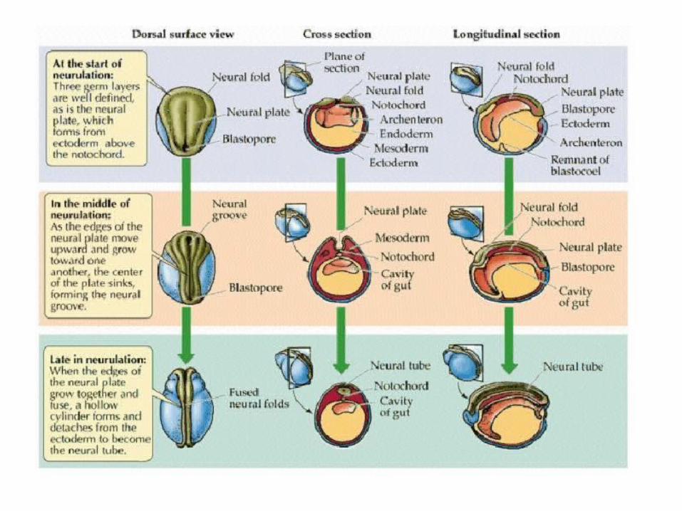
• Neurulation is the formation of the vertebrate nervous system in embryos.
• The notochord induces the formation of the CNS by signaling the ectoderm above it to form the thick and flat neural plate.
• The neural plate then folds in on itself to form the neural tube, which will then later differentiate into the spinal cord and brain.

Neurulation (cont’d)
• Different portions of the neural tube then form by 2 different processes in different species:
1. Primary Neurulation – the neural plate creases inward until the edges come into contact and then fuse.
2. Secondary Neurulation – the tube forms by hollowing out of the interior of a solid precursor

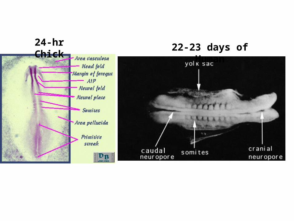
24-hr Chick 22-23 days of Human

23-26 days of Human

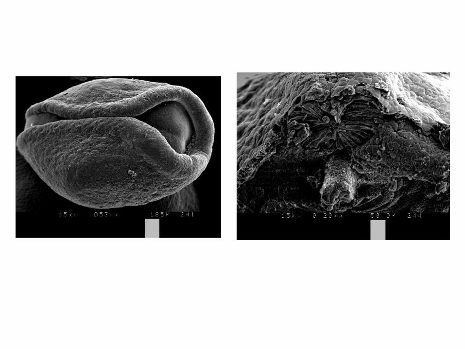
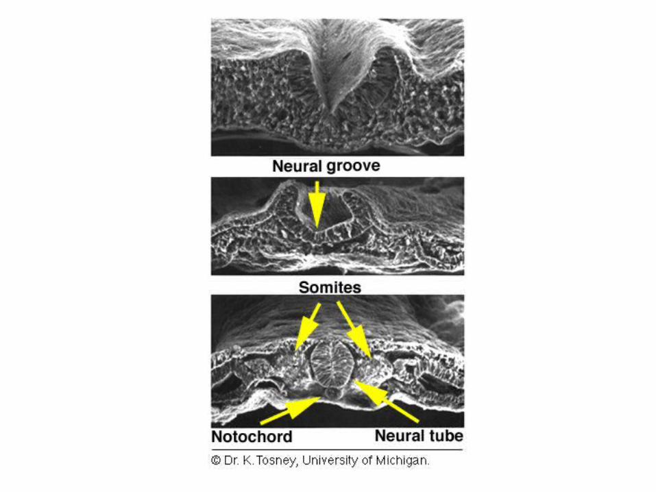
Formation of the Neural Tube
• Secondary Neurulation1. Occurs beyond the caudal neuropore 2. lumbar and tail region3. Exclusive mechanism for fish 4. Starts with formation of medullary cord5. Cavitation of cord to form hollow tube

Secondary Neurulation
Differentiation of Neural Tube
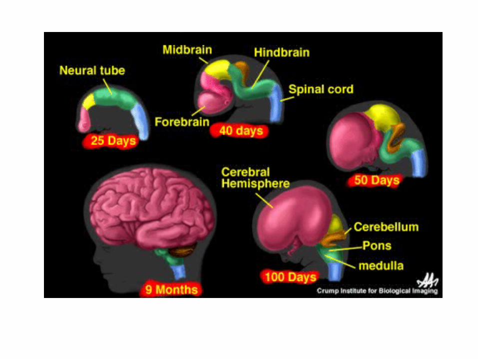
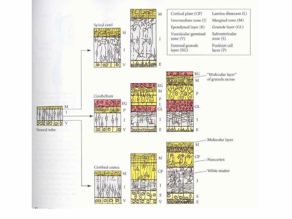
• Major morphological changes: differentiation of brain vesicles and spinal cord
• Differentiation of neural tube cells• Development of peripheral nervous system

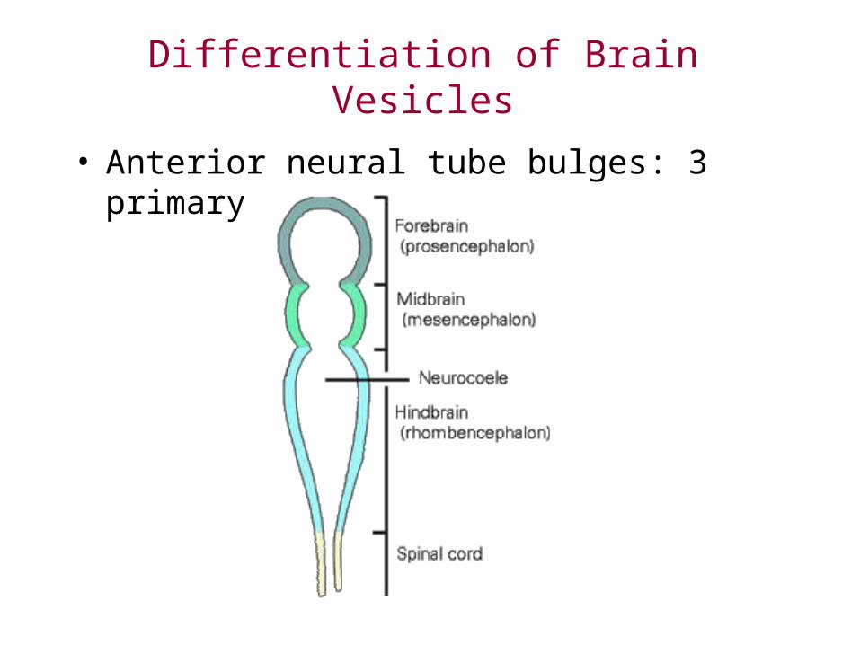
Differentiation of Brain Vesicles
• Anterior neural tube bulges: 3 primary vesicles:
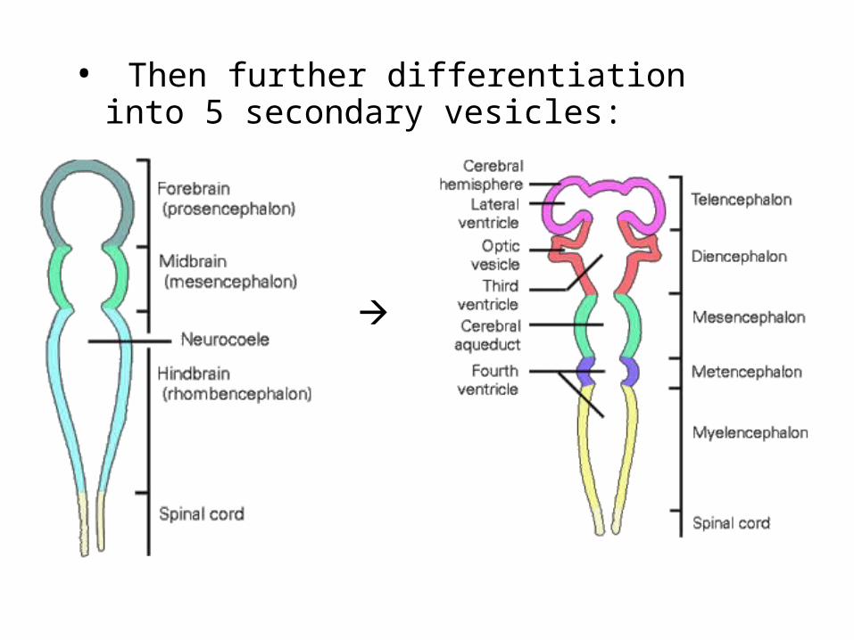
• Then further differentiation into 5 secondary vesicles:
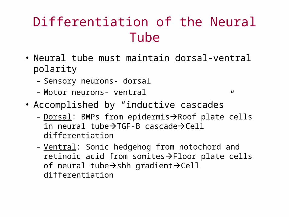
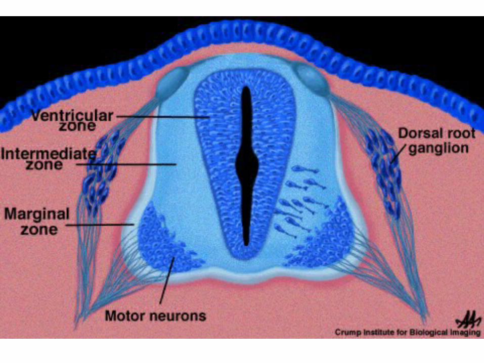
Differentiation of the Neural Tube
• Neural tube must maintain dorsal-ventral polarity– Sensory neurons- dorsal– Motor neurons- ventral
• Accomplished by “inductive cascades”– Dorsal: BMPs from epidermisRoof plate cells in
neural tubeTGF-B cascadeCell differentiation– Ventral: Sonic hedgehog from notochord and retinoic
acid from somitesFloor plate cells of neural tubeshh gradientCell differentiation

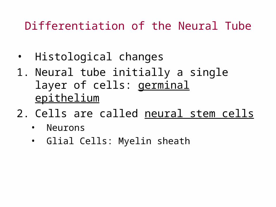
Differentiation of the Neural Tube
• Histological changes1. Neural tube initially a single layer of cells: germinal
epithelium2. Cells are called neural stem cells
• Neurons• Glial Cells: Myelin sheath


Glial Guidance


Development of Peripheral Nervous System• Divisions:– Autonomic NS
• Sympathetic vs. Parasympathetic– Somatic NS
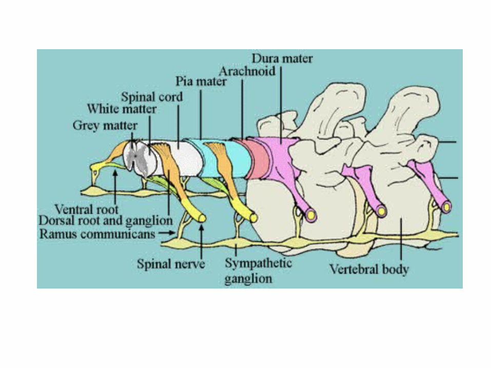
• Anatomy– Sensory Neurons
• Enter dorsal part of spinal cord• Soma located outside of cordDorsal ganglia• Form Dorsal Root of Spinal Nerve
– Motor Neurons• Soma in ventral gray matter• Somatic NS: Neurons run directly from SC to muscle• Autonomic: 2 Neurons
– Sympathetic: Ganglia near SC– Parasympathetic: Ganglia near or in or near organ


Development of Peripheral Nervous System• Anatomy– Spinal Nerves:Spinal Cord• Sensory fibers of somatic nervous system: Dorsal
root• Preganglionic neurons of sympathetic system:
Ventral root• Motor fibers of somatic nervous system: Ventral
root– Cranial Nerves: Brain stem• Sensory fibers of somatic nervous system: Dorsal
root• Preganglionic neurons of parasympathetic system:
Ventral root• Motor fibers of somatic nervous system: Ventral
root

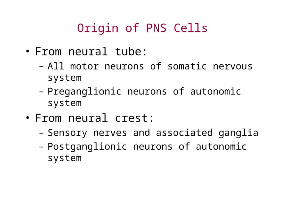
Origin of PNS Cells
• From neural tube:– All motor neurons of somatic nervous system– Preganglionic neurons of autonomic system
• From neural crest:– Sensory nerves and associated ganglia– Postganglionic neurons of autonomic system
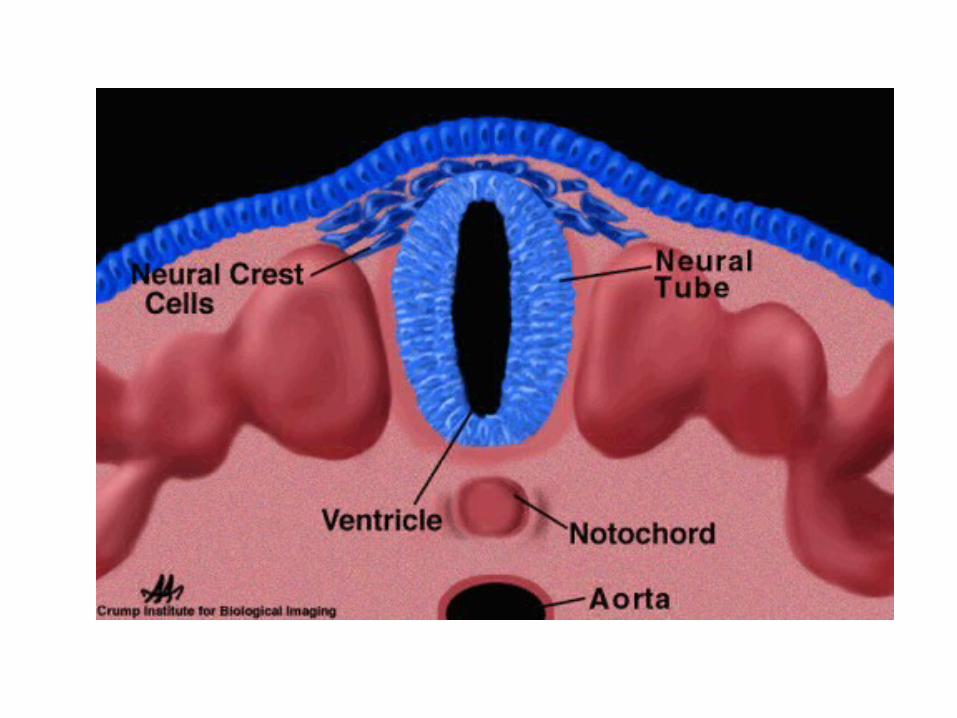


Neural Crest Cells
• Induced by organizing cells of notochord• Main functional groups:– Cranial neural crest:• Bones and connective tissue of face• Tooth primordia• Thymus, parathyroid, thyroid glands• Sensory cranial neurons• Parasympathetic ganglia and nerves• Parts of the heart (cardiac neural crest)

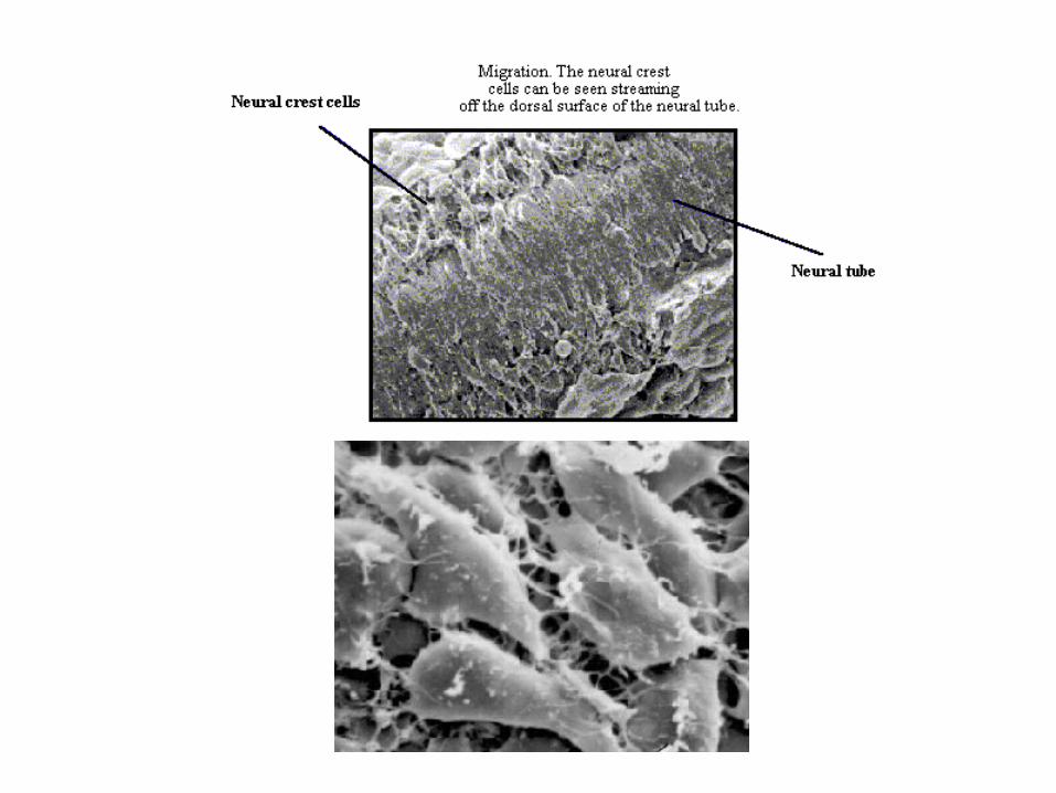
Neural Crest CellsA group of cells, which breaks away from the closing
neural tube and populate the periphery (as opposed to the CNS, which will develop from the tube).
• Neural crest will supply all neurons of the PNS and a variety of other peripheral structures, ranging from melanocytes to craniofacial bones to cells of the adrenals.
• This population of cells separates from the neural plate shortly after the fusion of the neural folds, and streams of dividing cells begin their journey through the embryo.
• The expression of which genes are turned off during this migratory stage?
• And, this occurs for individual cells as well as for groups of cells
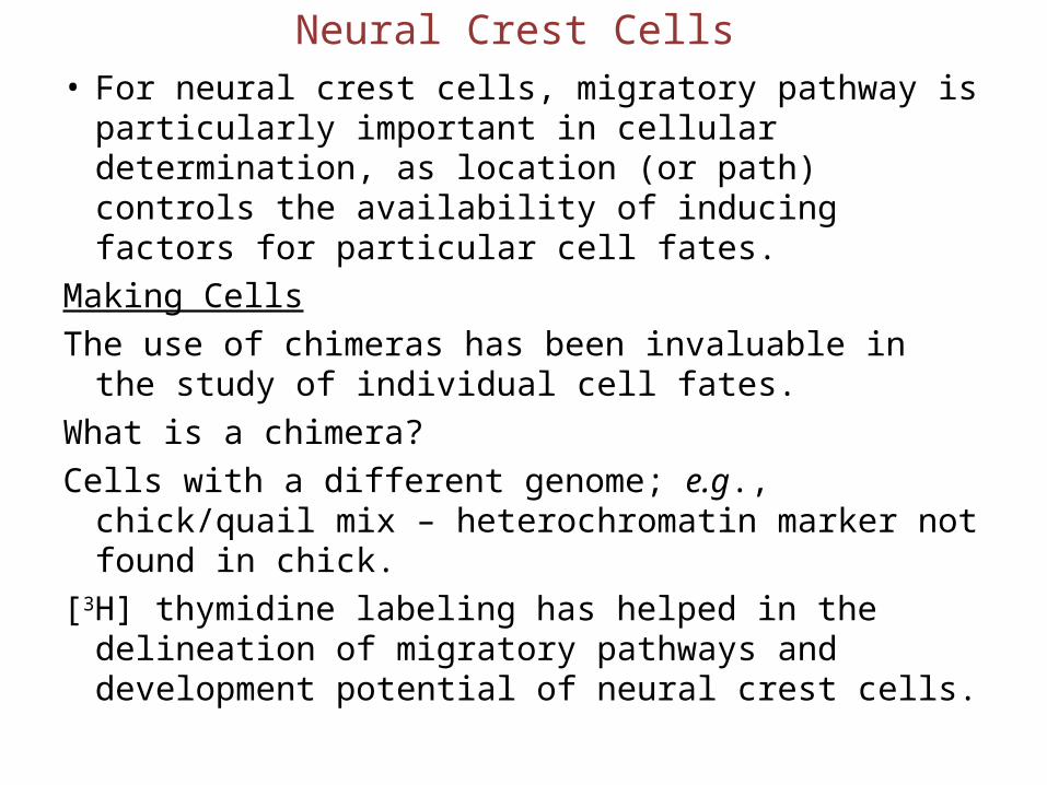
Neural Crest Cells• For neural crest cells, migratory pathway is
particularly important in cellular determination, as location (or path) controls the availability of inducing factors for particular cell fates.
Making CellsThe use of chimeras has been invaluable in the study of
individual cell fates.What is a chimera?Cells with a different genome; e.g., chick/quail mix –
heterochromatin marker not found in chick.[3H] thymidine labeling has helped in the delineation of
migratory pathways and development potential of neural crest cells.

Neural Crest Cells
Interaction:Neural crest migration/movement is rigid and occurs in
a ventral (1st cells give rise to ventral structures) - to- dorsal order in the head.
Migratory pathways are linked to neuronal fate.What is the mostly likely result of transplant
experiments when early cells will switch their fate?Extrinsic cues ?Whether these come from the pathway itself or the
final destination (target-derived cues) is not clear.As in the CNS, the earlier the cell is, the more
pleuripotent it is (the more flexibility of fate).

Neural Crest Cells
• Main functional groups: • The stream of neural crest cells migrates via a
ventral route to form:– Trunk neural crest:• Melanocytes (via the dorsal route)• Sensory neurons (DRG)• Sympathetic ganglia and nerves (ANS)• Medulla of adrenal glands (chromaffin cells)
Note that they migrate segmentally (sclerotome) – only in the rostral compartment.

Neural Crest Cells
• Migration:– Epithelial to mesenchyme transition– Migrational pathways are established by juxtacrine
signals:• Fibronection, laminin in ECM + integrins• Ephrin proteins: Restrict movement• Contact inhibition• Use of existing structures
– Migration ceases when these signals are reversed
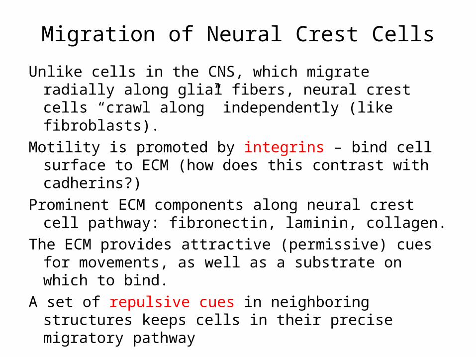
Migration of Neural Crest Cells
Unlike cells in the CNS, which migrate radially along glial fibers, neural crest cells “crawl along” independently (like fibroblasts).
Motility is promoted by integrins – bind cell surface to ECM (how does this contrast with cadherins?)
Prominent ECM components along neural crest cell pathway: fibronectin, laminin, collagen.
The ECM provides attractive (permissive) cues for movements, as well as a substrate on which to bind.
A set of repulsive cues in neighboring structures keeps cells in their precise migratory pathway

Migration of Neural Crest Cells
As the cells reach their destination, the expression of cadherins is once again activated (had been repressed during free movement) cells aggregate into ganglia when they undergo terminal neuronal and glial differentiation.
Side bar: retroviral labeling:A cell can be labelled permanently and heritably by
injection with a retrovirus carrying a gene (e.g., β-galactosidase) incorporated into cells’ DNA and then expressed.
A substance, which will turn blue from the action of the enzyme, can then be introduced in a histochemical test.

Neural Crest Cells
• Differentiation:– Largely based on location along neural tube and their
migration route:

Neural Crest Cells• Differentiation:
– Migration routes along trunk:– Ventral pathway: cells move through anterior portion of
somite toward ventral side of embryo• Cells become: sensory neurons, sympathetic ganglia,
medulla of adrenal gland– Dorsolateral pathway: cells move between epidermis and
somite• Cells become: melanocytes
• Basic organization of the PNS is established by the migratory pathways of the neural crest cells


Neural Crest Cells• Differentiation:
– How do they know what to become?– Most cells are pleuripotent- fate determined by position
– Paracrine factors play a role• Example: Endothelin-3 and Wnt
– Some exceptions: only NC cells from head make bone
– Individual cells may differentiate early in migration

Differentiation of Neurons
• Within nerve tube:– Dorsal Interneurons– Ventral Motor neurons

Differentiation of Neurons• Motor neurons:– Tissues they innervate depends on:– Anterior-posterior location along the nerve tube– When the cells were “born”


Axonal Pathways
1. Motor neurons from ventral root
2. Sensory ganglia
3. Autonomic motorneurons
Spinal Cord

Axonal Pathways
• Establishing pathways and connections:– Pathway selection– Target selection– Address selection

Axonal Pathways
1. Pathway Selection– Pathway axon takes influenced by
extracellular matrix and cells encountered: signals by both paracrine and juxtacrine factors:• Cell adhesion and contact guidance: Haptotaxis• Growth cone repulsion
– Ephrin and semaphorin proteins
• Labeled Pathways Hypothesis: Pioneer Neurons• Diffusible molecules

Axonal Pathways2. Target Selection:– Neurotrophins on target cells (muscle or another
neuron)
3. Address selection: constructing the synapse

