new chemistry with old elements
TRANSCRIPT

Dissertation zur Erlangung des Doktorgrades
der Fakultät für Chemie und Pharmazie
der Ludwig-Maximilians-Universität München
Matching Phosphorus and Chalcogens
– New Chemistry with Old Elements
Synthesis of Unusual Anions and Small Molecules
vorgelegt von
Christiane Rotter
aus
Augsburg
2009

Erklärung
Diese Dissertation wurde im Sinne von § 13 Abs. 3 bzw. 4 der Promotionsordnung vom 29. Januar 1998 von Herrn Professor Konstantin Karaghiosoff betreut.
Ehrenwörtliche Versicherung
Diese Dissertation wurde selbstständig, ohne unerlaubte Hilfe erarbeitet.
München, am 24.11.2009
__________________________________
Christiane Rotter
Dissertation eingereicht am: 24.11.2009
1. Gutachter: Prof. Dr. Konstantin Karaghiosoff
2. Gutachter: Prof. Dr. Thomas M. Klapötke
Mündliche Prüfung am: 11.12.2009

DanksagungKeine Schuld ist dringender, als die, DANKE zu sagen.
(Marcus Tullius Cicero)
Und diese Schuld möchte ich hiermit begleichen, denn ohne die fachliche, finanzielle und moralische Unterstützung vieler Personen, wäre die Anfertigung der vorliegenden Arbeit nicht möglich gewesen: V I E L E N D A N K …
… meinem Doktorvater, Herrn Prof. Dr. Konstantin Karaghiosoff, für die Geduld, den fachlichen Rat und das andauernde Interesse an meiner Arbeit.
… Herrn Prof. Dr. Thomas M. Klapötke, für die freundliche Aufnahme in den Arbeitskreis, die fortwährende Unterstützung in jedweder Form, das fachliche Interesse, und die exzellenten Berechnungen.
… meiner Familie und meinem Freund, die mich sowohl moralisch, als auch finanziell andauernd unterstützten, und mich immer wieder motiviert haben weiterzumachen.
… Dr. F. Xaver Steemann, Norbert Mayr und Richard Moll, die mir mit Rat und Tat, vor allem in der anstrengenden Schlusszeit, zur Seite gestanden sind, und immer ein offenes Ohr für meine Probleme hatten.
… meinen Praktikanten, Stefanie Schönberger, Mirijam Kohler, Jowita Humin, Camilla Evangelisti (ihr ein besonderer Dank auch für die Berechnungen), Sebastian Gebler, Katrin Beimler, sowie Alberto Maulu, die durch ihr großartiges Engagement wertvolle Ergebnisse für diese Arbeit beigetragen haben.
… meinen Laborkollegen aus D 3.103, Alexander Penger, Stefan Huber, Susanne
Scheutzow, und vor allem Karin Lux (die mir nicht nur während der Promotionszeit zur Seite gestanden hat) für das entspannte Arbeitsklima.
… Dr. Jörg Stierstorfer, Wolfgang Betzl, Dr. F. Xaver Steemann und Daniel
Maxien die meine Arbeit Korrektur gelesen haben.
... Leo Berlinger, für die Gestaltung des Titelbildes.
... Dr. Oliver Schön, für die angeregten Diskussionen, und fortwährende Unterstützung.
… dem gesamten Arbeitskreis Klapötke für die freundliche Atmosphäre.


Table of Content
Phosphorus the Living Element 13
Research objectives 19
1 Thiophosphates PnSmx- 20
2 Selenophosphates PnSemx- 20
3 Tellurophosphates PnTemx- 20
The Truth about Trithiometaphosphate PS3 – the History
of a Forbidden Anion 21
1 Introduction 22
2 Results and Discussion 26
2.1 Synthesis of [nBu4N]2[P2S6] • THF (1) and [Ph4P]2[P2S6] (2) 26
2.2 31P NMR spectroscopic investiagtion 30
2.3 Computational results 33
2.4 Chemical behavior of PS3– 38
2.5 Adduct formation 38
2.6 Comparison of the structures of the adduct stabilized trithiometaphosphate PS3– 44
3 Conclusion 45
4 Experimental Section 46
The Triselenometaphosphate Anion PSe3-: A Synthetic
Challenge 49
1 Introduction 50
2 Syntheses 51

3 Crystal and Molecular Structure of [pyH][pyPSe3] (1) 54
4 Comparison of the structures of [pyH][pyPS3] and [pyH][pyPSe3] 56
5 Quantum chemical calculation 57
6 Conclusion 61
7 Experimental Section 62
On the Existence of Dichalcogenophosphoryl
Monohalogenides PCh2X (Ch = S, Se, Te; X = Cl, Br, I) 65
1 The Problem with σ3λ5 Phosphorus 66
2 New route to pyPCh2X (Ch = S, Se, Te; X = Cl, Br, I) 69
3 Reactions of PX3 with Na2S2 (X = Cl, Br, I) 69
3.1 Reaction of PCl3 and Na2S2 69
3.2 Reaction of PBr3 and Na2S2 70
3.3 Reaction of PI3 and Na2S2 71
4 Reaction of PX3 with Na2Se2 (X = Cl, Br, I) 71
4.1 Reaction of PCl3 with Na2Se2 71
4.2 Reaction of PBr3 with Na2Se2 73
4.3 Reaction of PI3 with Na2Se2 74
4.4 Reaction of PI3 with Na2Te2 74
5 Conclusion 75
6 Experimental Section 76
Binary Neutral Phosphorus Sulfides py2P2S5 and py2P2S7 79
1 Introduction 80
2 Results and Discussion 81
3 Quantum chemical calculations 86
4 Conclusion 88
5 Experimental Section 89

Some News about P2S82– 93
1 Introduction 94
2 Results and Discussion 95
3 Conclusion 98
4 Experimental Section 99
Structural and NMR Spectroscopic Investigations of
Chair and Twist Conformers of the P2Se82–Anion 103
1 Introduction 104
2 Syntheses 104
3 31P and 77Se NMR Spectroscopy of the P2Se82– Anion 105
3.1 31P NMR Spectra of chair-P2Se82– 106
3.2 31P NMR spectra of twist-P2Se82– 107
3.3 77Se NMR Spectrum of twist – and chair-P2Se82– 107
4 Crystal and Molecular Structures of P2Se82– Salts 109
4.1 [Li(py)4]2[P2Se8] (1) 109
4.2 [nBu4N]2[P2Se8]·2MeCN (2) 111
4.3 [bmim]2[P2Se8] (3) 112
4.4 [Li(MeCN)4]2[P2Se8] (4): First Structure of twist-P2Se82– 114
4.5 Comparison of the Structures of P2Se82– Salts 116
5 Computational Results 117
6 Conclusion 122
7 Experimental Section 123

A Waterstable Selenophosphate Anion: P2Se64– 129
1 Introduction 130
2 Syntheses 131
3 [pyH]4[P2Se6] • H2O (2) 132
4 31P and 77Se Heteronuclear NMR Spectroscopy 134
5 Conclusion 136
6 Experimental Section 137
A New Synthesis and Crystal Structure of the Old
Hexathiohypodiphosphate P2S64– 141
1 Introduction 142
2 [py2Li]4[P2S6] • 2 py 143
3 Comparison of the structures of [py2Li]4[P2S6] • 2py and the corresponding
selenium compound [py2Li]4[P2Se6] • 2 py 145
4 Conclusion 147
5 Experimental Section 148
Phosphorus Rich Selenophosphates 151
1 Introduction 152
2 A New Route to Phosphorus Rich Selenophosphate Anions PnSemx– 153
3 Reaction of P4 with P4Se72- 153
5 Reaction of P4 with P4Se3 and Na2Se2 157
Tris(pentafluorphenylseleno)phosphine (C6F5Se)3P 161
1 Introduction 162
2 Results and Discussion 163
3 Conclusions 165
4 Experimental Section 166

Do Binary Phosphorus Tellurium Anions Exist? 167
1 Introduction 168
2 Synthesis and NMR analysis of P4Te22– 169
3 Quantum chemical calculations 174
4 Are there more phosphorus-tellurium anions? 175
5 Conclusion 177
6 Experimental Section 178
Some Unexpected Compounds in the Reaction of White
Phosphorus, Lithium and Tellurium 179
1 Introduction 180
2 Results and Discussion 180
3 Conclusion 189
4 Experimental Section 190
4.1 Syntheses of new P-Te anions outgoing from the elements 191
4.2 Syntheses of (N-MeIm)2P4Li2 191
4.3 Syntheses of (N-MeIm)2PLi 192
4.4 Reaction of (N-MeIm)2PLi with tellurium 193
Conclusion 195
1 σ3λ5 - An unusual bonding situation for the phosphorus atom 195
2 Alternative syntheses for chalcogenophosphates 196
3 New fields of chalcogenophosphate chemistry 196
– a first insight into P, Te anions 196


Phosphorus the Living Element
Mit Eifer hab’ ich mich der Studien beflissen;Zwar weiß ich viel, doch möcht‘ ich alles wissen
Goethe, Faust I
This citation is most probable the motivation of every chemist to make chemical research. It is the inquisitiveness to discover, learn about and understand new, exciting fields of chemistry.
One of the most fascinating elements is phosphorus, due to its versatile chemistry and properties.
Phosphorus was discovered by the alchemist, researched by the early chemists,
exploited by the industrialists of the nineteenth century and abused by the
combatants of the twentieth.1
Elemental white (or yellow) phosphorus was accidently discovered by Henning Brandt 1669 in Hamburg. Brandt searched for the philosopher’s stone, like every alchemist at that time. He dried urine in a glass vessel and added then some charcoal. After heating the vessel glowing fumes appeared and a liquid, which bursts into flames immediately when having contact with air, dripped out of the vessel opening – white phosphorus has been prepared for the first time.1
Figure 1. The Alchemist in Search of the Philosopher’s Stone, by Joseph Wright of Derby, 1771.
1 Emsley, J. The Sordid Tale of Murder, Fire and Phosphorus – The 13th Element, John Wiley & Sons, Inc., New York, 2000.
Chapter I

14 c h a P t E r I
Since that, phosphorus fascinated the chemists because of its incomparable properties. The glow of phosphorus gave the element its name: it originates from the greek word phosphoros, which means light bringing. Due to its glow phosphorus was believed to have healing powers, so it was widely used as medicine for almost every disease. But since white phosphorus is poisonous and may cause death, no one could ever be cured from any suffering, so suffering was most probable alleviated and definitely shortened, however.
Figure 2. Title page of Johann Heinrich Cohausen‘s book about phosphorus, 1717 (left) and illustration in the same book (right). It demonstrates that it was believed the glow was made by supernatural powers.
White phosphorus brought light to the people in the 18th century in the truest sense of the word in terms of friction matches called Lucifers. In times where only flint and tinder could be used to make fire the Lucifer matches were considered as blessing as it was now possible to light fire everywhere. Nevertheless the other side of the coin was called Phossy Jaw – a serious disease causing the degradation of the jaw bone.

15I N t r o d u c t I o N
Figure 3. Lucifer matches which could be struck everywhere.
The match-makers suffered from phossy jaw, especially those who dipped the matches. But it was not before the 2nd world war white phosphorus got disrepute due to its use in phosphorus bombs, which cause bad injuries and painful death.
So deadly phosphorus in its elemental form can be, so essential are phosphorus compounds for living organism, especially the phosphate moiety plays a major role as it is the limiting factor for life itself, being part of the DNA and ATP.
We may be able to substitute nuclear power for coal, and plastics for wood, and yeast for meat, and friendliness for isolation – but for phosphorus there is neither
substitute nor replacement.2
In the form of detergents and food additives phosphate compounds make our daily life easier. Of particular importance is the role of phosphate compounds as fertilizers as the natural availability of phosphate is the most important growth factor of plants. Only with the invention of phosphate fertilizers it is possible to yield a large crop.
Recently a very special phosphate, the lithium iron phosphate LiFePO4 attracts more and more attention, as its application as cathode material in highly efficient lithium ion accumulators is much appreciated.3
Life can multiply until all the phosphorus has gone and then there is an inexorable halt which nothing can prevent.2
2 asimov, I. Asimov on Chemistry, Macdonald & Jane’s, London, 1975.3 Padhi, a. K.; Nanjundaswamy, K. S.; Goodenough, J. B. J. Electrochem. Soc. 1997, 144(4), 1188.

16 c h a P t E r I
Consequently, oxophosphates PnOmx– are very well investigated and represent an
integral part in chemistry textbooks. In contrast chalcogenophosphates PnChmx–
containing the heavier chalcogens sulfur, selenium and tellurium are much less investigated and therefore still represent a challenge in the field of inorganic chemistry. Indeed only a limited number of thiophosphate anions PnSm
x– and only a few selenophosphate anions PnSem
x– have been described in the literature so far.4 In the case of tellurophosphate anions only one example has been described in literature. Mewis et al. found in crystalline BaP4Te2.5 Considering only the selenophosphates PnSem
x– many contributions to these compounds originate from the groups of Kanatzidis and Dorhout.6
The increasing interest in metal chalcogenophosphates with the heavier chalcogens sulfur and selenium due to their interesting material properties like ion conductivity3 or optoelectrical7 properties stimulates the search for new and in particular phosphorus rich chalcogenophosphate anions. The classical syntheses of metal chalcogenophosphates are mainly the domain of solid state chemistry. They involve reactions at high temperatures in the melt, under hydrothermal conditions or using polychalcogenide fluxes.4,5,6 So there is a need to investigate new syntheses routes under kinteical conditions.
In our research work group a new method to synthesize chalcogenophosphates has been developed outgoing from easily available educts in solution at ambient temperature or below, thus making the formation of metastable chalkogenophosphate anions possible.8 Remarkably the thio– and selenophosphates show a much larger structural diversity than the oxophosphates. Especially concerning the selenophosphates, structural motives can be found like in polyphosphides or neutral binary phosphorus selenides.
The use of organic cations like Ph4P+ or nBu4N+ makes it possible to isolate the chalcogenophosphate anions in the solid state. The yielded salts are soluble in common organic solvents thus opens up the possibility to investigate the further chemistry of those compounds.
4 (a) Klingen, W.; ott, r.; hahn, h. Z. Anorg. Allg. Chem., 1973, 396, 271. (b) Krause, W.; Falius, h. Z. Anorg. Allg. Chem., 1983, 496, 80. (c) chan, B. c.; hess, r. F.; Feng, P. L.; abney, K. d.; dorhout, P. K. Inorg. Chem., 2005, 44, 2106, and references therein.5 Mewis, a.; Jörgens, S.; Johrendt, d. Chem. Eur. J., 2003, 9, 2405.6 (a) chung, I.; Karst, a. L.; Weliky, d.; Kanatzidis, M. G. Inorg. Chem. 2006, 45, 2785. (b) chung, I.; Malliakas, c. d.; Jang, J. I.; canlas, c. G.; Weiky, d. P.; Kanatzidis, M. G. J. Am. Chem .Soc. 2009, 129(48), 14996. (c) chung, I.; Karst, a. L.; Weliky, c. G.; Kanatzidis, M. G. Inorg. Chem. 2004, 43, 2785. (d) Briggs Piccoli, P. M.; abney, K. d.; Schoonover, J. r.; dorhout, P. K. Inorg. Chem. 2000, 39, 2970. (e) chan, B. c.; hess, r. F.; Feng, P. L.; abney, K. d.; dorhout, P. K. Inorg. Chem. 2005, 44(6), 2106.7 Galdámez, a.; Manríquez, V.; Kasaneva, J.; avila, r. E. Mater. Res. Bull. 2003, 38, 1063.8 (a) Schuster, M. dissertation, LMu Munich, 1999. (b) Schuster, M.; Karaghiosoff, K. Phosphorus, Sulfur Silicon and Relat. Elem. 2001, 168, 117. (c) rotter, c.; Schuster, M.; Kidik, M.; Schön, o.; Klapötke, t. M.; Karaghiosoff, K. Inorg. Chem. 2008, 47, 1663.

17I N t r o d u c t I o N
Although the chalcogenophosphate chemistry is an already established field of research among phosphorus chemistry, there is still a big lack of knowledge concerning the following points:
• Small molecules with a central phosphorus atom in unusual bonding situations
(e.g. σ3λ5 or σ2λ3) are rarely described and even much less explored concerning
stability, reactivity and bonding situation
• Analogies and differences to the nitrogen chemistry should be examined
• As phosphorus containing compounds which are analogue to nitrogen-
chalcogen-compounds are forbidden by the double bond rule and by the classical
chemical view of the world, it is a particular challenge for a phosphorus chemist
to investigate this special, unanswered topic of phosphorus chemistry
• Nevertheless, scientific research should also always result in contributions to
solution of daily life problems. So it is important to gain knowledge about the
syntheses of compounds which could find practical applications
The above mentioned considerations are the starting point for the idea and concept of the following thesis. The formulated goals listed below should be received within this thesis.


Chapter II
Research Objectives
Main research objectives of the present thesis are the development of synthetic
procedures to prepare and isolate new simple neutral and anionic phosphorus
chalcogen species. Their identity should be established by means of heteronuclear
(31P, 77Se, 125Te) NMR spectroscopy and single crystal X-ray diffraction. Their
chemical properties should be investigated. The investigations should enable us to
compare in detail the chemistry of N-O compounds with the heavier homologues
P-S(Se, Te). Quantum chemical calculations (in collaboration with Prof. Thomas
M. Klapötke and Camilla Evangelisti, B. Sc.) should help us to understand
analogies and differences.

20 c h a P t E r I I
1 Thiophosphates PnSmx-
It was planed to put light on the mystery around the PS3- anion. Does it really exists and
is it in deed a “heavy nitrate”, or does it show completely different properties? For this synthesis of a salt containing the PS3
- anion and NMR spectroscopical investigations concerning the reaction behaviour in basic solvents should be carried out. In addition, the formation and reaction behaviour of the corresponding dimer P2S6
2- and trimer P3S9
3- should be investigated. At the same time, quantum chemical calculations should be made to help to explain the experimental results obtained.
In addition, the investigations should be extended to neutral compounds like P2S5 or PS2Cl, which have the same interesting bonding situation for the central phosphorus atom (σ3λ5) like in case of the PS3
- anion. Does the change from an anionic species to neutral affect the stability of these compounds?
2 Selenophosphates PnSemx-
The investigations on PS3- (see above) should be extended to the corresponding
selenium derivative PSe3-. Which analogies and differences can be observed for the
two systems? The planed investigations includes the development of a synthesis for PSe3
- salts, the possible isolation of acid-base adducts and again the elucidation of the bonding situation by means of quantum chemical calculations.
In addition, the syntheses of new phosphorus rich selenophosphates as well as of new soluble salts of selenium rich selenophosphates should be attempted.
3 Tellurophosphates PnTemx-
A particular intriguing question regards the existence of unstable soluble tellurophosphates. This point represents a central objective of the present thesis. Do phosphorus-tellurium anions exist at all? Different synthetic strategies, e. g. starting from the elements (P4, Li, Te), should be attempted, in order to give an answer to this challenging question.

Chapter III
The Truth about Trithiometaphosphate PS3 – the History of a
Forbidden Anion
Two new salts containing the P2S62– anion have been prepared as well as a new
salt of the donor-stabilized monotrithiometaphosphate PS3– starting from P4S3,
Na2S2 and elemental sulfur in common organic solvents like THF or acetonitrile. 31P, 31P EXSY NMR spectroscopy was used to proof the existence of free monomeric
trithiometaphosphate anion in solution, and to determine its 31P NMR chemical
shift, as well as the one of P2S62–. Temperature dependent 31P NMR spectroscopy
was used to reveal the equilibrium between the adduct stabilized monomeric
trithiometaphosphate and the free monomeric trithiometaphosphate in solution.
Quantum chemical calculations were performed to elucidate the bonding
situation in the trithiometaphosphate PS3– and the formation of the dimeric
(P2S62–) and trimeric (P3S9
3–) trithiometaphosphate in solution.
22 c h a P t E r I I I
1 Introduction
Binary nitrogen-oxygen anions are well known and represent an integral part of every textbook of inorganic chemistry.F
1F The most prominent exponent of representative
species with the general formula PnCh3– (Pn = N, P; Ch = O, S, Se) is the nitrate anion
NO3–, which has a trigonal planar structure. Oligomers of NO3
– are not known. Besides the trigonal planar NO3
– molecule a structural isomer, the unstable peroxonitrit consisting of a bent ONOO chain is known (XScheme 2X).
On introduction of sulfur in place of oxygen in NO3–, the corresponding NS3
– anion does not adopt a trigonal planar structure like NO3
– but has the structure of trithioperoxonitrit, constisting of a bent SNSS chain (XScheme 2X). A NSe3
– compound is still not described in literature.
The situation changes drastically if nitrogen is replaced by the higher homologue phosphorus. Due to the low tendency of the elements of the third period to form double bonds, small anions consisting of a central phosphorus atom in a low coordination number CN (CN = 2-3) are less favored. Stable cyclic oligomers are formed which could be isolated as metal salts.
A monomeric trioxometaphosphate PO3 – is only known as reactive intermediate
in the gas phase: instead cyclic oligomers (PO3–)n (n = 3-8) are thermodynamically
and structurally favored.X
1,2,3,4,5 In case of trithiometaphosphate anion PS3-, the
corresponding dimer, trimer and tetramer (PS3–)n (n = 2-4) are described in literature.
6,7,8,9,10
1 holleman, a.; Wiberg, E.; Wiberg, N. Lehrbuch der Anorganischen Chemie 102nd edition, 2007, Walter de Gruyter, Berlin – New York.2 henchman, M.; Viggiano, a. a.; Paulsen, J. F.; Freedman, a.; Wormhoudt, J. J. Am. Chem. Soc. 1985, 107(5), 265-266.3 Westheimer, F. h. Chem. Rev. 1981, 81, 313-326.4 Meisel, M. Multiple Bonds and Low Coordination in Phosphorus Chemistry, Georg thieme Verlag, Ed.: M. regitz, o. J. Scherer, Stuttgart, 1990, 415-442.5 Binnewies, M.; Schnöckel, h. Chem. Rev. 1990, 90, 321-330.6 roesky, h. W.; ahlrichs, r.; Brode, S. Angew. Chem. 1986, 98, 91-93.7 a) Gjikaj, M.; Ehrhardt, c.; Brockner, W. Z. Naturforsch. 2006, 61b(9), 1049-1053. b) Gjikaj, M.; Brockner, W. Z. Anorg. Allg. Chem. 2006, 632(2), 279-283. c) dimitrov, a.; hartwich, I.; Ziemer, B.; heidemann, d.; Meisel, M. Z. Anorg. Allg. Chem. 2005, 631(12), 2439-2444. d) Gjikaj, M.; Brockner, W. Vibr. Spec. 2005, 39(2), 262-265. e) Gjikaj, M.; adam, a.; duewel, M.; Brocknder, W. Z. Kristallograph. 2005, 220(1), 67-68. f) angenault, J.; cieren, X.; Wallez, G.; Quarton, M. J. Solid State Chem. 2000, 153(1), 55-65. g) Salmon, P. S.; Xin, S.; Fischer, h. E. Phys. Rev. 1998, 58B(10), 6115-6123. h) Menzel, F.; Brockner, W.; Ystenes, M. Vibrational Spec. 1997, 14(1), 59-70. i) Voroshilov, Y. V.; Potorii, M. V.; Gebesh, V. Y. Neorganicheskie Materialy 1994, 30(4), 479-483. j) Menzel, F.; Brockner, W.; Ystenes, M. J. Mol. Struct. 1993, 294, 53-56. k) andrae, h.; Blachnik, r. J. Alloys Compd. 1992, 189(2), 209-215. l) Lathrop, d.; Franke, d.; Maxwell, r.; tepe, t.; Flesher, r.; Zhang, Z.; Eckert, h. Solid State Nucl. Magn. Reson. 1992, 1(2), 73-83. m) Ystenes, M.; Brockner, W.; Menzel, F. Z. Naturforsch. 1992, 47a(4), 614-618. n) andrae, h.; Blachnik, r. J. Thermal Anal. 1989, 35(2), 595-607. o) ohse, L.; Brockner, W. Z. Naturforsch. 1988, 43(5), 494-496. p) Jandali, M. Z.; Eulenberger, G.; hahn, h. Z. Anorg. Allg. Chem. 1985, 530, 144-154. q) Brockner, W.; Becker, r.; Eisenmann, B.; Schaefer, h. Z. Anorg. Allg. Chem. 1985, 520, 51-58. r) Becker, r.; Brockner, W. Z. Naturforsch. 1984, 39A(11), 1120-1121. s) toffoli, P.; Khodadad, P.; rodier, N. Acta Crystallogr. 1978, B34(12), 3561-3564.
8 a) hanko, J. a.; Kanatzidis, M. G. J. Solid State Chem. 2000, 151(2), 326-329. b) hanko, J. a.; Sayettat, J.; Jobic, S.; Brec, r.; Kanatzidis, M. G. Chem. Mater. 1998, 10(10), 3040-3049. c) Wolf, G. u.; Meisel, M. Z. Anorg. Allg. Chem. 1982, 494, 49-54.9 Evain, M.; Brec, r.; ouvard, G.; rouxel, J. Mat. Res. Bull. 1984, 19, 41-48.10 Karaghiosoff, K.; Schuster, M. Phosphorus, Sulfur Silicon Relat. Elem. 2001, 168, 117-122.

23t h E t r I t h I o M E t a P h o S P h a t E a N I o N
The anion PS4– does not exist, in contrast to the corresponding dimer P2S8
2– (Scheme 1). The same situation could be found for the anion PS2
–. The twofold coordination of the phosphorus atom is avoided by forming the annular oligomers P4S8
4–, P5S105– and
P6S126-.1,2,3
Scheme 1. Binary phosphorus-sulfur anions.
1 a) dastychova, L.; Sotolarova, M.; dastych, d.; taraba, J.; Necas, M.; Prihoda, J. Polyhedron 2007, 26(15), 4250-4256. b) Katrusiak, a. J. Mol. Struct. 1999, 474, 125-133. c) Falius, h.; Schliephake, a.; Schomburg, d. Z. Anorg. Allg. Chem. 1992, 611, 141-148. d) Jones, P. G.; Weinkauf, a. Acta Crystallogr. 1991, C47(3), 686-687. e) Micu-Semeniuc, r.; Popse, S.; haiduc, I. Rev. Roum. Chim. 1983, 28(6), 605-614. f) Minshall, P. c.; Sheldrick, G. M Acta Crystallogr. 1978, B34(4), 1378.1380.2 a) Badeeva, E. K.; Krivolapov, d. B.; Gubaidullin, a. t.; Litvinov, I. a.; Batyeva, E. S.; Sinyashin, o. G. Mendeleev Commun. 2005, 1, 22-23. b) Falius, h.; Schliephake, a. B. Inorg. Synth. 1989, 25, 5-7. c) Buerger, h.; Pawelke, G.; Falius, h. Spectrochim. Acta 1981, 37A(9), 753-755. d) Falius, h.; Krause, W.; Sheldrick, W. S. Angew. Chem. 1981, 93(1), 121-122.3 Krause W.; Falius, h. Z. Allg. Anorg. Chem. 1983, 496, 80-93.

24 c h a P t E r I I I
The compound of our interest is the trithiometaphosphate anion PS3–, which is
structurally analogue to the well known nitrate anion NO3 – (XScheme 2X).
Scheme 2. Lewis formula of NO3–, NS3
–, PO3 – and PS3
–.
But why is the trithiometaphosphate anion PS3 – so unusual compared to the NO3
– anion?
In order to answer this question, we have to have a closer look at the bonding situation of the central phosphorus atom, which is in an unusual bonding situation. The coordination of the central phosphorus atom in PS3
– is planar. The phosphorus center has the formal oxidation state +V, but is only threefold coordinated and has therefore an unsaturated coordination sphere. One possibility to stabilize compounds with such a central phosphorus atom was investigated by Yoshifuji et al.4,5 They could show that big bulky substituents can stabilize a σ3λ5 phosphorus atom.
Scheme 3. Example for a dithiooxophosphorane stabilized with the bulky substituent supermesityl.
But this sterical protection is missing in the PS3– anion. Due to this reason, the
synthesis of the trithiometaphosphate anion PS3– is a very appealing preparative
challenge. Obviously, Roesky and coworkers had the same idea and tried to find an answer for the problem.6 They investigated the nucleophilic degradation of P4S10 with KCN/H2S in acetonitrile which resulted in the formation of the (NCPS2)2S2– anion, isolated as the nPr4N+ salt on addition of nPr4NBr in H2O/MeOH (Scheme 4).
4 Yoshifuji, M.; toyota, K.; ando, K.; Inamoto, N. Chemistry Lett., 1984, 317-318.5 Yoshifiuji, M.; Sangu, S.; Kamijo, K.; toyota, K. Chem. Ber. 1996, 129, 1049.

25t h E t r I t h I o M E t a P h o S P h a t E a N I o N
Scheme 4. Synthesis and isolation of the trithiometaphosphate anion PS3– by Roesky et al.
On further addition of Ph4AsCl in methanol yellow crystals formed, which the authors claim to be the tetraphenylarsonium salt of the trithiometaphosphate anion [Ph4As][PS3]. Evidence is provided in form of elemental analysis, mass spectra and 31P NMR spectra (δ31P = 52.3 ppm).
But there are some problems which raise some doubts about the correctness of the result: Roesky et al. claimed that it was not possible to solve the crystal structure for sure due to a disorder of the anion. In addition, they used water and methanol as reaction medium, but to our knowledge and experiences with thiophosphates in general, this kind of compounds are unlikely to be stable or preparable in water as they are extremely sensitive towards moisture and oxidation. But the most important hint that Roesky et al. could probably be wrong is the reported 31P NMR chemical shift of 52.3 ppm. For a σ3λ5 phosphorus atom, a 31P NMR resonance at much lower field is to be expected comparable to the ones reported by Yoshifuji et al. for thiooxophosphoranes at about δ31P = 300.14,15
Nevertheless, the corresponding cyclic oligomers P2S62–, P3S9
3– and P4S124– are
described in literature for sure.7,8,9 Most of the contributions to the investigation of the monomeric and dimeric trithiometaphosphate PS3
– and P2S62– originate from the
group of Brockner.7

26 c h a P t E r I I I
2 Results and Discussion
2.1 Synthesis of [nBu4N]2[P2S6] • THF (1) and [Ph4P]2[P2S6] (2)
PS3– was accessible by the reaction of P4S3 with Na2S2 and S8 in THF as developed in
course of this work (XScheme 5X).F
6
Scheme 5. Synthesis of PS3 – in solution.
After adding nBu4Br to the reaction solution, yellow rod shaped crystals could be obtained (XFigure 1).
Figure 1. Crystals of [nBu4N]2[P2S6] • THF (1).
Interestingly, the anion of the compound obtained is formed by the cyclic dimer P2S62–
of the trithiometaphosphate anion (Figure 2). The geometry of the anion P2S62– does
not differ from those reported in literature. It consists of a planar four-membered ring formed by two phosphorus and two sulfur atoms. Four further sulfur atoms are bonded exocyclic to the two phosphorus atoms standing orthogonal to the ring plane. The averaged endocyclic P-S distance is 214.9(1) pm while the averaged exocyclic P-S distance is shorter with 196.4(1) pm. Both central phosphorus atoms are distorted tetrahedrally surrounded by four sulfur atoms. The averaged angles are Sexo-P-Sexo 116.6(1)°, Sexo-P-Sendo 111.6(1)°, Sendo-P-Sendo 90.9(1)° and P-Sendo-P 88.9(1)°. Selected parameters of the structure are given in XTable 1X.
One complete P2S62– anion can be observed within the unit cell together with two
P2S62– anions which lie on an inversion center and therefore are in half generated
by symmetry. That is the reason why only four cations are shown in the molecular structure of 1 (XFigure 2X).
6 Karaghiosoff, K.; Schuster, M. Phosphorus, Sulfur Silicon Relat. Elem. 2001, 168, 117-122.

27t h E t r I t h I o M E t a P h o S P h a t E a N I o N
Figure 2. Molecular structure of [nBu4N]2[P2S6] • THF. Thermal ellipsoids of all non hydrogen atoms are drawn at the 50 % probability level; hydrogen atoms are omitted for clarity. View orthogonal to c axis; i = – x, 2-y, 2-z; ii = 1-x, 1-y, 2-z.
Table 1. Selected bond lengths (pm) and angles (°) of 1; i = – x, 2-y, 2-z; ii = 1-x, 1-y, 2-z.
Distances [pm] Angles [°]
P1-S1 196.8(1) P3-S7 197.0(1) S1-P1-S2 116.3(1) S7-P3-S8 117.0(1)
P1-S2 196.7(1) P3-S8 196.8(1) S2-P1-S3 111.3(1) S8-P3-S9 113.0(1)
P1-S3 214.9(1) P3-S9 214.7(1) S3-P1-S1 111.7(1) S9-P3-S7 110.4(1)
P1-S3(i) 213.9(1) P3-S10 214.8(1) S1-P1-S3(i) 111.5(1) S7-P3-S10 110.8(1)
P2-S4 196.8(1) P4-S9 214.7(1) S2-P1-S3(i) 112.2(1) S8-P3-S10 111.7(1)
P2-S5 196.1(1) P4-S10 214.8(1) S3-P1-S3(i) 91.1(1) S9-P3-S10 91.0(1)
P2-S6 213.9(1) P4-S11 197.2(1) P1(i)-S3-P1 88.9(1) P4-S10-P3 88.5(1)
P2-S6(ii) 214.4(1) P4-S12 196.8(1) S4-P2-S5 117.1(1) P4-S9-P3 88.4(1)
S5-P2-S6 112.8(1) S11-P4-S12 116.4(1)
S6-P2-S4 111.3(1) S12-P4-S9 111.0(1)
S4-P2-S6(ii) 111.0(1) S9-P4-S11 111.2(1)
S5-P2-S6(ii) 110.9(1) S11-P4-S10 112.4(1)
S6-P2-S6(ii) 90.8(4) S12-P4-S10 112.2(1)
P2(ii)-S6-P2 89.2(1) S9-P4-S10 91.0(1)

28 c h a P t E r I I I
Figure 3. Unit cell of 1. View along a axis. Ellipsoids are drawn at 50 % probability level. Hydrogen atoms are
omitted for clarity.
Yellow block shaped crystals of [Ph4P]2[P2S6] • 2 py (2) were isolated after the addition of tetraphenylphosphonium bromide to a solution containing P4S3, Na2S2 and elemental sulfur in pyridine. Compound 2 crystallizes in the monoclinic space group P21/c with four formula units in the unit cell.
The P2S62– anion lies on a crystallographic inversion center which is located in
the middle of the four-membered ring; therefore half of the anion is generated by symmetry. The four-membered ring consisting of both phosphorus atoms P2 and P2(i) and two sulfur atoms S3 and S3(i) has a angle sum of 360° and is therefore planar. The exocyclic bonded sulfur atoms are arranged orthogonal to the ring plane. The P-S bond lengths to the exocyclic sulfur atoms are 198.2(1) pm (P2-S1) and 196.4(1) pm (P2-S2) and therefore within a P-S single bond and a P=S double bond found in phosphorus(V) compounds.X
1,14,15 The endocyclic P-S distances correspond with 215.9(1) pm (P2-S3) and 214.3(1) pm (P2(i)-S3) to P-S single bonds. The phosphorus atom is surrounded in a distorted tetrahedral arrangement by four sulfur atoms. The endocyclic S3-P2-S3(i) angle is 91.5(1)°, whereas the exocyclic S-P-S angles range from 110.0° to 118.7°. The angle at the endocyclic sulfur atom P2-S3-P2(i) is 88.5(1)°. Selected structural parameters for the P2S6
2– anion in compound 2 are given in XTable 1X.

29t h E t r I t h I o M E t a P h o S P h a t E a N I o N
Four P2S62– anions are located on planes parallel to the ab – and ac plane. The
anions and cations are staggered separately along the a axis. There are no significant interactions in the structure than the electrostatic attraction between cations and anions. The pyridine solvent molecules are located between the cations. The position of the nitrogen atom in pyridine can exactly be determined.
Table 2. Selected bond lengths (pm) and angles (°) of [Ph4P]2[P2S6] • py (2); i = 1-x, – y, 1-z.
Bond lengths [pm] Bond angles [°] Torsion angles [°]S1-P2 198.2(1) P2-S3-P2(i) 88.5(1) P2(i)-S3-P2-S2 112.7(1)S2-P2 196.4(1) S3-P2-S3(i) 91.5(1) P2(i)-S3(i)-P2-S2 -112.1(1)S3-P2 215.9(1) S2-P2-S3(i) 110.7(1) P2(i)-S3(i)-P2-S1 113.6(1)S3-P2(i) 214.3(1) S2-P2-S3 110.0(1) P2(i)-S3-P2-S1 -113.7(1)
S1-P2-S3 111.3(1) P2(i)-S3(i)-P2-S3 0.0(1)S1-P2-S3(i) 111.4(1) P2(i)-S3-P2-S3(i) 0.0(1)S1-P2-S2 118.7(1)
Figure 4. Molecular structure of [Ph4P]2[P2S6] • py (2) in the solid state. Thermal ellipsoids are set at the 50 % probability level; hydrogen atoms are omitted for clarity. View along a axis; i = 1-x, – y, 1-z.

30 c h a P t E r I I I
Figure 5. Crystal structure of [Ph4P]2[P2S6] • py (2). View of the unit cell along the a axis; ellipsoids are drawn at the 50 % probability level. Hydrogen atoms are omitted for clarity.
2.2 31P NMR spectroscopic investiagtionBrockner et al.7 used high temperature syntheses starting from the elements
phosphorus, sulfur and metals yielding solids, which are insoluble in common solvents. In case of Na2P2S6 and K2P2S6, they succeeded in dissolving the solids in acetonitrile by adding crown ethers. Nevertheless, the compounds were characterized mostly using X-ray diffraction methods and Raman spectroscopy. Brockner et al. found that the formed anion was P2S6
2– in all cases. Interestingly, the Raman spectrum of a molten alkalibromide-Tl2P2S6 mixture measured at different temperatures indicated that the P2S6
2– anion dissociates into the monomeric trithiometaphosphate anion PS3–.
Brockner et al. were therefore the first, who postulated an equilibrium between the P2S6
2– anion and the PS3– anion.7
As [nBu4N]2[P2S6] • THF is soluble in common polar solvents like pyridine, acetonitrile or propionitrile, it was now possible to determine the 31P NMR chemical shift of P2S6
2–
which has not been reliably reported till now. Surprisingly, the 31P NMR spectrum of [nBu4N]2[P2S6] • THF dissolved in benzonitrile consists of two signals at a chemical shift of 297.5 ppm and 30.2 ppm (XFigure 6X).

31t h E t r I t h I o M E t a P h o S P h a t E a N I o N
Figure 6. Observed 31P {1H} NMR spectrum of [nBu4N]2[P2S6] • THF dissolved in benzonitrile (0.16 M, 25°C, 4096 scans with PD = 0.5 s, 2 h measuring time, υ0 = 161.8347 MHz, broadband 1H decoupling, 0.5 Hz line broadening).
The extremely unusual low field shift of 297.5 ppm could also be found in dithiooxophosphoranes containing a central phosphorus atom in a similar bonding situation like in the trithiometaphosphate PS3
–. Yoshifuji et al.14,15 reported the 31P NMR chemical shift of a dithiooxophosphorane to be 298.2 ppm (in CDCl3). Following the prediction of Brockner that the dimeric trithiometaphosphate anion P2S6
2– spontaneously dissociates into the monomeric trithiometaphosphate anion PS3
–, the 31P NMR signal at 297.5 ppm could be assigned to the monomeric trithiometaphosphate PS3
–, while the 31P NMR signal at 30.2 ppm could be assigned to the dimeric trithiometaphosphate P2S6
2– (Scheme 6).
Scheme 6: Equilibrium between the monomeric trithiometaphosphate PS3– and the corresponding dimer P2S6
2–.

32 c h a P t E r I I I
A 31P, 31P EXSY spectrum shown in Figure 7 reveals that – due to the existence of cross peaks between the two signals – interconversion between the trithiometaphosphate and the corresponding dimer is slow on NMR time scale at ambient temperature (Scheme 6). This experimental result is also in agreement with the reported dimer-monomer equilibrium of special dithiooxophosphoranes reported in literature.7
Figure 7: 31P, 31P EXSY NMR spectrum of 1 dissolved in propionitrile. Crosspeaks are marked with arrows (0.16 m, 25 °C, matrix 2048 x 2048, mixing time 0.5 sec).
So this is the final proof that a trithiometaphosphate anion PS3– really exists and is
stable in its monomeric form in solution. As it turns out, Roesky had indeed the right feeling that a monomeric trithiometaphosphate anion PS3
– does exist. But the real 31P NMR chemical shift of this compound is, as expected due to the structural analogy to thiooxophosphporanes, shifted to much lower field than reported by Roesky.
This knowledge generates a series of further questions:
• What is the reason for the stability of the PS3– anion in solution as there is no
sterical stabilization?
• How can the spontaneous dissociation in solution of P2S62– be explained?
• Which properties does the new anion have?
7 Beckmann, h.; Großmann, G.; ohms, G.; Sieler, J. Heteroat. Chem. 1994, 5(1), 73-83.

33t h E t r I t h I o M E t a P h o S P h a t E a N I o N
2.3 Computational resultsAll calculations were carried out using the Gaussian G03W program code.8 In order to
elucidate the structures, bonding and energies in the gas phase of PS3–, P2S6
2– and P3S93–
theoretically, computations were carried out with Becke’s B3 three parameter hybrid functional using the LYP correlation functional (B3LYP).9 For all atoms, Dunning’s correlation consistent polarized double-zeta basis set was used (cc-pVDZ) (Table 3).10,11,12,13 The DFT calculations reproduce the bond angles in the P2S6
2– and P3S93– anion
well, while the P-S bond lengths are overestimated due to the high concentration of negative charge in an isolated anion in the gas phase. Through observed anion-cation interaction in the condensed phase, the concentration of negative charge in the anion could be abated.
Table 3. Computational results at B3LYP level of theory for the anions PS3–, P2S6
2– and P3S93–.
[PS3]– [P2S6]
2– [P3P9]3–
–E / a.u. 1536.138465 3072.174876 4608.142015
Erel. / kcal mol-1 (per PS3
– moiety)0.0 + 32.0 + 57.1
symmetry D3h D2h C3v
NIMAG 0 0 0
zpe / kcal mol-1 3.63 7.97 12.3
calcul. / expt.a calcul. / exptl.8
d(P-Sr) / Å b 2.232 / 2.14 2.227 / 2.129
d(P-St) / Å c 1.991 2.016 / 1.97 ax: 2.004 / 1.964eq: 2.045 / 1.964
<(Sr-P-Sr) / ° 89.6 / 91.3 103.2 / 103.7
<(St-P-St) / ° 120.0 118.1 / 117.3 118.3 / 119.7
<(Sr-P-St) / ° 111.4 / 111.2 Sr-P-St.ax: 115.2Sr-P-St.eq: 101.3
<(P-Sr-P) / ° 90.5 / 88.6 115.5 / 111.2a average values; b Sr = sulfur atom within the ring; c terminal sulfur atom.
8 Gaussian 03, revision a.1, Frisch, M. J.; trucks, G. W.; Schlegel, h. B.; Scuseria, G. E.; robb, M. a.; cheeseman, J. r.; Montgomery, Jr., J. a.; Vreven, t.; Kudin, K. N.; Burant, J. c.; Millam, J. M.; Iyengar, S. S.; tomasi, J.; Barone, V.; Mennucci, B.; cossi, M.; Scalmani, G.; rega, N.; Petersson, G. a.; Nakatsuji, h.; hada, M.; Ehara, M.; toyota, K.; Fukuda, r.; hasegawa, J.; Ishida, M.; Nakajima, t.; honda, Y.; Kitao, o.; Nakai, h.; Klene, M.; Li, X.; Knox, J. E.; hratchian, h. P.; cross, J. B.; adamo, c.; Jaramillo, J.; Gomperts, r.; Stratmann, r. E.; Yazyev, o.; austin, a. J.; cammi, r.; Pomelli, c.; ochterski, J. W.; ayala, P. Y.; Morokuma, K.; Voth, G. a.; Salvador, P.; dannenberg, J. J.; Zakrzewski, V. G.; dapprich, S.; daniels, a. d.; Strain, M. c.; Farkas, o.; Malick, d. K.; rabuck, a. d.; raghavachari, K.; Foresman, J. B.; ortiz, J. V.; cui, Q.; Baboul, a. G.; clifford, S.; cioslowski, J.; Stefanov, B. B.; Liu, G.; Liashenko, a.; Piskorz, P.; Komaromi, I.; Martin, r. L.; Fox, d. J.; Keith, t.; al-Laham, M. a.; Peng, c. Y.; Nanayakkara, a.; challacombe, P,; Gill, M. W.; Johnson, B.; chen, W.; Wong, M. W.; Gonzalez, c.; and Pople, J. a.; Gaussian, Inc., Pittsburgh Pa, 2003.9 Miehlich, B.; Savin, a.; Stoll, h.; Preuss, h. Chem. Phys. Lett. 1989, 157, 200.10 Woon, d. E.; dunning Jr., t. h. J. Chem. Phys. 1993, 98, 1358.11 Kendall, r. a.; dunning Jr., t. h., harrison, r. J. J. Chem. Phys. 1992, 96, 6796.12 dunning Jr., t. h. J. Chem. Phys. 1989, 90, 1007.13 Peterson, K. a., Woon, d. E.; dunning Jr., t. h. J. Chem. Phys. 1994, 100, 7410.

34 c h a P t E r I I I
Lattice energiesAs we were able to isolate only the dimeric trithiometaphosphate anion P2S6
2– as tetrabutylammonium salt 1 and tetraphenylphosphonium salt 2, and so were Dimitrov et al., who isolated the P2S6
2– anion as tetraphenylarsonium salt in contrast to what has been reported by Roesky et al., we carried out computations of the enthalpies of formation of P2S6
2– and PS3– in the solid state.7c The enthalpy of formation of P2S6
2– (A) was computed in literature to be ΔH° (A) = 62.3 kcal mol-1.14,15 Using the data of the X-ray crystal analysis of 1, the cell volume of compound 1 could be determined to be Vcell = 1633.5 Å3. The molecular volume of THF could be calculated using the density of THF (ρ(THF) = 0.889 g cm-1) to be VTHF = 135 Å3. Therefore, for one formular unit of [nBu4N]2[P2S6] (A) the molecular volume could be estimated to be VA = 749 Å3.16 According to the Jenkins equation, the lattice energy UL for a M2X salt could be calculated for A:17,18
UL = |z+| |z-| v (α/(VM)0.33 + β) with VM in nm3
salt α [kJ/mol] β [kJ/mol]MX 117.3 51.9MX2 133.5 60.9M2X 165.3 -29.8
UL (A) = 913.0 kJ mol-1 = 218.2 kcal mol-1
Assuming that a hypothetical compound [nBu4N][PS3] B possesses exactly half the molecular volume of compound A, means VB = 374.5 Å3 then the Jenkins equation used above for a MX salt leads to a lattice energy for B of UL (B) = 429 kJ mol-1 = 102.5 kcal mol-1.
Now the lattice energies UL could be converted into the lattice enthalpies ΔHL using the following equation:27,
X
28
ΔHL(MpXq) = UL + [p (nM/2 – 2) + q (nx/2 – 2)] RT
with nM, nX: 3: monoatomic ions 5: linear polyatomic ions
6: non-linear polyatomic ionsHence the lattice enthalpy values for A and B are:
ΔHL(B) = 434.0 kJ mol-1 = 103.7 kcal mol-1
14 Klapötke, t. M.; Schulz, a.; harcourt, r. d. Quantum Chemical Methods in Main-Group Chemistry, Wiley: chichester, 1998.15 as [nBu4N]2[P2S6] • thF (1) is readily available, the computation is based on the results of the X-ray crystal analysis of (1).16 Aldrich, Handbuch für Feinchemikalien, 2007-2008.17 Jenkins, h. d. B.; roobottom, h. K.; Passmore, J.; Glasser, L. Inorg. Chem. 1999, 38, 3609.18 Jenkins, h. d. B.; tudela, d.; Glasser, L. Inorg. Chem. 2002, 41, 2364.

35t h E t r I t h I o M E t a P h o S P h a t E a N I o N
ΔHL(A) = 920.4 kJ mol-1 = 220.0 kcal mol-1
The enthalpy of the reaction of two [nBu4N][PS3] (B) to form one [nBu4N]2[P2S6] (A) can now be estimated to:
ΔH = + 49.7 kcal mol-1.
A lattice energy estimation revealed that a cation should have a volume of 1000 Å3, which means that it should consist of 55–60 non-hydrogen atoms (with the assumption that one non hydrogen atom has a volume of 18 Å3).
Valence bond structuresThe VB program package VB2000, version 1.8, was used for all VB calculations
employing a full D95 double-zeta basis set. VB2000 is an ab initio electronic structure package for performing modern VB calculations; it is based on a highly efficient VB algorithm – the so called algebrant algorithm – and the group function (GF) approach, in which a large molecule is described in terms of its constituent parts physically identifiable “electron groups”. A major feature of VB2000 is the implementation of modern VB theory at ab initio level using the algebrant alogarithm.7r,27,28,19,20,21,22,23
The VB method used in this study was the CASVB(6,4) one (complete active space) with six VB electrons (π electrons) spread over four π orbitals. The CASVB method implemented in the VB2000 program package generates the weights of all resonance structures. In VB2000 each “structure” becomes a spin-coupling scheme, involving pairs of electrons occupying rather localized, strongly overlapping orbitals. The overlapping orbitals of any bond are expected to bear some similarity to the AOs used in constructing a localized MO. In particular, the π-orbitals used in this study are almost one-center AOs with only a very small degree of “tailing”, i.e. spreading onto other atomic centers.
Scheme 7 shows all possible VB structures for the PS3– anion, which were generated
from a CASVB(6,4) calculation with the six π-electrons forming the active VB space (all σ and core electrons were treated with the HF method). As expected, the Hiberty weights of the standard Lewis structures of type I are the individually most important contributors to the resonance scheme (Tab. 4). According to investigations made by Schoeller, the set of π-MOs should be composed of the same found for trimethylene methane, thus generating Y aromaticity in PS3
– anion.24
19 Nakamoto, K.; Infrared and Raman Spectra of Inorganic and Coordination Compounds, 5th ed.; Part a, Wiley: New York, chichester, 1997, 228:20 Li, J.; McWeeny, r. VB2000, version 1.7, SciNet technologies: San diego, ca, (November 2005).21 Li, J; Pauncz, r. Int. J. Quantum. Chem. 1997, 62, 245.22 Li, J.; McWeeny, r. Int. J. Quantum. Chem. 2002, 89, 208.23 McWeeny, r. Adv. Quant. Chem. 1999, 31, 15.24 Schoeller, W. W.; Niemann, J. Phosphorus, Sulfur and Silicon and Relat. Elem. 1989, 46, 47.

36 c h a P t E r I I I
It is also interesting to point out that the Dewar-type (or long bond) structures (II) together with the sextet structure III are important resonance structures, whereas the structures with one sulfur atom carrying a positive charge (IV) are much less important (Tab. 4).
Scheme 7. VB structures for PS3– generated from a CASVB(6,4) calculation with the six π-electrons forming
the active VB space (all σ and core electrons were treated with the HF method).
Table 4. Hiberty structural weights25 of the individual Lewis structures depicted in Scheme 7.
Structure I II III IV
hiberty weight 3 x 0.22 3 x 0.07 0.12 3 x 0.001
25 The hiberty weights Wi are defined as follows: Wi = Ci2 / Σ Ci
2 (where Ci are the coefficient of the wavefunctions for VB structures I).

37t h E t r I t h I o M E t a P h o S P h a t E a N I o N
Another central question, which has to be answered is why there is a dynamic equilibrium between the monomeric trithiometaphosphate anion PS3
–, which is formed by the spontaneous dissociation of the corresponding dimeric anion P2S6
2–
in solution. This equilibrium can be observed using 31P, 31P EXSY NMR spectroscopy. Dimitrov et al. stated a similar equilibrium for the trimeric trithiometaphosphate anion P3S9
3– (Scheme 8),7c but we were not able to confirm that statement. We rather believe that the formation of the dimeric P2S6
2– (V) from monomeric PS3– (I) is straightforward
and can easily be explained (Scheme 8). However, the formation of the trimer P3S93–
(VII) does not seem to be easily possible from the monomer (I). It is more likely that the trimer (VII) forms from a reaction of the dimer (V) via VI which then reacts with the monomer as shown in Scheme 8.
The quantum chemical calculations showed that the dissociation of the dianion P2S6
2– into two monoanions PS3– is facilitated in solution, while in the solid state,
due to higher lattice energies, the formation of the dianion P2S62– is favored. Further
calculations showed that for a stabilization of the PS3– anion in the solid state, a cation
constisting of at least 50–60 non-hydrogen atoms is necessary.
Scheme 8. Possible VB mechanism for the formation of P2S62– and P3S9
3–.

38 c h a P t E r I I I
2.4 Chemical behavior of PS3–
Taking into consideration the experiences with neutral dithiooxophosphoranes, a highly electrophilic phosphorus center is to be expected, which should be easily attacked by nucleophiles to form adducts. But as the PS3
– compound is an anion, electrophiles are expected to attack the sulfur atoms to form neutral compounds. Consequently, the trithiometaphosphate anion PS3
– is expected to show an amphoteric acid-base behavior.
To proof this experimentally, the trithiometaphosphate anion was reacted with different nucleophiles (F–, Cl–, Br–, I–, OCN–, SCN–, CN–) in solution. But in all cases except one, no reaction could be observed. Only in case of F–, three species containing one fluorine atom could be observed in the 31P NMR spectrum.
In addition, the Mulliken charges of the trithiometaphosphate anion were also calculated. In almost all cases, the effective charge of the central phosphorus atom is less than one. This corresponds to the calculated Mulliken charges for dithiooxophosphoranes reported in literature.26
2.5 Adduct formationIn the course of the investigation of the reaction condition for the
trithiometaphosphate syntheses, we became aware that using polar basic solvents like pyridine or N-methylimidazole as reaction medium changes the resonance in the 31P NMR spectra. The signal at 30.2 ppm caused by the dimeric trithiometaphosphate P2S6
2– could still be found but no 31P NMR chemical shift at 297.5 ppm. Instead a new broad signal at higher field depending on the reaction medium used could be identified (Table 5). As already mentioned, the existence of the dimeric trithiometaphosphate P2S6
2– in the reaction solution always implicates the existence of the monomeric trithiometaphosphate PS3
– due to the spontaneous dissociation of P2S62– in solution.
To get more information about the new compound causing the high field shift in the 31P NMR spectrum, bases were added in stoichiometric amounts to a solution of [nBu4N]2[P2S6] • THF (1), yielding the identical 31P NMR spectra with a resonance at 30.2 ppm and a broad signal at higher field.
26 Peräklä, M.; Pakkanen, t. a.; Björkroth, J.-P.; Pohjala, E.; Leiras, o. h. J. Chem. Soc. Perkin Trans. 1992, 2, 1167-1171.

39t h E t r I t h I o M E t a P h o S P h a t E a N I o N
Table 5. 31P NMR chemical shift of the adduct stabilized PS3-.
base δ31P at 25 °C
4-dimethylaminopyridine 213.5
N-methyl imidazole 254.2
pyridine 223.9
This leads to the assumption that the base coordinates the trithiometaphosphate anion PS3
– forming an adduct. Dimitrov et al.7c were the first, who could isolate such adduct stabilized trithiometaphosphates in the solid state. They characterized the compounds using X-ray diffraction methods, 31P-MAS-NMR spectroscopy and 31P NMR spectroscopy in solution at different temperatures. They interpreted their results as equilibrium between the adduct stabilized trithiometaphosphate, the free monomeric trithiometaphosphate, the dimeric, and the trimeric trithiometaphosphate (Scheme 9).
Scheme 9. Interpretation of the 31P NMR spectra in solution by Dimitrov et al.
Interestingly, Dimitrov et al. observed in the 31P NMR spectrum at high temperature (70 °C) a signal at 129-130 ppm, which was assigned to be caused by the adduct stabilized trithiometaphosphate anion and another signal at 226.2 ppm which was assigned to be caused by the free monomeric trithiometaphosphate anion, which is definitely the wrong 31P NMR chemical shift. This implicates that the equilibrium between the adduct stabilized trithiometaphosphate anion and the free monomeric trithiometaphosphate anion is slow on the NMR time scale even at high temperature.
Temperature dependent 31P NMR spectroscopy
As already mentioned, the equilibrium between the adduct stabilized trithiometaphosphate anion and the free monomeric trithiometaphosphate anion in solution is temperature dependent. We investigated the behavior of 3 dissolved in propionitrile at different temperatures using 31P NMR spectroscopy. Surprisingly, we were not able to confirm the results of Dimitrov et al.,7c who reported that at higher temperatures the resonance for free monomeric trithiometaphosphate anion appears. The 31P NMR spectra of 3 recorded at different temperatures are shown in Figure 8.

40 c h a P t E r I I I
Figure 8. 31P{1H} NMR spectra of 3 dissolved in propionitrile at different temperatures (0.1 M, 1024 scans with PD = 0.5 s, broadband 1H decoupling).
The signal of 3 shifts from high field (δ31P = 138.1) at low temperature (–80 °C) to lower field (δ31P = 286.6) at high temperature (80 °C). Interestingly, the signal in the 31P NMR spectrum converges to the 31P NMR signal observed for the free monomeric trithiometaphosphate anion in propionitrile at 297.5 ppm. As propionitrile has a boiling point at about 90 °C, it was not possible to measure 31P NMR spectra above 80 °C. But nevertheless, we came to the conclusion that the resonance observed for 3 in the 31P NMR spectra measured at different temperatures can be interpreted as averaged signal between the adduct stabilized trithiometaphosphate anion and the free monomeric trithiometaphosphate anion. Depending on the temperature, the equilibrium between these two compound is either on the side of the adduct (at low temperatures) or on the side of the free monomeric trithiometaphosphate anion (at high temperatures) (Scheme 10).

41t h E t r I t h I o M E t a P h o S P h a t E a N I o N
Scheme 10. Temperature dependent equilibrium between the adduct stabilized trithiometaphosphate anion and the free monomeric trithiometaphosphate anion.
If N-methyl imidazole is added to a solution of 1 in propionitrile and 31P NMR spectra at different temperatures are measured of this solution, the observed 31P NMR spectra do not differ significantly from those observed for 3 (XFigure 11X).
Figure 9. 31P{1H} NMR spectra of 1 as N-methyl imidazole adduct dissolved in propionitrile at different temperatures (0.1 M, 1024 scans with a PD = 0.5 s, broadband 1H decoupling).
The 31P NMR resonance is shifted from high field (δ31P = 122.6) at low temperatures (–80 °C) to low field (δ31P = 297.0) at 80 °C. The 31P NMR chemical shift at 80 °C corresponds to the free monomeric trithiometaphosphate anion PS3
– as already shown before. This is the final evidence for the existence of the temperature dependent equilibrium between the adduct stabilized trithiometaphosphate anion and the free monomeric trithiometaphosphate anion in solution. So obviously, the adduct stabilization in case of the N-methyl imidazole stabilized trithiometaphosphate anion

42 c h a P t E r I I I
is not so strong compared to 3, as at 80 °C the equilibrium is almost completely on the side of the free monomeric trithiometaphosphate anion.
In contrast, at –80 °C the equilibrium is almost completely shifted to the side of the adduct stabilized trithiometaphosphate anion in case of 3 as well as in case of the N-methylimidazole stabilized trithiometaphosphate anion because the shift of the 31P NMR signals at –50 °C are almost the same as the 31P NMR chemical shifts of the signals observed at –80 °C.
[(CH3)2NC5NH4PS3][nBu4N] • EtCN (3)
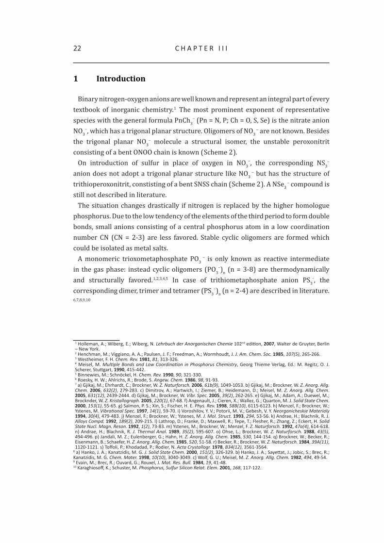
To a solution of 1 in propionitrile 4-dimethylaminopyridine was added and after storing the reaction solution at –25 °C, yellow block shaped crystals of 3 could be obtained. [(CH3)2NC5NH4PS3)][nBu4N] • EtCN (3) crystallizes in the triclinic space group P–1.
The phosphorus atom is coordinated distorted tetrahedral by three sulfur atoms and one 4-dimethylaminopyridine molecule. The three P-S bond lengths are in the range of 198 pm and therefore significantly shorter than a P-S single bond.1,15 The distance between the phosphorus atom and the nitrogen atom P1-N1 is 188.6(1) pm and therefore significantly longer than a P-N single bond but in the same range as Dimitrov et al. reported for the pyridine adducts.7c,27 The P-S-P bond angles have an average value of 115.6°. The sum of all three S-P-S angles is 347° indicating only a slight deviation from planarity. In Table 6X some selected bond parameters are summarized.
Table 6. Selected bond parameters of [(CH3)2NC5NH4PS3)][nBu4N] • EtCN (3)
Distances [pm] Bond angles [°]
S1-P1 198.3(1) S1-P1-S2 116.2(1)
S2-P1 198.6(1) S2-P1-S3 114.9(1)
S3-P1 198.4(1) S3-P1-S1 115.9(1)
N1-P1 188.6(2) S1-P1-N1 102.7(1)
S2-P1-N1 101.1(1)
S3-P1-N1 102.9(1)
27 allen, F. h.; Kennard, o.; Watson, d. G.; Brammer, L.; orpen, a. G.; taylor, r. J. Chem. Soc. Perkin Trans.II 1987, S1-S19.
43t h E t r I t h I o M E t a P h o S P h a t E a N I o N
Figure 10. Molecular structure of [(CH3)2NC5NH4PS3)][nBu4N] • EtCN (3) in the crystal. Thermal ellipsoids of all non hydrogen atoms are drawn at the 50 % probability level; hydrogen atoms are omitted for clarity.
View along c axis.
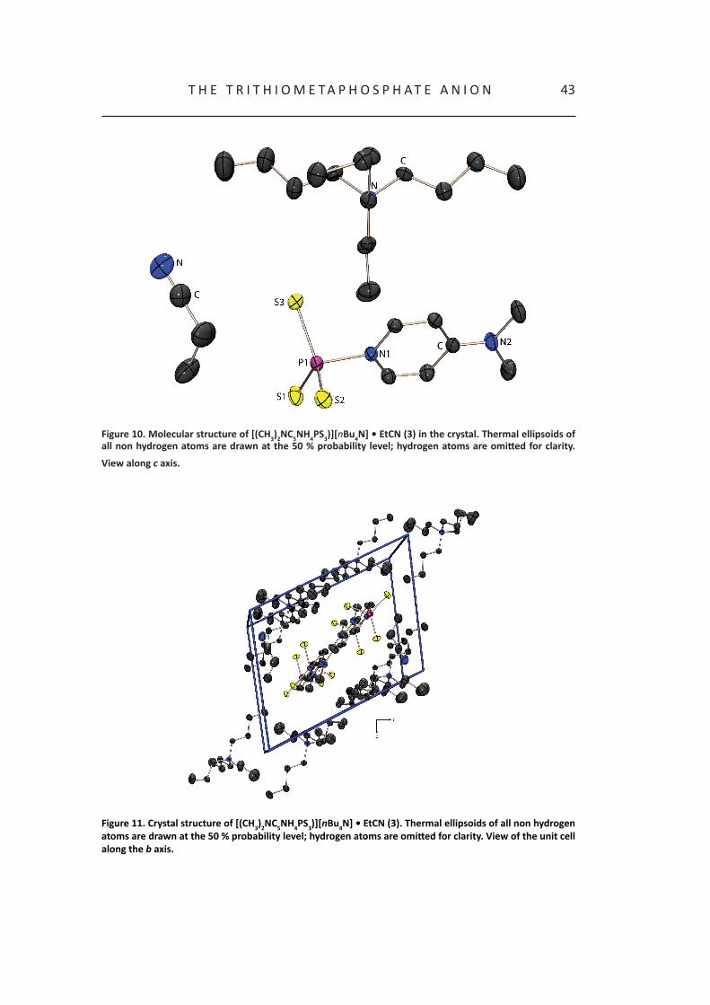
Figure 11. Crystal structure of [(CH3)2NC5NH4PS3)][nBu4N] • EtCN (3). Thermal ellipsoids of all non hydrogen atoms are drawn at the 50 % probability level; hydrogen atoms are omitted for clarity. View of the unit cell along the b axis.

44 c h a P t E r I I I
2.6 Comparison of the structures of the adduct stabilized trithiometaphosphate PS3
–
The obtained structural parameters of [(CH3)2NC5NH4PS3)][nBu4N] • EtCN (3) are compared with those reported in literature by Dimitrov et al.: [pyPS3][H2NMe2], [pyPS3][H2NEt2] and [pyPS3][pyH].7c
Table 7. Comparison of selected bond parameters of all known salts containing the adduct stabilized trithiometaphosphate anion.
3 [pyPS3][H2NMe2] [pyPS3][H2NEt2] [pyPS3][pyH]
P-S bond distances [pm]
198.3(1)
198.6(1)198.4(1)
197.5(1)198.4(1)198.6(1)
197.8(1)197.8(1)198.7(1)
198.0(1)198.6(2)200.4(1)
average value P-S distances [pm]
198.4 198.2 198.1 199.0
S-P-S bond angles [°]
116.2(1)114.9(1)115.9(1)
115.09(4)115.29(4)117.67(3)
115.69(5)116.42(2)115.69(2)
113.72(2)116.72(2)116.41(2)
average value S-P-S bond angles [°]
115.7 116.0 115.9 115.6
Σ S-P-S bond angles [°] 347 345.89 347.8 346.85
P-N distance [pm] 188.6(2) 191.5(2) 192.2(2) 190.6(2)
As shown in Table 7, neither the P-S bond lengths nor the S-P-S bond angles vary significantly in all four known salts. Furthermore the central phosphorus atom is only slightly distorted out of the plane, defined by the sulfur atoms in all cases. The P-N distance is somewhat shorter in 3 than in the pyridine stabilized adducts, probably due to the electron donating effect of the dimethylamino group. Another difference between 3 and the pyridine adducts of the trithiometaphosphate anion reported by Dimitrov et al. is the lack of hydrogen bridge bonds between the cation and the anion in 3. This is in contrast to Dimitrov et al.’s statement that hydrogen bridge bonds between cation and anion provide the essential contribution to stabilize the adduct in the solid state.7c

45t h E t r I t h I o M E t a P h o S P h a t E a N I o N
3 Conclusion
We were able to contribute new results to the chemistry of the trithiometaphosphate anion PS3
– and complete the work of Brockner, Dimitrov and Roesky by providing a new perspective on that inconspicuous but fascinating PS3
– anion. Quantum chemical calculations reveal that it is impossible to isolate the monomeric trithiometaphosphate anion PS3
– in the crystal by using conventional cations like nBu4N+ or Ph4P+. The synthesis of other promising cations, which could stabilize the monomeric trithiometaphosphate anion in the crystalline state is currently under work. Additionally, we were able to elucidate the behavior of the adduct stabilized trithiometaphosphate anion in solution. The isolation of 3 reveals that no stabilizing hydrogen bridges between anion and cation are necessary to stabilize the adduct of the trithiometaphosphate anion in the solid state.

46 c h a P t E r I I I
4 Experimental Section
General. All reactions were carried out under inert gas atmosphere using Schlenk techniques. Argon (Messer Griesheim, purity 4.6 in 50 L steel cylinder) was used as inert gas. The glass vessels used were stored in a 130 °C drying oven. Before use, the glass vessels were flame dried in vacuum at 10-3 mbar.
The sodium disulfide Na2S2 was prepared as described in literature and stored in a dry box under nitrogen atmosphere.28 Elemental sulfur was used as received (Acros Organics). P4S3 was obtained commercially (Riedel-de Häen) and was purified by extraction with CS2 before use. The solvents used were dried using commonly known methods and freshly destilled before use. Melting points were determined in capillaries using a Büchi B540 instrument and are uncorrected.
NMR Spectroscopy. NMR spectra were recorded with a Jeol EX 400 Eclipse instrument operating at 161.997 MHz (31P). Chemical shifts are referred to 85 % H3PO4 as external standard. All spectra were measured, if not mentioned otherwise, at 25 °C. The %-data correspond to the intensities in the 31P NMR spectra with respect to the total intensity. The difference to 100 % belongs to not determinable signals.
X-ray Cristallography. The molecular structures in the crystalline state were determined using an Oxford Xcalibur3 diffraction instrument equipped with a Spellman generator (voltage 50 kV, current 40 mA) and a Kappa CCD detector with a X-ray radiation wavelength of 0.71073 Å (MoΚα). The data collection was performed with the CrysAlis CCD software, the data reduction with the CrysAlis RED software.29,30 The structures were solved with SIR-92, SIR-97 and SHELXS-97 and refined with SHELXL-97 and finally checked using PLATON.31,32,33,34,35 The absorptions were corrected by SCALE3 ABSPACK multi-scan method.36 All relevant data and parameters of the X-ray measurements and refinements are given in Table 1.
28 (a) thompson, d. P.; Boudjouk, P. J. Org. Chem. 1988, 53, 2109. (b) Schuster, M. Phd thesis, LMu Munich, 1999.29 crysalis ccd, oxford diffraction Ltd., Version 1.171.27p5 beta (release 01-04-2005 crysalis171 .NEt), (compiled apr 1 2005,17:53:34).30 crysalis rEd, oxford diffraction Ltd., Version 1.171.27p5 beta (release 01-04-2005 crysalis171 .NEt), (compiled apr 1 2005,17:53:34).31 SIr-92, A Program for Crystal Structure Solution; altomare, a.; cascarano, G. L.; Giacovazzo, c.; Guagliardi, a. J. Appl. Cryst 1993, 26, 343.32 altomare, a.; Burla, M. c.; camalli, M.; cascarano, G. L.; Giacovazzo, c.; Guagliardi, a.; Moliterni, a. G. G.; Polidori, G.; Spagna, r. J. Appl. Crystallogr. 1999, 32, 115.33 Sheldrick, G. M. SHELXS-97, Program for Crystal Structure Solution; universität Göttingen, 1997.34 Sheldrick, G. M. SHELXL-97, Program for the Refinement of Crystal Structures; university of Göttingen, Germany, 1997. 35 Spek, L. a. PLATON, A Multipurpose Crystallographic Tool, utrecht university, utrecht, the Netherlands, 1999.36 ScaLE3 aBSPacK – An Oxford Diffraction program (1.0.4,gui:1.0.3) (c) 2005 oxford diffraction Ltd.

47t h E t r I t h I o M E t a P h o S P h a t E a N I o N
[nBu4N]2[P2S6]•THF (1): P4S3 (4.4 g, 20 mmol), Na2S2 (3.2 g, 40 mmol) and sulfur (3.2 g, 12.5 mmol) in 80 mL of THF were stirred at room temperature. After one day, a yellow solution was obtained. A solution of nBu4NBr (25.8 g, 80 mmol) in 20 mL of acetonitrile was added yielding a yellow solution and a white precipitate (NaBr). The NaBr was removed using a G4 frit. The filtrate was stored at –25 °C. Within 24 h, yellow crystals of [nBu4N]2[P2S6]•THF formed, which were separated using a G3 frit, washed twice with 10 mL of cold THF and dried under vacuum yielding 15.67 g (6.42 mmol, 32 % with respect to P4S3) of 1.
31P{1H} NMR in THF: δ = 30.2 (P2S62-, 36 %); 79.2 (P3S9
3-, 6 %); 105.0 (s, 3 %); 107.5 (s, 3 %); 128.8 (P2S7
2-, 7 %); 297.5 (PS3-, 38 %).
[Ph4P]2[P2S6]•py (2): P4S3 (880 mg, 4 mmol), Na2S2 (881 mg, 8 mmol) and sulfur (641 mg, 20 mmol) in 40 mL of pyridine were stirred for 24 h at room temperature yielding a yellow solution. Ph4PBr (6.708 g, 16 mmol) was added giving a white precipitate (NaBr) which was removed using a G4 frit. The orange filtrate was stored at +4 °C. After five days yellow crystals of [Ph4P]2[P2S6]•py were obtained. Yield: 2.83 g (5.2 mmol), m.p.: 173 °C (dec.).
31P{1H} NMR in pyridine: δ = 128.8 (P2S72-, 11 %); 237.9 (pyPS3
-, 83 %).
[nBu4N][(CH3)2NC5H4NPS3] (3): 0.5 g (0.64 mmol) [nBu4N]2[P2S6]•THF and 75 mg (0.62 mmol) (CH3)2NC5H4N were dissolved in 12 mL propionitrile and stirred at room temperature, yielding a yellow suspension which was refluxed for one hour (90 °C oil bath temperature). The resulting yellow solution was stored at – 25 °C. After one week, only few yellow crystals of [nBu4N][(CH3)2NC5H4NPS3] were obtained. Therefore a yield cannot be determined.
31P{1H} NMR in propionitrile: δ = 30.2 (P2S62-, 8 %); 128.8 (P2S7
2-, 7 %), 213.5 (3, 69 %), 104.9 (s, 6 %).
Reactions of PS3– with nucleophilies. To a solution of PS3
– (0.1 m) in propionitrile the corresponding nucleophile (1 eq.) was added at ambient temperature and investigated using 31P NMR spectroscopy. In case of Cl–, Br–, I–, OCN–, SCN– and CN– only PS3
– was observable.
In case of F-: 31P{1H} NMR in propionitrile: δ = 149.1 (d, 1JPF = 1.05 kHz, 16 %), 114.9 (d, 1JPF = 1.07 kHz, 2 %), 119.5 (twist-P2S8
2-, 68 %), 55.5 (chair-P2S82-, 14 %).

48 c h a P t E r I I I
Table 8. Crystal and structure refinement data.
[nBu4N]2[P2S6] • THF [Ph4P]2[P2S6] • py (2) [nBu4N][(CH3)2NC5H4NPS3] (3)
empirical formula (C16H36N)6 (C16H29N) (P2S6)4 C4o
C29H25NP2S3 C26H51N4PS3
formula mass 3000.98 545.65 546.86
temp (K) 100 100 200
cryst. size (mm) 0.4 x 0.12 x 0.08 0.3 x 0.3 x 0.25 0.3 x 0.15 x 0.1
cryst. descriptn. yellow rod yellow rod colourless block
cryst. system triclinic monoclinic triclinic
space group P-1 P21/c P-1
a (Å) 11.1073(8) 9.4174(1) 10.7269(5)
b (Å) 15.4662(11) 12.3123(1) 10.8430(4)
c (Å) 27.2591(19) 23.2297(3) 14.7239(6)
α (deg) 97.892(6) 90 98.584(3)
β (deg) 96.433(6) 96.014(1) 102.892(4)
γ (deg) 106.977(6) 90 105.500(4)
V (Å) 4378.7(6) 2678.66(5) 1568.40(13)
Z 1 4 2
ρcalc (g cm-1) 1.138 1.353 1.160
µ (mm-1) 0.409 0.416 0.308
F (000) 1628 1136 598
θ range (deg) 3.71-29.9 3.75-27.00 3.66-27.00
index ranges -14 ≤ h ≤ 14,-19 ≤ k ≤19,-34 ≤ l ≤ 34,
-12 ≤ h ≤ 12,-15 ≤ k ≤ 15,-29 ≤ l ≤ 29
-13 ≤ h ≤ 13,-13 ≤ k ≤ 13,-18 ≤ l ≤ 18
reflcns collcd 48107 28478 17264
reflcns obsd 19039 4832 4416
reflcns unique 11603 (Rint = 0.0450) 5827 (Rint = 0.0287) 6817 (Rint = 0.0345)
R1, wR2 (2σ data) 0.0428, 0.1013 0.0368, 0.0875 0.0365, 0.0810
R1, wR2 (all data) 0.0829, 0.1178 0.0483, 0.0950 0.0628, 0.0901
max/min transm 0.8483/1.0000 0.901/0.883 0.970/0.946
data/restr/params 19039/0/818 5827/0/396 6817/0/487
S on F2 0.976 1.052 0.913
larg. diff peak/hole (e/Å)
0.918/-0.815 1.203/-0.416 0.551/-0.285
remarks the hydrogen atoms of the thF molecule are missing due to the disorder within the molecule.

The Triselenometaphosphate Anion PSe3-: A Synthetic
Challenge
A new synthesis of the triselenometaphosphate anion PSe3– is presented.
Multinuclear NMR spectroscopy (31P, 77Se) is used for its characterization. The
reaction with nucleophiles like pyridine and N-methylimidazole is investigated.
Temperature dependent 31P NMR spectroscopy is used to examine the
equilibrium between the free monomeric triselenometaphosphate anion and its
base adducts, in analogy to the corresponding sulfur derivative. In the course
of the investigation, the crystal structure of the pyridine adduct of PSe3– ([pyH]
[pyPSe3] (1)), is obtained and structurally characterized by single crystal X-ray
diffraction. In addition, quantum chemical investigations are carried out to
examine the bonding situation in the triselenometaphosphate anion PSe3– in
the gas phase as well as the NMR chemical shift and the Mulliken charges. The
structure analysis of [pyH][pyPSe3] (1) is compared with the literature known
sulfur analogue [pyH][pyPS3].
chapter iv

50 c h a P t E r I V
1 Introduction
Now that the trithiometaphosphate anion PS3– and its dimer P2S6
2– have been discussed, let us have a closer look at P, Se anions comprising of a central phosphorus atom in a low coordination number CN (CN = 2, 3). In Scheme 1, the literature known monomers with a central phosphorus atom in a low coordination number (PSe2
–, PSe3–)
and the existing oligomers (P2Se62–, P3Se9
3–, P2Se82–) are shown.1,2,3,4
Scheme 1. Binary phosphorus-selenium anions.
In 1997, Brockner et al. characterized the formal dimer of the triselenometaphosphate anion P2Se6
2– by Raman spectroscopy for the first time. Nevertheless, no evidence for the existence of the monomeric triselenometaphosphate anion PSe3
– was found.1 Indications for the existence of a monomer, dimer and trimer of the triselenometaphosphate anion, (PSe3
–)n (n = 1-3), have been proofed by our group using 31P and 77Se NMR spectroscopy.3
1 Menzel, F.; Brockner, W.; Ystenes, M. Vibrational Spec. 1997, 14(1), 59.2 o’Neal, S. c.; Pennington, W. t.; Kolis, J. W. Angew. Chem. 1990, 102(12), 1502.3 (a) Karaghiosoff, K.; Schuster, M. Phosphorus, Sulfur Silicon Relat. Elem. 2001, 168, 117. (b) Schuster, M. dissertation, LMu Munich, 1999.4 (a) Zhao, J.; Pennington, W. t.; Kolis, J. W. J. Chem., Chem. Commun. 1992, 265. (b) rotter, c.; Schuster, M.; Kidik, M.; Schön, o.; Klapötke, t. M.; Karaghiosoff, K. Inorg. Chem. 2008, 632, 1663.

51t h E t r I S E L E N o M E t a P h o S P h a t E a N I o N
2 Syntheses
Scheme 2. Syntheses of PSe3– and the corresponding N-methyl imidazole and pyridine adducts (M = Li, Na).
The triselenometaphosphate anion PSe3– (2a) could be observed in all polar aprotic
solvents shown in Table 1. In case of using P4Se3, M2Se2 (M = Li, Na) as educts or starting from the elements P4, Li/Na and selenium, the ideal stoichiometry corresponds to the theoretically needed, which has to be maintained exactly (Scheme 2).3
Table 1. Ratio (in mol-%) of PSe3–, P2Se6
2– and P3Se93– depending on the used solvent. The maximum total
proportion (in mol-%) of all three anions in the reaction solution is also given.
solvent PSe3– (2a) P2Se6
2– (2b) P3Se93– (2c) total proportion
N-methyl imidazole 100 b) 0 0 100a)
HMPA 100 0 0 70a)
benzonitrile 100 0 0 19a)
dMPu 98 2 0 80a)
pyridine 90b) 0 10 35a)
THF 70 0 30 30a)
2-methyl pyridine 30 0 70 10a)
acetonitrile 0 0 100 20a)
a) the percentage ratio of the compounds is determined by using the integrals of the resonances in the 31P NMr spectra of the corresponding reaction solutions; b) as adduct

52 c h a P t E r I V
Alternatively, the triselenometaphosphate PSe3– is formed on deselenation of
[nBu4N]2[P2Se8] • 2 CH3CN using phosphanes R3P (R = Ph, C2H4CN, nBu) in DMPU or HMPA (Scheme 2).3 Therefore, two deselenation steps are required. In the first deselenation of P2Se8
2–, one endocyclic Se atom is removed, thus a fivemembered ring P2Se7
2– is formed. During the second deselenation, a second selenium atom is removed out of the endocyclic diselenide bridge, forming a fourmembered ring P2Se6
2–, which is the dimer of PSe3
–. In polar solvents like DMPU or HMPA, the equilibrium between P2Se6
2– and PSe3– is on the side of the monomeric PSe3
– (Scheme 3).3
Scheme 3. Formal reaction way of the deselenation of P2Se82– using phosphanes R3P (R = Ph, C2H4CN, nBu).
Upon dissolving [py2Li]4[P2Se6] in HMPA, surprisingly, the monomeric triselenometaphosphate anion PSe3
– can be observed in the reaction solution using 31P NMR spectroscopy. Besides P2Se6
4– HPSe32– is observable. The ratio of PSe3
–: HPSe32– is
1 : 1. The more HMPA is added, implying a dilution of the reaction solution, the higher is the observable rate of PSe3
– and HPSe32– in the 31P NMR spectrum. In a 0.5 m reaction
solution no P2Se64– is observed (Scheme 4).3
Scheme 4. Formation of PSe3– on dissolving P2Se6
4– in HMPA.

53t h E t r I S E L E N o M E t a P h o S P h a t E a N I o N
A new way of synthesizing the pyridine adduct stabilized PSe3– anion is starting from
PCl3 in pyridine with Na2Se2, which shows the high building tendency of pyPSe3– in
pyridine.The 31P NMR chemical shift of the free monomeric triselenometaphosphate PSe3
– is 212.2 ppm in HMPA (Figure 1). One pair of 77Se satellites could be observed. The 1JSeP coupling constant is –788.2 Hz which is an usual value for one coordinated 77Se atom. The ratio of the main signal to the corresponding 77Se satellite pair is 80.5:19.9 thus indicating that three equivalent selenium atoms are bonded to the central phosphorus atom. In the 77Se NMR spectrum, a doublet at 1251.9 ppm with the same 1JSeP coupling constant as in the
31P NMR spectrum (-788.2 Hz) could be observed for PSe3 – in HMPA.3
Figure 1. 31P NMR chemical shift of free monomeric PSe3– in comparison to the corresponding sulfur analogue
PS3– in HMPA. In the insert black box, the 77Se NMR spectrum of PSe3
¬ in HMPA is shown (31P NMR spectrum: 1100 scans with a PD = 0.3 s, 15 min measuring time, 0.1 m solution; 77Se NMR: 50000 scans with a PD = 0.4 s, 12 h measuring time, 0.1 m).

54 c h a P t E r I V
3 Crystal and Molecular Structure of [pyH][pyPSe3] (1)
The pyridinium salt [pyH][PSe3] (1) was obtained as block shaped orange crystals in the reaction of PCl3 with Na2Se2 in pyridine and characterized using single crystal X-ray diffraction. Compound 1 crystallizes in the monoclinic space group Cc.
The phosphorus atom is coordinated by three selenium atoms and one pyridine molecule. The P-Se bond distances have an average value of 217 pm and are between the values for a P-Se single bond and those found for the P-Se bond in phosphine selenides.5 The P1-N1 distance is 185.3(2) pm and therefore significantly longer than the distance for P-N single bond of 176 pm described in literature.6 The Se-P-Se bond angles have values of 112.4(1)° (Se1-P1-Se2), 114.8(1)° (Se2-P1-Se3) and 115.0(1)° (Se3-P1-Se1). The sum of all three bond angles is 342° which is between a perfect tetrahedral surrounding (324°) and a total planar arrangement (360°) (Table 2).
Remarkably, in contrast to the analogue PS3– salt, no hydrogen bonds could be found
between the anion and the cation. The position of the nitrogen atom in the pyH+ cation could be determined exactly, the position providing the best wR2 value and thermal parameters is shown in Figure 2.
Figure 2. Molecular structure of [pyH][pyPSe3] (1) in the solid state. Thermal ellipsoids of non hydrogen atoms are drawn at the 50 % probability level.
5 (a) cordes, a. W., Selenium; Zingaro, r. a.; cooper, W. c. (Ed.), Van Nostrand reinhold company: New York, 1974. (b) Shaw, r. a.; Woods, M.; cameron, I. S.; dahlen, B. Chem. Ind. 1971, 151. (c) Grand, a.; Martin, J.; robert, J. B.; tordjman, I. Acta Crystallogr. 1975, B31, 2523. (d) Galdecki, Z.; Glowka, M. L.; Michalski, J.; okruszek, a.; Stec, W. J. Acta Crystallogr. 1977, B33, 2322. (e) cameron, t. S.; howlett, K. d.; Miller, K. Acta Crystallogr. 1978, B34, 1639. (f) codding, P. W.; Kerr, K. a. Acta Crystallogr. 1979, B35, 1261. (g) romming, c.; Songstad, J. Acta Chem. Scand. 1979, A33, 187.6 holleman, a.; Wiberg, E.; Wiberg, N. Lehrbuch der anorganischen Chemie, 102nd ed.; Walter de Gruyter Verlag, Berlin, 2007.

55t h E t r I S E L E N o M E t a P h o S P h a t E a N I o N
Table 2. Selected bond parameters of [pyH][PSe3] (1)
Distances [pm] Bond angles [°]
Se1-P1 217.1(1) Se1-P1-Se2 112.4(1)
Se2-P1 217.6(1) Se2-P1-Se3 114.8(1)
Se3-P1 217.3(1) Se3-P1-Se1 115.0(1)
N1-P1 185.3(2) Se1-P1-N1 106.9(1)
Se2-P1-N1 104.3(1)
Se3-P1-N1 101.8(1)
Within the unit cell, the pyH+ cations are located between the pyPSe3 anions and arranged parallel to the pyridine ring of the anion. No other than electrostatic interactions between the cations and the anions can be found in the crystal.
Figure 3. Unit cell of 1. View along a axis. Thermal ellipsoids of all non hydrogen atoms are drawn at 50 % probability level.

56 c h a P t E r I V
4 Comparison of the structures of [pyH][pyPS3] and [pyH] [pyPSe3]
The obtained structural parameters of [pyH][pyPSe3] (1) are compared to those of the analogue sulfur containing salt [pyH][pyPS3] in Table 3.
Figure 4. Crystal structures of [pyH][pyPSe3] (left) and [pyH][pyPS3] (right).
Table 3. Comparison of selected bond parameters of the pyridine adduct stabilized trichalcogenometaphosphate anion [pyH][pyPCh3] (Ch = S or Se).
Ch = S or Se 1 [pyH][pyPS3]
P-ch bond distances [pm]217.1(1)217.6(1)217.3(1)
198.0(1)198.6(2)200.4(1)
average value P-ch distances [pm] 217 199.0
ch-P-ch bond angles [°]112.4(1)114.8(1)115.0(1)
113.72(2)116.72(2)116.41(2)
average value ch-P-ch bond angles [°] 114.1 115.6
Σ ch-P-ch bond angles [°] 342 347
P-N distance [pm] 185.3(2) 190.6(2)
In both cases, the P-Ch bond lengths have an average value, which is in between the values found for P-Ch single bonds and P=Ch double bonds.5 The Ch-P-Ch bond angles observed in 1 are only slightly smaller than those found in the corresponding sulfur analogue resulting in a slightly bigger deviation from planarity of about 5° in comparison to the sulfur analogue. The most interesting point is the big difference of the P-N distance between 1 (185.3 pm) and [pyH][pyPS3] (190.6 pm) of 5 pm.
According to the quantum chemical calculations presented in the previous chapter, equivalent calculations were carried out for the triselenometaphosphate anion PSe3
–, in order to elucidate the structure, bonding, energetic situation and NMR chemical shifts of the monomeric triselenometaphosphate PSe3
– in the gas phase, where solid states effects are not considered.

57t h E t r I S E L E N o M E t a P h o S P h a t E a N I o N
5 Quantum chemical calculation
The calculations were carried out with the Gaussian 03 program code using different basis sets (pseudo potential ECP28MWB/VTZ7, 6-31+G**, aug-cc-pVTZ and aug-cc-pVQZ).8
Table 4. Calculated bond lengths [pm] and angles [°] for PSe3 – in the gas phase.
pseudo potential ECP28MWB/VTZ
6-31+G** aug-cc-pVTZ aug-cc-pVQZ
d (P-Se) / pm 220.0 210.4 210.8 210.3
< P-Se-P / ° 120 120 120 120
point group D3h D3h D3h D3h
The geometry optimization of PSe3– yields D3h symmetry for this anion in all cases,
which is in good agreement with the symmetry determined in quantum chemical calculations for the corresponding PS3
–. Also the calculated Se-P-Se bond angles of 120° perfectly confirm the D3h symmetry of the triselenometaphosphate anion PSe3
– in the gas phase. In case of the calculated bond distances, the all-electron calculations (6-31+G**, aug-cc-pVTZ and aug-pVQZ) give the same value of 210 pm for the P-Se bond, while the pseudo potential ECP28MWB/VTZ calculation gives a value of 220 pm. As the pseudo potential calculation considers relativistic effects, it gives the more reliable value for P-Se bonds (Table 4).
Table 5. Mulliken charges for PSe3– and PS3
–.
PSe3– PS3
–
P Se P S
pseudo potential EcP28MWB/VtZ - - 0.631 -0.544
6-31+G** 0.409 -0.470 -0.015 -0.328
aug-cc-pVtZ 0.308 -0.436 0.631 -0.544
aug-cc-pVQZ -0.014 -0.329 0.709 -0.570
7 http://www.theochem.uni-stuttgart.de/pseudopotentials/index.en.html (14. Marz 2009).8 Gaussian 03, revision d.01, Frisch, M. J.; trucks, G. W.; Schlegel, h. B.; Scuseria, G. E.; robb, M. a.; cheeseman, J. r.;
Montgomery, Jr., J. a.; Vreven, t.; Kudin, K. N.; Burant, J. c.; Millam, J. M.; Iyengar, S. S.; tomasi, J.; Barone, V.; Mennucci, B.; cossi, M.; Scalmani, G.; rega, N.; Petersson, G. a.; Nakatsuji, h.; hada, M.; Ehara, M.; toyota, K.; Fukuda, r.; hasegawa, J.; Ishida, M.; Nakajima, t.; honda, Y.; Kitao, o.; Nakai, h.; Klene, M.; Li, X.; Knox, J. E.; hratchian, h. P.; cross, J. B.; Bakken, V.; adamo, c.; Jaramillo, J.; Gomperts, r.; Stratmann, r. E.; Yazyev, o.; austin, a. J.; cammi, r.; Pomelli, c.; ochterski, J. W.; ayala, P. Y.; Morokuma, K.; Voth, G. a.; Salvador, P.; dannenberg, J. J.; Zakrzewski, V. G.; dapprich, S.; daniels, a. d.; Strain, M. c.; Farkas, o.; Malick, d. K.; rabuck, a. d.; raghavachari, K.; Foresman, J. B.; ortiz, J. V.; cui, Q.; Baboul, a. G.; clifford, S.; cioslowski, J.; Stefanov, B. B.; Liu, G.; Liashenko, a.; Piskorz, P.; Komaromi, I.; Martin, r. L.; Fox, d. J.; Keith, t.; al-Laham, M. a.; Peng, c. Y.; Nanayakkara, a.; challacombe, P,; Gill, M. W.; Johnson, B.; chen, W.; Wong, M. W.; Gonzalez, c.; Pople, J. a.; Gaussian, Inc., Wallingford ct, 2004.

58 c h a P t E r I V
5.1 Valence bond structures
The VB program package VB2000, version 1.8 was used for all VB calculations employing STO-3G basis set for PSe3
– and additionally the D95 basis set for PS3–.9 VB2000
is an ab initio electronic structure package for performing modern VB calculations; it is based on a highly efficient VB algorithm – the so called algebrant algorithm – and the group function (GF) approach, in which a large molecule is described in terms of its constituent parts physically identifiable “electron groups”. A major feature of VB2000 is the implementation of modern VB theory at the ab initio level using the algebrant alogarithm.9, 10,11,12,13,14,15,16,17,18,19
The VB method used in this study was the CASVB(6,4) one (complete active space) with six VB electrons (π electrons) spread over four π orbitals. The CASVB method implemented in the VB2000 program package generates the weights of all resonance structures. In VB2000, each “structure” becomes a spin-coupling scheme, involving pairs of electrons occupying rather localized, strongly overlapping orbitals. The overlapping orbitals of any bond are expected to bear some similarity to the AOs used in constructing a localized MO. In particular, the π-orbitals used in this study are almost one-center AOs with only a very small degree of “tailing”, i.e. spreading onto other atomic centers.
Figure 4 shows all possible VB structures for the PSe3– anion, which were generated
from a CASVB(6,4) calculation with the six π electrons forming the active VB space (all σ and core electrons were treated with the HF method). As expected, the Hiberty weights of the standard Lewis structures of type I are the individually most important contributors to the resonance scheme for PSe3
– (Table 6) in comparison to the already presented ones for PS3
–. It is also interesting to point out that the Dewar-type (or long bond) structures (II) together with the sextet structure III are important resonance structures for both anions (PS3
– and PSe3–) whereas the structures with one chalcogen
atom carrying a positive charge (IV) is much less important (Table 6).
9 Li, J.; McWeeny, r. VB2000, version 1.7, SciNet technologies: San diego, ca, (November 2005).10 Li, J; Pauncz, r. Int. J. Quantum. Chem. 1997, 62, 245.11 Li, J. McWeeny, r. Int. J. Quantum. Chem. 2002, 89, 208.12 McWeeny, r. Adv. Quant. Chem. 1999, 31, 15.13 McWeeny, r. Proc. R. Soc. Lond. 1959, Ser A253, 242.14 angyan, J. a. Theor. Chem. Acc. 2000, 103, 238.15 McWeeny, r.; Sutcliffe, B. t. Proc. R. Soc. Lond. 1963, Ser. A 273, 103.16 Gallup, G. a.; Norbeck, J. M. Chem. Phys. Lett. 1973, 21, 495.17 hiberty, P. c.; ohanessian, G. Int. J. Quantum. Chem. 1985, 27, 245.18 Levine, I. N. in Quantum Chemistry, vol. I, sect. 15.10, allyn and Bacon: Boston, 1970, p 550.19 Bachler, V. Theor. Chem. Acc. 1997, 92, 223.

59t h E t r I S E L E N o M E t a P h o S P h a t E a N I o N
Figure 5. VB structures for PSe3
– generated from a CASVB(6,4) calculation with the six π electrons forming the active VB space (all σ and core electrons were treated with the HF method).
Table 6. Hiberty structural weights of the individual Lewis structures for PS3 – and PSe3
– depicted in Figure 5, calculated at CASVB(6,4)/STO-3G level of theory.16,17,18,19a,20
Structure I II III IV
hiberty weight for PS3– 3 x 0.21 3 x 0.09 0.10 3 x 0.003
hiberty weight for PSe3– 3 x 0.22 3 x 0.07 0.12 3 x 0.003
20 the hiberty weights Wi are defined as follows: Wi = Ci2 / Σ Ci
2. (Where Ci are the coefficients of the wavefunctions for VB structures i).

60 c h a P t E r I V
For the PS3– anion, also a CASVB calculation using the double-zeta D95 basis in order
to account for the relatively poor basis set used above were carried out. The results show (Table 7) that the Hiberty weights for the resonance structures of PS3
– remain essentially unchanged which gives credence that also for PSe3
– (for which there is no D95 basis implemented in the VB2000 code) the STO-3G calculated weights are reliable.
Table 7. Hiberty structural weights of the individual Lewis structures for PS3– depicted in Fig. 1, calculated
at CASVB(6,4)/D95 level of theory. 16,17,18,19a,21
Structure I II III IV
hiberty weight 3 x 0.22 3 x 0.07 0.12 3 x 0.001
Since the above calculations for PS3– at the CASVB(6,4) level of theory did not indicate
any relevance for structures containing “hypervalent” phosphorus, a VB(6)/3-21G(d) calculation, including explicitly (in addition to the ten structures shown in Figure 5) six further VB structures (using dz2 orbitals at the P center) with two double bonds between P and S and one single bond between P an S– was carried out. It turned out, as already expected from the CASVB calculation, that the three structures with two double bonds each carry a negligible (Hiberty) weight (0.0002).
The calculated 77Se NMR chemical shifts represent the observed 77Se NMR chemical shift for the PSe3
– anion quite well. To provide the exactitude of the NMR calculations, the 77Se NMR chemical shifts of the used reference Me2Se was also calculated. In Table 8, the calculated isotropic shielding of Me2Se and PSe3
– as well as the relative 77Se NMR chemical shifts are summarized and compared to the experimentally observed 77Se NMR chemical shifts.21
Table 8. Computed isotropic magnetic shieldings and relative 77Se NMR chemical shifts (ppm) referenced to Me2Se.
calc. isostropic shieldingrel. shift
[ppm]exptl. reported
chem. shift [ppm]
Me2Se 1833.9 0 0
PSe3– 537.3 1296.6 1251.9
21 31P NMr chemical shifts were also calculated, but differ significantly from the experimental determined ones (Δδ = 200).

61t h E t r I S E L E N o M E t a P h o S P h a t E a N I o N
6 Conclusion
The X-ray crystallographic study on [pyH][pyPSe3] demonstrates for the first time the existence of the PSe3
– anion in the solid state. In addition, quantum chemical cal-culations were carried out to elucidate the bonding situation, NMR chemical shift and Mulliken charges in the free monomeric triselenometaphosphate PSe3
–.

62 c h a P t E r I V
7 Experimental Section
General. All reactions were carried out under inert gas atmosphere using Schlenk techniques. Argon (Messer Griesheim, purity 4.6 in 50 L steel cylinder) was used as inert gas. The glass vessels used were stored in a 130 °C drying oven and were flame dried in vacuum at 10-3 mbar before use.
Na2Se2 was prepared as described in the literature and stored in a dry box under nitrogen atmosphere.22 Pyridine was dried following known methods. PCl3 (Aldrich) was distilled immediately before use.
NMR Spectroscopy. NMR spectra were recorded using a Jeol EX 400 Eclipse instrument operating at 161.997 MHz (31P) and 76.321 MHz (77Se). Chemical shifts are referred to 85 % H3PO4 and (CH3)2Se (77Se) as external standards. All spectra were measured, if not mentioned otherwise, at 25 °C. The %-data correspond to the intensities in the 31P NMR spectra with respect to the total intensity. The difference to 100 % belongs to not determinable signals.
X-ray Cristallography. The molecular structure of [pyH][pyPSe3] in the crystalline state was determined using an Oxford Xcalibur3 diffraction instrument equipped with a Spellman generator (voltage 50 kV, current 40 mA) and a Kappa CCD detector with a X-ray radiation wavelength of 0.71073 Å (MoΚα). The data collection was performed with the CrysAlis CCD software, the data reduction with the CrysAlis RED software.23,24 The structure was solved with SIR-92 and SHELXS-97 and refined with SHELXL-97 and finally checked using PLATON.25,26,27,28,29 The absorptions were corrected by the SCALE3 ABSPACK multi-scan method.30 All relevant data and parameters of the X-ray measurement and refinement are given in Table 9.
22 (a) thompson, d. P.; Boudjouk, P. J. Org. Chem. 1988, 53, 2109. (b) Schuster, M. dissertation, LMu Munich, 1999.23 crysalis ccd, oxford diffraction Ltd., Version 1.171.27p5 beta (release 01-04-2005 crysalis171 .NEt), (compiled apr 1 2005,17:53:34).24 crysalis rEd, oxford diffraction Ltd., Version 1.171.27p5 beta (release 01-04-2005 crysalis171 .NEt), (compiled apr 1
2005,17:53:34).25 SIr-92, 1993, A Program for Crystal Structure Solution; altomare, a.; cascarano, G. L.; Giacovazzo, c.; Guagliardi, a. J. Appl. Cryst. 26, 343.26 altomare, a.; Burla, M. c.; camalli, M.; cascarano, G. L.; Giacovazzo, c.; Guagliardi, a.; Moliterni, a. G. G.; Polidor, G.; Spagna, r. J. Appl. Crystallogr. 1999, 32, 115.27 Sheldrick, G. M. SHELXS-97, Program for Crystal Structure Solution; universität Göttingen, 1997.28 Sheldrick, G. M. SHELXL-97, Program for the Refinement of Crystal Structures; university of Göttingen, Germany, 1997. 29 Spek, L. a. PLATON, A Multipurpose Crystallographic Tool, utrecht university, utrecht, the Netherlands, 1999.30 ScaLE3 aBSPacK – An Oxford Diffraction program (1.0.4,gui:1.0.3) (c) 2005 oxford diffraction Ltd.

63t h E t r I S E L E N o M E t a P h o S P h a t E a N I o N
[pyH][pyPSe3] (1). Na2Se2 (408 mg, 2 mmol) was suspended in 2 mL of pyridine. PCl3 (161µL, 2 mmol) was slowly added during two minutes. The reaction was strongly exothermic yielding a brownish red solution. After one hour at ambient temperature, the reaction mixture was refluxed for two hours. The red brown suspension was centrifuged to remove the red precipitate. The clear orange brown solution was transferred into a NMR tube. After three months, orange crystals of [pyH][pyPSe3] were formed and characterized using X-ray crystallography.
31P{1H} NMR of the reaction solution in pyridine: δ = 206.0 (PCl3, 22 %), 200.6 (pyPSe3, 3%); 94.1 (py2PSe2
+, 1JSeP = 817 Hz, 3 %); 44.3 (pyPSe2Cl, 1JSeP = 891 Hz, 36 %); 30.5 (s, 1JSeP = 971 Hz, ratio of 77Se satellites: 6% ≈ 1 Se atom , 8 %); – 11.9 (s, 1JSeP = 816 Hz, ratio of 77Se satellites: 14.5% ≈ 2 Se atoms, 28 %) ppm.
77Se{1H} NMR of the reaction solution in pyridine: δ = 701.4 (d, 1JSeP = 816 Hz); 388.4 (pyPSe2Cl); 66.3 (d, 1JSeP = 972 Hz).

64 c h a P t E r I V
Table 9. Crystal data and structure refinement of 1.
[pyh][pyPSe3] (1)
empirical formula C10H11N2PSe3
formula mass 427.06
temp (K) 200
cryst. size (mm) 0.15 x 0.15 x 0.05
cryst. descriptn red plate
cryst. system monoclinic
space group Cc
a (Å) 7.7491(7)
b (Å) 13.0902(13)
c (Å) 13.5632(11)
α (deg) 90
β (deg) 94.479(8)
γ (deg) 90
V (Å) 1371.6(2)
Z 4
ρcalc (g cm-3) 2.068
µ (mm-1) 8.137
F (000) 808
θ range (deg) 4.1327-29.9897
index ranges -9≤ h ≤ 9,-16 ≤ k ≤ 16,-16 ≤ l ≤ 16
reflcns collcd 6811
reflcns obsd 1880
reflcns unique 2688 (Rint = 0.0569)
R1, wR2 (2σ data) 0.0358, 0.0643
R1, wR2 (all data) 0.0550, 0.0670
max/min transm 0.307/0.666
data/restr/params 2688/2/132
S on F2 0.802
larg. diff peak/hole (e/Å) 0.601/-0.370

On the Existence of Dichalcogenophosphoryl Monohalogenides PCh2X (Ch = S, Se, Te; X = Cl, Br, I)
The existence of dichalcogenophosphoryl monohalogenides PCh2X (Ch = S, Se,
Te; X = Cl, Br, I) is investigated. The new syntheses strategy occurred starting from
phosphorus trihalogenide (PCl3, PBr3, PI3); and sodium dichalcogenide (Na2S2,
Na2Se2, Na2Te2) in polar aprotic solvents. In addition, the new selenium containing
pyridine adducts of PSe2Cl and PSe2Br can be observed and characterized for the
first time using solution NMR spectroscopy.
Chapter V

66 c h a P t E r V
1 The Problem with σ3λ5 Phosphorus
Among the nitrylmonohalogenides and phosphorylmonohalogenides shown in Scheme 1, only the NO2X (X = F, Cl, Br) are described in literature. The dioxonitrylhalogenides NO2X are gases under normal conditions (25 °C, 1 bar) and, besides NO2F, endothermic compounds.1 Comparing the oxonirtylhalogenid to the isoelectronic nitrate anion NO3
–, the substitution of an oxygen atom through a halogen atom, fluorine, chlorine or bromine, obviously leads to the destabilization of the molecule NO2X, probably due to the loss of Y aromaticity.
Scheme 1. Lewis formula of NO2X, NS2X, PO2X, PS2X and PSe2X (X = F, Cl, Br, I).
But what is about the dichalcogenylphosphoryl monohalogenides PCh2X?The dichalcogenophosphoryl monohalogenides PCh2X are metaphosphate
derivatives, where the central phosphorus atom has a very unusual bonding situation, as it is planar with the formal oxidation state +V, but has an unsaturated coordination sphere as it is only tricoordinated. So a strong Lewis acidity is to be expected.
The way out of that uncomfortable situation is dimerisation (Scheme 2). Großmann observed a monomer-dimer equilibrium for perthiophosphonic acid anhydrides in solution. These compounds have an organic residue R like a phenyl or a methyl group.2
Scheme 2: Dimerisation of metaphosphate derivatives.
1 holleman, a.; Wiberg, E.; Wiberg, N. Lehrbuch der anorganischen Chemie, 102nd ed.; Walter de Gruyter Verlag, Berlin, 2007.2 Beckmann, h.; Großmann, G.; ohms, G. Heteroatom Chemistry, 1994, 5(1), 73-83.

67d I c h a L c o G E N o P h o S P h o r Y L M o N o h a L o G E N I d E S
Till now there are only two possibilities known to stabilize the monomeric form:One is described by Yoshifuji et al. where R is a bulky substituent like supermesityl
(Scheme 3).3,4
Scheme 3. Dithioxophosphorane.
The other possibility is if R is a negatively charged group like S– or Se–. In the first case the monomeric form is favored due to sterical reasons, while in the second case electronical reasons are responsible for the stabilization of the monomeric form as two anions have to get together during the dimerisation and due to Coulomb interactions this is a disadvantaged process.
So the question was what happens if the negatively charged chalcogen Ch– is substituted by a neutral group. And as the halogens are isoelectronical to negatively charged chalcogen anions Ch– investigations were made to examine the exchange of chalcogen against halogen atoms.
There are no reports in literature about PS2X (as monomer or oligomer), however there are reports about corresponding pyridine adducts pyPS2X (Scheme 4).
Scheme 4: Pyridine adduct stabilized dichalcogenphosphoryl monohalogenides pyPCh2X.
The access to PS2X chemistry is represented by pyPS2Cl, which is easily prepare able starting from P4S10 and PSCl3 in pyridine. Starting from pyPS2Cl the fluorine derivative can be synthesized, while the bromine analogue is made starting from pyPS2(NMe2) and PBr3. In solution an equilibrium between the cation py2PS2
+ X– and donorstabilized dithiometaphosphoric acid halides pyPS2X can be observed (Scheme 5).5,6,7,8
3 Yoshifuji, M.; toyota, K.; ando, K.; Inamoto, N. Chem. Lett. 1984, 317-318.4 Yoshifuji, M.; Shibayama, K.; Inamoto, N. Chem. Lett. 1984, 603-606.5 Meisel, M.; Grunze, h. Z. anorg. allg. Chem. 1968, 360, 277-285.6 Fluck, E.; retuert, P. J.; Binder, h. Z. anorg. allg. Chem. 1973, 397, 225-320.7 costisella, B.; Schulz, J.; teichmann, h.; donath, c.; Meisel, M. Phosphorus, Sulfur Silicon Relat. Elem. 1990, 53, 367-371.8 Meisel, M.; Lönnecke, P.; Grimmer, a.-r.; Molder-Wulff, d. Angew. Chem. Int. Ed. 1997, 36(17), 1869-1870.

68 c h a P t E r V
Scheme 5. Syntheses of literature known pyPS2X (X = F, Cl, Br, I).
As P4S10 is used as educt, no selenium containing compound is described, as the required starting material P4Se10 does not exists. In Table 1 all analytical data of the literature known compounds is summarized.
Table 1. Analytical data of all literature known pyPS2X (X = F, Cl, Br, I).
Ch X NMR data X-Ray
S F
δ31P = 112 [6] / 117.5 [7] (in CHCl3)δ19F = 76.5 [6] 1JPF = 1144 hz [6] / 1166 hz [7] (in CHCl3)1JPN = 20.6 hz [7]
S Clδ31P = 138 [5] / 97.5 [7] (in CHCl3)1JPN = 24.1 hz [7] [4,9]
S Brδ31P = 65.5 (in ch3cN) [6] 1JPN = 32.6 hz [8]
S Iδ31P = – 0.4 [8] 1JPN = 36.7 hz [8]
Therefore the aims of the investigations were:
• Analysis of the bonding situation in pyPS2X
• Exceeding the investigation to the corresponding selenium compounds

69d I c h a L c o G E N o P h o S P h o r Y L M o N o h a L o G E N I d E S
2 New route to pyPCh2X (Ch = S, Se, Te; X = Cl, Br, I)The easiest and straight forward way to synthesize compounds pyPCh2X should
start from PX3 and Na2Ch2 in pyridine as reaction medium.
Scheme 6: New synthesis of pyPCh2X.
The advantages of this reaction way are obvious:• Possibility to introduce iodine (PI3), selenium (Na2Se2) or tellurium (Na2Te2)• Formation of only NaX as by-product is expected, so purification should be
easily made
3 Reactions of PX3 with Na2S2 (X = Cl, Br, I)
3.1 Reaction of PCl3 and Na2S2
In the 31P NMR spectrum of reaction solution of PCl3 with Na2S2 in the stoichiometric ratio of 1:1 PCl3 can still be observed in quite huge quantities (39 % of the total 31P intensity). Nevertheless, the 31P NMR spectrum reveals that the compound pyPS2Cl is also formed, shows the characteristic 31P NMR chemical shift of 95.2 ppm (in pyridine; 53 % of the total 31P intensity). This is in accord with the 31P chemical shift reported of 97.5 ppm (in CHCl3).7 The small difference can be attributed to the different solvents used. In addition, some colourless crystals were isolated after storing the reaction solution at – 20 °C for six weeks. The determined cell parameters correspond to those reported in literature for pyPS2Cl.9
Based on the experience with the triseleno – and trithiometaphosphate anion, an equilibrium between the pyridine adduct (pyPS2Cl) and free PS2Cl (and pyridine) might be anticipated (Scheme 7).
Scheme 7: Possible equilibrium between the adduct pyPS2Cl and the free species PS2Cl.
9 averbuch-Pouchot, M.-t.; Meisel, M. Acta Crystallogr., Sect. C: Cryst. Struct. Commun. 1989, C45, 1937-1939.

70 c h a P t E r V
However temperature dependent 31P NMR spectroscopy in the temperature range between –40 °C to 100 °C revealed that this equilibrium is completely on the side of the adduct pyPS2Cl. Thus, PS2Cl seems to be less stable than the isoelectronic analogous species PS3
–.As PCl3 was still present in the reaction mixture the amount of Na2S2 was increased
up to a ratio of 1:3. At this stoichiometry no PCl3 is observed in the 31P NMR spectrum anymore. The main product is still pyPS2Cl (75 % of the total intensity). From the reaction solution at this stoichiometry crystals of py2P2S7 were obtained.
3.2 Reaction of PBr3 and Na2S2
As previously described for the reaction of PBr3 with Na2S2 the first stoichiometry investigated was PBr3 and Na2S2 of 1:1. The 31P NMR spectrum of the reaction mixture reveals the presence of pyPS2Br (37 % of the total 31P intensity) which can thus be obtained using PBr3 and Na2S2 as educts. However PBr3 is also still present in the reaction mixture (δ31P = 220; 16 % of the total 31P intensity). In addition to PBr3 and pyPS2Br also the signal caused by py2PS2
+ could be identified at δ31P = 104.7 (< 4 % of the total 31P intensity). This cation was isolated in the crystalline state as [py2PS2]Br salt and identified by its cell parameters using single crystal X-ray diffraction.10
The 31P NMR spectrum of the reaction solution displays two new signals (at 135.8 ppm and 298 ppm) the origin of which remains at the moment mysterious. Temperature dependent 31P NMR spectroscopy in the range between – 40 °C and 0 °C reveals that the signals at δ31P = 298 and 135.8 are both shifted to higher field with decreasing temperature (Δδ ≈ 23 ppm) and the same is true for the 31P NMR signal of PBr3. Interestingly the difference between the signals is always the same (Δδ = 78 ppm). Particularly interesting is the signal at δ31P = 298. This corresponds to the signal observed for the PS3
– anion. The presence of PS3– under the given reaction
conditions seems very unlikely, however. The 31P shift observed for PS3– in pyridine is
220 ppm, due to the equilibrium with pyPS3-. In addition, the coexistence of PS3
– and PBr3 (a base and an acid) seems improbable. For monomeric PS2Br a low field 31P NMR signal would be expected. Further investigations are needed, however, in order to clarify this point.
Increasing the amount of Na2S2 (ratio 1:3) leads to the formation of only sulfur containing compounds like P2S8
2–, P5S105– and P2S7
2–.10 Meisel, M.; Lönnecke, P.; Grimmer, a.-r.; Wulff-Molder, d. Angew. Chem. Int. Ed. Engl. 1997, 36(17), 1869-1870.

71d I c h a L c o G E N o P h o S P h o r Y L M o N o h a L o G E N I d E S
3.3 Reaction of PI3 and Na2S2
PI3 and Na2S2 were reacted in pyridine at ambient temperature in the molar ratios 1:1 and 1:3. According to the 31P NMR spectrum of the reaction solution formation of dithiophosphoryl monoiodide pyridine adduct pyPS2I does not occur. Instead signals at δ31 = 104.7, 119.2 and 54.5 were observed, which can tentatively be assigned to the cation py2PS2
+ and the anion P2S82–, respectively. This result might be interpreted
considering the weak P-I bond, compared to the P-Br and P-Cl bond. Assuming the intermediate formation of pyPS2I a nucleophilic attack of a second pyridine molecule would be favoured by the weak P-I bond, resulting in the formation of pyPS2
+ cation.
4 Reaction of PX3 with Na2Se2 (X = Cl, Br, I)
4.1 Reaction of PCl3 with Na2Se2
Reaction of equimolar amounts of PCl3and Na2Se2 in pyridine at ambient temperature yields a clear red solution. The 31P NMR spectrum shows the presence of a considerable amount of unreacted PCl3 (22 % of the total 31P intensity). Main product is the pyridine adduct of PSe2Cl, pyPSe2Cl, which is observed and characterized here for the first time. The identity of pyPSe2Cl results unambiguously from the pattern of the 31P NMR signal and is further supported by the 14N and 77Se NMR spectra. The 31P NMR signal (Figure 1) consists of a (1:1:1) triplet and a pair of 77Se satellites. The triplet is caused by scalar coupling of 31P with 14N (I = 1) and proofs the coordination of pyridine to phosphorus in pyPse2Cl. 1JPN (44 Hz) is typical for a one bond 31P, 14N coupling. The relative intensity of the 77Se satellites (12.7 %) corresponds to two selenium atoms bonded to phosphorus. 1JSeP = 892 Hz is in the same range as found for phosphorus selenides (500 – 1200 Hz).11 In the 77Se NMR spectrum a doublet at 388.4 ppm is observed, in a range typical for phosphorus selenides. The P coordinated nitrogen atom gives a signal at – 132.5 ppm.
11 Karaghiosoff, K. in: Encyclopedia of Nuclear Magnetic resonance, Vol. 6 Grant, M.; harris, r. K. Eds.: Wiley, chichester, 1996, pp. 3612-3619.

72 c h a P t E r V
Figure 1. Observed 31P{1H} NMR spectrum of pyPSe2Cl in pyridine at 25°C (0.1 m, 1023 scans with a PD = 0.5 sec, measuring time 28 min, ν0 = 161.9966 MHz, 1H broadband decoupling).
In addition to the signals of PCl3 and pyPSe2Cl three other signals with one pair of 77Se satellites each can be observed in the 31P NMR spectrum: 94.1, 30.5 and – 11.9 ppm. The signal at 94.1 ppm (2.5 % of the total intensity) can be attributed to the new cation py2PSe2
+. It is formed in much better yield when PBr3 is used instead of PCl3 (see below).
From the reaction solution red crystals of [pyH][pyPSe3] were isolated at ambient temperature and were investigated by single crystal X-ray diffraction.
As there is still PCl3 in the reaction mixture, the amount of Na2Se2 has to be increased up to an educt ratio of 1:3 (PCl3 : Na2Se2). pyPSe2Cl could be identified as main product according to the 31P NMR spectrum of the reaction mixture (58 % of the total intensity). The signal at δ31P = 94.1 ppm disappeared completely whereas the total intensity of the signal at δ31P = 30.5 ppm increased up to 27 % and that of the singlet at δ31P = –11.9 ppm decreased (14 %).

73d I c h a L c o G E N o P h o S P h o r Y L M o N o h a L o G E N I d E S
4.2 Reaction of PBr3 with Na2Se2
Addition of PBr3 to an equimolar amount of Na2Se2 in pyridine results in the formation of a clear red solution. The 31P NMR spectrum shows only two signals at 94.1 ppm and 4.5 ppm, each accompanied by a pair of 77Se satellites. Main product is pyPSe2Br (63 % of the total 31P intensity), the pyridine adduct of PSe2Br, described and characterized for the very first time. In analogy to the previously described chlorine compound pyPSe2Cl, the 31P NMR signal of pyPSe2Br consists of a 1:1:1 triplet (1JPN = 36 Hz) and one pair of 77Se satellites (1JSeP = 851 Hz) with a relative intensity of 13 %, indicating the existence of two selenium atoms bonded to the central phosphorus atom (Figure 2).
Figure 2. Observed 31P{1H} NMR spectrum of pyPSe2Br in pyridine at 25°C (0.1 M, 4096 scans with a PD = 0.5 sec, measuring time 97 min, ν0 = 161.8347 MHz, 1H broadband decoupling).
The singlet at 94.1 ppm, which can also be observed in the reaction of PCl3 and Na2Se2, is accompanied by one pair of 77Se satellites with a 1JSeP (817 Hz) corresponding to single coordinated selenium atoms, also found in phosphorus selenides.11 The relative intensity of the 77Se satellites (16 %) indicates the presence of two selenium nuclei bonded to the phosphorus atom. The signal can be assigned to the bispyridine adduct stabilized PSe2
+ cation, py2PSe2+.

74 c h a P t E r V
4.3 Reaction of PI3 with Na2Se2
In analogy to the reaction of PI3 and Na2S2, only selenium containing compounds like the P2Se8
2– anion can be observed in the 31P NMR spectrum. So this synthesis starting from PI3 is useless so prepare the iodine containing compound pyPSe2I.
4.4 Reaction of PI3 with Na2Te2
The formation of pyPTe2I cannot be observed in the 31P NMR spectrum. Instead the signal of only PI3 can be identified.

75d I c h a L c o G E N o P h o S P h o r Y L M o N o h a L o G E N I d E S
5 Conclusion
PCh2X (Ch = S, Se; X = Cl, Br) can be readily generated by the reaction of PX3 with Na2Ch2 and effectively stabilized as pyridine adducts in solution. The new selenium derivatives pyPSe2Cl, pyPSe2Br and py2PSe2
+ are prepared and characterized by NMR spectroscopy for the first time. It could be demonstrated that using PI3 as educt for the syntheses of the corresponding iodine compounds is not suitable as the weak P-I bonds are completely broken by sulfur and selenium atoms. In case of tellurium it has to be recognized that this is too unreactive to break the P-I bond.
It could also be shown that there is no equilibrium between the adduct stabilized dichalcogenophosphoryl monohalogenides pyPCh2X and the free monomeric compounds PCh2X.
In addition two new compounds can be obtained in the solid state and characterized using X-ray diffraction. They are discussed in separate chapters: [pyH][pyPSe3] and py2P2S7.
Table 2. Summary of analytical data of the observed compounds.
NMR data X-Ray
pyPS2Cl δ31P = 95.2 (in pyridine)
pyPS2Br δ31P = 65.5 (in pyridine)
pyPS2I not prepareable
pyPSe2Cl δ31P = 44.3 (in pyridine)δ77Se = 388.4δ14N = – 132.51JSeP = 892 hz
1JPN = 44 hz
pyPSe2Br δ31P = 4.51JSeP = 851 hz
1JPN = 36 hz
pyPSe2I not prepareable
pyPte2I not prepareable
py2PS2+ δ31P = 104.7 (in pyridine)
py2PSe2+ δ31P = 94.1
1JSeP = 817 hz

76 c h a P t E r V
6 Experimental section
General. All reactions were carried out under inert gas atmosphere using Schlenk techniques. Argon (Messer Griesheim, purity 4.6 in 50 L steel cylinder) was used as inert gas. The glass vessels used were stored in a 130 °C drying oven. Before use the glass vessels were flame dried in vacuum at 10-3 mbar.
The sodium dichalcogenides were prepared as described in the literature and stored in a dry box under nitrogen atmosphere.12 Pyridine (Aldrich) was dried using commonly known methods. PCl3 (Aldrich) was distilled before use. PBr3 (Aldrich) was used as received.
NMR Spectroscopy. NMR spectra were recorded using a Jeol EX 400 Eclipse instrument operating at 161.997 MHz (31P), 76.321 MHz (77Se) and 28.918 MHz (14N) if not mentioned otherwise. Chemical shifts are referred to 85 % H3PO4, (CH3)2Se (77Se) and CH3NO2 (14N) as external standards. All spectra were measured, if not mentioned otherwise, at 25 °C. The %-data correspond to the intensities in the 31P NMR spectra with respect to the total intensity. The difference to 100 % belongs to not determinable signals. For low temperature 31P NMR spectroscopy experiments: The reaction solution was directly used. Samples were measured in the temperature range of – 40 – 0 °C at steps of 10°C. The observed signals are listed in the tables below.
Syntheses. To a suspension of Na2Ch2 in pyridine a solution of PX3 in pyridine is added slowly at ambient temperature using a dropping funnel. The reaction suspension was allowed to stir for 24 h at room temperature. The used amounts of educts and solvent as well as the NMR analysis are provided in Table 3.
In the case of reaction number 3, a small quantity of colorless crystals of pyPS2Cl were obtained after storing the reaction solution at – 20°C for two weeks. They were identified by means of single crystal X-ray diffraction by comparison of the unit cell with that reported in the literature.
Similarly in the case of reaction number 4 colorless crystals of py2PS2Br were formed after storing the reaction solution at ambient temperature for 2 days. The compound was identified by means of single crystal X-ray diffraction by comparison of the unit cell with that reported in the literature.
12 (a) thompson, d. P.; Boudjouk, P. J. Org. Chem. 1988, 53, 2109. (b) Schuster, M. dissertation, LMu Munich, 1999.

77d I c h a L c o G E N o P h o S P h o r Y L M o N o h a L o G E N I d E S
Table 3. Used amounts of educts and solvent and NMR data.
Rct. PX3 Na2Ch2 pyridine NMR data
V[ml]/m[mg] n[mmol] m[mg] n[mmol] V [ml]
PCl3 [ml] Na2S2 δ31P
1 0.214 1 110 1 2 205.9 (Pcl3, 53 %), 95.2 (pyPS2Cl, 39 %), 81.2 (s, 4 %), 43.8 (s, 2 %), –10.4 (2 %).
2 0.214 1 220 2 3 205.9 (Pcl3, 22 %), 95.2 (pyPS2cl, 72 %), 82.3 (s, 3 %), 44.8 (s, 1 %), –9.3 (s, 2 %).
3 0.214 1 330 3 7 104.7 (py2PS2+, 19 %), 95.2 (pyPS2cl, 75 %),
82.3 (s, 3 %), 44.9 (s, 2 %), –18.9 (s, 1 %).
PBr3 [ml] Na2S2 δ31P
4 0.092 1 110 1 5 298.0 (s, br, 12 %), 222.7 (PBr3, 14 %), 135.8 (s, 23 %), 116.9 (s, 12 %), 104.7 (py2PS2
+, 2 %), 97.8 (s, 2 %); 65.5(pyPS2Br, 32 %), 10.0 (s, 2 %), –14.2 (s, 1% ).
5 0.092 1 330 3 7 127.0 (P2S72-, 9 %), 124.7 (s, 7 %), 119.2 &
54.5 (P2S82-, 17 %), 95.7 (P5S10
5-, 9 %), 93.7 (s, 29 %), 76.5 (s, 9 %), 46.4 (s, 20 %).
PI3 [mg] Na2S2 δ31P
6 313 0.76 84 0.76 4 119.2 & 54.5 (P2S82-, 10 %), 104.7 (py2PS2
+, 16 %),.
7 500 1.2 396 3.6 8 158.5 (s, br, 18 %), 127.0 (P2S72-, 2 %),
119.2 & 54.5 (P2S82-, 52 %), 112.8 (s, 4 %),
104.7 (py2PS2+, 11 %), 95.7 (P5S10
5-, 4 %), 86.8 (s, 6 %), 52.2 (s, 3 %).
PCl3 [ml] Na2Se2 δ31P / δ77Se / δ14N
8 0.180 2 408 2 2 δ31P = 206.0 (Pcl3, 22 %), 200.6 (pyPSe3, 3 %); 94.1 (py2PSe2
+, 1JSeP = 817 hz, 3 %); 44.3 (pyPSe2Cl, 1JSeP = 891 hz, 36 %); 30.5 (s, 1JSeP = 971 hz, 8 %); –11.9 (s, 1JSeP = 816 hz, 28 %) ppm.
9 0.214 1 617 3 7 δ31P = 44.3 (pyPSe2cl, 58 %), 30.5 (s, 1JSeP = 973 hz, 27%), –11.9 (s, 1JSeP = 816 hz, 14 %). δ77Se = 701.4 (d, 1JSeP = 816 hz), 388.4 (pyPSe2cl), 66.3 (d, 1JSeP = 972 hz). δ14N = –58.8 (pyridine), –132.5 (pyPSe2cl).
PBr3 [ml] Na2Se2 δ31P
10 0.092 1 206 1 5 94.1 (py2PSe2+, 37 %), 4.5 (pyPSe2Br, 63 %).
11 0.092 1 617 3 7 -56.2 (s, br, 100 %).
PI3 [mg] Na2Se2 δ31P / δ77Se
12 512 1.24 256 1.24 5 δ31P = –2.8 & 87.4 (P2Se82-, 58 %), –14.7
(P2Se92-, 25 %), –48.9 (s, br, 17 %).
13 700 1.7 1049 5.1 10 δ31P = 54.2 (P2Se64-, 33 %), –10.4 (s, 1JSeP =
478 hz, 14 %), –109.9 (PSe43-, 53 %).
δ77Se = 701.6 (PSe43-), 439.7 (s, br), 184.9
(P2Se64-).
PI3 [mg] Na2Te2 δ31P
14 360 0.87 260 0.87 5 185.5 (PI3, 100 %).

78 c h a P t E r V
Table 4. Temperature dependent 31P NMR spectroscopy of reaction nb. 4 (Table 3).
T [°C] δ 31P
– 40273.5 (17.4 %); 194.7 (10.4 %); 114.6 (46.5 %); 102.5 (13.9 %); 67.4 (1.5 %);
65.2 (6.7 %); 9.2 (3.4 %).
– 30281.3 (16.0 %); 203.4 (13.6 %); 122.2 (34.6 %); 114.9 (20.4 %); 67.4 (1.9 %);
65.2 (5.1 %); 9.4 (2.6 %).
– 20288.9 (15.4 %); 207.8 (18.2 %); 125.6 (35.5 %); 115.3 (20.4 %); 67.9 (2.5 %);
65.3 (5.4 %); 9.4 (2.7 %).
– 10291.2 (15.5 %); 211.9 (11.7 %); 128.5 (32.2 %); 115.7 (18.7 %); 68.0 (2.0 %);
65.4 (15.1 %); 9.6 (1.5 %).
0293.1 (18.3 %); 216.7 (11.9 %); 130.9 (31.9 %); 116.1 (18.5 %); 68.2 (1.8 %);
65.4 (14.8 %); 9.8 (2.7 %).

Binary Neutral Phosphorus Sulfides py2P2S5 and py2P2S7
In course of investigations on dichalcogenylphosphoryl monohalides Ch2PX (Ch =
S, Se; X = Cl, Br, I) a new linear binary stabilized phosphorus sulphide stabilized as
the bis(pyridine) adduct py2P2S7 (Millak) was obtained. The compound is formed
from the reaction of PCl3 and Na2S2 in pyridine and was characterized using X-ray
diffraction, 31P NMR spectroscopy and quantum chemical calculations.
In addition, the already known py2P2S5 (Millikino) was in the solid state and
characterized using X-ray diffraction for the first time.
Chapter VI

80 c h a P t E r V I
1 IntroductionBinary phosphorus sulfides have been known for a long time and belong to the basics
of inorganic chemistry, described in every chemistry textbook.1 Acyclic phosphorus sulfides (comparable to nitrogen oxides) would imply unusually coordinated and bonded phosphorus atoms (either σ5λ3 or σ2λ3 phosphorus atoms), and are not stable at ambient temperature.
Scheme 1. Binary acyclic N,O and P,S compounds.
Some examples illustrating this context are given in Scheme 1. In contrast to the well known dinitrogentetraoxide N2O4 and dinitrogenpentaoxide N2O5, the corresponding phosphorus sulfides P2S4 and P2S5 are not stable, due to the presence of σ3λ5 phosphorus atoms. Formally the dimers P4S8 and P4S10 in which phosphorus avoids this unfavourable bonding situation, are known and represent stable molecules. The “monomers” can be however stabilized by complexation (adduct formation) with (a base) pyridine, which fills the electronic gap at σ3λ5 phosphorus. Thus py2P2S4 and py2P2S5 are formed when refluxing P4S7 and P4S10 in pyridine respectively, isolated as stable compounds and characterized by 31P NMR (Scheme 2).
Scheme 2. Literature known bis(py)adduct stabilized phosphorus sulfides.
1 holleman, a.; Wiberg, E.; Wiberg, N. Lehrbuch der anorganischen Chemie, 102nd ed.; Walter de Gruyter Verlag, Berlin, 2007.

81M I L L a K & M I L L I K I N o
2 Results and Discussionpy2P2S4 was synthesized by Wolf via refluxing P4S7 in pyridine. No analytical data is
given in the literature for this compound.2 py2P2S5 was first described by Fluck et al. and characterized using 31P NMR spectroscopy (δ31P 105.5 ppm).3 Meisel et al. also investigated py2P2S5 31P NMR spectroscopically (δ31P (crystalline py2P2S5) 70±20; δ31P (glassy py2P2S5) 103±10).4 One might think of py2P2S5 being the first member of a series py2P2S4(S)x (x = 1), due to the tendency of sulfur to form chains. According to the procedure described in literature by Meisel et al., it was possible to obtain py2P2S5 in the solid state as colourless crystals and characterize the compound using single crystal X-ray diffraction.
py2P2S5 crystallizes in the triclinic space group P-1, with two molecules within the assymteric unit.
The phosphorus atoms are surrounded distorted tetrahedrally by three sulfur atoms and one pyridine molecule. The biggest values for S-P-S angles can be found between the one coordinated sulfur atoms with 123° (Table 1). The S-P-S angles between the one coordinated and two coordinated sulfur atoms are smaller with values around 102°. The P-S-P angles at the two coordinated sulfur atom forming the S1-bridge are 112.7(2)° (P1-S3-P2) respectively 111.4(2)° (P3-S8-P4). The angle sums at the phosphorus atoms are 341°. This indicates a slight deviation from planarity. The P-S distances are within the range expected for P-S single bonds (211 pm) and P=S double bonds (191 pm). The P-N distances are with an average value of 185 pm longer than reported P-N single bonds.1
Figure 1. Crystal structure of py2P2S5. View of the unit cell along a axis. Ellipsoids are drawn at the 50 % probability level. Hydrogen atoms are omitted for clarity.
2 Wolf, G. u. Patentnummer dd245878, 1987.3 Fluck, E.; Binder, h. Z. Anorg. Allg. Chem. 1967, 354(3-4), 114-129.4 Meisel, M.; Grunze, h. Z. Anorg. Allg.Chem. 1968, 360, 277-283.

82
Figure 2. Molecular structure of py2P2S5 shown along the a axis (upper picture) and along the c axis. Thermal ellipsoids are set at 50 % probability level. Hydrogen atoms are omitted for clarity.
c h a P t E r V I

83
Table 1. Structural parameters of py2P2S5.
Distances [pm]
P1-N1 186.2(6) P3-N3 186.5(3)
P1-S1 192.4(3) P3-S6 211.2(2)
P1-S2 194.2(3) P3-S7 192.4(1)
P1-S3 211.1(3) P3-S8 194.0(1)
P2-N2 185.0(6) P4-N4 185.3(6)
P2-S3 211.8(3) P4-S8 211.7(3)
P2-S4 193.1(3) P4-S9 192.3(3)
P2-S5 193.4(3) P4-S10 193.7(3)Angles [°]
S1-P1-S2 123.9(2) S6-P3-S7 123.6(2)
S2-P1-S3 102.5(2) S7-P3-S8 103.3(2)
S3-P1-S1 114.9(2) S6-P3-S8 114.4(2)
S1-P1-N1 106.8(2) S6-P3-N3 107.4(2)
S2-P1-N1 105.9(2) S7-P3-N3 105.6(2)
S3-P1-N1 100.0(2) S8-P3-N3 100.0(2)
P1-S3-P2 112.7(2) P3-S8-P4 111.4(2)
S3-P2-S4 114.2(2) S8-P4-S9 114.5(2)
S4-P2-S5 123.6(2) S9-P4-S10 122.3(2)
S3-P2-S5 103.8(2) S8-P4-S10 105.2(2)
S3-P2-N2 99.3(2) S8-P4-N4 99.1(2)
S4-P2-N2 106.6(2) S9-P4-N4 107.5(2)
S5-P2-N2 106.5(2) S10-P4-N4 105.5(2)
An alternative synthetic route to bis(pyridine) stabilized phosphorus sulphides might start from a phosphorus trihalide, which is reacted with a polysulfide Sn
2– (n= 2, 3, ...) in the presence of pyridine. The polysulfide fulfills two functions: it oxidizes the phosphorus (from PIII to PV) and at the same time it provides the sulfur chain, connecting the phosphorus atom. Following this strategy, the new phosphorus sulphide P2S7 could be prepared and stabilized as the bis(pyridine) adduct.
Crystals of py2P2S7 were obtained in the reaction of PCl3 and Na2S2 in pyridine in a molar ratio of 1:3.
Assuming that the used Na2S2 contains traces of S32– anions, the formation of py2P2S7
could be explained as shown in Scheme 2. In the first step the formation of pyPS2Cl is assumed. Two molecules pyPS2Cl then further reacts with S3
2– like shown in Scheme 3.
M I L L a K & M I L L I K I N o

84 c h a P t E r V I
Scheme 3. Possible formation of PCl3 and Na2S2 to form py2P2S7.
py2P2S7 was isolated in form of colourless block shaped crystals. Its structure was determined using single crystal X-ray diffraction. py2P2S7 crystallizes in the monoclinic space group P21/c. The molecular structure is depicted in Figure 3.
Figure 3. Molecular structure of py2P2S7 shown in two different orientations. Thermal ellipsoids are set at 50 % probability level. Hydrogen atoms are omitted for clarity.

85M I L L a K & M I L L I K I N o
Figure 4. Crystal structure of py2P2S7. View of the unit cell along b axis. Ellipsoids are drawn at the 50 % pro-bability level. Hydrogen atoms are omitted for clarity.
No interactions between the different molecules in the unit cell were observed (Figure 4).
In py2P2S7 each phosphorus atom is distorted tetrahedrally coordinated by three sulfur atoms and a pyridine molecule. The P-S distances (Table 2) are within the range expected for P-S single bonds (211 pm) and P=S double bonds (191 pm). The P-N distances (Table 2) with 186.9(3) pm (P1-N1) and 186.4(3) pm (P2-N2) are significantly longer than P-N single bonds (176 pm) described in literature. The S-S bond distance in the S3 bridge corresponds with 204.2(2) pm (S4-S5) and 206.3(2) pm (S3-S4) very well to the S-S bond distance found in elemental sulfur.
The S-P-S bond angles have values between 103° and 125°. The angle between the three sulfur atoms S3-S4-S5 is 107° thus forming a zick-zack conformation. The corresponding torsion angles P1-S3-S4-S5 [S3-S4-S5-P2] have values of 84.9(1)° [90.9(1)°].
The sum of all S-P-S angles for each central phosphorus atom is 344° thus indicating only a slight deviation from planarity for the central phosphorus atoms. This suggests that py2P2S7 might be viewed as the new acyclic phosphorus sulphide P2S7, which is stabilized by weak coordination of pyridine to the σ3λ5 phosphorus atoms. In order to further investigate this point quantum chemical calculations were performed.

86 c h a P t E r V I
Table 2. Structural parameter of py2P2S7.
Distances [pm]
P1-N1 186.9(3) P2-N2 186.5(3)
P1-S1 192.0(1) P2-S5 211.2(2)
P1-S2 194.0(1) P2-S6 192.4(1)
P1-S3 212.5(2) P2-S7 194.0(1)
S3-S4 206.3(2) S4-S5 204.2(2)Angles [°]
S1-P1-S2 125.6(1) S5-P2-S6 114.5(1)
S2-P1-S3 103.4(1) S5-P2-S7 103.1(1)
S3-P1-S1 113.8(1) S6-P2-S7 125.5(1)
S1-P1-N1 106.2(1) S5-P2-N2 97.1(1)
S2-P1-N1 106.1(1) S6-P2-N2 106.4(1)
S3-P1-N1 98.2(1) S7-P2-N2 106.3(1)
S4-S3-P1 106.2(1) S4-S5-P2 104.6(1)
S3-S4-S5 107.6(1)Torsion angles [°]
P1-S3-S4-S5 84.9(1) S3-S4-S5-P2 90.9(1)
3 Quantum chemical calculations
In order to elucidate the structure and bonding situation of py2P2S7, quantum chemical calculations at the MPW1PW91 level of theory using a polarised double-zeta basiset (aug-cc-pVDZ) were carried out, as well as a structure optimization for the hypothetical compound without pyridine P2S7 (Figure 5). The results for the structure optimization are summarized in Table 3. Interestingly the calculated values for the bond angles of P2S7 correspond very well to the determined values using single crystal X-ray diffraction. In contrast, the calculated values for py2P2S7 result in longer distances. This suggests that coordination of the pyridine is indeed weak and does not affect the bond lengths within the molecule.

87M I L L a K & M I L L I K I N o
Table 3. Calculated and experimentally observed distances [pm] and bond angles [°] of py2P2S7 (average values).
Distances [pm]
py2P2S7 (obs.) P2S7 (calc.) py2P2S7 (calc.)
P-N 187 - 196
P-Soc[a] 193 192 196
P-Stc[b] 212 213 217
S-S 205 207 208Angles [°]
py2P2S7 (obs.) P2S7 (calc.) py2P2S7 (calc.)
Soc-P-Soc 126 134 128
Soc-P-Stc 103/114 107/118 103/115
S3-S4-S5 107 106 105Torsion angles [°]
py2P2S7 (obs.) P2S7 (calc.) py2P2S7 (calc.)
P1-S3-S4-S5 85 85 96
S3-S4-S5-P2 91 85 96[a] oc: one coordinated [b] tc: twice coordinated
Figure 5. Optimized structure of P2S7 showing different orientations.
In the 31P NMR spectrum of py2P2S7 a singlet at 82.2 ppm is observed. To support the assignment of the 31P NMR chemical shift of py2P2S7 quantum chemical calculations at MPW1PW91 level of theory using a polarised double-zeta basisset (aug-cc-pVDZ) for phosphorus were performed. The calculated 31P NMR chemical shift of 84.2 ppm corresponds very well to the experimentally observed value of 82.2 ppm.

88 c h a P t E r V I
4 Conclusion
py2P2S5 can be prepared according to the literature procedure and structurally characterized using single crystal X-ray diffraction for the first time. In addition the new py2P2S7 can also be prepared and structurally characterized via single crystal X-ray diffraction.
Due to the calculated and observed structural parameters the present new molecule py2P2S7 can be regarded as bis(py)adduct stabilized neutral, linear, binary phosphorus sulphide py2P2S7 being a new representative of those bis(py)adduct stabilized binary phosphorus sulfides. Also the corresponding 31P NMR chemical shift could be experimentally determined and justified using quantum chemical calculations.

89M I L L a K & M I L L I K I N o
5 Experimental Section
General. All reactions were carried out under inert gas atmosphere using Schlenk techniques. Argon (Messer Griesheim, purity 4.6 in 50 L steel cylinder) was used as inert gas. The glass vessels used were stored in a 130 °C drying oven. Before use the glass vessels were flame dried in vacuum at 10-3 mbar.
Na2S2 was prepared as described in literature and stored in a dry box under nitrogen atmosphere.5 Elemental sulfur was used as received (Acros Organics). P4S10 was commercially obtained (Riedel-de Häen). PCl3 (Aldrich) was freshly destilled before use, to remove traces of HCl. The used pyridine was dried using commonly known methods and freshly destilled before use.
NMR Spectroscopy. NMR spectra were recorded using a Jeol EX 400 Eclipse instrument operating at 161.997 MHz (31P). Chemical shifts are referred to 85 % H3PO4 as external standard. All spectra were measured, if not mentioned otherwise, at 25 °C. The %-data correspond to the intensities in the 31P NMR spectra with respect to the total intensity. The difference to 100 % belongs to not determinable signals.
X-ray Cristallography. The molecular structures in the crystalline state were determined using an Oxford Xcalibur3 diffraction instrument with a Spellman generator (voltage 50 kV, current 40 mA) and a Kappa CCD detector with a X-ray radiation wavelength of 0.71073 Å (MoΚα). The data collection was performed with the CrysAlis CCD software, the data reduction with the CrysAlis RED software.6,7 The structures were solved with SIR-92, SIR-97 and SHELXS-97 and refined with SHELXL-97 and finally checked using PLATON.8,9,10,11,12 The absorptions were corrected by SCALE3 ABSPACK multi-scan method.13 All relevant data and parameters of the X-ray measurement and refinement are given in Table 4.
5 (a) thompson, d. P.; Boudjouk, P. J. Org. Chem. 1988, 53, 2109. (b) Schuster, M. Phd thesis, LMu Munich, 1999.6 crysalis ccd, oxford diffraction Ltd., Version 1.171.27p5 beta (release 01-04-2005 crysalis171 .NEt), (compiled apr 1 2005,17:53:34).7 crysalis rEd, oxford diffraction Ltd., Version 1.171.27p5 beta (release 01-04-2005 crysalis171 .NEt), (compiled apr 1
2005,17:53:34).8 SIr-92, 1993, A Program for Crystal Structure Solution; altomare, a.; cascarano, G. L.; Giacovazzo, c.; Guagliardi, a. J. Appl. Cryst. 26, 343.9 altomare, a.; Burla, M. c.; camalli, M.; cascarano, G. L.; Giacovazzo, c.; Guagliardi, a.; Moliterni, a. G. G.; Polidori, G.; Spagna, r. J. Appl. Crystallogr. 1999, 32, 115.10 Sheldrick, G. M. SHELXS-97, Program for Crystal Structure Solution; universität Göttingen, 1997.11 Sheldrick, G. M. SHELXL-97, Program for the Refinement of Crystal Structures; university of Göttingen, Germany, 1997. 12 Spek, L. a. PLATON, A Multipurpose Crystallographic Tool, utrecht university, utrecht, the Netherlands, 1999.13 ScaLE3 aBSPacK – An Oxford Diffraction program (1.0.4,gui:1.0.3) (c) 2005 oxford diffraction Ltd.

90 c h a P t E r V I
py2P2S5. P4S10 (315 mg, 14.2 mmol) was refluxed in 40 mL of pyridine for 1 h. The orange reaction mixture turned brown when cooling down to ambient temperature and the formation of a colorless crystalline precipitate consisting of py2P2S7 can be observed. The precipitate was separated and dried in vaccum.
py2P2S7. 214 µl (1 mmol) PCl3 was dissolved in 7 mL of pyridine. After the addition of 330 mg (3 mmol) Na2S2 the yellow reaction suspension stirred over night at room temperature. Afterwards the yellow suspension was filtered to remove the colorless (NaCl) precipitate yielding a yellow solution. 1 ml of the yellow reaction solution was removed and filled into a NMR tube for 31P NMR spectroscopical investigations. After one day colorless crystals of py2P2S7 were observed in the NMR tube.
31P{1H} NMR in pyridine: δ = 113.5 (s, 19.5 %), 95.2 (py2PS2Cl, 75 %) 82.3 (py2P2S7, 3.5 %), 44.9 (s, 1.5 %), – 18.9 (s, 0.7 %).

91M I L L a K & M I L L I K I N o
Table 4. Crystallographic and refinement data.
py2P2S5 py2P2S7
empirical formula C10H10N2P2S5 C10H10N2P2S7
formula mass 380.49 444.63
temp (K) 200 200
cryst. size (mm) 0.3 x 0.05 x 0.05 0.179 x 0.101 x 0.043
cryst. descriptn. colourless needle colourless block
cryst. system triclinic monoclinic
space group P-1 P21/c
a (Å) 9.3013(8) 13.000(3)
b (Å) 12.8702(12) 8.0185(16)
c (Å) 13.2736(13) 17.332(4)
α (deg) 88.536(8) 90
β (deg) 87.088(7) 97.68(3)
γ (deg) 85.690(7) 90
V (Å) 1582.1(3) 1790.5(6)
Z 4 4
ρcalc (g cm-3) 1.597 1.649
µ (mm-1) 0.920 1.051
F (000) 776 904
θ range (deg) 4.2043 – 28.9646 3.134 – 25.350
index ranges -10 ≤ h ≤ 11,-14 ≤ k ≤15,-15 ≤ l ≤ 15
-15 ≤ h ≤ 15,-9 ≤ k ≤9,-20 ≤ l ≤ 20
reflcns. collcd. 10924 11002
reflcns. obsd. 2759 2316
reflcns. unique 5534 (Rint = 0.0760) 3259 (Rint = 0.0617)
R1, wR2 (2σ data) 0.0644, 0.1465 0.0421; 0.0888
R1, wR2 (all data) 0.1243, 0.1583 0.0755; 0.0980
max/min transm. 0.759/0.955 0.880/0.956
data/restr./params. 5534/0/343 3259/0/190
S on F2 0.864 1.052
larg. diff peak/hole (e/Å) 1.073/-0.493 0.375/-0.455


Some News about P2S82–
The P2S82– anion is long known in literature. Nevertheless, a characterization
using the 2 dimensional 31P, 31P EXSY NMR spectroscopy is still missing, in order
to elucidate if the sixmembered ring contained in P2S82– is also existing in twist
and chair conformation in accordance to the results found for P2Se82–. In addition
a new salt containing the P2S82– anion could be isolated and characterized using
single crystal X-ray diffraction.
Chapter VII

94 c h a P t E r V I I
1 Introduction
The P2S82– anion represents an already long known and therefore quite well
investigated sulfur rich thiophosphate anion. Two isomers of P2S82– are known both
consisting of a sixmembered ring and exocyclic bonded sulfur atoms. The difference is the connectivity of the atoms within the ring (Scheme 1).1,2,3
Scheme 1. Two isomers of P2S82–.
The 31P NMR chemical shift of the 1,4-P2S82– is reported by Falius et al.1c M. Schuster3
also investigated the 31P NMR chemical shift of P2S82– and found that in analogy to
the P2Se82– anion4 two conformers of the 1,4-P2S8
2– anion are expected: a twist and a chair conformer, which should be in dynamic equilibrium with each other. In fact two signals are observed in the 31P NMR spectrum, when dissolved in an aprotic, polar solvent. Based on the experience with the P2Se8
2– anion, M. Schuster assigned the signal at δ31P = 119.5 to the twist conformer and the signal at δ31P = 55.5 to the chair conformer.3 But nevertheless the final proof is still missing, as it has not been possible till now to isolate the twist conformer in the solid state.
Scheme 2. Twist– and chair conformer of P2S82–.
1 (a) dastychova, L.; Sotolarova, M.; dastych, d.; taraba, J.; Necas, M.; Prihoda, J. Polyhedron, 2007, 26, 4250. (b) Katrusiak, a. J. Mol. Struct., 1999, 125. (c) Falius, h.; Schliephake, a., Schomburg, d. Z. Anorg. Allg. Chem. 1992, 611, 141. (d) Jones, P. G.; Weinkauf, a. Acta Crystallogr., Sect. C: Cryst. Struct. Commun., 1991, C47, 686. (e) Micu-Semeniuc, r.; Popse, S.; haiduc, I. Rev. Roum. Chim., 1983, 28(6), 605. (f) Minshall, P. c.; Sheldrick, G. M. Acta Crystallogr., Sect. B: Struct. Sci., 1978, B34, 1378.2 (a) hoppe, d.; Pfitzner, a. Z. Anorg. Allg. Chem. 2009, 635. (b) Gruber, h.; Müller, u. Z. Anorg. Allg. Chem. 1997, 623, 957.3 Schuster, M. dissertation, LMu Munich, 1999.4 rotter, c.; Schuster, M.; Kidik, M.; Schön, o.; Klapötke, t. M.; Karaghiosoff, K. Inorg. Chem. 2008, 632, 1663.

95o c t a t h I o d I P h o S P h a t E P 2 S 82 -
2 Results and Discussion
As already described by M. Schuster the synthesis of P2S82– is quite straight forward
reacting P4S3, Na2S2 and elemental sulfur in the appropriate stoichiometric ratio in an aprotic polar organic solvent like THF or acetonitrile (Scheme 3).
Scheme 3. Synthesis of P2S82– in solution.
To proof the assumption that a twist conformer of 1,4-P2S82– does exist, and that
there is an equilibrium between the twist and chair conformer, a 31P, 31P-EXSY-NMR experiment was carried out, using a solution of Na2P2S8 in THF. Crosspeaks between the signal at δ31P = 119.5 and δ31P = 55.5 revealed that there is indeed an equilibrium between the two species causing these signals (Figure 1).
Figure 1. 31P, 31P EXSY NMR spectrum of P2S82– dissolved in acetonitrile. (0.1 m, 25°C, matrix 2048 x2048,
mixing time 1 sec).
The exchange is slow on the NMR time scale (Scheme 4). The intensity ratio of both signals is 4 : 1 (twist : chair), like already observed for the corresponding selenium compound.

96 c h a P t E r V I I
Scheme 4. Equilibrium of twist and chair conformers of P2S82–.
Quantum chemical calculations were carried out at the B3LYP level of theory using 6-311+G(d) basissets in order to elucidate the total energies of twist and chair conformer.5 The calculations revealed that they are very close together, but the twist conformer is thermodynamically more favoured with a value of – 2.8 kcal/mol.
The P2S82– anion has a very high tendency of formation: it is formed even when
stoichiometries are used, which contain less sulfur than needed. Thus the P2S82– anion
is often observed during the synthesis of PS3– as a byproduct.
Scheme 5. Synthesis of PS3–.
In course of the investigations to isolate a salt of the PS3– anion in its monomeric
form, a new salt containing the P2S82– anion could be obtained and characterized by
single crystal X-ray diffraction.Colourless crystals of [PPN]2[P2S8] • MeCN were obtained after the addition of PPNCl
to a reaction solution of P4S3, Na2S2 and sulfur (ratio 1:2:5) in acetonitrile.6 The salt crystallizes in the monoclinic space group C2/c.
The crystal of [PPN]2[P2S8] • MeCN consists of the PPN+ cations and P2S82– anions with
no significant cation/anion interaction. The P2S82– anion adopts chair conformation. In
the crystal the P2S82– anions occupy two positions which differ in the positions of the
sulfur atoms, while there is almost no difference in the position of the phosphorus atoms. This disorder is shown in Figure 2. Bond lengths and angles are given in Table 1. They do not differ significantly from those of P2S8
2– in other salts reported in literature.1 The disorder of the anions can be explained by the fact, that the cation is very huge and bulky, thus the anion has much space within the crystal and therefore two orientations are possible.
5 – E/a.u (chair-P2S82-) = 3868.686151; – E/a.u (twist-P2S8
2-) = 3868.690552.6 PPN = Bis-(triphenylphosphino)-imminium
97o c t a t h I o d I P h o S P h a t E P 2 S 82 -
Figure 2. Right: molecular structure of [PPN]2[P2S8] showing the disorder of the P2S82– anions, view along the
b axis, hydrogen atoms are omitted for clarity, ellipsoids are drawn at 50 % probability level. Left: crystal structure of [PPN]2[P2S8], view of the unit cell along b axis, the PPN cation is omitted for clarity, ellipsoids are drawn at the 50 % probability level.
Table 1. Structural details of [PPN]2[P2S8] • MeCN, i = – x, – y, – z.
Distances [pm]
P3-S1 215.4(3) P3-S5 191.0(4)
P3-S2 217.3(5) P3-S6 220.2(3)
P3-S3 208.3(4) P3-S7 196.5(4)
P3-S4 220.2(4) P3-S8 168.5(6)
S1-S4(i) 197.8(5) S6-S7(i) 204.6(5)Angles [°]
S1-P3-S2 93.4(2) S5-P3-S6 80.9(2)
S2-P3-S3 119.1(2) S6-P3-S7 91.0(2)
S3-P3-S4 107.1(2) S7-P3-S8 127.8(3)
S1-P3-S4 94.2(2) S5-P3-S8 113.2(3)
S2-P3-S4 93.1(2) S6-P3-S8 123.3(3)
S1-P3-S3 139.0(2) S5-P3-S7 109.9(2)
S4(i)-S1-P3 96.8(2) S7(i)-S6-P3 100.1(2)
S1(i)-S4-P3 107.2(2) S6(i)-S7-P3 104.1(2)
S3-P3-S5 33.0(2) S3-P3-S8 128.6(2)
S3-P3-S6 93.8(2) S4-P3-S5 139.2(2)
S3-P3-S7 79.4(2) S4-P3-S6 99.8(2)
S1-P3-S5 113.9(2) S2-P3-S6 139.3(2)
S1-P3-S6 47.5(2) S2-P3-S7 116.7(2)
S1-P3-S7 109.1(2) S2-P3-S8 16.1(2)
S1-P3-S8 78.7(2) S4-P3-S7 29.8(2)
S2-P3-S5 113.4(2) S4-P3-S8 100.5(2)

98 c h a P t E r V I I
3 Conclusion
It could be shown that, according to the results for P2Se82–, P2S8
2– in fact exists in two conformers, a twist and a chair conformer. The equilibrium between both conformers is relatively slow on the NMR time scale. The high building tendency of P2S8
2– could be shown through the isolation of [PPN]2[P2S8] • MeCN using a stoichiometry with less sulfur than normally needed for the building of P2S8
2–. The characterization of [PPN]2[P2S8] • MeCN occurred using single crystal X-ray diffraction.

99o c t a t h I o d I P h o S P h a t E P 2 S 82 -
4 Experimental Section
General. All reactions were carried out under inert gas atmosphere using Schlenk techniques. Argon (Messer Griesheim, purity 4.6 in 50 L steel cylinder) was used as inert gas. The glass vessels used were stored in a 130 °C drying oven. Before use the glass vessels were flame dried in vacuum at 10-3 mbar.
Na2S2 and PPNCl were prepared as described in literature and stored in a dry box under nitrogen atmosphere.7,8 Elemental sulfur was used as received (Acros Organics). P4S3 was commercially obtained (Riedel-de Häen), and was purified by extraction with CS2
before use. The used solvents were dried using commonly known methods and freshly destilled before use.
NMR Spectroscopy. NMR spectra were recorded with a Jeol EX 400 Eclipse instrument operating at 161.997 MHz (31P). Chemical shifts are referred to 85 % H3PO4 as external standard. All spectra were measured, if not mentioned otherwise, at 25 °C. The %-data correspond to the intensities in the 31P NMR spectra with respect to the total intensity. The difference to 100 % belongs to not determinable signals.
X-ray Cristallography. The molecular structure in the crystalline state was determined using an Oxford Xcalibur3 diffraction instrument with a Spellman generator (voltage 50 kV, current 40 mA) and a Kappa CCD detector with a X-ray radiation wavelength of 0.71073 Å (MoΚα). The data collection was performed with the CrysAlis CCD software, the data reduction with the CrysAlis RED software.9,10 The structure was solved with SHELXS-97 and refined with SHELXL-97 and finally checked using PLATON.11,12,13,14,15 The absorptions were corrected by SCALE3 ABSPACK multi-scan method.16 All relevant data and parameters of the X-ray measurement and refinement are given in Table 2.
7 (a) thompson, d. P.; Boudjouk, P. J. Org. Chem. 1988, 53, 2109. (b) Schuster, M. dissertation, LMu Munich, 1999.8 (a) ruff, J. K.; Schlientz, W. J. Inorg. Synth. 1974, 15, 84. (b) chihara, t.; Yamazaki, h. J. Chem. Soc., Dalton Tarns. 1995, 1369.9 crysalis ccd, oxford diffraction Ltd., Version 1.171.27p5 beta (release 01-04-2005 crysalis171 .NEt), (compiled apr 1 2005,17:53:34).10 crysalis rEd, oxford diffraction Ltd., Version 1.171.27p5 beta (release 01-04-2005 crysalis171 .NEt), (compiled apr 1
2005,17:53:34).11 SIr-92, 1993, A Program for Crystal Structure Solution; altomare, a.; cascarano, G. L.; Giacovazzo, c.; Guagliardi, a. J. Appl. Cryst. 26, 343.12 altomare, a.; Burla, M. c.; camalli, M.; cascarano, G. L.; Giacovazzo, c.; Guagliardi, a.; Moliterni, a. G. G.; Polidori, G.; Spagna, r. J. Appl. Crystallogr. 1999, 32, 115.13 Sheldrick, G. M. SHELXS-97, Program for Crystal Structure Solution; universität Göttingen, 1997.14 Sheldrick, G. M. SHELXL-97, Program for the Refinement of Crystal Structures; university of Göttingen, Germany, 1997. 15 Spek, L. a. PLATON, A Multipurpose Crystallographic Tool, utrecht university, utrecht, the Netherlands, 1999.16 ScaLE3 aBSPacK – An Oxford Diffraction program (1.0.4,gui:1.0.3) (c) 2005 oxford diffraction Ltd.

100 c h a P t E r V I I
Na2P2S8: P4S3 (220 mg, 1 mmol), Na2S2 (220 mg, 2 mmol) and elemental sulfur (288 mg, 9 mmol) are suspended in 12 mL of THF. The reaction mixture was stirred over night at ambient temperature. A yellow solution was obtained, from which 0.7 mL were removed and transferred into a 5 mm NMR tube in order to use it for the 31P,31P EXSY NMR experiment.
31P{1H} NMR in THF: δ = 119.5 (twist P2S82-, 78.0 %), 55.5 (chair P2S8
2-, 19 %), 115 (s, br, 3 %).
[PPN]2[P2S8]: P4S3 (220 mg, 1 mmol), Na2S2 (220 mg, 2 mmol) and elemental sulfur (160 mg, 5 mmol) were suspended in 10 mL of acetonitrile. The reaction mixture was stirred for 24 h at ambient temperature. Then PPNCl (2.154 g, 4 mmol) dissolved in 5 mL of acetonitrile was added. The white precipitate (NaCl) was removed, and the filtrate was stored at room temperature for 24 h. [PPN]2[P2S8] could be isolated as colourless crystals.
31P{1H} NMR in MeCN: δ = 21.2 (PPN+, 65.8 %), 119.5 (twist P2S82-, 2.9 %), 55.5 (chair
P2S82-, 0.3 %), – 9.2 (s, 10.9 %), 0.85 (s, 10.5 %), 35.4 (s, 2.0 %).
MS (FAB+): m/z = 538 (PPN+, 100 %).
IR: ν [cm-1] = 3052 (90), 1587 (95), 1482 (93), 1439 (84), 1284 (85), 1267 (81), 1182 (95), 1162 (97), 1115 (80), 1026 (98), 997 (94), 797 (99), 748 (92), 724 (81), 694 (82), 676 (85), 548 (77), 532 (80), 500 (88).

101o c t a t h I o d I P h o S P h a t E P 2 S 82 -
Table 2. Crystal and refinement data.
[PPN]2[P2S8]
empirical formula C74H62N3P6S8
formula mass 1435.65
temp (K) 200
cryst. size (mm) 0.2 x 0.2 x 0.2
cryst. descriptn. colourless block
cryst. system monoclinic
space group C2/c
a (Å) 32.0246(14)
b (Å) 12.8498(5)
c (Å) 20.2496(8)
α (deg) 90
β (deg) 121.295(4)
γ (deg) 90
V (Å) 7120.5(5)
Z 4
ρcalc (g cm-3) 1.339
µ (mm-1) 0.431
F (000) 29800
θ range (deg) 3.69-29.99
index ranges -37 ≤ h ≤ 38,-15 ≤ k ≤ 13,-24 ≤ l ≤ 15
reflcns collcd 14586
reflcns obsd 3237
reflcns unique 6069 (Rint = 0.0764)
R1, wR2 (2σ data) 0.0750, 0.1041
R1, wR2 (all data) 0.1617, 0.1347
max/min transm 0.844/1.000
data/restr/params 6069/0/453
S on F2 1.041
larg. diff peak/hole (e/Å) 0.551/-0.454


Structural and NMR Spectroscopic Investigations of Chair and Twist Conformers of the P2Se8 2–Anion
Four salts of the P2Se82– anion have been prepared, starting from easily available
reagents using different reaction strategies including reaction of the elements
(Li, Se, Pred), oxidation of P4Se3 with alkalimetal diselenides and elemental
selenium, and the use of an ionic liquid as reaction medium. Multinuclear NMR
studies show the presence of both chair-P2Se82– and twist-P2Se8
2– in solution, with
twist-P2Se82– being the predominant conformer. The interconversion between
the two conformers is slow on the NMR time scale. Structural investigations of
the new salts by single-crystal X-ray diffraction reveal that chair-P2Se82– is the
conformer mostly found in the solid state. A first structural characterization of
twist-P2Se82– is reported. The bonding situation in the P2Se8
2– anion as well as
the relative stability of the chair, twist, and boat conformers was elucidated by
quantum chemical calculations.
Chapter VIII

104 c h a P t E r V I I I
1 Introduction
In the course of the investigations on chalcogenophosphates my interest was attracted by the anion P2Se8
2–. This cyclic anion with a six-membered P-Se ring was first synthesized 1992 by J. Kolis et al.1 The synthesis occurred via the reaction of P4Se4 glass with an excess of K2Se4 in DMF:
Scheme 1. Synthesis of [Ph4P]2[P2Se8] according to J. Kolis.
The P2Se82– anion was isolated as the [Ph4P]2[P2Se8] salt with a yield of 36 %.
The compound was characterized using 31P NMR spectroscopy and single-crystal X-ray diffraction. However, the synthesis of P2Se8
2– on a large scale by this route is problematic because of the laborious preparation of the starting materials. Here, the synthesis of new salts of the P2Se8
2– anion starting from easily available materials and its characterization in solution by 31P and 77Se NMR spectroscopy as well as in the solid state by single-crystal X-ray diffraction is presented.
2 Syntheses
The P2Se82– anion is readily obtained by the oxidation of white phosphorus
with an alkali metal diselenide M2Se2 (M = Li, Na) and grey selenium in the appropriate stoichiometric ratio using basic solvents like N-methyl imidazole, N,N-dimethylpropyleneurea or pyridine. Alternatively, P4Se3 can be used instead of P4 in the reaction with M2Se2 and grey selenium (Scheme 2). The reactions can be performed on a large scale and yield P2Se8
2– as the predominating phosphorus-containing species in solution. The use of M2Se2 has the advantage that common organic solvents like MeCN or THF can also be employed. Exchange of the alkali metal cation by nBu4N+ in acetonitrile yields [nBu4N]2[P2Se8]·2MeCN (2) as yellow crystals in gram quantities.2
Scheme 2. Syntheses of new salts with the P2Se82– anion (M = Li, Na).
1 Zhao, J.; Pennington, W. t.; Kolis, J. W. J. Chem., Chem. Commun. 1992, 265.2 (a) Schuster, M. dissertation, LMu Munich, 1999. (b) rotter, c.; Schuster, M.; Kidik, M.; Schön, o.; Klapötke, t. M.; Karaghiosoff, K. Inorg. Chem. 2008, 47, 1663.

105o c t a S E L E N o d I P h o S P h a t E P 2 S e 82 -
Lithium salts of the P2Se82– anion can be readily obtained by reaction of the elements.
Heating of red phosphorus together with the stoichiometric amount of lithium and elemental selenium at 240 °C for 24 h results in the formation of an orange-red solid, which readily dissolves in pyridine or acetonitrile. From these solutions, yellow crystals of the lithium salts [Li(py)4]2[P2Se8] (1) and [Li(MeCN)4]2[P2Se8] (4) are formed. In the second case, crystallization occurred only after the addition of a small amount of CuCl and yielded a first example of a salt with the P2Se8
2– anion in the twist conformation.
Ionic liquids like [bmim]SCN (bmim = 1-butyl-3-methylimidazolium cation) can also be used as a reaction medium of high polarity, which favors the formation of selenophosphate anions. When Na2Se2 is reacted with P4Se3 and elemental selenium in [bmim]SCN (Scheme 3), orange crystals of the new selenophosphate salt [bmim]2[P2Se8] (3) are isolated in high yield. In this case, the cation of the salt is provided by the ionic liquid.
Scheme 3. Synthesis of [bmim]2[P2Se8] in ionic liquid.
The isolated new salts of the P2Se82– anion are moderately sensitive toward air and
moisture and should be stored under an inert atmosphere. They are readily soluble in common organic solvents like acetonitrile, benzonitrile, propionitrile, and pyridine.
3 31P and 77Se NMR Spectroscopy of the P2Se82–
Anion
There are three possible conformations for the six-membered ring in P2Se82–: chair,
boat, and twist conformations (Scheme 4). In contrast to what has been reported in the literature, always two signals (at – 4.5 and – 89.8 ppm) could be observed in the 31P NMR spectrum of P2Se8
2–, which are attributed to the twist and the chair conformers, respectively. The ratio between the twist and the chair conformers is 4:1 in all of the solvents investigated. A 31P NMR signal, indicating the presence of a boat conformer, was not observed.1,2

106 c h a P t E r V I I I
Scheme 4. Twist (upper left), chair (upper right), and boat (middle) conformations of the P2Se8 dianion.
Interconversion between the conformers at ambient temperature is slow on the NMR time scale. The presence of a dynamic equilibrium between the twist and the chair conformers is clearly indicated by 31P,31P exchange NMR spectroscopy (EXSY). In the spectrum (Figure 1) cross peaks between the signals of the two conformers are clearly visible.
Figure 1. 31P,31P EXSY spectrum of P2Se82– in acetonitrile (0.16 M, matrix 2048 x 1024, mixing time 0.5 sec).
3.1 31P NMR Spectra of chair-P2Se82–
The 31P NMR signal for the chair conformer of the P2Se82– anion in acetonitrile was
observed at –89.8 ppm (Figure 2). The signal pattern consists of a singlet and three pairs of 77Se satellites. The observed pattern is in accord with the C2h symmetry of chair-P2Se8
2–, which implies the presence of two different exocyclic selenium nuclei. The singlet is caused by the isotopomer without 77Se nuclei in the anion. The three pairs of 77Se satellites display an intensity ratio of 2:1:1 and originate from isotopomers with one 77Se nucleus. They all show the typical pattern of the A part of AA'X spectra with 3JAA' = 3JPP of 8 Hz. The pair of satellites with the highest intensity is due to the isotopomer with one endocyclic 77Se nucleus (1JSeP = 348 Hz, 2JSeP = 33 Hz), while the other two pairs of satellites of equal intensity are caused by the isotopomers with a 77Se nucleus in the exocyclic position (axial or equatorial). The corresponding 1JSeP coupling constants are 604 and 733 Hz.2
107o c t a S E L E N o d I P h o S P h a t E P 2 S e 82 -
Figure 2. 31P{1H} NMR spectrum of chair-P2Se82–: (a) observed spectrum at –20 °C in acetonitrile (0.16 M,
1200 scans with a PD = 0.5 s, 30 min measuring time, υ0 = 109.365 MHz, broadband 1H decoupling, 0.5 Hz line broadening, satellite signals enhanced), (b-d) calculated spectra for the isotopomers with one 77Se nu-cleus.
3.2 31P NMR spectra of twist-P2Se82–
For twist-P2Se82–, the 31P NMR spectrum shows a singlet at – 4.5 ppm, which is
accompanied by two pairs of 77Se satellites of equal intensity (Figure 3). The two pairs of satellites both display the line pattern typical for the A part of AA'X spectra and are caused by the isotopomers with one 77Se nucleus in endocyclic (1JSeP = 372 Hz) and exocyclic (1JSeP = 659 Hz) position. The two exocyclic selenium nuclei are isochronous, which is in accord with the twist conformation of the anion. The P,P coupling constant 3JPP = 3JAA' is 4 Hz smaller compared to that of chair-P2Se8
2–.2
3.3 77Se NMR Spectrum of twist – and chair-P2Se82–
In the 77Se NMR spectrum (Figure 4), the signal for the endocyclic selenium atoms of the chair conformer appears at 910 ppm as a doublet of doublets (1JSeP = 348 Hz). The observed fourline pattern (instead of the anticipated five lines for the X part of AA'X) results from the small P,P coupling constant, which implies only a small deviation from the first-order splitting. For the exocyclic selenium atoms, two doublets at 390 (1JSeP = 733 Hz) and 242 ppm (1JSeP = 604 Hz) are observed. On the basis of the quantum chemical calculations (see below), they are assigned to the selenium atoms in axial and equatorial positions, respectively. In the 77Se NMR spectrum of the twist conformer, signals at 762 ppm (1JSeP = 372 Hz, 2JSeP = 33 Hz) and at 454 ppm (1JSeP = 659 Hz) are observed for the endocyclic and exocyclic selenium atoms, respectively.2

108 c h a P t E r V I I I
Figure 3. 31P{1H} NMR spectrum of twist-P2Se82–: (a) observed spectrum at –20 °C in acetonitrile (0.16 M,
1200 scans with a PD = 0.5 s, 30 min measuring time, υ0 = 109.365 MHz, broadband 1H decoupling, 0.5 Hz line broadening, satellite signals enhanced), (b and c) calculated spectra for the isotopomers with one 77Se nucleus, (d-i) calculated spectra for the isotopomers with two 77Se nuclei.
Figure 4. 77Se{1H} NMR spectrum of twist – and chair-P2Se82–: observed spectrum at 20 °C in N-methylimidazol
(0.1 M, 92500 scans with a PD ) 0.3 s, 19 h measuring time, υ0 = 51.525 MHz, broadband decoupling, 2 Hz
line broadening).

109o c t a S E L E N o d I P h o S P h a t E P 2 S e 82 -
4 Crystal and Molecular Structures of P2Se82– Salts
The structure of the P2Se82– anion in four different salts was determined using single-
crystal X-ray diffraction. In the following section, the structures of the different salts are first discussed individually and are then compared with each other.
4.1 [Li(py)4]2[P2Se8] (1)
Block-shaped single crystals of [Li(py)4]2[P2Se8] (1) suitable for X-ray diffraction were obtained by keeping a pyridine solution of the salt for several days at ambient temperature. The compound crystallizes in the triclinic space group P–1 with two formula units in the unit cell.
The P2Se82– anion adopts a chair conformation with C2h symmetry (Figure 5). The
P-Se bond lengths to the exocyclic selenium atoms are with 214.3(1) pm (P1-Se4) and 210.3(1) pm (P1-Se3) between the values for a P-Se single bond and those found for the P-Se bond in phosphine selenides.3 The endocyclic P-Se distances correspond with 227.6(1) pm (P1-Se1) and 227.4(1) pm (P1(i)-Se2) to P-Se single bonds. The Se-Se bond length of 234.2(1) pm is in good agreement with the value expected for a Se-Se single bond.4 The phosphorus atom is surrounded in a distorted tetrahedral arrangement by four selenium atoms. The endocyclic Se1-P1-Se2(i) angle is 104.4(1)°, whereas the exocyclic Se-P-Se angles range between 99° and 124°. Selected structural parameters for the P2Se8
2– anion in compound 1 are given in Table 1. The angles at the endocyclic selenium atoms are 103.0(1)° and 102.0(1)°. For the torsion angles P1-Se1-Se2-P1(i) and Se2-Se1-P1-Se2(i), values of 71.9(1)° and 73.0(1)° are observed.
3 (a) cordes, a. W., Selenium; Zingaro, r. a.; cooper, W. c. (Ed.), Van Nostrand reinhold company: New York, 1974. (b) Shaw, r. a.; Woods, M.; cameron, I. S.; dahlen, B. Chem. Ind. 1971, 151. (c) Grand, a.; Martin, J.; robert, J. B.; tordjman, I. Acta Crystallogr. 1975, B31, 2523. (d) Galdecki, Z.; Glowka, M. L.; Michalski, J.; okruszek, a.; Stec, W. J. Acta Crystallogr. 1977, B33, 2322. (e) cameron, t. S.; howlett, K. d.; Miller, K. Acta Crystallogr. 1978, B34, 1639. (f) codding, P. W.; Kerr, K. a. Acta Crystallogr. 1979, B35, 1261. (g) romming, c.; Songstad, J. Acta Chem. Scand. 1979, A33, 187.4 holleman, a. F.; Wiberg, E.; Wiberg, N. Lehrbuch der Anorganischen Chemie, 101st ed.; Walter de Gruyter Verlag: Berlin, 1995.

110 c h a P t E r V I I I
Figure 5. Left: Molecular structure of one ion pair in [Li(py)4]2[P2Se8] (1) in the crystal. View along the b axis; ellipsoids are drawn at the 50 % probability level; i = 1-x, 1-y, z. Right: Crystal structure of [Li(py)4]2[P2Se8] (1). View of the unit cell along the a axis; ellipsoids are drawn at the 50 % probability level. In both figures are the hydrogen atoms omitted for clarity.
Table 1. Selected bond lengths (pm) and angles (°) of [Li(py)4]2[P2Se8] (1).
Bond lengths [pm]
P1-Se1 227.6(1) P1-Se3 214.3(1)
P1(i)-Se2 227.4(1) Se1-Se2 234.2(1)
P1-Se4 210.3(1)
Bond angles [°]
Se1-P1-Se2(i) 104.4(1) Se2(i)-P1-Se3 98.96(1)
Se2(i)-P1-Se4 113.69(1) Se4-P1-Se1 113.2(1)
Se4-P1-Se3 124.2(1) P1-Se1-Se2(i) 102.0(1)
Se3-P1-Se1 99.7(1) P1(i)-Se2-Se1 103.0(1)
Torsion angles [°]
P1-Se1-Se2-P1(i) 71.9(1) Se2-Se1-P1-Se2(i) -73.0(1)

111o c t a S E L E N o d I P h o S P h a t E P 2 S e 82 -
4.2 [nBu4N]2[P2Se8]·2MeCN (2)
Yellow block-shaped single crystals of [nBu4N]2[P2Se8]·2 MeCN (2) were obtained after the addition of tetra(n-butyl)ammonium bromide to a solution of Na2P2Se8 in acetonitrile. Compound 2 crystallizes in the monoclinic space group P21/c with four formula units in the unit cell.2
The P2Se82– anion lies on a crystallographic inversion center which is located in the
middle of the chair-shaped P2Se82– ring; therefore, half of the anion is generated by
symmetry (Figure 6). The P-Se bond lengths between the phosphorus atom and the axial exocyclic Se atoms (210.8(1) pm) is shorter than that to the equatorial exocyclic selenium atoms (213.5(1) pm). The P-Se distances to the endocyclic selenium atoms are, at 228.0(1) pm, for P1-Se1 and P1(i)-Se2 practically identical. The Se-Se distance within the diselenide moiety is 233.5(1) pm (Table 2). Bond lengths and angles of the P2Se8
2– anion in compound 2 do not differ significantly from those observed in compound 1. Again, the largest Se-P-Se angle at phosphorus (123.1(1)°) is that involving both exocyclic selenium atoms. The endocyclic angle Se1-P1-Se2(i) is, at 103.3(1)°, smaller. The P1-Se1-Se2 angle within the ring is 102.4(1)°. The two torsion angles are 73.1(1)° (P1-Se1-Se2-P1)and -73.8(1)° (Se2-Se1-P1-Se2).2
Two seleniumphosphate anions are centered on the bc plane, while six other anions are located at the vertices of the unit cell (Figure 7). There are no other significant interactions in the structure than the electrostatic attraction between the cation and anion. It is worthwhile to mention the loose packing of the ions within the unit cell, and the presence of the acetonitrile solvent molecules in the space between the ions.2
Table 2. Selected bond lengths (pm) and angles (°) of [nBu4N]2[P2Se8] • 2 MeCN (2).
Bond lengths [pm]
P1-Se1 227.9(1) P1-Se3 210.8(1)
P1(i)-Se2 228.0(1) Se1-Se2 233.6(1)
P1-Se4 213.5(1)
Bond angles [°]
Se1-P1-Se2(i) 103.3(1) Se2(i)-P1-Se3 113.1(1)
Se2(i)-P1-Se4 101.4(1) Se4-P1-Se1 100.0(1)
Se4-P1-Se3 123.1(1) P1-Se1-Se2 102.4(1)
Se3-P1-Se1 113.4(1) P1(i)-Se2-Se1 102.4(1)
Torsion angles [°]
P1-Se1-Se2-P1(i) 71.9(1) Se2-Se1-P1-Se2(i) -73.0(1)

112 c h a P t E r V I I I
Figure 6. Left: Molecular structure of [nBu4N]2[P2Se8]·2 MeCN (2) in the crystal. View along the a axis; ellip-soids are drawn at the 50 % probability level; i = 2 – x, 1 – y, 1 – z. Right: Crystal structure of [nBu4N]2[P2Se8]·2 MeCN (2). View of the unit cell along the a axis; ellipsoids drawn at the 50 % probability level. In both figures are the hydrogen atoms omitted for clarity.
4.3 [bmim]2[P2Se8] (3)
Orange block-shaped single crystals were isolated after one day from a solution containing P4Se3, Na2Se2, and grey selenium in 1-butyl-3-methyl-imidazoliumrhodanide [bmim][SCN]. Compound 3 crystallizes in the monoclinic space group P21/n with four formula units in the unit cell.
The P2Se82– anion adopts a chair conformation and is located on a center of inversion;
thus, half of the anion is generated by symmetry (Figure 7). The P-Se distances to the axial selenium atoms are with 210.7(1) pm slightly shorter than those to the equatorial exocyclic selenium atoms with values of 213.1(1) pm. The P-Se distances within the ring, Se1-P1 227.7(1) pm and Se2-P1(i) 228.4(1) pm, as well as the Se-Se distance of 234.4(1) pm fit well to those observed for the other P2Se8
2– salts (Table 3).
Table 3. Selected bond lengths (pm) and angles (°) of [C8H15N2]2[P2Se8] (3).
Bond lengths [pm]
P1-Se1 227.7(1) P1-Se3 210.7(1)
P1(i)-Se2 228.4(1) Se1-Se2 234.4(1)
P1-Se4 213.1(1)
Bond angles [°]
Se1-P1-Se2(i) 104.7(1) Se2(i)-P1-Se3 112.9(1)
Se4-P1-Se2(i) 101.5(1) Se4-P1-Se1 98.5(1)
Se3-P1-Se4 123.9(1) P1-Se1-Se2(i) 103.7(1)
Se3-P1-Se1 112.9(1) Se1-Se2-P1(i) 101.5(1)
Torsion angles [°]
P1-Se1-Se2-P1(i) 71.6(1) Se2-Se1-P1-Se2(i) -73.9(1)

113o c t a S E L E N o d I P h o S P h a t E P 2 S e 82 -
Figure 7. Molecular structure of [C8H15N2]2[P2Se8] (3) in the crystal. View along the c axis; ellipsoids are drawn at the 50 % probability level; i = 1-x, 1-y, – z. Hydrogen atoms are omitted for clarity.
The phosphorus atom is coordinated in a distorted tetrahedral arrangement by four selenium atoms with the largest Se-P-Se angle (123.9(1)°) involving the exocyclic selenium atoms. The Se2-Se1-P1 angle is 103.7(1)°, and the torsion angles adopt values of – 73.9(1)° (Se2-Se1-P1-Se2(i)) and 71.6(1)° (P1-Se1-Se2-P1(i)).
In the crystal, the P2Se82– anions lie on special positions within the unit cell (Figure 8).
Two anions are located in the middle of the ab plane. Four other anions are placed on the edges of the unit cell along the c axis at a height of c/2. Anions and cations are arranged along the a axis. There are only electrostatic interactions between the cations and the anions.

114 c h a P t E r V I I I
Figure 8. Crystal structure of [C8H15N2]2[P2Se8] (3). View of the unit cell along the a axis; ellipsoids are drawn at the 50 % probability level. Hydrogen atoms are omitted for clarity.
4.4 [Li(MeCN)4]2[P2Se8] (4): First Structure of twist-P2Se82–
In all of the crystal structures of P2Se82– salts investigated so far, the anion was always
found to adopt a chair conformation. In contrast, according to NMR spectroscopic investigations, the twist conformer predominates in solution. With [Li(MeCN)4]2[P2Se8] (4), a first salt with the P2Se8
2– anion adopting a twist conformation in the solid state was isolated and structurally characterized.
Yellow rod-shaped single crystals of compound 4 were obtained from an acetonitrile solution of Li2P2Se8 after the addition of a small amount of CuCl at +4 °C. The crystals have a melting point around 15 °C. Compound 4 crystallizes in the orthorhombic space group Pbam with four formula units in the unit cell.
In the crystal, the P2Se82– anion adopts twist conformation with D2 symmetry and is
located on a 2-fold axis of symmetry, which intercepts the Se-Se bond (Figure 9).Thus, half of the anion is generated by symmetry. The two P-Se distances to the
exocyclic selenium atoms bonded to the same phosphorus atom are with 212.5(1) pm (P1-Se4) and 212.9(1) pm (P1-Se3) very similar (Table 4). The observed values are closer to the distance generally found for phosphine selenides and are significantly smaller than a P-Se single-bond distance.3,4 The endocyclic P-Se distances are with

115o c t a S E L E N o d I P h o S P h a t E P 2 S e 82 -
Figure 9. Molecular structure of [Li(MeCN)4]2[P2Se8] (4) in the crystal. View along the c axis; ellipsoids are drawn at the 50 % probability level, i = – x, – y, – z.
Table 4. Selected bond lengths (pm) and angles (°) of [Li(MeCN)4]2[P2Se8] (4).
Bond lengths [pm]
P1-Se1 228.0(1) P1-Se3 212.9(1)
P1-Se2 228.9(1) Se1-Se1(i) 232.4(1)
P1-Se4 212.5(1) Se2-Se2(i) 232.3(1)
Bond angles [°]
Se1-P1-Se2 105.1(1) Se2-P1-Se3 99.8(1)
Se4-P1-Se2 116.9(1) Se4-P1-Se1 99.3(1)
Se3-P1-Se4 120.1(1) P1-Se2-Se2(i) 102.5(1)
Se3-P1-Se1 115.5(1) P1-Se1-Se1(i) 101.2(1)
Torsion angles [°]
P1-Se1-Se1(i)-P1(i) 88.4(1) Se1(i)-Se1-P1-Se2 -40.7(1)
P1-Se2-Se2(i)-P1(i) 84.5(1) Se2(i)-Se2-P1-Se1 -37.2(1)
228.0(1) pm for P1-Se1 and 228.9(1) pm for P1-Se2 typical for P-Se single bonds.4 The Se-Se distances within the diselenide units are with 232.3(1) pm for Se2-Se2(i) and 232.4(1) pm for Se1-Se1(i) as expected.The phosphorus atom displays a distorted tetrahedral coordination. The endocyclic angle at phosphorus is 105.1(1)°. As expected, the torsion angles within the ring (88.4(1)° for P1-Se1-Se1(i)-P1(i), 84.5(1)° for P1-Se2-Se2(i)-P1(i), – 40.7(1)° for Se1(i)-Se1-P1-Se2, and – 37.2(1)° for Se2(i)-Se2-P1-Se1) differ considerably from those observed in the structures of chair-P2Se8
2-.

116 c h a P t E r V I I I
In the crystal, cations and anions form stacks along the c axis (Figure 10). The unit cell of compound 4 comprises two complete P2Se8
2– anions related by a mirror plane to each other, which is oriented orthogonal with respect to the c axis. Eight more P2Se8
2– anions are located on the edges of the unit cell along the c axis. The lithium atoms are tetrahedrally coordinated, each by four acetonitrile molecules.
Figure 10. Crystal structure of [Li(MeCN)4]2[P2Se8] (4). View of the unit cell along the c axis; ellipsoids are drawn at the 50 % probability level.
4.5 Comparison of the Structures of P2Se82–
Salts
For a comparison of the structural features of the P2Se82– anion also, the only
structurally characterized P2Se82– salt described in the literature so far, [Ph4P]2[P2Se8],
is included.1 The bond lengths and angles of the P2Se82– anion in all of the salts
structurally investigated do not differ significantly. Even in the twist conformation, there are no significant deviations of the bond lengths and angles as compared to those observed for the chair conformation. The P2Se4 six-membered ring of the P2Se8
2– anion is remarkable, as to the best of our knowledge, there is only one example in the literature for a comparable six-membered ring with two diselenide units incorporated.5 The Se-Se distances in this compound, twist-(SiMe2Ph)4C2Se4, are 231.2(1) and 230.9(1) pm and compare well with those found in the P2Se8
2– salts.4 The same applies also to the C-Se-Se-C torsion angles in twist-(SiMe2Ph)4C2Se4, which
are with –78.7(2)° and – 81.9(2)°, in the same range as those found for twist-P2Se82– in
compound 4.5 Klapötke, t. M.; Krumm, B.; Polborn, K.; Scherr, M. Eur. J. Inorg. Chem. 2006, 2937.

117o c t a S E L E N o d I P h o S P h a t E P 2 S e 82 -
5 Computational ResultsAll calculations were carried out using the Gaussian G03W program code.6 For
the [P2Se8]2– anion, both the chair (A) and twist (B) conformers were observed experimentally by X-ray crystallography. In order to elucidate the structure, bonding, and energetics in the gas phase theoretically, computations were carried out using ab initio Hartree–Fock (HF) and hybrid density functional theory (DFT) at the B3LYP level (Becke’s three-parameter hybrid functional, where the nonlocal correlation is provided by the LYP correlation functional, i.e. the correlation functional of Lee, Yang, and Parr).7,8 Initially, the chair (A), twist (B), and boat (C) conformers were calculated without any symmetry constraints employing an all-electron 3-21G(d) basis set.9 At this level, the chair and twist conformers were found to be stable minima (NIMAG = 0) on the potential energy hypersurface, whereas the boat conformer was found to optimize to the chair structure. Consequently, all subsequent calculations were performed for the chair and twist conformers in C2h and D2 symmetry, respectively, at the HF and B3LYP levels of theory. Calculations employing a D95V basis for phosphorus and a quasirelativistic Stuttgart/Dresden10 pseudopotential (SDD) for the core electrons of selenium and a double-zeta basis for the valence electrons of Se resulted in optimized structures with overestimated (0.10–0.25 Å) bond lengths.
In order to obtain more reliable structural and thermochemical data, the chair and twist conformers of the [P2Se8]2– anion were then calculated at the B3LYP level of theory using an augmented polarized triple-zeta basis (6-311+G(d)) set for phosphorus and selenium (all-electron calculation).11 The results are summarized in Table 4.
6 Frisch, M. J.; trucks, G. W.; Schlegel, h. B.; Scuseria, G. E.; robb, M. a.; cheeseman, J. r.; Montgomery, J. a., Jr.; Vreven, t.; Kudin, K. N.; Burant, J. c.; Millam, J. M.; Iyengar, S. S.; tomasi, J.; Barone, V.; Mennucci, B.; cossi, M.; Scalmani, G.; rega, N.; Petersson, G. a.; Nakatsuji, h.; hada, M.; Ehara, M.; toyota, K.; Fukuda, r.; hasegawa, J.; Ishida, M.; Nakajima, t.; hon-da, Y.; Kitao, o.; Nakai, h.; Klene, M.; Li, X.; Knox, J. E.; hratchian, h. P.; cross, J. B.; adamo, c.; Jaramillo, J.; Gomperts, r.; Stratmann, r. E.; Yazyev, o.; austin, a. J.; cammi, r.; Pomelli, c.; ochterski, J. W.; ayala, P. Y.; Morokuma, K.; Voth, G. a.; Salvador, P.; dannenberg, J. J.; Zakrzewski, V. G.; dapprich, S.; daniels, a. d.; Strain, M. c.; Farkas, o.; Malick, d. K.; rabuck, a. d.; raghavachari, K.; Foresman, J. B.; ortiz, J. V.; cui, Q.; Baboul, a. G.; clifford, S.; cioslowski, J.; Stefanov, B. B.; Liu, G.; Liashenko, a.; Piskorz, P.; Komaromi, I.; Martin, r. L.; Fox, d. J.; Keith, t.; al-Laham, M. a.; Peng, c. Y.; Nanayakkara, a.; challacombe, P,; Gill, M. W.; Johnson, B.; chen, W.; Wong, M. W.; Gonzalez, c.; and Pople, J. a.; Gaussian 03, revision a.1; Gaussian, Inc.: Pittsburgh, Pa, 2003.
7 (a) roothan, c. c. J. ReV. Mod. Phys. 1951, 23, 69. (b) Pople, J. a.; Nesbet, r. K. J. Chem. Phys. 1954, 22, 571. (c) McWeeny, r.; dierksen, G. J. Chem. Phys. 1968, 49, 4852.
8 (a) Lee, c.; Yang, W.; Parr, r. G. Phys. ReV. B: Condens. Matter Mater. Phys. 1988, 37, 785. (b) Miehlich, B.; Savin, a.; Stoll, h.; Preuss, h. Chem. Phys. Lett. 1989, 157, 200. (c) Becke, a. d. J. Chem. Phys. 1993, 98, 5648.
9 (a) Binkley, J. S.; Pople, J. a.; hehre, W. J. J. Am. Chem. Soc. 1980, 102, 939. (b) Gordon, M. S.; Binkley, J. S.; Pople, J. a.; Pietro, W. J.; hehre, W. J. J. Am. Chem. Soc. 1982, 104, 2797. (c) Pietro, W. J.; Francl, M. M.; hehre, W. J.; defrees, d. J.; Pople, J. a.; Binkley, J. S. J. Am. Chem. Soc. 1982, 104, 5039. (d) dobbs, K. d.; here, W. J. J. Comput. Chem. 1986, 7, 359. (e) dobbs, K. d.; here, W. J. J. Comput. Chem. 1987, 8, 861. (f) dobbs, K. d.; here, W. J. J. Comput. Chem. 1987, 8, 880.
10 (a) dunning, t. h., Jr.; hay, P. J. In Modern Theoretical Chemistry; Schaefer, h. F., III, Ed.; Plenum: New York, 1976. (b) Institut für theoretische chemie. http://www.theochem.uni-stuttgart.de/ (accessed dec 2007).
11 (a) McLean, a. d.; chandler, G. S. J. Chem. Phys. 1980, 72, 5639. (b) Krishnan, r.; Binkley, J. S.; Seeger, r.; Pople, J. a. J. Chem. Phys. 1980, 72, 650. (c) Waters, a. J. h. J. Chem. Phys. 1970, 52, 1033. (d) hay, P. J. J. Chem. Phys. 1977, 66, 4377. (e) raghavachari, K.; trucks, G. W. J. Chem. Phys. 1989, 91, 1062. (f) Binning, r. c., Jr.; curtiss, L. a. J. Comput. Chem. 1990, 11, 1206. (g) curtiss, L. a.; McGrath, M. P.; Blaudeau, J.-P.; davis, N. E., Jr.; radom, L. J. Chem. Phys. 1995, 103, 6104.

118 c h a P t E r V I I I
Table 5. Computational results at the HF/6-311+G(d) and B3LYP/6-311+G(d) level of theory for the chair (A) and twist (B) conformers of [P2Se8]
2–.
Chair-[P2Se8]2– (A) Twist-[P2Se8]
2– (B)
HF/6-311+G(d)]
symmetry C2h D2
-E / a.u. 19880.11168 19880.112539
Erel. / kcal mol-1 + 0.9 0.0
calculated experimental calculated experi-mental
d(Seterm-P) / Å 2.15 (ax.), 2.17(eq.) 2.10 (ax.), 2.14 (eq.) 2.16 2.13
d(Sering-P) / Å 2.31 2.27 2.31 2.29
d(Se-Se) / Å 2.34 2.34 2.33 2.32
<(Seterm-P-Seterm) / ° 120.7 124.2 119.0 120.2
<(Seterm-P-Sering) / ° 113.9 (ax.), 102.1 (eq.) 113.7(ax,), 99.0 (eq.) 102.3, 114.5 99.3, 115.5
<(P-Sering-Sering) / ° 104.9 103.0 102.9 102.6
B3LYP/6-311+G(d)
symmetry C2h D2
-E / a.u. 19895.465679 19895.469325
Erel. / kcal mol-1 + 2.3 0.0
calculated experimental calculated experi-mental
d(Seterm-P) / Å 2.14 (ax.), 2.18 (eq.) 2.10 (ax.), 2.14 (eq.) 2.17 2.13
d(Sering-P) / Å 2.37 2.27 2.37 2.29
d(Se-Se) / Å 2.37 2.34 2.36 2.32
<(Seterm-P-Seterm) / ° 122.5 124.2 120.0 120.2
<(Seterm-P-Sering) / ° 114.5 (ax.), 101.0 (eq.) 113.7, 99.0 100.4, 116.7 99.3, 115.5
<(P-Sering-Sering) / ° 105.6 103.0 104.7 102.6
Both DFT calculations reproduce well the angles in the chair and twist conformers of the [P2Se8]2– dianion (Table 5). Whereas the SDD calculation always overestimated the Se-P and Se-Se bond lengths, the all-electron calculation reproduces the experimentally observed structural parameters reasonably well (Table 5).
It also has to be stressed that all Se-Se and Se-P bond lengths were calculated for the isolated dianion in the gas phase where solid state effects are not taken into account. The fact that both calculations (SDD and all-electron) overestimated to some extent the bond lengths can be explained by the high negative charge concentration for an isolated (gas-phase) dianion, which is not necessarily the case for the condensed

119o c t a S E L E N o d I P h o S P h a t E P 2 S e 82 -
phase where significant cation-anion interactions result in a lowering of the negative charge on the dianion.
It is interesting to point out that the twist and chair conformers are very close in their total energies, with the twist conformer being thermodynamically more favorable (after zpe correction) by only 3.3 kcal mol-1 (B3LYP/SDD calculation) or 2.3 kcal mol-1 (B3LYP/6-311+G(d)):12
chair – [P2Se8]2– → twist-[P2Se8]2– (1)
ΔH(1) = – 3.3 kcal/mol (B3LYP/SDD), – 2.3 kcal/mol (B3LYP/6-311+G(d))
The calculated similar relative energies of the twist and chair conformers are consistent with a recent combined theoretical and experimental study on the six-membered Se4C2 heterocycle twist-(SiMe2Ph)4C2Se4.4 For this system, the twist conformer was calculated to be 1.9 kcal mol-1 more stable than the chair form.
With the reasonable assumption that ΔS(1) ≈ 0 (no significant change in entropy for the chair f twist conformation change; i.e., ΔG ≈ ΔH ≈ 2.3–3.3 kcal mol-1), the equilibrium constant for eq 1 can be estimated according to eq 2 to be 2.5–3.7. This indicates that solutions of the chair or twist conformer may often show detectable amounts of the other species present, with the twist concentration being 2.5–3.7 times higher than the concentration of the chair conformer. This is in good accord with the experimental value (see above), which suggests a 1:4 ratio.
K = – exp{ΔG/RT} ≈ 2.5-3.7 (2)
The 77Se NMR chemical shifts differ quite substantially for the chair and twist conformers of the [P2Se8]2– anion. In order to compute the NMR chemical shifts for both species, the isotropic magnetic shieldings were computed using the gauge-independent atomic orbital (GIAO) method implemented in Gaussian03.5,13 Since the DFT functionals do not include a magnetic field dependence, and therefore the DFT method does not provide systematically better NMR results than Hartree–Fock, the NMR shielding tensors were calculated at the HF/6-311+G(d) level of theory using the GIAO method.12 Table 6 summarizes the computed isotropic magnetic shieldings. Relative 77Se NMR chemical shifts (ppm) are referenced to Me2Se. Table 5 also contains the experimentally reported and calculated chemical shifts of Me2Se, MeSeH, and (CF3)2Se to provide an assessment of the accuracy of the calculated values.
12 Klapötke, t. M.; Schulz, a.; harcourt, r. d. Quantum Chemical Methods in Main-Group Chemistry; Wiley: chichester, u.K., 1998.13 (a) Wolinski, K.; hilton, J. F.; Pulay, P. J. Am. Chem. Soc. 1990, 112, 8251. (b) dodds, J. L.; McWeeny, r.; Sadlej, a. J. Mol.
Phys. 1980, 41, 1419. (c) ditchfield, r. Mol. Phys. 1974, 27, 789. (d) McWeeny, r. Phys. ReV. 1962, 126, 1028.

120 c h a P t E r V I I I
Table 6. Computed isotropic magnetic shieldings (GIAO method, HF/6-311+G(d))5, 12 and relative 77Se NMR chemical shifts (ppm) referenced to Me2Se.
abs. anisostropic shielding rel. shift, δ / ppm
exptl. reported chem. shift, δ / ppm
Me2Se 1946 0 0
MeSeH 2031 -85 -115 15
(CF3)2Se 1308 638 717 15
chair-[P2Se8]2–
116015641689
786382262
ring 910axial 390
equatorial 242
twist-[P2Se8]2– 1252
1534694412
ring 762terminal 454
The 31P chemical shifts are also calculated using the GIAO method at the same level of theory as for the 77Se NMR shifts (HF/6-311+G(d)). The computed 31P chemical shifts clearly predict a downfield shift of the twist conformer with respect to the chair conformer of 61 ppm, which is qualitatively in good agreement with the experimentally observed chemical shifts of δ = – 89.8 ppm (chair) and δ = – 4.5 ppm (twist) (Δδ = 85 ppm).
For the chair and twist conformers of the [P2Se8]2– anion, a natural bond orbital (NBO) analysis was performed at the RHF/6-311+G(d) level of theory. The most important natural Lewis structures (according to the NBO analysis) are shown in Figure 8. It is interesting to note that for the chair conformer the four most important (identical) intramolecular donor–acceptor interactions are of the following types:
Figure 10. Natural Lewis structures for the chair (left) and twist (right) conformers of [P2Se8]2–.
π-LP(Seaxial) → σ*(P3-Se2ring)π-LP(Seaxial) → σ*(P3-Se4ring)π-LP(Seaxial) → σ*(P6-Se1ring)π-LP(Seaxial) → σ*(P6-Se5ring)
with a donor–acceptor interaction energy of 15.1 kcal mol-1 each. The fact that the axial terminal Se atoms interact more strongly with the antibonding σ*(P-Sering) bonds than the equatorial terminal Se atoms do is in good accord with the findings for sixmembered organic ring compounds in the chair conformation (anomeric effect).11

121o c t a S E L E N o d I P h o S P h a t E P 2 S e 82 -
The computed NBO natural charges are summarized in Table 7, and computational details for the chair and twist conformations are given in Table 8.
Table 7. NBO natural charges for the chair (A) and twist (B) conformers of [P2Se8]2–.
chair-[P2Se8]2– (A) twist-[P2Se8]
2–(B)
Se, terminal axial: – 0.62equatorial: – 0.68 -0.66
Se, ring -0.10 -0.09
P + 0.50 0.50
Table 8. Computational details for the chair (A) and twist (B) conformers of P2Se82–.
chair-[P2Se8]2– (A) twist-[P2Se8]
2– (B)
symmetry C2h D2
-E / a.u. 757.652644 757.657825
Erel. / kcal mol-1 + 3.3 0.0
NIMAG a 0 0
zpe b / kcal mol-1 5.6 5.7
ν IR / Raman / cm-1c (intensity) d
d(Seterm-P) [X-ray] / Å 2.25 (ax.), 2.29 (eq.)
[2.10 (ax.), 2.14 (eq.)]
2.27 [2.13]
d(Sering-P) [X-ray] / Å 2.54 [2.27] 2.54 [2.29]
d(Se-Se) [X-ray] / Å 2.43 [2.34] 2.42 [2.32]
<(Seterm-P-Seterm) [X-ray] / ° 122.4 [124.2] 120.3 [120.2]
<(Seterm-P-Sering) [X-ray] / ° 114.5 (ax.), 101.8
(eq.) [113.7, 99.0]
101.8, 116.0
[99.3, 115.5]
<(P-Sering-Sering) [X-ray] / ° 107.2 [103.0] 106.7 [102.6]
νas(Sering-Sering) 234 (2 / 0) 238 (1 / 11)
νsym(Sering-Sering) 244 (0 / 9) 250 (0 / 3)
νas(Sering-P) 254 (0 / 38) 253 (1 / 14)
νsym(Sering-P) 282 (45 / 0) 288 (34 / 4)
δas(Seterminal-P) 326 (237 / 0) 333 (270 / 6)
δsym(Seterminal-P) 335 (0 / 17) 343 (0 / 12)
νsym(Seterminal-P) 428 (168 / 0) 417 (162 / 16)
νas(Seterminal-P) 429 (0 / 55) 421 (3 / 39)a) number of imaginary frequencies; b) zero point energy; c) only vibrations with ν > 200 cm-1 have been included into the table; d) Ir intensities in km mol-1; raman activities in Å4 amu-1.

122 c h a P t E r V I I
For the twist conformer, there are again four energetically identical intramolecular donor–acceptor interactions:
π-LP(Seterminal) → σ*(P3-Se2ring)π-LP(Seterminal) → σ*(P3-Se4ring)π-LP(Seterminal) → σ*(P6-Se1ring)π-LP(Seterminal) → σ*(P6-Se5ring)
In this case, all four terminal Se atoms donate lone-pair π electron density into the antibonding σ*(P-Sering) bonds, which results in larger (116.0° and 116.7°; Table 8) Seterminal-P-Sering angles. The donor–acceptor interaction energies were all calculated to be 18.5 kcal mol-1 each. The computed NBO natural charges are also summarized in Table 7.
6 ConclusionStarting from readily available reagents and following the synthetic routes
described in this study, stable and in common organic solvents soluble salts of the selenophosphate anion P2Se8
2– become available in preparatively useful quantities. The availability of P2Se8
2– salts on a large scale opens the door to a systematic study of its coordination chemistry, which might lead to new transition metal selenophosphate based materials prepared by “wet” synthesis under kinetically controlled conditions, and which is under current investigation in our laboratory. The synthetic strategies applied for the synthesis of the P2Se8
2– salts, and in particular the use of ionic liquids, provide useful methods for the preparation of selenophosphates in general.
The X-ray crystallographic studies on [Li(py)4]2[P2Se8], [bmim]2[P2Se8], and [Li(MeCN)4]2[P2Se8] demonstrate the mobility of the system investigated. Small changes in the cation and in the crystallization conditions are sufficient to stabilize the chair conformer of P2Se8
2– in the solid state, which is found in most cases, although the twist conformer predominates in solution. The results indicate only small energy differences between the conformations of the P2Se8
2– anion, and this is supported by quantum chemical calculations.

123o c t a S E L E N o d I P h o S P h a t E P 2 S e 82 -
7 Experimental Section
General. All reactions were carried out under an inert gas atmosphere using Schlenk techniques. Argon (Messer Griesheim, purity 4.6 in 50 L steel cylinder) was used as an inert gas. The glass vessels used were stored in a 130 °C drying oven. Before use, the glass vessels were flame-dried in a vacuum at 10-3 mbar.
P4Se3, the alkali metal selenides, and 1-butyl-3-methylimidazolium rhodanide were prepared as described in the literature and stored in a dry box under a nitrogen atmosphere.14,15,16 Elemental selenium and lithium were used as received (Aldrich). The solvents were dried using commonly known methods and were freshly distilled before use.
Melting points were determined in capillaries using a Büchi B540 instrument and are uncorrected.
NMR Spectroscopy. NMR spectra were recorded using a Jeol EX 400 Eclipse instrument operating at 161.997 MHz (31P), 76.321 MHz (77Se), and 155.526 MHz (7Li) and a Jeol GSX 270 Delta Eclipse instrument operating at 109.365 MHz (31P) and 51.525 MHz (77Se). Chemical shifts are referred to 85 % H3PO4 (31P), (CH3)2Se (77Se), and 0.1 M solution of LiCl in D2O (7Li) as external standards. All spectra were measured, if not mentioned otherwise, at 25 °C. The %-data correspond to the intensities in the 31P NMR spectra with respect to the total intensity. The difference to 100 % belongs to not determinable signals. For the simulation of the 31P NMR spectra, the PERCH program package was used.17
X-ray Crystallography. The molecular structures in the crystalline state were determined using an Oxford Xcalibur3 diffraction instrument equipped with a Spellman generator (voltage 50 kV, current 40 mA) and a Kappa CCD detector with an X-ray radiation wavelength of 0.71073 Å (MoΚα). The data collection was performed with the CrysAlis CCD software and the data reduction with the CrysAlis RED software.18,19 The structures were solved with SIR-92, SIR-97, and SHELXS-97 and
14 (a) Stoppioni, P.; Peruzzini, M. Gazz. Chim. Ital. 1988, 118, 581. (b) Schön, o. Ph.d. thesis, LMu Munich, Munich, Germany, 2007.15 (a) thompson, d. P.; Boudjouk, P. J. Org. Chem. 1988, 53, 2109. (b) Schuster, M. Ph.d. thesis, LMu Munich, Munich, Ger-many, 1999.16 (a) dyson, P. J.; Grossel, M. c.; Srinivasan, N.; Vine, t.; Welton, t.; Williams, d. J.; White, a. J. P.; Zigras, t. J. Chem. Soc.,
Dalton Trans. 1997, 3465. (b) Kamal, a.; chouhan, G. Tetrahedron Lett. 2005, 46, 1489.17 Laatikainen, r.; Niemitz, M.; Weber, u.; Sundelin, t.; hasinen, t.; Vepsäläinen, J. J. Magn. Reson. A 1996, 120, 1.18 CrysAlis CCD, version 1.171.27p5 beta (release 01–04–2005 crysalis171.NEt; compiled apr 1 2005,17:53:34); oxford
diffraction Ltd.: oxfordshire, u.K.19 CrysAlis RED, version 1.171.27p5 beta (release 01–04–2005 crysalis171.NEt; compiled apr 1 2005, 17:53:34); oxford
diffraction Ltd.: oxfordshire, u.K.

124 c h a P t E r V I I I
refined with SHELXL97 and finally checked using PLATON.20,21,22,23,24 The absorptions were corrected by a SCALE3 ABSPACK multiscan method.25 All relevant data and parameters of the X-ray measurements and refinements are given in Table 8. Further information on the crystal structure determinations has been deposited with the Cambridge Crystallographic Data Centre as supplementary publication numbers 654161 (1), 654159 (2), 654162 (3), and 654160 (4).
[Li(py)4]2[P2Se8] (1). P4Se3 (1.443 g, 4 mmol), lithium powder (111.1 mg, 16 mmol), and grey selenium (2.842 g, 36 mmol) in 8 mL of pyridine were stirred for 24 h. From the resulting yellow suspension, the solid was separated by filtration using a G4 frit. The yellow solution was stored at room temperature. After three days, yellow crystals of [Li(py)4]2[P2Se8] were formed, which were separated by filtration and dried in vacuo. Yield: 2.9 g (90 % with respect to P4Se3).
Alternatively, red phosphorus (62 mg, 2 mmol), lithium powder (14 mg, 2 mmol), and gray selenium (632 mg, 8 mmol) were heated in a glass flask for 24 h at 240 °C. After cooling to room temperature, the orange-red solid was dissolved in 10 mL of pyridine yielding an orange solution. The solution was stored at room temperature. After five days, yellow crystals of [Li(py)4]2[P2Se8] were obtained. Yield: 1.89 g (71 % with respect to P4Se3).
m.p.: 108 – 110 °C.
31P{1H} NMR (pyridine): δ = – 89.8 (chair-P2Se82-, 21 %), – 4.5 (twist-P2Se8
2-, 79 %).
77Se{1H} NMR (pyridine): δ = 241.8 (Seexo, chair-P2Se82-, 1JSeP = 604 Hz), 390.0 (Seexo,
chair-P2Se82-, 1JSeP = 733 Hz), 453.7 (Seexo, twist-P2Se8
2-, 1JSeP = 659Hz), 762.2 (Seendo, twist-P2Se8
2-, 1JSeP = 372 Hz, 2JSeP = 33 Hz), 910.0 (Seendo, chair-P2Se82-, 1JSeP = 348 Hz).
7Li{1H} NMR (pyridine): δ = 5.5.
[nBu4N]2[P2Se8]·2 MeCN (2). P4Se3 (1.303 g, 3.6 mmol), Li2Se2 (1.293 g, 7.2 mmol), and grey selenium (2.567 g, 32.5 mmol) in 40 mL of acetonitrile were stirred at room temperature. After three days, a red reaction solution and small amounts of a dark solid (probably unreacted elemental selenium) were obtained. The dark solid 20 SIr-92, a Program for crystal Structure Solution: altomare, a.; cascarano, G. L.; Giacovazzo, c.; Guagliardi, a. J. Appl.
Crystallogr. 1993, 26, 343.21 altomare, a.; Burla, M. c.; camalli, M.; cascarano, G. L.; Giacovazzo, c.; Guagliardi, a.; Moliterni, a. G. G.; Polidori, G.;
Spagna, r. J. Appl. Crystallogr. 1999, 32, 115.22 Sheldrick, G. M. SHELXS-97, Program for Crystal Structure Solution; universität Göttingen: Göttingen, Germany, 1997.23 Sheldrick, G. M. SHELXL-97, Program for the Refinement of Crystal Structures; university of Göttingen: Göttingen, Ger-many, 1997.24 Spek, L. a. PLATON, A Multipurpose Crystallographic Tool; utrecht university: utrecht, the Netherlands, 1999.25 SCALE3 ABSPACK – An Oxford Diffraction program (1.0.4, gui:1.0.3); oxford diffraction Ltd.: oxfordshire, u.K., 2005.

125o c t a S E L E N o d I P h o S P h a t E P 2 S e 82 -
was removed using a G4 frit. A solution of nBu4NBr (4.6 g, 14.3 mmol) in 20 mL of acetonitrile was added to the filtrate, and the resulting mixture was stored at room temperature. Within 24 h, yellow crystals of [nBu4N]2[P2Se8]·2MeCN formed, which were separated using a G3 frit, washed with 3 × 5 mL of cold acetonitrile and dried under vacuum. Yield: 5 g (91%, with respect to P4Se3).
Alternatively, P4 (1.92 g, 15.5 mmol), Li2Se2 (5.32 g, 31.0 mmol), and gray selenium (14.48 g, 186 mmol) in 60 mL of acetonitrile were stirred at room temperature. After five days, a red-brown solution over a dark gray solid (probably unreacted elemental selenium) was formed. The solid was removed by filtration using a G4 frit. Subsequently, a solution of nBu4NBr (19.65 g, 60.95 mmol) in 40 mL of acetonitrile was added to the yellow filtrate, and the reaction mixture was stored at room temperature. Within one day, yellow crystals of [nBu4N]2[P2Se8]·2MeCN were formed, which were separated via a G3 frit, washed with 2 × 3 mL of cold acetonitrile and dried under vacuum. Yield: 22.3 g (87 % with respect to P4Se3).
m.p.: 99-101 °C.
31P{1H} NMR (CH3CN): δ = – 4.5 (twist-P2Se82-, 78 %), – 89.8 (chair-P2Se8
2-, 22 %).
77Se{1H} NMR (CH3CN): δ = 242.0 (Seexo, chair-P2Se82-, 1JSeP = 604 Hz), 390.0 (Seexo,
chair-P2Se82-, 1JSeP = 733 Hz), 453.6 (Seexo, twist-P2Se8
2-, 1JSeP = 659 Hz), 762.2 (Seendo, twist-P2Se8
2-, 1JSeP = 372 Hz, 2JSeP = 33 Hz), 910.3 (Seendo, chair-P2Se82-, 1JSeP = 348 Hz, 2JSeP
= 33 Hz).
Anal. Calcd for C36H78N4P2Se8: C, 34.79; H, 6.24; N, 4.44. Found: C, 34.83; H, 6.27; N 4.39.
[bmim]2[P2Se8] (3). P4Se3 (360 mg, 1 mmol) Na2Se2 (815 mg, 4 mmol), and grey selenium (711 mg, 9 mmol) in 1-butyl-3-methylimidazolium thiocyanate (10 mL) were stirred for 12 h at room temperature. During this time, the reagents dissolved completely and an orange solution was obtained. From this solution, orange crystals of [bmim]2[P2Se8] separated after one day, which were isolated by filtration, washed with 7 mL of cold CH2Cl2, and dried in vacuo. Yield: 261 mg (54 % with respect to P4Se3).
m.p.: 109-112 °C.

126 c h a P t E r V I I I
[Li(MeCN)4]2[P2Se8] (4). P4Se3 (1.443 g, 4 mmol), Li2Se (742.7 mg, 8 mmol), and gray selenium (2.21 g, 28 mmol) in 8 mL of acetonitrile were stirred at room temperature for a period of 12 h. The red-brown suspension was filtrated using a G4 frit, and to the orange clear filtrate was added a spatula-point of CuCl. The solution turned red and was stored in the fridge at +4 °C. After one day, [Li(MeCN)4]2[P2Se8] separated as yellow crystals. Yield: 1.21 g (58 % with respect to P4Se3).
m.p.: 14–16 °C.
31P{1H} NMR (MeCN): δ = – 4.5 (twist, 58 %), – 89.8 (chair, 14 %) ppm.

127o c t a S E L E N o d I P h o S P h a t E P 2 S e 82 -
Table 8. Crystal and refinement Data.
[Li(py)4]2[P2Se8] (1) [(nBu)4N]2[P2Se8] · 2 MeCN (2)
[C8H15N2]2[P2Se8] (3) [Li(MeCN)4]2[P2Se8] (4)
empirical formula C20H20LiN4PSe4 C36H78N4P2Se8 C8H15N2PSe4 C16H24Li2N8P2Se8
formula mass 670.15 1260.67 486.03 1035.93
temp (K) 200 200 100 100
cryst. size (mm) 0.2 x 0.1 x 0.05 0.3 x 0.3 x 0.2 0.25 x 0.2 x 0.15 0.3 x 0.2 x 0.2
cryst. descriptn. yellow rod yellow rod orange block yellow rod
cryst. system triclinic monoclinic monoclinic orthorhombic
space group P-1 P21/c P21/n Pbam
a (Å) 10.1582(8) 9.6284(5) 7.6642(10) 20.0535(6)
b (Å) 10.7716(8) 16.9553 (8) 10.9948(2) 12.1200(5)
c (Å) 12.7499(10) 16.0899(7) 17.0380(3) 15.0831(6)
α (deg) 95.271(6) 90 90 90
β (deg) 94.847(6) 96.875(4) 91.211(2) 90
γ (deg) 114.219(7) 90 90 90
V (Å) 1255.40(17) 2607.8(2) 1435.41(4) 3665.9(2)
Z 2 2 4 4
ρcalc (g cm-3) 1.773 1.605 2.249 1.877
µ (mm-1) 5.922 5.694 10.308 8.081
F (000) 644 1248 912 1936
θ range (deg) 3.98-30.06 4.01-27.50 3.71-27.50 3.76-26.00
index ranges -14 ≤ h ≤ 14,-15 ≤ k ≤ 15,-17 ≤ l ≤ 17
-12 ≤ h ≤ 12,-22 ≤ k ≤ 22,-20 ≤ l ≤ 20
-9 ≤ h ≤ 9,-14 ≤ k ≤ 14,-22 ≤ l ≤ 22
-24 ≤ h ≤ 24,-14 ≤ k ≤ 14,-18 ≤ l ≤ 18
reflcns collcd 17142 29439 15990 35613
reflcns obsd 2885 4638 2547 3427
reflcns unique 7312 (Rint = 0.0792) 5975 (Rint = 0.0730) 3287 (Rint = 0.0337) 3732 (Rint = 0.1607)
R1, wR2 (2σ data) 0.0481, 0.0534 0.0409, 0.0677 0.0281, 0.0715 0.1075, 0.1295
R1, wR2 (all data) 0.1739, 0.0802 0.0649, 0.0770 0.0425, 0.0763 0.1207, 13.42
max/min transm 1.1604/0.7733 1.1938/0.7294 1.0000/0.7163 0.8572/0.4122
data/restr/params 7312/0/271 5975/0/227 3287/0/136 3732/0/190
S on F2 0.897 1.060 1.160 1.364
larg. diff peak/hole (e/Å)
0.556/-0.439 0.616/-0.447 1.176/-0.658 1.535/-0.655


Chapter IX
A Waterstable Selenophosphate Anion: P2Se64–
A new structure of the P2Se64– anion as well as 7Li NMR spectroscopical
investigations on [Li(py)2]4[P2Se6] • 2 py are presented. The P2Se64– anion is
remarkably stable in water.

130 c h a P t E r I X
1 Introduction
One of the few selenophosphate well known anions is the hexaselenohypodiphosphate anion P2Se6
4–. Transition metal salts of this anion (e.g. K2MgP2Se6 or Cd2P2Se6 salts) have been the subject of extensive investigations due to their interesting physical properties like photoconductivity or ferroelectricity. Most of the contributions to the chemistry of the hexaselenohypodiphosphate anion originate from solid state chemistry.1 Typical syntheses of metal hexaselenohypodiphosphates involve reaction of the elements (the metal, red phosphorus and elemental selenium) at high temperatures resulting in the formation of thermodynamically favoured modifications of the corresponding salts. Alkali metal hexaselenohypodiphosphates and, in particular, the lithium salt are of current interest due to possible ion conductivity. In the case of the lithium salt it might be of interest as a material for lithium ion batteries.1
However, no syntheses of metal hexaselenohypodiphosphates in solution have been described in the literature. The only exception is the recently reported reaction of P4Se10 glass with metal carbonates in water, resulting in the formation of [Na4(P2Se6)(H2O)6], [A4(P2Se6)(H2O)4] (A = K, Rb, Cs) and [(NH4)4(P2Se6(H2O)2] (Scheme 1).2
Scheme 1. Synthesis of A4P2Se6 [A = Na, K, Rb, Cs, NH4] according to literature.1
The use of water as reaction medium causes always the formation of toxic H2Se. Therefore the development of a general synthesis of metal hexaselenohypodiphosphates in solution, avoiding protic solvents, would be highly desirable. The results of our current investigations in this respect are presented below.
1 (a) X. Bourdon, a.-r. Grimmer, V. B. cajipe Chem. Mater. 1999, 11, 2680. (b) J. d. Breshears, M. G. Kanatzidis J. Am. Chem. Soc. 2000, 122, 7839. (c) a. carnabuci, V. Grasso, L. Silipigni J. Appl. Phys. 2001, 90(9), 4526. (d) P. M. Briggs-Piccoli, K. d. abney, P. K. dorhout J. Nucl. Sci. Technol. 2002, 3, 611. (e) S. Jörgens, a. Mewis, r.-d. hoffmann, r. Pöttgen, B. d. Mosel Z. Anorg. Allg. Chem. 2003, 629, 429. (f) S. Jörgens, a. Mewis Z. Anorg. Allg. Chem. 2004, 630, 51. (g) J. a. aitken, M. Evain, L. Ioranidis, M. G. Kanatzidis Inorg. Chem. 2002, 41(2), 180.
2 rothenberger, a.; Malliakas, c. d.; Kanatzidis, M. G. Z. Anorg. Allg. Chem. 2009, 635.

131h E X a S E L E N o h Y P o d I P h o S P h a t E P 2 S e 64 -
2 Syntheses
Alkali metal salts of the P2Se64– anion can be prepared in solution under mild
conditions (20 – 130 °C) starting from the corresponding alkali metal diselenide M2Se2 (M = Li, Na), P4Se3 and elemental selenium or alternatively using the elements (the alkali metals, white phosphorus and grey selenium) as educts. A polar and preferably basic solvent like pyridine, N-methyl imidazole, THF or DMPU is required (Scheme 2).3
Scheme 2. Syntheses of P2Se64– anion (M = Li, Na).
The lithium salt [Li(py)2]4[P2Se6] • 2 py is readily obtained in form of yellow crystals on keeping the reaction solution (in pyridine) at ambient temperature over night. The best solvent for 1 is water. In contrast to our expectations the P2Se6
4– anion resulted, is remarkably stable towards hydrolysis. A solution of 1 in water shows after three days no indication of hydrolysis in 31P NMR. This unexpected stability is important for the possible use of salts of P2Se6
4– in drugs as source of selenium when healing diseases caused by selenium deficiency. It also enabled us to determine the complete 31P and 77Se NMR spectroscopical data of the P2Se6
4– anion, which are reported here for the first time (see below).3
From the water solution single crystals of the hitherto unknown pyridinium salt [pyH]4[P2Se6] • H2O, were obtained. The structure of the salt was determined by single crystal X-ray diffraction (Scheme 3). Compound 2 is remarkably stable in water at +4 °C for several months.
Scheme 3. Syntheses of 1 and 2.
3 Schuster, M. dissertation, LMu Munich, 1999.

132 c h a P t E r I X
3 [pyH]4[P2Se6] • H2O (2)
Yellow single crystals of [pyH]4[P2Se6] • H2O (2) were obtained from a water solution of 1 after 2 days standing at room temperature. Compound 2 crystallizes in the monoclinic space group P21/c with four formula units in the unit cell (Figure 1).
The crystal structure consists of isolated P2Se64– anions and pyH+ cations (Figure 1).
The P2Se64– anion adopts an ethan like conformation. Its structural parameters do not
deviate much from those of other salts reported in literature.1 Despite the presence of water molecules and pyH+ cations in the crystal no hydrogen bonds were found between the P2Se6
4– anions and the water molecules or the pyH+ cations. To the best of my knowledge, this is the first time that the P2Se6
4– anions are observed in the solid state without any coordination to the cation.
Figure 1. Molecular structure of 2 in the crystal. View along c axis. Ellipsoids are drawn with 50 % probability level. i = 1-x, 1-y, 1-z.
There are separated anion – and cation stacks staggered along the b axis. The solvent molecule H2O is localized between the anion layers. Only electrostatic interactions between the pyH+ cations and the P2Se6
4– anions can be observed within the unit cell (Figure 2).

133h E X a S E L E N o h Y P o d I P h o S P h a t E P 2 S e 64 -
Table 1. Selected bond distances [pm], bond – and torsion angles [°]. i = 1-x, 1-y, 1-z.
Bond lengths [pm] Bond angles [°] Torsion angles [°]
Se1-P1 218.4(1) Se1-P1-Se2 113.8(1) Se1-P1-P1(i)-Se1(i) 180.0(1)
Se2-P1 218.5(1) Se1-P1-Se3 112.2(1) Se1-P1-P1(i)-Se2(i) 58.3(1)
Se3-P1 217.9(1) Se2-P1-Se3 110.2(1) Se1-P1-P1(i)-Se3(i) -60.5(1)
P1-P1(i) 227.3(1) Se1-P1-P1(i) 104.9(1) Se2-P1-P1(i)-Se1(i) -58.3(1)
Se2-P1-P1(i) 108.1(1) Se2-P1-P1(i)-Se2(i) 180.0(1)
Se3-P1-P1(i) 107.4(1) Se2-P1-P1(i)-Se3(i) 61.2(1)
Se3-P1-P1(i)-Se1(i) 60.5(1)
Se3-P1-P1(i)-Se2(i) -61.2(1)
Se3-P1-P1(i)-Se3(i) -180.0(1)
Figure 2. Unit cell of 2. View along the c axis (left) and along the a axis (right, hydrogen atoms are omitted for clarity). Thermal ellipsoids of all non hydrogen atoms are drawn at 50 % probability level.

134 c h a P t E r I X
4 31P and 77Se Heteronuclear NMR Spectroscopy
The hexaselenohypodiphosphate anion P2Se64– could be characterized via 31P –
and 77Se NMR spectroscopy in solution. The compound consists of two isochrone phosphorus atoms causing a singlet in the 31P NMR spectrum for the isotopomer without 77Se nuclei.3
The isotopomer with one NMR active 77Se nucleus causes an AA’X spin system as the two phosphorus atoms are now magnetically inequivalent. The P,Se coupling constants as well as the P,P coupling constant could be obtained. The 1JPP coupling constant is – 210 Hz which is within the range of other known 1JPP coupling constants. The 1JSeP coupling constant is – 532 Hz which is in between the values for 1JSeP coupling constants for one and two coordinated selenium atoms. The 2JSeP coupling constant is 9 Hz. Besides the isotopomer with one 77Se nuclei, the isotopomers with two 77Se nuclei could also be observed in the 31P NMR spectrum. The isotopomer where each of the two 77Se nuclei are bounded to one phosphorus atom cause an AA’XX’ spin system. The isotopomer where both 77Se nuclei are bounded to one phosphorus atom could also be observed in the 31P NMR spectrum but with less intensity.3
The 77Se NMR spectrum consists mainly of the isotopomer with one 77Se nuclei (81.31 %) causing the X part of the AA’X spin system. The isotopomer, in which two 77Se atoms are bonded to two different phosphorus atoms gives the 3JSeSe coupling constant which is 11.8 Hz and thus remarkably high.3
The 31P NMR chemical shift of the P2Se64– anion in solutions of compound 1 depends
strongly on the solvent used. The phosphorus NMR signal is shifted to lower field in the series of solvents H2O, MeOH, HMPT, DMPU, pyridine, N-methyl imidazole, 2-methyl pyridine and DMF (Table 2).3
One reason for this rather huge change in the 31P NMR chemical shift (Δδ = 15) might be the coordination of the lithium cations by the solvent used, which would destroy the Li4P2Se6 clusters found for the solid state. Therefore, the 7Li NMR spectra of solutions of compound 1 in different solvents as well as the 7Li NMR spectra of LiCl in the same solvents for comparison were measured. First results are presented in Table 2 and Figure 4.

135h E X a S E L E N o h Y P o d I P h o S P h a t E P 2 S e 64 -
Figure 3. 31P{1H} and 77Se{1H} NMR spectra of P2Se64-: (a) observed 31P NMR spectrum in D2O (0.3 m, 1655
scans, 40 min measuring time); (b) calculated 31P NMR spectrum for the isotopomer with one 77Se nucleus; (c and d) calculated 31P NMR spectra for the isotopomers with two 77Se nuclei; (e) observed 77Se NMR spectrum in D2O (0.3 m, 60000 scans, 18 h measuring time); (f) calculated 77Se NMR spectrum for the isotopomer with one 77Se nucleus; (g and h) calculated 77Se NMR spectra for the isotopomers with two 77Se nuclei.3
Table 2. 31P and 7Li NMR chemical shifts of P2Se64– and 7Li NMR chemical shifts of LiCl in different solvents.
Solvent δ31P δ7Li δ7Li (LiCl) Δδ7Li
water 40.3 -1.02 -0.09 -0.93
HMPT 50.3 -0.53 -0.22 -0.31
dMPu 52.1 -1.30 -0.03 -1.27
pyridine 52.3 2.82 3.06 -0.24
N-methyl imidazole 52.6 2.67 2.89 -0.22
dMF 55.3 0.79 1.07 -0.28
In all cases δ 7Li of the salt [Li(py)2]4[P2Se6] • 2 py is at higher field compared to δ7Li of LiCl in the same solvent. The difference Δδ 7Li are largest in the case of DMPU and water and smallest in the case of N-methyl imidazole and pyridine. Further investigations are needed, however, in order to clarify the coordination sphere of the lithium cations of [Li(py)2]4[P2Se6] • 2 py in solution.

136 c h a P t E r I X
Figure 4. 7Li NMR spectra of 1 (black) and LiCl (grey) in different solvents.
5 Conclusion
Herein a new salt containing the P2Se64– anion isolated in the solid state as
[pyH]4[P2Se6] • H2O (2) is presented. The compound could be characterized using single crystal X-ray diffraction. 2 is the second example of a salt of the P2Se6
4– anion with a non metal cation yielding isolated anions in the solid state which are amazingly stable in water over several months.
In addition, 7Li NMR spectroscopy was used to examine the behaviour of 1 in different solvents.

137h E X a S E L E N o h Y P o d I P h o S P h a t E P 2 S e 64 -
6 Experimental Section
General. All reactions were carried out under inert gas atmosphere using Schlenk techniques. Argon (Messer Griesheim, purity 4.6 in 50 L steel cylinder) was used as inert gas. The glass vessels used were stored in a 130 °C drying oven and were flame dried in vacuum at 10-3 mbar before use.
White phosphorus (ThermPhos) was peeled under water, washed with dry THF and dried under vacuum. The alkali metal diselenides were prepared according to a literature procedure and stored in a dry box under nitrogen atmosphere.4 Elemental selenium (Aldrich) was used as received. The solvents used were dried applying known methods and freshly distilled before use.
NMR Spectroscopy. NMR spectra were recorded with a Jeol EX 400 Eclipse instrument operating at 161.997 MHz (31P), 76.321 MHz (77Se) and 155.526 MHz (7Li). Chemical shifts are referred to 85 % H3PO4 (31P), (CH3)2Se (77Se) and 0.1 m solution of LiCl in D2O (7Li) as external standards. All spectra were measured at 25 °C. The %-data correspond to the intensities in the 31P NMR spectra with respect to the total intensity. The difference to 100 % belongs to not determinable signals.
X-ray Cristallography. The molecular structure of [pyH]4[P2Se6] • H2O in the crystalline state was determined using an Oxford Xcalibur3 diffraction instrument equipped with a Spellman generator (voltage 50 kV, current 40 mA) and a Kappa CCD detector with a X-ray radiation wavelength of 0.71073 Å (MoΚα). The data collection was performed with the CrysAlis CCD software, the data reduction with the CrysAlis RED software.5,6 The structure was solved with SIR-92 and SHELXS-97 and refined with SHELXL-97 and finally checked using PLATON.7,8,9,10,11 The absorptions were corrected by SCALE3 ABSPACK multi-scan method.12 All relevant data and parameters of the X-ray measurement and refinement are given in Table 3.
4 P. Boudjouk, d. P. thompson J. Org. Chem,. 1988, 53, 2109.5 crysalis ccd, oxford diffraction Ltd., Version 1.171.27p5 beta (release 01-04-2005 crysalis171 .NEt), (compiled apr 1 2005,17:53:34).6 crysalis rEd, oxford diffraction Ltd., Version 1.171.27p5 beta (release 01-04-2005 crysalis171 .NEt), (compiled apr 1
2005,17:53:34).7 SIr-92, 1993, A Program for Crystal Structure Solution; altomare, a.; cascarano, G. L.; Giacovazzo, c.; Guagliardi, a. J. Appl. Cryst. 26, 343.8 altomare, a.; Burla, M. c.; camalli, M.; cascarano, G. L.; Giacovazzo, c.; Guagliardi, a.; Moliterni, a. G. G.; Polidori, G.; Spagna, r. J. Appl. Crystallogr. 1999, 32, 115.9 Sheldrick, G. M. SHELXS-97, Program for Crystal Structure Solution; universität Göttingen, 1997.10 Sheldrick, G. M. SHELXL-97, Program for the Refinement of Crystal Structures; university of Göttingen, Germany, 1997. 11 Spek, L. a. PLATON, A Multipurpose Crystallographic Tool, utrecht university, utrecht, the Netherlands, 1999.12 ScaLE3 aBSPacK – An Oxford Diffraction program (1.0.4,gui:1.0.3) (c) 2005 oxford diffraction Ltd.

138 c h a P t E r I X
[py2Li]4[P2Se6] • 2 py (1). 1 was prepared according to literature.3 Li2Se2 (1.508 g, 8.77 mmol), P4Se3 (792 mg, 2.19 mmol) and grey selenium (173 mg, 2.19 mmol) were suspended in 30 mL of pyridine. Alternatively, elemental lithium (382 mg, 55.04 mmol), P4 (853 mg, 6.88 mmol) and grey selenium (6.523 g, 82.61 mmol) were suspended in 60 mL of pyridine. An exothermic reaction was observed. The reaction mixture became green. Afterwards the suspension was refluxed for 2 h (130 °C oil bath temperature). Yellow crystals of 1 were obtained on cooling the reaction mixture to room temperature. The crystals were separated using a G3 frit and washed twice with 5 mL of cold pyridine and twice with 5 ml of diethylether. The yellow crystals of 1 are very sensitive towards light and air. Yield: 2.4 g (40.4 %, 1.7 mmol). For further information concerning the analytical data of compound 1 see reference 3.
[pyH]4[P2Se6] • H2O (2). 0.2 mmol (135 mg) of [Li(py)2]4[P2Se6] • 2 py are dissolved in 2 mL of water. On keeping the solution for 2 days in a closed glass vessel at +4 °C, only few crystals of [pyH]4[P2Se6] • H2O were obtained.

139h E X a S E L E N o h Y P o d I P h o S P h a t E P 2 S e 64 -
Table 3. Crystallographic data.
[pyH]4[P2Se6] • H2O (2)
empirical formula C20 H26 N4 o P2 Se6
formula mass 846.15
temp (K) 200
cryst. size (mm) 0.2 x 0.15 x 0.1
cryst description yellow block
cryst. system monoclinic
space group P21/c
a (Å) 9.4755(2)
b (Å) 13.0642(3)
c (Å) 8.4883(2)
α (deg) 90
β (deg) 92.009(2)
γ (deg) 90
V (Å3) 1050.12(4)
Z 4
ρcalcd (g cm-3) 2.315
µ (mm-1) 10.610
F(000) 680
θ range (deg) 3.7850-33.3985
index ranges -8 ≤ h ≤ 11-16 ≤ k ≤ 16-10 ≤ l ≤ 9
reflns collected 5237 (Rint = 0.0192)
reflns observed 1759
reflns unique 2046
R1, wR2 (all data) 0.0277, 0.0516
R1, wR2 (2σ data) 0.0201, 0.0469
max/min transm 0.346/0.161
data/restr/params 2046/2/112
S on F2 1.051
larg. diff peak/hole (e/Å) 0.600/-0,464
comments the position of the nitrogen atom in the pyh cation could be determined exactly due to the best yielded wR2 value and thermal parameters for the shown position of the nitrogen atom within the pyridine ring.


A New Synthesis and Crystal Structure of the Old Hexathiohypodiphosphate P2S6
4–
A new wet chemical synthesis of the hexathiohypodiphosphate anion P2S64–
starting from P4S3, Li2S and elemental sulfur is presented. This reaction in pyridine
yields crystals which were analyzed using single crystal X-ray diffraction. The
crystals are the hitherto unknown [py2Li]4[P2S6] • 2 py compound, which is
isostructural to the corresponding selenium compound [py2Li]4[P2Se6] • 2 py. A
comparison of both structures is presented.
Chapter X

142 c h a P t E r X
1 Introduction
Scheme 1. Known hexachalcogenohypodiphosphate anions P2Ch64– (Ch = O, S, Se).
The hexathiohypodiphosphate anion P2S64– was described first by Falius in 1968.1 It
was synthesized starting from PCl3 and Na2S in water as the sodium salt Na4P2S6 • 6 H2O. Therefore, the 31P NMR chemical shift of P2S6
4– in water was also determined with δ31P = 110.7.1 From this time on, salts of the hexathiohypodiphosphate anion P2S6
4– are subject of continuing research (1032 publications using the SciFinder database from 1968-2009) due to its physical properties like semiconductivity, ferroelectricity or luminescence (in dependence on the metal cation used).2 But in most cases, the compounds are synthesized using common solid state methods.3 In literature, there is already described another possible synthesis of the lithium salt Li4P2S6 starting from Li2S, phosphorus and sulfur or P2S5 in the melt at 900 °C (Scheme 2).4
Scheme 2. Solid state synthesis of Li4P2S6.
The newly developed synthesis starts from P4S3, Li2S and elemental sulfur and proceeds in pyridine as reaction medium at ambient temperature (Scheme 3).
Scheme 3. Synthesis of P2S64-.
1 Falius, h. Z. Anorg. Allg. Chem. 1968, 356 (3-4), 189-194.2 (a) Mys, o.; Martynyuk-Lototska, I.; Grabar, a.; Vlokh, r. J. Phys.: Condens. Matter, 2009, 21(26), 265401/1-265401/7. (b)
Wu, Yuandong, Bensch, W. J. Solid State Chem. 2009, 182(3), 471-478.3 (a) cyin, S. J.; cyvin, B. N.; Wibbelmann, c.; Becker, r.; Brockner, W.; Parensen, M. Z. Naturforsch., A: Phys. Sci. 1985, 40A(7),
709-713. (b) Simon, a.; hahn, h.; Peters, K. Z. Naturforsch., B: Chem. Sci. 1985, 40B(6), 730-732.4 Mercier, r.; Malugani, J. P.; Fahys, B.; douglade, J.; robert, G. J. Solid State Chem. 1982, 43, 151-162.

143h E X a t h I o h Y P o d I P h o S P h a t E P 2 S 64 -
2 [py2Li]4[P2S6] • 2 py
The salt [py2Li]4[P2S6] • 2 py was obtained in excellent yield as yellow crystals from the reaction of P4S3, Li2S and elemental sulfur in pyridine. [py2Li]4[P2S6] • 2 py crystallizes in the monoclinic space group P21/c.
It is soluble in highly polar solvents like HMPT, DMPU or water. It is remarkably stable in water over several days. In HMPT and DMPU decomposition takes place.
The molecular structure of [py2Li]4[P2S6] • 2 py comprises P2S64– anions, surrounded
by four lithium cations. Each lithium cation is coordinated by two sulfur atoms bonded to the two different phosphorus atoms. The coordination involves for every lithium cation two sulfur atoms of one and one sulfur atom of the other phosphorus atom forming a hetero-norbonan skeleton. As a consequence, two of the sulfur atoms bounded to one phosphorus atom are two coordinated, while the third is threefold coordinated. The coordination sphere of the lithium cations is completed by two pyridine molecules, building isolated, neutral [py2Li]4[P2S6] clusters, which form the crystal. Two additional uncoordinated pyridine molecules can be found completing the unit cell (Figure 1).
The sulfur atoms of the P2S64– anion are arranged in a staggered conformation
forming an ethane like structure. The P-P bond distance (225.1(1) pm) as well as the P-S bond distances (average value: 202 pm) do not deviate significantly from the bond distances reported in literature for isolated hexathiohypodiphosphate anions like Na4P2S6 • 6 H2O or K4P2S6 • 4 H2O.5,6
The S-Li distances are found with an average value of 247 pm significantly shorter than the S-Li distances in Li4P2S6 (263 pm) described by Mercier et al.4 In this structure, the coordination number of the lithium cations is six. The lithium cations are surrounded by six sulfur atoms forming a LiS6 octaeder.4
5 Fincher, t.; LeBret, G.; cleary, d. a. J. Solid State Chem. 1998, 141, 274-281.6 Gjikaj, M.; Ehrhardt, c. Z. Anorg. Allg. Chem. 2007, 633, 1048-1054.

144 c h a P t E r X
Figure 1. Molecular structure of [py2Li]4[P2S6] • 2 py in the crystal. Ellipsoids are drawn at 50 % probability level. In the upper right corner the molecular structure is shown without the pyridine molecules. Hydrogen atoms are omitted for clarity. i = 1-x, – y, – z.
Table 1. Selected bond distances [pm], bond – and torsion angles [°]. i = 1-x, – y, – z
Bond lengths [pm] Bond angles [°] Torsion angles [°]S1-P1 201.2(1) S1-P1-S2 112.5(1) S1-P1-P1(i)-S1(i) -180.0(1)
S2-P1 203.9(1) S1-P1-S3 114.0(1) S1-P1-P1(i)-S2(i) 61.0(1)
S3-P1 201.4(1) S2-P1-S3 111.4(1) S1-P1-P1(i)-S3(i) -57.2(1)
P1-P1(i) 225.1(1) S1-P1-P1(i) 106.7(1) S2-P1-P1(i)-S1(i) -61.0(1)
S1-Li1 245.3(2) S2-P1-P1(i) 103.9(1) S2-P1-P1(i)-S2(i) -180.0(1)
S2-Li2 248.8(3) S3-P1-P1(i) 107.7(1) S2-P1-P1(i)-S3(i) 61.8(1)
S2-Li2(i) 249.1(3) P1-S1-Li1 99.8(1) S3-P1-P1(i)-S1(i) 57.2(1)
S3-Li2(i) 244.1(2) P1-S2-Li2 93.5(1) S3-P1-P1(i)-S2(i) -61.8(1)
P1-S2-Li1(i) 92.5(1) S3-P1-P1(i)-S3(i) 180.0(1)
Li1(i)-S2-Li2 103.1(1)
P1-S3-Li2(i) 98.0(1)

145h E X a t h I o h Y P o d I P h o S P h a t E P 2 S 64 -
3 Comparison of the structures of [py2Li]4[P2S6] • 2 py and the corresponding selenium compound [py2Li]4[P2Se6] • 2 py
[py2Li]4[P2Se6] • 2 py was synthesized and characterized first by M. Schuster.7 The synthesis of [py2Li]4[P2Se6] • 2 py proceeds under very mild conditions (20 °C – 130 °C) starting from Li2Se2, P4Se3 or white phosphorus and elemental selenium or, starting from the elements in pyridine (Scheme 4) alternatively.7
Scheme 4. Syntheses of [py2Li]4[P2Se6] • 2 py.
The selenium compound is isostructural to the new salt of the hexathiohypodiphosphate anion [py2Li]4[P2S6] • 2 py described previously.
Both hexachalcogenohypodiphosphate anions have an ethan-like structure with a staggered arrangement of the chalcogen atoms. In addition, the coordination spheres of the lithium cations are in both cases formed by two chalcogen atoms and two pyridine molecules. In case of the selenium compound, the Se-Li distances have an average value of 258 pm and are therefore about 10 pm longer than in the thio-analogue (S-Li 247 pm). Nevertheless, the Ch-Li distances correspond in both cases to those found in chalcogenolates where the lithium cation has a coordination sphere similar to the discussed compounds (py2Li-Ch) (Table 2).
Table 2. Ch-Li distances in [py2Li]4[P2Ch6] • 2 py compared to found distances in chalcogenolates (Ch = S, Se).8,9,10
d (S-Li) [pm] d (Se-Li) [pm]
[py2Li]4[P2S6] • 2 py 247 [py2Li]4[P2Se6] • 2 py 258
[py2Li][PhS]8 251; 246 [py2Li][PhSe]2py2Yb(SePh)29 257; 269
[bipyLi][PhSe]10 255; 259
7 Schuster, M. dissertation, LMu Munich, 1999.8 Banister, a. J.; clegg, W.; Gill, W. r. J. Chem. Soc., Chem. Commun. 1987, 850-852.9 Berardini, M.; Emga, t. J.; Brennan, J. G. J. Chem. Soc., Chem. Commun. 1993, 1537-1538.10 Khasnis, d. V.; Buretea, M.; Emge, t. J.; Brennan, J. G. J. Chem. Soc. Dalton Trans. 1995, 45-48.

146 c h a P t E r X
Figure 2. Molecular structure of [py2Li]4[P2Se6] • 2 py in the crystal. Ellipsoids are drawn at 50 % probability level. Hydrogen atoms are omitted for clarity. i = – x, – y, – z.

147h E X a t h I o h Y P o d I P h o S P h a t E P 2 S 64 -
4 Conclusion
It could be shown that starting from P4S3, Li2S and elemental sulfur as educts in pyridine as solvent [py2Li]4[P2S6] • 2 py can be prepared in excellent yield. The single crystal X-ray diffraction of the new lithium thiophosphate salt revealed that [py2Li]4[P2S6] • 2 py is isostructural to the selenium analogue [py2Li]4[P2Se6] • 2 py.
The bond parameters and conformation determined for P2S64– in [py2Li]4[P2S6] • 2
py does not deviate significantly from those reported in literature. The found Li-S distances correspond to S-Li distances in the thiolate [py2Li][PhS]. Nevertheless it is the first time that the coordination sphere of the lithium cations is not completed by only P2S6
4– molecules.The stability and solubility of [py2Li]4[P2S6] • 2 py in water makes it possible to use
this salt as educt for further applications.

148 c h a P t E r X
5 Experimental Section
General. All reactions were carried out under inert gas atmosphere using Schlenk techniques. Argon (Messer Griesheim, purity 4.6 in 50 L steel cylinder) was used as inert gas. The glass vessels used were stored in a 130 °C drying oven. Before use the glass vessels were flame dried in vacuum at 10-3 mbar.
Li2S was prepared as described in literature and stored in a dry box under nitrogen atmosphere.11 Elemental sulfur was used as received (Acros Organics). P4S3 was commercially obtained (Riedel-de Häen), and was purified by extraction with CS2
before use. The used solvents were dried using commonly known methods and freshly destilled before use.
NMR Spectroscopy. NMR spectra were recorded using a Jeol EX 400 Eclipse instrument operating at 161.997 MHz (31P). Chemical shifts are referred to 85 % H3PO4 as external standard. All spectra were measured, if not mentioned otherwise, at 25 °C. The %-data correspond to the intensities in the 31P NMR spectra with respect to the total intensity. The difference to 100 % belongs to not determinable signals.
X-ray Cristallography. The molecular structure in the crystalline state was determined using an Oxford Xcalibur3 diffraction instrument with a Spellman generator (voltage 50 kV, current 40 mA) and a Kappa CCD detector with a X-ray radiation wavelength of 0.71073 Å (MoΚα). The data collection was performed with the CrysAlis CCD software, the data reduction with the CrysAlis RED software.12,13 The structures were solved with SIR-92, SIR-97 and SHELXS-97 and refined with SHELXL-97 and finally checked using PLATON.14,15,16,17,18 The absorptions were corrected by SCALE3 ABSPACK multi-scan method.19 All relevant data and parameters of the X-ray measurement and refinement are given in Table 3.
11 (a) thompson, d. P.; Boudjouk, P. J. Org. Chem. 1988, 53, 2109. (b) Schuster, M. Phd thesis, LMu Munich, 1999.12 crysalis ccd, oxford diffraction Ltd., Version 1.171.27p5 beta (release 01-04-2005 crysalis171 .NEt), (compiled apr 1 2005,17:53:34).13 crysalis rEd, oxford diffraction Ltd., Version 1.171.27p5 beta (release 01-04-2005 crysalis171 .NEt), (compiled apr 1
2005,17:53:34).14 SIr-92, 1993, A Program for Crystal Structure Solution; altomare, a.; cascarano, G. L.; Giacovazzo, c.; Guagliardi, a. J. Appl. Cryst. 26, 343.15 altomare, a.; Burla, M. c.; camalli, M.; cascarano, G. L.; Giacovazzo, c.; Guagliardi, a.; Moliterni, a. G. G.; Polidori, G.; Spagna, r. J. Appl. Crystallogr. 1999, 32, 115.16 Sheldrick, G. M. SHELXS-97, Program for Crystal Structure Solution; universität Göttingen, 1997.17 Sheldrick, G. M. SHELXL-97, Program for the Refinement of Crystal Structures; university of Göttingen, Germany, 1997. 18 Spek, L. a. PLATON, A Multipurpose Crystallographic Tool, utrecht university, utrecht, the Netherlands, 1999.19 ScaLE3 aBSPacK – An Oxford Diffraction program (1.0.4,gui:1.0.3) (c) 2005 oxford diffraction Ltd.

149h E X a t h I o h Y P o d I P h o S P h a t E P 2 S 64 -
[py2Li]4[P2S6] • 2 py. P4S3 (220 mg, 1 mmol), Li2S (184 mg, 4 mmol) and sulfur (160 mg, 5 mmol) in 10 mL of pyridine were stirred at 120 °C for two hours. The reaction suspension turns yellow. After cooling down to room tmeperature a yellow reaction solution and a yellow precipitate were obtained and separated from each other via centrifugation. The yellow precipitate consists of [py2Li]4[P2S6] • 2 py, which is good soluble in water. The yellow solution was stored at ambient temperature. Over night crystals of [py2Li]4[P2S6] • 2 py were formed, which were isolated by filtration and dried in vacuum. Yield: 1.07 g (0.99 mmol, 99 % with respect to P4S3).
31P{1H} NMR in pyridine of the reaction solution: δ = 233.5 (s, br, 28. %), 122.1 (s, 6.6 %), 113.5 (P2S6
4-, 17.7 %), 107.2 (s, 10.1 %), 103.1 (s, 7.9 %), 96.3 (s, 7.9 %), 47.2 (s, 3.1 %), 34.2 (s, 3.6 %).
31P{1H} NMR in water of the yellow precipitate: δ = 111.1 (P2S64-, 100 %).

150 c h a P t E r X
Table 3. Crystal and structure refinement data.
[py2Li]4[P2S6] • 2 py
empirical formula C40H40N8Li4P2S6, 2(c5H5N)
formula mass 1073.12
temp (K) 200
cryst. size (mm) 0.3 x 0.3 x 0.25
cryst. descriptn. colourless block
cryst. system monoclinic
space group P21/c
a (Å) 10.6899(2)
b (Å) 17.9376(4)
c (Å) 14.5311(3)
α (deg) 90
β (deg) 91.524(2)
γ (deg) 90
V (Å) 2785.37(10)
Z 2
ρcalc (g cm-1) 1.280
µ (mm-1) 0.346
F (000) 1116
θ range (deg) 4.17-27.00
index ranges -13 ≤ h ≤ 13,-22 ≤ k ≤ 22,-18 ≤ l ≤ 18
reflcns collcd 46090
reflcns obsd 4681
reflcns unique 6064 (Rint = 0.0347)
R1, wr2 (2σ data) 0.0298, 0.0751
R1, wr2 (all data) 0.0411, 0.0774
max/min transm 0.992/1.000
data/restr/params 6064/0/385
S on F2 0.981
larg. diff peak/hole (e/Å) 0.301/-0.288

Phosphorus Rich Selenophosphates
PnSemx- (n>m)
Phosphorus rich selenophosphates are rarely known in literature till now. As
most of the syntheses of selenophosphate anions occur under thermodynamical
controll, the degradation of the phosphorus frame work is the consequence.
Herein two new possibilities are described to synthesize phosphorus rich
selenophosphates under kinetical conditions.
Chapter XI

152 c h a P t E r X I
1 Introduction
The representatives of binary chalcogenophosphates PnChmx– reported in the
literature comprise mainly chalcogen rich (n > m) anions, except P7S3–, which
was reported by Baudler and Floruss and characterized in solution using 31P NMR spectroscopy.1
In the case of selenophosphate anions the only known examples of phosphorus rich representatives have been prepared in our group by M. Schuster. He used as educts M2Se (M = Li, Na), P4Se3 or the elements P4, Na and selenium and characterized the new anions by 31P and 77Se NMR spectroscopy (Scheme 1).2 The reaction medium is essential for the synthesis of phosphorus rich selenophosphates, as these compounds can only be observed in highly polar basic solvents like DMPU or N methyl imidazole.
Scheme 1. Examples of phosphorus rich selenophosphate anions.
1 Baudler, M.; Floruss, a. Z. Anorg. Allg. Chem. 1994, 620, 2070–2076.2 Schuster, M. dissertation, LMu Munich, 1999.

153P h o S P h o r u S r I c h S E L E N o P h o S P h a t E S
2 A New Route to Phosphorus Rich Selenophosphate Anions PnSemx–
Our concept of an alternative synthesis of phosphorus rich selenophosphates anions is based on the straight forward preparation of selenium rich selenophosphate anions as well as on the availability of P4Se3 and uses the reactivity of white phosphorus. Reaction of P4 with selenium rich anions PnSem
x– (m > n) might result in a comproportionation to give phosphorus rich anions (m < n). Starting from P4Se3 in addition to P4 an anionic species ( Se2–, Se2
2–) is required to furnish the negative charge.This alternative route is anticipated to proceed also in common organic solvents
(THF, pyridine, MeCN) due to the solubility of the tetraalkylammonium salts of e. g. P2Se8
2–, which would be an advantage compared to the route developed by M. Schuster (Scheme 2).
Scheme 2. New syntheses of phosphorus rich selenophosphates PnSemx– (n > m).
3 Reaction of P4 with P4Se72-
To a THF solution of white phosphorus an equimolar amount of Na2P4Se7 was added and the resulting orange yellow suspension was investigated by 31P NMR spectroscopy. The 31P NMR spectrum showed broad signals at 128.9 ppm, 92.4 ppm and – 23.2 ppm (13,7 %, 6.3 % and 16.7 % of the total intensity). Interestingly all the white phosphorus had reacted, while P4Se7
2– was still present in the reaction solution. In addition the formation of the sixmembered ring P2Se8
2– can be observed (Figure 1).

154 c h a P t E r X I
Figure 1. 31P{1H} NMR spectrum of the reaction solution of P4 and P4Se72– in THF at 25°C (0.1 m; 2048 scans
with a PD = 1 s, 73 min measuring time, ν0 = 161.9966 MHz, broadband 1H decoupling).
The reaction suspension was continued stirring at room temperature for 24 h and again 31P NMR spectroscopical investigated. The educt signals of P4Se7
2– could still be observed besides new signals (δ31P = 116.6 & 118.9) which split in the 1H coupled 31P NMR spectrum into quintets. This is most probably due to reaction products with the solvent THF.
As the formation of an orange yellow precipitate was observed, crown ether is added, to increase the solubility of the precipitate, as it is expected, that phosphorus rich selenophosphates are badly soluble in solvents like THF or acetonitrile. But as this does not work properly, the precipitate was separated and solved in pyridine under reflux. The reaction solution turned green, and during cooling down to ambient temperature the color changed to yellow and once again the formation of a yellow precipitate could be observed. The reaction suspension was investigated by 31P NMR spectroscopy whereas signals corresponding to three compounds can be observed.
At a 31P NMR chemical shift of – 19.8 ppm a singlet with two pairs of 77Se satellites can be observed. The 1JSeP coupling constants correspond with values of 370 Hz and 666 Hz to twice respectively once coordinated selenium atoms.3 The intensity ratio of the two 77Se satellite pairs is 1:1. The proportion of the satellites pairs to the total 31P NMR intensity of the signal is 33 % which leads to the assumption that 4 selenium nuclei are contained in the compound (Figure 2). 3 Karaghiosoff, K. In Encyclopedia of Nuclear Magnetic Resonance; Grant, d. M., harris, r. K., Eds.: Wiley: chichester, u.K.,
1996; Vol. 6, pp 3612-3619.

155P h o S P h o r u S r I c h S E L E N o P h o S P h a t E S
Figure 2. 31P{1H} NMR spectrum in pyridine at 25°C. (7429 scans with a PD = 1 s, 266 min measuring time, 1H broad band decoupling).
Figure 3. 31P{1H} NMR spectrum in pyridine at 25°C (7429 scans with a PD = 1 s, 266 min measuring time, 1H broad band decoupling).

156 c h a P t E r X I
The second signal has a 31P NMR chemical shift of – 20.1 ppm and is accompied by three pairs of 77Se satellites with an intensity ratio of 1:2:2. The proportion to the total intensity of the 31P NMR signal is 37 % which implies the existence of 5 selenium nuclei. The two more intense 77Se satellite pairs have 1JSeP coupling constants of 386 Hz and 666 Hz indicating a PSe4 surrounding. Due to the existence of the weaker satellite pair with a 2JSeP coupling constant of 186 Hz it can be concluded that a P-Se-Se-Se-P motive is existing (Figure 3).
The singlet at δ31P = – 32.4 is accompied by two pairs of 77Se satellites (intensity ratio 1:1) with 1JSeP coupling constants of 346 Hz and 669 Hz which corresponds to twice and single coordinated 77Se nuclei. The proportion of the 77Se satellites intensity to the total intensity is 23 % thus indicating the existence of three selenium atoms.
Further investigations are in course in order to determine the identity of these compounds.
Figure 4. 31P{1H} NMR spectrum in pyridine (7429 scans with a PD = 1 s, 266 min measuring time, 1H broad band decoupling).

157P h o S P h o r u S r I c h S E L E N o P h o S P h a t E S
5 Reaction of P4 with P4Se3 and Na2Se2
Scheme 3. Used educt stoichiometries and observed product mixture according to the 31P NMR spectra of the reaction solutions.
As afore mentioned, it has not been investigated till now what products are available using P4Se3, Na2Se2 and P4 as educts together. It is expected, that new phosphorus rich selenophosphates should be preparable with this educt combination. Due to the experience with the preparation of phosphorus rich chalcogenophosphates N-methyl imidazole was used as reaction medium.2
All investigated stoichiometries (Scheme 3) provided the same result: It was not possible to observe signals corresponding to hitherto unknown compounds, instead already known2 but nevertheless mostly phosphorus rich compounds could be observed according to the 31P NMR spectrum: P8Se5
2–, P6Se32–, P9Se6
3– and P2Se64–
(Figure 5).Thus it can be concluded that using the syntheses described in Scheme 2, it is not
possible to yield new phosphorusrich selenophosphates.
Figure 5. 31P{1H} NMR spectrum of the reaction solution (4 P4 + P4Se3 + 1.5 Na2Se2) (8192 scans with a PD = 0.5 s, 226 min measuring time, 1H broad band decoupling). x = P8Se3
2– Δ = P6Se32–; o = P9Se6
3–.

158 c h a P t E r X I
6 Conclusion
There are indications for the formation of new anions with phosphorus and selenium, in particular when P4Se7
2– is used as starting material. This opens the possibility for further investigations in order to identify the products.

159P h o S P h o r u S r I c h S E L E N o P h o S P h a t E S
7 Experimental Section
General. All reactions were carried out under inert gas atmosphere using Schlenk techniques. Argon (Messer Griesheim, purity 4.6 in 50 L steel cylinder) was used as inert gas. The glass vessels used were stored in a 130 °C drying oven and were flame dried in vacuum at 10-3 mbar before use.
White phosphorus (ThermPhos) was peeled under water, washed with dry THF and dried under vacuum. The sodium diselenide, P4Se3, Na2P4Se7 and Na2P2Se8 were prepared according to a literature procedure and stored in a dry box under nitrogen atmosphere.4,5,2 The solvents used were dried applying known methods and freshly distilled before use.
NMR Spectroscopy. NMR spectra were recorded with a Jeol EX 400 Eclipse instrument operating at 161.997 MHz (31P). Chemical shifts are referred to 85 % H3PO4 as external standard. All spectra were measured, if not mentioned otherwise, at 25 °C. The difference to 100 % belongs to not determinable signals. The %-data correspond to the intensities in the 31P NMR spectra with respect to the total intensity. The difference to 100 % belongs to not determinable signals.
P4 + Na2P4Se7
To a solution of Na2P4Se7 in THF (4.84 mmol), 150 mg (4.84 mmol) white phosphorus was added and stirred at ambient temperature till all P4 was dissolved. An orange yellow suspension was yielded. 15-crown-5 ether was added to dissolve the precipitate. It was continued stirring for 3 d at room temperature. Still the yellow precipitate could be observed. Therefore the suspension was fritted using a G4 frit to remove the solid. The solid is soluble in pyridine under reflux for 5 h. At higher temperatures the reaction solution turned green, and after cooling down to ambient temperature it turned yellow.
31P-{1H}-NMR (THF): P2Se82– (chair, 1.8 %), P2Se8
2– (twist, 7.2 %), P4Se72– (16.3 %); δ =
128.9 (br, 13.7 %), 92.4 (6.3 %), 64.8 (s, 1JSeP = 930 Hz, 9.4 %), – 21.4 - –23.4 (16.7 %).after 24 h: 31P-{1H}-NMR (THF): P2Se8
2– (chair, 0.5 %), P2Se82– (twist, 7.5 %), P4Se7
2– (3.5 %), δ = 118.9 (s, 2.1 %), δ = 116.5 (s, 2.3 %).
precipitate in pyridine:31P-{1H}-NMR: δ = – 19.8 – –20.1 (82.5 %), – 32.4 (s, 8.9 %).4 Boudjouk, P.; thompson, d. P. J. Org. Chem,. 1988, 53, 2109 – 2112.5 (a) Stoppioni, P.; Peruzzini, M. Gazz. Chim. Ital. 1988, 118, 581. (b) Schön, o. dissertation, LMu Munich, Munich, Germany, 2007.
2 P4 + 1.5 P4Se3 + 1.5 Na2Se2
61 mg (1.9 mmol) of P4 was dissolved in 5 mL N methyl imidazole. Then 348 mg (1.44 mmol) P4Se3 and 294 mg (1.44 mmol) were added. After stirring at room temperature for 3 h a red solution is obtained.
31P-{1H}-NMR (N methyl imidazole): P8Se52– (18.3 %), P6Se3
2– (11.3 %), P9Se63–
(53.6 %), P2Se64– (7.2 %).
4 P4 + P4Se3 + 1.5 Na2Se2
White phosphorus (101 mg, 3.16 mmol) was dissolved in 6 mL N methyl imidazole and subsequently P4Se3 (288 mg, 0.79 mmol) and Na2Se2 (242 mg, 1.18 mmol) were added. The color of the reaction suspension immediately turned into red. After continuous stirring at ambient temperature a clear, red solution is obtained.
31P-{1H}-NMR (N-MeIm, RT): P8Se52– (25 %), P6Se3
2– (9.4 %), P9Se63– (49.9 %),
δ = – 85.8 (s, 4.2 %).
c h a P t E r X I160

Tris(pentafluorphenylseleno)phosphine (C6F5Se)3P
(C6F5Se)3P was obtained by the reaction of (C6F5Se)2 with white phosphorus
in THF at ambient temperature. The compound is a new representative of the
almost unexplored tris(aryl – or alkylseleno)phosphines (RSe)3P. It is the second
derivative with a perflourated organic substituent R bonded to selenium.
(C6F5Se)3P is identified via multinuclear magnetic resonance spectroscopy (31P, 77Se, 19F).
Chapter XII

162 c h a P t E r X I I
1 Introduction
There are only a few examples known in literature for tris(aryl – or alkylseleno)phosphines and characterization of the known representatives by multinuclear NMR spectroscopy is incomplete.1,2,3,4,5,6,7
Rosenbaum was the first who succeeded in synthesizing a trisalkylselenophosphine. He used PCl3 and potassiumdiselenoxanthogenate as starting materials.1
Scheme 1. Synthesis according to Rosenbaum.
Most of the known tris(aryl – or alkylseleno)phosphines are synthesised using PCl3 and an alkali salt of the selenoligand as educts, except Maier, who used white phosphorus and the corresponding organodiselenides as starting materials.2-5 A full NMR spectroscopical characterization of the described phosphines is missing in most cases, however (Table 1).
Table 1. All known tris(aryl – or alkylseleno)phosphines, the syntheses and analytical characterization.
phosphine starting materials analytical methods
NMR chemical shifts [ppm]
coupling constants [Hz]
(rocSe2)3P 1 PCl3 + rocSe2Kelemental analysis
-
(MeSe)3P 2 PCl3 + Me3SiSeMe NMr δ1h 2.19JPH 6.8, JSeH 11.3, JCH 145.4
(MeSe)3P 3 P4 + (MeSe)2
elemental analysis, NMr
δ1h 2.17, δ31P 1073JPH 7,
2JSeH 11, 1JSeP 233
(PhSe)3P 3 P4 + (PhSe)2
elemental analysis, NMr
δ1h 7.14, 7.5
(PhSe)3P 5 PCl3 + NaSec6H5 X-ray structure
(CF3Se)3P 4 PBr3 + hg(SecF3)2 Ir, NMrδ31P 65.05, δ19F 30.4, δ77Se 960.0
1JSeP 202.5, 3JPF 14.9
(CF3Se)3P 7 - NMr δ77Se 541
{Fe(C5H4Se)P[(Sec5H4)2Fe]}2
PCl3 + Fe(C5H4Se-SnMe3)2
NMr
1 rosenbaum, a. Journal. für praktische Chemie, 1968, 137, 200-205 and references cited therein.2 anderson,J.W.; drake, J. E.; hemmings, r. t.; Nelson, d. L. Inorg. Nucl. Chem. Letters, 1975, 11, 233-237.3 Maier, L. Helv. Chim. Acta 1976, 59 (28), 252-256.4 darmadi, a.; haas, a.; Kaschani-Motlach, M. Z. Anorg. Allg. Chem. 1979, 488, 35-39.5 Keder, N. L.; Shibao, r. K.; Eckert, h. Acta Cryst. 1992, C48, 1670-1671.6 herberhold, M.; dörnhöfer, c.; Scholz, a.; G.-X. Jin, Phosphorus, Sulfur Silicon Relat. Elem. 1992, 64, 161-168.7 W. Gombler, Z. Naturforsch., B: Chem. Sci. 1981, 36, 535-543.

163( c 6 F 5 S e ) 3 P
2 Results and Discussion
Scheme 2. Reaction of white phosphorus with (C6F5Se)2 in THF.
The new compound (C6F5Se)3P was obtained by the reaction of white phosphorus with (C6F5Se)2 in THF. (C6F5Se)3P was unequivocally identified using 31P-, 19F – and 77Se NMR spectroscopy. In the 31P{19F} NMR spectrum of (C6F5Se)3P a singlet at 121.1 ppm is observed, which is accompanied by a pair of 77Se satellites (1JSeP = 205 Hz, 20 % of the total 31P signal intensity). The relative intensity of the satellites corresponds to three selenium atoms bonded to phosphorus. When fluorine coupling to phosphorus is allowed, the main signal and the satellites split into a septet due to coupling of phosphorus with the six o-fluorine atoms (Figure 1).
Figure 1. 31P-19F NMR spectrum of (C6F5Se)3P: observed spectrum at 24°C in THF (0.1 M, 3640 scans with a PD = 0.5 s, 91 min measuring time, υ0 = 161.9967 MHz, broadband 1H decoupling).
The coupling of the phosphorus atom to the fluorine atoms in meta – and para position is smaller than the line width of the NMR spectrum and therefore cannot be observed. There are three signals for the fluorine atoms in the 19F NMR spectrum. The 19F NMR chemical shift of both fluorine atoms in ortho position is -126.9 ppm (doublet) with a 3JFF coupling constant to the fluorine atoms in meta position of 22 Hz.

164 c h a P t E r X I I
The signal pattern of 19F atoms located in meta position is a doublet of doublets at a chemical shift of -161.8 ppm. The 3JFF coupling constants are 22 Hz (19Fortho-19Fmeta) and 20 Hz (19Fmeta-19Fpara). The fluorine atom in para position has a NMR chemical shift of – 151.9 Hz. The signal splits into a triplet with a 3JFF coupling constant of 20 Hz. The signal pattern of the 77Se atoms consists of a doublet of triplets at 246.5 ppm. The 3JSeF coupling constant is 20 Hz (Figure 2).
Figure 2. 77Se-19F NMR spectrum of (C6F5Se)3P: observed spectrum at 24°C in THF (0.1 M, 24184 scans with a PD = 0.5 s, 10.5 h measuring time, υ0 = 76.3207 MHz, broadband decoupling).
The 31P NMR resonance (C6F5Se)3P is in the same range found for comparable compounds (Table 2), as well as the 19F NMR resonances. In contrast, the 77Se NMR signal of (C6F5Se)3P is shifted to higher field compared to (CF3Se)3P.

165( c 6 F 5 S e ) 3 P
Table 2. Known tris(aryl – or alkylchalkogeno)phosphines with perfluorinated aryl – or alkyl substituents.891011
phosphine δ 31P δ 77Se δ 19Fcoupling
constants [Hz](CF3S)3P 8 85.0 - 33.5 3JPF 20
(CF3Se)3P 4 65.5 960.0 -
1JSeP 2023JPF 14.9
(CF3Se)3P 7 - 541 - -
(C6F5CH2o)3P 9 142-149.1 (o) a
-167.9 (m) a
-157.0 (p) a
3J 22.1 b
4J 7.7 b
3J 20.6 b
4J 7.4 b
3J 22 b
(C6F5o)3P 10 - - -
(C6F5S)3P 11 --130.2 (o) a
-163.1 (m) a
-149.2 (p) a
3JFF 20 hz
a) o = ortho, m = meta, p = para; b) no comment given which coupling constant is meant.
3 Conclusions
(C6F5Se)3P is a new representative of the class of tris(aryl-or alkylseleno)phosphines. It is also the second example with perfluorinated substitutes at selenium (Table 2). (C6F5Se)3P could be unequivocally characterized using 31P-, 19F-, and 77Se NMR spectroscopy.
8 haas, a.; Winkler, d. Z. Anorg. Allg. Chem. 1980, 468, 68-76.9 denney, d. B.; denney, d. Z.; Lin, L.-t. Phosphorus, Sulfur Silicon Relat. Elem. 1982, 13, 1-7.10 Furin, G. G.; Krupoder, S. a.; rezvukhin, a. I.; Kilina, t. M.; Yakobsen, G. G. J. Fluorine Chem. 1983, 22(4), 345-375.11 Peach, M. E.; Spinney, h. G. Can. J. Chem. 1971, 49, 644-648.

166 c h a P t E r X I I
4 Experimental Section
General. All reactions were carried out under inert gas atmosphere using Schlenk techniques. Argon (Messer Griesheim, purity 4.6 in 50 L steel cylinder) was used as inert gas. The glass vessels used were stored in a 130 °C drying oven and were flame dried in vacuum at 10-3 mbar before use.
White phosphorus (ThermPhos) was peeled under water, washed with dry THF and dried under vacuum. Elemental selenium (Aldrich) was used as received. THF was dried over sodium/benzophenone and freshly distilled before use. The pentafluorphenyldiselenide was prepared according to literature.12
NMR Spectroscopy. NMR spectra were recorded with a Jeol EX 400 Eclipse instrument operating at 161.997 MHz (31P), 76.321 MHz (77Se) and 376.548 MHz (19F). Chemical shifts are referred to 85 % H3PO4 (31P), (CH3)2Se (77Se) and CCl3F (19F) as external standards. All spectra were measured at 25 °C.
The synthesis of (C6F5Se)3P was adapted from literature. White phosphorus (0.25 mmol, 8 mg) was solved in 2 mL of THF and pentaflourphenyldiselenide (1.48 mmol, 730 mg) was added in one portion at ambient temperature yielding a clear yellow solution.
The 31P NMR spectrum of the reaction solution showed two signals at 121.1 ppm ((C6F5Se)3P, 89 %) and – 526.8 (P4, 11 %).
12 Klapötke, t. M.; Krumm, B.; Mayer, P. Z. Naturforsch., B: Chem. Sci. 2004, 59, 547-553.

Chapter XIII
Do Binary Phosphorus Tellurium Anions Exist?
The first discrete P-Te anion, P4Te22–, which is readily obtained by oxidation of
white phosphorus with Te22– in N-methyl imidazole at ambient temperature, is
presented. According to the 31P and 125Te NMR spectra, the anion P4Te22– has a
bicyclo[1.1.0]tetraphosphane (“butterfly”) structure with the tellurium atoms
in the exo positions. The anion is remarkably stable in solution at ambient
temperature and disproportionates only slowly with formation of elemental
tellurium. P4Te22– is the first P-Te anion with a bicyclo[1.1.0]tetraphosphane
structure. The synthesis of P4Te22– demonstrates that binary P-Te anions do exist
as stable species in solution. Furthermore quantum chemical calculations give an
insight into the charge distribution within this anion.
In addition it is possible to observe some new phosphorus rich P-Te compounds
after a longer reaction time. The results provide first information on the stability
of P-Te anions.

168 c h a P t E r X I I I
1 IntroductionThe increasing interest in metal chalcogenophosphates with the heavier chalcogens
sulfur and selenium, due to their interesting material properties like ion conductivity or optoelectrical properties, stimulates the search for new and, in particular, phosphorus-rich chalcogenophosphate anions.1 Surprisingly, while oxo–, thio– and selenophosphate anions are known in literature and are, as in the case of the oxophosphate anions, well investigated, tellurophosphates are practically unknown.
There is only one tellurophosphate anion, which has been described in the literature. Mewis et al. found in crystalline BaP4Te2 the polymeric anion (P4Te2
2–)x, which consists of chains of P6 rings in the chair conformation with the tellurium atoms covalently bonded to phosphorus in the 1,4 position (Scheme 1, Figure 1).2
Scheme 1: Synthesis of BaP4Te2 reported in literature.
Figure 1. Part of the molecular structure of BaP4Te2.
Here, we report the synthesis and multinuclear (31P, 125Te) NMR spectroscopic characterization of the new anion P4Te2
2–, which represents formally the “monomer” of the polymeric (P4Te2
2–)x and which is the first discrete P-Te anion ever observed. Quantum chemical calculations complete the characterization of Na2P4Te2 with respect to its structure and charge distribution.
1 davies, r. chalcogen-Phosphorus (and heavier congeners) chemistry. In Handbook of Chalcogen Chemistry; devillanova, F. a., Ed.; rSc Publishing: London, 2007; pp 286–343.
2 Mewis, a.; Jörgens, S.; Johrendt, d. Chem.; Eur. J. 2003, 9, 2405–2410.

169B I N a r Y P, te - a N I o N S
2 Synthesis and NMR analysis of P4Te22–
The classical syntheses of metal chalcogenophosphates are mainly the domain of solid-state chemistry. They use the elements as starting materials and involve reactions at high temperatures in the melt, under hydrothermal conditions, or using polychalcogenide fluxes.3,4,5,6 One synthetic strategy uses reactions in solution at ambient temperature or below, thus making the formation of metastable chalcogenophosphate anions possible. In particular, P-Te anions, which suffer from the weak phosphorus-tellurium bond, should become accessible under these conditions. Chivers et al. have demonstrated that an anionic tellurium atom bonded to a phosphorus atom stabilizes the phosphorus tellurium bond, thus preventing tellurium elimination.7,8
In this context, I investigated the reaction of white phosphorus with Na2Te2 in N-methyl imidazole at ambient temperature. The reaction of P4 with 1.5 equivalents of Na2Te2 is completed within 12 h, yielding a dark purple solution. According to the 31P NMR spectrum, the anion P4Te2
2– (1) is formed almost quantitatively (98 %; Scheme 2) together with traces (2 %) of P5
– (δ31P = 4719). No other phosphorus-containing species are observed in the solution.
Scheme 2. Synthesis of P4Te22–.
The stoichiometry (1 : 1.5) as well as the solvent seem to be essential. With less Na2Te2, additional phosphorus-containing products are formed. In less basic solvents than N-methyl imidazole, for example in pyridine, only broad 31P NMR signals are observed. The anion P4Te2
2– is remarkably stable in N-methyl imidazole solution: it can be kept for 1 day in daylight at ambient temperature or for 4 days at 0 °C without detectable decomposition. After a longer period of time, however, the signals of 1 disappear and new broad signals are observed in the 31P and 125Te NMR spectra, most probably due to anions with still higher phosphorus content. This will be discussed in detail later in this chapter.
3 chung, I.; Malliakas, c. d.; Jang, J. I.; canlas, c. G.; Weliky, d. P.; Kanatzidis, M. G. J. Am. Chem. Soc., 2007, 129 (48), 14996-15006 and references cited therein.
4 Klingen, W.; ott, r.; hahn, h. Z. Anorg. Allg. Chem. 1973, 396, 271–278.5 Krause, W.; Falius, h. Z. Anorg. Allg. Chem. 1983, 496, 80–93.6 chan, B. c.; hess, r. F.; Feng, P. L.; abney, K. d.; dorhout, P. K. Inorg. Chem. 2005, 44, 2106-2113 and references cited therein.7 Briand, G. G.; chivers, t.; Parvez, M. Angew. Chem., Int. Ed. 2002, 41, 3468–3470.8 chivers, t.; Eisler, d. J.; ritch, J. S. Dalton Trans. 2005, 2675–2677.9 Baudler, M.; düster, d.; ouzounis, d. Z. Anorg. Allg. Chem. 1987, 544, 87–94.

170 c h a P t E r X I I I
Obviously in the series of anions P4S22–, P4Se2
2– and P4Te22-, the phosphorus-tellurium
anion 1 seems to be the most stable species under the prevailing reaction conditions.In case of the selenium analogue, other products like PSe4
3–, P2Se62–, HPSe3
2–, HP2Se53–
and P213– were identified using 31P and 77Se NMR spectroscopy (Scheme 3) .10
Scheme 3. Synthesis of P4Se22–.
P4S22– could only be observed in small amounts (4 %) in the reaction solution. Besides
P4S22–, P4S5
–, PS43–, P19
3–, P162– and some protonated species whose identity could not be
determined, was identified in the 31P NMR spectrum (Scheme 4).
Scheme 4. Synthesis of P4S22–.
Identity and structure of P4Te22– result clearly from the 31P and 125Te NMR spectra.
They reveal a bicyclo[1.1.0]tetraphosphane (butterfly) structure with the tellurium atoms in the sterically more favorable exo positions (Figure 2).
Figure 2. Bicyclo[1.1.0]tetraphosphane structure. 1, X = Te-; 2, X = Se-; 3, X = S-; 4, X = Cl; 5, X = Br; 6, X = SiMe3; 7, X = N(SiMe3)2.
The symmetrical bicyclic framework is suggested by the 31P NMR spectrum of A2M2 type for the isotopomer without magnetically active tellurium11 (Figure 3) and is further supported by the satellite pattern for the signal of P-2(4), caused by the isotopomer with one magnetically active 125Te nucleus (A part of the AA’M2X spectrum). From the satellite pattern, a large and positive value for the “hidden” coupling constant 2JPP between the isochronous nuclei P2 and P4 is determined (Table 1), in accord with the exo position of the tellurium nuclei. No signals corresponding to an exo-endo or endo-endo isomer can be detected in the 31P NMR spectrum (Figure 4).
10 Schuster, M. dissertation, LMu Munich, 1999.11 natural abundace of 125te = 7.14 %, I = ½.

171B I N a r Y P, te - a N I o N S
Figure 3. 31P NMR spectrum of P4Te22– (1). Signal of P2(4), A part of AA’M2X spin system: (a) enlarged satellite
pattern of the observed spectrum, (b) observed spectrum in N-methyl imidazole (9000 scans with relaxation delay of 0.5 s, 274 min measuring time), (c) calculated12 spectrum for the isotopomer without NMR-active tellurium (86.2 %), (d) calculated spectrum for the isotopomer with one 125Te nucleus (6.6 %).
Figure 4. 31P NMR spectrum of P4Te22– (1). Signal of P1(3), M part of AA’M2X spin system: (a) observed
spectrum in N-methyl imidazole (9000 scans with relaxation delay of 0.5 s, 274 min measuring time), (b) calculated11 spectrum for the isotopomer with one 125Te nucleus.
12 Laatikainen, r.; Niemitz, M.; Weber, u.; Sundelin, t.; hasinen, t.; Vepsäläinen, J. J. Magn. Reson., A 1996, 120, 1–10.
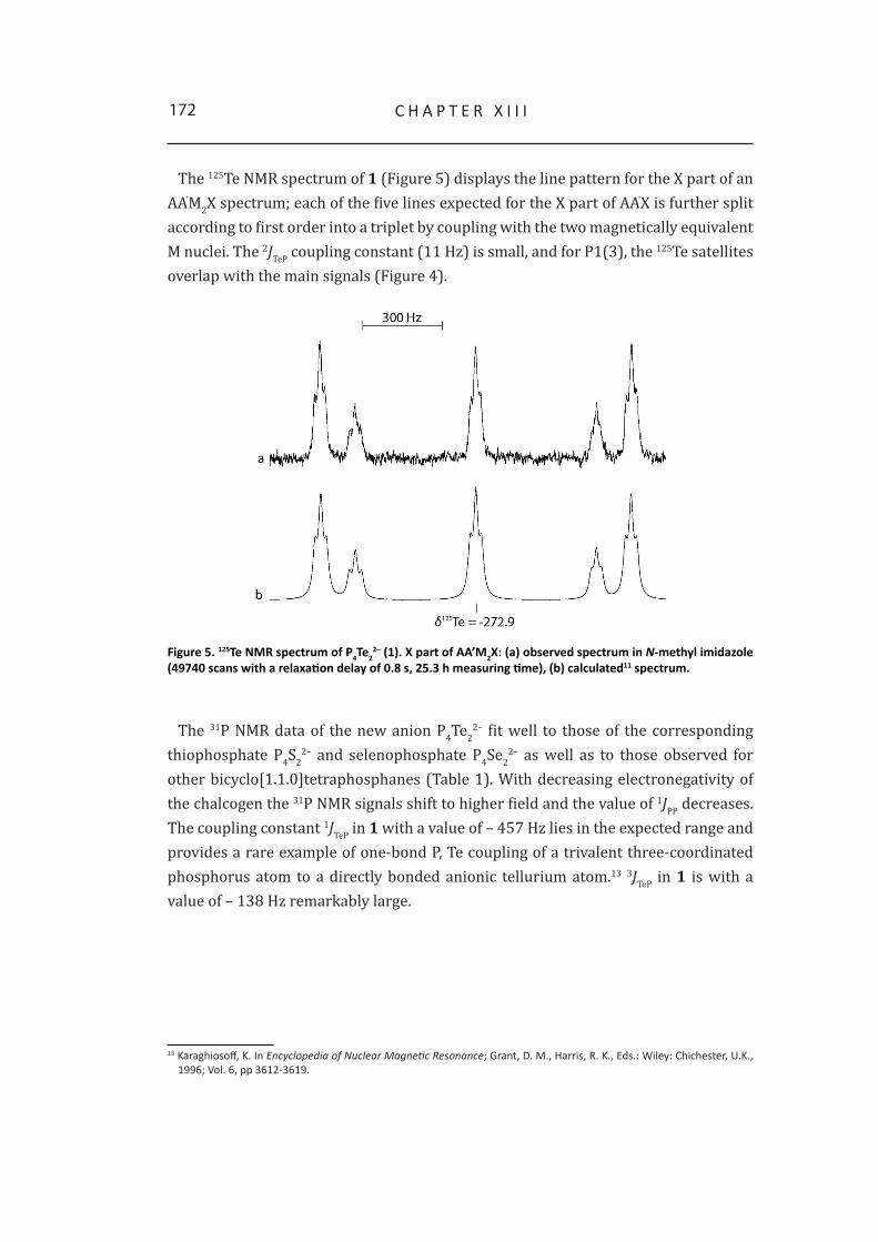
172 c h a P t E r X I I I
The 125Te NMR spectrum of 1 (Figure 5) displays the line pattern for the X part of an AA’M2X spectrum; each of the five lines expected for the X part of AA’X is further split according to first order into a triplet by coupling with the two magnetically equivalent M nuclei. The 2JTeP coupling constant (11 Hz) is small, and for P1(3), the 125Te satellites overlap with the main signals (Figure 4).
Figure 5. 125Te NMR spectrum of P4Te22– (1). X part of AA’M2X: (a) observed spectrum in N-methyl imidazole
(49740 scans with a relaxation delay of 0.8 s, 25.3 h measuring time), (b) calculated11 spectrum.
The 31P NMR data of the new anion P4Te22– fit well to those of the corresponding
thiophosphate P4S22– and selenophosphate P4Se2
2– as well as to those observed for other bicyclo[1.1.0]tetraphosphanes (Table 1). With decreasing electronegativity of the chalcogen the 31P NMR signals shift to higher field and the value of 1JPP decreases. The coupling constant 1JTeP in 1 with a value of – 457 Hz lies in the expected range and provides a rare example of one-bond P, Te coupling of a trivalent three-coordinated phosphorus atom to a directly bonded anionic tellurium atom.13 3JTeP in 1 is with a value of – 138 Hz remarkably large.
13 Karaghiosoff, K. In Encyclopedia of Nuclear Magnetic Resonance; Grant, d. M., harris, r. K., Eds.: Wiley: chichester, u.K., 1996; Vol. 6, pp 3612-3619.

173B I N a r Y P, te - a N I o N S
Table 1. NMR chemical shifts and coupling constants of the new (1) and literature-known (2-8) bicyclo[1.1.0]tetraphosphanes.14,15
δ [ppm] J [hz]1[a] (X = te-)
P-2,4P-1,3Te
-143.1-301.9-272.9
1JPP2JP2P41JteP2(4)2JTeP1(3)3JteP2(4)
-180168
-45711
-1382 [a] (X = Se-)
P-2,4 P-1,3 Se
-90.5-284.0-160.0
1JPP2JP2P41JSeP2(4)2JSeP1(3)3JSeP2(4)
-206181
-336< 8 -51
3 (X = S-)
P-2,4 P-1,3
-64.9-271.6
1JPP -222
4[17]
(X = cl)P-2,4 P-1,3
-54.5-290.4
1JPP -215
5[17] (X = Br)
P-2,4 P-1,3
-72.7-294.4
1JPP -203
6[14] (X=SiMe3)
P-2,4P-1,3
-155.0-329.2
1JPP 162
7[15] (X=N(SiMe3)2)
P-2,4P-1,3
-79.1-287.3
1JPP 222[a] the signs of the coupling constants result from the iterative fitting of the NMr spectra assuming a negative sign for 1JPP, 1JTeP and 1JSeP.
12
A bicyclo[1.1.0]tetraphosphane has been postulated as a reaction intermediate for the nucleophilic degradation of white phosphorus16,17 and has been shown to be the initial product of the oxidation of white phosphorus with Cl2 and Br2.18 The formation of the new binary anion 1 implies the oxidative addition of the isoelectronic ditelluride Te2
2– to one of the P-P bonds in the P4 tetrahedron and fits well into this picture.
14 cappello, V.; Baumgartner, J.; dransfeld, a.; Flock, M.; hassler, K. Eur. J. Inorg. Chem. 2006, 2393–2405.15 Niecke, E.; rüger, r.; Krebs, B. Angew. Chem. 1982, 94(7), 553–554.16 Brown, c.; hudson, r. F.; Wartew, G. a. Phosphorus, Sulfur Silicon Relat. Elem. 1978, 5, 67–80.17 Baudler, M.; adamek, c.; opiela, S.; Budzikiewicz, h.; ouzounis, d. Angew. Chem. 1988, 100, 1110–1111.18 tattershall, B. W.; Kendall, N. L. Polyhedron 1994, 13(10), 1517–1521.

174 c h a P t E r X I I I
3 Quantum chemical calculations
In order to elucidate the structure and charge distribution of P4Te22– quantum
chemical calculations at the MPW1PW91 level of theory using a polarized triple-zeta basis set (aug-cc-pVTZ) for phosphorus and a pseudo potential ECP4GMWB/AVTE for tellurium were carried out.
Table 2. Calculated and experimentally observed structural parameters for new and literature known bicyclo[1.1.0]tetraphosphanes.19,20
Distances [pm]P4Te2
2– [a] P4(P[NiPr2]2)2[19] P4(N(SiMe3)2)2
[b, 14] P4(sMes)2[d,20]
P1-P3 217.36 216.10(12) 212.9 216.6(2)P1-P4/P2 224.5 221.21(11)/220.70(11) 221.7/222.7 222.3(4)/222.2(3)P3-P4/P2 224.5 221.76(11)/221.23(11) 222.3/223.0 223.5(7)/223.6(9)P2-P4 298.4 - 281.0 -P-te 246.4 - - -
Bond angles [°]P1-P4-P3/P1-P2-P3 57.9 58.40(4)/58.55(4) 57.2/57.1 58.1(1)/58.1(1)P4-P1-P3/P2-P1-P3 61.0 60.93(4)/60.60(4) 61.8/61.5 61.2(1)/61.3(1)P4-P3-P1/P2-P3-P1 61.0 60.68(4)/60.85(4) 61.0/61.4 60.7(1)/60.6(1)P4-P1-P2/P4-P3-P2 83.3 80.70(4)/80.46(4) 95.2 80.9(1)/80.4(1)P-P-X[c] 106.4 96.2 107.3 106.9[a] calculated values, [b] Experimentally determined values by X-ray diffraction, [c] averaged values for the experimentally
determined angles, [d] sMes = super mesityl = 2,4,6-tri-tert-butyl phenyl.
The calculated structural parameters (bond distances and angles) of the bicyclo[1.1.0]tetraphosphane framework of P4Te2
2– correspond well to those reported in literature for compounds containing the same structural motive (Table 2). The P-P distance of the bridgehead phosphorus atoms P1 and P3 is in all cases shorter, than the distances to the other phosphorus atoms bonded to tellurium. The values for the fold angles are all in the same range. The P-Te distance is calculated to be 246.4 pm in P4Te2
2– which is in the same order of magnitude reported for BaP4Te2 (246.9 pm). So the geometry optimization corroborates the NMR spectroscopic deduced bicyclo[1.1.0]tetraphosphane framework of P4Te2
2–.In addition the Mulliken charges of the phosphorus and tellurium atoms in P4Te2
2– are calculated in order to elucidate the charge distribution within this anion. This is quite an interesting point as the calculated P-Te distance is 246.4 pm indicating a covalent P-Te bonding (Σ covalent radii of P and Te: 247 pm).21 So it is expected that the negative charge is distributed all over the P4Te2
2– molecule. This expectation 19 Lapczuk-Krygier, a.; Baranowska, K.; Pikies, J. Acta Crystallogr., Sect. E: Struct. Rep. Online 2006, 64, o2427-o2440.20 riedel, r.; hausen, h.-d.; Fluck, E. Angew. Chem. Int. Ed. Engl. 1985, 24(12), 1056-1057.21 Pauling, L. Die Natur der Chemischen Bindung, 3rd ed., Vch, Weinheim, 1968, p. 379.

175B I N a r Y P, te - a N I o N S
is confirmed by the calculations. Interesstingly, the main part of the negative charge is localized at the tellurium atoms. In contrast, there is only a small amount of the negative charge localized at the bridgehead phosphorus atoms P1 and P3. In Table 3 the calculated Mulliken charges for P4Te2
2– in the gas phase are summarized.
Table 3. Mulliken charges of the phosphorus and tellurium atoms of P4Te22– in the gas phase.
P1/P3 P2/P4 Te-0.029 -0.344 -0.627
4 Are there more phosphorus-tellurium anions?
As obviously the only possibility to synthesize P-Te anions is starting from P4 and Na2Te2, other stoichiometries were examined:
• P4 + 2 Na2Te2
• P4 + 3 Na2Te2
• 2 P4 + Na2Te2
In all cases the main product after 24 h is P4Te22– according to the corresponding 31P
NMR spectra. Interestingly, also traces of other phosphorus rich compounds can be identified in the 31P NMR spectra, however. After 24 h only traces of new phosphorus rich compounds could be identified in the 31P NMR spectrum (Figure 5).
After 5 days – the reaction solution was stored in the refrigerator at +4 °C – the signals of P4Te2
2– decreased, while the intensity of the signals for the new compounds increased.
After 18 days the signals caused by P4Te22– disappeared. In the 31P NMR spectrum
broad signals appeared and the already observed new ones increased in intensity.A 31P, 31P-COSY 45 NMR spectrum revealed cross peaks between the signal groups at
δ31P = 168.8, 79.4, – 6.0, – 113.9, – 138.6 and – 199.2 with an intensity ratio of 1:1:1:1:1:1. Therefore, the corresponding new compound must contain six phosphorus atoms obviously arranged in a polycyclic structure. In further investigations the connectivity of the phosphorus atoms has to be determined.
After 4 weeks a further 31P NMR spectrum of the reaction solution was measured. This time no additional signals were observed.
So it can be concluded that the observed P-Te anions are metastable compounds which decompose within several weeks. The same results are found for the other stoichiometries investigated. No further P-Te containing compounds can be identified for sure.

176 c h a P t E r X I I I
Figure 6. Observed 31P NMR spectra of the reaction mixture during a period of time in N-methyl imidazole: (a) after 24 h; (b) enlarged baseline of the observed 31P NMR spectrum of the reaction mixture after 24 h; (c) after 5 d; (d) after 18 d.
Figure 7. 31P NMR spectrum of the reaction mixture (32768 scans with a relaxation delay of 0.5 sec., 16.7 h measuring time). Signals which belong to the P6 compound are marked in blue.
Scheme 5. Decomposition of P4Te22–.

177B I N a r Y P, te - a N I o N S
5 Conclusion
With the synthesis of the first discrete P-Te anion P4Te22– (1) and its full
characterization by 31P and 125Te NMR spectroscopy, described here, it could be demonstrated that binary phosphorus tellurium anions can exist as stable species in solution. The results suggest the possible existence of further phosphorus-rich P-Te anions like P6Te4
2–.The quantum chemical calculations confirm the structure of P4Te2
2– and elucidate the charge distribution within the P4Te2
2– anion in the gas phase which is found as expected.

178 c h a P t E r X I I I
6 Experimental Section
General procedure. All reactions were carried out under inert gas atmosphere using Schlenk techniques. Argon (Messer Griesheim, purity 4.6 in 50 L steel cylinder) was used as inert gas. All glass vessels used were stored in a 130°C drying oven and were flame-dried in vacuum at 10-3 mbar before use.
White phosphorus (ThermPhos) was peeled under water, washed with dry THF and dried under vacuum. The sodium dichalcogenides were prepared according to a literature procedure and stored in a dry box under nitrogen atmosphere.1 N-methyl imidazole (Aldrich) was used as received.
NMR Spectroscopy. NMR spectra were recorded with a Jeol EX 400 Eclipse instrument operating at 161.997 MHz (31P), and 126.256 MHz (125Te). Chemical shifts are referred to 85 % H3PO4 (31P) and (CH3)2Te (125Te). All spectra were measured at 25 °C. For the simulation of the 31P NMR spectra, the PERCH program package was used.11
From the resulting reaction solutions a sample was transferred into a 5 mm NMR tube closed with a ground-glass stopper for multinuclear NMR spectroscopic investigations.
Na2P4S2. The yellow solution of P4 in N-methyl imidazole became green after the addition of Na2S2. After five minutes stirring at room temperature the reaction solution turned yellow and the formation of an orange-yellow precipitate was observed.
Na2P4Se2. After the addition of Na2Se2 a red brown suspension formed immediately.Na2P4Te2. To a solution of P4 (1 mmol) in 5 mL of N-methyl imidazole Na2Te2
(1.5 mmol) was added with stirring at ambient temperature, which was continued for 12 h, thus a purple colored solution could be obtained.
P4 + 2 Na2Te2
To a solution of P4 (2.8 mmol) in 10 mL of N-methyl imidazole Na2Te2 (5.6 mmol) was added at ambient temperature. A dark red colored reaction solution was obtained.
P4 + 3 Na2Te2
1.98 mmol of white phosphorus were disolved in 10 mL of N-methyl imidazole. 5.95 mmol Na2Te2 were added at ambient temperature, and a dark red reaction suspension could be obtained.
2 P4 + Na2Te2
To a solution of white phosphorus (1.46 mmol) in 4 mL of N-methyl imidazole Na2Te2 (0.73 mmol) were added in one portion at room temperature. The reaction mixture turned orange brown.11 Boudjouk, P.; thompson, d. P. J. Org. Chem,. 1988, 53, 2109 – 2112.

Chapter XIV
Some Unexpected Compounds in the Reaction of White Phosphorus, Lithium and Tellurium
In course of the investigations on the syntheses of new phosphorus tellurium
anions in solution starting from the elements white phosphorus, lithium and
tellurium in N-methyl imidazole, some new and interessting compounds can
be observed. Consequently, the syntheses of these new compounds containing
N-methylimidazole were investigated.

180 c h a P t E r X I V
1 Introduction
In the course of the investigation on the syntheses of phosphorus-tellurium anions one strategy I tried, was the reaction of the elements white phosphorus, lithium and tellurium in N-methyl imidazole or THF as reaction medium (Scheme 1).
Scheme 1. Synthetic strategy for LixPnTem.
In case of N-methyl imidazole as reaction medium, I could identify some new and unexpected compounds in the corresponding 31P NMR spectra. These compounds are described in the present chapter.
2 Results and Discussion
White phosphorus, lithium and tellurium in a molar ratio of 1 : 1.6 : 3.1 were stirred in N-methyl imidazole for one hour at ambient temperature. A purple solution was obtained. The 31P NMR spectrum of the reaction solution did not indicate the formation of binary phosphorus-tellurium anions. However, the signals of some new and unexpected species were observed, which are discussed in detail below:
At a 31P NMR chemical shift of δ = –158.4 a singlet accompanied by one pair of 125Te satellites was observed. The value of the 1JTeP coupling constant is 1.28 kHz. The intensity ratio of the satellite pair to the total intensity of the signal is 12.2 %, which indicates the presence of two tellurium atoms directly bonded to phosphorus. 31P chemical shift and 1JTeP coupling constant fit well to those of [Ph2PTe2][Li(THF)3.5(TMEDA)0.25] reported by Davies et al.: δ31P = –123.8 ppm, 1JTeP = 1.53 kHz.1 Based on this similarity the structure 1 of a ditellurophosphonate with two N-methyl imidazole moieties bonded to phosphorus via C2 is suggested (Figure 1).
Figure 1. 31P{1H} NMR spectrum of 1 in N-methyl imidazole. (0.1 m; 29214 scans, 14.9 h measuring time); 125Te satellites are marked with o.
1 davies, r. P.; Martinelli, M. G.; Wheatley, a. E. h.; White, J. P.; Williams, d. J. Eur. J. Inorg. Chem. 2003, 3409.

181P 4 , L i , Te a N d N - M E t h Y L I M I d a Z o L E
Another singlet with one pair of 125Te satellites can be observed at δ31P = –32.7. The 1JTeP coupling constant is 1.68 kHz. The intensity ratio of the satellite pair is 8.5 %. Thus it can be concluded that only one tellurium atom directly bonded to phosphorus must be present in the molecule. In addition, one pair of 13C satellites is clearly visible (Figure 2). The relative intensity of the 13C satellites can only be roughly estimated (4.2 %) it indicates, however, the presence of more than one carbon atom bonded to phosphorus. Thus, structures 2 and 3 seem possible. The values of 1JTeP and 1JPC allow distinguishing between the two structures. The value of 1.68 kHz of 1JTeP corresponds well to those observed for phosphorus tellurides R3PTe (1663 – 2153 Hz)2, while for R2PTe with a trivalent phosphorus much smaller values are reported, e.g. 1JTeP = 747 Hz of [Ph2PTe][Li(TMEDA)1.33(THF)1.33] (δ31P = –32.5 ppm).1 In addition, the value of 120 Hz for 1JPC indicates clearly the presence of a tetracoordinate phosphorus atom, while for 1JPC to a threecoordinate phosphorus again much smaller values are typical (565 – 775 Hz).1,3 Both coupling constants thus clearly support structure 2 of a phosphorus telluride.
Figure 2. 31P{1H} NMR spectrum of 2 in N-methyl imidazole. (0.1 m; 29214 scans, 14.9 h measuring time); 13C
satellites are marked with x; 125Te satellites are marked with o.
Both compounds discussed above imply reactions of the solvent and N-methyl imidazole in position 2 with formation of a P-C bond. In order to put more light on the mechanism of formation of compounds 1 and 2, the reaction of P4 with lithium in N- methyl imidazole was investigated.
2 Jones, c. h. W.; Sharma, r. d. Organometallics, 1987, 6(7), 1419-1423.3 Bildstein, B.; Sladky, F. Phosphorus, Sulfur Silicon and Relat. Elem. 1990, 47, 341-347.

182 c h a P t E r X I V
Possible reaction mechanism
The formation of the compounds 1 and 2 described above, can be explained if we assume metallation of N-methyl imidazole in 2-position with lithium to compete effectively with the reduction of P4 to polyphosphides. Metallation of N-methyl imidazole in 2-position has been described in literature. Nucleophilic degradation of P4 by this carbanionic species 4 might lead to the bis(imidazolyl)phosphide 5, telluration of which should give the anion 1. Formation of phosphides like 5 by nucleophilic degradation of P4 has first been described by A. Schmidpeter. The phosphine telluride 2 might result from the oxidative coupling of 4 and 5 (by elemental tellurium) and successive telluration (Scheme 2).
Scheme 2. Reaction scheme of P4 and lithium in N-methyl imidazole.
The observed reaction of lithium with the solvent N-methyl imidazole might result from the fact, that white phosphorus, lithium and tellurium were first put into the Schlenk flask and N-methyl imidazole was added afterwards. As white phosphorus dissolves quite slowly in N-methyl imidazole, lithium would have the chance to react with the solvent before it reacts with phosphorus, due to the only small amounts of P4 present in solution in the beginning.
Thus, P4 was first dissolved in N-methyl imidazole and then lithium was added. The yellow solution warmed up and its color changed into brownish-red. The 31P NMR spectrum of the reaction mixture is shown in Figure 3.

183
Figure 3. 31P{1H} NMR spectrum in N-methyl imidazole. (0.1 m; 7507 scans, 3.8 h measuring time).
Three groups of resonances at δ31P = 177, –109.5 and –218 can be assigned to P113–
described by M. Schuster in his dissertation.4
Figure 4. Lewis formula of P113-.
The singlet at a 31P NMR chemical shift of –80.5 ppm is accompied by one pair of 13C satellites. The 1JPC coupling constant of 72 Hz indicates a direct bond of the carbon atoms to phosphorus. Thus the 31P NMR resonance at –80.5 ppm can be attributed to the phosphide 5 (Figure 5).
Figure 5. Lewis formula of compound 5.
In literature there are similar compounds described: Ph2P–, PhP(CN)– and (CN)2P–. 5,6,7 The corresponding 31P NMR chemical shifts of these compounds are related to the organic substituents present. The more electron withdrawing the substituent is, the more the 31P NMR chemical shift is shifted to higher field. Thus, the 31P NMR chemical shift found for (CN)2P– is –193 ppm, in contrast to the 31P NMR chemical shift assigned for Ph2P– –20 ppm. It can be concluded that N-methyl imidazole is an electron donating substituent, but not of that quality like phenyl groups.4 Schuster, M. dissertation, LMu Munich, 1999.5 Fluck, F.;Issleib, K. Z. Naturforsch. Teil B, 1965, 20, 11236 Schmidpeter, a.; Zirzow, K.-h.; Burget, G.; huttner, G.;. Jibril, I. Chem. Ber., 1984, 117, 1695-1706.7 (a) Schmidpeter, a.; Zwaschka, F. Angew. Chem., Int. Ed. Engl., 1977, 16, 704. (b) Sheldrick, W. .; Kroner, J.; Zwaschka, F.; Schmidpeter, a. Angew. Chem. 1979, 91, 998. (c) Sheldrick, W. S.; Kroner, J.; Zwaschka, F.; Schmidpeter, a. Angew. Chem., Int. Ed. engl. 1979, 18, 934.
P 4 , L i , Te a N d N - M E t h Y L I M I d a Z o L E

184 c h a P t E r X I V
At a chemical shift of δ31P = 358 the A-part of an AA’XX’ spin system was identified. Signals at such low field are typical for the dicoordinate phosphorus atom in diphosphenes.8 A similar chemical shift and splitting pattern was also observed for the central two phosphorus atoms in 7, described by A. Wörner et al.9 Although the X-part of this system could not yet be identified with certainty, the observed signal strongly indicates the presence of an analogous N-methyl imidazole substituted compound in the solution (Figure 6)
Figure 6. Left: Lewis structure of compound 6; right: Lewis structure of compound 7[9].
Elemental tellurium was added to the reaction solution described above to give a dark purple reaction solution. The 31P NMR spectrum of the reaction solution revealed that the compounds described above have reacted and resonances of new compounds have appeared, as well as the already described resonance of 2.
In addition signal groups at δ31P = 100.8, 62, 36, –51, –115, –142 can be observed, which are most probably due to new binary phosphorus-tellurium anions.
Because of the great interest in the anion 6 attempts were made to synthesize it as the main product. Stoichiometric amounts of white phosphorus, lithium and N-methyl imidazole were reacted in THF at ambient temperature. In the 31P NMR spectrum the following compounds were identified without any doubt: (N-MeIm)2PLi [10], P11
3– and P5
– (δ31P = 467).11 The signals of [(N-MeIm)2P4)]Li2 were not observed.Also another stoichiometry (P4 : Li : N-MeIm 1:1:1) as well as other reaction conditions
(–80 °C) were applied, according to literature procedure.8,12 A red reaction solution was obtained and NMR spectroscopically investigated. The products observed are P5
–, P16
2- (δ31P = 60, 39, 6, –34, –132, –172)13 and P4.I also tried to selectively prepare bis(N-methyl imidazolyl)phosphide 5 as the
main product. Stoichiometric amounts of P4, lithium and N-methyl imidazole were reacted in THF. A green reaction solution was obtained and 31P NMR spectroscopically investigated. The anion 5 as well as P7
3– and other not further identified products, the signals of which split into multipletts in the 31P-1H coupled NMR spectrum were observed.8 Schrödel, h.-P.; Schmidpeter, a. Phosphorus, Sulfur, Silicon Relat. Elem. 1997,129, 69.9 Wiberg, N.; Wörner, a.; Karaghiosoff, K.; Fenske, d. Chem. Ber. Recueil, 1997, 130, 135.10 N-MeIm = N-methyl imidazole11 Baudler, M.; düster, d.; ouzounis, d. Z. Anorg. Allg. Chem. 1987, 544, 87.12 Wiberg, N.; Wörner, a. Eur. J. Inorg. Chem., 1998, 118, 833.13 Baudler, M. Z. Anorg. Allg. Chem., 1985, 529, 7.

185
Attempts were made to isolate the phosphide 5 in the solid state by slow diffusion of diethylether into the reaction mixture. After some days, colorless crystals were obtained and characterized using single crystal X-ray diffraction.
The crystal consists of [Me2(O)Si(N-MeIm)]Li, which crystallizes in the trigonal space group R–3.
The silicium atoms are distorted tetrahedrally coordinated by three carbon and one oxygen atom. The corresponding bond angles range from 113° to 104°. The averaged Si-C bond distances of 187 pm are in accord with literature known values of Si-C single bonds.14 The Si-O distance is 158 pm long, and therefore in between the values for Si-O single bonds and Si=O double bonds.15
The lithium and oxygen atoms in the asymmetric unit are arranged along the b axis in a planar fourmembered ring, as the angle sum is 360°. The lithium atoms are tetra coordinated by three oxygen and one nitrogen atom. The coordination sphere of Li1 is built by O1, O2, O1(i) and N2; the coordination sphere of Li2 is formed by O1, O2, O2(ii) and N3. The distances between the lithium atoms and the oxygen or nitrogen atoms correspond to those found in bis[(µ2-benzyldiethylphosphonato-O,O)-(1,4-diazabicyclo(2.2.2)octane-N,N’)-lithium].16
Along the c axis, the oxygen and lithium atoms are forming two sixmembered rings lying eclipsed one above the other in chair conformation. The two sixmembered ring are formed by O1, Li1, O1(i), Li1(i), O1(ii) Li1(ii) respectively Li2, O2, Li2(i), O2(i), Li2(ii), O2(ii). As the two sixmembered rings are eclipsed, there is always one lithium atom of the one ring placed over the oxygen atom of the other ring, so there are coordinative interactions between the two rings.
Furthermore, one lithium atom is connected to one neighbored oxygen atom not only through electrostatic interactions, but also due to a N-C-Si bridge, forming a non planar five membered ring (O1, Si2, C2, N2, Li1 and Li2, O2, Si1, C8, N3).
Despite the described interactions and bonds within one molecule, there are no other interactions found between the molecules within the unit cell.
The formation of this compound can be explained by the reaction of ground joint fat containing a polymeric methylated silicon compound.
14 allen, F. h.; Kennard, o.; Watson, d. G.; Brammer, I.; orpen, a. G.; taylor, r. J. Chem. Soc. Perkin Trans. II 1987, S1.15 holleman, a.F.; Wiberg, F.; Wiberg, N. Lehrbuch der anorganischen Chemie, de Gruyter, Berlin 1995, 101. auflage16 Zarges, W.; Marsch, M.; harms, K.; haller, F.; Frenking, G.; Boche, G. Chem. Ber. 1991, 124, 861.
P 4 , L i , Te a N d N - M E t h Y L I M I d a Z o L E

186 c h a P t E r X I V
Figure 7. Top: asymmteric unit of [Me2(O)Si(N-MeIm)]Li, view along b axis. Bottom: molecular structure of [Me2(O)Si(N-MeIm)]Li in the crystal, view along c axis, i = -y, x-y, z, ii = -x+y, -x, z. Ellipsoids are drawn at the 50 % probability level, hydrogen atoms are omitted for clarity.

187
Figure 8. Molecular structure of [Me2(O)Si(N-MeIm)]Li in the unit cell. View along the c axis. The ellipsoids are drawn at the 50 % probability level.
Figure 9. Molecular structure of [Me2(O)Si(N-MeIm)]Li in the unit cell. View along the a axis (left) and along the c axis (right). The ellipsoids are drawn at the 50 % probability level. Hydrogen, nitrogen and carbon atoms are omitted for clarity.
Table 1. Selected bond parameters of [(Me2(O)Si(N-MeIm)]Li, i = -y, x-y, z, ii = -x+y, -x, z.
Distances [pm] Angles [°] Angles [°]Si1-o2 158.4(2) o1-Si2-c1 104.76(17) c8-N3-Li2 108.1(3)Si1-c8 189.9(4) o1-Li1-o1(i) 120.3(3) Si1-c8-N3 119.6(2)Si2-o1 158.3(2) o1-Li1-o2 99.8(3) Si1-o2-Li1 137.4(2)Si2-c1 189.2(5) o1-Li1-N2 93.8(3) Si1-o2-Li2 111.33(19)Si1-c6 185.5(4) o1(i)-Li1-N2 125.0(4) Si1-o2-Li2(i) 124.5(2)Si1-c7 186.9(5) o1(i)-Li1-o2 98.2(3) Si2-c1-N2 118.9(3)Si2-c4 187.2(5) o1-Li2-o2 100.8(3) Si2-o1-Li1 112.1(2)Si2-c5 185.3(5) o1-Li2-N3 121.1(3) Si2-o1-Li1(ii) 126.5(3)o1-Li1 198.3(8) o1-Li2-o2(ii) 98.8(3) Si2-o1-Li2 130.2(2)o1-Li2 194.3(5) o2-Si1-c8 104.74(14) Li1-o1-Li2 79.9(3)o1-Li1(ii) 190.6(7) o2-Li1-N2 118.4(3) Li1-o1-Li1(ii) 115.5(3)o2-Li1 196.6(6) o2-Li2-N3 94.6(3) Li1(ii)-o1-Li2 81.7(3)o2-Li2 197.9(8) o2(ii)-Li2-N3 120.9(3) Li1-o2-Li2 79.5(3)o2-Li2(i) 191.2(8) o2-Li2-o2(ii) 120.4(3) Li1-o2-Li2(i) 81.0(2)N2-Li1 207.8(7) c1-N2-Li1 109.7(3) Li2-o2-Li2(i) 114.8(3)N3-Li2 208.8(6)
P 4 , L i , Te a N d N - M E t h Y L I M I d a Z o L E

188 c h a P t E r X I V
As it is possible to synthesize (N-MeIm)2PLi in solution as main product, the reaction behavior with tellurium is investigated. Therefore (N-MeIm)2PLi is synthesized starting from P4, BuLi17 and N-methyl imidazole in THF, then elemental tellurium is added. So a red reaction suspension is obtained and investigated using 31P NMR spectroscopy. According to the 31P NMR spectrum, (N-MeIm)2PLi has abreacted and P5
- and P73- can be identified as by-products. In addition new signals can be observed,
but it is not possible to determine the corresponding compounds:At δ31P = –58.7 a singlet with three pairs of satellites can be identified. The coupling
constants are 109, 468 and 871 Hz. The values of the coupling constants are within the range of the values for 1JTeP couplings constants (84 - 2290 Hz) reported in literature.18 Another singlet at δ31P = –60.4 is accompanied by two pairs of satellites with values of 143 and 294 Hz. As already mentioned, in both cases it is not possible to suggest structures for the compounds causing these signals. Furthermore, some not interpretable spin systems appear in the spectrum. The 125Te NMR spectrum shows a singlet at a 125Te NMR chemical shift of –335.0 ppm.
17 BuLi = butyl lithium18 Karaghiosoff, K. In Encyclopedia of Nuclear Magnetic Resonance; Grant, d. M., harris, r. K., Eds.: Wiley: chichester, u.K., 1996; Vol. 6, pp 3612.

189
3 Conclusion
New reaction products of white phosphorus, lithium, tellurium and N-methyl imidazole can be observed and structurally charcterized using 31P NMR spectroscopy. It was also possible to formulate a way of formation of the new compounds.
In addition, the molecular structure of [Me2(O)Si(N-MeIm)]Li can be determined using single crystal X-ray diffraction.
P 4 , L i , Te a N d N - M E t h Y L I M I d a Z o L E

190 c h a P t E r X I V
4 Experimental Section
General procedure. All reactions were carried out under inert gas atmosphere using Schlenk techniques. Argon (Messer Griesheim, purity 4.6 in 50 L steel cylinder) was used as inert gas. All glass vessels were stored in a 130 °C drying oven and were flame-dried in vacuum at 10-3 mbar before use.
N-methyl imidazole, tellurium and elemental lithium (Aldrich) were used as received. Butyl lithium (Acros) was used as 2.5 m solution in hexane. White phosphorus (ThermPhos) was peeled under water, washed with dry THF and dried under vacuum.
NMR spectroscopy. NMR spectra were recorded using a Jeol EX 400 Eclipse instrument operating at 161.997 MHz (31P), and 126.256 MHz (125Te). Chemical shifts are referred to 85 % H3PO4 (31P) and (CH3)2Te (125Te). All spectra were measured at 25 °C. The %-data correspond to the intensities in the 31P NMR spectra with respect to the total intensity. The difference to 100 % belongs to not determinable signals.
X-ray crystallography. The molecular structure of Me2(O)Si(N-MeIm)Li in the crystalline state was determined using an Oxford Xcalibur3 diffraction instrument with a Spellman generator (voltage 50 kV, current 40 mA) and a Kappa CCD detector with a X-ray radiation wavelength of 0.71073 Å. The data collection was performed with the CrysAlis CCD software, the data reduction with the CrysAlis RED software.19,20 The structures were solved with SIR-92, SIR-97 and SHELXS-97, refined with SHELXL-97 and finally checked using PLATON.21,22,23,24,25 The absorptions were corrected by a SCALE3 ABSPACK multi-scan method.26
19 crysalis ccd, oxford diffraction Ltd., Version 1.171.27p5 beta (release 01-04-2005 crysalis171 .NEt), (compiled apr 1 2005,17:53:34).
20 crysalis rEd, oxford diffraction Ltd., Version 1.171.27p5 beta (release 01-04-2005 crysalis171 .NEt), (compiled apr 1 2005,17:53:34).
21 SIr-92, 1993, A Program for Crystal Structure Solution; altomare, a.; cascarano, G. L.; Giacovazzo, c.; Guagliardi, a. J. Appl. Cryst. 26, 343.
22 altomare, a.; Burla, M. c.; camalli, M.; cascarano, G. L.; Giacovazzo, c.; Guagliardi, a.; Moliterni, a. G. G.; Polidori, G.; Spagna, r. J. Appl. Crystallogr. 1999, 32, 115.23 Sheldrick, G. M. SHELXS-97, Program for Crystal Structure Solution; universität Göttingen, 1997.24 Sheldrick, G. M. SHELXL-97, Program for the Refinement of Crystal Structures; university of Göttingen, Germany, 1997. 25 Spek, L. a. PLATON, A Multipurpose Crystallographic Tool, utrecht university, utrecht, the Netherlands, 1999.26 ScaLE3 aBSPacK - An Oxford Diffraction program (1.0.4,gui:1.0.3) (c) 2005 oxford diffraction Ltd.

191
4.1 Syntheses of new P-Te anions outgoing from the elementsP4 + 1.6 Li + 3.1 Te
White phosphorus (267 mg, 8.61 mmol) was dissolved in 10 mL N-methyl imidazole. Then elemental lithium (96 mg, 13.79 mmol) and tellurium (3.4 g, 26.69 mmol) were suspended in 25 mL N-methyl imidazole and added to the solution of P4. The reaction suspension turned red, and heated up a little bit.
31P-{1H}-NMR (N-MeIm, RT): (N-MeIm)2PTe2- (11.8 %), (N-MeIm)3PTe (24.5 %),
δ = 120.5 (s, 6.6 %), 27.3 (s, 19.4 %), 20.2 (s, 3.5 %), -2.9 (s, 5.6 %), -10.4 (s, 2.5 %), -38.5 (s, 2.7 %), -115.4 (s, 11.2 %).
125Te-{1H}-NMR (N-MeIm, RT): δ = 26.3 (s, 86.3 %), -283.3 (s, 11.7 %), -428.1 (s, 1.9 %).
P4 + 1.6 Li + 3.6 Te
140 mg (4.52 mmol) white phosphorus was dissolved in 14 mL N-methyl imidazole. 50 mg (7.23 mmol) lithium were added at ambient temperature. The yellow solution turned brownish red and heated up a little bit. Afterwards elemental tellurium (1.2 mL, 8 mmol) was added and the reaction suspension turned dark purple. The suspension was continued stirring at ambient temperature for 24 h.
31P-{1H}-NMR (N-MeIm, RT): (N-MeIm)2P- (12.7 %), P113- (33 %), (N-MeIm)2P4Li2
(1.4 %), δ = -96 (d, 15.9 %), -103.7 (s, 1.98 %), -109.5 (q, 6.8 %), -129.9 (s, 1.7 %).
After the addition of tellurium:
31P-{1H}-NMR (N-MeIm, RT): (N-MeIm)3PTe (4.5%), δ = 100.8 (t, 8.5 %), 62.0 (t, 2.2 %), 36.0 (t, 1.6 %), 27.4 (s, 8 %), 20.5 (s, 3.4 %), -10.5 (s, 1.2 %), -32.7 (s, 4.5 %), -115 (s, 14 %), -142.0 (m, 6.3 %), -156.5 (s, 4.1 %).
4.2 Syntheses of (N-MeIm)2P4Li2
P4 + 2 Li + 2 N-MeIm
White phosphorus (51 mg, 1.58 mmol) were dissolved in 10 mL THF. In another glass vessel, lithium (20 mg, 3.16 mmol) and N-methyl imidazole (259 mg, 3.16 mmol) were reacted in 5 mL THF yielding a green solution. This solution is added at once to the solution of white phosphorus. The reactions suspension immediately turned orange.
31P-{1H}-NMR (N-MeIm, RT): (N-MeIm)2PLi (10.6 %), P5- (1.4% ), δ = -2.46 (quintett,
21.9 %), -98.7 (s, 15.3 %), -176.7 (s, 12.3 %).
P 4 , L i , Te a N d N - M E t h Y L I M I d a Z o L E

192 c h a P t E r X I V
P4 + Li + N-MeIm
58 mg (1.81 mmol) P4 were dissolved in 4 mL THF and cooled down to -78°C afterwards. 1.81 mmol (1.81 mL) of an already prepared solution of N-MeImLi was added.27 The suspension turned immediately brownish red.
31P-{1H}-NMR (THF, -80°C): (N-MeIm)2PLi (12.7 %), δ = -451.3 (s, 87.3 %).31P-{1H}-NMR (THF, RT): P11
3- (99.1 %), (N-MeIm)2PLi (0.5 %), P5- (0.4 %).
4.3 Syntheses of (N-MeIm)2PLiP4 + 8 Li + 8 N-MeIm
43 mg (1.39 mmol) white phosphorus were dissolved in 10 mL THF. In another flask 71 mg (11.1 mmol) lithium and 911 mg (11.1 mmol) N-MeIm were suspended in 5 mL THF. The greenish solution was added to the solution of P4, so an orange reaction suspension could be obtained. The reaction mixture was stirred 24 h at ambient temperature and the formation of a yellow precipitate could be observed and removed. So a green solution can be obtained of which 2 mL were transferred into another glass vessel for crystallization and 10 mL diethylether were allowed to diffuse slowly into the reaction suspension. After some days colourless crystals of Me2(O)Si(N-MeIm)Li could be obtained and analyzed using single crystal X-ray diffraction.
31P-{1H}-NMR (N-MeIm, RT): (N-MeIm)2PLi (27.9 %), P73- (18.2 %), δ = 37.1 (q, 11.8
%), -19.7 (t, 10.4 %), -2.9 (s, 5.6 %), -102.3 (t, 10.1 %), -133.4 (s, 21.7 %).
4 days later:31P-{1H}-NMR (N-MeIm, RT): (N-MeIm)2PLi (30%), P7
3- (43 %), δ = -133.42 (s, 27 %).
P4 + 8 Li + 8 N-MeIm
128 mg (4 mmol) white phosphorus were dissolved in 35 mL THF, and then lithium (222 mg, 32 mmol) and 2.63 g (32 mmol) N-MeIm were added. The reaction mixture turned red. It was continued stirring at ambient temperature for 24 h yielding a greenish reaction suspension.
31P-{1H}-NMR (THF, RT): (N-MeIm)2PLi (23.2 %), P73- (67.7 %), δ = -133.3 (s, 9.1 %).
After 24 h:31P-{1H}-NMR (THF, RT): (N-MeIm)2PLi (16.7 %), P7
3- (65.6 %), δ = 106.6 (m, 5.2 %).
27 35 mg (5 mmol) lithium and 398 µL (5 mmol) N-methyl imidazole were reacted in 10 mL thF.

193
4.4 Reaction of (N-MeIm)2PLi with telluriumWhite phosphorus (80 mg, 2.5 mmol) were dissolved in 7.5 mL THF. In another flask,
319 µL (4 mmol) N-methyl imidazole and 1.6 mL BuLi were reacted in 8 mL THF. The two solutions were combined, yielding an orange-red solution.
31P-{1H}-NMR (THF/Hexan, RT): (N-MeIm)2PLi (44.1 %), δ = 53.8 (s, 1.5 %), 32.5 (q, 6.2 %), -16.2 (t, 5.7 %), -109.1 (t, 6 %), -134.4 (s, 35.6 %).
Elemental tellurium (319 mg, 2.5 mmol) were added to the reaction solution at ambient temperature. After one hour, a red solution was obtained, and all tellurium has abreacted.
31P-{1H}-NMR (THF, RT): P5- (0.2%), P7
3- (26.9%), δ = 9.5 (q, 6.7 %), -9.5 (m, 15.9 %), -58.7 (s, 6.6 %), -60.4 (s, 3.8 %), -75.5 (t, 8.6 %), -127.2 (m, 6 %), -137.5 (m, 13.2 %).
125Te-{1H}-NMR (THF, RT): δ = -335 (s, 100 %).
P 4 , L i , Te a N d N - M E t h Y L I M I d a Z o L E

194 c h a P t E r X I V
Table 2. Crystal and structure refinement data.
Me2(O)Si(N-MeIm)Li
empirical formula C36H66N12Li6o6Si6
formula mass 973.16
temp (K) 200
cryst. size (mm) 0.2 x 0.15 x 0.1
cryst. descriptn. colourless block
cryst. system trigonal
space group R–3
a (Å) 21.8036(10)
c (Å) 20.6909(8)
V (Å) 8518.6(6)
Z 6
ρcalc (g cm-1) 1.138
µ (mm-1) 0.194
F (000) 3132
θ range (deg) 3.74-23.98
index ranges-24 ≤ h ≤ 24,-24 ≤ k ≤ 24,-23 ≤ l ≤ 23
reflcns collcd 22172
reflcns obsd 2154
reflcns unique 2966 (Rint = 0.0439)
R1, wr2 (2σ data) 0.0495, 0.1126
R1, wr2 (all data) 0.0822, 0.1337
max/min transm 0.995/1.000
data/restr/params 2966/0/232
S on F2 1.055
larg. diff peak/hole (e/Å) 0.265/-0.289

195Chapter XV
Conclusion
1 σ3λ5 - An unusual bonding situation for the phosphorus atom
Trithiometaphosphate PS3- and triselenometaphosphate PSe3
- anions do exist as monomers in solution and are stable as monomers at ambient temperature. The results in the present thesis definitively put light on the controversies on the existence of PS3
- anion. They also show for PS3- a low nucleophilicity at phosphorus. So we may
call PS3- as a “heavy nitrate”. It was possible through the synthesis of [nBu4N]2[P2S6]
• THF, a soluble salt in organic solvents, to observe an equilibrium between the monomeric PS3
- anion and the corresponding dimer P2S62- in solution. The 31P NMR
chemical shift of PS3- was determined reliably for the first time. Quantum chemical
calculations justify the experimental results.The stabilization of binary P, S compounds containing σ3λ5 phosphorus atoms in
the solid state can be achieved via adduct formation with a base like pyridine. The P-N distance gave much longer P-N single bond, which indicates the presence of weak adducts. The isolation of py2P2S7, py2P2S5, [pyH][pyPS3] and [pyH][pyPSe3] in the crystalline state and the NMR spectroscopic identification of pyPCh2X (Ch = S, Se; X = Cl, Br) is the final evidence for this. In addition, quantum chemical calculations revealed that the double bond rule is set out of order for this kind of compounds with a σ3λ5 phosphorus atom. The examined compounds py2P2S5 and py2P2S7 can therefore be regarded as new bis(pyridine) adduct stabilized neutral binary phosphorus sulfides.

196 c h a P t E r X V
2 Alternative syntheses for chalcogenophosphates
The synthesis of chalcogenophopshates at ambient or lower temperature makes it possible to isolate metastable compounds like in the case of twist-P2Se8
2-, which melts at 15 °C. The use of organic cations like Ph4P+ or nBu4N+ results in the formation of salts which are soluble in common aprotic organic solvents, like in the case of P2S6
2-. In case of the anions P2S8
2- and P2Se82-, consisting of sixmembered rings, it was shown that
two conformers - twist- and chair- do exist in solution. There is a dynamic equilibrium between them. The twist conformer is favored in solution, while in the solid state, the chair conformer is isolated.
3 New fields of chalcogenophosphate chemistry – a first insight into P, Te anions
Binary P, Te, anions do exist and are stable in solution at ambient temperature. The nucleophilic degradation of white phosphorus in highly polar solvents like N methylimidazole with Na2Te2 at ambient temperature yields the first P, Te anion P4Te2
2- in solution. It is characterized unequivocally by 31P and 125Te NMR spectroscopy. Further investigations revealed that P4Te2
2- is just an intermediate during the degradation of white phosphorus, it disproportionate with formation of other phosphorus rich tellurophosphates.


199Curriculum vitae
Christiane Angelika Rotter
Geburtsdatum 13.12.1981 Geburtsort augsburg Familienstand ledigStaatsangehörigkeit deutsch
Schulausbildung
1992 bis 2001 abitur Gymnasium Wertingen
1988 bis 1992 Grundschule Biberbach
Wissenschaftlicher Werdegang 2007 bis jetzt Promotion im Fachbereich Ludwig-Maximilians- anorganische chemie unter universität München der anleitung von Prof. dr. K. Karaghiosoff
2005 bis 2006 Studium chemie Ludwig-Maximilians- Master universität München
thema der Masterarbeit: Systematische Untersuchung der Chalkogenophosphate PX3
- und P2X82- mit X = S, Se
2001 bis 2005 Studium chemie Ludwig-Maximilians- Bachelor universität München
Besuchte Tagungen
april 2009 12th International Seminar: New trends in research of Ener-getic Materials, Pardubice (tschechien)
März 2009 6th European Workshop on Phosphorus chemistry, Florenz (Italien):
Moderation einer Vortragsreihe

200 c u r r I c u L u M V I t a E
September 2008 13. deutscher Fluortag, Schmitten
März 2008 5th European Workshop on Phosphorus chemistry, regens-burg (deutschland):Vortrag mit dem thema New Chalcogenophosphates from White Phosphorus
März 2007 4th Phd Workshop on Phosphorus chemistry, Zandvoort (Niederlande): Moderation einer Vortragsreihe
Auszeichnungen
bester Vortrag (thema: New Chalcogenophosphates from White Phosphorus, regensburg 2008)
römer-Preis 2007 für die Masterarbeit (thema: Systematische Untersuchung der Chalkogenophosphate PX3
- und P2X82- mit X = S, Se)
Publikationen
An Unusual Binary Phosphorus-Tellurium Anion and its Seleno- and Thio Analogues: P4Ch2
2- (Ch = S, Se, Te): c. rotter, M. Schuster, K. Karaghiosoff; im druck.
Structural and NMR Spectroscopic Investigations of Chair and Twist Conformers of the P2Se8
2- Anion: c. rotter, M. Schuster, M. Kidik, o. Schön, t. M. Klapötke, K. Karaghiosoff; Inorg. Chem. 2008, 47(5), 1663-1673.
