newsletter january 2018 - iop.org · expertise and experience in scanning electron microscopy of...
TRANSCRIPT

NEWSLETTER
January 2018
Bright Field-STEM image of porosity in a beta-zeolite (FEI Titan Themis G2 300
image courtesy of James Cattle, PhD Student, University of Leeds. Sample provided by Dr Stig Helveg, Haldor Topsoe A/S).
See http://emag.iop.org for further details
EMAG Group newsletter Jan 2018
2
CONTENTS
EMAG COMMITTEE 3
LETTER FROM THE CHAIR 4
FORTHCOMING EVENTS 5
EMAG 2018 CALL FOR PAPERS 5 EMAG ANNUAL GENERAL MEETING 2018 6
NEWS 7
PROF. PRATIBHA GAI DBE 7 UNIVERSITY OF GLASGOW PLASMA-FIB 8 ELECTRON MICROSCOPY AT DARESBURY LABS 10
MEETING REPORTS 11
MSM-XX, OXFORD 11 VISION AND OPHTHALMOLOGY, BALTIMORE 12 MMC17, MANCHESTER 14 M&M, ST LOUIS 15
APPLY FOR IOP RESEARCH STUDENTS CONFERENCE FUND
15
EMS MEMBERSHIP 16
ADDITIONAL FUTURE MEETINGS OF INTEREST 17

EMAG Group newsletter Jan 2018
3
EMAG COMMITTEE
Chair Dr Sarah Haigh School of Materials, University of Manchester, Manchester, M13 9PL Tel: 0161 306 3618 Email: [email protected] Secretary & Honorary Treasurer Dr Andy Brown School of Chemical and Process Engineering, University of Leeds, Leeds, LS2 9JT Tel: 0113 343 2382 Email: [email protected] Ordinary Members Dr ZiYou Li, University of Birmingham, [email protected] Dr Ana Sanchez, University of Warwick, [email protected] Dr Cornelia Rodenburg, University of Sheffield, [email protected] Dr Larry Stoter, Retired, [email protected] Mr Michael Dixon, Hitachi Europe, [email protected] Prof Jun Yuan, University of York, [email protected] Dr Donald MacLaren, University of Glasgow, [email protected] Dr Sarah Karimi, JEOL Ltd, [email protected]
Co-opted Dr. Caterina Ducati, University of Cambridge, [email protected] Dr Thomas Slater, University of Manchester, [email protected]

EMAG Group newsletter Jan 2018
4
LETTER FROM THE CHAIR Dear Friends and Colleagues,
My main task here is to thank the many people who have made EMAG such a success this year. I’d like to start by thanking the many people who volunteered to be part of the EMAG committee last summer and to everyone who voted. I’m delighted to welcome our new members, Cornelia Rodenburg and Larry Stoter. Larry brings to the committee vital industrial insights while Cornelia brings new expertise and experience in scanning electron microscopy of soft matter. We also welcome back Jun Yuan and Michael Dixon who have done a great job and been re-elected for a further 4 years. Special thanks are also due to our outgoing committee members, Budhika Mendis, Caterina Ducati and David Beamer for their fantastic contributions to EMAG over many years. We further decided to co-opt from our nominated candidates, Thomas Slater, who is a postdoctoral research. Tom will take on the new role to represent the interests of our younger members (so please do get in touch with him if you have ideas/suggestions!). I would also like to thank the team at the Royal Microscopy Society for all their support in the organisation of our full 3.5 day biennial EMAG conference which was again held as part of the MMC conference in Manchester from 3rd-6th July 2017 (www.mmc2017.org.uk). I really enjoyed the meeting and the feedback we have had from attendees has been great. We had over 200 abstracts submitted to EMAG alone, 1340 attendees at the MMC conference overall and > 70 companies represented at the exhibition. The pre-meeting schools were oversubscribed and all the poster and oral presentations were exceptionally high quality showing the excellence and diversity of the EMAG community. For example, you may be interested to know that of the 66 EMAG speakers 20 were female and 35 international. The flash presentations worked really well for increasing the visibility of posters and encouraging students to present. I’d like to encourage students to consider requesting an oral contribution when submitting to our exciting EMAG 2018 workshop which focusses on applications of electron microscopy to beam sensitive materials (page 5). Finally I’d like to thank Andy Brown for putting together this excellent newsletter – we welcome your contributions for future issues!
Best wishes,
Sarah Haigh, University of Manchester, EMAG Chair

EMAG Group newsletter Jan 2018
5
FORTHCOMING EVENTS
EMAG 2018: Applications of Electron Microscopy to Beam Sensitive Materials
Call for Papers
Abstract submission deadline 7 April 2018.
Submit through http://emag2018.iopconfs.org/home
The EMAG conference in 2018 will be held at the University of Warwick on 4-6 July 2018. This single session format conference will focus on Applications of Electron Microscopy to Beam Sensitive Materials. We have a fantastic group of invited speakers (see below) who will present alongside contributed oral presentations as well as poster sessions. Prizes will be awarded to the best student contributions. A table-top trade exhibition will be held for delegates to explore the latest advances in instrumentation. Conference delegates will be able to enjoy a dinner in the historic Warwick Castle.
There will also be a set of company workshops with experimental and software demonstrations running from 1 pm on Wed 4th July – check the conference websites for more detail on these as they develop…
Invited Speakers
• Professor Elena Besley, University of Nottingham, UK
• Dr Stuart Boden, University of Southampton, UK
• Professor Nigel Browning, University of Liverpool, UK
• Professor Ray Egerton, University of Alberta, Canada
• Dr Lewys Jones, Trinity College Dublin, Ireland
• Professor Nico Sommerdijk, Technische Universiteit Eindhoven, Holland
• Professor Kazu Suenaga, National Institute of AIST, Japan
Abstract submission closes 7 April 2018.

EMAG Group newsletter Jan 2018
6
EMAG Annual General Meeting 2018
The AGM of the EMAG group will be held as part of EMAG 2018 in Warwick (4 - 6th of July 2018).
http://emag2018.iopconfs.org/home
No fee is charged to attend the Annual General Meeting. Agenda and more details will be circulated nearer the time. If you cannot attend the AGM but have any issues you would like to be raised at the meeting, please contact the honorary secretary ([email protected]).
EMAG Group newsletter Jan 2018
7
NEWS Congratulations to Prof. Pratibha Gai, FREng, FRS, FRMS and
founding director of the York-JEOL Nanocentre, on being made a Dame Commander of the Order of the British Empire (DBE) for services to Chemical Sciences and Technology in the Queen’s New Year Honour List. Prof. Pratibha Gai is an early pioneer of research into atomic processes important to the chemical industry, particularly using in-situ electron microscopy techniques. With Prof. Edward D. Boyes, they custom modified the pole pieces of a conventional high-resolution analytical electron microscope in 1995 to enable interaction between gas molecules and catalysis to be probed directly in real-time, at the atomic scale, under a controlled environment. Their innovation has now been commercialized as Environmental Transmission Electron Microscopes by several major electron microscope manufacturers. After many years working in DuPont, USA, Profs Gai and Boyes joined the University of York to found the York-JEOL Nanocentre in 2006, bringing their innovative approach to newly-developed aberration corrected electron microscopy. They have converted a JEOL double-aberration corrected 2200FS (S)TEM into the world’s only environmental electron microscope capable of both aberration corrected high resolution transmission electron microscopy and high resolution scanning transmission electron microscopy, including high angle annular dark field imaging capabilities with single atom sensitivity. Her work has already been recognized by the IoP’s award of the Gabor medal and prizes. She has also been named as L’Oréal-UNESCO Women in Science European Laureate for 2013 and now fittingly, has royal recognition. In-situ electron microscopy at the atomic scale has become an indispensable characterization tool, thanks to pioneering work such as those by Prof Gai and others. We will expect to see more fundamental understanding of the dynamical processes in advanced materials reported using such approaches. Prof Jun Yuan, University of York
EMAG Group newsletter Jan 2018
8
Glasgow Plasma-FIB Now Open For New Users ----------------------------------------- Following a successful inauguration event last summer, the new Xenon Plasma Focused Ion Beam Microscope (PFIB) at the University of Glasgow is now fully operational. The system, a Helios DualBeam microscope supplied by Thermo Fischer Scientific, has a unique configuration that enables the preparation, manipulation and analysis of materials in three dimensions and across micron to nanometre length scales. Funded by the EPSRC, the system is open to external users through a simple access scheme that will be free over the coming year. We are keen to facilitate as wide a variety of nanoscience as possible. The PFIB's primary role is in the fabrication of large-area, low-damage cross-sections suitable for electron microscopy analysis. The instrument uses a noble gas beam in place of established Gallium (Ga) ion-beam technology, thereby reducing artefacts and damage in specimens. A 50-100 times increase in beam current also enables the site-specific preparation of far larger areas and volumes than were previously feasible. It has already been exploited in studies of microelectronic devices, thermoelectric materials and meteorites and is expected to be of tremendous benefit to the study of a far wider range of materials. Coupled with detectors for electron backscattered diffraction (EBSD) and energy dispersive spectroscopy (EDS), the new instrument facilitates three-dimensional chemical and structural analysis of specimens. Tomography is enabled using serial sectioning, to create a stack of images, elemental maps and diffraction patterns that can be processed to produce a 3D representation of the sample. Perhaps the most exciting capability - which is unique to the UK - is lithographic deposition of magnetic nanostructures. The PFIB is equipped with multiple gas injector systems that enable the localised deposition of C, Pt, Co and Fe. Using the electron beam induced deposition (EBID) technique, it is now possible to pattern arbitrary metallic structures in three dimensions, enabling a variety of new magnetic studies. If you are interested in making use of this new technology, please contact [email protected] for further details or download an application form directly from https://www.gla.ac.uk/schools/physics/research/groups/mcmp/facilities/ , following the link to the PFIB. Dr Donald MacLaren, University of Glasgow

EMAG Group newsletter Jan 2018
9
(a) The Glasgow PFIB; (b) a 3D section of a geological specimen; (c) wide-area cross-section preparation; (d) 3D EBID patterning of free-standing magnetic wires and structures; (d) EBSD mapping of a polycrystalline heusler alloy.
EMAG Group newsletter Jan 2018
10
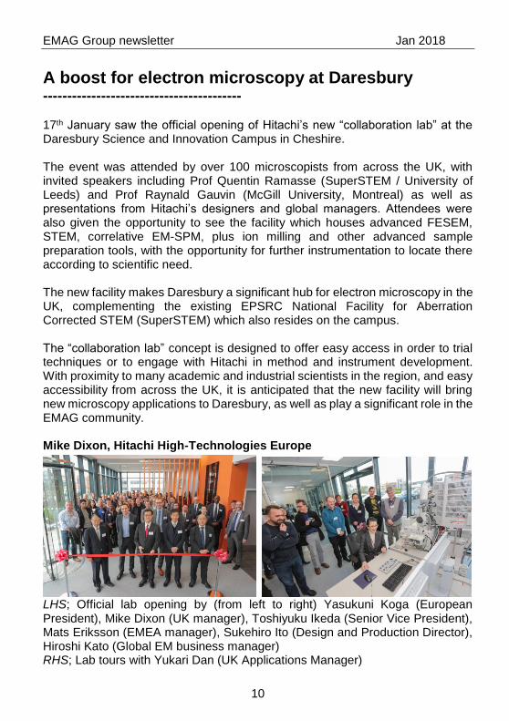
A boost for electron microscopy at Daresbury ----------------------------------------- 17th January saw the official opening of Hitachi’s new “collaboration lab” at the Daresbury Science and Innovation Campus in Cheshire. The event was attended by over 100 microscopists from across the UK, with invited speakers including Prof Quentin Ramasse (SuperSTEM / University of Leeds) and Prof Raynald Gauvin (McGill University, Montreal) as well as presentations from Hitachi’s designers and global managers. Attendees were also given the opportunity to see the facility which houses advanced FESEM, STEM, correlative EM-SPM, plus ion milling and other advanced sample preparation tools, with the opportunity for further instrumentation to locate there according to scientific need. The new facility makes Daresbury a significant hub for electron microscopy in the UK, complementing the existing EPSRC National Facility for Aberration Corrected STEM (SuperSTEM) which also resides on the campus. The “collaboration lab” concept is designed to offer easy access in order to trial techniques or to engage with Hitachi in method and instrument development. With proximity to many academic and industrial scientists in the region, and easy accessibility from across the UK, it is anticipated that the new facility will bring new microscopy applications to Daresbury, as well as play a significant role in the EMAG community. Mike Dixon, Hitachi High-Technologies Europe
LHS; Official lab opening by (from left to right) Yasukuni Koga (European President), Mike Dixon (UK manager), Toshiyuku Ikeda (Senior Vice President), Mats Eriksson (EMEA manager), Sukehiro Ito (Design and Production Director), Hiroshi Kato (Global EM business manager) RHS; Lab tours with Yukari Dan (UK Applications Manager)
EMAG Group newsletter Jan 2018
11
MEETING REPORTS Microscopy of Semiconducting Materials, MSM-XX 2017 Oxford
Veerendra C Angadi, PhD student, University of Sheffield I had an opportunity to attend the 20th Microscopy of Semiconducting Materials 2017 (MSM-XX 2017) held in Oxford from 9 - 13th April 2017 which is about a 2 hour drive from Sheffield. The conference included multiple sessions, in which each session is focused on various aspects of semiconductor microscopy like analytical TEM, cathodoluminescence, lattice defects, nanowires, strained layers and quantum wells etc. Lady Margaret Hall of University of Oxford was chosen as the venue. The venue has a very historic significance as it is the first women’s college in Oxford founded in 1878. The number of attendees for the conference were relatively low as it was very specific to microscopy of semiconductors. As a PhD student I had to present my poster on the topic “Determination of bandgap onset by blind deconvolution of electron energy-loss spectra (EELS): 1D EELS vs 2D EEL spectrum images”. My session was on 10th April, Monday 17:00 - 19:00 in Symposium A: Analytical TEM poster session. It was a great opportunity for me to present among fellow researchers. The discussions and questions posed by the attendees were really enlightening. The interactions and suggestions gave a lot of opportunity to improve my work. In the mean time I attended many talks by the invited speakers and the industrial exhibitors. The one that I found very interesting was a talk by Dr Peng Wang on the topic of 2D and 3D electron ptychographic reconstruction. On Wednesday evening the congress banquet was arranged in the dining hall of Lady Margaret Hall, which was right in front of the conference venue. At the banquet, a MSM traditional pub quiz was held. Overall the conference was a fantastic learning experience for me and I am sure everyone felt the same. I would like to thank IoP for the bursary support and an opportunity to write a report on my experience of the conference.

EMAG Group newsletter Jan 2018
12
Association for Research in Vision and Ophthalmology Annual Meeting 2017, Baltimore Matthew G. Pilgrim, PhD student, University College London The Association for Research in Vision and Ophthalmology annual meeting (ARVO) is the largest and most significant conference for any researcher working in the field of ophthalmology. This year the conference took place from the 7-11th May 2017 in the city of Baltimore, United States of America. By attending this year’s ARVO annual meeting I was able to disseminate my current research to both researchers and clinicians working in the field of ophthalmology. Currently, my research is focused on determining the composition of sub-retinal pigment epithelial deposits, the earliest pathological lesion of the blinding disease age-related macular degeneration (AMD). To understand the elemental and molecular composition of these lesions I am utilizing a number of high-resolution imaging modalities and molecular analysis techniques including scanning electron microscopy (SEM), density dependent colour SEM, SEM coupled energy dispersive X-ray analysis, secondary ion mass spectrometry, synchrotron micro focus X-ray fluorescence and transmission electron microscopy coupled with selected area electron diffraction. This novel approach to answering a biological question in the field of vision and ophthalmology has proven highly successful. The conference provided the perfect opportunity to showcase these methodologies and demonstrate how they can assist ophthalmic research. I presented this study in the form of a poster that was entitled “A mineralomic study of the retinal pigment epithelium – Bruch’s membrane complex in human eyes with age-related macular degeneration”. As a PhD student, attendance to ARVO 2017 was extremely beneficial. It provided a rare opportunity to discuss my research with the international community of AMD researchers. These discussions were extremely useful as I was able to discuss my results in the context of other areas of AMD research. This was invaluable as it highlighted additional lines of enquiry, in my case another inorganic compound. The poster session also provided the chance to discuss the recent publication “Sub retinal pigment epithelial deposition of drusen components including hydroxyapatite in a primary cell culture model”. This study was completed and co-authored with Professor Christine Curcio of the University of Alabama at Birmingham, who I met at ARVO 2015, and my research group at the UCL Institute of Ophthalmology. Receiving feedback about our model system as well as discussing potential experiments was extremely insightful and motivating. One of the highlights of the conference was a presentation by Dr Ruchira Singh

EMAG Group newsletter Jan 2018
13
from the University of Rochester Medical Center, United States of America. Dr Singh gave a 15-minute presentation on her current research investigating the role of the retinal pigment epithelium in macular dystrophies, including AMD. To understand the role of this cell type in disorders like AMD and Sorsby’s fundus dystrophy, Dr Singh and colleagues utilized human induced pluripotent stem cells (hiPSC), these are of great interest in ophthalmic research as they have been shown to differentiate into retinal pigment epithelial cells, the support cells of the light sensitive photoreceptor cells of the retina. Dr Singh and her group showed that hiPSCs in culture were sufficient to promote drusen formation. Cells cultured for >90 days produced sub-RPE deposits containing lipids and known drusen markers like APO-E. This study was particularly interesting as Dr Singh used Transwell inserts identical to the ones used in the aforementioned cell culture model. In our model system we found that primary RPE cells produced deposits containing both lipid and APO-E, like Dr Singh’s model, as well as hydroxyapatite. Whilst Dr Singh didn’t show data or appear to investigate whether hydroxyapatite was present in her model system, this study was of great interest as hiPSCs have been used in a recent clinical trial aiming to restore or prevent further vision loss. However, if these cells can still produce deposits containing the key molecular components of drusen, including inorganic hydroxyapatite, then perhaps this is not an optimal treatment. Overall, the annual meeting for the Association for Research in Vision and Ophthalmology was a huge success. Through discussions with the international community of AMD researchers I was able to identify possible new lines of research. Furthermore, the various talks and posters that were available each day provided a rich source of knowledge on the biological question I am currently investigating as well as providing technical information.

EMAG Group newsletter Jan 2018
14
Microscience and Microscopy Congress 2017, Manchester Julie. A. Watts, PhD, student, University of Nottingham
The conference chairs, Peter O’Toole and Rik Brydson, set the tone for a relaxed, welcoming, and informative international congress, which was the biggest yet with more attendees than previous events, 6 plenary lectures, 6 parallel sessions and a large exhibition hall packed with demonstration kit alongside knowledgeable technical experts. Three words summed up the event for me: aspirational, fast-paced, and inspirational. Aspirational because the Plenary lectures from world leaders in their varied subject areas were packed with information and brilliant concepts, yet were understandable and entertaining. I particularly enjoyed the historical perspectives and scientific breakthroughs discussed by the speakers, and the unexpected interactivity: I can see where my individual research findings fit into the bigger picture. Fast-paced, since with six parallel sessions running, not only was there scope to attend lectures highly relevant to my topic, but also to find out about different research areas and techniques. The exhibition was the place to go for advice and tips from an array of companies, and included a learning zone and workshop areas which were good places to hear about the latest microscopes, equipment and detectors. Inspirational by virtue of the variety and number of opportunities to connect with other individuals from around the world, informally at drinks receptions, the EMAG dinner or at the congress banquet, or more formally during one of the two poster sessions. The breadth of research on display was astounding. The EMAG lecture from Professor Angus Kirkland, 2017 winner of the Alan Agar Medal for Electron Microscopy, explaining how reconstruction of exit wave function phase data projected onto image data results in super-resolution was definitely inspirational. Attending the Microscience Microscopy Congress 2017 has benefitted me in practical ways thanks to helpful discussions with researchers at all levels who kindly participated in the poster sessions, and the pre-congress workshop (ImageJ/FIJI) that has enabled me to process data with vastly improved efficiency. I am very grateful to have been given the opportunity to attend this very successful four-day congress, held at Manchester Central, to the EPSRC for funding my research and to the IOP for providing a bursary towards the cost of attending the event.
EMAG Group newsletter Jan 2018
15
Microscopy and Microanalysis 2017, St Louis Anuj Pokle, PhD student, Trinity College Dublin The Microscopy and Microanalysis conference is an annual international conference held in US in the field of electron microscopy. This year it was in St. Louis, Missouri (6-10th August 2017). Fortunately, this time I got the opportunity to give two platform presentations. It was very exciting but at the same time hectic to prepare yourself for two talks. The best plenary talk that I enjoyed was on ‘Detecting Massive Black Holes via Attometry’ by Keith Riles from University of Michigan. Prior to the conference there were pre-meetings scheduled for students and scientists across the fields followed by a welcome reception on Sunday evening (6th Aug). It was an interesting informal meeting where you could get in touch with the people and have a chat over a beer and good food! There were numerous microscopy sessions organized over cellular, food, pharmaceuticals semiconductors and energy related materials. Various characterization techniques within the microscopy world were presented including In-situ experiments, FIB (Focussed Ion Beam), Cryo, Tomography, Aberration-corrector, machine learning and implementing advanced computation in microscopy. The parallel sessions going on made it pretty tough to choose the talks to attend. My prime focus of work is on aberration corrected STEM (Scanning Transmission Electron Microscopy), EELS (Electron Energy Loss Spectroscopy, EDX (Energy-dispersive X-ray Spectroscopy) over two –dimensional materials. Presentations on low loss vibrational spectroscopy (equivalent of doing optical spectroscopy at high spatial resolution) and quantified imaging which allows you to count the number atoms in a nanoparticle were very interesting. These are pretty recent developments in the field of electron microscopy. Poster presentations were much helpful to discuss the theoretical and experimental aspects in more details. The Microscopy Society of America (MSA) and the Student council organized meetings to update on the current society situation. The student council elected their new board members and put across plans of providing further travel grants for students from next year. The MSA had also organized a lunch meeting especially for early stage researchers to get proper guidance from mentors and to share their views. Thanks to the IOP Research funds, I could take care of some of my travel expenses. This was really helpful. I would strongly recommend the M&M 2017 Conference not only for electron microscopists but people from other fields including materials, biology etc. It’s a perfect way to learn new things in a friendly environment, meet the experts and networking for future collaborations and career planning.

EMAG Group newsletter Jan 2018
16
APPLY FOR IoP RESEARCH STUDENTS CONFERENCE FUND
If you are a student member and are looking for funding to attend a meeting or conference, please apply for an RSCF bursary, which may give you up to £300 towards your costs. We have several of these bursaries to give away each year. Check eligibility criteria and download the form at
http://www.iop.org/about/grants/research_student/page_38808.html
EMS MEMBERSHIP
EMAG members are reminded that they are all automatically members of the European Microscopy Society, at no cost to themselves. However, in order to receive information from the EMS, it is essential to send your e-mail address to the EMS secretary - this cannot be sent by the IOP due to the Data Protection Act. This is important, since almost all communications from the EMS are sent by e-mail, including information for voting for the next Executive Board.
Send your e-mail address (and preferably your other details, postal address, phone & fax numbers) to: [email protected] and indicate whether you agree to include this information in the EMS Yearbook. If you do NOT wish to appear in the Yearbook, your e-mail address will be used solely for the dispatch of information by the EMS secretary ([email protected]).
The EMS web page can be viewed at: http://www.eurmicsoc.org/
EMAG members are also reminded of the availability of EMS Bursaries. For more details, see http://www.eurmicsoc.org/scholarships.htm
EMAG Group newsletter Jan 2018
17
ADDITIONAL FUTURE MEETINGS OF INTEREST Microscopy characterization of organic-inorganic Interfaces 22-23rd February 2018, London https://www.rms.org.uk/discover-engage/event-calendar/microscopy-characterisation-2018.html Electron Back Scatter Diffraction Meeting 2018 10-11th April 2018, Oxford https://www.rms.org.uk/discover-engage/event-calendar/ebsd-2018.html 19th International Microscopy Congress (IMC19) 9-14th September 201, Sydney, Australia http://imc19.com/ mmc2019: Microscience Microscopy Congress 2019 1-4th July 2019, Manchester https://www.rms.org.uk/discover-engage/event-calendar/mmc2019-microscience-microscopy-congress-2019.html For more microscopy events see: http://www.eurmicsoc.org/en/meeting-calendar/calendar/

EMAG Group newsletter Jan 2018
18
EMAG contact points IOP: Institute of Physics, 80 Portland Place, London, W1B 1NT, UK Tel: +44 (0)20 7470 4800, Fax: +44 (0) 20 7470 4900 For conferences: Email: [email protected] http://www.iop.org/events/scientific/conferences/index.html Group matters: Science Support Officer Email: [email protected] EMS: European Microscopy Society Email: [email protected] http://www.eurmicsoc.org/index.html MRS: Materials Research Society, 9800 McKnight Road, Pittsburgh, PA 15237, USA. Tel: +1 412 779 3003, Fax: +1 412 779 8313 http://www.mrs.org/meetings/ MSA: Microscopy Society of America, 12100 Sunset Hills Rd., Suite 130,
Reston, VA 20190, USA. Tel: +1 703 234 4115, Fax: +1 703 435 4390 http://www.microscopy.org/ RMS: Royal Microscopical Society, 37/38 St. Clements, Oxford, OX4 1AJ. Tel: +44 1865 248 768, Fax: +44 1865 791 237 Email: [email protected] http://www.rms.org.uk/events/
This newsletter is also available on the web and in larger print sizes The contents of this newsletter do not necessarily represent the views or policies of the Institute of Physics, except where explicitly stated. The Institute of Physics, 80 Portland Place, W1B 1NT, UK. Tel: 020 7470 4800 Fax: 020 7470 4848