nih public access 1 , and thomas e. scammell syndrome
TRANSCRIPT

Neural Circuitry Engaged by Prostaglandins during the SicknessSyndrome
Clifford B. Saper1, Andrej A. Romanovsky2, and Thomas E. Scammell11Department of Neurology and Division of Sleep Medicine, Harvard Medical School, Beth IsraelDeaconess Medical Center, Boston, MA, 022152Systemic Inflammation Laboratory, Trauma Research, St. Joseph’s Hospital and Medical Center,Phoenix, AZ 85013
AbstractDuring illnesses caused by infectious disease or other sources of inflammation, a suite of brain-mediated responses called the “sickness syndrome” occurs, including fever, anorexia, sleepiness,hyperalgesia, and elevated corticosteroid secretion. Much of the sickness syndrome is mediated byprostaglandins acting on the brain, and can be prevented by non-steroidal anti-inflammatory drugs,such as aspirin or ibuprofen, that block prostaglandin synthesis. By examining whichprostaglandins are produced at which sites and how they interact with the nervous system,researchers have identified specific neural circuits that underlie the sickness syndrome.
Most people experience several episodes of acute infectious disease each year. Despite themultiplicity of organisms that cause these illnesses, and the different body tissues that theyinfect (e.g., upper respiratory, urinary, gastrointestinal systems) a similar set ofconstitutional symptoms typically occurs. These include fever, achiness, loss of appetite, andsleepiness so that most people want to crawl into bed, pull the covers over their heads, andgo to sleep. Known as the “sickness syndrome”, these autonomic, endocrine, and behavioralchanges are an adaptive CNS response to help fight infectious diseases1–4. Fever (elevationof body temperature, Tb) helps the immune system fight infections by increasing activity ofwhite blood cells and inhibiting growth of many microorganisms1, 5; achiness, fatigue, andsleepiness conserve energy that is needed to fight the infection and elevate Tb; and loss ofappetite minimizes blood glucose6 which is a preferred fuel for many microorganisms.
Over the last two decades, it has become clear that the sickness syndrome is mediated by thebrain response to inflammatory molecules produced during infection and other inflammatorydiseases (such as arthritis, autoimmune disorders, and some types of cancers). Researchershave found that prostaglandins play a key role in linking systemic inflammation to brainresponse by their ability to signal across the blood-brain barrier (BBB). The availability ofmolecular tools and mouse models for manipulating prostaglandin synthetic enzymes andreceptors has allowed investigators to begin mapping the brain circuitries that are engagedby prostaglandins to cause the sickness syndrome. In this review, we will first provide anoverview of how immune mediators affect the brain, and then examine in detail the circuitrymediating each aspect of the sickness syndrome.
Address correspondence to: Dr. C. B. Saper, Dept. of Neurology, Beth Israel Deaconess Medical Center, 330 Brookline Avenue,Boston, MA 02215, [email protected], Telephone: 617-667-2622; Fax: 617-975-5161.
NIH Public AccessAuthor ManuscriptNat Neurosci. Author manuscript; available in PMC 2013 August 21.
Published in final edited form as:Nat Neurosci. ; 15(8): 1088–1095. doi:10.1038/nn.3159.
NIH
-PA Author Manuscript
NIH
-PA Author Manuscript
NIH
-PA Author Manuscript

Many aspects of the sickness syndrome are mediated by prostaglandinsDuring an infection, a number of signals activate immune response. First, many immunesystem cells bear receptors for common macromolecules present on invading organisms. Forexample, CD14 is a protein that binds lipopolysaccharide (LPS), a common constituent ofbacterial cell walls, and activates Toll-like receptor 4 which triggers an intracellular cascadeof events activating immune responses7. In this innate immune response the activated cellsrelease an array of hormones called cytokines such as interleukin1 (IL-1β), interleukin 6,and tumor necrosis factor α (TNFα). Some cytokines attract other immune cells to the siteof the inflammation, while others act on a variety of other peripheral tissues to releaseadditional chemical mediators of inflammation.
Among these mediators are the prostaglandins. Prostaglandins are derivatives of arachidonicacid, which is converted to prostaglandin H2 by cyclo-oxygenase (COX). There are twoforms of COX: COX1 is constitutively active in many tissues, and COX2 is mainly inducedduring inflammatory responses8. Although COX2 is expressed constitutively by someneurons in the brain, systemic LPS induces COX2 in the brain only in perivascular andendothelial cells along small venules, and IL-1β primarily activates COX2 in perivascularcells9–11. These venules are found throughout the brain, but are densest in the preoptic area,the base of the hypothalamus, and in the ventrolateral medulla and nucleus of the solitarytract. A number of additional enzymes then convert prostaglandin H2 to otherprostaglandins, prostacyclins, or leukotrienes. For the purpose of this review, we willconcentrate on two of these molecules, prostaglandin E2 (PGE2, produced by microsomalPGE synthase 1, or mPGES1) and prostaglandin D2 (PGD2, produced by lipocalin PGDsynthase, or L-PGDS), as for reasons reviewed below, these molecules appear to be mostrelevant for producing the sickness syndrome. PGE2 acts on four different EP receptors(EP1-4), all of which are expressed in different parts of the central nervous system12. PGD2acts on the DP1 receptor, which is mainly found in the meninges along the surface of thebrain, especially in the area ventral to the hypothalamus. A DP2 receptor has recently beendescribed, but is present at only very low levels in the brain, and as yet no functions havebeen attributed to it in that site.
For many years, researchers have debated whether the sickness syndrome is due to theaction of cytokines directly on the brain or via an intermediary such as prostaglandins.Inflammatory cytokines such as IL-1β, interleukin-6, and TNFα, which can cause feverresponses (and are therefore sometimes called endogenous pyrogens) and other aspects ofthe sickness syndrome, are too bulky to cross the BBB in large amounts directly13, 14. Thus,although they can cause local inflammation if injected directly into the brain, theseresponses are more akin to encephalitis, whereas in this review we will concentrate oncentral nervous system (CNS) responses caused by systemic immune activation.
Cytokines can, however, enter the brain in small quantities at the circumventricular organs,small islands of neural tissue along the surface of the cerebral ventricles that lack a BBB,allowing circulating proteins to interact with nearby neurons13. In addition, there is evidencethat the inflammatory cytokines can be actively transported into the CNS14, but thephysiological significance of this process is not clear. The strongest argument that cytokinesrequire prostaglandin intermediates to activate many of the components of the sicknesssyndrome is one that is exploited by millions of people on a daily basis: COX inhibitorssuch as aspirin, ibuprofen, or naproxen can substantially reduce fever, sleepiness, pain, andanorexia. The neural circuits underlying these prostaglandin-dependent components of thesickness syndrome have been intensively studied, and will be the focus of the remainder ofthis review.
Saper et al. Page 2
Nat Neurosci. Author manuscript; available in PMC 2013 August 21.
NIH
-PA Author Manuscript
NIH
-PA Author Manuscript
NIH
-PA Author Manuscript
FeverFever is a brain-regulated elevation of Tb that occurs during an inflammatory response.After peripheral administration of LPS, fever occurs in several phases, each representing adistinct burst of activity of thermoregulatory effectors and a distinct rise in Tb15. Feverresponses are blocked by systemic administration of COX inhibitors and are absent inanimals in which the mPGES1 gene has been deleted16, indicating that they are mediated byPGE2. However, the PGE2 that mediates different phases of fever may come from differentsources. The first (early) phase of LPS fever is triggered by PGE2 of peripheral origin, asneutralization of circulating PGE2 by an anti-PGE2 antibody that does not cross the BBBblocks this response17. LPS acts on Toll-like receptor 4 on hepatic (Kupffer cells) andpulmonary macrophages, in which there is upregulation of mRNA and protein forphospholipase A2 (which produces arachidonic acid), COX-2, and mPGES1, which in turnresults in an increase in PGE2 in both venous and arterial blood17, 18. The PGE2 binds toalbumin, which transports it and may protect it from enzymatic inactivation18. PGE2 maydissociate from albumin and then be carried across the BBB to its site of action byspecialized transporters that are expressed in the hypothalamus18.
The more prominent later phases of fever, which start at about 1 hour after LPSadministration and last for several hours15, are mediated by PGE2 produced by COX-2 inperivascular and endothelial cells2, 9, 10, 19. IL-1βand low doses of LPS upregulate COX2expression in perivascular cells, whereas higher doses of LPS also increase COX2 inendothelial cells, mainly along venules9, 11. However, only endothelial cells have beenfound to produce mPGES1 in adult brain20, so they are the source of PGE2 in the laterphases of fever. Induction of COX-2 and mPGES1 in brain occurs simultaneously with thelater febrile phases18, 21. Whereas studies in knockout mice have demonstrated theindispensable roles of COX-222 and mPGES116 in LPS fever, studies with cell type-specificmodulation of expression of these enzymes are still needed to determine the cellular sourceof febrigenic PGE218. The interplay of PGE2-degrading and transporting systems may alsoplay an important role in regulating brain levesl of PGE2 during fever18.
PGE2 acts on neuronal receptors of the EP family within the thermoregulation circuitry totrigger fever. The EP3 receptor is now viewed as the principle type responsible forfever23–25, but EP1 receptors may also contribute under some conditions25. The EP3receptor occurs in several isoforms, and in rodents isoforms α and γ are strongly expressedin the median preoptic nucleus (MnPO)24–26 and are thought to mediate fever27. The MnPOis also the most responsive part of the brain to both the pyrogenic action of PGE228 and theantipyretic action of intracerebral injection of ketorolac (a COX inhibitor) after LPSadministration29. Focal deletion of EP3 receptors in the MnPO has shown that their presenceis required for fever following systemic LPS or intracerebroventricular (i.c.v.) PGE224.Preoptic EP3-expressing neurons produce γ-aminobutyric acid (GABA)30, and are thoughtto inhibit downstream neurons that drive increases in Tb. Typically, α and γ EP3 receptorsinhibit neuronal function through Gimediated inhibition of adenylate cyclase. Hence, it islikely that PGE2 binding to EP3 receptors reduces the activity of MnPO neurons, resultingin disinhibition of downstream targets that elevate Tb31.
What targets do EP3-expressing preoptic neurons engage to increase Tb during fever? Theautonomic contribution to fever after systemic LPS32or i.c.v. PGE33 relies primarily on tworesponses: cutaneous vasoconstriction (particularly of the tail skin, the principle means bywhich rodents elevate Tb in a warm environment), and activation of thermogenesis in brownadipose tissue (BAT, which rodents recruit to elevate Tb in a cool environment). Theseresponses are controlled by two populations of GABAergic preoptic neurons that are warm-sensitive (i.e., fire faster when warmed). One population located mainly in the MnPO
Saper et al. Page 3
Nat Neurosci. Author manuscript; available in PMC 2013 August 21.
NIH
-PA Author Manuscript
NIH
-PA Author Manuscript
NIH
-PA Author Manuscript

regulates tail skin vasoconstriction by means of projections to the rostral medullary raphe(RMR), which in turn directly innervates sympathetic preganglionic neurons. These EP3-expressing MnPO neurons are probably also involved in baseline thermoregulation34. Theother group is located more caudally and laterally in the dorsolateral preoptic area andregulates BAT thermogenesis through projections to the dorsomedial hypothalamus (DMH),which also projects to the RMR35, 36. Hence, in rodents, PGE2 triggers autonomic heat-conservation and heat-production responses by acting on EP3 receptors to reduce activity ofpreoptic GABAergic neurons, thus disinhibiting sympathetic activation of tail skinvasoconstriction and BAT thermogenesis.
The fever response to LPS is opposed by central pathways containing α -melanocytestimulating hormone (α-MSH) acting on melanocortin (MC) 3 and 4 receptors37. TheMC3/4 blocker SHU-9119 increases fever due to intraperitoneal (i.p.) LPS but blocksanorexia (see below), suggesting that there is tonic action of α-MSH on MC3/4 receptorsthat opposes hyperthermia but mediates anorexia. As the DMH neurons that project to theRMR have high levels of MC4 receptors38, which are inhibitory, this is a likely site whereα-MSH might inhibit fever. LPS fever also involves several thermoregulatory behaviors,including selection of a higher ambient temperature, or warmth seeking39. Neural pathwayscontrolling thermoregulatory behaviors differ from those controlling autonomicthermoeffectors31, 36, 40. They do not travel through the preoptic area but instead may followthalamo-cortical pathways for discriminatory thermal sensitivity41. Rats with largeelectrolytic lesions of the preoptic area exhibit excellent behavioral thermoregulation,including the ability to generate fever after LPS administration by moving to a higherambient temperature, even though the same rats are incapable of mounting autonomicthermoregulatory responses39. The locus of PGE2 action for behavioral thermoregulationremains unknown.
SleepinessIrresistible, prolonged sleep is a major aspect of the sickness syndrome. Normally, about25% of the sleep period in humans is spent in rapid eye movement (REM) sleep, a statecharacterized by dreaming, fast cortical activity, and muscle paralysis, and the remaining75% is spent in non-REM (NREM) sleep, a state of slow cortical activity and lowmetabolism. Wakefulness is driven by monoaminergic and cholinergic neurons that activatethe forebrain, but during sleep these wake-promoting systems are inhibited by GABAergicneurons in the ventrolateral preoptic nucleus (VLPO) and adjacent parts of the preopticarea42. This action of the VLPO is necessary to produce adequate amounts of sleep, and akey target of the VLPO is the histaminergic tuberomammillary nucleus (TMN), which is amajor wake-promoting site. Researchers are now beginning to understand how inflammatorymediators act through these pathways to alter sleep.
In healthy human subjects, as in rats and mice, low to moderate doses of intravenous LPSincrease deep NREM sleep and reduce REM sleep43–45. Some components of the sleepresponse to systemic inflammation may be mediated by direct effects of cytokines onneurons or via other mediators such as nitric oxide on the brain46, 47. However, theimportance of prostaglandins as mediators of the sleep response is demonstrated by the factthat even after i.c.v. administration of TNF i.c.v. injection of a COX2 inhibitor blocks theincrease in NREM sleep48, 49.
Hayaishi, Urade, and colleagues have identified PGD2 as a major sleep-promoting factor.Incubation of preoptic brain slices with LPS induces production of PGD250, and infusion ofPGD2 in the subarachnoid space just ventral to the VLPO produces large and sustainedincreases in NREM sleep51. The origin of PGD2 is likely to be in the meninges, as the
Saper et al. Page 4
Nat Neurosci. Author manuscript; available in PMC 2013 August 21.
NIH
-PA Author Manuscript
NIH
-PA Author Manuscript
NIH
-PA Author Manuscript

synthetic enzyme L-PGDS is found in the meninges and choroid plexus, but not in the brainparenchyma52. PGD2 then activates target cells through DP1 receptors, which are mostabundant in the meninges just ventral to the VLPO53.
DP1 signaling then induces release of adenosine which acts as a paracrine signalingmolecule to increase sleep54. Adenosine may act in part by inhibiting wake-active neuronsthrough A1 receptors55–57, but excitatory adenosine A2a receptors also play a role. Infusionof an adenosine A2a agonist in the subarachnoid space just ventral to the preoptic areapromotes sleep, and the sleep-promoting response to PGD2 can be reduced by an A2aantagonist58, 59. It remains unclear how adenosine activates VLPO neurons, which showfewer inhibitory synaptic inputs but no direct response to adenosine56, but this question cannow be addressed by focally expressing or deleting the A2a receptor. For example, deletionof the A2a receptors in the nucleus accumbens was recently shown to block the wake-promoting effects of caffeine, which is an adenosine antagonist60. These observationssuggest that A2a receptors in the nucleus accumbens shell promote sleep, but it has not beenexplored whether this signaling pathway mediates the increase in NREM sleep withsystemic inflammation.
Whereas PGD2 ultimately activates sleep-promoting neurons in the VLPO, PGE2 activateshistaminergic, wake-promoting TMN neurons. Infusion of PGE2 in the TMN markedlyincreases wakefulness61. In addition, TMN neurons express EP4 receptors, and infusion ofan EP4 agonist in this region increases histamine release and wakefulness62. While thiseffect may seem to run counter to the sleep-promoting effects of PGD2, sleep becomes morefragmented in people treated with relatively high doses of LPS43. Hence, with mild tomoderate levels of inflammation, PGD2 may activate the VLPO sufficiently to suppressactivity in the TMN and increase sleep, whereas with more severe inflammation, PGE2 mayactivate TMN neurons sufficiently to overcome VLPO inhibition, and produce fragmented,fitful sleep, or even an agitated delirium.
AnorexiaIn the 24 hours after a single dose of i.p. LPS, both rats and mice eat and drink less and loseweight63–65. The anorexia and weight loss are eliminated by systemic administration of aCOX inhibitor (e.g., indomethacin) prior to the LPS, so these responses are likely to bemediated predominantly if not exclusively by prostaglandins. Interestingly, a relativelysmall, early component of the anorexia appears to be sensitive to COX1 inhibitors and is lostin COX1-/- mice, while the later and larger component is sensitive to COX2 inhibitors63.Similar to LPS, IL-1 administered i.p. produces anorexia which is attenuated in COX2 -/-mice63. Thus, the bulk of the response (i.e., after the first hour) is likely to be due to COX2-mediated production of prostaglandins.
The anorexic response can be replicated by i.c.v. injection of PGE2, and the response to i.p.IL-1β administration is eliminated in mice that lack mPGES166, thus suggestinginvolvement of PGE2. The anorexic effect of PGE2 i.c.v. can be reversed with the EP4receptor antagonist, ONO-AE3-208, whereas i.c.v administration of the EP4 agonist ONO-AE1-329 mimics the effect of PGE2 on feeding67. Because the EP4 receptor is present athigh levels in the paraventricular nucleus of the hypothalamus (PVH), and its expression isincreased by intravenous (i.v.) LPS68, some of the CNS effects of PGE2 may be mediatedby the PVH. Because i.c.v. PGD2 causes an increase in feeding in mice69, it is unlikely toplay a role in the anorexia seen after systemic LPS (except perhaps to limit it).
After i.v. LPS, there is activation of expression of cFos (an early-response protein that isused as a marker of neuronal activity) in neurons in the PVH, as well as in the arcuatenucleus, the DMH, and the lateral hypothalamic area70. Within the PVH, many of the cFos+
Saper et al. Page 5
Nat Neurosci. Author manuscript; available in PMC 2013 August 21.
NIH
-PA Author Manuscript
NIH
-PA Author Manuscript
NIH
-PA Author Manuscript

neurons are immunoreactive for corticotropin-releasing hormone (CRH), whereas in thearcuate nucleus many of the cFos+ neurons stain with antibodies against pro-opiomelanocortin (POMC) peptides71. The POMC neurons are thought to inhibit feedingduring the first few hours after LPS, whereas the CRH neurons are thought to mediate anincrease in corticosteroid production (see below) which may increase feeding at later times.
Recent studies have emphasized the presence of two populations of neurons in the arcuatenucleus with opposite effects on feeding. The POMC neurons express cFos afteradministration of leptin, a hormone that signifies adequate availability of energysubstrates72. They project to the PVH, the DMH, and the lateral hypothalamic area, wherethey act on MC4 receptors which inhibit feeding. Other arcuate neurons, which produceneuropeptide Y (NPY) and agouti-related protein (AgRP), project to the same targets73,where AgRP acts as an endogenous antagonist of the MC4 receptor74. The arcuate NPY/AgRP neurons, which are activated by high levels of the appetite-promoting hormoneghrelin, help drive feeding75. They also contain GABA, with which they directly inhibit thePOMC neurons and other brainstem feeding circuitry76. This network of structures isthought to modulate feeding as well as energy expenditure and body weight through avariety of CNS projections to autonomic, endocrine, and behavioral targets. Thus theactivation of neurons in these structures after i.v. LPS likely reflects their roles in regulatingfeeding. For example, the blockade of MC3/4 receptors by SHU-9119 is known to preventthe anorexia after i.p. LPS37, indicating that the POMC/α-MSH projections may play a keyrole in the response. It will be important to explore the roles of the targets of this pathwaythat express MC3/4 receptors, and to identify other chemically-defined populations ofneurons to determine which of them contribute to the anorexia.
On the other hand, i.p. LPS also causes a fall in levels of ghrelin, a peptide secreted bygastric cells that increases feeding77. Replacing the ghrelin restores feeding after i.p. LPS.The effect of LPS on ghrelin release could be blocked by i.p. indomethacin77, so it isprostaglandindependent, but whether the prostaglandins that suppress ghrelin secretion acton the brain or periphery has not been studied. Hence it is possible that the anorexiaassociated with i.p. LPS administration may be mediated by PGE2 acting at a number ofdifferent levels within the system that regulates feeding and energy metabolism.
HyperalgesiaLike anorexia, the influence of inflammation on pain perception may also be mediated atdifferent levels of the neuraxis. The classic signs of inflammation, “tumor, calor, rubor, anddolor” (swelling, warmth, redness, and pain), are due to the local effects of inflammatorymediators, which cause vasodilation, edema, and can sensitize the terminals of nociceptiveneurons causing local pain. However, the central response to pain is also susceptible tomodulation at multiple levels, from the spinal cord to the brainstem and the forebrain. Thetests used to measure pain responses are dependent upon different levels of the neuraxis, andtherefore can produce different results, even in the same model of inflammation. In addition,the effects of inflammation on pain are dynamic, showing hyperalgesia initially, but in someassays a later analgesic effect4. The later analgesia may be due to cascades of additionalmediators, such as corticosteroids, and so we will focus here on the initial, hyperalgesicresponse to acute inflammation.
Some aspects of the hyperalgesic response to inflammation apparently are not mediated byprostaglandins. For example, after i.p. injection of LPS, there is a reduction in the latency ofthe tail-flick response, a pain assay that measures tail movement in response to local heatingand is generally agreed to be spinally mediated. This response is unaffected by systemicadministration of the COX inhibitor indomethacin78. This is understandable, as i.p. LPS
Saper et al. Page 6
Nat Neurosci. Author manuscript; available in PMC 2013 August 21.
NIH
-PA Author Manuscript
NIH
-PA Author Manuscript
NIH
-PA Author Manuscript
causes expression of inflammatory mediators that can irritate local peripheral nerves. ThusCOX inhibitors may be unable to eliminate the local pain and the effects on the associatedspinal reflexes. On the other hand, the latency to paw withdrawal from noxious heat, whichis sensitive to supraspinal influences, is also reduced after i.p. LPS, but it is restored to near-normal levels by systemic administration of drugs that inhibit prostaglandin synthesis79.
The i.v. LPS model is better for examining the effect of inflammation on overall CNSresponse to pain, as there is no local site of inflammation, and LPS enters the circulationimmediately, rather than by gradual hematogenous and lymphatic uptake80. In this model,initial hyperalgesia is followed in a few hours by hypoalgesia4, 81. After i.v. LPS even atrelatively low doses (e.g., 10–100µg/kg), there is initially a reduction in the latency to pawwithdrawal to noxious heating82. This hyperalgesic response is abolished by injecting a non-selective COX inhibitor (diclofenac) or a COX2 selective inhibitor (NS-398) into thepreoptic area (but not other parts of the hypothalamus). The medial preoptic region of thehypothalamus has extensive projections to the midbrain periaqueductal gray matter83, 84,which is known to mediate antinociceptive responses.
There is some evidence that LPS-induced hyperalgesia may be reduced in animals withdeletion of EP3 (but not EP1, 2, or 4) receptors85, but whether this effect was central vsperipheral was not examined. On the other hand, after i.c.v. injection of PGE2, there is alsoa reduction in latency to paw withdrawal, and an increase in the firing rate of spinaltrigeminal neurons to noxious toe pinching81. This hyperalgesic effect was replicated byi.c.v. injection of an EP3 agonist (M&B28767), but not an EP1 (17-phenyl-ω-trinorPGE2)or EP2 (butaprost) agonist, and the most sensitive site for either PGE2 or the EP3 agonists tocause hyperalgesia was the preoptic area. Given that EP3 receptors in the preoptic area arehighly concentrated in the MnPO24, 68, which along with the adjacent medial preopticnucleus projects extensively to the periaqueductal gray matter83, this site is a likely locus forPGE2 to produce not only fever, but also hyperalgesia. However, this hypothesis remains tobe tested.
Elevated corticosteroid levelsCorticosteroid secretion is elevated by a variety of stressful stimuli, including acuteinflammation. However, the degree to which this response is dependent upon prostaglandinshas been controversial, as some studies have found complete inhibition by systemicadministration of COX inhibitors86, and others only partial inhibition71, 87, or none atall88–90. These discrepancies may be explained by differences in route of administration orspecies, as well as the time course of the effect. For example, in mice lacking either EP1 orEP3 receptors, the expected elevation of adrenocorticotropic hormone (ACTH) secretionmice was absent at 1 hour and 3 hours after i.p. LPS administration, but present at 2 hoursafter administration91. Other workers found that i.c.v. administration of COX1 (but notCOX2) inhibitors caused reductions in ACTH and corticosterone in rats only at 30 minutes,but not at later time points92. Such findings suggest a complex dynamic response, in whichsome components are prostaglandin-dependent and some are not. Most of the work onneural circuitry has been done on this prostaglandin-dependent early component of thecorticosteroid response, which will be the focus here.
Sawchenko and colleagues avoided peritoneal irritation by examining the mechanisms forelevation of CRH mRNA in neurons in the PVH after acute administration of i.v. LPS orIL-1β in rats93, 94. They found increased cFos expression in catecholaminergic neurons inthe ventrolateral medulla (VLM), and showed that the elevation of CRH mRNA wasblocked by interrupting noradrenergic and adrenergic afferents from the VLM to the PVH.They then showed increased expression of COX1 and COX2 mRNA in blood vessels in the
Saper et al. Page 7
Nat Neurosci. Author manuscript; available in PMC 2013 August 21.
NIH
-PA Author Manuscript
NIH
-PA Author Manuscript
NIH
-PA Author Manuscript
VLM after IL- 1β administration, and that injection of PGE2 into the VLM could increaseCRH mRNA expression in the PVH. However, i.c.v. administration of a COX1 inhibitoronly partially blocked the early ACTH and corticosterone responses to i.v. LPS or IL-1β,and a COX2 inhibitor had no effect92. These findings are consistent with the observationsthat a COX1 inhibitor prevents i.v. LPS from activating cFos expression in VLMcatecholamine neurons that express EP3 receptors, and which in turn activate PVH CRHneurons92, 95, but indicate that there are additional steps, at least some of which do notdepend on prostaglandins, to cause secretion of ACTH during an inflammatory response.
Matsuoka and colleagues examined the roles of different EP receptors in the corticosteroidresponse to LPS by measuring levels of ACTH in blood one hour after injecting i.p. LPS inmice91. They found that the elevation of ACTH after LPS injection was absent in EP1 andEP3 knockout mice, but unaffected in EP2 or EP4 knockout mice. LPS also increased cFosexpression in the PVH, especially in CRH neurons. Surprisingly, the increased c-Fos in thePVH still occurred in EP1 and EP3 knockout mice, although it was prevented byadministering an EP1 antagonist to EP3 knockout mice, suggesting that either EP1 or EP3receptors can activate pathways that cause cFos expression in PVH neurons, but neither onealone is sufficient to increase CRH secretion. EP1, but not EP3 receptors, were found in thecentral nucleus of the amygdala, a source of afferents to the PVH.
Another potential site of action for PGE2 would be the MnPO, which contains both EP1 andEP3 receptors, and which projects to the PVH83. Both amygdala and MnPO neurons thatproject to the PVH also receive extensive noradrenergic inputs from the medulla. However,whether the cells projecting to the PVH from these sites bear EP receptors, and whatneurotransmitters they use are not known.
In summary, the cascade of mediators that are released during inflammation can act atmultiple levels of the nervous system. Though peripheral nerves trigger some responses,there is strong evidence that the penetration of the inflammatory signal into the CNSrequires the mediation of prostaglandins, especially for fever and feeding responses, and thatprostaglandin-activated circuitry plays an important role in regulating sleep, hyperalgesia,and corticosteroid responses as well. This linkage provides an opportunity to probe the CNScircuitry that is engaged by the different prostaglandin receptors. Studies in the last decadehave identified many of these circuits, and suggest testable hypotheses about others. Inaddition, drugs that can differentially alter these responses may be of value in alleviatingsuffering due to the sickness syndrome, without diminishing its valuable adaptive effects.
AcknowledgmentsThe authors wish to acknowledge support from USPHS grants NS055367 (TES), HL095491 (CBS, TES),NS072337 (CBS), and NS41233 (AAR).
Reference List1. Kozak W, Conn CA, Kluger MJ. Lipopolysaccharide induces fever and depresses locomotor activity
in unrestrained mice. Am. J. Physiol. 1994; 266:R125–R135. [PubMed: 8304533]
2. Elmquist JK, Scammell TE, Saper CB. Mechanisms of CNS response to systemic immunechallenge: the febrile response. Trends Neurosci. 1997; 20:565–570. [PubMed: 9416669]
3. Konsman JP, Parnet P, Dantzer R. Cytokine-induced sickness behaviour: mechanisms andimplications. Trends Neurosci. 2002; 25:154–159. [PubMed: 11852148]
4. Romanovsky AA, et al. First and second phases of biphasic fever: two sequential stages of thesickness syndrome? Am. J. Physiol. 1996; 271:R244–R253. [PubMed: 8760227]
5. Kluger MJ, Kozak W, Conn CA, Leon LR, Soszynski D. Role of fever in disease. Ann. N. Y. Acad.Sci. 1998; 856:224–233. 224–233. [PubMed: 9917881]
Saper et al. Page 8
Nat Neurosci. Author manuscript; available in PMC 2013 August 21.
NIH
-PA Author Manuscript
NIH
-PA Author Manuscript
NIH
-PA Author Manuscript
6. Yates DT, et al. Effects of bacterial lipopolysaccharide injection on white blood cell counts,hematological variables, and serum glucose, insulin, and cortisol concentrations in ewes fed low- orhigh-protein diets. J. Anim Sci. 2011; 89:4286–4293. [PubMed: 21788428]
7. Bode JG, Ehlting C, Haussinger D. The macrophage response towards LPS and its control throughthe p38(MAPK)-STAT3 axis. Cell Signal. 2012
8. Kalinski P. Regulation of immune responses by prostaglandin E2. J. Immunol. 2012; 188:21–28.[PubMed: 22187483]
9. Schiltz JC, Sawchenko PE. Distinct brain vascular cell types manifest inducible cyclooxygenaseexpression as a function of the strength and nature of immune insults. J. Neurosci. 2002; 22:5606–5618. [PubMed: 12097512]
10. Breder CD, Saper CB. Expression of inducible cyclooxygenase mRNA in the mouse brain aftersystemic administration of bacterial lipopolysaccharide. Brain Res. 1996; 713:64–69. [PubMed:8724976]
11. Serrats J, et al. Dual roles for perivascular macrophages in immune-to-brain signaling. Neuron.2010; 65:94–106. [PubMed: 20152116]
12. Woodward DF, Jones RL, Narumiya S. International Union of Basic and Clinical Pharmacology.LXXXIII: classification of prostanoid receptors, updating 15 years of progress. Pharmacol. Rev.2011; 63:471–538. [PubMed: 21752876]
13. Maness LM, Kastin AJ, Banks WA. Relative contributions of a CVO and the microvascular bed todelivery of blood-borne IL- 1alpha to the brain. Am. J. Physiol. 1998; 275:E207–E212. [PubMed:9688620]
14. Banks WA, Erickson MA. The blood-brain barrier and immune function and dysfunction.Neurobiol. Dis. 2010; 37:26–32. [PubMed: 19664708]
15. Romanovsky AA, Simons CT, Kulchitsky VA. "Biphasic" fevers often consist of more than twophases. Am. J. Physiol. 1998; 275:R323–R331. [PubMed: 9688995]
16. Engblom D, et al. Microsomal prostaglandin E synthase-1 is the central switch during immune-induced pyresis. Nat. Neurosci. 2003; 6:1137–1138. [PubMed: 14566340]
17. Steiner AA, et al. Cellular and molecular bases of the initiation of fever. PLoS. Biol. 2006; 4:e284.[PubMed: 16933973]
18. Ivanov AI, Romanovsky AA. Prostaglandin E2 as a mediator of fever: synthesis and catabolism.Front Biosci. 2004; 9:1977–1993. 1977–1993. [PubMed: 14977603]
19. Matsumura K, et al. Brain endothelial cells express cyclooxygenase-2 during lipopolysaccharide-induced fever: light and electron microscopic immunocytochemical studies. J. Neurosci. 1998;18:6279–6289. [PubMed: 9698320]
20. Yamagata K, et al. Coexpression of microsomal-type prostaglandin E synthase withcyclooxygenase-2 in brain endothelial cells of rats during endotoxin-induced fever. J. Neurosci.2001; 21:2669–2677. [PubMed: 11306620]
21. Inoue W, et al. Brain-specific endothelial induction of prostaglandin E(2) synthesis enzymes andits temporal relation to fever. Neurosci. Res. 2002; 44:51–61. [PubMed: 12204293]
22. Steiner AA, et al. Expanding the febrigenic role of cyclooxygenase-2 to the previously overlookedresponses. Am. J. Physiol Regul. Integr. Comp Physiol. 2005; 289:R1253–R1257. [PubMed:16081878]
23. Ushikubi F, et al. Impaired febrile response in mice lacking the prostaglandin E receptor subtypeEP3. Nature. 1998; 395:281–284. [PubMed: 9751056]
24. Lazarus M, et al. EP3 prostaglandin receptors in the median preoptic nucleus are critical for feverresponses. Nat. Neurosci. 2007; 10:1131–1133. [PubMed: 17676060]
25. Oka T, et al. Characteristics of thermoregulatory and febrile responses in mice deficient inprostaglandin EP1 and EP3 receptors. J. Physiol. 2003; 551:945–954. [PubMed: 12837930]
26. Nakamura K, et al. Immunohistochemical localization of prostaglandin EP3 receptor in the ratnervous system. J. Comp Neurol. 2000; 421:543–569. [PubMed: 10842213]
27. Vasilache AM, Andersson J, Nilsberth C. Expression of PGE2 EP3 receptor subtypes in the mousepreoptic region. Neurosci. Lett. 2007; 423:179–183. [PubMed: 17706357]
Saper et al. Page 9
Nat Neurosci. Author manuscript; available in PMC 2013 August 21.
NIH
-PA Author Manuscript
NIH
-PA Author Manuscript
NIH
-PA Author Manuscript

28. Scammell TE, Elmquist JK, Griffin JD, Saper CB. Ventromedial preoptic prostaglandin E2activates fever-producing autonomic pathways. J. Neurosci. 1996; 16:6246–6254. [PubMed:8815905]
29. Scammell TE, Griffin JD, Elmquist JK, Saper CB. Microinjection of a cyclooxygenase inhibitorinto the anteroventral preoptic region attenuates LPS fever. Am. J. Physiol. 1998; 274:R783–R789. [PubMed: 9530246]
30. Nakamura K, et al. The rostral raphe pallidus nucleus mediates pyrogenic transmission from thepreoptic area. J. Neurosci. 2002; 22:4600–4610. [PubMed: 12040067]
31. Nakamura K. Central circuitries for body temperature regulation and fever. Am. J. Physiol Regul.Integr. Comp Physiol. 2011; 301:R1207–R1228. [PubMed: 21900642]
32. Szekely M, Szelenyi Z. Endotoxin fever in the rat. Acta Physiol Acad. Sci. Hung. 1979; 53:265–277. [PubMed: 396758]
33. Szelenyi Z, Bartho L, Szekely M, Romanovsky AA. Cholecystokinin octapeptide (CCK-8) injectedinto a cerebral ventricle induces a fever-like thermoregulatory response mediated by type B CCK-receptors in the rat. Brain Res. 1994; 638:69–77. [PubMed: 8199877]
34. Tanaka M, McKinley MJ, McAllen RM. Roles of two preoptic cell groups in tonic and febrilecontrol of rat tail sympathetic fibers. Am. J. Physiol Regul. Integr. Comp Physiol. 2009;296:R1248–R1257. [PubMed: 19211726]
35. Yoshida K, Li X, Cano G, Lazarus M, Saper CB. Parallel preoptic pathways for thermoregulation.J. Neurosci. 2009; 29:11954–11964. [PubMed: 19776281]
36. Morrison SF. 2010 Carl Ludwig Distinguished Lectureship of the APS Neural Control andAutonomic Regulation Section: Central neural pathways for thermoregulatory cold defense. J.Appl. Physiol. 2011; 110:1137–1149. [PubMed: 21270352]
37. Huang QH, Hruby VJ, Tatro JB. Role of central melanocortins in endotoxin-induced anorexia. Am.J. Physiol. 1999; 276:R864–R871. [PubMed: 10070149]
38. Kishi T, et al. Expression of melanocortin 4 receptor mRNA in the central nervous system of therat. J Comp Neurol. 2003; 457:213–235. [PubMed: 12541307]
39. Almeida MC, Steiner AA, Branco LG, Romanovsky AA. Cold-seeking behavior as athermoregulatory strategy in systemic inflammation. Eur. J. Neurosci. 2006; 23:3359–3367.[PubMed: 16820025]
40. Romanovsky AA. Thermoregulation: some concepts have changed. Functional architecture of thethermoregulatory system. Am. J. Physiol Regul. Integr. Comp Physiol. 2007; 292:R37–R46.[PubMed: 17008453]
41. Craig AD. How do you feel? Interoception: the sense of the physiological condition of the body.Nat. Rev. Neurosci. 2002; 3:655–666. [PubMed: 12154366]
42. Saper CB, Fuller PM, Pedersen NP, Lu J, Scammell TE. Sleep state switching. Neuron. 2010;68:1023–1042. [PubMed: 21172606]
43. Mullington J, et al. Dose-dependent effects of endotoxin on human sleep. Am. J. Physiol Regul.Integr. Comp Physiol. 2000; 278:R947–R955. [PubMed: 10749783]
44. Morrow JD, Opp MR. Diurnal variation of lipopolysaccharide-induced alterations in sleep andbody temperature of interleukin-6-deficient mice. Brain Behav. Immun. 2005; 19:40–51.[PubMed: 15581737]
45. Krueger JM, Kubillus S, Shoham S, Davenne D. Enhancement of slow-wave sleep by endotoxinand lipid A. Am. J. Physiol. 1986; 251:R591–R597. [PubMed: 3529990]
46. Krueger JM, et al. Involvement of cytokines in slow wave sleep. Prog. Brain Res. 2011; 193:39–47. 39–47. [PubMed: 21854954]
47. Imeri L, Opp MR. How (and why) the immune system makes us sleep. Nat. Rev. Neurosci. 2009;10:199–210. [PubMed: 19209176]
48. Yoshida H, Kubota T, Krueger JM. A cyclooxygenase-2 inhibitor attenuates spontaneous andTNF-alpha-induced non-rapid eye movement sleep in rabbits. Am. J. Physiol Regul. Integr. CompPhysiol. 2003; 285:R99–R109. [PubMed: 12623776]
49. Terao A, Matsumura H, Yoneda H, Saito M. Enhancement of slow-wave sleep by tumor necrosisfactor-alpha is mediated by cyclooxygenase-2 in rats. Neuroreport. 1998; 9:3791–3796. [PubMed:9875706]
Saper et al. Page 10
Nat Neurosci. Author manuscript; available in PMC 2013 August 21.
NIH
-PA Author Manuscript
NIH
-PA Author Manuscript
NIH
-PA Author Manuscript

50. Ueno R, et al. Role of prostaglandin D2 in the hypothermia of rats caused by bacteriallipopolysaccharide. Proc. Natl. Acad. Sci. U. S. A. 1982; 79:6093–6097. [PubMed: 6964402]
51. Matsumura H, et al. Prostaglandin D-2-sensitive, sleep-promoting zone defined in the ventralsurface of the rostral basal forebrain. Proc Natl Acad Sci USA. 1994; 91:11998–12002. [PubMed:7991572]
52. Urade Y, et al. Dominant expression of mRNA for prostaglandin D synthase in leptomeninges,choroid plexus, and oligodendrocytes of the adult rat brain. Proc. Natl. Acad. Sci. U. S. A. 1993;90:9070–9074. [PubMed: 8415655]
53. Mizoguchi A, et al. Dominant localization of prostaglandin D receptors on arachnoid trabecularcells in mouse basal forebrain and their involvement in the regulation of non-rapid eye movementsleep. Proc. Natl. Acad. Sci. U. S. A. 2001; 98:11674–11679. [PubMed: 11562489]
54. Urade Y, Hayaishi O. Prostaglandin D2 and sleep/wake regulation. Sleep Med. Rev. 2011; 15:411–418. [PubMed: 22024172]
55. Bjorness TE, Greene RW. Adenosine and sleep. Curr. Neuropharmacol. 2009; 7:238–245.[PubMed: 20190965]
56. Chamberlin NL, et al. Effects of adenosine on gabaergic synaptic inputs to identified ventrolateralpreoptic neurons. Neuroscience. 2003; 119:913–918. [PubMed: 12831851]
57. Oishi Y, Huang ZL, Fredholm BB, Urade Y, Hayaishi O. Adenosine in the tuberomammillarynucleus inhibits the histaminergic system via A1 receptors and promotes non-rapid eye movementsleep. Proc. Natl. Acad. Sci. U. S. A. 2008; 105:19992–19997. [PubMed: 19066225]
58. Scammell TE, et al. An adenosine A2a agonist increases sleep and induces Fos in ventrolateralpreoptic neurons. Neuroscience. 2001; 107:653–663. [PubMed: 11720788]
59. Satoh S, Matsumura H, Suzuki F, Hayaishi O. Promotion of sleep mediated by the A2a-adenosinereceptor and possible involvement of this receptor in the sleep induced by prostaglandin D2 in rats.Proc. Natl. Acad. Sci. U. S. A. 1996; 93:5980–5984. [PubMed: 8650205]
60. Lazarus M, et al. Arousal effect of caffeine depends on adenosine A2A receptors in the shell of thenucleus accumbens. J. Neurosci. 2011; 31:10067–10075. [PubMed: 21734299]
61. Onoe H, Watanabe Y, Ono K, Koyama Y, Hayaishi O. Prostaglandin E2 exerts an awaking effectin the posterior hypothalamus at a site distinct from that mediating its febrile action in the anteriorhypothalamus. J. Neurosci. 1992; 12:2715–2725. [PubMed: 1613554]
62. Huang ZL, et al. Prostaglandin E2 activates the histaminergic system via the EP4 receptor toinduce wakefulness in rats. J. Neurosci. 2003; 23:5975–5983. [PubMed: 12853415]
63. Swiergiel AH, Dunn AJ. Distinct roles for cyclooxygenases 1 and 2 in interleukin-1-inducedbehavioral changes. J. Pharmacol. Exp. Ther. 2002; 302:1031–1036. [PubMed: 12183660]
64. Wieczorek M, Swiergiel AH, Pournajafi-Nazarloo H, Dunn AJ. Physiological and behavioralresponses to interleukin- 1beta and LPS in vagotomized mice. Physiol Behav. 2005; 85:500–511.[PubMed: 15996692]
65. Kozak W, et al. Thermal and behavioral effects of lipopolysaccharide and influenza ininterleukin-1 beta-deficient mice. Am. J. Physiol. 1995; 269:R969–R977. [PubMed: 7503324]
66. Pecchi E, et al. Involvement of central microsomal prostaglandin E synthase- 1 in IL-1beta-induced anorexia. Physiol Genomics. 2006; 25:485–492. [PubMed: 16554545]
67. Ohinata K, Suetsugu K, Fujiwara Y, Yoshikawa M. Activation of prostaglandin E receptor EP4subtype suppresses food intake in mice. Prostaglandins Other Lipid Mediat. 2006; 81:31–36.[PubMed: 16997129]
68. Oka T, et al. Relationship of EP(1–4) prostaglandin receptors with rat hypothalamic cell groupsinvolved in lipopolysaccharide fever responses. J. Comp Neurol. 2000; 428:20–32. [PubMed:11058222]
69. Ohinata K, et al. Central prostaglandin D(2) stimulates food intake via the neuropeptide Y systemin mice. FEBS Lett. 2008; 582:679–684. [PubMed: 18258196]
70. Elmquist JK, Scammell TE, Jacobson CD, Saper CB. Distribution of Fos-like immunoreactivity inthe rat brain following intravenous lipopolysaccharide administration. J. Comp Neurol. 1996;371:85–103. [PubMed: 8835720]
Saper et al. Page 11
Nat Neurosci. Author manuscript; available in PMC 2013 August 21.
NIH
-PA Author Manuscript
NIH
-PA Author Manuscript
NIH
-PA Author Manuscript
71. Rorato R, et al. Prostaglandin mediates endotoxaemia-induced hypophagia by activation of pro-opiomelanocortin and corticotrophin-releasing factor neurons in rats. Exp. Physiol. 2009; 94:371–379. [PubMed: 19074588]
72. Elias CF, et al. Leptin differentially regulates NPY and POMC neurons projecting to the lateralhypothalamic area. Neuron. 1999; 23:775–786. [PubMed: 10482243]
73. Elias CF, et al. Chemically defined projections linking the mediobasal hypothalamus and thelateral hypothalamic area. J Comp Neurol. 1998; 402:442–459. [PubMed: 9862320]
74. Williams G, et al. The hypothalamus and the control of energy homeostasis: different circuits,different purposes. Physiol Behav. 2001; 74:683–701. [PubMed: 11790431]
75. Wang L, Saint-Pierre DH, Tache Y. Peripheral ghrelin selectively increases Fos expression inneuropeptide Y - synthesizing neurons in mouse hypothalamic arcuate nucleus. Neurosci. Lett.2002; 325:47–51. [PubMed: 12023064]
76. Wu Q, Palmiter RD. GABAergic signaling by AgRP neurons prevents anorexia via amelanocortin-independent mechanism. Eur. J. Pharmacol. 2011; 660:21–27. [PubMed: 21211531]
77. Wang L, et al. LPS inhibits fasted plasma ghrelin levels in rats: role of IL-1 and PGs and functionalimplications. Am. J. Physiol Gastrointest. Liver Physiol. 2006; 291:G611–G620. [PubMed:16959954]
78. Watkins LR, et al. Characterization of cytokine-induced hyperalgesia. Brain Res. 1994; 654:15–26.[PubMed: 7982088]
79. Schmelzer KR, et al. Enhancement of antinociception by coadministration of nonsteroidal anti-inflammatory drugs and soluble epoxide hydrolase inhibitors. Proc. Natl. Acad. Sci. U. S. A. 2006;103:13646–13651. [PubMed: 16950874]
80. Romanovsky AA, et al. Lipopolysaccharide transport from the peritoneal cavity to the blood: is itcontrolled by the vagus nerve? Auton. Neurosci. 2000; 85:133–140. [PubMed: 11189020]
81. Hori T, Oka T, Hosoi M, Abe M, Oka K. Hypothalamic mechanisms of pain modulatory actions ofcytokines and prostaglandin E2. Ann. N. Y. Acad. Sci. 2000; 917:106–120. 106–120. [PubMed:11268335]
82. Abe M, Oka T, Hori T, Takahashi S. Prostanoids in the preoptic hypothalamus mediate systemiclipopolysaccharide-induced hyperalgesia in rats. Brain Res. 2001; 916:41–49. [PubMed:11597589]
83. Uschakov A, Gong H, McGinty D, Szymusiak R. Efferent projections from the median preopticnucleus to sleep- and arousal-regulatory nuclei in the rat brain. Neuroscience. 2007; 150:104–120.[PubMed: 17928156]
84. Rizvi TA, Murphy AZ, Ennis M, Behbehani MM, Shipley MT. Medial preoptic area afferents toperiaqueductal gray medullo-output neurons: a combined Fos and tract tracing study. J. Neurosci.1996; 16:333–344. [PubMed: 8613800]
85. Ueno A, et al. Major roles of prostanoid receptors IP and EP(3) in endotoxin-induced enhancementof pain perception. Biochem. Pharmacol. 2001; 62:157–160. [PubMed: 11389873]
86. Parsadaniantz SM, et al. Effects of the inhibition of cyclo-oxygenase 1 or 2 or 5- lipoxygenase onthe activation of the hypothalamic-pituitary-adrenal axis induced by interleukin-1beta in the maleRat. J. Neuroendocrinol. 2000; 12:766–773. [PubMed: 10929089]
87. Gadek-Michalska A, Spyrka J, Bugajski J. Psychosocial stress affects the involvement ofprostaglandins and nitric oxide in the lipopolysaccharide-induced hypothalamic-pituitary-adrenalresponse. J. Physiol Pharmacol. 2005; 56:287–298. [PubMed: 15985709]
88. Dunn AJ, Chuluyan HE. The role of cyclo-oxygenase and lipoxygenase in the interleukin-1-induced activation of the HPA axis: dependence on the route of injection. Life Sci. 1992; 51:219–225. [PubMed: 1614286]
89. Roth J, Hubschle T, Pehl U, Ross G, Gerstberger R. Influence of systemic treatment withcyclooxygenase inhibitors on lipopolysaccharide-induced fever and circulating levels of cytokinesand cortisol in guinea-pigs. Pflugers Arch. 2002; 443:411–417. [PubMed: 11810211]
90. Nadjar A, Sauvant J, Combe C, Parnet P, Konsman JP. Brain cyclooxygenase-2 mediatesinterleukin-1-induced cellular activation in preoptic and arcuate hypothalamus, but not sicknesssymptoms. Neurobiol. Dis. 2010; 39:393–401. [PubMed: 20470889]
Saper et al. Page 12
Nat Neurosci. Author manuscript; available in PMC 2013 August 21.
NIH
-PA Author Manuscript
NIH
-PA Author Manuscript
NIH
-PA Author Manuscript
91. Matsuoka Y, et al. Impaired adrenocorticotropic hormone response to bacterial endotoxin in micedeficient in prostaglandin E receptor EP1 and EP3 subtypes. Proc. Natl. Acad. Sci. U. S. A. 2003;100:4132–4137. [PubMed: 12642666]
92. Garcia-Bueno B, Serrats J, Sawchenko PE. Cerebrovascular cyclooxygenase-1 expression,regulation, and role in hypothalamic-pituitary-adrenal axis activation by inflammatory stimuli. J.Neurosci. 2009; 29:12970–12981. [PubMed: 19828811]
93. Schiltz JC, Sawchenko PE. Specificity and generality of the involvement of catecholaminergicafferents in hypothalamic responses to immune insults. J. Comp Neurol. 2007; 502:455–467.[PubMed: 17366612]
94. Ericsson A, Kovacs KJ, Sawchenko PE. A functional anatomical analysis of central pathwayssubserving the effects of interleukin-1 on stress-related neuroendocrine neurons. J. Neurosci. 1994;14:897–913. [PubMed: 8301368]
95. Zhang YH, Lu J, Elmquist JK, Saper CB. Specific roles of cyclooxygenase-1 andcyclooxygenase-2 in lipopolysaccharide-induced fever and Fos expression in rat brain. J. CompNeurol. 2003; 463:3–12. [PubMed: 12811798]
96. Romanovsky AA. Signaling the brain in the early sickness syndrome: are sensory nerves involved?Front Biosci. 2004; 9:494–504. [PubMed: 14766385]
97. Romanovsky AA, Kulchitsky VA, Simons CT, Sugimoto N, Szekely M. Febrile responsiveness ofvagotomized rats is suppressed even in the absence of malnutrition. Am. J. Physiol. 1997;273:R777–R783. [PubMed: 9277568]
98. Romanovsky AA, Simons CT, Szekely M, Kulchitsky VA. The vagus nerve in thethermoregulatory response to systemic inflammation. Am. J. Physiol. 1997; 273:R407–R413.[PubMed: 9249579]
99. Luheshi GN, et al. Vagotomy attenuates the behavioural but not the pyrogenic effects ofinterleukin-1 in rats. Auton. Neurosci. 2000; 85:127–132. [PubMed: 11189019]
100. Hansen MK, et al. Subdiaphragmatic vagotomy does not block intraperitoneallipopolysaccharide-induced fever. Auton. Neurosci. 2000; 85:83–87. [PubMed: 11189031]
Saper et al. Page 13
Nat Neurosci. Author manuscript; available in PMC 2013 August 21.
NIH
-PA Author Manuscript
NIH
-PA Author Manuscript
NIH
-PA Author Manuscript
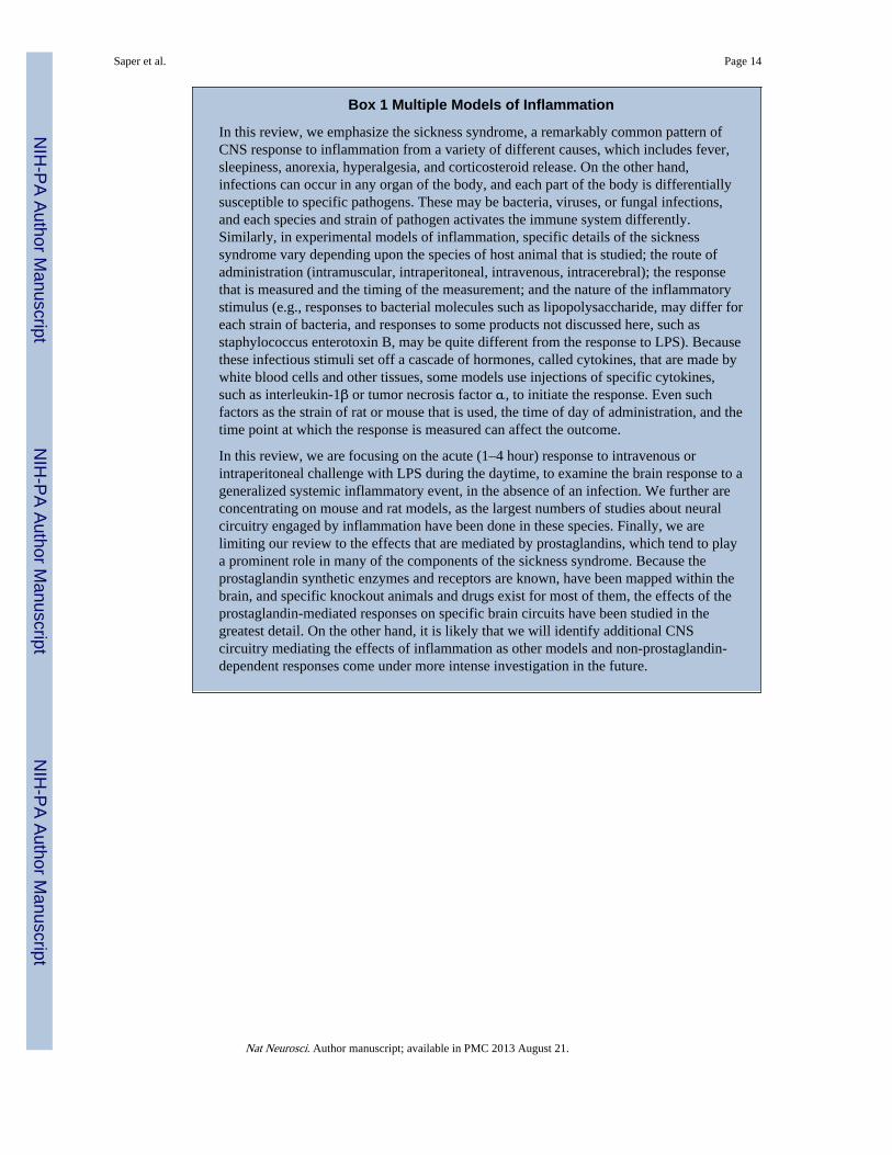
Box 1 Multiple Models of Inflammation
In this review, we emphasize the sickness syndrome, a remarkably common pattern ofCNS response to inflammation from a variety of different causes, which includes fever,sleepiness, anorexia, hyperalgesia, and corticosteroid release. On the other hand,infections can occur in any organ of the body, and each part of the body is differentiallysusceptible to specific pathogens. These may be bacteria, viruses, or fungal infections,and each species and strain of pathogen activates the immune system differently.Similarly, in experimental models of inflammation, specific details of the sicknesssyndrome vary depending upon the species of host animal that is studied; the route ofadministration (intramuscular, intraperitoneal, intravenous, intracerebral); the responsethat is measured and the timing of the measurement; and the nature of the inflammatorystimulus (e.g., responses to bacterial molecules such as lipopolysaccharide, may differ foreach strain of bacteria, and responses to some products not discussed here, such asstaphylococcus enterotoxin B, may be quite different from the response to LPS). Becausethese infectious stimuli set off a cascade of hormones, called cytokines, that are made bywhite blood cells and other tissues, some models use injections of specific cytokines,such as interleukin-1β or tumor necrosis factor α, to initiate the response. Even suchfactors as the strain of rat or mouse that is used, the time of day of administration, and thetime point at which the response is measured can affect the outcome.
In this review, we are focusing on the acute (1–4 hour) response to intravenous orintraperitoneal challenge with LPS during the daytime, to examine the brain response to ageneralized systemic inflammatory event, in the absence of an infection. We further areconcentrating on mouse and rat models, as the largest numbers of studies about neuralcircuitry engaged by inflammation have been done in these species. Finally, we arelimiting our review to the effects that are mediated by prostaglandins, which tend to playa prominent role in many of the components of the sickness syndrome. Because theprostaglandin synthetic enzymes and receptors are known, have been mapped within thebrain, and specific knockout animals and drugs exist for most of them, the effects of theprostaglandin-mediated responses on specific brain circuits have been studied in thegreatest detail. On the other hand, it is likely that we will identify additional CNScircuitry mediating the effects of inflammation as other models and non-prostaglandin-dependent responses come under more intense investigation in the future.
Saper et al. Page 14
Nat Neurosci. Author manuscript; available in PMC 2013 August 21.
NIH
-PA Author Manuscript
NIH
-PA Author Manuscript
NIH
-PA Author Manuscript
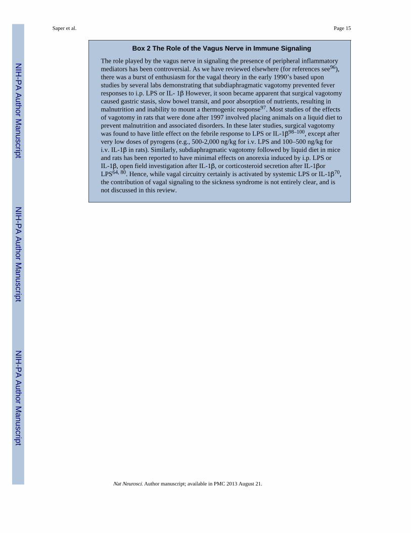
Box 2 The Role of the Vagus Nerve in Immune Signaling
The role played by the vagus nerve in signaling the presence of peripheral inflammatorymediators has been controversial. As we have reviewed elsewhere (for references see96),there was a burst of enthusiasm for the vagal theory in the early 1990’s based uponstudies by several labs demonstrating that subdiaphragmatic vagotomy prevented feverresponses to i.p. LPS or IL- 1β However, it soon became apparent that surgical vagotomycaused gastric stasis, slow bowel transit, and poor absorption of nutrients, resulting inmalnutrition and inability to mount a thermogenic response97. Most studies of the effectsof vagotomy in rats that were done after 1997 involved placing animals on a liquid diet toprevent malnutrition and associated disorders. In these later studies, surgical vagotomywas found to have little effect on the febrile response to LPS or IL-1β98–100, except aftervery low doses of pyrogens (e.g., 500-2,000 ng/kg for i.v. LPS and 100–500 ng/kg fori.v. IL-1β in rats). Similarly, subdiaphragmatic vagotomy followed by liquid diet in miceand rats has been reported to have minimal effects on anorexia induced by i.p. LPS orIL-1β, open field investigation after IL-1β, or corticosteroid secretion after IL-1βorLPS64, 80. Hence, while vagal circuitry certainly is activated by systemic LPS or IL-1β70,the contribution of vagal signaling to the sickness syndrome is not entirely clear, and isnot discussed in this review.
Saper et al. Page 15
Nat Neurosci. Author manuscript; available in PMC 2013 August 21.
NIH
-PA Author Manuscript
NIH
-PA Author Manuscript
NIH
-PA Author Manuscript
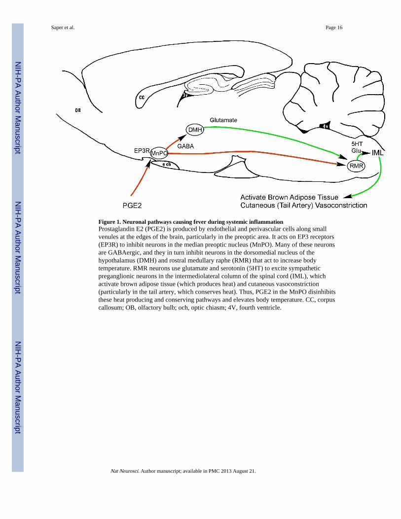
Figure 1. Neuronal pathways causing fever during systemic inflammationProstaglandin E2 (PGE2) is produced by endothelial and perivascular cells along smallvenules at the edges of the brain, particularly in the preoptic area. It acts on EP3 receptors(EP3R) to inhibit neurons in the median preoptic nucleus (MnPO). Many of these neuronsare GABAergic, and they in turn inhibit neurons in the dorsomedial nucleus of thehypothalamus (DMH) and rostral medullary raphe (RMR) that act to increase bodytemperature. RMR neurons use glutamate and serotonin (5HT) to excite sympatheticpreganglionic neurons in the intermediolateral column of the spinal cord (IML), whichactivate brown adipose tissue (which produces heat) and cutaneous vasoconstriction(particularly in the tail artery, which conserves heat). Thus, PGE2 in the MnPO disinhibitsthese heat producing and conserving pathways and elevates body temperature. CC, corpuscallosum; OB, olfactory bulb; och, optic chiasm; 4V, fourth ventricle.
Saper et al. Page 16
Nat Neurosci. Author manuscript; available in PMC 2013 August 21.
NIH
-PA Author Manuscript
NIH
-PA Author Manuscript
NIH
-PA Author Manuscript
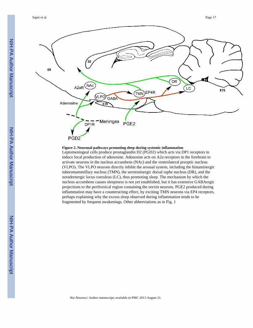
Figure 2. Neuronal pathways promoting sleep during systemic inflammationLeptomeningeal cells produce prostaglandin D2 (PGD2) which acts via DP1 receptors toinduce local production of adenosine. Adenosine acts on A2a receptors in the forebrain toactivate neurons in the nucleus accumbens (NAc) and the ventrolateral preoptic nucleus(VLPO). The VLPO neurons directly inhibit the arousal system, including the histaminergictuberomammillary nucleus (TMN), the serotoninergic dorsal raphe nucleus (DR), and thenoradrenergic locus coeruleus (LC), thus promoting sleep. The mechanism by which thenucleus accumbens causes sleepiness is not yet established, but it has extensive GABAergicprojections to the perifornical region containing the orexin neurons. PGE2 produced duringinflammation may have a counteracting effect, by exciting TMN neurons via EP4 receptors,perhaps explaining why the excess sleep observed during inflammation tends to befragmented by frequent awakenings. Other abbreviations as in Fig. 1
Saper et al. Page 17
Nat Neurosci. Author manuscript; available in PMC 2013 August 21.
NIH
-PA Author Manuscript
NIH
-PA Author Manuscript
NIH
-PA Author Manuscript

Figure 3. Neuronal pathways that may cause anorexia during systemic inflammationPGE2 produced by vascular and perivascular cells in the region of the median eminence actsvia EP4 receptors to activate pro-opiomelanocortin (POMC) expressing neurons in thearcuate nucleus. These neurons produce α-melanocyte stimulating hormone (a-MSH) whichacts via melanocortin 4 (MC4) receptors to inhibit neurons in the DMH, paraventricularnucleus (PVH), and lateral hypothalamic area (LHA) that otherwise promote feeding, bymeans of descending projections to autonomic regions that control the gastrointestinalsystem, and by ascending orexin (ORX) containing projections to the cerebral cortex.During systemic inflammation, there is also a prostaglandin-mediated fall in levels of serumghrelin, a peptide made in the stomach that promotes feeding by acting on neurons in theDMH and PVH as well as the arcuate nucleus neurons containing agouti-related peptide(AgRP). Other abbreviations as in previous figures.
Saper et al. Page 18
Nat Neurosci. Author manuscript; available in PMC 2013 August 21.
NIH
-PA Author Manuscript
NIH
-PA Author Manuscript
NIH
-PA Author Manuscript

Figure 4. Neuronal pathways that may cause hyperalgesia in the first few hours of systemicinflammationPGE2 made by vascular and perivascular cells along venules in the preoptic area acts onEP3 receptors in the MnPO. This results in disinhibition of descending inhibitory projectionsfrom the medial preoptic area (MPO) to the brainstem anti-nociceptive system, includingneurons in the periaqueductal gray matter (PAG) that promote analgesia by activatingdescending serotoninergic neurons in the RMR, which in turn inhibit nociceptive neurons inthe spinal dorsal horn. Other abbreviations as in previous figures.
Saper et al. Page 19
Nat Neurosci. Author manuscript; available in PMC 2013 August 21.
NIH
-PA Author Manuscript
NIH
-PA Author Manuscript
NIH
-PA Author Manuscript
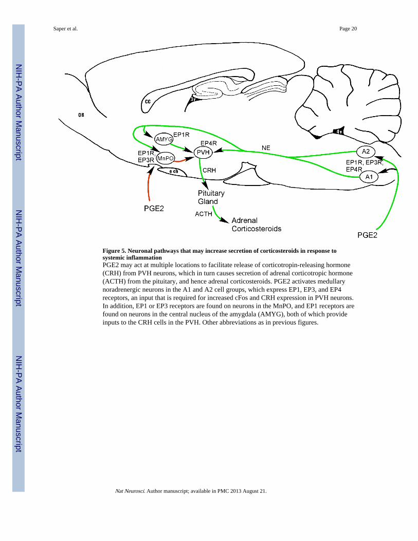
Figure 5. Neuronal pathways that may increase secretion of corticosteroids in response tosystemic inflammationPGE2 may act at multiple locations to facilitate release of corticotropin-releasing hormone(CRH) from PVH neurons, which in turn causes secretion of adrenal corticotropic hormone(ACTH) from the pituitary, and hence adrenal corticosteroids. PGE2 activates medullarynoradrenergic neurons in the A1 and A2 cell groups, which express EP1, EP3, and EP4receptors, an input that is required for increased cFos and CRH expression in PVH neurons.In addition, EP1 or EP3 receptors are found on neurons in the MnPO, and EP1 receptors arefound on neurons in the central nucleus of the amygdala (AMYG), both of which provideinputs to the CRH cells in the PVH. Other abbreviations as in previous figures.
Saper et al. Page 20
Nat Neurosci. Author manuscript; available in PMC 2013 August 21.
NIH
-PA Author Manuscript
NIH
-PA Author Manuscript
NIH
-PA Author Manuscript