nih public access kimberly a. rosenthal imtiaz a. siddiqui ... · mage-c2 promotes growth and...
TRANSCRIPT
MAGE-C2 Promotes Growth and Tumorigenicity of MelanomaCells, Phosphorylation of KAP1, and DNA Damage Repair
Neehar Bhatia1, Tony Z. Xiao2, Kimberly A. Rosenthal2, Imtiaz A. Siddiqui2, SaravananThiyagarajan3, Brendan Smart2, Qiao Meng4, Cindy L. Zuleger1, Hasan Mukhtar2, ShannonC. Kenney4, Mark R. Albertini1,5, and B. Jack Longley2
1Department of Medicine, University of Wisconsin, Carbone Cancer Center, Madison, Wisconsin,USA2Department of Dermatology, University of Wisconsin, Madison, Wisconsin, USA3Mazumdar-Shaw Cancer Center, Bangalore, India4McArdle Laboratory for Cancer Research, Madison, Wisconsin, USA5Medical Service, William S. Middleton Memorial Veterans Hospital, Madison, Wisconsin, USA
AbstractMelanoma-associated antigen-encoding (MAGE) genes are expressed in melanoma and othercancers but not in normal somatic cells. MAGE expression is associated with aggressive tumorgrowth, poor clinical outcome, and resistance to chemotherapy, but the mechanisms have not beencompletely elucidated. In this study, we show that downregulation of MAGE-C2 in A375melanoma cells and low-passage cultures from human metastatic melanomas (MRA cells) resultsin increased apoptosis and decreased growth of tumor xenografts in athymic nude mice.Previously, we showed that MAGE-C2 binds KAP1, a scaffolding protein that regulates DNArepair. Phosphorylation of KAP1-Serine 824 (Ser824) by ataxia-telangiectasia–mutated (ATM)kinase is necessary for repair of DNA double-strand breaks (DSBs); now we show that MAGE-C2knockdown reduces, whereas MAGE-C2 overexpression increases, ATM kinase–dependentphosphorylation of KAP1-Ser824. We demonstrate that MAGE-C2 increases co-precipitation ofKAP1 with ATM and that binding of MAGE-C2 to KAP1 is necessary for increased KAP1-Ser824 phosphorylation. Furthermore, ectopic expression of MAGE-C2 enhances repair of I-SceIendonuclease–induced DSBs in U-2OS cells. As phosphorylation of KAP1-Ser824 facilitatesrelaxation of heterochromatin, which is necessary for DNA repair and cellular proliferation, ourresults suggest that MAGE-C2 can promote tumor growth by phosphorylation of KAP1-Ser824and by enhancement of DNA damage repair.
INTRODUCTIONMelanoma is the most deadly form of skin cancer, accounting for <5% of skin cancer casesbut for more than 75% of skin cancer deaths. Unlike other common types of cancers, theincidence of melanoma continues to rise and the American Cancer Society estimates that~114,900 new cases of melanoma were diagnosed in the United States in 2010— 46,770
© 2012 The Society for Investigative Dermatology
Correspondence: Neehar Bhatia, Department of Medicine, University of Wisconsin, Carbone Cancer Center, 4040 WI Institute ofMedical Research, 1111 Highland Avenue, Madison, Wisconsin 53705, USA. [email protected]; or B. Jack Longley,Department of Dermatology, University of Wisconsin, Madison, Wisconsin, USA. [email protected].
CONFLICT OF INTERESTThe authors state no conflict of interest.
NIH Public AccessAuthor ManuscriptJ Invest Dermatol. Author manuscript; available in PMC 2013 March 01.
Published in final edited form as:J Invest Dermatol. 2013 March ; 133(3): 759–767. doi:10.1038/jid.2012.355.
NIH
-PA Author Manuscript
NIH
-PA Author Manuscript
NIH
-PA Author Manuscript

noninvasive (in situ) and 68,l30 invasive—with nearly 8,700 resulting in death(Surveillance, Epidemiology, and End Results Program and the National Center for HealthStatistics, http://www.skincancer.org/Skin-Cancer-Facts/#melanoma).
The first melanoma-associated antigen-encoding (MAGE) gene was identified in melanomain 1991 as a gene encoding tumor-specific antigens recognized by cytolytic T lymphocytes(Chomez et al., 2001). MAGE proteins have been classified into two subfamilies: MAGE-Iand MAGE-II. The MAGE-II family members are ubiquitously expressed in somatic tissue.The MAGE-I family members (collectively called MAGE hereon) are encoded by 28 genesclustered on the X chromosome (Chomez et al., 2001). MAGE proteins are normallyexpressed only in developing sperm, trophoblasts, fetal ovarian germ cells, and placenta(Takahashi et al., 1995; Jungbluth et al., 2002; Rajpert-De Meyts et al., 2003; Yakirevich etal., 2003; Gaskell et al., 2004; Pauls et al., 2006; Zhuang et al., 2006). MAGE I genes areunusual in that their expression is normally suppressed in all somatic tissues byhypermethylation of promoter region CpG islands (De Smet et al., 1999; Sigalotti et al.,2004). Genome-wide hypomethylation may occur in neoplastic transformation and oftencauses MAGE protein expression in tumor cells. Thus, MAGE proteins are peculiar for theirabsence in normal adult somatic tissues but are aberrantly expressed in variousmalignancies, including melanoma (Hofbauer et al., 1997; Basarab et al., 1999; Jungbluth etal., 2000, 2005; Park et al., 2002; Dhodapkar et al., 2003; Riker et al., 2008). Recently,MAGE expression has been identified as an independent prognostic variable associated withmetastasis and extent of relapse-free survival, comparable to Breslow thickness andulceration in melanoma (Svobodova et al., 2011).
KRAB domain–associated protein 1 (KAP1), also known as TIF1β (transcriptionalintermediary factor 1), TRIM28, or Krip1, is a universal corepressor protein and acts as amolecular scaffold (Iyengar and Farnham, 2011). KAP1 induces chromatin condensationand histone modification, resulting in repression of specific genes (Groner et al., 2010).KAP1 localizes to sites of DNA damage, in a sequence-independent manner, where it iscritical for the DNA repair process (Goodarzi et al., 2009). Phosphorylation of KAP1-Serine824 (Ser824) by ataxia-telangiectasia–mutated (ATM) kinase (Li et al., 2007) initiates andpropagates chromatin relaxation necessary for the repair of damaged DNA (White et al.,2006; Goodarzi et al., 2008). In addition, KAP1 is increasingly being recognized as a criticalmultifunctional molecule (Monte et al., 2006; Yang et al., 2007; Liu et al., 2008;Atanackovic et al., 2010; Doyle et al., 2010; Nardiello et al., 2011). Recently, we and othershave shown that MAGE proteins bind to KAP1, suppress p53 and apoptosis, and increasetumor survival in vivo (Yang et al., 2007; Doyle et al., 2010).
MAGE proteins can be considered specific cancer molecular biomarkers because of theirselective expression in cancer cells and association with aggressive tumor growth, poorprognosis, and relapse. Although recent studies have sought to understand the role of MAGEproteins in cancer, the cellular and physiological functions of MAGE proteins have not beenfully elucidated. In this study, we demonstrate that MAGE proteins are important factors inthe survival and growth of melanoma cells. Here we establish that MAGE proteins induceATM kinase–dependent phosphorylation of KAP1-Ser824 and promote DNA damagerepair, establishing another mechanism by which Class I MAGE proteins promote tumordevelopment and providing a rationale for combining anti-MAGE therapy with cytotoxicregimens, which to our knowledge has not been reported previously.
Bhatia et al. Page 2
J Invest Dermatol. Author manuscript; available in PMC 2013 March 01.
NIH
-PA Author Manuscript
NIH
-PA Author Manuscript
NIH
-PA Author Manuscript
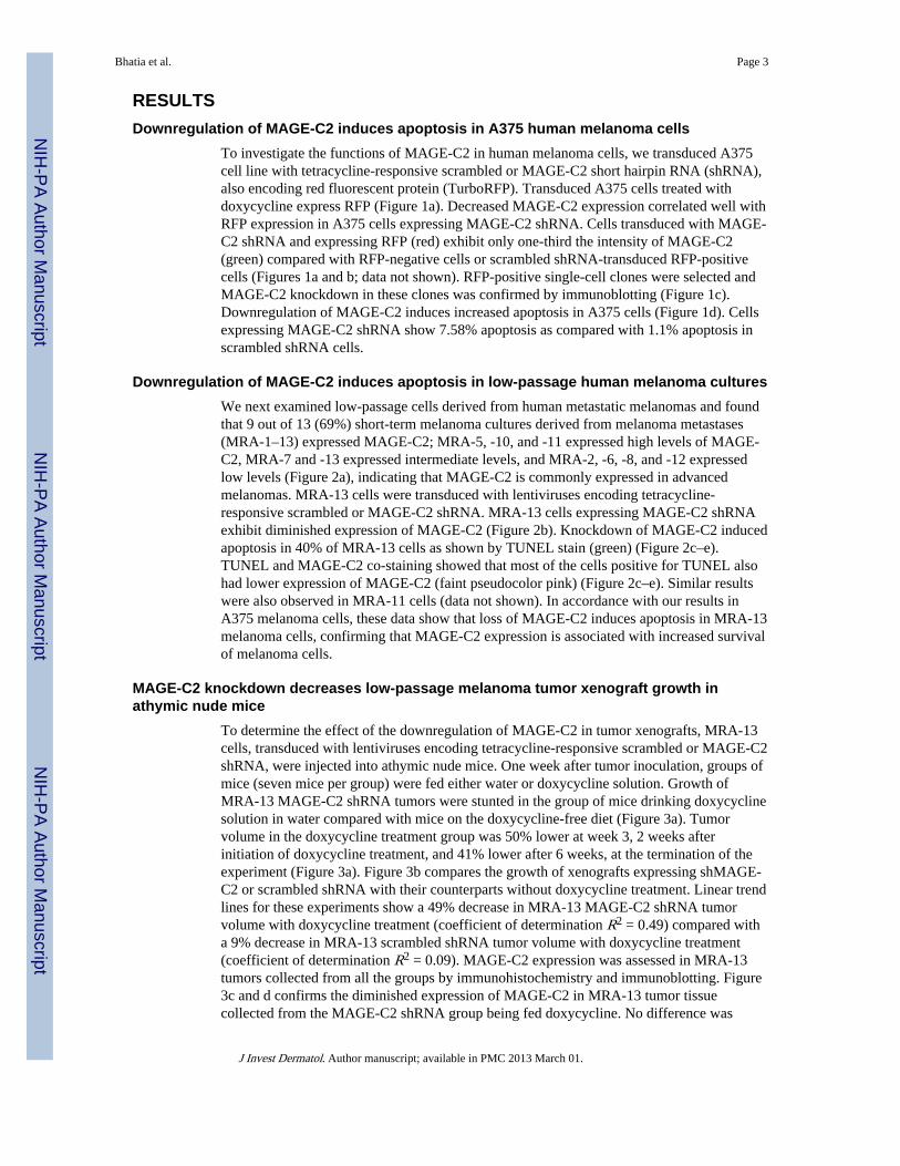
RESULTSDownregulation of MAGE-C2 induces apoptosis in A375 human melanoma cells
To investigate the functions of MAGE-C2 in human melanoma cells, we transduced A375cell line with tetracycline-responsive scrambled or MAGE-C2 short hairpin RNA (shRNA),also encoding red fluorescent protein (TurboRFP). Transduced A375 cells treated withdoxycycline express RFP (Figure 1a). Decreased MAGE-C2 expression correlated well withRFP expression in A375 cells expressing MAGE-C2 shRNA. Cells transduced with MAGE-C2 shRNA and expressing RFP (red) exhibit only one-third the intensity of MAGE-C2(green) compared with RFP-negative cells or scrambled shRNA-transduced RFP-positivecells (Figures 1a and b; data not shown). RFP-positive single-cell clones were selected andMAGE-C2 knockdown in these clones was confirmed by immunoblotting (Figure 1c).Downregulation of MAGE-C2 induces increased apoptosis in A375 cells (Figure 1d). Cellsexpressing MAGE-C2 shRNA show 7.58% apoptosis as compared with 1.1% apoptosis inscrambled shRNA cells.
Downregulation of MAGE-C2 induces apoptosis in low-passage human melanoma culturesWe next examined low-passage cells derived from human metastatic melanomas and foundthat 9 out of 13 (69%) short-term melanoma cultures derived from melanoma metastases(MRA-1–13) expressed MAGE-C2; MRA-5, -10, and -11 expressed high levels of MAGE-C2, MRA-7 and -13 expressed intermediate levels, and MRA-2, -6, -8, and -12 expressedlow levels (Figure 2a), indicating that MAGE-C2 is commonly expressed in advancedmelanomas. MRA-13 cells were transduced with lentiviruses encoding tetracycline-responsive scrambled or MAGE-C2 shRNA. MRA-13 cells expressing MAGE-C2 shRNAexhibit diminished expression of MAGE-C2 (Figure 2b). Knockdown of MAGE-C2 inducedapoptosis in 40% of MRA-13 cells as shown by TUNEL stain (green) (Figure 2c–e).TUNEL and MAGE-C2 co-staining showed that most of the cells positive for TUNEL alsohad lower expression of MAGE-C2 (faint pseudocolor pink) (Figure 2c–e). Similar resultswere also observed in MRA-11 cells (data not shown). In accordance with our results inA375 melanoma cells, these data show that loss of MAGE-C2 induces apoptosis in MRA-13melanoma cells, confirming that MAGE-C2 expression is associated with increased survivalof melanoma cells.
MAGE-C2 knockdown decreases low-passage melanoma tumor xenograft growth inathymic nude mice
To determine the effect of the downregulation of MAGE-C2 in tumor xenografts, MRA-13cells, transduced with lentiviruses encoding tetracycline-responsive scrambled or MAGE-C2shRNA, were injected into athymic nude mice. One week after tumor inoculation, groups ofmice (seven mice per group) were fed either water or doxycycline solution. Growth ofMRA-13 MAGE-C2 shRNA tumors were stunted in the group of mice drinking doxycyclinesolution in water compared with mice on the doxycycline-free diet (Figure 3a). Tumorvolume in the doxycycline treatment group was 50% lower at week 3, 2 weeks afterinitiation of doxycycline treatment, and 41% lower after 6 weeks, at the termination of theexperiment (Figure 3a). Figure 3b compares the growth of xenografts expressing shMAGE-C2 or scrambled shRNA with their counterparts without doxycycline treatment. Linear trendlines for these experiments show a 49% decrease in MRA-13 MAGE-C2 shRNA tumorvolume with doxycycline treatment (coefficient of determination R2 = 0.49) compared witha 9% decrease in MRA-13 scrambled shRNA tumor volume with doxycycline treatment(coefficient of determination R2 = 0.09). MAGE-C2 expression was assessed in MRA-13tumors collected from all the groups by immunohistochemistry and immunoblotting. Figure3c and d confirms the diminished expression of MAGE-C2 in MRA-13 tumor tissuecollected from the MAGE-C2 shRNA group being fed doxycycline. No difference was
Bhatia et al. Page 3
J Invest Dermatol. Author manuscript; available in PMC 2013 March 01.
NIH
-PA Author Manuscript
NIH
-PA Author Manuscript
NIH
-PA Author Manuscript

observed in MAGE-C2 expression in MRA-13 tumors collected from scrambled shRNAgroups with or without doxycycline. Similar results were observed with tumor xenografts ofMRA-11 cells (data not shown). TUNEL staining in xenograft tumors showed increasedapoptosis in tumors with MAGE-C2 knockdown, as was seen with in vitro cultures (Figure3e). These results demonstrate and corroborate our in vitro results that MAGE-C2 promotessurvival of melanoma cells both in vitro and in vivo.
MAGE-C2 expression increases and induces phosphorylation of KAP1Phosphorylation of KAP1-Ser824 can be induced by DNA damage and is essential for DNArepair. To study the effect of MAGE-C2 downregulation on KAP1-Ser824 phosphorylation,A375 cells transduced with scrambled or MAGE-C2 shRNA were treated with doxycyclinein conjunction with doxorubicin, a DNA-damaging agent. As demonstrated in Figure 4a,MAGE-C2 knockdown in A375 cells resulted in reduced phosphorylation of KAP1-Ser824.A375 cells expressing scrambled shRNA and treated with doxorubicin did not show anydifference in levels of phospho-KAP1-Ser824. Immunoprecipitation with KAP1 antibodyand immunoblotting for phospho-KAP1-Ser824 showed lower levels of phospho-KAP1-Ser824 in A375 cells with MAGE-C2 downregulation, even without doxorubicin-inducedDNA damage (data not shown). Overall, our data suggest that inhibition of MAGE-C2expression causes a reduction in the basal levels of KAP1-Ser824 phosphorylation anddecreases KAP1-Ser824 phosphorylation in response to doxorubicin-induced DNA double-strand breaks (DSBs).
To further validate the effect of MAGE-C2 expression on phosphorylation of KAP1-Ser824,MAGE-C2 was ectopically expressed in HEK293T cells. In accordance with the results ofMAGE-C2 knockdown in A375 cells, overexpression of MAGE-C2 inducedphosphorylation of KAP1-Ser824, whereas no effect was observed with an empty vector(Figure 4b). As shown in Figure 4b, MAGE-C2 expression also enhanced doxorubicin-induced phosphorylation of KAP1-Ser824. These results demonstrate that MAGE-C2expression induces phosphorylation of KAP1-Ser824 and also enhances DNA damage–induced phosphorylation of KAP1-Ser824. As phosphorylation of KAP1-Ser824 is requiredfor DNA damage repair, these results further suggest that MAGE-C2 expression maypromote DNA repair.
MAGE-C2-induced KAP1 phosphorylation is ATM kinase–dependentATM kinase acts as a key regulator of the DNA repair cascade (Roos and Kaina, 2006;Shanbhag et al., 2010). In response to DNA damage, ATM is activated andautophosphorylated on Ser1981 and initiates a cascade of phosphorylation events, includingp53, leading to DNA repair or apoptosis (Goodarzi et al., 2008). Similar to p53phosphorylation, ATM-induced phosphorylation of KAP1-Ser824 is an early event in DNArepair, inducing relaxation of heterochromatin, which is necessary for access to sites ofDNA breaks and critical for progression of the DNA damage repair response (White et al.,2006; Goodarzi et al., 2009; Smeenk and Lohrum, 2010).
To determine whether MAGE-C2-induced phosphorylation of KAP1-Ser824 is dependenton ATM kinase, we inhibited ATM kinase activity in HEK293T cells expressing ectopicMAGE-C2. For inhibition of ATM kinase activity, we used KU55933 (2-(4-morpholinyl)-6-(1-thianthrenyl)-4H-pyran-4-one), a potent and selective inhibitor of ATM kinase. Asexhibited in Figure 5a, treatment with KU55933 reduces MAGE-C2- and doxorubicin-induced phosphorylation of KAP1-Ser824, whereas KAP1 expression remains constant. Toconfirm inhibition of ATM kinase activity by KU55933, we probed for phospho-p53-Ser15,another substrate of ATM kinase. Figure 5a confirms inhibition of p53 phosphorylation atSer15 in response to KU55933, validating the inhibition of ATM kinase activity. No change
Bhatia et al. Page 4
J Invest Dermatol. Author manuscript; available in PMC 2013 March 01.
NIH
-PA Author Manuscript
NIH
-PA Author Manuscript
NIH
-PA Author Manuscript
in p53 expression was observed. These results demonstrate that MAGE-C2-inducedphosphorylation of KAP1-Ser824 is ATM kinase–dependent.
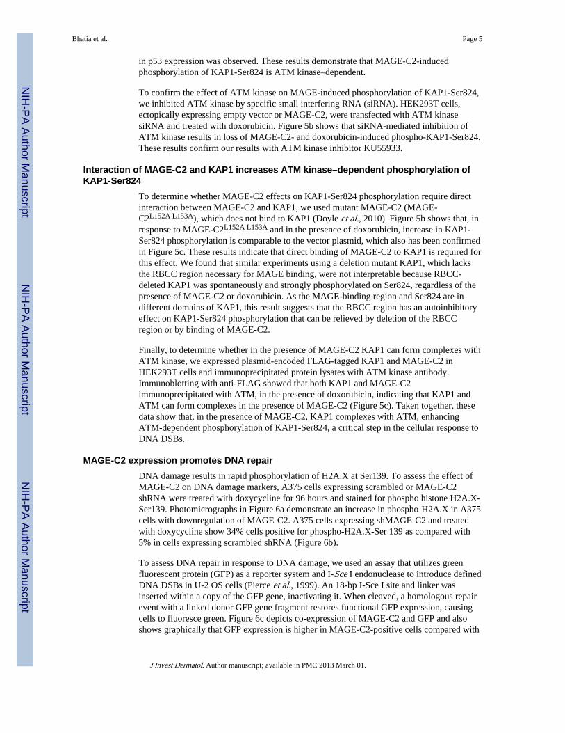
To confirm the effect of ATM kinase on MAGE-induced phosphorylation of KAP1-Ser824,we inhibited ATM kinase by specific small interfering RNA (siRNA). HEK293T cells,ectopically expressing empty vector or MAGE-C2, were transfected with ATM kinasesiRNA and treated with doxorubicin. Figure 5b shows that siRNA-mediated inhibition ofATM kinase results in loss of MAGE-C2- and doxorubicin-induced phospho-KAP1-Ser824.These results confirm our results with ATM kinase inhibitor KU55933.
Interaction of MAGE-C2 and KAP1 increases ATM kinase–dependent phosphorylation ofKAP1-Ser824
To determine whether MAGE-C2 effects on KAP1-Ser824 phosphorylation require directinteraction between MAGE-C2 and KAP1, we used mutant MAGE-C2 (MAGE-C2L152A L153A), which does not bind to KAP1 (Doyle et al., 2010). Figure 5b shows that, inresponse to MAGE-C2L152A L153A and in the presence of doxorubicin, increase in KAP1-Ser824 phosphorylation is comparable to the vector plasmid, which also has been confirmedin Figure 5c. These results indicate that direct binding of MAGE-C2 to KAP1 is required forthis effect. We found that similar experiments using a deletion mutant KAP1, which lacksthe RBCC region necessary for MAGE binding, were not interpretable because RBCC-deleted KAP1 was spontaneously and strongly phosphorylated on Ser824, regardless of thepresence of MAGE-C2 or doxorubicin. As the MAGE-binding region and Ser824 are indifferent domains of KAP1, this result suggests that the RBCC region has an autoinhibitoryeffect on KAP1-Ser824 phosphorylation that can be relieved by deletion of the RBCCregion or by binding of MAGE-C2.
Finally, to determine whether in the presence of MAGE-C2 KAP1 can form complexes withATM kinase, we expressed plasmid-encoded FLAG-tagged KAP1 and MAGE-C2 inHEK293T cells and immunoprecipitated protein lysates with ATM kinase antibody.Immunoblotting with anti-FLAG showed that both KAP1 and MAGE-C2immunoprecipitated with ATM, in the presence of doxorubicin, indicating that KAP1 andATM can form complexes in the presence of MAGE-C2 (Figure 5c). Taken together, thesedata show that, in the presence of MAGE-C2, KAP1 complexes with ATM, enhancingATM-dependent phosphorylation of KAP1-Ser824, a critical step in the cellular response toDNA DSBs.
MAGE-C2 expression promotes DNA repairDNA damage results in rapid phosphorylation of H2A.X at Ser139. To assess the effect ofMAGE-C2 on DNA damage markers, A375 cells expressing scrambled or MAGE-C2shRNA were treated with doxycycline for 96 hours and stained for phospho histone H2A.X-Ser139. Photomicrographs in Figure 6a demonstrate an increase in phospho-H2A.X in A375cells with downregulation of MAGE-C2. A375 cells expressing shMAGE-C2 and treatedwith doxycycline show 34% cells positive for phospho-H2A.X-Ser 139 as compared with5% in cells expressing scrambled shRNA (Figure 6b).
To assess DNA repair in response to DNA damage, we used an assay that utilizes greenfluorescent protein (GFP) as a reporter system and I-Sce I endonuclease to introduce definedDNA DSBs in U-2 OS cells (Pierce et al., 1999). An 18-bp I-Sce I site and linker wasinserted within a copy of the GFP gene, inactivating it. When cleaved, a homologous repairevent with a linked donor GFP gene fragment restores functional GFP expression, causingcells to fluoresce green. Figure 6c depicts co-expression of MAGE-C2 and GFP and alsoshows graphically that GFP expression is higher in MAGE-C2-positive cells compared with
Bhatia et al. Page 5
J Invest Dermatol. Author manuscript; available in PMC 2013 March 01.
NIH
-PA Author Manuscript
NIH
-PA Author Manuscript
NIH
-PA Author Manuscript

MAGE-C2-negative cells. As demonstrated in Figure 6d, 89% more cells undergo DNArepair in the presence of ectopically expressed MAGE-C2 than do cells expressing emptyvector. Figure 6e validates the expression of MAGE-C2 in U-2 OS cells by immunoblotting.These data demonstrate that MAGE-C2 expression promotes repair of DNA DSBs.
DISCUSSIONMAGE proteins are expressed in many types of human tumors including at least 50% ofadvanced malignant melanomas (Barker and Salehi, 2002). Although normal melanocytesand most melanocytic nevi do not express MAGE proteins, MAGE expression inmelanomas is associated with advanced and aggressive growth (Hofbauer et al., 1997;Basarab et al., 1999; Riker et al., 2008). Gene expression profiling shows an increase inMAGE-A3/6 expression over the entire range of melanoma thickness, a factor that correlateswith poorer prognosis (Riker et al., 2008). MAGE proteins are important prognostic factorsand have been associated with metastases, poor tumor differentiation, and clinical relapse inmelanoma; however, the mechanisms behind these associations have not been completelydetermined (Hoek et al., 2004; Bellati et al., 2007; Svobodova et al., 2011).
The work presented here confirms the effects of MAGE-C2 on the viability of A375 cells, awidely used established cell line, and with low-passage human melanoma cells frommetastases, using both in vitro and in vivo approaches. Here, our work with doxycycline-inducible shRNA encoded in integrated lentiviruses shows that established humanmelanoma xenografts can respond to MAGE-C2 knockdown triggered by a systemicallyadministered agent.
We and others have previously reported that MAGE proteins promote the growth andsurvival of tumor cells, at least in part, by suppressing p53 and apoptosis (Monte et al.,2006; Yang et al., 2006, 2007; Liu et al., 2008). More recently, Doyle et al. (2010)confirmed and extended our results, showing that MAGE proteins bind to KAP1 andenhance the E3 ubiquitin ligase activity of KAP1, causing p53 polyubiquitination anddegradation. Most recently, we have shown that MAGE proteins are master regulators ofKRAB domain containing zinc finger transcription factors (KZFs), the largest group oftranscription factors in vertebrates (Xiao et al., 2011). KZFs require KAP1 for generepression and their targets include many oncogenes and tumor suppressors.
KAP1 is critical for DNA DSB repair. In response to DNA damage, ATM kinase isactivated and phosphorylates KAP1-Ser824. Phosphorylated KAP-Ser824 localizes to DNAdamage foci and causes chromatin relaxation required for access by DNA repair proteins.Substitution of mutated KAP1S824A completely aborts the repair response and renders cellshypersensitive to DNA damage (White et al., 2006; Goodarzi et al., 2008). After the DNA isrepaired, KAP1-Ser824 is dephosphorylated, causing chromatin condensation.
We report here a function of MAGE, which to our knowledge has not been reportedpreviously, that induces ATM kinase–dependent phosphorylation of KAP1-Ser824 andactively promotes DNA repair. Previous studies and our current data support an emergingmodel in which Class I MAGE expression contributes to tumor growth and survival byfavoring DNA repair over apoptosis. As proliferation by neoplastic cells can by itself induceDNA damage, MAGE expression may provide a growth advantage in accordance withclinical observations that MAGE expression is associated with tumor progression andaggressive growth (Svobodova et al., 2011). This concept also fits nicely with in vitro and invivo observations that Class I MAGE expression is associated with resistance tochemotherapy. We also suggest that MAGE suppression, which will render tumor cells
Bhatia et al. Page 6
J Invest Dermatol. Author manuscript; available in PMC 2013 March 01.
NIH
-PA Author Manuscript
NIH
-PA Author Manuscript
NIH
-PA Author Manuscript
sensitive to DNA damage, in combination with DNA-damaging agents may enhance tumorresponse to therapy.
MATERIALS AND METHODSTissue culture
A375 melanoma cells and Human Embryonic Kidney 293T (HEK293T) cells were obtainedfrom ATCC (Manassas, VA). MRA melanoma cells, low-passage cultures derived frommetastatic melanoma patients, were provided by Dr Mark R. Albertini. Cells werepropagated in DMEM medium supplemented with 10% tet-system-approved fetal bovineserum (Clontech, Mountain View, CA) and 1% penicillin–streptomycin.
Transfections and lentiviral transductionTetracycline-responsive TRIPZ shRNAmir, MAGE-C2 or scrambled, and the Trans-Lentiviral packaging system were purchased from Open Biosystems (ThermoFisherScientific, Huntsville, AL). Lentiviruses were produced, as per manufacturer’srecommendations, and concentrated with Lentivirus Concentrator (Clontech). A375 andMRA cells were transduced with concentrated lentiviruses expressing scrambled or MAGE-C2 shRNA and selected with puromycin. Transduction efficiency, determined by expressionof TurboRFP, which is also encoded by the lentivirus vector, was ~70% in A375 cells and~90–100% in MRA-13 cells. MRA-13 cultures were used as bulk cultures, whereas single-cell clones of transduced A375 cells were selected on the basis of inducible RFP expression.MAGE-C2 knockdown was confirmed by immunoblotting. HEK293T cells were transfectedwith vector, MAGE-C2, or mutant MAGE-C2 by the calcium phosphate method and ATM472 siRNA (Dharmacon, Lafayette, CO) by Lipofectamine 2000 (Life Technologies, GrandIsland, NY).
Immunoprecipitations and immunoblottingA375 and MRA cells, transduced with scrambled or MAGE-C2 shRNA, were treated withdoxycycline and collected after 72 hours. HEK293T cells were transfected with emptyplasmid, MAGE-C2, or mutant MAGE-C2-expression plasmid. Protein lysates wereimmunoprecipitated with ATM antibody (Abcam, Cambridge, MA). The expression ofMAGE-C2, Actin (Santa Cruz Biotechnology, Santa Cruz, CA), phospho-KAP1, KAP1(Novus Biologicals, Littleton, CO), M2 FLAG (Sigma-Aldrich, St Louis, MI), and ATM(Abcam) was analyzed by immunoblotting. Density analysis was performed using ImageJsoftware (National Institutes of Health, Bethesda, MD) and was calculated for loading of thecontrols as actin or total protein expression for phosphorylated proteins. Density of thebands is shown as numbers under the blots.
Cytostaining and TUNEL assayA375 cells, transduced with lentivirus encoding scrambled or MAGE-C2 shRNA and treatedwith 0.5 μgml−1 doxycycline for 72 hours, were stained with MAGE-C2 antibody (SantaCruz Biotechnology) or phospho histone H2A.X (Cell Signaling, Danvers, MA) and FITC-labeled secondary antibody (Jackson ImmunoResearch Laboratories, West Grove, PA).MRA-13 and A375 melanoma cells, transduced with lentivirus encoding scrambled orMAGE-C2 shRNA and treated with doxycycline for 96 hours, were stained for TUNELusing the FITC-labeled DeadEnd Fluorometric TUNEL System (Promega, Madison, WI).These cells were co-stained with MAGE-C2 (Santa Cruz Biotechnology). Mouse MRA-13tumor xenograft sections were also stained with TUNEL. Quantitative analysis of TUNELstain and fluorescent MAGE-C2 or phospho-H2AX was carried out using NIS Elementssoftware (Nikon, Melville, NY).
Bhatia et al. Page 7
J Invest Dermatol. Author manuscript; available in PMC 2013 March 01.
NIH
-PA Author Manuscript
NIH
-PA Author Manuscript
NIH
-PA Author Manuscript

Athymic nude mice xenograftsMRA-13 cells, transduced with lentivirus encoding tetracycline-responsive scrambled orMAGE-C2 shRNA and selected with puromycin, were used for xenograft studies.Transduction efficiency was 90–100% as detected by RFP expression after treatment withdoxycycline. Four million MAGE-C2 or scrambled shRNA tumor cells were injectedsubcutaneously into 4-week-old athymic nude mice (Harlan Laboratories, Indianapolis, IN).A week later, half of the mice were given 2mgml−1 doxycycline in drinking water, withseven mice per group. The xenograft experiment was terminated 6 weeks later or whentumors were 1,400mm3 in volume, as per protocol.
Immunohistochemical stainingTumors collected from athymic nude mice xenografts were fixed in formalin and embeddedin paraffin. Paraffin sections were stained with MAGE-C2 antibody and developed by 3,3-diaminobenzidine (Sigma-Aldrich, St. Louis, MO).
ATM kinase inhibitor (KU55933) treatmentHEK293T cells, transfected with empty vector or MAGE-C2 expression plasmid, weretreated with KU55933 ATM kinase inhibitor (Tocris Biosciences, Ellisville, MO) for 24hours. Cells were collected and lysed as mentioned before. The expression of MAGE-C2,phospho-KAP1, KAP1, phospho p53, p53, and actin was evaluated by immunoblotting.
DNA damage and repair assayU-2 OS cells, containing an integrated modified gene for GFP, were used to measure DNAdamage repair (Pierce et al., 1999). Cells were co-transfected with I-SceI pCbASce plasmidand empty vector or MAGE-C2. After 48 hours, cells were immunostained for MAGE-C2expression (red). The expression of MAGE and GFP was enumerated and compared,including measurement of staining intensity using NIS Elements software.
AcknowledgmentsWe acknowledge Dr Maria Jasin for providing us modified U2-OS cells for DNA repair assay, and Dr AndrewSimpson and Dr Otavia Caballero for reagents. We also thank Dr V.S. Setaluri for critical suggestions and support.We are grateful to Tisha Kawahara for support with the IRB protocol. This work was supported by the Universityof Wisconsin Carbone Cancer Center, the Office of Research and Development, Biomedical Laboratory Researchand Development Service, Department of Veterans Affairs; by Grant P30 CA014520 from the National CancerInstitute; the Gretchen and Andrew Dawes Melanoma Research Fund; Ann’s Hope Foundation; the Jay Van SloanMemorial from the Steve Leuthold Family; and the Tim Eagle Memorial. The contents do not represent the viewsof the Department of Veterans Affairs or of the United States Government. Melanoma patient samples were usedunder UW-Madison Health Sciences IRB protocol number M-2009-1157.
Abbreviations
ATM ataxia-telangiectasia–mutated
DSBs double-strand breaks
GFP green fluorescent protein
MAGE melanoma-associated antigenencoding
RFP red fluorescent protein
Ser824 Serine 824
shRNA short hairpin RNA
Bhatia et al. Page 8
J Invest Dermatol. Author manuscript; available in PMC 2013 March 01.
NIH
-PA Author Manuscript
NIH
-PA Author Manuscript
NIH
-PA Author Manuscript
ReferencesAtanackovic D, Hildebrandt Y, Jadczak A, et al. Cancer-testis antigens MAGE-C1/CT7 and MAGE-
A3 are central survival factors for multiple myeloma cells. Haematol. 2010; 95:785–93.
Barker PA, Salehi A. The MAGE proteins: emerging roles in cell cycle progression, apoptosis, andneurogenetic disease. J Neurosci Res. 2002; 67:705–12. [PubMed: 11891783]
Basarab T, Picard JK, Simpson E, et al. Melanoma antigen-encoding gene expression in melanocyticnaevi and cutaneous malignant melanomas. Br J Dermatol. 1999; 140:106–8. [PubMed: 10215777]
Bellati F, Napoletano C, Tarquini E, et al. Cancer testis antigen expression in primary and recurrentvulvar cancer: association with prognostic factors. Eur J Cancer. 2007; 43:2621–7. [PubMed:17950595]
Chomez P, De Backer O, Bertrand M, et al. An overview of the MAGE gene family with theidentification of all human members of the family. Cancer Res. 2001; 61:5544–51. [PubMed:11454705]
De Smet C, Lurquin C, Lethe B, et al. DNA methylation is the primary silencing mechanism for a setof germ line- and tumor-specific genes with a CpG-rich promoter. Mol Cell Biol. 1999; 19:7327–35. [PubMed: 10523621]
Dhodapkar MV, Osman K, Teruya-Feldstein J, et al. Expression of cancer/testis (CT) antigens MAGE-A1, MAGE-A3, MAGE-A4, CT-7, and NY-ESO-1 in malignant gammopathies is heterogeneousand correlates with site, stage and risk status of disease. Cancer Immun. 2003; 3:9. [PubMed:12875607]
Doyle JM, Gao J, Wang J, et al. MAGE-RING protein complexes comprise a family of E3 ubiquitinligases. Mol Cell. 2010; 39:963–74. [PubMed: 20864041]
Gaskell TL, Esnal A, Robinson LL, et al. Immunohistochemical profiling of germ cells within thehuman fetal testis: identification of three subpopulations. Biol Reprod. 2004; 71:2012–21.[PubMed: 15317684]
Goodarzi AA, Noon AT, Deckbar D, et al. ATM signaling facilitates repair of DNA double-strandbreaks associated with heterochromatin. Mol Cell. 2008; 31:167–77. [PubMed: 18657500]
Goodarzi AA, Noon AT, Jeggo PA. The impact of heterochromatin on DSB repair. Biochem SocTrans. 2009; 37:569–76. [PubMed: 19442252]
Groner AC, Meylan S, Ciuffi A, et al. KRAB-zinc finger proteins and KAP1 can mediate long-rangetranscriptional repression through heterochromatin spreading. PLoS Genet. 2010; 6:e1000869.[PubMed: 20221260]
Hoek K, Rimm DL, Williams KR, et al. Expression profiling reveals novel pathways in thetransformation of melanocytes to melanomas. Cancer Res. 2004; 64:5270–82. [PubMed:15289333]
Hofbauer GF, Schaefer C, Noppen C, et al. MAGE-3 immunoreactivity in formalin-fixed, paraffin-embedded primary and metastatic melanoma: frequency and distribution. Am J Pathol. 1997;151:1549–53. [PubMed: 9403705]
Iyengar S, Farnham PJ. KAP1: An enigmatic master regulator of the genome. J Biol Chem. 2011;791:265–86.
Jungbluth AA, Busam KJ, Kolb D, et al. Expression of MAGE-antigens in normal tissues and cancer.Int J Cancer. 2000; 85:460–5. [PubMed: 10699915]
Jungbluth AA, Chen YT, Busam KJ, et al. CT7 (MAGE-C1) antigen expression in normal andneoplastic tissues. Int J Cancer. 2002; 99:839–45. [PubMed: 12115486]
Jungbluth AA, Ely S, Diliberto M, et al. The cancer-testis antigens CT7 (MAGE-C1) and MAGE-A3/6are commonly expressed in multiple myeloma and correlate with plasma-cell proliferation. Blood.2005; 106:167–74. [PubMed: 15761016]
Li X, Lee Y-K, Jeng J-C, et al. Role for KAP1 serine 824 phosphorylation and sumoylation/desumoylation switch in regulating KAP1-mediated transcriptional repression. J Biol Chem. 2007;282:36177–89. [PubMed: 17942393]
Liu W, Cheng S, Asa SL, et al. The melanoma-associated antigen A3 mediates fibronectin-controlledcancer progression and metastasis. Cancer Res. 2008; 68:8104–12. [PubMed: 18829569]
Bhatia et al. Page 9
J Invest Dermatol. Author manuscript; available in PMC 2013 March 01.
NIH
-PA Author Manuscript
NIH
-PA Author Manuscript
NIH
-PA Author Manuscript
Monte M, Simonatto M, Peche LY, et al. MAGE-A tumor antigens target p53 transactivation functionthrough histone deacetylase recruitment and confer resistance to chemotherapeutic agents. ProcNatl Acad Sci USA. 2006; 103:11160–5. [PubMed: 16847267]
Nardiello T, Jungbluth AA, Mei A, et al. MAGE-A inhibits apoptosis in proliferating myeloma cellsthrough repression of bax and maintenance of survivin. Clin Cancer Res. 2011; 17:4309–19.[PubMed: 21565982]
Park JW, Kwon TK, Kim IH, et al. A new strategy for the diagnosis of MAGE-expressing cancers. JImmunol Methods. 2002; 266:79–86. [PubMed: 12133624]
Pauls K, Schorle H, Jeske W, et al. Spatial expression of germ cell markers during maturation ofhuman fetal male gonads: an immunohistochemical study. Hum Reprod. 2006; 21:397–404.[PubMed: 16210381]
Pierce AJ, Johnson RD, Thompson LH, et al. XRCC3 promotes homology-directed repair of DNAdamage in mammalian cells. Genes Dev. 1999; 13:2633–8. [PubMed: 10541549]
Rajpert-De Meyts E, Jacobsen GK, Bartkova J, et al. The immunohistochemical expression pattern ofChk2, p53, p19INK4d, MAGE-A4 and other selected antigens provides new evidence for thepremeiotic origin of spermatocytic seminoma. Histopathology. 2003; 42:217–26. [PubMed:12605640]
Riker AI, Enkemann SA, Fodstad O, et al. The gene expression profiles of primary and metastaticmelanoma yields a transition point of tumor progression and metastasis. BMC Med Genomics.2008; 1:13. [PubMed: 18442402]
Roos WP, Kaina B. DNA damage-induced cell death by apoptosis. Trends Mol Med. 2006; 12:440–50. [PubMed: 16899408]
Shanbhag NM, Rafalska-Metcalf IU, Balane-Bolivar C, et al. ATM-dependent chromatin changessilence transcription in cis to DNA double-strand breaks. Cell. 2010; 141:970–81. [PubMed:20550933]
Sigalotti L, Fratta E, Coral S, et al. Intratumor heterogeneity of cancer/testis antigens expression inhuman cutaneous melanoma is methylation-regulated and functionally reverted by 5-aza-2′-deoxycytidine. Cancer Res. 2004; 64:9167–71. [PubMed: 15604288]
Smeenk L, Lohrum M. Behind the scenes: unravelling the molecular mechanisms of p53 target geneselectivity (Review). Int J Oncol. 2010; 37:1061–70. [PubMed: 20878053]
Svobodova S, Browning J, MacGregor D, et al. Cancer-testis antigen expression in primary cutaneousmelanoma has independent prognostic value comparable to that of Breslow thickness, ulcerationand mitotic rate. Eur J Cancer. 2011; 47:460–9. [PubMed: 21115342]
Takahashi K, Shichijo S, Noguchi M, et al. Identification of MAGE-1 and MAGE-4 proteins inspermatogonia and primary spermatocytes of testis. Cancer Res. 1995; 55:3478–82. [PubMed:7627949]
White DE, Negorev D, Peng H, et al. KAP1, a novel substrate for PIKK family members, colocalizeswith numerous damage response factors at DNA lesions. Cancer Res. 2006; 66:11594–9.[PubMed: 17178852]
Xiao TZ, Bhatia N, Urrutia R, et al. MAGE I transcription factors regulate KAP1 and KRAB domainzinc finger transcription factor mediated gene repression. PLoS One. 2011; 6:e23747. [PubMed:21876767]
Yakirevich E, Sabo E, Dirnfeld M, et al. Morphometrical quantification of spermatogonial germ cellswith the 57B anti-MAGE-A4 antibody in the evaluation of testicular biopsies for azoospermia.Appl Immunohistochem Mol Morphol. 2003; 11:37–44. [PubMed: 12610355]
Yang B, O’Herrin S, Wu J, et al. Select cancer testes antigens of the MAGE-A, -B, and -C families areexpressed in mast cell lines and promote cell viability in vitro and in vivo. J Invest Dermatol.2006; 127:267–75. [PubMed: 16960553]
Yang B, O’Herrin SM, Wu J, et al. MAGE-A, mMage-b, and MAGE-C proteins form complexes withKAP1 and suppress p53-dependent apoptosis in MAGE-positive cell lines. Cancer Res. 2007;67:9954–62. [PubMed: 17942928]
Zhuang R, Zhu Y, Fang L, et al. Generation of monoclonal antibodies to cancer/testis (CT) antigenCT10/MAGE-C2. Cancer Immun. 2006; 6:7. [PubMed: 16594646]
Bhatia et al. Page 10
J Invest Dermatol. Author manuscript; available in PMC 2013 March 01.
NIH
-PA Author Manuscript
NIH
-PA Author Manuscript
NIH
-PA Author Manuscript
Figure 1. Downregulation of melanoma-associated antigen-encoding (MAGE)-C2 inducesapoptosis in A375 melanoma cellsA375 cells were transduced with lentiviruses expressing tetracycline-responsive scrambledor MAGE-C2 short hairpin RNA (shRNA) and red fluorescent protein (RFP). Transductionefficiency was assessed by RFP expression and stained for MAGE-C2 (green). (a) A375cells expressing MAGE-C2 shRNA before selection with puromycin were treated with 0.5μgml−1 doxycycline for 72 hours and showed decreased MAGE-C2 expression (green) inRFP-positive cells. Bar = 50 μm. (b) A histogram of MAGE-C2 intensity in RFP-expressingversus non-expressing cells using the NIS Elements software, *P<0.05. (c) Single-cellclones were selected on the basis of RFP and MAGE-C2 expression. MAGE-C2 expressionin a representative clone is shown by immunoblotting after 72 hours of 0.5 μgml−1
doxycycline treatment. (d) Downregulation of MAGE-C2 induces apoptosis in A375 cells asanalyzed by TUNEL assay, *P<0.02.
Bhatia et al. Page 11
J Invest Dermatol. Author manuscript; available in PMC 2013 March 01.
NIH
-PA Author Manuscript
NIH
-PA Author Manuscript
NIH
-PA Author Manuscript
Figure 2. Short-term cell cultures of metastatic melanomas express melanoma-associatedantigen-encoding (MAGE)-C2, and downregulation of MAGE-C2 induces apoptosis(a) Immunoblotting detects variable expression of MAGE-C2 in 9 of 13 MRA cells, low-passage cultures from human melanoma metastases. (b) Immunoblotting shows thatdoxycycline-responsive MAGE-C2 short hairpin RNA (shRNA) downregulates MAGE-C2in MRA-13 cells. (c) Photomicrographs show MAGE-C2 downregulation (faint pseudocolorpink) in MRA-13 cells is associated with apoptosis, as measured by TUNEL staining(green). Control shRNA cells show high MAGE expression and only rare apoptotic cells.Inset shows TUNEL-positive cells magnified × 60. The arrows point to TUNEL positive(apoptotic) cells. Bar = 50 μm. (d) MAGE-C2 expression was determined by quantitativeimmunofluorescence (pseudocolor pink). The histogram shows that MAGE-C2 expression isdecreased in shMAGE-C2-expressing cells as opposed to cells expressing scrambledshRNA, confirming MAGE-C2 knockdown, *P<0.01. (e) The histogram compares TUNEL-positive (green) cells as a percentage of total 4′6-diamidino-2-phenylindole-positive cells incontrol (scrambled) and MAGE-C2 shRNA-expressing cells. The histogram shows thatTUNEL staining in MRA-13 cells is increased with MAGE-C2 knockdown as comparedwith scrambled shRNA, *P<0.02.
Bhatia et al. Page 12
J Invest Dermatol. Author manuscript; available in PMC 2013 March 01.
NIH
-PA Author Manuscript
NIH
-PA Author Manuscript
NIH
-PA Author Manuscript
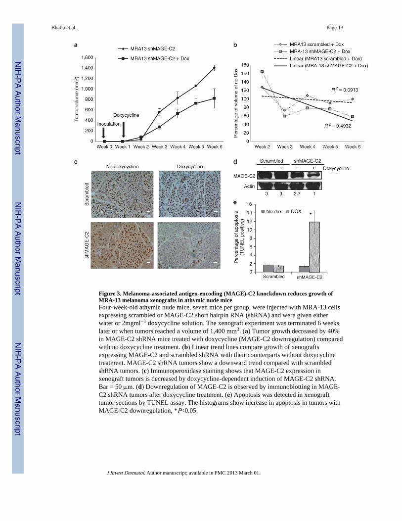
Figure 3. Melanoma-associated antigen-encoding (MAGE)-C2 knockdown reduces growth ofMRA-13 melanoma xenografts in athymic nude miceFour-week-old athymic nude mice, seven mice per group, were injected with MRA-13 cellsexpressing scrambled or MAGE-C2 short hairpin RNA (shRNA) and were given eitherwater or 2mgml−1 doxycycline solution. The xenograft experiment was terminated 6 weekslater or when tumors reached a volume of 1,400 mm3. (a) Tumor growth decreased by 40%in MAGE-C2 shRNA mice treated with doxycycline (MAGE-C2 downregulation) comparedwith no doxycycline treatment. (b) Linear trend lines compare growth of xenograftsexpressing MAGE-C2 and scrambled shRNA with their counterparts without doxycyclinetreatment. MAGE-C2 shRNA tumors show a downward trend compared with scrambledshRNA tumors. (c) Immunoperoxidase staining shows that MAGE-C2 expression inxenograft tumors is decreased by doxycycline-dependent induction of MAGE-C2 shRNA.Bar = 50 μm. (d) Downregulation of MAGE-C2 is observed by immunoblotting in MAGE-C2 shRNA tumors after doxycycline treatment. (e) Apoptosis was detected in xenografttumor sections by TUNEL assay. The histograms show increase in apoptosis in tumors withMAGE-C2 downregulation, *P<0.05.
Bhatia et al. Page 13
J Invest Dermatol. Author manuscript; available in PMC 2013 March 01.
NIH
-PA Author Manuscript
NIH
-PA Author Manuscript
NIH
-PA Author Manuscript
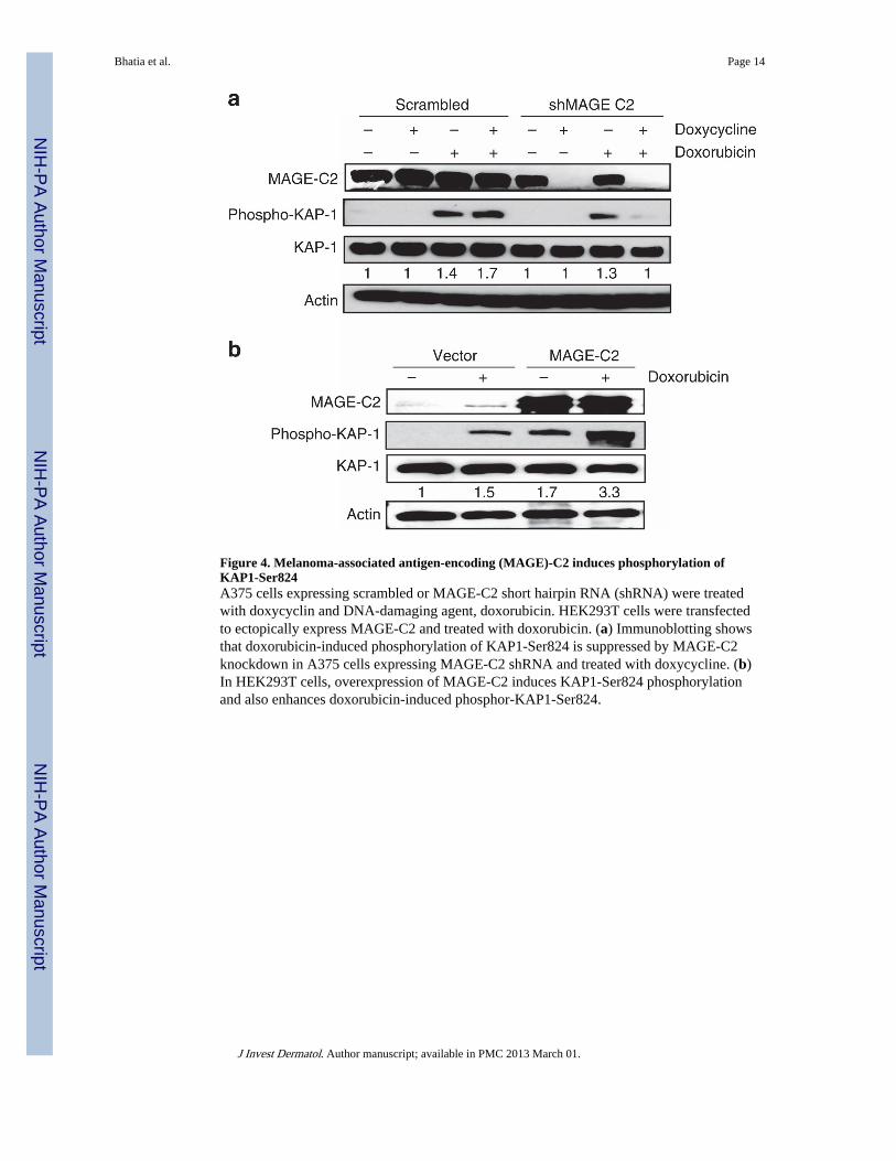
Figure 4. Melanoma-associated antigen-encoding (MAGE)-C2 induces phosphorylation ofKAP1-Ser824A375 cells expressing scrambled or MAGE-C2 short hairpin RNA (shRNA) were treatedwith doxycyclin and DNA-damaging agent, doxorubicin. HEK293T cells were transfectedto ectopically express MAGE-C2 and treated with doxorubicin. (a) Immunoblotting showsthat doxorubicin-induced phosphorylation of KAP1-Ser824 is suppressed by MAGE-C2knockdown in A375 cells expressing MAGE-C2 shRNA and treated with doxycycline. (b)In HEK293T cells, overexpression of MAGE-C2 induces KAP1-Ser824 phosphorylationand also enhances doxorubicin-induced phosphor-KAP1-Ser824.
Bhatia et al. Page 14
J Invest Dermatol. Author manuscript; available in PMC 2013 March 01.
NIH
-PA Author Manuscript
NIH
-PA Author Manuscript
NIH
-PA Author Manuscript

Figure 5. Melanoma-associated antigen-encoding (MAGE)-C2 binds to KAP-1 and inducesataxia-telangiectasia–mutated (ATM) kinase–dependent phosphorylation of KAP1-Ser824(a) HEK293T cells, ectopically expressing MAGE-C2, were treated with doxorubicin andKU55933, a specific ATM kinase inhibitor. MAGE-C2-induced phosphorylation of KAP1was blocked by the ATM kinase inhibitor KU55933. KU55933 treatment also causesreduction in phosphorylation of p53, another substrate of ATM kinase, which confirmsKU55933 activity. (b) HEK293T cells, with ectopic expression of wild-type MAGE-C2 ormutant MAGE-C2L152A L153A (unable to bind KAP1), were transfected with ATM kinasesmall interfering RNA (siRNA) and treated with doxorubicin. Immunoblotting shows thatATM siRNA inhibits MAGE-C2-induced as well as DNA damage–induced phosphorylationof KAP1-Ser824, confirming the effect of KU55933. (c) Immunoblotting shows that wild-type MAGE-C2, but not mutant MAGE-C2L152A L153A, increases KAP1-Ser824phosphorylation, in response to doxorubicin. Wild-type MAGE-C2, in the presence ofdoxorubicin treatment, increases co-precipitation of ATM kinase and KAP-1. Unable to bindto KAP-1, mutant MAGE-C2L152A L153A has no effect on ATM and KAP-1 co-precipitation. IP, immunoprecipitation.
Bhatia et al. Page 15
J Invest Dermatol. Author manuscript; available in PMC 2013 March 01.
NIH
-PA Author Manuscript
NIH
-PA Author Manuscript
NIH
-PA Author Manuscript

Figure 6. MAGE-C2 downregulation induces DNA damage, whereas melanoma-associatedantigen-encoding (MAGE)-C2 expression induces DNA damage repair(a) A375 melanoma cells expressing scrambled or MAGE-C2 short hairpin RNA (shRNA)and treated with doxycycline were stained for phospho histone H2A.X-Ser139, a marker forDNA damage. Fluorescent photomicrographs show increased expression of phospho histoneH2A.X-Ser139 in A375 cells with MAGE-C2 downregulation. Bar = 50 μm. (b) Histogramsshow quantitative analysis of phospho Histone H2A.X-Ser139-positive cells, demonstratingincreased expression of phospho Histone H2A.X-Ser139 in A375 cells with downregulationof MAGE-C2. Modified U-2 OS cells were transfected to ectopically express MAGE-C2and I-Sce I endonuclease. Cells were stained for MAGE-C2 expression, and greenfluorescent protein (GFP) was assessed, *P<0.02. (c) DNA repair, indicated by GFPexpression, is highest in MAGE-positive cells (red pseudo color). Bar = 50 μm. (d)Histogram shows that the percentage of cells undergoing DNA repair (GFP positive) ishigher in MAGE-positive cells, *P<0.05. (e) The intensity of GFP (DNA repair) is alsohigher in MAGE-positive cells, *P<0.05. (f) Immunoblotting confirms the expression ofMAGE-C2 in transfected U-2OS cells.
Bhatia et al. Page 16
J Invest Dermatol. Author manuscript; available in PMC 2013 March 01.
NIH
-PA Author Manuscript
NIH
-PA Author Manuscript
NIH
-PA Author Manuscript