non-thermal plasma assisted lithography for biomedical ... · pdf filenon-thermal plasma...
TRANSCRIPT

Int. J. Nanotechnol., Vol. 13, Nos. 10/11/12, 2016 695
Copyright © 2016 Inderscience Enterprises Ltd.
Non-thermal plasma assisted lithography for biomedical applications: an overview
Pieter Cools*, Nathalie De Geyterand Rino Morent Department of Applied Physics, Research Unit Plasma Technology, Ghent University, Sint-Pietersnieuwstraat 41 b4, 9000 Ghent, Belgium Email: [email protected] Email: [email protected] Email: [email protected] *Corresponding author
Abstract: Plasma assisted surface patterning is becoming a well-established technique that introduces both alternating surface texture and chemistry on substrates for biomedical applications. This review consists of an overview on the developments in this field over the last 15 years. Special attention is given to the different kinds of chemistry that can be introduced and their effect on the surface-cell interaction, as well as their feasibility for possible applications in the field of regenerative medicine.
Keywords: non-thermal plasma technology; lithography; micro-patterning; plasma treatment; plasma grafting; plasma polymerisation; plasma patterning.
Reference to this paper should be made as follows: Cools, P., De Geyter, N. and Morent, R. (2016) ‘Non-thermal plasma assisted lithography for biomedical applications: an overview’, Int. J. Nanotechnol., Vol. 13, Nos. 10/11/12, pp.695–715.
Biographical notes: Pieter Cools has graduated Cum Laude as a Master of Science in Chemistry (2013) at Ghent University (Belgium), specialised in surface analysis and biopolymers. He has started his PhD in the same year at the Faculty of Engineering and Architecture in the group of Professor Rino Morent and Professor Nathalie De Geyter. His PhD is focused on non-thermal plasma technology for the improvement of porous structures for tissue engineering. In the first two years of his PhD, he has contributed to 13 papers published in journals featured in ISI Web of Science and has written four chapters in books.
Nathalie De Geyter obtained a Materials Engineering degree at Ghent University (Belgium) in 2004. She received her PhD in Physical Engineering on Cold Plasma Treatment of Polymers in 2008. Since 2014, she is an Associate Professor at the Research Unit Plasma Technology of the Department of Applied Physics at the Faculty of Engineering and Architecture at Ghent University. In 2014, she obtained an ERC Starting Grant PLASMATS entitled “Plasma-assisted development and functionalisation of electrospun mats for tissue engineering purposes”. Her current research interests are focused on non-thermal plasmas for textiles and medical applications.

696 P. Cools et al.
Rino Morent obtained a degree in Physics at Ghent University (Belgium) in 2000. He received his PhD in Physical Engineering on VOC Abatement with Atmospheric Pressure DC Discharges in 2004. Since 2012, he is an Associate Professor at the Research Unit Plasma Technology of the Department of Applied Physics at the Faculty of Engineering and Architecture at Ghent University. In 2012, he obtained an ERC Starting Grant PLASMAPOR entitled “Plasma penetration into porous materials for biomedical, textile and filtration applications”. His current research interests are focused on cold atmospheric pressure plasmas for materials and environmental applications.
1 Introduction
The development of biomaterials for tissue engineering and transplant medicine has become increasingly important in the field of regenerative medicine. To successfully construct advanced bio-artificial tissues and organs, it does not suffice to develop materials that are passively being tolerated by cells. The main focus of modern biomaterials research is therefore centred around actively controlling the cell behaviour via external stimuli [1,2]. To mimic and manipulate the natural intercellular and cell-matrix interactions during their different stages (adhesion, proliferation, migration, orientation, differentiation and apoptosis) is extremely complex, as it involves up to a 100 different proteins, growth factors, and ligands. A schematic representation can be seen in Figure 1. Recreating a proactive extracellular matrix environment, using a biocompatible matrix, requires a well-defined combination of physical-chemical surface properties and surface structure [2–4].
Figure 1 Adherens junction (AJ) and focal adhesion (FA) as mechanosensors. Calcium-dependent homophilic interactions between cadherins results in binding of the actin cytoskeleton via a-catenin (a), b-catenin (b) and vinculin (Vin) complexes. Heterodimeric integrin receptors bind ECM proteins via their extracellular domains, while their cytoplasmic domains are associated with a supramolecular plaque containing talin (Tal), vinculin (Vin), paxillin (Pax), focal adhesion kinase (FAK), etc. The plaque, in turn, is connected to the termini of actin filament bundles (see online version for colours)
Source: Girard et al. [5]

Non-thermal plasma assisted lithography for biomedical applications 697
1.1 Surface wettability, charge and chemistry
Wettability and surface free energy are two well-known surface properties that greatly influence cell behaviour and in most cases a moderately hydrophilic surface is recommended. Using too hydrophilic surfaces hinders a strong cell adhesion as the cell adhesion mediating molecules bind relatively weak on such surface. Hydrophobic surfaces on the other hand, adsorb these mediating proteins, such as fibronectin or collagen, in their denatured and rigid state, significantly hampering the cell adhesion and its subsequent processes [1,6–11]. Besides surface free energy, surface chemistry plays an equally important role. Surfaces with a similar wettability, but different chemistry, can manipulate cells in very different ways. A well-known example is the osteogenic differentiation of mesenchymal stem cells (MSC). Seeding MSCs on -COOH rich surfaces results in a differentiation towards cartilage, while a –NH2 rich surface stimulates the osteogenic phenotype and a –CH3 surface enhances the MSC phenotype as such [12–14]. Other studies focusing on neural stem cell differentiation and protein adsorption have also stipulated the importance of the surface chemistry and functional group density [15–17]. Indirectly linked to the surface chemistry is the effect of charge and conductivity of the substrate. Certain functional groups such as primary amines and carboxylic acid moieties can become positively/negatively charged when stored in solutions, such as culture media, which in turn stimulates the adsorption of certain peptides and proteins [7,11,17–20].
1.2 Surface structure
Already in the beginning of the 20th century scientists were experimenting with surface structure and its effect on cell properties [22]. Today, partially stimulated by techniques originally developed in the semi-conductor industry, the patterning of biomaterials for tissue engineering has become a highly relevant research field. Depending on the scale of the applied modification, different categories can be distinguished, each with their specific purpose [22,23]:
• 100 µm < macro-roughness
• 1 µm < micro-roughness <100 µm
• 100 nm < sub-micro-roughness < 1 µm
• 5 nm < nano-roughness < 100 nm.
Macro-roughness is mainly used to increase the surface area of implant materials to enhance the mechanical anchorage to bone. With dimensions over 100 µm, the surface structural changes are too big to influence cell behaviour as such. Nano-roughness and sub-micro-roughness are generally known to stimulate the adhesion, proliferation and differentiation of cells compared to completely smooth surfaces [24–27]. Micro-roughness has a more ambiguous effect, depending on the cell type, size, surface texture and surface topography [28]. Most cells studied for tissue engineering applications have a diameter between 10 µm and 50 µm when stored in suspension. Upon adhesion on a surface, they can cover up several 100 or even 1000’s µm². Therefore they are inherently influenced by surface texture (ridges, grooves, steps,

698 P. Cools et al.
pillars and pits) and symmetry (orthogonal vs. hexagonal packing of pits) with similar dimensions [29].
1.3 Surface patterning strategies
Many different techniques are used today to generate µm and nm dimension surface textures. Some of these techniques solely introduce a physical pattern, while others combine it with an alternating chemistry as well. Many of these techniques can be combined with non-thermal plasma technology, forming the field of plasma lithography. It is a category of processing methods that combines both chemical and structural patterning. In this paragraph those strategies will be discussed that have already been combined with non-thermal plasma technology in the past. Some excellent reviews have already been written specifically on this subject and this paragraph is mainly based on these sources [1,4,13,28,30–33].
Plasma patterning strategies involving stencils/masks are the most straightforward and easy to use. An initial coating is applied to the entire substrate surface, either via plasma polymerisation, spin coating or other techniques. After that, a stencil with the wanted pattern is placed on top of the initial coating and a second plasma polymerised coating is applied. In the final step the stencil is removed and the patterned surface can be used as such. If the substrate already exhibits the wanted surface chemistry, then the initial coating step can be skipped. The resolution of the pattern mainly depends on the quality of the mask and is usually limited to the µm range [34–36].
Plasma assisted photolithography is also a stencil-based technique where geometric features drawn on a mask are transferred onto the substrate via UV-illumination. The first step in this two-step process consists of the deposition of a photoresist via spin coating (see Figure 2). After covering the photoresist with the mask of choice, the photoresist is exposed to UV-illumination for a positive photoresist and the exposed parts will dissolve. For a negative photoresist, a cross-linking reaction will occur, generating the ‘negative’ of the mask. After this step, either a plasma activation/etching step can be applied or a plasma coating can be deposited, depending on the wanted chemistry. In the final step, the lift-off, the mask is removed and the patterned surface can be used. The generated patterns can be as small as 1 µm2. If even smaller patterns are required, other techniques such as colloidal lithography are recommended.
Plasma assisted colloidal lithography uses the ability of nanoparticles to self-organise in clusters on a surface [36]. These clusters can then be used as a mask for lithography. These metal or polymer particles are typically distributed over the surface via spin-coating. Depending on the spin parameters and the functionalities of the spheres, different patterns can be generated in the nm-range and this over relatively large areas (cm2), making them ideal for biomedical applications. The fabrication process, as shown in Figure 3, goes as follows: after the deposition of an initial plasma polymerised thin film, similar to the photolithography process, the particles are deposited. This step is followed by a plasma etching step to generate the physical pattern, after which a second plasma polymerised thin film is then deposited. Finally, the sample is submersed in an ultrasonic bath to remove the nanoparticles of the surface, resulting in the exposure of the pattern.

Non-thermal plasma assisted lithography for biomedical applications 699
Figure 2 The photolithography process: 1. A spin-coated photoresist (PR) is locally exposed with UV light through a mask; 2. PR development to provide local access to the underlying substrate. One of two routes is generally followed from that stage. Route a: Deposition (3a) of a thin layer of either metal or bioactive molecules (peptides, proteins, polymers) and lift-off in an organic solvent (4a). Alternative route 3b: utilising the patterned PR layer as a mask for local dry etching of the metal (oxide) layer (which was deposited prior to PR spin coating) down into the underlying substrate and 4b lift-off the residual PR (see online version for colours)
Source: Falconnet et al. [30]
Figure 3 Schematic illustration of colloidal lithography combined with plasma-polymer patterning
Source: Singh et al. [37]
700 P. Cools et al.
1.4 Plasma technology
The term ‘plasma’, also referred to as the fourth state of matter, was introduced in 1929 by Langmuir [38]. Plasma is a partially ionised gas and consists of a mixture of electrons, ions, radicals and neutrals (atoms and molecules). Some of these particles can occur in their excited states and upon return to their ground state cause photoemission. This results in a luminescence that is characteristic for plasma [39]. Plasma are typically generated using DC, RF, MW or hot filament sources, of which a description can be found elsewhere [40]. Often plasma are classified as cold/non-thermal/non-equilibrium and hot/thermal/equilibrium plasma. Plasmas in thermal equilibrium have the same temperature for all their active species, with a range lying between 4000 K and 20,000 K [41]. Plasmas in thermal inequilibrium have electron temperatures that are several orders higher compared to the ‘heavy’ species. This results in an overall gas temperature of only a few 100 K. For the treatment of biomaterials with delicate surface textures, the use of cold plasma is recommended, as a hot plasma operating at several 1000 K will inevitably destroy the surface, if not the complete substrate [42]. Therefore this entire overview will be limited to non-thermal plasma technology.
Non-thermal plasma technology for the modification of biomaterials can then further be subdivided into two categories depending on their interactions at the plasma-surface interface: plasma activation + plasma etching and plasma polymerisation/grafting.
1.4.1 Plasma treatment and etching
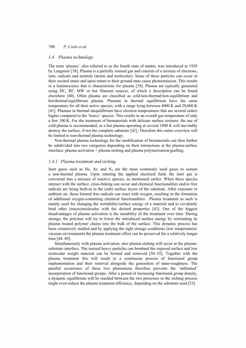
Inert gases such as He, Ar, and N2 are the most commonly used gases to sustain a non-thermal plasma. Upon entering the applied electrical field, the inert gas is converted into a mixture of reactive species, as mentioned earlier. When these species interact with the surface, cross-linking can occur and chemical functionalities and/or free radicals are being built-in in the (sub) surface layers of the substrate. After exposure to ambient air, these formed free radicals can react with oxygen, resulting in the formation of additional oxygen-containing chemical functionalities . Plasma treatment as such is mainly used for changing the wettability/surface energy of a material and to covalently bind other (macro)molecules with the desired properties [43]. One of the biggest disadvantages of plasma activation is the instability of the treatment over time. During storage, the polymer will try to lower the introduced surface energy by reorienting its plasma treated polymer chains into the bulk of the surface. This dynamic process has been extensively studied and by applying the right storage conditions (low temperatures/ vacuum environment) the plasma treatment effect can be preserved for a relatively longer time [44–49].
Simultaneously with plasma activation, also plasma etching will occur at the plasma-substrate interface. The ionised heavy particles can bombard the exposed surface and low molecular weight material can be formed and removed [50–52]. Together with the plasma treatment this will result in a continuous process of functional group implementation and their removal alongside the generation of nano-roughness. The parallel occurrence of these two phenomena therefore prevents the ‘unlimited’ incorporation of functional groups. After a period of increasing functional group density, a dynamic equilibrium will be reached between the two processes or the etching process might even reduce the plasma treatment efficiency, depending on the substrate used [53].

Non-thermal plasma assisted lithography for biomedical applications 701
It is a well-studied fact that plasma treated surfaces have a benign effect on the adhesion, proliferation and differentiation of certain cell-types [10,17,39,53]. But as the introduced chemistry is rather limited, plasma treatment as such is confined in its potential as a biomaterial surface modification technique.
1.4.2 Plasma grafting
Plasma grafting is a technique similar to other radical grafting polymerisation strategies used in chemistry (UV, ATRP, RAFT…) and consists of a two-step process. In a first step, the substrate is exposed to a plasma treatment (see previous paragraph). Secondly, after the plasma is turned off, a gaseous monomer is sent over the substrate [54]. The radicals sites formed on the substrate surface during the plasma treatment are used as initiation sites for a radical polymerisation of relatively low molecular weight polymer chains. The big advantage of this technique lies in the fact that it is a solvent-free polymerisation reaction with an excellent functional group retention. These newly introduced groups can then be used in any number of consecutive reaction pathways. As it is a radical polymerisation, the technique is limited to monomers that can be radically initiated as such.
1.4.3 Plasma polymerisation
The main difference between plasma grafting and plasma polymerisation is the exposure of the precursors to the plasma during polymerisation. Owing to this exposure the monomer will either be radicalised, ionised, partially fragmented or completely broken down to the atomic level, depending on the applied plasma conditions. This initiation step is then followed by poly-recombination of these fragments into randomly structured and cross-linked thin films [55,56]. The use of the term ‘plasma polymerisation’ is therefore rather misleading, as the structure of the deposited film is highly branched and the repetitive monomer structure is no longer present [57]. Moreover, precursors can be used for the deposition of thin films that cannot be polymerised using other conventional polymerisation techniques. The reaction mechanisms behind the plasma polymerisation process have been a source of discussion among the leading scientists in the field for the last 50 years and many different models have been proposed. In 2011, Friedrich [58] wrote an excellent review on the subject, collecting and comparing the most popular models.
The primary goal for biomedical applications is to deposit stable thin films with good functional group retention. To do so, the right set of plasma parameters has to be selected. Neiswender and Blaustein [59] were the first to introduce an energy related dose factor that allowed for a better comparing between different plasma conditions. Yasuda adopted and improved this model to what is known today as the Yasuda factor [60,61].
/YF W MFα= Φ = (1)
where,
W: the discharge power in J/s F: the monomer flow rate in mol/s M: the molecular weight of the precursor in kg/mol.
702 P. Cools et al.
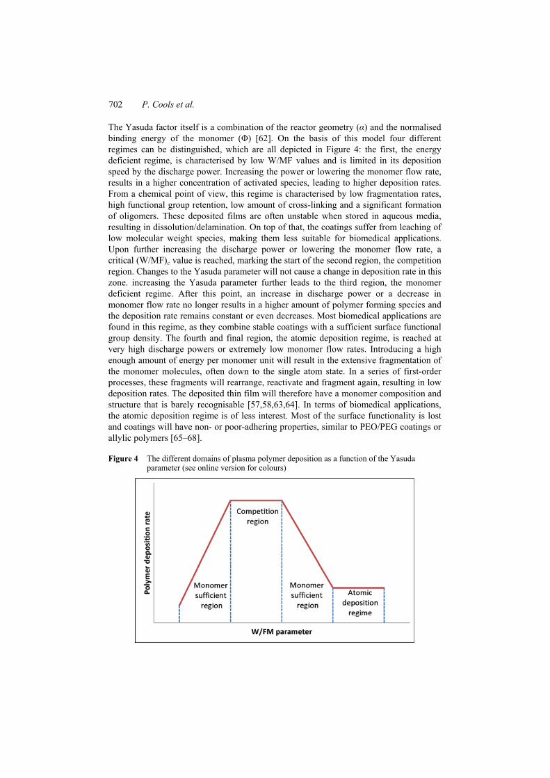
The Yasuda factor itself is a combination of the reactor geometry ( ) and the normalised binding energy of the monomer ( ) [62]. On the basis of this model four different regimes can be distinguished, which are all depicted in Figure 4: the first, the energy deficient regime, is characterised by low W/MF values and is limited in its deposition speed by the discharge power. Increasing the power or lowering the monomer flow rate, results in a higher concentration of activated species, leading to higher deposition rates. From a chemical point of view, this regime is characterised by low fragmentation rates, high functional group retention, low amount of cross-linking and a significant formation of oligomers. These deposited films are often unstable when stored in aqueous media, resulting in dissolution/delamination. On top of that, the coatings suffer from leaching of low molecular weight species, making them less suitable for biomedical applications. Upon further increasing the discharge power or lowering the monomer flow rate, a critical (W/MF)c value is reached, marking the start of the second region, the competition region. Changes to the Yasuda parameter will not cause a change in deposition rate in this zone. increasing the Yasuda parameter further leads to the third region, the monomer deficient regime. After this point, an increase in discharge power or a decrease in monomer flow rate no longer results in a higher amount of polymer forming species and the deposition rate remains constant or even decreases. Most biomedical applications are found in this regime, as they combine stable coatings with a sufficient surface functional group density. The fourth and final region, the atomic deposition regime, is reached at very high discharge powers or extremely low monomer flow rates. Introducing a high enough amount of energy per monomer unit will result in the extensive fragmentation of the monomer molecules, often down to the single atom state. In a series of first-order processes, these fragments will rearrange, reactivate and fragment again, resulting in low deposition rates. The deposited thin film will therefore have a monomer composition and structure that is barely recognisable [57,58,63,64]. In terms of biomedical applications, the atomic deposition regime is of less interest. Most of the surface functionality is lost and coatings will have non- or poor-adhering properties, similar to PEO/PEG coatings or allylic polymers [65–68].
Figure 4 The different domains of plasma polymer deposition as a function of the Yasuda parameter (see online version for colours)
Non-thermal plasma assisted lithography for biomedical applications 703
2 Plasma-assisted lithography
2.1 Alternating cell-adhesive and cell-repulsive microstructures
Most of the plasma-assisted chemically patterned surfaces for cell studies are based on alternating non-fouling and cell-adhesive domains. The number of polymers exhibiting efficient non-fouling properties is limited and three major groups can be distinguished. The first, most popular group, is the PEO/PEG (polyethylene oxide/polyethylene glycol) family. The variety of easily accessible and relatively cheap low molecular weight monomers combined with a simple chemical structure make them ideal for plasma polymerisation and surface analysis. A good control of the plasma parameters is essential, as a (partial) loss of the PEO repeating unit drastically lowers the non-fouling behaviour of the coating [69]. The second group of monomers is the fluorocarbon family. Fluorine-containing polymers are generally known to exhibit low-fouling behaviour and are not only used in biomedical applications, but can also be found in other industries and consumer products such as non-adhesive cooking utensils [70,71]. The two main disadvantages of fluorine-rich polymers are the toxicity of the monomers and the low-adhesive behaviour of their surface towards other polymer films, even after plasma treatment. The third group is the hydrocarbon family, which exhibits low-fouling behaviour. In combination with a good cell-selective pattern, these coatings will work, but most likely not for all cell-lines. The plasma polymers defining the cell-adhesive domains can be fabricated using a broad variety of precursors, introducing functional groups such as NH2, COOH, SH, COOR… In what follows, an overview will be given on plasma assisted patterning according to the chemistry of the cell-adhesive domains. Indirectly this also gives an overview according to what types of cells are being studied.
2.2 Amine-rich geometric domains
Surfaces protruding (primary) amines are well-sought after, as they efficiently bind with peptides (RGD) and proteins (lamilin, fibronectin, vitronectin…), thus greatly stimulating the cell-surface interactions [6,42,72,73]. This good selectivity towards macromolecules makes them ideal for surface patterning. Brétagnol et al. [74] used photolithography to deposit alternating 70 µm stripes of plasma polymerised (pp) PEO and allylamine. Seeding of L929 mouse fibroblasts showed a selective adhesion on the pp-allylamine domains. Thissen et al. [75] used to same process, albeit on a larger scale, to selectively bind Collagen I. Seeding of bovine corneal epithelial tissue showed excellent 2D control up to 21 days. Paik et al. [76] used a similar strategy, depositing pp-allylamine on a PDMS background, using a microstencils of different geometric shapes. An airbrushing technique was used to deposit fibronectin, which adhered selectively on the amine-rich domains. Six different cell types, including stem cells, fibroblasts and cardiomyocytes, were seeded onto the patterned surfaces with an excellent selectivity towards the N-containing domains, as shown in Figure 5. The pattern deposited on the flexible surfaces remained faithful even after elastic deformation, as evidenced in Figure 5. Singh et al. [37] used the same chemistry with binary colloidal printing as a masking method, generating egg-carton like patterns. A selective BSA adsorption was found for the coated regions. Several other authors used the same plasma polymers, but different masking techniques or pattern sizes as proof of concept, excluding any in-vitro testing [77–80].

704 P. Cools et al.
Figure 5 Micropatterning of complex designs. Micrographs showing (A) micro-stencils with complex pattern features. B: The corresponding eGFP labelled NIH-3T3 cell micropatterns on PDMS membranes. C: The entire surface cell micropattern. Images were taken 1 h post-patterning. D: PDMS sheets can be manually rolled to form tubes. E: Tubes remain patterned and cells remain viable. Image of tube was taken 2 h post-patterning and an extra 2 h post-rolling (see online version for colours)
Source: Paik et al. [76]
Albeit being the most used precursor, allylamine is certainly not the only monomer available to deposit N-rich surfaces. Muir et al. [81] used a pp-heptylamine as a base layer and covered it with a pp-PEG coating. Scanning probe nanolithography was then used to selectively ‘plough’ patterns of 300 nm wide, exposing the underlying pp-heptylamine layer. The selective immobilisation of fluorescently labelled igG proteins on the amine regions proved the viability of the technique for biomedical applications. Dai et al. [82] deposited a pp-n-hexane coating on mica plates. Using a solid mask, the pp-layer was exposed to an H2O plasma, introducing oxygen functionalities and creating a nm depth profile. Pyrrole was then plasma polymerised onto the activated layers, thus generating tracks of conductive polymers. These patterned polymers have the right properties to be used as flexible biosensors.
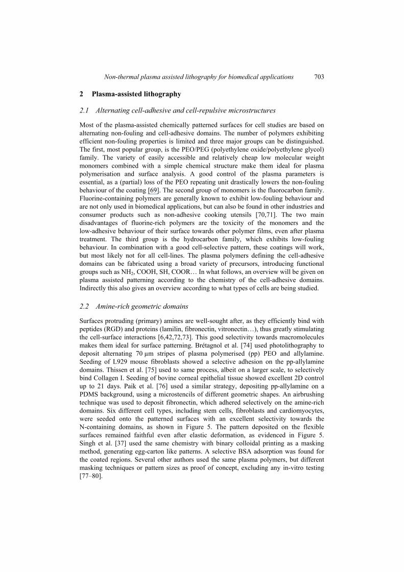
Several authors combined a plasma treatment with the adsorption of polylysine (PLL), a homopolymer normally synthesised by bacteria via fermentation [83–85]. PpPEO or ppCF4 were used as non-fouling backgrounds and a stencil technique was used to generate the patterns. In all cases the one month stable samples were tested for the micropatterning of neural stem cells. The neuron cells respected the artificially created boundaries. Both cell spreading and differentiation were studied as a function of cell density and the addition of fibronectin was tested (see Figure 6). These micro-labs are able to help understanding what mechanisms can lead to random and guided neuron outgrowth and possible applications lie in high-throughput drug and toxicology screening.
Non-thermal plasma assisted lithography for biomedical applications 705
Figure 6 (coloured) while close islands (b) and islands connected by lines (c) are used for monitoring short migrations and outgrowth projections. ToF-SIMS mapping showed good chemical contrast between PLL positive areas (d, f) recognised by C–N, and PEO-like positive background (e.g.) ascribed to C3–H3–O (see online version for colours)
Source: Ruiz et al. [84]
2.3 COOH-rich geometric domains
After (primary) amines, COOH-rich surfaces are the most sought after groups to incorporate onto a biomaterial’s surface. Amongst other beneficial properties, they effectively induce stem cell differentiation and readily interact with different macromolecules (collagen, heparin, insulin, fibronectin…) [13,86–88]. As the interfacial chemistry for COOH is complementary to amines, they are often used when NH2-rich surfaces are not triggering the desired cell-response.
Brétagnol et al. [89] and Sardella et al. [36] applied the same strategy as before, depositing alternating layers (70 µm) of ppPEO and ppPolyacrylic acid (ppPAcAc). Nanometre patterns with a higher resolution (100–500 nm) were obtained through a combination of plasma polymerisation and colloidal lithography [36,89]. L929 mouse fibroblasts were seeded onto the micropatterned surfaces and results showed that the cells were again restrained within the COOH-rich domains. The nanopatterned surfaces were used to immobilise BSA and well defined zones with diameters down to 100 nm were obtained. Favia et al. [90] used the exact same strategy to test the growth of the migration behaviour of keratinocytes and 3T3 fibroblasts as a function of the pattern width. Narrow patterns (50 µm) guided the fibroblasts to grow along the pattern, while wider patterns (two or more fibroblast cells wide) resulted in a random growth pattern. These results have important implications for guided cell tissue engineering.
Ploux et al. [91] combined photolithography and plasma polymerisation to deposit 1.6 µm wide pp maleic anhydride patterns on silicon wafers. After deposition, the samples were exposed to a UV-sterilisation process. Antibacterial (E. Coli) tests and osteoblast cell (HOP) tests were conducted in parallel and the combination of the altered texture and the UV treatment resulted in a lower adhesion efficiency of the E. Coli.The osteoblasts ignored the pattern and a good adhesion and spreading was found, similar to completely coated samples. These results show that there are effective alternatives to the traditional antibacterial coatings containing silver or cupper particles.
Barbier et al. [92] deposited 200 µm lines of ppPAcAc onto PDMS, using a masking technique similar to what is described in earlier paragraphs. These patterned surfaces were then successfully implemented into a microfluidic device to transport direct and double emulsions, which was first tried for PDMS samples and is shown in Figure 7. Sciarratta et al. [93] used the same strategy to generate 200 µm hexagonal domains of

706 P. Cools et al.
ppPAcAc with a plasma deposited coating of CHF3 as a background. Different tumour cells (HEK-293, RINm5f, KYM-1) were seeded onto the patterned surface in different culture media. Results showed an effective selection of the RINm5f tumour cells. Further analysis revealed that the adsorption of Ca2+ played a critical role in the selection and that only cell-lines, such as the RINm5f tumour cells, which express Ca2+ dependant membrane proteins are susceptible to the applied pattern.
Figure 7 Single O/W (a) and double W/O/W (b–e) emulsions made in a polydimethylsiloxane (PDMS) microsystem; Droplets are produced at T-junctions where both continuous and dispersed phases meet. The cross section of microchannels is 200 × 50 µm2. Fluids are driven by syringe pumps with flow rates varying from 0.1 to 20 µL/min. Observations are made using fluorescence video microscopy (water phases are labelled with fluorescein). For single O/W emulsions (a) tetradecane was used as dispersed phase and water with sodium dodecyl sulphate (SDS, 0.4%) as continuous phase. The whole microsystem is coated with PPAA. For O/W/O double emulsions (b–e), primary droplets of water in tetradecane (+SPAN 80, 0.75%) are produced at a first T-junction (uncoated, hydrophobic) and are then encapsulated with water and Tween (2%) at a second T-junction (coated with PPAA, hydrophilic). The hydrophobic/hydrophilic patterning was achieved by masking the area of the first T-junction with a glass slide according to the dotted line on picture (b)
Source: Barbier et al. [92]
Finally Filova et al. [94] did an extensive study on the cell response of four different cell types (rat smooth muscle cells, human and porcine skeletal muscle cells and bovine endothelial cells) to patterns generated via the plasma polymerisation of ppAcAc and 1,7-octadiene. A TEM grid was used to generate tracks of approximately 100 µm. The non-fouling efficiency of the 1,7-octadiene is considerably lower compared to PEO or fluorine containing layers and in some cases the selectivity of the cells dropped as low as 55%, with an average selectivity around 85%. Further analyses revealed though that there were significant differences between the cells adhered on either pattern. ELISA tests and immunostaining showed that for all cell-types there was a much better cell morphology and different cell parameters ( -actin and - actin levels, Weibel-Palade bodies…) indicated a much better maturation of the seeded cells. These results show the
Non-thermal plasma assisted lithography for biomedical applications 707
influence that surface chemistry can indirectly have on the different cell processes and opens doors for actual 3D tissue engineering.
2.4 Other surface chemistries
Although amine- and carboxylic acid-rich surfaces are the most studied interfaces with respect to cell adhesion, proliferation and maturation, they are definitely not the only functional groups available and other types of surface chemistry do have an important role in the regenerative medicine.
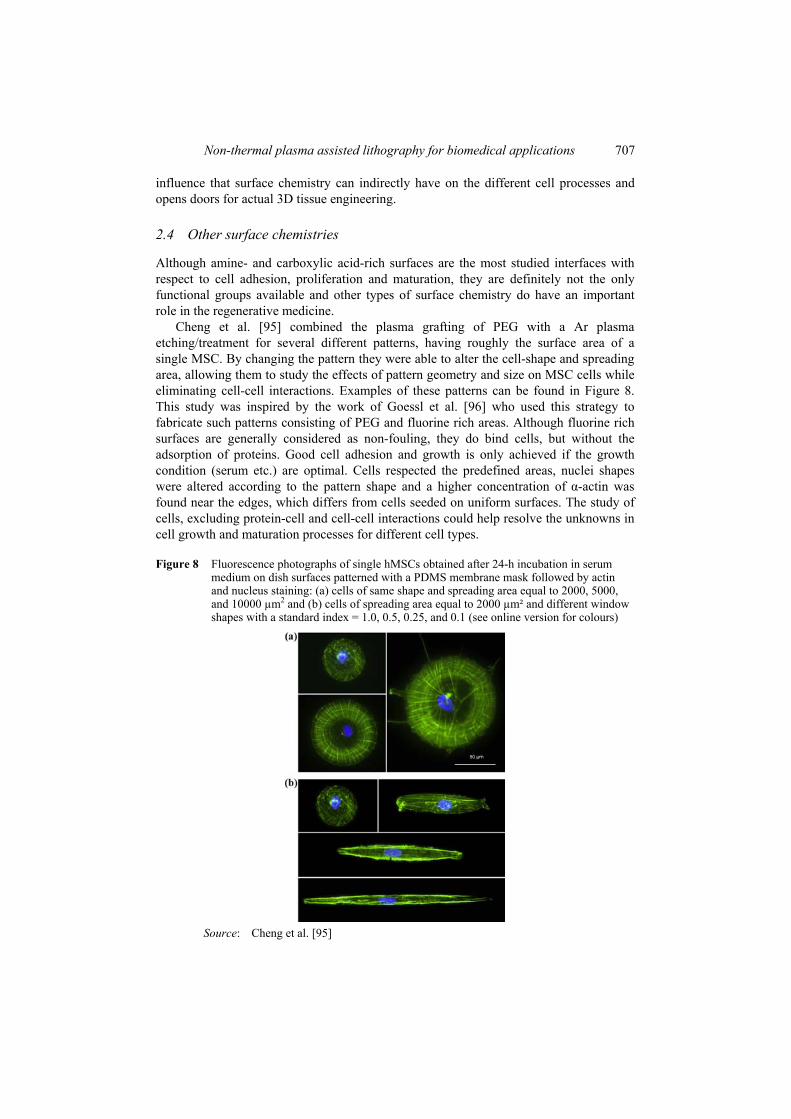
Cheng et al. [95] combined the plasma grafting of PEG with a Ar plasma etching/treatment for several different patterns, having roughly the surface area of a single MSC. By changing the pattern they were able to alter the cell-shape and spreading area, allowing them to study the effects of pattern geometry and size on MSC cells while eliminating cell-cell interactions. Examples of these patterns can be found in Figure 8. This study was inspired by the work of Goessl et al. [96] who used this strategy to fabricate such patterns consisting of PEG and fluorine rich areas. Although fluorine rich surfaces are generally considered as non-fouling, they do bind cells, but without the adsorption of proteins. Good cell adhesion and growth is only achieved if the growth condition (serum etc.) are optimal. Cells respected the predefined areas, nuclei shapes were altered according to the pattern shape and a higher concentration of -actin was found near the edges, which differs from cells seeded on uniform surfaces. The study of cells, excluding protein-cell and cell-cell interactions could help resolve the unknowns in cell growth and maturation processes for different cell types.
Figure 8 Fluorescence photographs of single hMSCs obtained after 24-h incubation in serum medium on dish surfaces patterned with a PDMS membrane mask followed by actin and nucleus staining: (a) cells of same shape and spreading area equal to 2000, 5000, and 10000 µm2 and (b) cells of spreading area equal to 2000 µm² and different window shapes with a standard index = 1.0, 0.5, 0.25, and 0.1 (see online version for colours)
Source: Cheng et al. [95]
708 P. Cools et al.
Marchesan et al. [97] used photolithography to fabricate 50 µm deep microwells that were plasma polymerised with 1-bromopropane exclusively inside the wells. This patterned bromide layer was then activated to covalently bind amines and acid groups, which were then used to immobilise either enzymes or peptide promoters. L929 cells were seeded and results showed a preferential growth within the microwells. This technique could be implemented into microfluidic devices to simultaneously test for a variety of analytes.
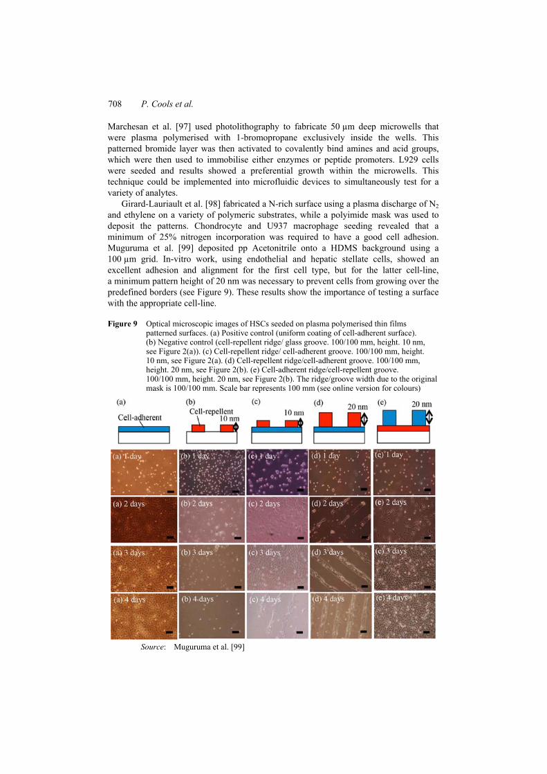
Girard-Lauriault et al. [98] fabricated a N-rich surface using a plasma discharge of N2and ethylene on a variety of polymeric substrates, while a polyimide mask was used to deposit the patterns. Chondrocyte and U937 macrophage seeding revealed that a minimum of 25% nitrogen incorporation was required to have a good cell adhesion. Muguruma et al. [99] deposited pp Acetonitrile onto a HDMS background using a 100 µm grid. In-vitro work, using endothelial and hepatic stellate cells, showed an excellent adhesion and alignment for the first cell type, but for the latter cell-line, a minimum pattern height of 20 nm was necessary to prevent cells from growing over the predefined borders (see Figure 9). These results show the importance of testing a surface with the appropriate cell-line.
Figure 9 Optical microscopic images of HSCs seeded on plasma polymerised thin films patterned surfaces. (a) Positive control (uniform coating of cell-adherent surface). (b) Negative control (cell-repellent ridge/ glass groove. 100/100 mm, height. 10 nm, see Figure 2(a)). (c) Cell-repellent ridge/ cell-adherent groove. 100/100 mm, height. 10 nm, see Figure 2(a). (d) Cell-repellent ridge/cell-adherent groove. 100/100 mm, height. 20 nm, see Figure 2(b). (e) Cell-adherent ridge/cell-repellent groove. 100/100 mm, height. 20 nm, see Figure 2(b). The ridge/groove width due to the original mask is 100/100 mm. Scale bar represents 100 mm (see online version for colours)
Source: Muguruma et al. [99]

Non-thermal plasma assisted lithography for biomedical applications 709
The pattern strategy used by Malkov et al. [79] has been described already in the NH2section. Next to allylamine, hexylamine and acrylic acid the research group also tested less conventional precursors such as hydroxy-ethyl methacrylate, N-vinyl-2-pyrrolidinone and N-vinyl formamide to form patterns on the fluorine-rich background. The introduction of other functionalities such as esters, amides and heterocyclic groups opens up opportunities for new series of labs on a chip. No cell-work was conducted in this work and no follow-up studies could be found testing their concept in vitro or in-vivo.
3 Conclusion
This review paper gives an overview of the developments in plasma assisted lithography in the last 15 years. Well-established surface texturing techniques such as photolithography, colloidal lithography and the use of stencils have been successfully combined with non-thermal plasma grafting and polymerisation. The combination of these two fields has led to the introduction of fast and clean patterning strategies implementing both topographical and chemical alterations. Most of the research is focused on alternating non- or low-fouling plasma coatings with plasma polymers that have a high receptivity towards cells. Different research groups were able to fabricate so-called labs on a chip, allowing for the fast study of single-cells, successfully eliminating cell-cell interactions. This gave valid information to further unravel the cell-interface interactions. Others deposited conductive patterns, showing valid alternatives for today’s flexible electric circuits. To this day, the applied plasma chemistry is almost exclusively limited to amine and carboxylic acid rich surfaces and a lot of advances can still be made exploring other functional groups such as thiols, oxazolines, imides, imines, phosphoric groups… Furthermore, the combination of different cell-interactive layers has not been studied, which could help progress the field of tissue engineering. When it comes to cells, many different cell types/lines were tested and the same conclusions can be drawn compared to non-patterned surfaces: each cell line responds differently to a certain surface. Therefore, when developing a new application, the tissue environment should always be kept in mind. This said, some differences were found for cells seeded on patterned surfaces. Cells were guided more easily, nuclei could change shape (depending on the pattern geometry) and certain factors (such as -actin levels) could significantly differ both in concentration and distribution. With respect to stem cell differentiation, plasma assisted patterning can play an important role in the high throughput scanning of the response to different surface chemistries and topographies, which could tremendously help stem cell research. Overall it can be said that plasma assisted patterning has proven its merits but its potential has only been touched and should be further explored in depth.
Acknowledgements
This review has received funding from the European Research Council under the European Union’s Seventh Framework Program (FP/2007-2013)/ERC Grant Agreement No. 279022.
710 P. Cools et al.
References 1 Bacakova, L., Filova, E., Parizek, M., Ruml, T. and Svorcik, V. (2011) ‘Modulation of cell
adhesion, proliferation and differentiation on materials designed for body implants’, Biotechnol. Adv., Vol. 29, pp.739–767.
2 Lutolf, M.P., Gilbert, P.M. and Blau, H.M. (2009) ‘Designing materials to direct stem-cell fate’, Nature, Vol. 462, pp.433–441.
3 Magnani, A., Priamo, A., Pasqui, D. and Barbucci, R. (2003) ‘Cell behaviour on chemically microstructured surfaces’, Mater. Sci. Eng., C, Vol. 23, pp.315–328.
4 Martinez, E., Engel, E., Planell, J. and Samitier, J. (2009) ‘Effects of artificial micro-and nano-structured surfaces on cell behaviour’, Ann. Anat., Vol. 191, pp.126–135.
5 Girard, P.P., Cavalcanti-Adam, E.A., Kemkemer, R. and Spatz, J.P. (2007) ‘Cellular chemomechanics at interfaces: sensing, integration and response’, Soft Matter, Vol. 3, p.307.
6 Jacobs, T., Morent, R., De Geyter, N., Dubruel, P. and Leys, C. (2012) ‘Plasma surface modification of biomedical polymers: influence on cell-material interaction’, Plasma Chem. Plasma Process., Vol. 32, pp.1039–1073.
7 Arima, Y. and Iwata, H. (2007) ‘Effect of wettability and surface functional groups on protein adsorption and cell adhesion using well-defined mixed self-assembled monolayers’, Biomaterials, Vol. 28, pp.3074–3082.
8 Van Kooten, T., Schakenraad, J., Van der Mei, H. and Busscher, H. (1992) ‘Influence of substratum wettability on the strength of adhesion of human fibroblasts’, Biomaterials,Vol. 13, pp.897–904.
9 Van Wachem, P., Hogt, A., Beugeling, T., Feijen, J., Bantjes, A., Detmers, J. and Van Aken, W. (1987) ‘Adhesion of cultured human endothelial cells onto methacrylate polymers with varying surface wettability and charge’, Biomaterials, Vol. 8, pp.323–328.
10 Lee, J.H., Park, J.W. and Lee, H.B. (1991) ‘Cell adhesion and growth on polymer surfaces with hydroxyl groups prepared by water vapour plasma treatment’, Biomaterials, Vol. 12, pp.443–448.
11 Webb, K., Hlady, V. and Tresco, P.A. (1998) ‘Relative importance of surface wettability and charged functional groups on NIH 3T3 fibroblast attachment, spreading, and cytoskeletal organization’, J. Biomed. Mater. Res., Vol. 41, p.422.
12 Curran, J.M., Stokes, R., Irvine, E., Graham, D., Amro, N., Sanedrin, R., Jamil, H. and Hunt, J.A. (2010) ‘Introducing dip pen nanolithography as a tool for controlling stem cell behaviour: unlocking the potential of the next generation of smart materials in regenerative medicine’, Lab Chip, Vol. 10, pp.1662–1670.
13 Curran, J.M., Chen, R. and Hunt, J.A. (2006) ‘The guidance of human mesenchymal stem cell differentiation in vitro by controlled modifications to the cell substrate’, Biomaterials, Vol. 27, pp.4783–4793.
14 Benoit, D.S., Schwartz, M.P., Durney, A.R. and Anseth, K.S. (2008) ‘Small functional groups for controlled differentiation of hydrogel-encapsulated human mesenchymal stem cells’, Nat. Mater., Vol. 7, pp.816–823.
15 Saha, K., Keung, A.J., Irwin, E.F., Li, Y., Little, L., Schaffer, D.V. and Healy, K.E. (2008) ‘Substrate modulus directs neural stem cell behavior’, Biophys. J., Vol. 95, pp.4426–4438.
16 Keselowsky, B.G., Collard, D.M. and Garcıa, A.J. (2004) ‘Surface chemistry modulates focal adhesion composition and signaling through changes in integrin binding’, Biomaterials, Vol. 25, pp.5947–5954.
17 Lee, J.H., Jung, H.W., Kang, I-K. and Lee, H.B. (1994) ‘Cell behaviour on polymer surfaces with different functional groups’, Biomaterials, Vol. 15, pp.705–711.
18 Bodhak, S., Bose, S. and Bandyopadhyay, A. (2009) ‘Role of surface charge and wettability on early stage mineralization and bone cell–materials interactions of polarized hydroxyapatite’, Acta Biomater., Vol. 5, pp.2178–2188.
Non-thermal plasma assisted lithography for biomedical applications 711
19 Ratner, B.D. (2004) Biomaterials Science: An Introduction to Materials in Medicine,Academic Press.
20 Thian, E., Ahmad, Z., Huang, J., Edirisinghe, M., Jayasinghe, S., Ireland, D., Brooks, R., Rushton, N., Bonfield, W. and Best, S. (2010) ‘The role of surface wettability and surface charge of electrosprayed nanoapatites on the behaviour of osteoblasts’, Acta Biomater., Vol. 6, pp.750–755.
21 Harrison, R.G. (1912) ‘The cultivation of tissues in extraneous media as a method of morpho-genetic study’, Anat. Rec., Vol. 6, pp.181–193.
22 Vagaská, B., Bacáková, L., Filová, E. and Balík, K. (2010) ‘Osteogenic cells on bio-inspired materials for bone tissue engineering’, Physiol. Res., Vol. 59, pp.309–322.
23 Price, R.L., Ellison, K., Haberstroh, K.M. and Webster, T.J. (2004) ‘Nanometer surface roughness increases select osteoblast adhesion on carbon nanofiber compacts’, J. Biomed. Mater. Res., Part A, Vol. 70, pp.129–138.
24 Zhao, G., Zinger, O., Schwartz, Z., Wieland, M., Landolt, D. and Boyan, B.D. (2006) ‘Osteoblast-like cells are sensitive to submicron-scale surface structure’, Clin. Oral Implants Res., Vol. 17, pp.258–264.
25 Yamashita, D., Machigashira, M., Miyamoto, M., Takeuchi, H., Noguchi, K., Izumi, Y. and Ban, S. (2009) ‘Effect of surface roughness on initial responses of osteoblast-like cells on two types of zirconia’, Dent. Mater. J., Vol. 28, p.461.
26 Hatano, K., Inoue, H., Kojo, T., Matsunaga, T., Tsujisawa, T., Uchiyama, C. and Uchida, Y. (1999) ‘Effect of surface roughness on proliferation and alkaline phosphatase expression of rat calvarial cells cultured on polystyrene’, Bone, Vol. 25, pp.439–445.
27 Hwang, S.Y., Kwon, K.W., Jang, K-J., Park, M.C., Lee, J.S. and Suh, K.Y. (2010) ‘Adhesion assays of endothelial cells on nanopatterned surfaces within a microfluidic channel’, Anal.Chem., Vol. 82, pp.3016–3022.
28 Lim, J.Y. and Donahue, H.J. (2007) ‘Cell sensing and response to micro-and nanostructured surfaces produced by chemical and topographic patterning’, Tissue Eng.,Vol. 13, pp.1879–1891.
29 Stevens, M.M. and George, J.H. (2005) ‘Exploring and engineering the cell surface interface’, Science, Vol. 310, pp.1135–1138.
30 Falconnet, D., Csucs, G., Grandin, H.M. and Textor, M. (2006) ‘Surface engineering approaches to micropattern surfaces for cell-based assays’, Biomaterials, Vol. 27, pp.3044–3063.
31 Folch, A. and Toner, M. (2000) ‘Microengineering of cellular interactions’, Annu. Rev. Biomed. Eng., Vol. 2, pp.227–256.
32 Fink, J., Théry, M., Azioune, A., Dupont, R., Chatelain, F., Bornens, M. and Piel, M. (2007) ‘Comparative study and improvement of current cell micro-patterning techniques’, Lab Chip,Vol. 7, pp.672–680.
33 Hasirci, V. and Kenar, H. (2006) ‘Novel surface patterning approaches for tissue engineering and their effect on cell behavior’, Nanomed., Vol. 1, No. 1, pp.73–90.
34 Goessl, A., Golledge, S.L. and Hoffman, A.S. (2001) ‘Plasma lithography – thin-film patterning of polymers by RF plasma polymerization II: Study of differential binding using adsorption probes’, J. Biomater. Sci. Polym. Ed., Vol. 12, pp.739–753.
35 Wang, X. and Grundmeier, G. (2006) ‘Morphology and patterning processes of thin organosilicon and perfluorinated bi-layer plasma polymer films’, Plasma Processes Polym.,Vol. 3, pp.39–47.
36 Sardella, E., Favia, P., Gristina, R., Nardulli, M. and d’Agostino, R. (2006) ‘Plasma-aided micro-and nanopatterning processes for biomedical applications’, Plasma Processes Polym.,Vol. 3, pp.456–469.
37 Singh, G., Griesser, H.J., Bremmell, K. and Kingshott, P. (2011) ‘Highly ordered nanometer-scale chemical and protein patterns by binary colloidal crystal lithography combined with plasma polymerization’, Adv. Funct. Mater., Vol. 21, pp.540–546.

712 P. Cools et al.
38 Tonks, L. and Langmuir, I. (1929) ‘A general theory of the plasma of an arc’, Phys. Rev.,Vol. 34, p.876.
39 Chu, P.K., Chen, J., Wang, L. and Huang, N. (2002) ‘Plasma-surface modification of biomaterials’, Mater. Sci. Eng., R, Vol. 36, pp.143–206.
40 Cools, P., De Geyter, N. and Morent, R. (2014) ‘PLA enhanced via plasma technology: a review’, in Winthrop, C. (Ed.): New Developments in Polylactic Acid Research,Nova Science, New York, p.218.
41 Kogelschatz, U. (2004) ‘Atmospheric-pressure plasma technology’, Plasma Phys. Controlled Fusion, Vol. 46, p.B63.
42 Desmet, T., Morent, R., Geyter, N.D., Leys, C., Schacht, E. and Dubruel, P. (2009) ‘Nonthermal plasma technology as a versatile strategy for polymeric biomaterials surface modification: a review’, Biomacromolecules, Vol. 10, pp.2351–2378.
43 Morent, R., De Geyter, N., Verschuren, J., De Clerck, K., Kiekens, P. and Leys, C. (2008) ‘Non-thermal plasma treatment of textiles’, Surf. Coat. Technol., Vol. 202, pp.3427–3449.
44 Friedrich, J., Loeschcke, I., Frommelt, H., Reiner, H-D., Zimmermann, H. and Lutgen, P. (1991) ‘Ageing and degradation of poly (ethylene terephthalate) in an oxygen plasma’, Polym. Degrad. Stab., Vol. 31, pp.97–114.
45 Banik, I., Kim, K., Yun, Y., Kim, D., Ryu, C. and Park, C. (2002) ‘Inhibition of aging in plasma-treated high-density polyethylene’, J. Adhes. Sci. Technol., Vol. 16, pp.1155–1169.
46 Occhiello, E., Morra, M., Cinquina, P. and Garbassi, F. (1992) ‘Hydrophobic recovery of oxygen-plasma-treated polystyrene’, Polymer, Vol. 33, pp.3007–3015.
47 Weikart, C.M. and Yasuda, H.K. (2000) ‘Modification, degradation, and stability of polymeric surfaces treated with reactive plasmas’, J. Polym. Sci., Part A: Polym. Chem., Vol. 38, pp.3028–3042.
48 Morra, M., Occhiello, E., Gila, L. and Garbassi, F. (1990) ‘Surface dynamics vs. adhesion in oxygen plasma treated polyolefins’, J. Adhes., Vol. 33, pp.77–88.
49 Van Deynse, A., Cools, P., Leys, C., Morent, R. and De Geyter, N. (2014) ‘Influence of ambient conditions on the aging behavior of plasma-treated polyethylene surfaces’, Surf. Coat. Technol., Vol. 258, pp.359–367.
50 Flamm, D.L., Auciello, O. and d’Agostino, R. (2012) Plasma Deposition, Treatment, and Etching of Polymers: The Treatment and Etching of Polymers, Elsevier, Academic Press, San Diego.
51 Egitto, F.D. (1990) ‘Plasma etching and modification of organic polymers’, Pure Appl. Chem.,Vol. 62, pp.1699–1708.
52 Coburn, J. and Winters, H.F. (1979) ‘Plasma etching – a discussion of mechanisms’, J. Vac. Sci. Technol., Vol. 16, pp.391–403.
53 Slepi ka, P., Trostová, S., Slepi ková Kasálková, N., Kolská, Z., Sajdl, P. and Švor ík, V. (2012) ‘Surface modification of biopolymers by argon plasma and thermal treatment’, PlasmaProcesses Polym., Vol. 9, pp.197–206.
54 Cools, P., Morent, R. and De Geyter, N. (2015) ‘Plasma modified textiles for biomedical applications’, Adv. Bioeng., pp.117–148.
55 Kylián, O., Choukourov, A. and Biederman, H. (2013) ‘Nanostructured plasma polymers’, Thin Solid Films, Vol. 548, pp.1–17.
56 Cools, P., Sainz-García, E., Geyter, N.D., Nikiforov, A., Blajan, M., Shimizu, K., Alba-Elías, F., Leys, C. and Morent, R. (2015) ‘Influence of DBD inlet geometry on the homogeneity of plasma-polymerized acrylic acid films: the use of a microplasma–electrode inlet configuration’, Plasma Processes Polym., Vol. 12, pp.1153–1163.
57 Yasuda, H. (2012) Plasma Polymerization, Academic Press, San Diego. 58 Friedrich, J. (2011) ‘Mechanisms of plasma polymerization – reviewed from a chemical point
of view’, Plasma Processes Polym., Vol. 8, pp.783–802.

Non-thermal plasma assisted lithography for biomedical applications 713
59 Neiswender, D. and Blaustein, B. (1969) ‘Chemical reactions in electrical discharges’, Adv. Chem. Ser.
60 Yasuda, H. and Lamaze, C. (1973) ‘Polymerization in an electrodeless glow discharge. II. Olefinic monomers’, J. Appl. Polym. Sci., Vol. 17, pp.1519–1531.
61 Yasuda, H. and Hirotsu, T. (1978) ‘Critical evaluation of conditions of plasma polymerization’, J. Polym. Sci., Part A: Polym. Chem., Vol. 16, pp.743–759.
62 Gilliam, M.A., Yu, Q. and Yasuda, H. (2007) ‘Plasma polymerization behavior of fluorocarbon monomers in low-pressure AF and RF discharges’, Plasma Processes Polym.,Vol. 4, pp.165–172.
63 Yasuda, H. (1981) ‘Macromolecular reviews Part DJ’, Polym. Sci., Vol. 16, p.199. 64 Friedrich, J., Gähde, J., Frommelt, H. and Wittrich, H. (1976) ‘Textiltechn’,
Z. Polymerenforsch, Vol. 27, p.517. 65 Kingshott, P. and Griesser, H.J. (1999) ‘Surfaces that resist bioadhesion’, Curr. Opin. Solid
State Mater. Sci., Vol. 4, pp.403–412. 66 Wu, Y.J., Timmons, R.B., Jen, J.S. and Molock, F.E. (2000) ‘Non-fouling surfaces produced
by gas phase pulsed plasma polymerization of an ultra low molecular weight ethylene oxide containing monomer’, Colloids Surf., B, Vol. 18, pp.235–248.
67 Aziz, G., Cools, P., De Geyter, N., Declercq, H., Cornelissen, R. and Morent, R. (2015) ‘Dielectric barrier discharge plasma treatment of ultrahigh molecular weight polyethylene in different discharge atmospheres at medium pressure: a cell-biomaterial interface study’, Biointerphases, Vol. 10, p.029502.
68 Aziz, G., De Geyter, N., Declercq, H., Cornelissen, R. and Morent, R. (2015) ‘Incorporation of amine moieties onto ultra-high molecular weight polyethylene (UHMWPE) surface via plasma and UV polymerization of allylamine’, Surf. Coat. Technol., Vol. 271, pp.39–47.
69 Sardella, E., Gristina, R., Senesi, G.S., d’Agostino, R. and Favia, P. (2004) ‘Homogeneous and micro-patterned plasma-deposited PEO-like coatings for biomedical surfaces’, PlasmaProcesses Polym., Vol. 1, pp.63–72.
70 Sarra-Bournet, C., Turgeon, S., Mantovani, D. and Laroche, G. (2006) ‘A study of atmospheric pressure plasma discharges for surface functionalization of PTFE used in biomedical applications’, J. Phys. D: Appl. Phys., Vol. 39, p.3461.
71 Leclair, A.M., Ferguson, S.S. and Lagugné-Labarthet, F. (2011) ‘Surface patterning using plasma-deposited fluorocarbon thin films for single-cell positioning and neural circuit arrangement’, Biomaterials, Vol. 32, pp.1351–1360.
72 Truica-Marasescu, F. and Wertheimer, M.R. (2008) ‘Nitrogen-rich plasma-polymer films for biomedical applications’, Plasma Processes Polym., Vol. 5, pp.44–57.
73 Puleo, D., Kissling, R. and Sheu, M-S. (2002) ‘A technique to immobilize bioactive proteins, including bone morphogenetic protein-4 (BMP-4), on titanium alloy’, Biomaterials, Vol. 23, pp.2079–2087.
74 Brétagnol, F., Ceriotti, L., Lejeune, M., Papadopoulou-Bouraoui, A., Hasiwa, M., Gilliland, D., Ceccone, G., Colpo, P. and Rossi, F. (2006) ‘Functional micropatterned surfaces by combination of plasma polymerization and lift-off processes’, Plasma Processes Polym.,Vol. 3, pp.30–38.
75 Thissen, H., Johnson, G., Hartley, P.G., Kingshott, P. and Griesser, H.J. (2006) ‘Two-dimensional patterning of thin coatings for the control of tissue outgrowth’, Biomaterials, Vol. 27, pp.35–43.
76 Paik, I., Scurr, D.J., Morris, B., Hall, G., Denning, C., Alexander, M.R., Shakesheff, K.M. and Dixon, J.E. (2012) ‘Rapid micropatterning of cell lines and human pluripotent stem cells on elastomeric membranes’, Biotechnol. Bioeng., Vol. 109, pp.2630–2641.
77 Im, S.G., Bong, K.W., Kim, B-S., Baxamusa, S.H., Hammond, P.T., Doyle, P.S. and Gleason, K.K. (2008) ‘Patterning nanodomains with orthogonal functionalities: solventless synthesis of self-sorting surfaces’, J. Am. Chem. Soc., Vol. 130, pp.14424–14425.

714 P. Cools et al.
78 Bullett, N., Short, R., O’Leary, T., Beck, A., Douglas, C., Cambray-Deakin, M., Fletcher, I., Roberts, A. and Blomfield, C. (2001) ‘Direct imaging of plasma-polymerized chemical micropatterns’, Surf. Interface Anal., Vol. 31, pp.1074–1076.
79 Malkov, G.S., Martin, I.T., Schwisow, W.B., Chandler, J.P., Wickes, B.T., Gamble, L.J., Castner, D.G. and Fisher, E.R. (2008) ‘Pulsed-plasma-induced micropatterning with alternating hydrophilic and hydrophobic surface chemistries’, Plasma Processes Polym.,Vol. 5, pp.129–145.
80 Slocik, J.M., Beckel, E.R., Jiang, H., Enlow, J.O., Zabinski, J.S., Bunning, T.J. and Naik, R.R. (2006) ‘Site-specific patterning of biomolecules and quantum dots on functionalized surfaces generated by plasma-enhanced chemical vapor deposition’, Adv. Mater., Vol. 18, pp.2095–2100.
81 Muir, B.W., Fairbrother, A., Gengenbach, T.R., Rovere, F., Abdo, M.A. and McLean KM (2006) ‘Scanning probe nanolithography and protein patterning of low-fouling plasma polymer multilayer films’, Adv. Mater., Vol. 18, pp.3079–3082.
82 Dai, L., Mau, A.W., Gong, X. and Griesser, H.J. (1997) ‘Electrochemical generation of conducting polymer patterns on plasma modified surfaces’, Synth. Met., Vol. 85, pp.1379–1380.
83 Chang, W.C. and Sretavan, D.W. (2008) ‘Novel high-resolution micropatterning for neuron culture using polylysine adsorption on a cell repellant, plasma-polymerized background’, Langmuir, Vol. 24, pp.13048–13057.
84 Ruiz, A., Buzanska, L., Gilliland, D., Rauscher, H., Sirghi, L., Sobanski, T., Zychowicz, M., Ceriotti, L., Bretagnol, F. and Coecke, S. (2008) ‘Micro-stamped surfaces for the patterned growth of neural stem cells’, Biomaterials, Vol. 29, pp.4766–4774.
85 Zychowicz, M., Mehn, D., Ruiz, A., Colpo, P., Rossi. F., Frontczak-Baniewicz, M., Domanska-Janik, K. and Buzanska, L. (2011) ‘Proliferation capacity of cord blood derived neural stem cell line on different micro-scale biofunctional domains’, Acta Neurobiol. Exp. (Wars.), Vol. 71, pp.12–23.
86 Kim, Y.J., Kang, I-K., Huh, M.W. and Yoon, S-C. (2000) ‘Surface characterization and in vitro blood compatibility of poly (ethylene terephthalate) immobilized with insulin and/or heparin using plasma glow discharge’, Biomaterials, Vol. 21, pp.121–130.
87 Lu, H., Guo, L., Kawazoe, N., Tateishi, T. and Chen, G. (2009) ‘Effects of poly (L-lysine), poly (acrylic acid) and poly (ethylene glycol) on the adhesion, proliferation and chondrogenic differentiation of human mesenchymal stem cells’, J. Biomater. Sci. Polym. Ed., Vol. 20, pp.577–589.
88 Mattioli-Belmonte, M., Lucarini, G., Virgili, L., Biagini, G., Detomaso, L., Favia, P., d’Agostino, R., Gristina, R., Gigante, A., Bevilacqua, C. and Bioact, J. (2005) ‘Mesenchymal stem cells on plasma-deposited acrylic acid coatings: an in vitro investigation to improve biomaterial performance in bone reconstruction’, J Bioact. Compat. Polym., Vol. 20, pp.343–360.
89 Brétagnol, F., Valsesia, A., Ceccone, G., Colpo, P., Gilliland, D., Ceriotti, L., Hasiwa, M. and Rossi, F. (2006) ‘Surface functionalization and patterning techniques to design interfaces for biomedical and biosensor applications’, Plasma Processes Polym., Vol. 3, pp.443–455.
90 Favia, P., Sardella, E., Gristina, R. and d’Agostino, R. (2003) ‘Novel plasma processes for biomaterials: micro-scale patterning of biomedical polymers’, Surf. Coat. Technol., Vol. 169, pp.707–711.
91 Ploux, L., Anselme, K., Dirani, A., Ponche, A., Soppera, O. and Roucoules, V. (2009) ‘Opposite responses of cells and bacteria to micro/nanopatterned surfaces prepared by pulsed plasma polymerization and UV-irradiation’, Langmuir, Vol. 25, pp.8161–8169.
92 Barbier, V., Tatoulian, M., Li, H., Arefi-Khonsari, F., Ajdari, A. and Tabeling, P. (2006) ‘Stable modification of PDMS surface properties by plasma polymerization: application to the formation of double emulsions in microfluidic systems’, Langmuir, Vol. 22, pp.5230–5232.

Non-thermal plasma assisted lithography for biomedical applications 715
93 Sciarratta, V., Sohn, K., Burger-Kentischer, A., Brunner, H. and Oehr, C. (2006) ‘Controlled cell attachment, using plasma deposited polymer microstructures: a novel study of cells-substrate interactions’, Plasma Processes Polym., Vol. 3, pp.532–539.
94 Filová, E., Bullett, N., Ba aková, L., Grausova, L., Haycock, J. and Hlu ilova, J. (2009) ‘Regionally-selective cell colonization of micropatterned surfaces prepared plasma polymerization of acrylic acid and 1, 7-octadiene’, Physiol. Res., Vol. 58, p.669.
95 Cheng, Q., Li, S. and Komvopoulos, K. (2009) ‘Plasma-assisted surface chemical patterning for single-cell culture’, Biomaterials, Vol. 30, pp.4203–4210.
96 Goessl, A., Bowen-Pope, D.F. and Hoffman, A.S. (2001) ‘Control of shape and size of vascular smooth muscle cells in vitro by plasma lithography’, J. Biomed. Mater. Res., Vol. 57, pp.15–24.
97 Marchesan, S., Easton, C.D., Styan, K.E., Leech, P., Gengenbach, T.R. and Forsythe, J.S. (2013) ‘SU-8 photolithography on reactive plasma thin-films: coated microwells for peptide display’, Colloids Surf., B, Vol. 108, pp.313–321.
98 Girard-Lauriault, P.L., Mwale, F., Iordanova, M., Demers, C., Desjardins, P. and Wertheimer, M.R. (2005) ‘Atmospheric pressure deposition of micropatterned nitrogen-rich plasma-polymer films for tissue engineering’, Plasma Processes Polym., Vol. 2, pp.263–270.
99 Muguruma, H., Hoshino, T., Fujita, R., Sumii, T. and Kudo, S. (2015) ‘Adhesion and alignment of nonparenchymal cells onto a patterned surface with a two-step plasma polymerization process’, Plasma Processes Polym., Vol. 12, pp.746–754.
The author has requested enhancement of the downloaded file. All in-text references underlined in blue are linked to publications on ResearchGate.The author has requested enhancement of the downloaded file. All in-text references underlined in blue are linked to publications on ResearchGate.