normal chest x-ray - medfreecon.files.wordpress.com · hydropneumothorax •air in pleural
TRANSCRIPT
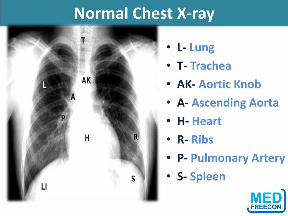
Normal Chest X-ray
• L- Lung
• T- Trachea
• AK- Aortic Knob
• A- Ascending Aorta
• H- Heart
• R- Ribs
• P- Pulmonary Artery
• S- Spleen
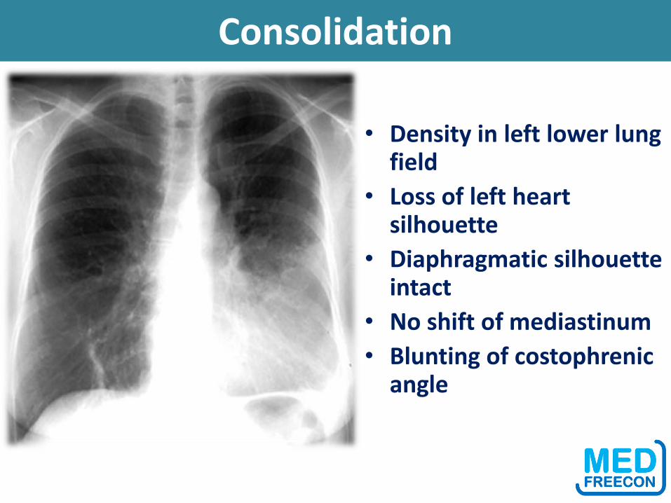
• Density in left lower lung field
• Loss of left heart silhouette
• Diaphragmatic silhouette intact
• No shift of mediastinum
• Blunting of costophrenic angle
Consolidation
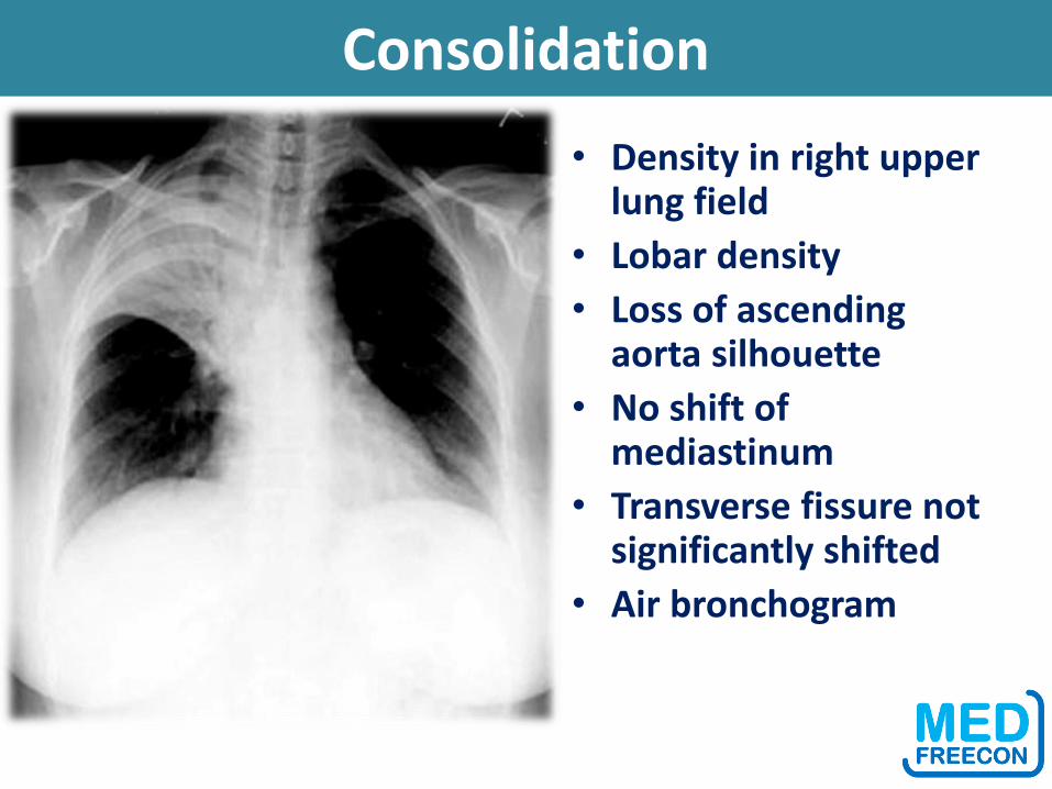
• Density in right upper lung field
• Lobar density
• Loss of ascending aorta silhouette
• No shift of mediastinum
• Transverse fissure not significantly shifted
• Air bronchogram
Consolidation
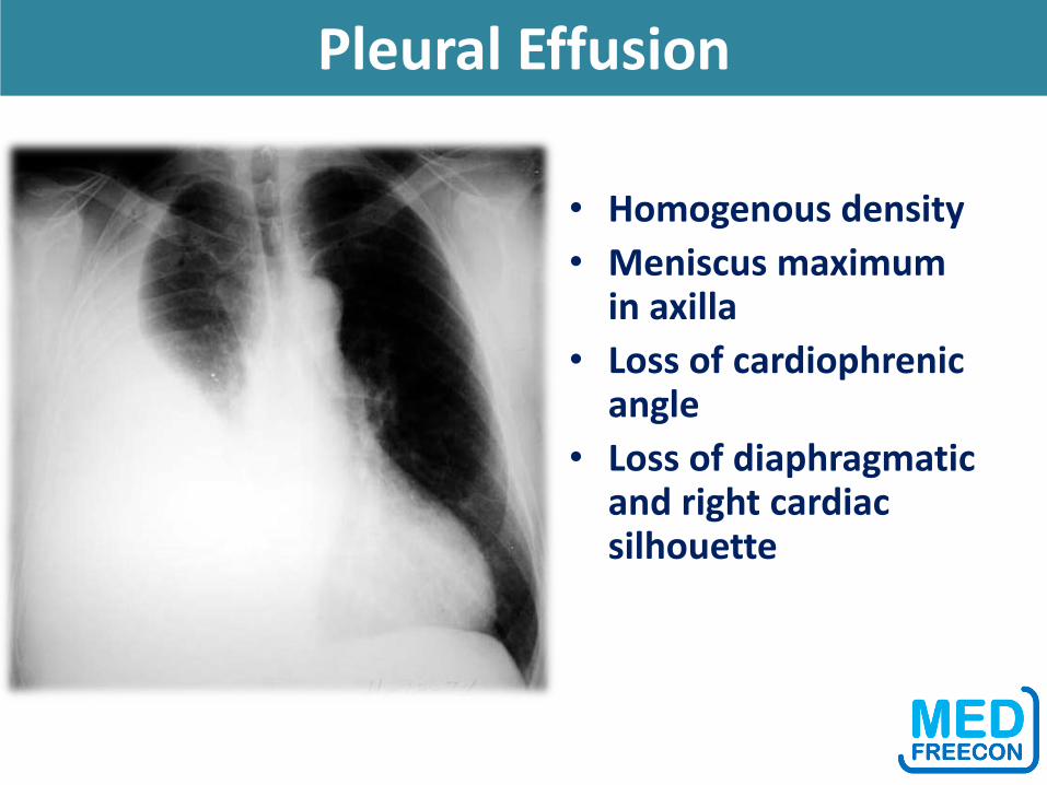
Pleural Effusion
• Homogenous density
• Meniscus maximum in axilla
• Loss of cardiophrenic angle
• Loss of diaphragmatic and right cardiac silhouette
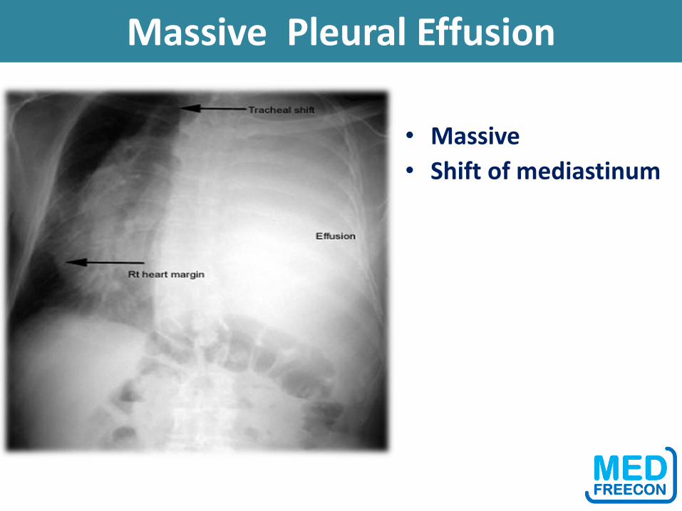
Massive Pleural Effusion
• Massive
• Shift of mediastinum
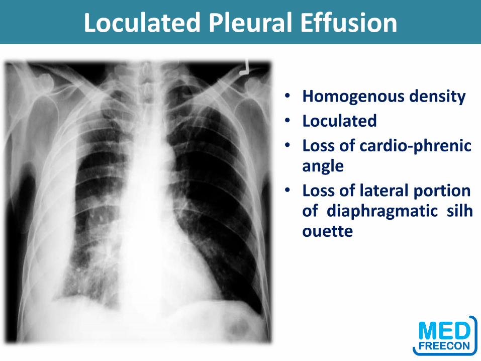
Loculated Pleural Effusion
• Homogenous density
• Loculated
• Loss of cardio-phrenic angle
• Loss of lateral portion of diaphragmatic silhouette
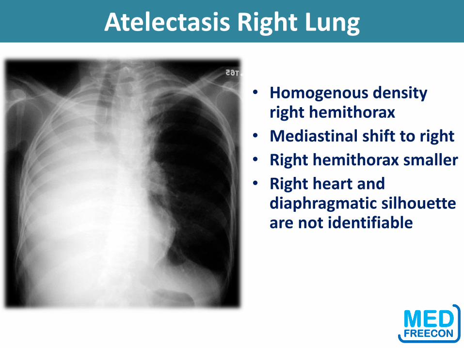
Atelectasis Right Lung
• Homogenous density right hemithorax
• Mediastinal shift to right
• Right hemithorax smaller
• Right heart and diaphragmatic silhouette are not identifiable
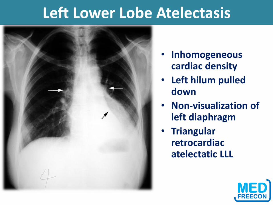
Left Lower Lobe Atelectasis
• Inhomogeneous cardiac density
• Left hilum pulled down
• Non-visualization of left diaphragm
• Triangular retrocardiac atelectatic LLL
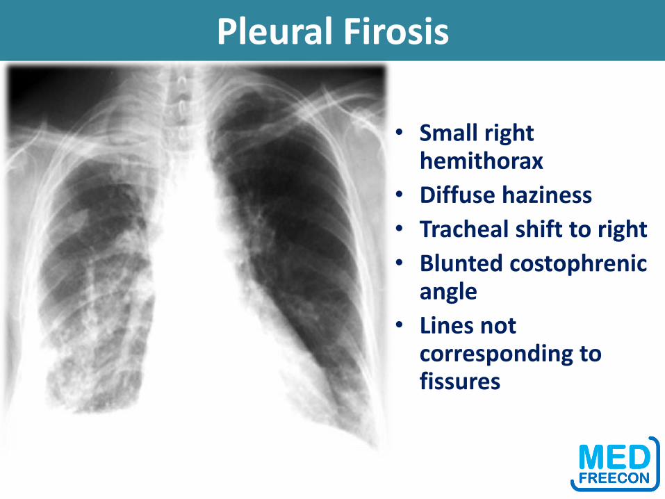
Pleural Firosis
• Small right hemithorax
• Diffuse haziness
• Tracheal shift to right
• Blunted costophrenic angle
• Lines not corresponding to fissures
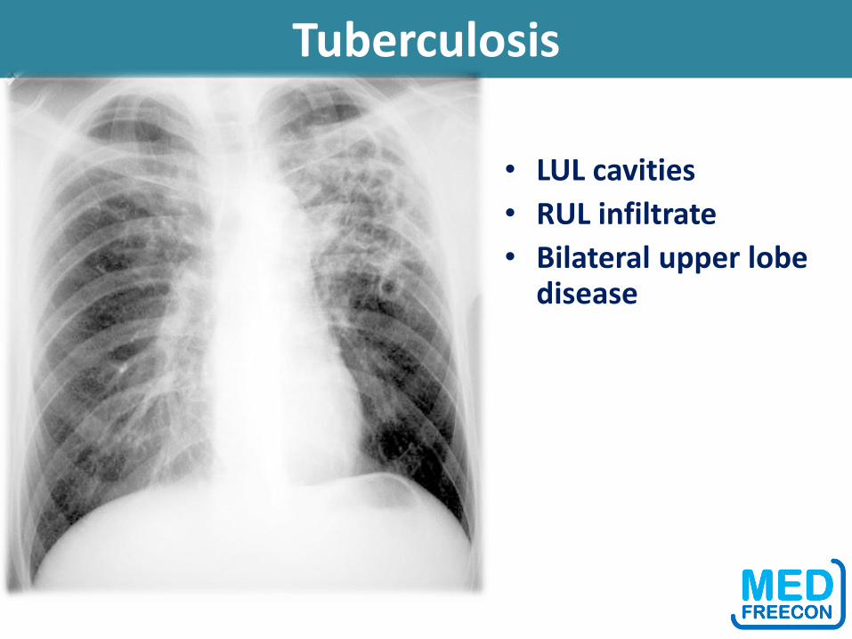
Tuberculosis
• LUL cavities
• RUL infiltrate
• Bilateral upper lobe disease

Tuberculosis
• LUL cavity
• Cavity behind clavicle - note increased density of clavicle in the region over lying cavity
• Pleural effusion on right
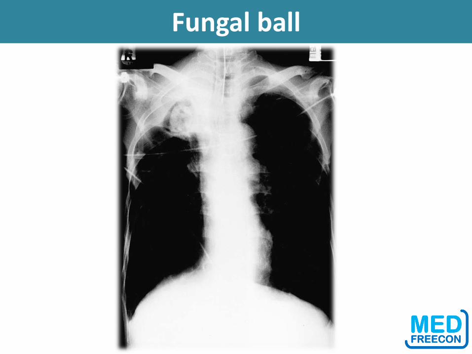
Fungal ball
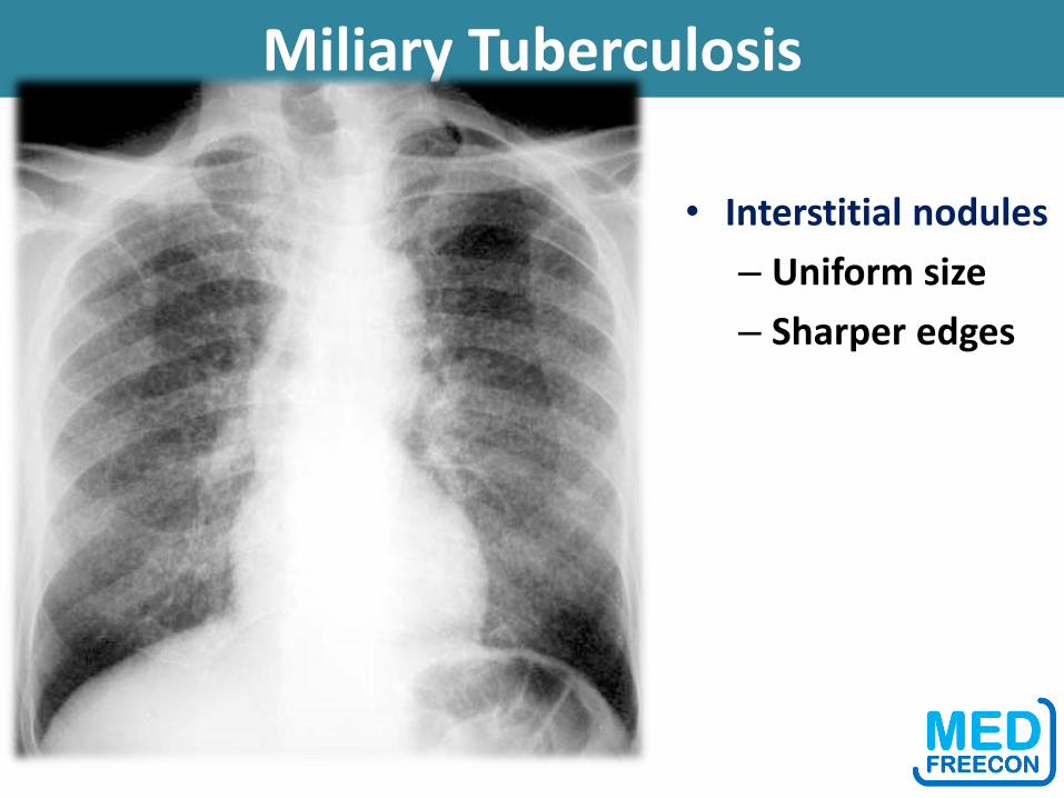
Miliary Tuberculosis
• Interstitial nodules
– Uniform size
– Sharper edges

Pneumothorax
• No vascular markings on right
• No shift of mediastinum to left
• Deep sulcus • Atelectatic right lung • Increased haziness on
left: Diversion of entire cardiac output
• Small fluid level near costophrenic angle: Hydro pneumothorax
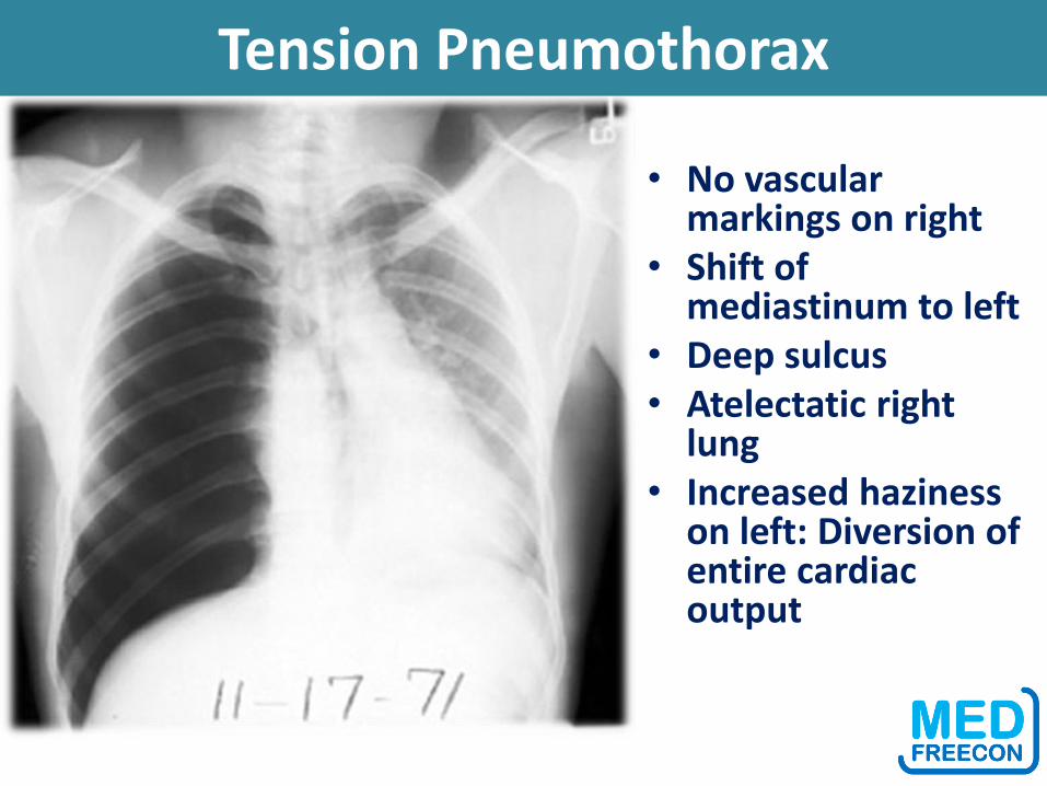
Tension Pneumothorax
• No vascular markings on right
• Shift of mediastinum to left
• Deep sulcus • Atelectatic right
lung • Increased haziness
on left: Diversion of entire cardiac output
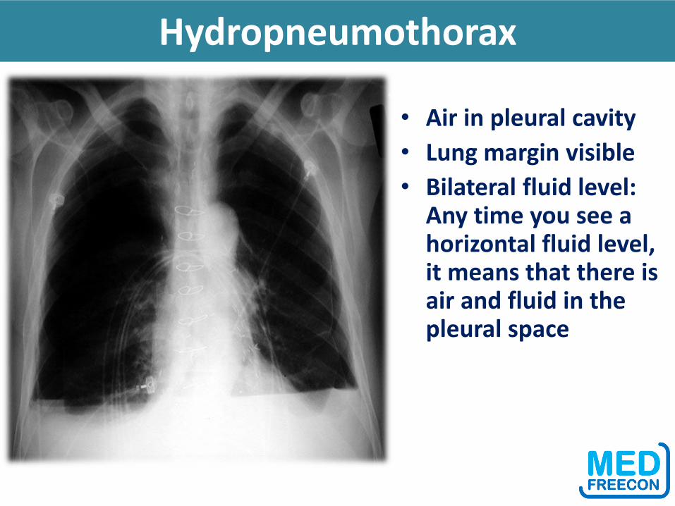
Hydropneumothorax
• Air in pleural cavity
• Lung margin visible
• Bilateral fluid level: Any time you see a horizontal fluid level, it means that there is air and fluid in the pleural space
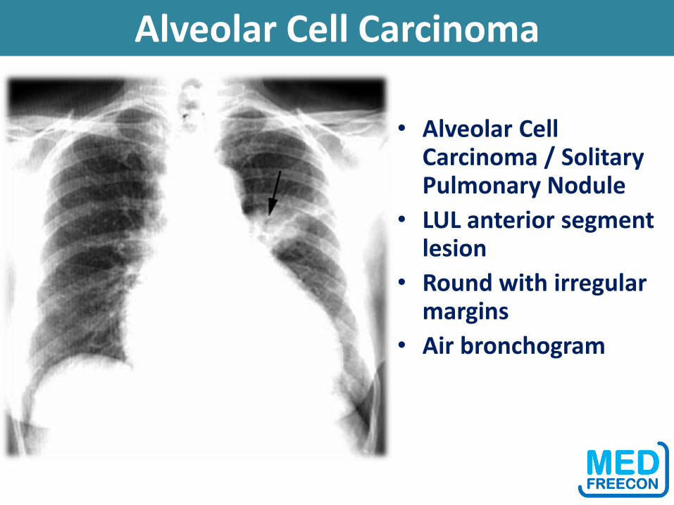
Alveolar Cell Carcinoma
• Alveolar Cell Carcinoma / Solitary Pulmonary Nodule
• LUL anterior segment lesion
• Round with irregular margins
• Air bronchogram
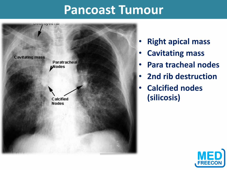
Pancoast Tumour
• Right apical mass
• Cavitating mass
• Para tracheal nodes
• 2nd rib destruction
• Calcified nodes (silicosis)
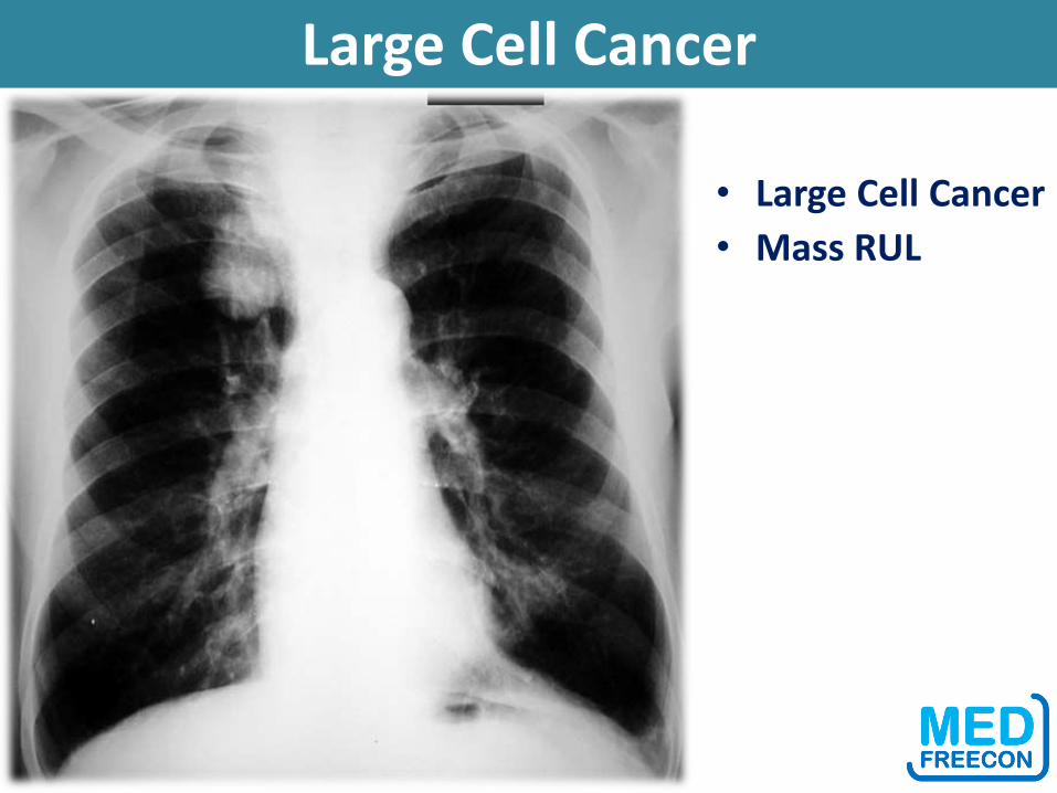
Large Cell Cancer
• Large Cell Cancer
• Mass RUL

Lung Mass
• Mass
• Round or oval
• Sharp margin
• Homogenous
• No respect for anatomy
• Lung Cancer: Large cell
•
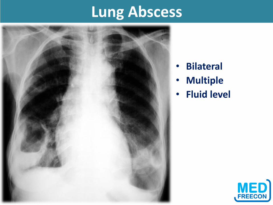
Lung Abscess
• Bilateral
• Multiple
• Fluid level

Lung Abscess
• Anterior segment of LUL
• Atypical location for aspiration lung abscess
• Thick wall
• Fluid level
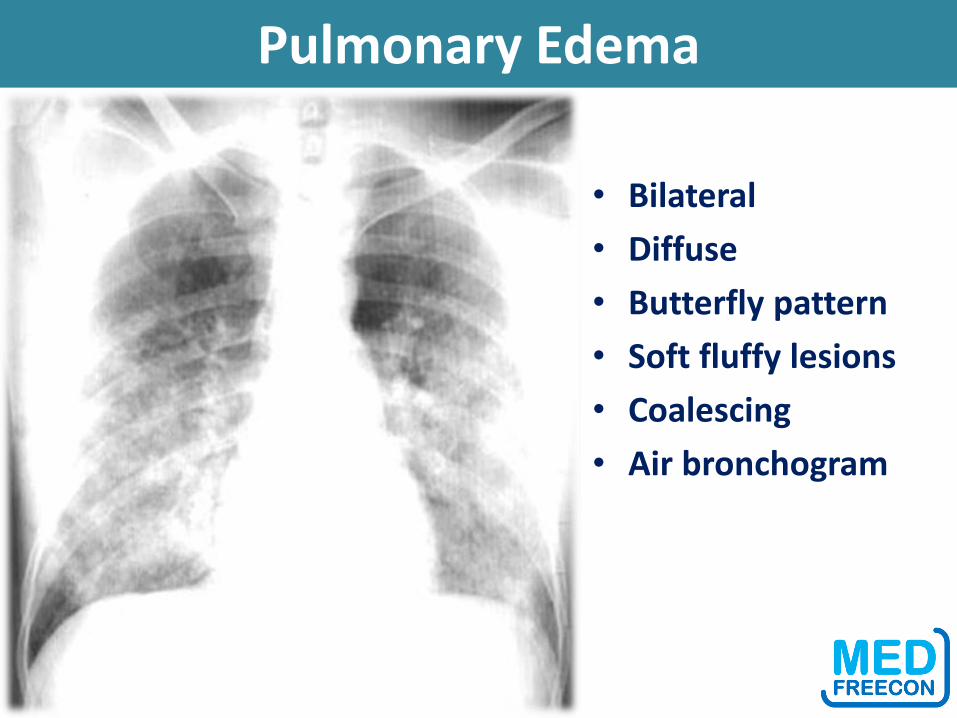
Pulmonary Edema
• Bilateral
• Diffuse
• Butterfly pattern
• Soft fluffy lesions
• Coalescing
• Air bronchogram
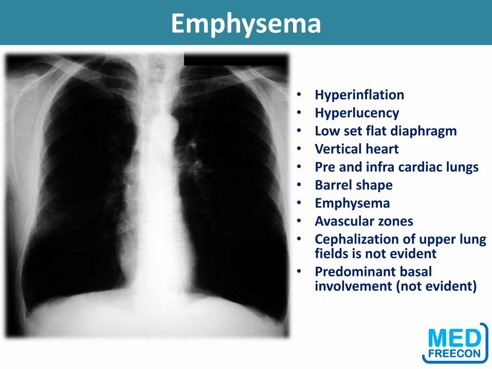
Emphysema
• Hyperinflation• Hyperlucency• Low set flat diaphragm • Vertical heart • Pre and infra cardiac lungs • Barrel shape • Emphysema• Avascular zones • Cephalization of upper lung
fields is not evident • Predominant basal
involvement (not evident)