not always be absolutely definite but the typical examples of each
TRANSCRIPT

HYGROMA CYSTICUM COLLIITS STRUCTURE AND ETIOLOGY.
BY CHARLES N. DOWD, M.D.,
OF NEW YORK.
Professor of Clinical Surgery in Columbia University.
SCArrERED through the books on surgery and pathologyone may find pictures of children with enormous swellingsof the neck which are described as hygromas. Although thereare a few definite descriptions the statements about thesegrowths are often indefinite, sometimes contradictory, and giveevidence of having been copied from one author by anotherwithout the opportunity of extensive personal observation.At least three types of growth have been included in some ofthe descriptions.
i. Cystic tumors which have endothelial linings and serouscontents and which grow with much power through the tissuesof the neck or downward under the clavicle into the axilla orpectoral region.
2. Lymphangiomas.3. Branchial cysts.The term hydroma should be confined to the former class.
They are usually described as cysts of lymphatic origin. Theloculi are sometimes described as dilated lymph spaces.
The dividing line between them and lymphangiomas maynot always be absolutely definite but the typical examples ofeach are distinct.
Since branchial cysts are now well understood one hardlysees how they need be confounded with hygromas.
The etiology of hygromas is usually referred to as un-known, although several theories for their origin have beengiven.
*Read before the American Surgical Association, May 8, 1913.112

HYGROMA CYSTICUM COLLI.
These growths are rare, the total number described insurgical literature is small. Conversation with friends whowould have been likely to see them indicates that they are veryuncommon.
Rare cases however come in groups, and after havingdone neck surgery for many years without seeing a single casethe writer has within the last year operated on three un-doubted cases and a fourth which was probably such a casebut in which inflammation had obliterated the finer structureof the cyst wall.
Most of the peculiarities which have been described in thereported cases have been present in one or more of those casesand their histories are here given in detail. The first one,a child of two and three-quarter years, gave the followinghistory.
CASE I.-J. K., age two and three-quarter years. AdmittedAugust 22, 19I2. Died Sept. 30, I9I2. Nationality, U; S.(Russian). Roosevelt Hospital, History No. B3034.
Chief Complaint.-Swelling of right neck and shoulder.Present Illness.-When patient was three months old the
mother first noticed a small swelling, the size of the tip of thelittle finger, just above the middle of the clavicle on the rightside of the neck. The skin over it was normal, it was not pain-ful or tender and could be made to disappear by firm pressuredownward. The lump has been gradually growing larger inspite of firm bandages, which used to make it disappear but nolonger do so. Following whooping-cough last winter it grewrapidly larger. Four days before admission patient had a fall,striking the lump, since which time it has been somewhatreddened and slightly tender. Otherwise a healthy normal child.Bowels regular, appetite rather poor.
Past History.-Has had whooping-cough, pneumonia twice,had " yellow jaundice " one year ago. Is rather subject tocoughs. Had a similar lump egg-sized in right axilla, with somesymptoms of reducibility, removed at a dispensary last year.
Family History.-No history of tumors or fistulae about theneck. No "lung trouble " in family.
II 3

CHARLES N. DOWD.
Physical Examination.-Patient is a well-nourished, healthylooking girl baby, appears restless but not ill.
Local Condition: On the right side of the neck is a globularswelling about 4 inches in diameter extending from the acromionto the anterior border of the sternomastoid and from the spineof the scapula to well in front of the clavicle. The skin over itis smooth but somewhat ecchymotic as from a contusion. Onpalpation it is soft, fluctuating and can be somewhat reducedin size by firm pressure. It is apparently not tender. On forcedexpiration there is an expansile impulse, percussion flat.
Eyes, Ears, Nose, and Throat: Negative.Skin: Reddish maculopapular eruptions on chest and back.Chest: There is marked inspiratory retraction of lower ribs
in front on both sides, sternum very prominent.Lungs: Clear.Heart: Not enlarged. Sounds of good quality. There is
a rough systolic murmur heard over the left half of base, trans-mitted upward toward both clavicles.
Abdomen: Rather a pot-belly with an umbilical hernia.Liver and spleen not felt.
Extremities and Genitals: Apparently normal.Operation (August 23, 1912).-A transverse incision was
made above the clavicle and parallel to it. The cyst was enu-cleated through this incision as far as the pedicle which came justin front of insertion of sternocleidomastoid muscle at the inneredge of sternum. When the cyst was opened it was foundthat it ran in under the sternum into the thoracic cavity butextrapleurally. It was shaped like an hour-glass and theconstricted portion was large enough to admit the finger, theinner dilated portion seemed to have a content of 3-4 oz. andwent up as far as thyroid and downward under the sternum forabout 2 inches, outward under the clavicle for an inch or more.As much of this lining membrane as could be dissected awaywas drawn out into the wound and cut off. No attempt wasmade to lay open this entire inner dilated portion, the conditionof child would not warrant so extensive a procedure. Woundclosed without drainage. Time of operation 45 minutes.
The recovery from the operation itself was prompt andsatisfactory, but the swelling quickly recurred and on SeptemberI7, a second operation was done.
II4

HYGROMA CYSTICUM COLLI. "5Second Operation (September 17, I912).-Anaesthetic, ether.
A transverse incision was made from inner end of clavicle wellback to posterior portion of neck. The skin and superficialfascia were dissected up and the cyst found to have reformedin the position where it previously existed. The prolongationwhich extended down under the sternum could not be found.The cyst was 3 x 4 inches long and 2 inches wide. It presentedthe same characteristics as the one previously removed. It wasdissected out in its anterior and lower and upper portions andwas found to have a pedicle which extended inward and back-ward and posterior to one of the scaleni muscles. After carefuldissection this pedicle was divided but it frayed out and therewas no evidence of an opening which extended further. Thecyst contained clear serous fluid; there was considerable fattytissue on its outer side, also some lymphatic tissue. The wallwas thin and similar to one previously removed. There was noevidence of epithelium or communication with the deeperstructures.
The child showed considerable reaction after the second opera-tion. There was free serous discharge, on the seventh day therewas hemorrhage from the wound, some oozing on the followingday and on the tenth day she died.
Pathologist's Report (R. H. Pat. No. B-5o6)i.-J. K., August 23, 1912.Specimen: Cyst wall.Gross Examination: Specimen consists of a thin-walled smooth
cyst, with an opening about 2 cm. in diameter at one end. Some areasare red in color, others thin and upon holding to light are translucent.A small thin-walled cyst containing a few drops of serous fluid wasfound within the cyst wall.
Microscopic Examination: Section shows a thick-walled cyst whichis involved in a chronic inflammatory reaction. There is an increase infibrous connective tissue especially around the blood-vessels which arenumerous. Areolar tissue is present in the outer portions in which thereare scattered aggregations of lymphoid follicles. No definite type of cellcan be made out lining the cyst though in some places they have theappearance of much flattened endothelial cells. This innner part of thewall shows a marked infiltration of round cells.
Diagnosis: Chronic inflammation of cyst wall.Pathologist, Baldwin Mann, M.D.Pathologist's Report, second specimen. (R. H. No. B-786).-J. K.,
age two and three quarter years, Sept. 17, 1912.Specimen: Cyst.

CHARLES N. DOWD.
Gross Examination: Specimen consists of a mass of fibrous andfatty tissue containing a few lympli nodes, and a small smooth lining ofa cyst.
Microscopic Examnination: Section is composed of muscle andconnective tissue, containing numerous rather congested blood-vessels,considerable fat and a few nerves. Round cell infiltration is presentthroughout. There is a definite cyst lined by a single or double layerof rather large cells with elongated nuclei. Lymph node shows moderatehyperplasia.
Diagnosis: Cyst wall showing chronic inflammation. Moderatehyperplasia of lymph node.
Pathologist, Mortimer Warren, M.D.
CASE II.-J. A., age twenty months. St. Mary's Free Hospi-tal for Children. History No. S. IO979. January i8, I9I3. Sentby Dr. John McBarron. Ever since birth the mother had noticedwhat she called a " bubble " just beneath the left clavicle. Thisapparently diminished on pressure and the mother had notnoticed any apparent change in it until two weeks or so ago.Then decided enlargement appeared over the upper left side ofthe chest in front. The child was sent to the hospital and foundto have a very prominent cystic tumor in this locality. Justbeneath the clavicle the cyst seemed thin walled, further downit had apparently thicker walls. It extended from the sternumto the outer edge of the pectoralis major and from the sixthrib to the clavicle.
The child's parents were first cousins and the entire familyof six children had been defective in some way. This one seemsidiotic. It was impossible to keep him quiet long enough to get agood picture of him and finally the picture had to be takenwhile he was under an anmesthetic.
Operation (January 2I).-A curved incision with its con-vexity outward was made from above the clavicle to the sixthrib and axillary line. The pectoralis major was found to be infront of the cyst. Its fibres were therefore split with the hopeof extracting the cyst through this split; this however was notpossible. It was found that a portion of the cyst projectedbetween the upper fibres of the pectoralis major and the clavicle.Finally the entire width of the pectoralis major was divided.The cyst was then found to press forward from beneath theclavicle. The axillary vessels and the brachial plexus werepushed forward. The cyst had extended beneath the pectoralisminor, and also in front of it so that it enveloped it. It
I I6

HYGROMA CYSTICUM COLLI.
was multilocular, the locules having walls of different thick-nesses and varying in size from a pullet's egg down. In someplaces the walls were very thin indeed so that these cystsruptured on pressure. It was found that this growth extendedwell upward into the neck, portions of it being found beneaththe lower portion of the sternomastoid muscle. The generalappearance of the growth at the time of operation is indicated inFig. 7, which was constructed from a sketch made by Dr. E. H.Pool at the time of operation. The cysts which broke containedthin serous fluid slightly yellowish in tinge. The growth ex-tended over so far to the left as to involve the capsule of theshoulder-joint, and in the course of the dissection this jointwas opened. It was manifest that it had the power of inde-pendent growth. The manner in which the clavicle was pushedforward, the axillary vessels and the brachial plexus wereseparated from it, the pectoralis major was pushed forwardunder strong tension, the pectoralis minor was enveloped both infront and behind, all gave evidence of a mass which waspushing its way along these tissues and following the line ofleast resistance.
This child made a reasonably good recovery from the opera-tion and went home in a month.
At a later time in the winter he developed a sore throat withlaryngeal obstruction and died.
Dr. Wm. C. Clarke, the pathologist of the hospital, waspresent at the operation and stained a part of the cyst wall withprotargol as soon as it was removed, thus hoping to determinethe character of the lining cells. A very beautiful endotheliallining was thus shown (Fig. 8). His report is here given.
Pathological Report.-J. A., 2293. February S, 1913.Gross Examination: Specimen consists of a sharply outlined lobulated
oval tumor mass measuring 6 x 8 cm. Attached to this mass are twocollapsed sacs of tissue; their walls are at points thin, elsewhere rein-forced by considerable adventitious tissue.
Extending inward from the walls of the cavities are folds or re-duplications that form compartments. These compartments or recessesconnect with the niain cavities of the sacs and are also crossed bynumerous trabecula. There are other smaller cysts in these partitions.The content of these smaller unopened cysts is straw colored fluid.
The lining membrane of all the cavities is smooth and glistening.The substance of the sac wall is soft and resilient. This tissue is red,does not contain exudate, and there are no inflammatory signs present.
II 7

CHARLES N. DOWD.
The uncollapsed mass on cut section is also found to be a cyst. Thewalls are 0.4 cm. in thickness. The tissue forming the wall is light incolor and contains a small amount of inflammatory exudate. The cystitself contains coagulated, clear, jelly-like exudate with radiating striationsin the mass. This coagulated exudate is firmly adherent to the liningof the cyst cavity. Because of this, the lining of the wall is roughenedand shaggy in appearance, not smooth and glistening as in the othercysts of the specimen. This portion of the specimen suggests that thecyst wall had been recently inflamed. Since the exudate as it pouredout contained fibrinogen, fibrin was thrown down and the jelly-likecoagulum resulted.
Microscopic Examination: Flat sections were stripped from thelining of the cyst cavities and were inpregnated with protargol. Follow-ing precipitation of the silver salt in the sunshine, it was found that allbut the cyst that contained coagulated exudate was lined by completelyspecialized endothelium.
The salt was precipitated in such a manner that a mosaic, wasformed of black lines of silver. This mosaic defined the margins of thelining cells, showing that they covered the surface in an even and distinctlayer characteristic of the lining endothelium of established blood orlymph channels, and not characteristic of connective tissue lined cavities,as in a joint or bursa for example.
Sections from wall of uncollapsed cyst show a definite connectivetissue stroma resting on striated muscle bundles. The lining of the cystis covered by dense masses of fibrin in the meshes of which are manyleucocytes and well preserved red blood cells. The actual lining cellsof the cyst are elongated fibroblasts which infiltrate the attached fibrin.Where the fibrin is deficient the lining cells are flattened and evenlydisposed with no tendency to project outward into the cyst cavity..
Sections from the walls of the sacs which had contained clear fluidshow thiat the cells are flattened out and present a distinct cell wall corre-sponding to the tangential sections of the silver preparations. At pointsin the cyst walls are many cavities containing dense masses of red bloodcells and apparently no fibrin. The outline of these cavities is sharp anddistinct with no tendency on the part of the lining cells to grow inamong the red blood cells.
Pathologist,' William C. Clarke, M.D.
CASE III.-W. F., aged eleven months. St. Mary's FreeHospital for Children, History No. S. IO949. Jan. 18, 1913. Pa-tient sent by Dr. Leonard Adair. At birth a slight protrusion wasnoticed on right side of neck. It has been gradually increasing,especially since a cold appeared ten days ago. Now there is alarge cystic swelling on the right side of neck extending fromclavicle, acromion and border of scapula up two-thirds of the wayto ear, looks as though it contains 6 oz. of fluid. Child looks
I I8

HYGROMA CYSTICUM COLLI. II9
fairly well nourished. The child went to the hospital with atemperature of I030, this temperature subsided, but at the timeof operation he had a temperature of IOIo
Operation (January 2).-A long transverse incision wasmade and the fatty subcutaneous tissue was found to be cede-matous. The cyst wall was not as definitely limited as if thiscedema had not existed but it was fairly well dissected out.It extended forward to the sternomastoid muscle above theclavicle and behind its border. It extended backward under thetrapezius and was so intimately adherent to the scaleni musclesthat they were separated from it with great difficulty. Someof the branches of the cervical plexus were adherent in the wallof the growth. The posterior branch of the eleventh nerve couldnot be found.
An opening was made into the cyst wall fairly early in theoperation and several ounces of pus exuded, this was reasonablythick, greenish in color and contained no coagule. When themass was removed a smooth friable lining was found in itscavity. There were no lymph nodes apparent. The wound waspartially closed, and drained. It healed satisfactorily and thechild left the hospital two weeks after the operation.
Bacteriological examination of the pus showed both pneumo-cocci and streptococci.
The microscopical examination of the cyst wall is givenbelow.
Pathological Report.-W. F., 2292. February 5, I913.Gross Examination: Specimen consists of a sac wall the outer struc-
tures of which are fat and recent tissue. At one point the wall is distinctand free from fat.
The sac wall over the larger part of the specimen is i cm. in thick-ness diminishing from this extreme measurement to 2 mm.
The inner surface is exceedingly smooth and presents several foldsand trabeculx which partially subdivide the cavity. This inner lining isalso exceedingly friable.
Cut section of the more solid portions of the wall show that there aretwo minute white areas in the lining of the main cavity. The tissueitself is yellowish pink with, areas of fat.
There are no areas of definite degeneration. Sections of wall removedin fresh state and impregnated with protargol. After precipitation ofthe silver salt by sunshine, microscopic examination of lining showedthat there were indifferent markings of silver but no distinct cell outline.
Hamatoxylin and eosin stain of cross section of cyst wall shows that

CHARLES N. DOWD.
it consists of granulation tissue. This granulation tissue is infiltratedwith countless leucocytes and fibrin strands.
There are no areas of degeneration, no giant-cells and no epithelioidcells.
Microscopic examination of lymph node adjacent to cyst wall showssimple hyperplasia.
Diagnosis: Cyst of neck acute inflammation.Pathologist, William C. Clarke, M.D.
CASE IV.-J. F., aged two years. St. Mary's Free Hospitalfor Children, History No. S. II450. April i, I913. At birththere had been a slight " bubble like " swelling in the left side ofthe child's neck. This had increased during the last few monthsand had been treated by several doctors. One of them haddrawn off considerable clear fluid by aspiration but the cyst hadquickly filled again.
Physical Examination.-There was a cystic tumor on theleft side of the neck which extended from behind the sterno-mastoid muscle forward almost to the median line and from themastoid process nearly to the clavicle. This was very near theskin in some places and was apparently very thin walled. Itfluctuated on pressure; there was no inflammation and the skinwas not discolored over it.
Operation (April I, I9I3).-Transverse incision which hadto be carried across the sternomastoid muscle. This muscle wasdivided. The cyst was multilocular, very thin walled, dark look-ing in places and extended up into the interstices of that portionof the neck. In removing it the internal carotid and internaljugular veins had to be separated for two inches or more. Theinternal jugular vein was liberated from it with great difficultyas it was densely adherent to it. The pneumogastric, phrenicand spinal accessory nerves were exposed for long distances.The cyst walls were attached firmly to the deep aponeuroticstructures of the neck and the dissection went well down to thetransverse processes of the cervical vertebra, exposing thebranches of the cervical plexus as well as the nerves previouslymentioned. Apparently it was completely removed, but certainportions of the hygroma were so thin walled and the loculeso small that their delicate structure could not always be dis-tinguished from the normal tissues and it is possible that somelittle portions remained behind.
I20

HYGROMA CYSTICUM COLLI. I21
Wound was closed and child made a good recovery.
Pathological Report.-J. F., age two years. Specimen No. 2444.St. M., No. 1371. April 2, I9I3.
Gross Examination: Specimen consists of a sharply outlined lobulatedtumor mass somewhat oval in shape and measuring about 5.5 + 4 cm.Extending from the surface of this mass are a number of small thin andtransparent walled cysts which in one area are joined together forminga chain. These small cysts are bound together as well as to the maintumor mass by strong bands of adventitious tissue. Attached to thetumor are two greatly enlarged lymph nodes.
The main cavity of cyst is divided by means of folds into a seriesof smaller cavities or compartments. Traversing these compartments area number of fine strands or trabeculae. The cysts contained a thinserous straw colored fluid.
The main cyst is lined by a smooth, glistening, pearly membrane.The sac wall is soft, elastic and very dense.
On sectioning one of the uncollapsed and smaller cysts the wall,though very thin, is found to be extremely dense and containing underconsiderable pressure a thin serous fluid.
The lining membrane of these cysts is similar to that of the largerone, namely, consisting of a pearly white membrane very smooth andglistening. There is no inflammatory reaction around the tumor. Speci-men dissected from lining wall of cyst was impregnated with protargol.
Microscopic Examination: Tangential sections show the silver saltsdeposited in the intercellar cement substance giving to the tissue amosaic appearance. The cells are seen to be highly specialized endothelial,similar to that seen lining the inner surface of blood and lymph vessels.The cyst wall is composed of a very definite connective tissue stroma,with no signs of inflammation.
Diagnosis: Hygroma.Pathologist, Wm. C. Clarke, M.D.
One who studies these cysts must be impressed by twofacts:
i. Their endothelial structure and multilocular develop-ment differentiate them from any of the other growths whichare found in this locality.
2. They had an independent power of growth which wassufficient to force them with great power into the surround-ing tissues.
The beautiful endothelial cells which existed in the secondand fourth cases are shown in Figs. 8, I4 and I5, and a crosssection of the endothelial structure which existed in the firstcase is shown in Fig. 3. Although a cross section does notshow all the details of endothelium the cyst lining shown in

CHARLES N.-DOWD.
Fig. 3 very closely resembles that shown in Fig. 9. Theselinings differ from the epithelial linings which are found inbranchial cysts and do not correspond to the tumors which arefound in connection with ordinary lymph nodes. The struc-ture is much more definite than one would find in an ordinarymalignant tumor. The endothelial structure resembles thatwhich is found in lymphatic vessels or in certain blood-vessels.It does not look like that which is found in joints or burstor which is developed in ordinary connective tissue.
We may dwell a little more at length on the enormousgrowing power which these cysts showed. The first one hadworked its way forward above the clavicle and pushing aroundthe internal border of the sternomastoid muscle had pusheddownward behind the clavicle and behind the sternum, form-ing a saddle bag shaped cyst. When the child coughed andforced the fluid from the internal into the external part ofthis cyst the effect was startling. The external part of this cystis indicated in Figs. i and 2. The communication betweenthe two parts was large enough to easily admit the finger andthe internal part extended for a wide distance behind thesternum into the mediastinum. The pressure force whichcarried it there must have been very great. When the externalpart of the cyst was removed and the pressure thus relieved,its internal part collapsed and healed and was not to be foundat the second operation.
The growing power was shown again by the rapid re-currence between the two operations. Apparently all but themediastinal part of the growth had been removed at the firstoperation, yet at the second operation, twenty-five days later,there was a multilocular thin walled cyst almost as large asthe first one. The growth was as rapid as that of a verymalignant tumor. The growth of the cyst in the second casehad progressed with similar force. The way in which itpressed down into the axilla and enveloped the pectoralisminor muscle was remarkable: The first one formed a saddlebag cyst around the sternomastoid muscle, the second oneformed a similar saddle bag cyst around the pectoralis minormuscle and pushed the pectoralis major forward so as to make
122

FIG. I.
Cystic hygroma of neck. This cyst was multilocular. One portion extended aroundthe lower inner border of the sternomastoid muscle into the mediastinum and formed asaddle-bag shaped cyst. Expansion could be noticed in the outer part when the childcoughed. It contained clear fluid and was apparently lined with endothelium.
FIG. 2.
Posterior view of cyst shown in Fig. I.

FIG. 3.
Cross section of cyst wall, Case I, showing lining which is believed to be endothelium. afibrous cyst wall, and round-celled infiltration.
FIG. 4.
Case II. Cystic hygroma of neck and chest. The iodine mark indicates the boundariesof the cyst.

FIG. 5-
Case IL. Cystic hygroma of neck and chest, side view. The growth extended under thepectoralis major muscle and pushed it forward.
FIG.6
l..I!.
CaseII. Diagram of cyst from drawing made at time of operation (by Dr. Eugene H.Pool). The pectoralis major muscle has been divided and laid back from each side so as toexpotse the cyst in situ. The thoracic portion of the cyst seemed more recent than thecervical portion.

FIG. 7.c
4
ri~~~~~~~~~~~~~~~~~~~'
:~~~~~~~~~
. __
r Diagram of vertical section of anterior chest wall and hygroma. (From drawing madeby Dr. Wm. E. Clarke.) I, Clavicle; 2, ribs; 3, axillary vessels; 4, pectoralis major muscle-5,rpectoralis minor muscle; 6, wall of cystic hygroma; A, small cysts in neck; B, larger cystsbetween clavicle and chest wall; C, cyst which had pushed in front and behind pectoralisminor muscle in saddle-bag shape. There were cocci and small round-cell infiltration inthe wall of this portion of the cyst and there was fibrin in its content. Inflammation wasthus indicated.
FIG. 8.
z;
Case II. Cyst lining stained with protargol, showing delicate outlining of endothelial cells.

2 . 4
'22(22*
. )(
4
Pn Mqc
-.00(DC
0
* 00
0
(!0
0D
2+ W
C+ rn
cFCD
0PA _
~2.
00 0
(D
CDnD<
-rA
0 0(0D 00',(1
* _.:r
oF3 -
2+ S _.
0 2+(r
0.o
0 S,
fD
00
III0.
0-

cd 41t)oB- = G*-
3rz4
Q w U CCC*UCab
o
CUO.4CCC
¢+C()rQ
C-
CC._
c-'U
CdcdIajV
0
bo
4)3-
X.
CUu
Cd
_ 41
.^
Cd-F4
cn0

-4.A
C1
ly-74'*S bA.'
.4j . r..ff
CD
CD
00
-Cty
oD o
Pp
CA
CDC
CD
CA
CD-0+
t,
;21
1* 0
"1,

FIG. I5.
p4
Portion of cyst wall from Case IV. The outlines of the endothelial cells are stained byprotargol, their nuclei are stained by hrematoxylin.

HYGROMA CYSTICUM COLLI. I23
a great protrusion on the anterior chest wall (See Figs. 4,5 and 6).
The peculiarities of the cyst growth were well shown in thesecond case. The translucent loculi, which apparently werethe most recently formed, contained clear serum and hadvery thin walls with delicate endothelial lining; on the otherhand, there was at least one other loculus which showed inits wall inflammatory infiltration of small round cells and thepresence of cocci; its contents were coagulated and jelly-like.
The wall of the cyst in the first case also showed inflamma-tory thickening.
The presence of inflammation in the walls of these twocysts suggests an explanation for the more extensive inflam-mation in the wall of the cyst in the third case.
All of these cysts corresponded to the ordinary method ofdevelopment which has been described, in that there wasa small bubble-like growth at birth which was almost quiescentfor many months and which then took on rapid growth.
Etiology.-One's curiosity must be excited by the presenceof these cysts. In order to explain them one must accountfor cystic lymphatic tissue which was present at birth, whichwas nearly quiescent for a long period and which then tookon rapid forceful growth.
Max Borst, has given a resume of our knowledge on thesubject. He states that their cause is not entirely clear. Luschkaand Boucher consider them as arising from the intercarotidganglion. Arnold opposed this and thought it a cystic develop-ment of the connective tissue. Others thought they wereconnected with the thyroid. Gurlt and Rohitansky, a sub-cutaneous hydrops. Lucke, Koster, Klelos, an endothelialcyst arising from lymph Vessels.
He suggests the presence of a congenital sequestration asthe cause.
Arnold2 who studied the subject most thoroughly as farback as I865 thought that a congenital defect underlay thecondition.
'Die Lehre von den Geschwulsten, Weisbaden, i902, p. 204.'Virchow's Archiv, x865.

CHARLES N. DOWD.
It is to this theory of an embryonic sequestration thatI will particularly ask attention. A sequestration of lymphatictissue which had in it an independent power of irregulargrowth offers a satisfactory explanation for all the conditionsfound.
It is unnecessary before this audience to review the entirequestion of embryonal sequestrations or rests. All here knowthat in the growth of the body fragments may be separatedfrom the main portion of any one of the organs and that theclosure of embryonic ducts or the development of other em-bryonic structures may be incomplete. The sequestrations orrests thus remaining may under certain conditions develop inirregular ways. Thyroglossal, branchial, dermoid, ovarian,parovarian, mesenteric or urachal cysts may be examples ofsuch growth. Sometimes the growth seems little more thanthe distention of closed ducts owing to the activity of theepithelial cells which line it. At other times the growth ismore active than this, e.g., the semi-malignant growth ofmisplaced portions of the suprarenal bodies.
If we study the comparative anatomy of the lymphaticsystem we find many reasons for expecting occasionallymphatic rests. In fishes3 for instance the lymph vesselsare not so plainly differentiated from the venous system as inthe higher forms-a lymph sinus connected with a veinoccurs on either side in the scapular region and into itlymphatic trunks from the head and body open.
Huntington, of Columbia, and McClure, of Princeton,have given much study to the comparative anatomy of thelymphatic system. The jugular lymph sac, depicted in Fig.i6, is a structure found in many animals and might well giverise to a sequestration in its development.
Professor McClure who has seen some of my specimenswrites as follows:
" I think there can be no doubt about the cystic growths youspeak of being due to embryonic sequestrations of lymphatictissue.
8 Wiedersheim, Comparative anatomy of Invertebrates, MacMillan,1907, p. 432.
I24

HYGROMA CYSTICUM COLLI.
" Both HIuntington and I have found that the lymphatics inthe neck region of mammals develop as independent structureswhich secondarily connect with the veins. In certain cases some ofthese independent structures may never join the vein and giverise to the structure you mention. I have found the same condi-tion in fishes, so one may regard this principle of developmentas uniform for vertebrals in general."
Fig. I7 shows an arrangement well adapted for the pro-duction of sequestrations.
The literature of the subject of hygromas throws muchFIG. I6.
It.wasni Iuge3w V
juagwwI LypI. Ike
_4o ~~~~~~~~~~~~~~~~~4h
A reconstruction of the left jugullar lymph sac of a IIx mm. cat embryo (Felis domesticgs).Reproduced from Anatomical Record, vol iii, p. s,3s (McClure and Silvester).
light on the peculiarities of their occurrence and strengthensour belief in the method of development above given.
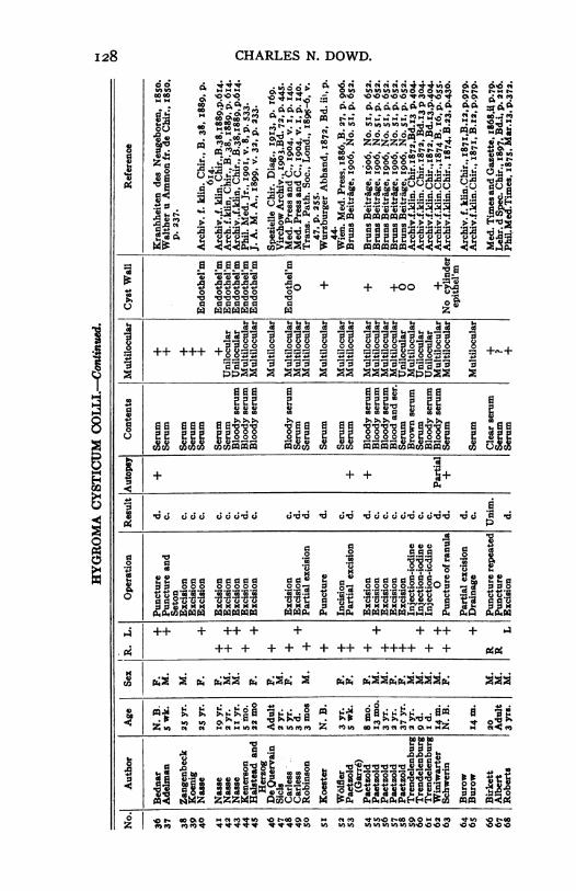
Dr. Farr has made out the following tables endeavoringto include only those cases which were definitely hygromas,according to the definition above given. It is not possibleto make a list which is altogether satisfactory, e.g., in onlyI5 of the 9I cases in the first table is there a definite state-
I25

CHARLES N. DOWD.
ment of the endothelial lining of the cysts and we do notknow how well the staining was done in most of these I5cases.
The group division here given is about what would beexpected. The neck is the part of the body most richly sup-plied with lymphatics and hence the most common site of these
FIG. 17.rnnv
Showing the structure of the lymphatics and their communications with the veins in thePig-tailed Macque (Macacuts uomestrinus Linn). Reproduced from Anatomical Record,vol iii, p. ssr, Fig. g (type xi) (McClure and Silvester).
growths. The axilla which also has a rich lymphatic supplyshows the next largest number. Without doubt some of theaxillary growths pressed through from the neck as in thewriter's second case or were associated with the neck cystsas in Case I.
It is a little strange that so few cases are reported in thegroin but there are enough to establish their existence.
It is also noteworthy that so little of the literature isrecent. Most of the recent cases are described under the
126

HYGROMA CYSTICUM COLLI.
CaCaC A) bba b ) b a-C bam 0VGo '-30' us.n C3 0V'DO 'OCnnAICA bAO'OOo 0Cn.OCiIA
P01-40 u > d 9 W>1<<"NSQN1tt>mt g W° g0g g f g g o .r;; :g;° z [ 2, g .CD 'Cn' 4
CD o Ci' D8 D C
1S v H zo z zv 4 " P OPo w
.1 2 w :1 to 9 w1 ww 2 M FI-Pp- B ,f BCD:4
MZDPIZ zZ m m
(.rI P ' r
e.'0 CD aDo irl.~C ".
0
'C-.~~~~~~~~.0 0
aCDDUPP P CDCD~~~~~~~t
t
CD CD-~~~~~~~~~~~~~~~~t0 D C 'AD D CO
VnCa V
* Z
127
10
;.P
0
3soa,
.0-
0
0
p9
0rvf
m
'4
8P
¢
a
p
40
CHARLES N. DOWD.
. t.. . .o . . . . .
00l H l* le HH T * * * * * J J,*CS c; Ci 4, ° , o." 's 6 S; m *.o" _" C4 _C.,<0 ,'In n V"t n n % in%n
c00 | sm . OIl)Z ZZ N
co~~~~~~~~~~~~~~~~~~~~~~~~~~~~~~~~~~~~~~~~~~~~~~~~~~~~~~~~~~~~~~~~m
B 6 5~~~~~oQo o) M Ma°++ M
)0000 0)0 n.in.~0oo
M M0 6CO 6C006 00l
0oV) et c44, Z .;-
0 0 00
10o 01 0 10 0c
>~~~~~~~~~~~~~~~~~~a% 0%a%atat
++ +++ +o8a; a °..a a~aa. aXa08gau 7 a
02 PP224 44+'4 4)a
0 0 0 CO Qo 0 al Z a - Z e0
101010c10S. 0 mm
+4 4 4O + + 0 +
IX ++ +4P
rA -d ;c ; c ;* ; c;c c ; t; 'dc d d c;-d-dc;c;c c; -dc;cJod d -d c) 'a -
~~ 1 0 3.0~~~~~~~~~~~060 0 0 0 0200 -20 0 4C)
4)6646NINN Cd 64464~~~ 10k20 020 r1 5 p 4p.5p p4.
I4++ + ++ + + + + ++ +
+++++++++++ + ++++ + +
* UU 0o.~4 ' a'0A
>Z I C4 CC)V( 40U C Mf 00 f KCi0)'i)OZ
aIV2 4''
04) 10 10010ZI 0W OM 4 uZ01 e
o '0 000)0 Mc4 D -000%0 elm *%n o0. oo0 cqm RU)MI M M V v V V V VVV U) I WW) W) in in WN
I28
i
8
0
34¢4
No 0-cNOa 0o

HYGROMA CYSTICUM COLLI.
o 0
ia -a-~~~~~4:-
+ ++ ++++ +++ +++ + +++++++
0
CU O Wlug gg! S S0 e.S SE:
rfl WA~f4
~~~~~~~~~~~~.
+ + + + +++
R4 0
0'0 UN~~~~~~~~
4 a -go o to~~~~~~~~~~~~o*44 dQ al aoaE4 oo 0'-. ".080
PkU 000 *c *
A. .4>X.44. .2
.U
Oa4
v o " ""wvo be v ^ " eq U>-b Cho -s b b r~~~~~N w MaOOro o OOOO0 Ws~ ~ ~~'~a'0
I29

130 CHARLES N. DOWD.
so o444c *c
Hoaoww va
(44& t ° ' * c
p *;00 00 P w
-&
CC
fi,!* *,6Io00
coa
U2(V.. . .,.
wq-V._,d1gom_p0*Q .
-ncom
:;++++++++++ + + +++ +++ Il++++ +++++X% .
tio A+_++++ + ++J0 ;
*a
d~~~~~~~~~~~~~~~
d 0aaa E
0 0I
o at 'co0mmtn0fis u cn uzank cnm m M Meanm M esm
X~~~~~~U N
~~ ,CCCC~~~~~~~CCC~~~~~,0 0N(#00 ~ ~ ~ ~ ~ 0 A.
o a gd dl0 0 0
o~~~~~ ~~~~ t IIju]]u| 3w|o jil$Pe~~~~~ ~~~03immiM 0M:Xmmm M MO:S R @@
.ir1. . 14.....4 . ... .... ..
X X X ;nn G n ;n - X; - Xn <-G~~1
% |t gtD - t C'°" 'aa
" CfS CCt C'C -a.D-.~~~~~~~~~0O... .!kciz
C)
IU
aC4
14
0
0
oo HO-w c mf VinNo r^eo o 04 Ml*"ooov t"~ ~ ~ m m m "N MH H N ZH" " "M "f . . . ."e nMeMe ienI

HYGROMA CYSTICUM COLLI.
8
4)
.0
00~~~~~>0
.) . .I
oCo Co -i.:z; ~ m
m .s,,,0W W 00
Q @
9 0 X̂ Wmo > m~O'
U)
mc
p4
'(.6
Co 0
NO
0d.
o m
co0
.-4 .
_ Co] -.
'up
mo COCoCm
.00
00' 00 4 0
4'+++++ + ++ ++
0fl aaaaa aa: a a
Asdony | + ++
a) a'0a o o . ° a0~~~~~
0004)30 0 00
22g..X2 2 o 0
. " k 4 3- .P
0 ._
4
a a ' 3.
be+
N~~~~~~~~~~4)~~~~~~~~~~~~c
4)
I3'
z
mrzn
0
0
04
I -Wqt=N I m cA m V w) % so Ch O -

CHARLES N. DOWD.
heading of lymphangioma and it is difficult to sift out thecases which are really hygromas. Winglowski's 4 recent arti-cle should be referred to, he studied various neck anomaliesand dissected I5O half necks in children and adolescents andfound rests 23 times.
It would be very gratifying to explain the remarkablepower of growth which these cysts have. Unfortunately wecannot do this with absolute accuracy any more than we cantell why some people grow to be large and others remainsmall. There are, however, many other instances of some-what similar growth. Ovarian cysts, parovarian cysts, der-moid cysts, both abdominal and subcutaneous, hypernephro-mas, mesenteric cysts, thyroglossal and branchial cysts,lipomata, exostoses, growths of the carotid body, commonwarts are all examples of individual power of growth whichcomes in tissues otherwise benign. In view of these examplesit is not strange that sequestrations of lymphatic tissue inthe neck should occasionally show this great power of growth.
SUMMARY.-I. Cystic hygromas of the neck have beendescribed for many years and their existence is undoubted.
2. The term should be restricted to cysts lined withendothelium and having a marked power of growth.
3. Such cystic growths are uncommon. A careful searchof the literature has so far revealed records of only 9I caseslocated distinctly in the neck and 35 cases located principallyin the axilla, but in part at least extending there from the neck.
4. The writer records three cases of undoubted hygromaand a fourth case which is believed to have been a hygromabut in which inflammation had destroyed the finer structureof the cyst walls.
5. The most satisfactory explanation of the existence ofthese hygromas is that embryonic sequestrations of lymphatictissue existed and that they had the power of persistentirregular growth.
6. Excision is the best treatment. If this is impracticablepartial excision is the next best.
'Archiv f. Klin. Chir., 1912, vol. 98, p. I51.
I32