novel methods for the control of phlebotomine sand flies
TRANSCRIPT

Louisiana State UniversityLSU Digital Commons
LSU Doctoral Dissertations Graduate School
2008
Novel methods for the control of phlebotominesand flies (Diptera: Psychodidae)Thomas Michael MascariLouisiana State University and Agricultural and Mechanical College
Follow this and additional works at: https://digitalcommons.lsu.edu/gradschool_dissertations
Part of the Entomology Commons
This Dissertation is brought to you for free and open access by the Graduate School at LSU Digital Commons. It has been accepted for inclusion inLSU Doctoral Dissertations by an authorized graduate school editor of LSU Digital Commons. For more information, please [email protected].
Recommended CitationMascari, Thomas Michael, "Novel methods for the control of phlebotomine sand flies (Diptera: Psychodidae)" (2008). LSU DoctoralDissertations. 2709.https://digitalcommons.lsu.edu/gradschool_dissertations/2709

NOVEL METHODS FOR THE CONTROL OF PHLEBOTOMINE SAND FLIES (DIPTERA: PSYCHODIDAE)
A Dissertation
Submitted to the Graduate Faculty of the Louisiana State University and
Agricultural and Mechanical College in partial fulfillment of the
requirements for the degree of Doctor of Philosophy
in
The Department of Entomology
by Thomas Michael Mascari
B.A. Louisiana State University, 2001 M.S. University of London, 2002
December 2008

ii
DEDICATION
This dissertation is dedicated to my parents, Kate and Walter Mascari, and to my brother David
Mascari. Their love, support, patience, and encouragement over the years made all of this
possible.

iii
ACKNOWLEDGEMENTS
During my time at LSU, I have benefitted tremendously from the guidance and
mentorship of Dr. Lane Foil. In science and grant writing, he’s the one who showed me the
ropes. I would like to thank the late Dr. Michael Perich for planting the seed for this research,
and for his friendship and kindness during the short time that I knew him. Thanks to Dr. Wayne
Kramer, an essential member of my committee, for his advice and for allowing me to take up
some of his precious lab-space. Thanks to Dr. Mark Mitchell for his support and thoughtful help,
and for his assistance in the development of the animal use protocol for my research (and
navigating the IACUC). I also would like to thank the other members of my committee, Dr. Jim
Ottea and Dr. Timothy Schowalter, for all their invaluable advice and support. Thanks to Dr. Ed
Rowton, Dr. Phil Lawyer, and COL Scott Gordon for helping me learn the ins and outs of
working with phlebotomine sand flies; they helped lay the groundwork for my research. Special
thanks to the original gang of students in the mosquito lab (Isidra Sabio, Dr. Andrew Mackay,
Dr. Isik Unlu, Ana Maria Sanchez, and Brett Collier), whose support and friendship encouraged
me to stick with sand flies when things looked bleak. Also thanks to Jessica Brauch for her
friendship both in the lab and the great outdoors. Thanks to Jeremy Colonna for his assistance
with the sand fly colony; his reliable help allowed me to leave the sand flies alone for brief
Christmas vacations without too much separation anxiety. Also thanks to the rest of the
industrious team of student workers in Dr. Foil’s lab who helped me over the past years. Finally,
a hearty thanks to Dr. Mileah Kromer for her continuing assistance and emotional support.
Thanks buddy!

iv
TABLE OF CONTENTS DEDICATION…………………………………………………………………………………...ii ACKNOWLEDGEMENTS………………………………………………………………….…iii LIST OF TABLES………………………………………………………………………………vi LIST OF FIGURES……………………………………………………………………………viii ABSTRACT………………………………………………………………………………………x INTRODUCTION …...……………………………………………………………………..…...1 CHAPTER 1. LITERATURE REVIEW……………………………………………………….3 1.1 Taxonomy of Phlebotomine Sand Flies…..…………………………………………...3 1.2 Sand Fly Biology, Ecology, and Sampling……………………………………….…...4 1.3 Disease Agents Transmitted by Sand Flies………………………………………..…..9
1.4 Rodent/Sand Fly Associations.……………………………………………………....15 1.5 Control of Leishmaniasis……...……………………………………………..………21
CHAPTER 2. LABORATORY EVALUATION OF DIFLUBENZURON AS A FEED-THROUGH FOR CONTROL OF IMMATURE SAND FLIES (DIPTERA: PSYCHODIDAE)………………………………………………………………………….……36 2.1 Introduction………………………………………………………………………..…36 2.2 Materials and Methods…………………………………………………………...…..37 2.3 Results…………………………………………………………………………..……40 2.4 Discussion……………………………………………………………………..……..42 CHAPTER 3. EVALUATION OF NOVALURON AS A FEED-THROUGH INSECTICIDE FOR CONTROL OF IMMATURE SAND FLIES (DIPTERA: PSYACHODIDAE)…………………………………………………………………….……….44
3.1 Introduction……………………………………………………………………..……44 3.2 Materials and Methods……………………………………………………………….45 3.3 Results…………………………………………………………………………..……47 3.4 Discussion…………………………………………………………………………....49 CHAPTER 4. IVERMECTIN AS A RODENT FEED-THROUGH INSECTICIDE FOR CONTROL OF IMMATURE SAND FLIES (DIPTERA: PSYCHODIDAE)………...……51
4.1 Introduction……………………………………………………………………..……51 4.2 Materials and Methods……………………………………………………………….52 4.3 Results………………………………………………………………………………..54 4.4 Discussion………………………………………………………………………..…..57 CHAPTER 5. EVALUATION OF JUVENILE HORMONE ANALOGUES AS RODENT FEED-THROUGH INSECTICIDES FOR CONTROL OF IMMATURE SAND FLIES (DIPTERA: PSYCHODIDAE)…………………………………………………………..…….59
5.1 Introduction………………………………………………………………………..…59

v
5.2 Materials and Methods…………………………...…………………………………..60 5.3 Results………………………………………………..………………………………63 5.4 Discussion………………………………………………..…………………………..66 CHAPTER 6. EVALUATION OF NOVALURON AS A RODENT FEED-THROUGH UNDER SIMULATED FIELD CONDITIONS FOR CONTROL OF SAND FLY LARVAE (DIPTERA: PSYCHODIDAE) ….………………...…………………..………………………68
6.1 Introduction……………………………………………………..……………………68 6.2 Materials and Methods……………………………………………...………………..69 6.3 Results…………………………………………………………………..……………73 6.4 Discussion…………………………………………………………………..………..78 CHAPTER 7. EFFECT OF ORAL IVERMECTIN TREATMENT OF RODENTS ON SURVIVAL OF SAND FLY (DIPTERA: PSYCHODIDAE) LARVAE FED ON THE RODENT FECES AND FEMALE SAND FLIES FED ON THE RODENTS……………..82
7.1 Introduction……………………………………………..……………………………82 7.2 Materials and Methods……………………………………...………………………..83 7.3 Results…………………………………………………………..……………………88 7.4 Discussion…………………………………………………………..………………..91 CHAPTER 8. EVALUATION OF RHODAMINE B AS AN ORALLY DELIVERED BIOMARKER FOR RODENTS AND A FEED-THROUGH TRANS-STADIAL BIOMARKER FOR PHLEBOTOMINE SAND FLIES (DIPTERA: PSYCHODIDAE)....95
8.1 Introduction…………………………………………………………………….….…95 8.2 Materials and Methods……………………………………………………………….97 8.3 Results………………………………………………………………………………101 8.4 Discussion…………………………………………………………………………..107 SUMMARY AND CONCLUSIONS…………………………………………………………112 REFERENCES……………………………………………………………………………...…116 APPENDIX A. COPYRIGHT PERMISSION LETTERS...……………………………….135 VITA…………………………………………………………………………………………...137

vi
LIST OF TABLES Table 1.1 Burrowing Rodent Reservoirs of Leishmania spp. and Associated Sand Fly Vectors………………………………………………………………………………………...…18 Table 2.1 Percent Mortality and Age at Death of 2nd Instar (13±1 Days Old) P. papatasi Fed Feces from Three Treatment Groups of Syrian Hamsters Receiving Different Oral Doses of Diflubenzuron, Feces from Untreated Syrian Hamsters, or an Untreated Laboratory Larval Diet (a 1:1 Rabbit Feces-Rabbit Chow Diet)……………………………………….…………………42 Table 3.1 Percent Mortality and Longevity of 2nd Instar (13 ± 1 Day Old) P. papatasi Larvae Fed Feces of Syrian Hamsters That Had Been Fed a Diet Containing 0, 9.88, 98.8, and 988 mg/kg, or an Aged 1:1 Rabbit Feces-Rabbit Chow Larval Diet Containing 0 and 988 mg/kg Novaluron………………………………………………………………………………….….…48 Table 4.1 Mortality of Second Instar Sand Flies Fed Feces Voided by Ivermectin-Treated or Untreated Hamsters, and Ivermectin-Treated or Untreated Laboratory Larval Diet (1:1 Rabbit Feces-Rabbit Chow w:v)……………………………………………………………………..…..56 Table 5.1 Percent Pupation and Adult Emergence of Immature Sand Flies Fed Feces of Hamsters Fed Diets Containing Methoprene or Pyriproxyfen, or Control Diets…………...……………....64 Table 6.1 Mortality and Longevity of 2nd Instar Sand Flies Fed Hamster Feces Directly Treated with Novaluron Solutions………………………………………………………………………..73 Table 6.2 Means (±SE) of Body Weight, Food Intake, and Daily Dosages of Novaluron for Syrian Hamsters………………………………………………………………………………….74 Table 6.3 Mortality and Longevity of 2nd Instar Sand Flies Fed Feces of Hamsters Fed Diets Containing Novaluron……………………………………………………………………………75 Table 6.4 Means (±SE) of Body Weight, Food Intake, and Daily Dosages of Novaluron for Syrian Hamsters……………………………………………………………………………….…75 Table 6.5 Means (±SE) of Body Weight, Food Intake, and Daily Doses of Novaluron for Syrian Hamsters Offered Food Containing Novaluron as All, Part, or None of Their Diet………….....77 Table 6.6 Mortality and Longevity of 2nd Instar Sand Flies Fed Feces of Hamsters Fed Diets Containing Novaluron. Hamsters Were Fed Novaluron-Treated Food as All, Part, or None of Their Daily Diet………………………………………………………………………………….78 Table 7.1 Means (±SE) of Body Weight, Food Intake, and Daily Dosages of Ivermectin for Syrian Hamsters………………………………………………………………………………….88 Table 7.2 Post-Bloodmeal (24 h) Survival of Sand Flies Fed on Ivermectin-Treated Hamsters……………………………………………………………………………………...….89

vii
Table 7.3 Post-Bloodmeal (48 h) Survival of Sand Flies Fed on Ivermectin-Treated Hamsters……………………………………………………………………………………...….89 Table 7.4 Mean Number and Viability of Eggs Deposited by Sand Flies That Had Taken Bloodmeals from Hamsters 14 d after Being Withdrawn from an Untreated or Ivermectin-Treated Diets………………………………………………………………………………..…....90 Table 8.1 Means (±SE) of Body Weight, Food Intake, and Daily Dosages of Rhodamine B for Syrian Hamsters Fed Rhodamine B-Treated or Untreated Diets for 9 d……………………….101 Table 8.2 Duration and Location of Markings after Hamsters Were Withdrawn from Untreated Diets or Diets Containing Rhodamine B……………………………………………………….102 Table 8.3 Results of the Rhodamine B Feed-Through Larval Bioassay (Percent Adult Emergence, Age at Adult Emergence, and Fluorescence of Adult Sand Flies That Were Fluorescent when Observed Using Fluorescence Microscopy). Second Instar Sand Flies Were Fed Feces of Hamsters That Had Been Fed a Diet Containing 0, 50, 500, or 5,000 mg/kg Rhodamine B…………………….……………………………………………………………..104

viii
LIST OF FIGURES Figure 2.1 Cumulative Per Cent Survival of 2nd Instar (13±1-d old) P. papatasi Larvae Fed Feces from Three Treatment Groups of Syrian Hamsters Receiving Diets Containing Different Concentrations of Diflubenzuron, Feces from Untreated Control Syrian Hamsters, or an Untreated Control Laboratory Larval Diet (a 1:1 Rabbit Feces-Rabbit Chow Diet). Vertical Reference Lines Indicate the First Appearance of Pupae (13 d) and Adults (17 d) in Control Vials……………………………………………………………………………...………………41 Fig. 5.1 A Pupa-Form Larva That Had Been Fed Feces of Hamsters Fed a Diet Containing 9.82 mg/kg Pyriproxyfen as a Larva (A), and a Normal Pupa That Had Been Fed Feces of Untreated Hamsters as a Larva (B)………………………………………………………………….………65 Figure 6.1 Percent Adult Emergence of Sand Flies Fed Feces of Control or Novaluron-Treated Hamsters; Feces Were Aged under Simulated Field Conditions (28 °C, 90% RH) for up to 150 d…………………………………………………………………………………………………..76 Figure 7.1 Sand Flies Taking a Bloodmeal from a Chemically Immobilized Ivermectin-Treated Syrian Hamster……………………………………………………………………………..…….86 Figure 7.2 Percent Adult Emergence (Mean ± SE) of Sand Flies Fed as 2nd Instars the Feces of Untreated or Ivermectin-Treated Hamsters; Feces Used in This Bioassay Were Voided by Hamsters 0, 3, 7, or 14 d after the Hamsters Were Withdrawn from Their Respective Diets…………………………………………………………………………………………...…90 Fig. 8.1 A Hamster That Had Been Fed a Diet Containing Rhodamine B. Pink Coloration Was Visible on the Dorsal Pelage (A), Oral Mucosa (B), Paws (C), and Urogenital Area and Tail (D)……………………………………………………………………………………………....102 Fig. 8.2 Images of Feces of Four Hamsters Taken under Incandescent Lighting (A, B, C, and D), and Using Fluorescence Microscopy with a 1 sec Exposure Time (E, F, G, and H). The Feces Pictured Are from a Hamster Fed a Control Diet (A and E), or a Diet Containing 50 (B and F), 500 (C and G) or 5,000 mg/kg Rhodamine B (D and H)…………………………………….…103 Fig. 8.3 Images of Two Female Sand Flies Taken under Incandescent Lighting (A and B), and Using Fluorescence Microscopy (C and D). The Sand Fly Pictured in the First Column (A and C) Had Been Fed as a Larva the Feces of a Hamster That Had Been Fed a Diet Containing 5,000 mg/kg Rhodamine B, and the Sand Fly Pictured in the Second Column (B and D) Had Been Fed as a Larva the Feces of a Hamster That Had Been Fed a Control Diet………………………...105 Fig. 8.4 Images of Two Male Sand Flies Taken under Incandescent Lighting (A and B), and Using Fluorescence Microscopy (C and D). The Sand Fly Pictured in the First Column (A and C) Had Been Fed as a Larva the Feces of a Hamster That Had Been Fed a Diet Containing 5,000 mg/kg Rhodamine B, and the Sand Fly Pictured in the Second Column (B and D) Had Been Fed as a Larva the Feces of a Hamster That Had Been Fed a Control Diet……………………...…105 Fig. 8.5 A Female Sand Fly Taking a Bloodmeal from the Hind Foot of an Anesthetized, Rhodamine B-Treated Hamster………………………………………………………………...106

ix
Fig. 8.6 Images of Two Bloodfed Female Sand Flies Taken under Incandescent Lighting (A and B), and Using Fluorescence Microscopy (C and D). The Sand Fly Pictured in the First Column (A and C) Had Taken a Bloodmeal from a Hamster That Had Been Fed a Diet Containing Rhodamine B, and the Sand Fly Pictured in the Second Column (B and D) Had Taken a Bloodmeal from a Hamster Fed a Control Diet……………...…………………………………107

x
ABSTRACT
In arid and semi-arid parts of the Old World, Phlebotomus paptasi is a significant biting
pest of man and is the primary vector of Leishmania major, the causative agent of zoonotic
cutaneous leishmaniasis (ZCL). Phlebotomus papatasi exhibits a close association with the
burrowing rodents that serve as the reservoirs of L. major. Rodent burrows are considered to be
the primary habitat of immature P. papatasi in ZCL foci, and sand fly larvae have been observed
feeding on rodent feces. In laboratory studies, five insecticides (diflubenzuron, novaluron,
methoprene, pyriproxyfen, or ivermectin) were incorporated into the diet of Syrian hamsters and
evaluated as feed-throughs to control immature sand flies. Feces of hamsters fed a diet
containing approximately 10 mg/kg diflubenzuron, novaluron, or pyriproxyfen, or 20 mg/kg
ivermectin killed 100% of sand fly larvae that consumed these feces. Feces of hamsters fed a diet
containing up to 978.8 mg/kg methoprene caused significant, but not complete, mortality of sand
fly larvae. Feces of novaluron-treated hamsters also were held under simulated field conditions
for up to 30 d, and all larvae that consumed these feces died before pupation; a significant
reduction in treated larval survival relative to control was observed when the feces were aged for
up to 150 d. Novaluron also was shown to be effective as a feed-though larvicide when
novaluron-treated food made up only a portion of the diet of hamsters. Ivermectin also was
evaluated as a systemic insecticide; ivermectin treatment of hamsters was 100% effective against
bloodfeeding sand flies for up to 7 d after hamsters were withdrawn from ivermectin-treated
diets. In the final study, proof of concept was established for a novel biomarker system using a
feed-through fluorescent dye. The value of this method is that it can mark rodents and their feces
to establish the consumption of treated-baits, mark adult female sand flies that feed rodents for
the duration of persistence of the dye in rodents, and mark adult male and female sand flies that
had fed on feces of bait-fed rodents as larvae.

1
INTRODUCTION
Phlebotomine sand flies are major biting pests of man and are the vectors of several
viruses, the bacterium Bartonella bacilliformis, and, most importantly, the protozoan parasites
that cause leishmaniasis. Worldwide, there are an estimated 2 million new cases of leishmaniasis
annually, and 12 million people are currently believed to be infected (WHO 2006). Throughout
North Africa, the Middle East and Southwest Asia, Phlebotomus papatasi is the primary vector
of Leishmania major, the causative agent of zoonotic cutaneous leishmaniasis (ZCL).
While larvicides are commonly used to control mosquitoes and many other flies of
medical and veterinary importance, there is no current use of larvicides for phlebotomine sand
fly control. In arid and semi-arid foci, P. papatasi exhibits a close association with several
burrowing rodent reservoirs of L. major (Neronov and Gunin 1971). In ZCL foci in the Old
World, rodent burrows are considered to be the primary immature habitats for P. papatasi, but
introducing an insecticide into the burrows is generally precluded by the length and complexity
of the tunnels which comprise the burrows (Seyedi-Rashti and Nadim 1973, Karapet’ian et al.
1983).
In Old World ZCL foci, sand fly larvae also have been observed feeding on the feces of
rodents (WHO 1968). Because of this fact, rodent feed-through insecticides are a potential means
of controlling sand fly larvae. Therefore, the chitin synthesis inhibitors diflubenzuron and
novaluron, the juvenile hormone analogs methoprene and pyriproxyfen, and the macrocyclic
lactone ivermectin were evaluated as rodent feed-through insecticides to control sand fly larvae.
The development and survival of P. papatasi larvae fed feces of Syrian hamsters, Mesocricetus
auratus, that had been fed a diet containing an insecticide were measured. Additional studies
were conducted to determine the effectiveness of novaluron as a feed-though larvicide to control
sand flies under simulated field conditions.

2
Because populations of P. papatasi that live in burrows rely upon rodent reservoirs of L.
major as a bloodmeal source, incorporating a systemic insecticide into rodent bait could be a
potential way to control this epidemiologically important group of adult sand flies. Therefore,
experiments were conducted to determine whether the post-bloodmeal survival of adult sand flies
would be affected by feeding their rodent hosts a diet containing ivermectin. In this study the
insecticidal effect of ivermectin treatments against bloodfed sand flies was monitored for 14 d
after rodents were withdrawn from their ivermectin-treated diets, and bioassays with larval sand
flies were conducted using feces voided by ivermectin-treated rodents over this same time
period.
Prior to or simultaneous with field evaluations of feed-through or systemic control of
sand flies in the different sand fly/rodent associations that exist, establishing whether the larvae
of different species of sand fly feed exclusively on the feces of rodents must be demonstrated.
There are currently no available methods to directly demonstrate if the larval diet of
phlebotomine sand flies is exclusively rodent feces. Although sand fly larvae have been
recovered from rodent burrows and have been observed feeding on the feces of rodents, larval
sampling is an impractical method to demonstrate the larval diet of sand flies. An objective of
this research was to establish a fluorescent tracer technique using rhodamine B as a rodent feed-
through to identify adult sand flies that had fed on the feces of rhodamine-B treated hamsters as
larvae. We also evaluated rhodamine B as a biomarker of bait-fed rodents and the female flies
that fed upon them.

3
CHAPTER 1. LITERATURE REVIEW
1.1 Taxonomy of Phlebotomine Sand Flies
1.1.1 Family Psychodidae
Phlebotomine sand flies belong to the family Psychodidae, which is among the most
primitive families of Diptera (Young and Duncan 1994). The family Psychodidae is
characterized by their wing venation (the presence of numerous parallel veins running to wing
margin), and the presence of dense hairs on the wings and thorax (Triplehorn and Johnson 2005).
1.1.2 Subfamily Phlebotominae
Phlebotomine sand flies are classified within the subfamily Phlebotominae, and are called
phlebotomine sand flies to distinguish them from other flies that are sometimes referred to as
sand flies (such as members of families Simulidae or Ceratopogonidae). Phlebotomine sand flies
are differentiated from other subfamilies within Psychodidae by the presence of biting
mouthparts that are longer than the head, five-segmented palps, nearly cylindrical antennae, a
five-branched radial vein on the wing, and the absence of an eye-bridge (Triplehorn and Johnson
2005). Some general attributes that can often be used to distinguish sand flies from other small
flies include their size (1.5 to 2.5 mm in length), characteristic hopping flight, and the “V”
position in which they hold their wings while resting.
1.1.3 Phlebotomine Sand Fly Genera
There are three New World genera within subfamily Phlebotominae: Brumptomyia
França & Parrot, Warileya Hertig, and Lutzomyia França (Young and Duncan 1994). Sand flies
in the genus Brumptomyia have not been reported feeding on humana, and are distinguished
from sand flies in other genera by differences in the morphology of male external genitalia
(Young and Duncan 1994). Sand flies in the genus Warileya are reported to be anthropophilic,
but they have not been implicated in the transmission of any human pathogens (Young and

4
Duncan 1994). Sand flies in the genus Lutzomyia feed on mammals and are the only medically
important genus of sand flies in the New World. Lutzomyia is distinguished from Brumptomyia
by the number of rows of teeth on the cibarium (Lutzomyia has 1 row of transverse teeth,
Brumptomyia has 4 horizontal rows of teeth), and from Warileya by the presence of episternal
setae (Lutzomyia has episternal setae, and Warileya does not).
There are two Old World genera within the subfamily Phlebotominae: Sergentomyia
França and Phlebotomus Rondani & Berté (Lewis 1982). Sand flies in the genus Sergentomyia
feed primarily on lizards, and may be the vectors of the agents of saurian leishmaniasis. Sand
flies of the genus Phlebotomus feed on mammals, and represent all of the medically important
sand flies in the Old World. Sand flies of the genus Phlebotomus can often be distinguished from
those within Sergentomyia by the cibarium; Phlebotomus does not have a row of teeth and
usually does not have a patch of pigment (Lewis 1982).
1.2 Sand Fly Biology, Ecology, and Sampling
1.2.1 Immature Stages
The eggs of phlebotomine sand flies are dark brown or black and elliptical in shape. The
eggs have ridges in species-specific patterns that potentially could be used for identification. The
number of eggs laid by a single female at one time varies greatly by species and by factors such
as species of bloodmeal host or ambient temperature, but typically is between 40 to 70 eggs
(Young and Duncan 1994). Eggs are laid in batches on moist substrates, and the presence of
conspecific eggs can serve as an oviposition attractant and stimulant (Elnaiem and Ward 1991,
Srinivasan et al. 1995). The hatching of eggs usually occurs within 10 d after oviposition, but
hatching of some eggs in a batch is sometimes delayed for as long as 30 d (Young and Duncan
1994).
5
Sand fly larvae have four instars. Sand fly larvae are covered in setae along the length of
their bodies, and have four caudal setae by the time they reach 4th instar. Sand fly larvae feed on
organic matter near the site of oviposition. The larval stage of phlebotomine sand flies is
completed in as few as 18 d, but typically lasts longer and can be dependent on temperature
(Young and Duncan 1994). Before pupation, sand fly larvae cease feeding and some species may
travel a short distance upward to a drier location. Pupae sometimes attach to rocks or other fixed
objects.
The sand fly larval habitats have been identified for only a handful of species. In the Old
World, immature stages of P. argentipes, P. martini, P. papatasi, P. celiae, P. ariasi, P.
perfiliewi, and P. langeroni have been recovered from soil taken from inside of structures
housing humans or domesticated animals (Dhiman et al. 1983, Mutinga et al. 1989, Killick-
Kendrick 1987, Bettini 1989, Doha et al. 1990). Larvae of P. martini, P. papatasi, and P.
duboscqi have consistently been recovered from soil taken from inside of rodent burrows
(Mutinga et al. 1986, Mutinga et al. 1989, Doha et al. 1990, Dedet et al. 1982, Perfil'ev 1968,
Artemiev et al. 1972, Morsy et al. 1993). Larvae of the sand flies P. martini and P. celiae have
been recovered from termite mounds in East Africa (Mutinga et al. 1989).
In the New World, structures housing livestock have been shown to be a larval habitat for
L. longipalpis, and L. intermedia (Deane and Deane 1957, Forattini 1954). Larvae of other
species, including many of medical importance (including, L. trapidoi, L. umbratalis, L. anduzei,
and L. whitmani), have been found among soil and leaf litter on the forest floor (Rutledge and
Ellenwood 1975, Arias and Freitas 1982, Casanova 2001).
For many of the species listed above, very few immature specimens have been recovered,
and thus little can be stated about the importance of their larval habitats. However, for some
species, enough evidence has been compiled to make more definitive conclusions about their

6
larval habitat. For example, the primary immature habitat of P. papatasi outside of urbanized
areas is considered to be rodent burrows. Similarly, larvae of P. duboscqi have been recovered
consistently from inside of rodent burrows; this is considered to be the principle larval habitat for
this species.
Several methods have been employed for sampling immature sand flies. However, the
process remains time consuming and frequently unproductive regardless of the method used. To
illustrate this point, researchers in Central Asia processed over 6 tons of soil and recovered only
around 150 immature sand flies (Petrischeva and Izyumskaya 1941). The first sand fly larva (P.
mascittii) recovered in nature was found by direct examination of a soil sample taken from a
cellar in Rome (Grassi 1908). Direct examination of soil to find sand fly larvae was the method
used throughout the early 20th century and is still the preferred method of some more recent
researchers (Dhiman et al. 1983). A method of extracting immature sand fly larvae from soil
samples though differential flotation in salt or sugar solutions also has been used, but there is no
improvement in the rate of success and it is no less labor intensive (McCombie-Young et al.
1926). This method has been modified by combining differential flotation with passing the soil
samples through a series of nested sieves, but the modified method still was no simpler or
productive than flotation or direct examination (Hansen 1961). The larvae of P. papatasi also
have been extracted from soil samples through dessication with some success in Iran (Seyedi-
Rashti and Nadim 1972). This method was validated in the laboratory by extracting larvae from
soil samples that had been spiked with larvae from a laboratory colony (Killick-Kendrick 1987).
Breeding sites also have been identified by isolating soil samples and recovering adult sand flies
as they emergea either through the incubation of soil samples in the laboratory, or by placing
emergence traps over suspected breeding sites in the field (Mutinga and Kamau 1986, Bettini et
al. 1986).

7
1.2.2 Adults
Male adult sand flies typically emerge before females from the same egg batch, and they
become sexually mature within 1 d (Young and Duncan 1994). Male sand flies can find potential
mates through the use of pheromones, or by locating vertebrate hosts or resting sites to which
female sand flies also may be attracted. Both specific pheromones and wing-beat rhythms have
been identified for mate location for the sand fly L. longipalpis (Phillips et al. 1986, Ward and
Morton 1991).
Adult male and female sand flies obtain energy by ingesting sugars. Sugar meals can be
obtained from a variety of sources, including the sap of plants and honeydew from aphids
(Schlein and Warburg 1986, Killick-Kendrick and Killick-Kendrick 1987, Cameron et al. 1995).
In arid areas where sand flies are found, the available sources of sugar can be limited to a
handful of plant species (Schlein and Yuval 1987). Female sand flies also are required to feed on
the blood of vertebrate hosts for the production of eggs. Females of most species take
bloodmeals only once per gonotrophic cycle, though females of some species, such as L.
shannoni, will feed multiple times throughout the gonotrophic cycle (Young and Duncan 1994).
Because of their characteristic short, hopping flight, sand flies are often perceived as
weak fliers unable to travel long distances. For many species this holds true: the longest recorded
dispersal distance for a P. papatasi sand fly was 280 m. Sand flies in forested areas of the New
World also do not have long flight ranges; in one study in Panama in which 20,000 sand flies
were marked with fluorescent powder and released, the majority of re-captured sand flies were
collected within about 50 m of the release site; four sand flies were recaptured 200 m away
(Chaniotis et al. 1984). However, P. ariasi sand flies have been shown to fly as far as 2 km in
southern France (Killick-Kendrick et al. 1984).
8
Adult sand flies of all species are active at night. During the day, adults of the majority of
New World sand fly species have been found resting in tree holes or the buttresses of trees.
Adults of the majority of Old World species and some New World species have been found
resting in rock crevices, caves, or in man-made structures such as cellars, wells, or animal sheds.
Adults of L. anthophora, P. papatasi, and P. duboscqi are all frequently recovered from the
burrows or nests of rodents.
In addition to collecting adult sand flies through direct examination of potential resting
sites, sand flies can be sampled using either interception traps or attraction traps. Trapping by
interception samples the population of sand flies that is active in an area with little bias. Malaise
traps (mesh, tent-like devices placed across the suspected flight paths of insects) are often used in
New World forests to collect sand flies. This method collects sand flies of both sexes, but
generally collects low numbers of sand flies and many non-target insects that may damage sand
fly specimens (Alexander 2000). Sticky traps are the most commonly used tool in the Old World
for sampling sand fly by interception. The typical design of a sticky trap is a sheet of paper
dipped in castor oil and placed in an area where sand flies are thought to be active, including
man-made structures, fields, rock crevices, or at the openings of animal burrows and nests
(Alexander 2000). Sticky traps are used less frequently in Central and South America because
the traps are less effective in areas with high humidity.
Sampling sand flies by attraction can be conducted using animal baited traps. The Disney
trap is an effective and simple animal baited trap in which a small animal (often a rodent) is
placed in a cage on a tray coated with castor oil (Disney 1966). As sand flies approach the caged
animal in short hops, they are trapped in the castor oil. A cone trap has been developed for
attracting sand flies to larger animals (Montoya-Lerma and Lane 1996). An animal, such as a
9
horse, is tethered inside a mesh cage with concave cones that allow sand flies to enter the cage
but not exit. The trapped sand flies then can be collected off of the interior walls of the cage.
Battery-operated light traps also have been used to sample sand flies. Light traps are not
attractive to sand flies over a great distance; the maximum distance was 2m for P. ariasi, 6 m for
L. youngi, and about 2.5 m for L. intermedia and L. whitmani (Killick-Kendrick et al. 1985,
Valenta et al. 1995, Campbell-Lendrum et al. 1999). Light traps have been shown to
preferentially sample females of certain species. This sampling bias is particularly present in the
some New World sand flies; for example, over 75% of adult L. whitmani collected by light traps
were female, but females made up less than 25% of the catch when the bulbs were removed from
the traps (Campbell-Lendrum et al. 1999). Using carbon dioxide in conjunction with light traps
can be used to increase the number of sand flies collected as well as the range of attraction
(Gillies 1980).
1.3 Disease Agents Transmitted by Sand Flies
Sand flies of more than 30 species in the genus Lutzomyia and 40 species in the genus
Phlebotomus are vectors of human pathogens. Phlebotomine sand flies are the vectors of several
viruses, the bacterium Bartonella bacilliformis, and, most importantly, nearly 20 species of
protozoan parasites in the genus Leishmania.
1.3.1 Viruses
Sand flies have been shown to be vectors of medically important viruses in three families:
Bunyaviridae, Reoviridae, and Rhabdoviridae. The most important viruses transmitted to man by
sand flies are in the family Bunyaviridae and genus Phlebovirus. In the New World, more than
30 serotypes of the genus Phlebovirus have been identified, but their medical importance is not
fully known (Tesh et al. 1989). However, in the Old World, two viruses in the genus Phlebovirus
are of significant public health importance: Sandfly fever Sicilian virus (SFSV) and Toscana

10
virus (TOSV, species Sandfly fever Naples virus), and. Human infections with SFSV have been
confirmed in Italy, Cyprus, Egypt, Iran, and Pakistan; SFSV antibodies have been found in
humans in Israel, Jordan, Algeria, Tunisia, Sudan, and Bangladesh (Karabatsos 1985, Papa et al.
2006, Batieha et al. 2000, Cohen et al. 1999, McCarthy et al. 1996, Chastel et al. 1983,
Gaidamovich 1984, Izri et al. 2008). The vector of SFSV has been shown to be the sand fly P.
papatasi, and it is suspected that the distribution of SFSV coincides with the distribution of P.
papatasi (Karabatsos 1985). The symptoms of infection with SFSV typically are pyrexia and
myalgia, and cases usually resolve within a week.
Toscana virus has been found in many countries around the Mediterranean including
Italy, Spain, Portugal, France, Slovenia, Cyprus, Greece, and Turkey (Hemmersbach-Miller et al.
2004, Peyrefitte et al. 2005, Mendoza-Montero et al. 1998, Echevarria et al. 2003, Eitrem et al.
1985). Two species of sand flies have been incriminated as vectors of TOSV: P. perniciosus and
P. perfiliewi (Charrel et al. 2005). Unlike human infections with SFSV, infection with TOSV can
be life-threatening. In Italy, TOSV is considered to be a leading etiological agent of aseptic
meningitis (Charrel et al. 2005).
In the New World, a number of viruses in the genus Orbivirus and family Reoviridae
have been shown to be transmitted to man and other mammals by sand flies (Rosa et al. 1984). In
man, these little-studied viruses are believed to produce symptoms similar to infection with
Phlebovirus.
Chandipura virus (CHPV) is in the genus Vesiculovirus and family Rhabdoviridae and
has been isolated from sand flies in India and West Africa (Dhanda et al. 1970, Fontenille et al.
1994). Human infections with CHPV typically involve fever, but encephalopathy was reported in
one fatal case. The sand fly P. papatasi is believed to be the vector of CHPV in India, but the
vector remains unknown in West Africa. Venereal transmission of CHPV in P. papatasi has been

11
demonstrated in the laboratory, and P. argentipes has been shown to be a competent vector
(Mavale et al. 2006, Mavale et al. 2007).
On an uninhabited island in the Atlantic Ocean off the coast of Georgia, USA, the sand
fly L. shannoni serves as the vector of another virus in the family Rhabdoviridae, the New Jersey
serotype of Vesicular stomatitis virus (VSV-NJ). The virus has been isolated from the sand fly
vector, and from swine and other mammals; because the island is uninhabited, humans are not at
risk of infection (Clarke et al. 1996).
1.3.2 Bartonella bacilliformis
The bacterium Bartonella bacilliformis is transmitted by the sand fly L. verrucarum in
Peru and parts of Ecuador. There is no known non-human reservoir for B. bacilliformis. The
disease resulting from infection with B. bacilliformis is called bartonellosis or Carrión’s Disease
(named after Daniel Carrión, who died in 1885 after inoculating himself with infectious material
taken from a patient). There are two distinct clinical forms of disease: verruga peruana and
Oroya fever. Verruga peruana, the benign form of bartonellosis, is characterized by the
appearance of numerous painless nodules on the skin of patients, which, if untreated, resolve
within a year. Oroya fever is characterized by fever, arthralgia, hemolytic anemia, and jaundice,
and if untreated has a mortality rate of up to 90% (Grey et al. 1990). Both clinical forms of
bartonellosis can be treated successfully with antibiotics, such as chloramphenicol.
1.3.3 Leishmania spp.
Leishmania is a genus of heteroxenous parasites in the family Trypanosomatidae.
Leishmania parasites are the etiological agents of a complex of diseases with a broad clinical
spectrum called leishmaniasis. Nearly 20 species of Leishamania have been shown to cause
human disease (Desjeux 2004). Worldwide, 2 million new cases of leishmaniasis are believed to
occur annually, and as many as 12 million people currently may be infected (WHO 2006).

12
Traditionally, species within the genus Leishmania have been categorized according to the form
of leishmaniasis they cause: visceral leishmaniasis (VL) or cutaneous leishmaniasis (CL).
Visceral leishmaniasis (fever, wasting, anemia, and enlargement of the liver and spleen) is often
fatal if untreated, and CL, while not life-threatening, can cause long-lasting lesions that can leave
disfiguring scars after they heal. The species of Leishmania also are further categorized
according to the whether or not non-human reservoirs are important in the transmission cycle:
zoonotic leishmaniasis (ZCL and ZVL) or anthroponotic leishmaniasis (ACL and AVL).
Leishmania parasites are transmitted to humans by phlebotomine sand flies of around 30 species
in the genus Lutzomyia in the New World, and of the genus Phlebotomus in the Old World
(Desjeux 2004).
New World
In the New World, the main etiological agents of ZCL are L. mexicana and L.
amazonensis. Infections with L. mexicana occur primarily among people working or living in
forested areas in Central America and Mexico. Climbing rats (Ototylomys phyllotis) and other
forest rodents serve as the primary reservoirs of L. mexicana parasites (Disney 1968). The vector
species for L. mexicana in Central America and Mexico are L. olmeca and L. ayacuchensis,
respectively (Eduardo 1991). Human cases of ZCL due to infection with L. mexicana have been
reported in Texas, where the Southern Plains woodrat (Neotoma micropus) serves as the enzootic
host (Kerr et al. 1995). The sand fly L. anthophora frequently is collected in and around woodrat
nests and has been incriminated as the vector of L. mexicana in Texas.
Infections with L. amazonensis occur in northern South America (Bolivia, Colombia,
Ecuador, Venezuela Brazil, and French Guyana) and, like L. mexicana, occur primarily in
inhabitants of settlements that encroach into forests or in visitors to these areas. The incriminated
vector of L. amazonensis is the sand fly L. flaviscutellata, and the reservoir of L. amazonensis is

13
believed to be the spiny rat (Proechimys spp.) and a large number of other small mammals
(Dedet et al. 1989). Human infections with several other Leishmania species that cause ZCL
have been reported, including L. guyanensis, L. peruviana, L. lainsoni, L. panamensis, L. shawi,
L. naiffi, L. colombiensis, and L. venezuelensis (Young and Arias 1992).
Infection with L. braziliensis causes a primary lesion that occurs at the site of infection
and a delayed secondary lesion that occurs in the buccal and nasal mucosa. The cartilage and
surrounding tissue degenerate and often become necrotic and subject to secondary bacterial
infection. This condition can last for several years and can result in severe deformity, removing
the palate, lips, and nose. Infections with L. braziliensis occur in Brazil, Colombia, Venezuela,
and Bolivia, where it is transmitted by several species of sand flies including L. wellcomei, L.
complexus, L. whitmani, and L. ovalessi (De Souza et al. 1996, De Queiroz et al. 1994,
Feliciangeli and Rabinovich 1998, Warburg et al. 1991, Young and Arias 1992). Nearly a dozen
other sand fly species are suspected to be vectors of L. braziliensis. The reservoirs of L.
braziliensis parasites are believed to be sloths and other forest-dwelling mammals (Dedet 1992).
New World ZVL is caused by L. infantum. Infections with L. infantum occur throughout
Central and South America, where the sand fly L. longipalpis (an abundant, peridomestic
species) serves as the vector (Young and Arias 1992). Many sylvatic animals, particularly foxes,
are suspected as important reservoirs of L. infantum parasites. However, the role of dogs in the
transmission cycle is well established, and they are considered to be the most important reservoir
host (Dedet 1992).
Old World
In the Old World, ACL is caused by L. tropica. Human infections with L. tropica have
been reported in the Middle East, Southwest Asia, and North and East Africa. Transmission
generally occurs in densely populated areas, where the peridomestic sand flies P. sergenti and P.

14
guggisbergi serve as vectors (Lawyer et al 1991, Al-Zahrani et al. 1988, Killick-Kendrick et al.
1995). Transmission of L. tropica appears to be maintained indefinitely in humans without the
involvement of non-human reservoirs, although some possible non-human reservoirs such as the
rock hyrax have been suggested (Sang et al. 1992).
The primary etiological agent of ZCL in the Old World is L. major. Human infections
with L. major have been reported throughout the arid zone stretching from North Africa through
the Middle East and into Central and Southwest Asia, and also in arid areas of Sub-Saharan
Africa. Leishmania major exists as a zoonosis among populations of burrow-dwelling rodents in
the family Muridae. Humans are infected with L. major when they encroache into enzootic foci
(for example, during development projects, urban expansion, or military movements). In Central
and Southwest Asia and Iran the rodent reservoirs of L. major are Rhombomys opimus and
Meriones spp., and the sand fly vector is P. papatasi (Yaghoobi-Ershadi et al. 2004). In North
Africa and the Middle East, Psammomys obesus, Meriones spp., and Gerbillus spp. are the main
rodent reservoirs, and P. papatasi serves as the vector (Saliba et al 1994, Rioux et al. 1982,
Rioux et al. 1992, Morsy et al 2001, Morsy et al. 1996, Fichet Calvet 2003). In Sub-Saharan
Africa, a number of agricultural and peridomestic rodent pests serve as the reservoirs, and P.
duboscqi is the only incriminated vector species (Gebre-Michel et al. 1993, Githure et al. 1984,
Githure et al. 1986).
Leishmania aethiopica also is an etiological agent of ZCL in the Old World. Human
infections with L. aethiopica have been reported in Kenya and Ethiopia. Cases of ZCL due to L.
aethiopica often present with multiple lesions, and the disease is sometimes called diffuse
cutaneous leishmaniasis. Hyraxes (Procavia spp. and Heterohyrax spp.) have been implicated as
reservoirs of L. aethiopica, and two species of sand flies have been incriminated as vectors: P.
pedifer and P. longipes (Gemetchu 1990).

15
In the Old World, as in the New World, ZVL is caused by L. infantum. Human cases
have been reported primarily in the Mediterranean littoral, but also in Southwest and Central
Asia. As in the New World, the primary reservoir for L. infantum is the dog. The sand fly species
that have been incriminated as vectors of L. infantum include P. ariasi, P. langeroni, P.
neglectus, P. perfiliewi, and P. perniciosus (Rioux et al. 1979, Pires et al. 1984, Maroli et al.
1987, Doha and Shehata 1992).
Leishmania donovani is the causative agent of AVL in the Old World. Cases of AVL due
to L. donovani have been reported in Kenya, Ethiopia, Sudan, and the Indian subcontinent.
Infection with L. donovani is similar to infection with L. infantum, and AVL often is fatal if
untreated. After treatment, a small percentage of patients develop post-kala-azar dermal
leishmaniasis: a condition in which the skin is covered in large nodules that can be disfiguring.
Humans are thought to be the only reservoirs for L. donovani, but several animals such as the
mongoose have been suggested as potential non-human reservoirs (Elnaiem et al 2001). The
vectors of L. donovani in Africa are the sand flies P. orientalis, P. martini, and P. celiae
(Elnaiem et al. 1996, Gebre-Michel and Lane 1993), while Phlebotomus argentipes serves as the
vector of L. donovani in the Indian subcontinent (Joshi et al 1986).
1.4 Rodent/Sand Fly Associations
1.4.1 Sand Flies Associated with Rodent Reservoirs of New World ZCL and Old World VL
Phlebotomine sand flies of many species are associated with rodents. The closeness of
this association varies by habitat and the involvement of other (non-rodent) mammals in the
transmission of a particular Leishmania parasite. In Central and South America, the known
reservoirs of Leishmania parasites in ZCL foci include rodents such as the spiny rat, Proechimys
spp., and climbing rat, Ototylomys phyllotis. However, many other forest mammals also are
suspected to be reservoirs including rodents (Sciurus vulgaris, Heteromys desmarestianus,

16
Oryzomys capito, Nyctomys sumichrasti, Akodon sp., Sigmodon hispidus, Rattus rattus, Coendu
sp., and Agouti paca), marsupials, edentates, carnivores, and non-human primates (Dedet 1992).
The larval habitats of many of the sand fly vectors of ZCL in Central and South America have
been shown to be leaf litter and other organic debris dispersed throughout the forest floor
(Hanson 1961, Hanson 1968, Arias and Freitas 1982, Vieira et al. 2000, Casanova 2001,
Rutledge and Ellenwood 1975). The presence of many alternative hosts and the widely dispersed
habitats for immature sand flies make control measures that target reservoir hosts and sand fly
larvae improbable.
In the Old World, some sand fly species that are vectors of the agents that cause VL are
associated with rodents. Rodents have not been shown to be reservoirs of L. infantum or L.
donovani, and adult sand flies have not been associated with rodents or the rodents’ nests or
burrows. However, larvae of P. martini and P. langeroni, have been recovered from inside
rodent burrows (Mutinga et al. 1989; Doha et al. 1990). Each of these sand fly species also has
many alternative larval habitats in a single VL focus. In Kenya, larvae of P. martini have been
recovered with greater frequency from termite mounds, and also from inside houses and tree
holes (Mutinga et al 1989). A single specimen of P. langeroni was recovered from soil inside of
a rodent burrow; larvae of P. langeroni are much more commonly recovered from piles of rocks
and garbage, animal sheds, and wells (Doha et al. 1990).
1.4.2 Sand Flies Associated with Rodent Reservoirs of Old World ZCL
Three species of medically important sand flies exhibit a very close association with the
rodents that serve as reservoirs of Leishmania parasites in ZCL foci: L. anthophora, P. duboscqi,
and P. papatasi (Table 1.1). Each of these rodent/sand fly associations occurs in arid or semi-arid
habitats and involves rodents that construct burrows and sand flies that are frequently collected
from rodent burrows.

17
Adult female sand flies require nutrients from mammalian blood for reproduction, and by
sharing a burrow with rodents they have continuous access to a source of blood. This relationship
creates an environment suitable for the intense transmission of Leishmania parasites among
rodent populations.
In arid and semi-arid areas, rodents construct burrows as refuges from the high diurnal
temperatures (and a number of other external stresses including predation and fire). The air
temperature within the burrows of desert rodents remains relatively stable, and the burrows can
serve as a heat-sink to remove the animal’s excess metabolic heat (Grenot 2001). In one study
the soil temperature within the burrow of P. obesus was shown to be 27 °C and constant
throughout the day, while the temperature of the soil outside the burrow reached over 60 °C
(Grenot 2001). Sand flies also benefit from the temperature moderating effects of rodent
burrows; laboratory colonies of sand flies are kept between 24 and 29 °C.
The relative humidity within rodent burrows in arid environments has been found to be
very high or near saturation (Grenot 2001, Shenbrot et al. 2002). The concentration of fine earth
and organic matter lining the burrows of rodents increases the water-holding capacity of the soil,
and on a larger scale, burrows also may affect the hydrology of the surrounding area by allowing
rainfall to infiltrate the soil (Shenbrot et al. 2002). Both adult and immature sand flies benefit
from the humid microhabitat created within rodent burrows; sand fly colonies are typically
maintained in conditions with a relative humidity between 75 and 100%.
In arid environments, the burrows of desert rodents often are in close proximity to
vegetation. The rodents benefit from constructing their burrows beneath the root systems of
plants by gaining structural integrity and soil retention, which helps prevent tunnel collapse
(Hole 1981). The plants also serve as a food source for the rodents, and by building their burrows
nearby plants, rodents can avoid extended intervals outside foraging in high temperatures and

18
under threat of predation (Hole 1981). Adult sand flies also benefit from the proximity to plants,
from which they obtain sugar meals (Schlein and Warburg 1986).
Table 1.1 Burrowing Rodent Reservoirs of Leishmania spp. and Associated Sand Fly Vectors Reservoir species Sand fly
vector Location
Muridae Murinae Aethomys kaiseri P. duboscqi Kenya Arvicanthis spp P. duboscqi Kenya, Senegal, Sudan Mastomys spp P. duboscqi Kenya, Nigeria, Senegal Nesokia indica P. papatasi Iran, Palestine Gerbillinae Gerbillus pyramidum P. papatasi Egypt Meriones crassus P. papatasi Israel Meriones hurriannae P. papatasi India Meriones libycus P. papatasi Iran, Libya, Saudi Arabia, Tunisia, Uzbekistan Meriones persicus P. papatasi Iran Meriones rex P. papatasi Saudi Arabia Meriones sacramenti P. papatasi Egypt Meriones shawi P. papatasi Algeria, Morocco, Tunisia Psammomy obesus P. papatasi Algeria, Egypt, Israel, Jordan, Libya, Palestine,
Saudi Arabia, Syria, Tunisia Rhombomys opimus P. papatasi Afghanistan, Iran, Kazakhstan, Tajikistan,
Turkmenistan, Uzbekistan Tatera gambiana P. duboscqi Nigeria, Senegal Tatera robusta P. duboscqi Kenya Taterillus emini P. duboscqi Kenya Cricetidae Neotominae Neotoma micropus L. anthophora Texas
The availability of habitats and food for immature sand flies is severely limited in rural
arid environments, and may be limited to rodent burrows. In nature, rodent burrows contain
feces, nest material, and other organic detritus, which support sand fly larval development. In a
ZCL focus in Central Asia, sand fly larvae have been observed feeding on the feces of rodents
(WHO 1968). The larval diet used in laboratory colonies of sand flies typically includes the feces

19
of rodents or other small mammals (Young et al. 1981). For example, Mascari et al. (2007) have
reared sand fly larvae using a 1:1 mixture of rabbit feces and rabbit food, or the feces of hamsters
alone.
Association between Lutzomyia anthophora and Neotoma micropus
In the semi-arid ZCL foci in Southern Texas, L. mexicana parasites are transmitted by the
sand fly L. anthophora among populations of the southern plains woodrat, N. micropus (Table
1.1; McHugh et al. 1991). Woodrat nests typically consist of subterranean tunnels beneath a
small constructed pile of woody debris and cactus. The burrows are simple, with a common
chamber for food storage and bedding; feces are scattered throughout the burrow. Adult L.
anthophora were first collected from woodrat nests near San Antonio, Texas, USA in 1965
(Young 1972). Since then, adult L. anthophora have been collected in and around woodrat nests
throughout Southern Texas (Young and Duncan 1994, McHugh et al. 2001). Bloodfed female L.
anthophora sand flies have been found resting among the bedding inside of woodrat nests
(Young 1972). Soil samples taken from woodrat nests have been examined for immature stages
of L. anthophora, but none have been recovered (Young 1972). Because there are believed to be
no alternative micro-environments appropriate for the development of sand fly larvae in these
arid and semi-arid ZCL foci in Texas, woodrat nests are considered to be the likely habitat for
immature L. anthophora.
Association between Phlebotomus duboscqi and Burrowing Rodents
In the arid belt south of the Sahara Desert, P. dubsocqi is the vector of L. major parasites
among populations of different burrowing rodents (Table 1.1). The ecology of the sand flies,
vectors, and rodents have been studied extensively in an enzootic of L. major focus in Baringo
District, Kenya, and there is considerable evidence promoting the idea that both adult and
immature P. duboscqi use rodent burrows as their primary habitat. Adult P. duboscqi sand flies

20
have been recovered from the burrows of rodent reservoirs of L. major by direct aspiration,
sticky paper traps, and updraft traps (Mutero et al. 1991). During entomological surveys of
potential diurnal resting sites, the majority of adult P. duboscqi sand flies typically are collected
from rodent burrows; in one study, the number of adult P. duboscqi sand flies collected from
rodent burrows was more than 19-times greater than the number collected from termite mounds
(Basimike 1992). Larvae of P. duboscqi also have been collected from the burrows of rodents in
ZCL foci in Kenya. The mean temperature of the soil inside of rodent burrows from which P.
duboscqi sand flies had been recovered was 25.6 °C; the optimum temperature shown to promote
the development and survival of P. duboscqi in laboratory colonies is 27 °C (Basimike et al.
1990, Beach et al. 1986).
Association between Phlebotomus papatasi and Burrowing Rodents
In North Africa and the Middle East, Psammomys obesus, Nesokia indica, Gerbillus
pyramidum, and Meriones spp. have been identified as the reservoirs of L. major (Table 1.1;
Desjeux 1991, Gunders et al. 1968, Schlein et al. 1984). In Iran and Southwest and Central Asia,
Rhombomys opimus replaces P. obesus as the most ubiquitous reservoir of L. major (Kellina
1981, Nadim et al. 1979). In India, the rodent implicated in the enzootic cycle of L. major is
Meriones hurrianae (Mohan and Suri 1975). All of these rodents are in the family Muridae, and
all are within the subfamily Gerbillinae except for Nesokia indica (subfamily Murinae). Rodents
in each of these species construct burrows: from simple burrows constructed in sand and loose
soil by Meriones crassus, to the expansive burrow complexes constructed by R. opimus that are
used by many generations over a period of decades (Shenbrot et al. 2002).
Adult P. papatasi sand flies are collected from diverse habitats. Around human
settlements in arid areas, adult P. papatasi are recovered from animal sheds and cellars, and also
from burrows of rodents. Similarly, in areas of human habitation, larvae of P. papatasi have been

21
recovered from animal sheds, stone piles, and rodent burrows (Artemiev et al. 1971, Doha et al.
1990). However, in less-developed or natural habitats, adult and immature P. papatasi are
recovered almost exclusively from rodent burrows (Desjeux 1991). Because of the very close
association between P. papatasi (the sole vector species for L. major in the region) and
burrowing rodents that serve as reservoirs of L. major, the prevalence of infection with L. major
parasites can be as high as 21% in P. papatasi and 85% in the rodent population (Wasserberg
2003, Nadim and Amini 1970). Populations of P. papatasi sampled in rodent burrows and in
villages in an area do not appear to be genetically distinct (Parvizi et al. 2003).
1.5 Control of Leishmaniasis
1.5.1 Introduction
The World Health Organization considers leishmaniasis to be an emerging and
uncontrolled disease (WHO 2005). As a vector-borne zoonosis, control of leishmaniasis could be
achieved through: A) control of Leishmania parasites, B) control of mammalian reservoirs, C)
control of sand fly vectors, D) or protection of humans against infection.
1.5.2 Control of Leishmania Parasites
Control of Leishmania parasites could be achieved through treatment of all infected
human and non-human hosts. However, current chemotherapy for leishmaniasis is with sodium
stibogluconate, meglumine antimonite, amphotericin B, or liposomal amphotericin administered
by injection daily for at least 28 d (Abramowitz 2004). Therefore, this approach would not be
cost effective for use in domestic animals nor practical for use in wildlife.
1.5.3 Host-Targeted Control
There are two notable cases where host-targeted control of mammalian reservoirs of
leishmaniasis has brought about a reduction in the incidence of leishmaniasis: A) canine
reservoirs of L. infantum, and B) rodent reservoirs of L. major. Host-targeted control methods
22
have played an integral part in campaigns to reduce the incidence of VL in South America, the
Mediterranean littoral, and in Iran, where dogs serve as the primary non-human reservoir of L.
infantum parasites. The cornerstone in the current approach to control of VL is the use of
insecticide-impregnated dog collars. In Brazil, polyvinylchloride collars impregnated with
deltamethrin reduced the feeding rates of L. longipalpis and L. migonei sand flies on treated dogs
for up to eight months (David et al. 2001). Furthermore, the survival of sand flies exposed to
dogs wearing deltamethrin-impregnated collars also was reduced for up to eight months (David
et al. 2001). Deltamethrin-treated dog collars also had anti-feeding and insecticidal effects
against P. perniciousus sand flies in Southern France and P. papatasi sand flies in Iran for up to
8 months, which could protect a dog throughout the entire annual period of sand fly activity
(Killick-Kendrick et al. 1997, Halbig et al. 2000). A large scale (multiple village) evaluation of
the use of deltamethrin-impregnated dog collars also was conducted in Iran; children in villages
in which all domestic dogs were fitted with insecticide-treated collars had a significantly lower
seroconversion rate for L. infantum (Gavgani et al. 2002). Topical treatment of dogs with
insecticides also has been evaluated as a potential control method against VL. Spot-on treatments
of imidacloprid and permethrin showed significant repellent and insecticidal effect against P.
papatasi sand flies for up to a month after treatment (Mencke et al. 2003).
In parts of the former Soviet Union in Central Asia, attempts to control the great gerbil ,
R. opimus, (the primary reservoir of L. major in the area) and their burrows through plowing or
crushing with heavy machinery was conducted. Zonal control of the great gerbil (eliminating all
burrows within a 2 to 3 km radius of all towns) was found to be inadequate due to re-invasion of
the controlled areas by great gerbils (Sergiev 1978, Eliseev 1980). On the other hand, massive
campaigns to eradicate the great gerbil and their burrows were carried out over vast areas
surrounded by natural borders such as mountain ranges and rivers, and yielded lasting reductions

23
in the sand fly population (by a factor of 270) and no cases of ZCL reported within the areas for
at least 4 years afterwards (Sergiev 1978, Eliseev 1980). In Central Asia, large scale eradication
of the great gerbil through poisoned baits successfully eliminated the rodent, but had no effect on
the population of P. papatasi (Dergacheva and Zherikhina 1980).
Attempts to reduce the incidence of ZCL in Isfahan, Iran by treating the burrows of the
rodent reservoirs (R. opimus and M. libycus) with dichloro-diphenyl-trichloroethane (DDT)
powder were unsuccessful (Seyedi-Rashti and Nadim 1974). In a concurrent study, rodents also
were poisoned with baits containing zinc phosphide, yielding a reduction in the number of
rodents, but having no effect on the incidence of human infection with L. major (Seyedi-Rashti
and Nadim 1974). A successful campaign to reduce incidence of ZCL was undertaken in
Badrood, Iran. All rodent burrows within 500 m of several villages were systematically
excavated (and any surviving rodents were killed with bait containing zinc phosphide).
Subsequently, the incidence of L. major infection in humans was significantly lower than the
incidence in untreated villages.
1.5.4 Control of Sand Fly Vectors
Adult Control
Control measures targeting the sand fly vectors remain a major component of control of
leishmaniasis and other sand fly-borne diseases. Control measures against phlebotomine sand
flies include chemical control measures (contact insecticides and larvicides) and control through
environmental modification.
The earliest report of chemical control of sand flies was carried out in a bartonellosis
endemic region of Peru in 1944 (Hertig and Fairchild 1948). Spraying houses with DDT
protected inhabitants from sand fly bites for around one week after treatment. This approach was
attempted on a small scale in Italy, Greece, and Palestine to prevent new infections of sand fly

24
fever and anthroponotic CL with some degree of success (Hertig 1949, Jacusiel 1947, Hertig and
Fisher 1945). In India, residual spraying of houses and cattlesheds with DDT and benzene
hexachloride (BHC) reduced the number of sand flies (P. papatasi and other medically important
species); reductions lasted for up to 8 months for DDT and less than one month for BHC (Ghosh
1950). On a larger scale, ACL due to L. tropica was eliminated in the Central Asian republics of
the former Soviet Union through the use of residual BHC and DDT (Nadzharov 1966,
Nadzharov and Gasan-Zade 1980). Initially, entire villages were treated with the residual
insecticides; after a few years of control, the disease was eliminated by follow-up treatment of
the houses of leishmaniasis cases.
Control of sand flies and sand fly-borne diseases also was achieved in several countries
as a collateral effect of intense large-scale campaigns to eradicate malaria. All successes in
reducing the incidence of sand fly-borne diseases through residual insecticide spraying were with
pathogens for which humans can serve as an important reservoir of infection (L. tropica, L.
donovani, and Sand Fly Fever virus). This suggests that control using residual house spraying
may be successful only under certain epidemiological circumstances, such as with certain species
of sand fly or with certain peri-domestic populations of sand fly. In Pakistan and India in the
1950s and 1960s, visceral leishmaniasis was nearly eliminated during the anti-malaria campaign,
which involved spraying all houses with DDT (Sanyal et al. 1979). However, a resurgence of the
disease was observed immediately following the cessation of the campaign. In Greece, the
antimalaria campaign significantly (and temporarily) reduced the incidence of sand fly fever but
not of visceral leishmaniasis (Tesh and Papaevangelou 1977).
In foci of zoonotic leishmaniasis in Iran, control of malaria with DDT yielded no effect
on incidence of leishmaniasis or the sand fly population (Seyedi-Rashti and Nadim 1975).
Similarly, in South America, residual spraying of houses with DDT for control of malaria and

25
chagas disease had no detectable effect on the incidence of zoonotic leishmaniasis (Viokov
1987).
In the aftermath of the global campaign to eradicate malaria with DDT, the infrastructure
to spray houses with residual insecticides to control sand flies rarely is present. Nevertheless,
several residual insecticides have since been evaluated against sand flies in different foci around
the world. In India, cattlesheds were treated with a single application of malathion, leading to a
reduction in the sand fly population for up to 8 months (Pandya 1983). In Egypt, the residual
effect of propoxur, permethrin, malathion, and BHC after 75 d was evaluated against P. papatasi
(Morsy et al. 1993). The results were not encouraging; after sand flies were exposed to treated
surfaces for 30 minutes, mortality was around 75% for propoxur, and 50% for permethrin,
malathion, or BHC. In Bolivia, the effect of treating houses and animal sheds with deltamethrin
differed among sand fly species. The vector of L. infantum (L. longipaplis) was eliminated for up
to 10 months, while the population density of the vector of the parasites that cause CL (L.
nuneztovari) was unchanged (Le Pont et al. 1989). The authors pointed out that this difference
likely was due to the endophilic behavior of L. longipalpis and the exophilic behavior of L.
nuneztovari. This point was further demonstrated by Alexander et al. (1995), who showed that
treating houses in a village in a Colombian forest with deltamethrin had no effect on the number
of sand flies collected in and around the houses, even though the treated surfaces of the houses
were shown to be insecticidal to sand flies.
Spraying residual insecticides to form a protective barrier around a human settlement has
been evaluated in sylvatic areas of Central and South America where leishmaniasis is associated
with human encroachment into forests. In an early attempt at barrier spraying in the 1950s in a
forested region in French Guiana, tree trunks (which were known to be resting sites of sand flies
in the area) were sprayed with DDT (Floch 1957). There was no reduction in the number of sand

26
flies collected inside of the treated area. However, another study in French Guyana in the 1980s
found that clear-cutting the forest to create a 400 m wide barrier around human settlements
effectively reduced the number sand flies collected and the incidence of human cases of
leishamaniasis, and eliminated all mammals, removing all potential reservoirs of Leishmania
parasites from the area (Esterre et al. 1986). In Panama, spraying trees and vegetation bimonthly
with malathion to form a 100 m diameter treated area in a forest yielded a small reduction in the
number of sand flies (approximately 30%) collected off of human bait or on tree trunks within
the barrier (Chaniotis et al. 1982). Perich et al. (1995) reported the successful application of
barrier spraying to control sand flies in a small-scale trial conducted in Guatamala. Cyfluthrin
was sprayed on vegetation, forming a 100 m treated band around a simulated human settlement,
and the number of sand flies collected inside the treated area was significantly lower than outside
the area for more than 80 d.
Larval Control
The larval habitat for many sand fly species is unknown, and therefore larval control has not
played a large part in sand fly control. Nevertheless, larval control methods could play a role in
certain situations where a larval habitat is well defined.
The possibility of sand fly larval control in houses and cattlesheds has been shown on a
small-scale in India (Dhiman 1995). Crevices suspected of harboring larvae of P. papatasi were
covered with cement, and a reduction in the number of sand flies collected inside of the buildings
(up to 70% reductions) was reported. However, this species is known to have alternative larval
habitats outside of human settlements (such as in rodent burrows). Therefore this control
measure may have a limited impact on the transmission of ZCL.
The sporulating bacterium Bacillus thuringiensis israelensis was the first larvicide
evaluated in the laboratory for sand flies, and it was found to cause significant mortality when

27
fed to larvae of P. papatasi and L. longipalpis (De Barjac et al. 1982). Bacillus sphaericus also
has been evaluated as a control agent for sand fly larvae. A high level of mortality was observed
for larvae of P. papatasi that had been fed a diet treated with B. sphaericus, and sand fly eggs
treated with B. sphaericus were significantly less likely to hatch than control eggs (Pener and
Wilamowski 1996, Robert et al. 1998). An elaborate system using adult sand flies that had
ingested sugar baits containing B. sphaericus to deliver the insecticide to the larval habitat of
sand flies also has been evaluated (Robert et al. 1997). The authors of this study reported a
reduction in the sand fly population for up to 12 weeks after treatment.
1.5.5 Protection of Humans against Infection
Since adequate parasite, reservoir, and vector control measures are currently not available
for many epidemiological settings, humans could be protected against sand fly bites and
infection with sand fly-borne pathogens by using vaccines or personal protective measures such
as insecticide treated materials (clothing, curtains, wall cloths, bed sheets, screens, and bednets)
or repellents (topical and area-wide).
Vaccines
The development of an effective vaccine against any of the Leishmania spp. presents an
ongoing challenge. The earliest attempt to induce immunity to Leishmania parasites was by
inoculating people with infectious material taken from patients infected with L. major, a process
called leishmanization. Leishmanization was intended to cause a single, self-healing lesion that
would confer lifelong immunity against re-infection, and was carried out throughout the Middle
East and Soviet Union from the 1940s until the 1990s (Palatnik-de-Sousa 2008). However,
leishmanization largely has been discontinued because of the risk of complications resulting
from infection with L. major including the development of multiple and persistent lesions, the
potential migration of parasites to the spleen and liver, and ethical concerns. A live vaccine for

28
humans has been licensed for use in Uzbekistan and is used in some circumstances in Iran, such
as the movement of troops into areas of high risk of infection (Nadim et al. 1997, Kamesipour et
al. 2006). In Brazil, a killed-parasite vaccine for the agents that cause cutaneous leishmaniasis
has shown some efficacy when used in conjunction with antimony chemotherapy, but it is not
used as a monotherapy vaccine for the prevention of leishmaniasis (Mayrink et al. 2006).
Currently there are several vaccine candidates in different phases of clinical trial, and stable
multiple-gene DNA vaccines are considered to be a promising line of investigation (Palatnik-de-
Sousa 2008).
Insecticide Treated Materials
Anecdotal evidence suggests that sand fly bites (and subsequently, CL lesions) occur on
parts of the body where skin is exposed because sand flies, unlike some other hematophagous
flies, do not bite through clothing. Evidence that clothing may provide a physical barrier against
sand fly bites was provided by Dedet et al (1987) who reported the location of multiple CL
lesions relative to clothed and exposed areas on patients who had travelled to French Guyana. As
no lesions were present in places that had been covered by the patients’ clothing at the time of
infection, the authors concluded that sand flies were unable to bite through the fabric. As a short-
term preventive measure Dedet et al. (1987) recommended that travelers to areas of CL
transmission wear long pants and long-sleeved shirts.
Laboratory studies have been conducted to determine whether the protection against sand
fly bites conferred by clothing could be enhanced by impregnating the fabric with permethrin. In
one published study, permethrin-treated clothing did not cause significant “knock-down” in sand
flies (P. papatasi) exposed to the material for short periods of time (as would occur during
feeding attempts in a field setting); the majority of sand flies exposed for up to 3 minutes were
unaffected and potentially able to feed (Fryauff et al. 1996).

29
Field trials evaluating the effectiveness of insecticide-treated clothing to prevent sand fly
bites and leishmaniasis also have been conducted. In a field evaluation in a lowland tropical
forest in Panama, human subjects wearing permethrin-treated uniforms received fewer sand fly
bites per hour than control subjects (Schreck et al. 1982). However, the number of sand fly bites
received by treated subjects was high (16 bites per hour), and the sand flies did not appear to
experience quick “knock-down” after exposure to the permethrin-treated uniforms. Sand flies
exposed to treated uniforms for 15 min were still able to feed to repletion (Schreck et al. 1982).
A double-blind placebo controlled study of insecticide-treated clothing as preventive measure
against CL was conducted with Iranian soldiers (Asilian et al. 2003). Soldiers in the intervention
group were provided with permethrin-treated uniforms (shirts, undershirts, pants, socks, and a
hat), and their use was strictly monitored over a period of 3 months. The attack rates of CL in
soldiers in the intervention and control groups were not significantly different, and the authors
concluded that permethrin-treated uniforms alone were not sufficient to prevent sand fly bites or
CL (Asilian et al. 2003). While results of these two studies suggest that insecticide-treated
uniforms do not effectively prevent sand fly bites, the use of insecticide-treated uniforms has
been adopted by the U.S. Armed Forces, as well as other militaries around the world, as a
personal protective measure against sand fly bites and leishmaniasis.
The use of insecticide-treated curtains as a means of protection against sand flies has
been evaluated in diverse ecological settings with varying degrees of success. The use of
insecticide-treated curtains to prevent sand flies from entering houses was first evaluated in
Burkina Faso against P. duboscqi and several Segentomyia spp (Majori et al 1989). Cotton
curtains impregnated with permethrin were placed in doorways and under the eaves of houses,
and a nearly 100% reduction in the number of sand flies collected inside of treated houses was
observed. Similarly, in Italy the number of specimens of P. papatasi and P. perniciosus

30
collected in light traps inside stables was significantly reduced by placing permethrin-
impregnated curtains over windows (Maroli and Majori 1991). In Khartoum, the number of sand
flies obtained by pyrethrum knockdown collection was significantly different between rooms
with permethrin-treated curtains and rooms without curtains or with untreated curtains (Elnaiem
et al. 1999).
Insecticide-treated curtains also have been evaluated in the New World. In one field trial
in Colombia, the number of sand flies collected during human landing catches in rooms with
deltamethrin-treated or untreated curtains was not significantly different (Alexander et al. 1995).
However, a study in Venezuela found that using curtains treated with lambdacyhalothrin
provided significant reductions in the number of sand flies collected inside of houses as well as a
reduction in the incidence of cutaneous leishmaniasis (Kroeger et al. 2002).
The use of insecticide-treated wall cloth has been evaluated as a control measure against
endophilic sand flies in Kenya (Mutinga et al. 1992). In this study, cotton cloth was treated with
permethrin and used to cover the interior walls of houses. The number of sand flies (P. duboscqi
and P. martini) collected using sticky paper traps inside of treated houses was significantly lower
(more than 75% lower) than control houses.
Insecticide-treated bednets, which are widely used for protection against mosquito
vectors of malaria, have been evaluated as a personal protective measure in many different foci
of leishmaniasis. In Kabul, which is an endemic area for ACL, significant differences in the
incidence of infection with L. tropica have been observed between people using permethrin-
treated bednets (2.4%) and people receiving no intervention (7.2%): approximately 65%
protective efficacy was reported (Reyburn et al. 2000). In Southeastern Anatolia, another ACL
endemic area, people using deltamethrin-treated bednets also had a significantly lower incidence
of infection than controls; no impact was detected on the overall abundance of sand flies (Alten

31
et al. 2003). A large-scale study was conducted in over a dozen villages near Aleppo suggested
that the use of deltamethrin-treated bednets reduced incidence of ACL (Jalouk et al. 2007). The
study also found that when the use of bednets was interrupted, incidence of ACL rebounded to
pre-intervention levels within 1 to 2 years, suggesting that the use of insecticide-treated bednets
as a personal protective measure must be sustainable in order to be effective.
In a ZCL hyperendemic area near Isfahan, the use of deltamethrin-treated bednets also
has been evaluated as a personal protective measure against sand fly bites and protection against
infection with Leismania parasites (Yaghoobi-Ershadi et al. 2006). Rates of infection with L.
major were significantly different after insecticide-treated bednets were used, and the incidence
also was significantly lower than the control. There was no reduction in the mean total density of
P. papatasi in areas that received insecticide-treated bednets, presumably because this sand fly
feeds on a number of non-human hosts.
Insecticide-treated bednets also have been used to prevent sand fly bites and infection
with the causative agents of visceral leishmaniasis (L. infantum and L. donovani). In a case
control study in Nepal, analysis of several potential risk factors indicated that the ownership and
use of a bednet was a significant protective factor against visceral leishmaniasis (Bern et al.
2000). In a VL endemic area of Sudan, lambdacyhalothrin-treated bednets were found to
completely protect people sleeping beneath them, as indicated by human landing catches
(Elnaiem et al. 1999). In practice, however, it was found that few men were protected against
sand fly bites when they used insecticide-treated bednets because the peak biting-time of vector
sand flies was earlier than most men went to bed. The authors suggested that children (the group
with the highest incidence of VL) could be protected against VL by going to bed earlier.
In a New World VL endemic area in Brazil, deltamethrin-treated bednets have been
evaluated as a protective measure against L. longipalpis sand flies, the primary vector of L.

32
infantum parasites (Courtenay et al. 2007). Compared with untreated nets, insecticide-treated
nets significantly decreased the human-landing rate and increased the 24-h mortality rate of sand
flies inside the bednets. However, as indicated by human landing catches, the peak activity
period of host seeking L. longipalpis sand flies is between 19 – 23 h, and a substantial part of this
period is before people have gone to bed. Furthermore, despite the 24-h mortality observed for
sand flies that contacted or penetrated insecticide-treated bednets, death was not immediate and
would allow time for sand flies to bite and potentially transmit Leishmania infantum parasites.
In parts of the Middle East and Southwest Asia, people frequently sleep outdoors during
warmer months, and in the absence of any potential protection against sand flies conferred by
residual insecticides or insecticide treated materials, expose themselves to sand fly bites (Alten et
al. 2003). To address this issue, a control trial of top sheets (chaddars) treated with permethrin
was conducted (Reyburn et al. 2000). The incidence of cutaneous leishmaniasis for people
provided with treated sheets was 65% lower than controls, and was found to be equally effective
as insecticide treated bednets, and more effective than residual spraying. However, insecticide
treated top sheets were the least popular intervention (compared to residual spraying or bednets)
among people included in the study.
Repellents
The first documented evaluations of a chemical repellent used to protect against sand
flies and sand fly-borne diseases were conducted during World War II. United States Army
personnel in Egypt (and the Eastern Mediterranean as a whole) experienced a large number of
cases of sand fly fever; the sand fly fever case rate at the study site in Egypt was reported to be
25% in 1943 (Philip et al. 1944). A placebo-controlled trial of topical applications of dimethyl
phthalate (DMP) as a repellent against the vector species, P. papatasi, was conducted, and a
higher percentage (43%) of treated participants reported a relief from sand fly bites compared to

33
patients receiving placebo (12%). The treatments also appeared to protect against infection, and
six times as many cases were reported among participants receiving placebo than among
participants receiving DMP treatments. Around the same time, Soviet researchers determined
that xanthic disulphide (“K preparation”) was an effective topical repellent against sand flies
(Jukova 1944).
The first laboratory comparison of the efficacy of repellents against sand flies was
conducted in 1969 (Schmidt and Schmidt 1969). Nine repellents (diethyltoluamide (DEET); O-
ethoxy-N,N-diethylbenzamide; dimethyl carbate; ethyl hexanediol; dimethyl phthalate; O-
chloro-N,N-diethylbenzamide; N-butyryl-1,2,3,4-tetrahydroquinoline; indalone, and 2,2,4-
tetramethyl-1,3-pentanediol) were applied to the forearms of participants, and the repellent effect
against P. papatasi sand flies was evaluated. The mean duration of the repellent effect of three of
the nine compounds (DEET; O-ethoxy-N,N-diethylbenzamide; and O-chloro-N,N-
diethylbenzamide) was found to be greater than four hours when used at 5% concentrations.
The first laboratory evaluations of different repellent compounds against a New World
sand fly species were conducted more than a decade later (Buescher et al. 1982). When applied
to human skin, Indalone, DEET, and Citronyl were found to be effective repellents against L.
longipalpis sand flies. Four other experimental compounds were found to be more repellent than
DEET when they were applied to rabbits: Rohm & Haas 398; 3-[N-(n-butyl)-N-
acetyl]aminoproprionic acid-ethyl ester; N-(u-hexyl)-2-oxazolidine; and methyl N,N-di-(n-
hexyl)-ethylenediamine monocarbamate. The sand fly L. longipalpis was found to be more
sensitive to DEET and other repellents than mosquitoes and other insect pests (Buescher et al.
1982).
Repellents against species of New World sand flies were first evaluated in the field in
Panama in the 1980s (Schreck et al. 1982). In addition to DEET, four other compounds (p-

34
isopropyl-N,N-dimethylbenzamide; 1-(3-cyclohexen-1-ylcarbonyl)piperidine; hexahydro-1-
[(2methylcyclohexyl) carbonyl]-1H-azepine; and N,N-dipropylcyclohexanecarboxamide)
provided significant protection against bites of Lutzomyia spp., including two that are known
vectors of Leishmania parasites in Panama. However, only one of the compounds (hexahydro-1-
[(2-methylcyclohexyl)carbonyl]-1H-azepine) provided complete protection against sand fly bites
even in situations of low sand fly biting pressure on control subjects (fewer than 30 bites per
hour).
Three repellents (DEET, indalone, and MGK11) have been evaluated against P.
perniciosus, an important sand fly vector of L. infantum in Europe (Fossati and Maroli 1986).
Comparing median effective dosages (ED50) of the repellents, the authors found that indalone
and MGK11 were similarly effective against P. perniciosus, while the ED50 of DEET was
significantly higher, indicating that it was less repellent.
The repellent and deterrent effects of DEET, picaridin, and SS220 have been evaluated
for P. papatasi (Klun et al. 2006). As shown previously, when applied to the skin of human
participants, the compounds deterred sand flies from feeding. Also, when the compounds were
applied to cloth, sand flies were repelled from (and never landed on) the treated surfaces. These
three compounds were shown to act primarily through affecting olfactory sensation of sand flies;
biting activity of sand flies on treated participants was reduced by approximately one-half
compared to control participants (Klun et al. 2006).
Naucke et al. (2006) evaluated the laboratory efficacy of the repellents IR3535 and DEET
against the sand flies P. duboscqi and P. mascittii. Both IR3535 and DEET provided
approximately 6 h protection against P. duboscqi. The mean protection time against P. mascittii
was around 9 h with DEET, and more than 10 h with IR3535.

35
Several compounds also have been evaluated as area repellents for sand flies. In a CL
endemic area of Rajasthan, India, neem oil on a heated electric mat was shown to significantly
reduce the number of sand flies (P. papatasi) collected in rooms compared to control rooms
(Dhiman and Sharma 1994). In Turkey, allethrin was found to act as an area repellent and
significantly reduced the number of sand flies collected during human landing catches (Alten et
al. 2003). In Israel, citronella, linalool, and geraniol candles were evaluated as indoor area
repellents for sand flies (Muller et al. 2008). Citronella candles were found to have only a slight
repellent effect against sand flies (24.7%), while linalool and geraniol candles significantly
reduced the number of sand flies collected during human landing catches (55.2% and 79.7%,
respectively).

36
CHAPTER 2. LABORATORY EVALUATION OF DIFLUBENZURON AS A FEED-THROUGH FOR CONTROL OF IMMATURE SAND FLIES (DIPTERA: PSYCHODIDAE)* 2.1 Introduction
∗Phlebotomine sand flies (Diptera: Psychodidae) are the vectors of the protozoan
parasites that cause leishmaniasis. Sand flies also are vectors of the disease agents Bartonella
bacilliformis and sandfly fever virus, and are notorious pests of humans. Worldwide, there are an
estimated 400 000 cases of leishmaniasis annually, and a population of almost 350 million at risk
of infection (Ashford et al. 1991). Throughout North Africa, the Middle East, and Asia,
Phlebotomus papatasi Scopoli is the primary vector of Leishmania major, which is the causative
agent of zoonotic cutaneous leishmaniasis (ZCL).
In arid and semi-arid foci, P. papatasi exhibits a close association with the semi-fossorial
rodents that serve as the reservoirs of L. major (Neronov and Gunin 1971). The temperatures
within rodent burrows in arid environments are both cooler in the summer and warmer in the
winter than outside the burrow, and the relative humidity is near saturation, creating conditions
that are ideal for survival of all life stages of sand flies (Kay and Whitfield 1978). In ZCL foci in
the Old World, rodent burrows are considered the primary immature habitats for P. papatasi, and
larvae have been consistently recovered from organic detritus within burrow chambers
(Artemiev et al. 1972, Morsy et al. 1993).
The chemical control of sand flies in ZCL foci has rarely been successful due to the
difficulty of delivering insecticides to their precise microhabitats (Seyedi-Rashti and Nadim
1973, Karapet’ian et al. 1983). Introducing an insecticide into the burrows is generally precluded
by the length and complexity of the tunnels that comprise the burrows. Additionally, even
successful treatments are short-lived and would require frequent reapplication (Seyedi-Rashti
∗ Reprinted by permission of the Journal of Medical Entomology

37
and Nadim 1973, Karapet’ian et al. 1983). Therefore, the development of new methods for the
control of the vectors of ZCL is considered a priority for endemic countries.
Diflubenzuron is a benzoylurea that has an arthropod-specific inhibitory effect on chitin
formation and deposition in the cuticle. It has pathological effects on the terrestrial larvae of
several species of Diptera including house flies (Musca domestica), face flies (Musca
autumnalis), stable flies (Stomoxys calcitrans), and horn flies (Haematobia irritans) (Miller
1974, Wright 1974, Kunz et al. 1977). Diflubenzuron also prevents the development of immature
stages of Psychoda alternata, which is in the same family as P. papatasi (Ali and Kok-Yokomi
1990).
Phlebotomine sand fly larvae have been observed feeding on the feces of rodents (WHO
1968), and incorporating larvicides into rodent bait as a method for sand fly larval control has
been suggested (Perich, personal communication). The objective of this study was to assess
diflubenzuron as a rodent feed-through. Thus, the development and survival of P. papatasi larvae
fed feces from Syrian hamsters, Mesocricetus auratus, fed a diet containing diflubenzuron was
evaluated.
2.2 Materials and Methods
2.2.1 Feeding Protocol
Adult Syrian hamsters were housed individually in micro-isolator cages and maintained
and used as described in Animal Care and Use Protocol 05-074, which was approved by the
Institutional Animal Care and Use Committee at Louisiana State University, Baton Rouge, LA.
Diflubenzuron [89.7% active ingredient (a.i.), Crompton Corporation, Middlebury, CT] was
added to a meal form rodent food (5001 Rodent Diet, LabDiet®, PMI Nutrition International,
Brentwood, MO). A stock of 100 g of food was prepared daily in a glass beaker for each diet

38
group, and diflubenzuron was added to achieve three diet concentrations: 8.97, 89.7, and 897
mg/kg. The treated food was thoroughly mixed.
To avoid the inclusion of hamsters that were refractory to eating a powdered diet in this
study, the daily food intake of 43 hamsters was recorded for three days. The hamsters were
ranked by mean daily food intake, and the twelve hamsters with the highest daily food intake
were included in this study. Three hamsters were then randomly assigned to each of four diet
groups (0, 8.97, 89.7, and 897 mg/kg diflubenzuron). The body weight of each hamster was
recorded once on the day before treated diets were administered.
All hamsters were provided 25 g of their respective diets in a ceramic bowl daily for nine
days. Remaining food was removed every 24 h, and food intake was calculated. Daily doses of
diflubenzuron were calculated by multiplying the daily food intake by the diet concentration.
Body weight and daily food intake of hamsters in different diet groups were compared using a
repeated measures analysis of variance (ANOVA), performed with the general linear model
(GLM) procedure of SAS (SAS Institute 2001). The Tukey multiple comparison procedure was
used to separate significantly different means. Within the four hamster diet groups, the ingested
doses of diflubenzuron also were compared with a repeated measures ANOVA performed with
the GLM procedure (SAS Institute 2001).
All fecal pellets were removed from the hamster cages each day for nine days, placed in
uncovered plastic cups, and dried at room temperature for one week. The samples of hamster
feces were stored at -80 °C until used.
2.2.2 Bioassay
A colony of P. papatasi was established from larvae obtained from a long-standing
colony at the Walter Reed Army Institute of Research (WRAIR, Department of Entomology, 503
Robert Grant Ave., Silver Spring, MD). The colony originated from specimens collected in

39
Jordan. Immature sand flies were reared using a standard larval diet comprised of equal parts by
weight of dried, decomposed rabbit chow (5321 Rabbit Diet, LabDiet®, PMI Nutrition
International, Brentwood, MO) and rabbit feces (Young et al. 1981). Hamster feces collected
from each diet group after the ninth day of treatment was used in these assays. Fecal pellets from
the three hamsters in each diet group were pooled, ground with a pestle, and thoroughly mixed.
A portion of the feces (approximately 0.1 g) was then placed in the vials. A second control group
was provided with 0.1 g of the rabbit feces-rabbit chow standard larval diet to allow comparisons
between the survival of sand fly larvae fed the two control diets. Bioassays of the 5 larval groups
(3 treated and 2 control groups) were conducted in 26 mL (7 dram) polystyrene vials with a 1 cm
thick basal layer of plaster of Paris extending through a hole drilled in the bottom. The plaster
was saturated with distilled water prior to the experiment, and was blotted with filter paper to
remove standing water immediately before use.
Ten 2nd instars (13±1-d old) were transferred to each vial using a moistened wooden
applicator stick. Each vial was closed with a polyethylene cap that was pierced ten times with an
18-gauge needle. There were six replications (60 larvae total) for each larval diet group. The
vials were placed in an environmental chamber at 28 °C, 90% relative humidity (RH), 14:10
(L:D) photoperiod.
Larval mortality was recorded daily; larvae were considered dead if they did not respond
within 15 sec to prodding with a blunt probe. Alimentation was noted by observation of the
presence of frass in the vials and dark material in the guts of the larvae. All larvae were observed
for abnormal behavioral and morphological characteristics.
The percent survival of sand flies and the age of the sand flies at death in each larval diet
group were compared with a repeated measures ANOVA performed with the GLM procedure
(SAS Institute 2001). The Tukey multiple comparison procedure was used to separate

40
significantly different means. The mean number of days until adult emergence for larvae fed
each larval diet was compared using Student’s t-test (SAS Institute 2001). The percent survival
of sand flies fed feces from untreated hamsters and the rabbit feces-rabbit chow standard larval
diet also were compared using Student’s t-test (SAS Institute 2001).
2.3 Results
2.3.1 Feeding Protocol
The mean body weight of the Syrian hamsters was 102.6±6.2 g, and there were no
significant differences in mean body weight among diet groups. The mean daily food intake was
7.68±1.04, 8.38±1.24, 7.67±0.99, and 7.36±0.82 g for hamsters receiving diets containing 0,
8.97, 89.7, and 897 mg/kg diflubenzuron, respectively. The mean daily food intake of hamsters
was not significantly different between diet groups (F = 1.27, df = 3, P = 0.29). The estimated
mean daily doses of diflubenzuron for hamsters were 0.68 ± 0.09, 6.26 ± 0.66, and 62.28 ± 7.03
mg/kg body weight for hamsters receiving 8.97, 89.7, and 897 mg/kg diflubenzuron,
respectively.
2.3.2 Bioassay
Evidence of food ingestion was found for all larvae in each larval diet group. The mean
percent survival from 2nd instar to adult was 95±5.5% for the control hamster feces larval group
and 96.7±5.2% for the rabbit feces-rabbit chow larval group. Mean percent survival was not
significantly different between sand flies fed feces from untreated hamsters and the rabbit feces-
rabbit chow standard larval diet groups (t = 0.54, df = 10, P = 0.5995; Table 2.1). Similarly, the
time to adult emergence was not significantly different between the two control groups (larval
diet: 21.48±2.73 d, feces 22.19±3.14 d; t = 1.30, df = 113, P = 0.20).
All sand fly larvae that were fed feces from hamsters fed diets containing diflubenzuron
failed to emerge as adults. Larvae fed feces from hamsters fed diflubenzuron began to die around

41
the same time as the first appearance of pupae in the sand flies fed either untreated hamster feces
or the standard larval diet (13 d after treatment). Larvae fed feces from hamsters that had been
fed diflubenzuron had malformed exoskeletons (translucent and fragile), were ataxic, and did not
feed. None of the larvae successfully pupated in the groups fed feces from hamsters fed diets
containing diflubenzuron (Table 2.1; Fig. 2.1). The mean age at death was 30.4±3.6, 30.0±2.5,
and 27.6±2.5 d for larvae reared on feces from hamsters fed 8.97, 89.7, and 897 mg/kg
diflubenzuron, respectively (Table 2.1). There was no significant difference in the age at death of
the sand flies in the three diflubenzuron treatment groups (Table 2.1).
Days after treatment0 2 4 6 8 10 12 14 16 18 20 22 24 26 28 30
Per
cent
surv
ival
0
20
40
60
80
100
Control (larval diet)Control (feces)8.97 mg/kg89.70 mg/kg897.00 mg/kg
Figure 2.1 Cumulative Per Cent Survival of 2nd Instar (13±1-d old) P. papatasi Larvae Fed Feces from Three Treatment Groups of Syrian Hamsters Receiving Diets Containing Different Concentrations of Diflubenzuron, Feces from Untreated Control Syrian Hamsters, or an Untreated Control Laboratory Larval Diet (a 1:1 Rabbit Feces-Rabbit Chow Diet). Vertical Reference Lines Indicate the First Appearance of Pupae (13 d) and Adults (17 d) in Control Vials

42
Table 2.1 Percent Mortality and Age at Death of 2nd Instar (13±1 Days Old) P. papatasi Fed Feces from Three Treatment Groups of Syrian Hamsters Receiving Different Oral Doses of Diflubenzuron, Feces from Untreated Syrian Hamsters, or an Untreated Laboratory Larval Diet (a 1:1 Rabbit Feces-Rabbit Chow Diet)
Treatment group Sand fly mortality
% (mean* ± SE)#
Sand fly age at death d
(mean* ± SE)# Diflubenzuron 0.68 ± 0.09 mg/kg body weight 100.0a 30.4 ± 3.6a
6.26 ± 0.66 mg/kg body weight 100.0a 30.0 ± 2.5a
62.28 ± 7.03 mg/kg body weight 100.0a 27.6 ± 2.5a
Control Hamster feces 5.0 ± 5.5b 32.0 ± 1.4a
Laboratory larval diet 3.3 ± 5.2b 30.3 ± 3.1a
* Six replicates, ten larvae per replicate # Values within a column followed by the same letter are not significantly different. P> 0.05
2.4 Discussion
Sand fly larvae fed feces from hamsters fed a diet containing diflubenzuron began to
show morphological abnormalities and began to die at nearly the same time that control sand
flies began to pupate suggesting a specific effect of diflubenzuron on the pupation of sand flies.
Wright (1974) observed that larvae of M. domestica and S. calcitrans that had been treated with
diflubenzuron died during the transformation from larvae to pupae. Wright (1974) reported that
the larvae of M. domestica and S. calcitrans that had been exposed to diflubenzuron also
possessed malformed cuticles that appeared very thin a delicate.
The food intake of the tested hamsters was not affected by the diflubenzuron treatments,
suggesting that diflubenzuron treated diets are palatable to hamsters. The bait preferences of
some of the rodent reservoirs of L. major are known. Rhombomys opimus and Meriones libycus,
important reservoirs of L. major in parts of the Middle East and Asia, are commonly baited with
grains (Yaghoobi-Ershadi 2000, Yaghoobi-Ershadi 2005). Reservoirs of L. major in Sub-Saharan

43
Africa, such as Arvicanthis spp, Mastomys spp, and Tatera spp., are granivorous and could be
targeted with treated baits.
The results of this study indicate that rodents could be used effectively as a vehicle to
deliver insecticides to the larval habitats of sand flies that are otherwise difficult to locate and
reach by conventional means. Diflubenzuron has pharmacokinetic characteristics that make it an
appropriate feed additive to control immature flies that live in and feed on feces. Diflubenzuron
has low mammalian toxicity, and the majority of the compound is excreted from mammalian
systems unchanged in the feces (FAO 1981). It has been used successfully as a feed additive for
cattle and chickens (Miller 1974, Miller 1975, Cook and Gerhardt 1977). Diflubenzuron also is
relatively persistent in the environment. Miller et al. (1976) found that more than half of the
original amount of diflubenzuron was present after 45 d in the feces of cattle fed 16 mg/kg body
weight.
The results of this study provide the proof of concept for the future development of feed-
through rodent baits containing diflubenzuron for field use for sand fly control. If shown to be
effective in field trials, this new method of controlling sand fly larvae also may play a vital role
in the prevention of ZCL.

44
CHAPTER 3. EVALUATION OF NOVALURON AS A FEED-THROUGH INSECTICIDE FOR CONTROL OF IMMATURE SAND FLIES (DIPTERA: PSYACHODIDAE)∗ 3.1 Introduction
Phlebotomine sand flies (Diptera: Psychodidae) are the vectors of the protozoan parasites
that cause leishmaniasis and are notorious pests of humans. Worldwide, there are an estimated 2
million new cases of leishmaniasis annually, and an estimated 12 million people are currently
infected (WHO 1991). Throughout Asia and North Africa, the sand fly Phlebotomus papatasi
Scopoli is the primary vector of Leishmania major, which is the causative agent of zoonotic
cutaneous leishmaniasis (ZCL).
Semi-fossorial rodents serve as the primary reservoir hosts of ZCL in arid and semi-arid
Old World foci. In these ZCL foci, which have high diurnal temperatures and low relative
humidity, populations of sand flies aggregate in the burrows of the rodent hosts of L. major.
Sand fly larvae and adults thrive in the microclimate within the burrows where the abundant
organic debris serves as the food source for sand fly larvae. In Old World ZCL foci, the larvae of
P. papatasi frequently have been recovered from animal burrows (Artemiev et al. 1972, Morsy
et al. 1993).
The only historical successes in suppressing the transmission of L. major have involved
the destruction of large areas of natural habitat to eliminate reservoirs, and vector breeding and
resting places (Faizulin 1980). The use of insecticides to control sand flies in Old World ZCL
foci has not been successful because insecticide applications introduced into rodent burrows do
not reach the microhabitats of adult and immature sand flies due to the length and complexity of
the tunnels that make up the burrows (Seyedi-Rashti and Nadim 1973, Karapet’ian et al. 1983).
The development of new, efficacious methods for the control of the vectors of ZCL is needed.
∗ Reprinted by permission of the Journal of Medical Entomology

45
Phlebotomine sand fly larvae have been observed feeding on the feces of rodents (WHO
1968). Feed-through rodent baits that contain insecticides have been suggested as a novel method
for sand fly larval control, and the feasibility of this method has been established using
diflubenzuron, a benzoylurea chitin synthesis inhibitor, to control larvae of P. papatasi (Mascari
et al. 2007b). The objective of this study was to assess novaluron, which also is a benzoylurea
chitin synthesis inhibitor, as a rodent feed-through to control sand fly larvae. The development
and survival of P. papatasi larvae fed feces of Syrian hamsters, Mesocricetus auratus, which had
been fed a diet containing novaluron, was evaluated.
3.2 Materials and Methods
3.2.1 Feeding Protocol
Twelve Syrian hamsters were housed individually in micro-isolator cages. The
maintenance of the hamsters and the experimental procedures of this research followed Animal
Care & Use Protocol No. 05-074, which was approved by the Institutional Animal Care and Use
Committee at Louisiana State University, Baton Rouge, LA.
Four hamster diets were prepared by adding novaluron (98.8% a.i., Makhteshim Agan
Industries Ltd., Tel Aviv, Israel) to a meal form laboratory rodent diet (5001 Rodent Diet,
LabDiet®, PMI Nutrition International, Brentwood, MO). Novaluron was added to the meal form
hamster diet to achieve four concentrations in the diet (0, 9.88, 98.8 and 988 mg/kg) and was
thoroughly mixed.
Three hamsters were randomly assigned to each of the four diet groups (0, 9.88, 98.8 or
988 mg/kg novaluron). The initial body weight of the hamsters was measured on the day before
the experiment. The body weight of hamsters in different diet groups was compared using
analysis of variance (ANOVA), performed with the general linear model (GLM) procedure of
SAS (SAS Institute 2001).

46
At 12:00 h each day for 9 d, each hamster was provided 25 g of their respective diet. The
uneaten portion of the food was collected the following day at 12:00 h, and the daily food intake
for each hamster was calculated. The daily doses of novaluron that were ingested by the hamsters
were calculated by multiplying the daily food intake by the concentration of novaluron in the
hamster’s diet. Both the daily food intake and the daily doses of novaluron for individual
hamsters were compared within hamster diet groups; daily food intake and the daily dose of
novaluron also were compared between hamster diet groups. Each comparison was performed
using a repeated measures ANOVA, performed with the GLM procedure of SAS (SAS Institute
2001). The Tukey multiple comparison procedure was used to separate significantly different
means.
The feces produced by each hamster were collected daily for 9 d. The feces were placed
in uncovered glass vials and dried at room temperature for seven days. Once dry, the feces were
stored at -80 °C until used.
3.2.2 Bioassay
A laboratory colony of sand flies was established at Louisiana State University using
specimens obtained from a long-standing colony of a Turkish strain of P. papatasi at the Walter
Reed Army Institute of Research (WRAIR, Department of Entomology, 503 Robert Grant Ave.,
Silver Spring, MD). The sand flies in the colony were reared using a larval diet composed of a
dried and decomposed 1:1 mixture of rabbit feces and rabbit chow (Young et al. 1981). The
colony was maintained in environmental chambers at 28 °C, 90% RH, 14:10 (L:D) photoperiod.
Six larval diets were used in sand fly larval bioassays. The feces collected from hamsters
on day nine were pooled by treatments and crushed using a glass mortar and pestle. Four groups
of larvae were fed feces of hamsters in each hamster diet groups. Two additional groups of sand
fly larvae were fed the rabbit feces-rabbit chow larval diet containing either 0 or 988 mg/kg

47
novaluron. This allowed comparisons between the survival of sand fly larvae fed feces of
hamsters that had been fed diets without novaluron and the untreated rabbit feces-rabbit chow
larval diet, as well as comparisons between the survival of sand fly larvae fed feces of hamsters
that had been fed diets containing novaluron and a larval diet treated directly with novaluron.
The larval bioassays were conducted according to the methods described by Mascari et
al. (2007a) A 0.1 g portion of the larval diets was transferred to the plaster surface of each
bioassay vial. Ten second instars (13±1-d old) were transferred to each bioassay vial and held in
an environmental chamber at 28 °C, 90% RH, 14:10 (L:D) photoperiod. Six bioassay vials were
used for each of the six larval diet groups.
The larvae were observed under magnification daily. Larval mortality, defined as the lack
of response to prodding with a blunt probe after 15 s, was recorded, and the larvae were observed
for abnormal behavioral and morphological characteristics. Evidence of feeding, the presence of
frass in the vials and dark material in the guts of larvae, also was monitored.
The percent survival of sand flies and the age of the sand flies at death in each larval diet
group were compared with a repeated measures ANOVA performed with the GLM procedure
(SAS Institute 2001). The Tukey multiple comparison procedure was used to separate
significantly different means. The mean number of days until adult emergence for larvae fed
each larval diet was compared using Student’s t-test (SAS Institute 2001). The percent survival
of sand flies fed feces of untreated hamsters and the untreated rabbit feces-rabbit chow standard
larval diet also was compared using Student’s t-test (SAS Institute 2001).
3.3 Results
3.3.1 Feeding Protocol
The mean body weight of the twelve Syrian hamsters was 136.0±20.1 g, and the mean
body weights of hamsters in the different hamster diet groups were not significantly different (F

48
= 0.57, df = 3, P = 0.65). The mean daily food intake of the hamsters was 7.6±1.7, 8.2±1.7,
7.7±1.3, and 7.6±0.8 g for hamsters receiving diets containing 0, 9.88, 98.8, and 988 mg/kg
novaluron, respectively, and was not significantly different (F = 1.00, df = 3, P = 0.40). The
estimated mean daily doses of novaluron for hamsters were 0.6±0.1, 6.2±0.9, and 56.6±7.7
mg/kg body weight for hamsters receiving 9.88, 98.8, and 988 mg/kg novaluron, respectively.
3.3.2 Bioassay
Larvae in each of the larval diet groups were observed feeding, and frass was found in
each bioassay vial. The mean percent survival from 2nd instar to adult for the sand flies in the
untreated hamster feces larval diet group was 100% and was not significantly different from the
98.3±4.2% survival for sand flies in the rabbit feces-rabbit chow larval diet group (t = -1.00, df =
10, P = 0.34; Table 3.1).
Table 3.1 Percent Mortality and Longevity of 2nd Instar (13 ± 1 Day Old) P. papatasi Larvae Fed Feces of Syrian Hamsters That Had Been Fed a Diet Containing 0, 9.88, 98.8, and 988 mg/kg, or an Aged 1:1 Rabbit Feces-Rabbit Chow Larval Diet Containing 0 and 988 mg/kg Novaluron
Larval diet group Mortality
% (mean* ± SE)#
Longevity d
(mean* ± SE)# Hamster feces 988 mg/kg 100.0a 4.7 ± 1.9a
98.8 mg/kg 100.0a 4.9 ± 2.0a
9.88 mg/kg 100.0a 4.8 ± 1.7a
0 mg/kg 0.0b n/a§ Aged rabbit feces-rabbit chow 988 mg/kg 100.0a 4.4 ± 1.6a
0 mg/kg 1.7 ± 4.18b n/a§ * Six replicates, ten larvae per replicate # Values within a column followed by the same letter are not significantly different from each other, P> 0.05 § Not applicable
Sand fly larvae that were fed feces of hamsters that had consumed diets containing
novaluron and larvae that had been fed the rabbit feces-rabbit chow larval diet containing 988

49
mg/kg novaluron were ataxic, ceased feeding, and died before pupation (Table 3.1). The mean
longevity of sand fly larvae fed feces of hamsters that had been fed 9.88, 98.8, and 988 mg/kg
novaluron, or the rabbit feces-rabbit chow larval diet containing 988 mg/kg novaluron was not
significantly different (Table 3.1).
3.4 Discussion
The quantity of food eaten by the hamsters in this study was not affected by the
incorporation of novaluron in a powdered diet. This suggests that novaluron treated diets are
palatable to hamsters, and that novaluron could be incorporated into baits for other rodents.
Some important rodent reservoirs of L. major in parts of the Middle East and Asia, including
Rhombomys opimus and Meriones libycus, are readily attracted to grain-based baits (Yaghoobi-
Ershadi 2000, Yaghoobi-Ershadi 2005). In Sub-Saharan Africa rodent reservoirs of L. major,
such as Arvicanthis spp, Mastomys spp, and Tatera spp., are granivorous and also could be
targeted with treated baits.
Sand fly larvae fed feces of hamsters that had been fed a diet containing novaluron began
to die at a time when the control sand flies were molting from second to third instar. This
observation is consistent with second instar spined soldier bugs (Podisus maculiventris) that had
been exposed to a novaluron-treated substrate, and later exhibited ataxia and died as larvae
(Cutler et al. 2006). The mortality of 2nd instar Culex quinquefasciatus and Aedes aegypti
principally occurred during the larval stage when they were exposed to 1 ppb novaluron in water
(Mulla et al. 2003; Su et al. 2003).
Previously, diflubenzuron was evaluated as a rodent feed-through for sand fly larvae
(Mascari et al. 2007a). Unlike the present findings with novaluron, 2nd instar sand flies that were
fed feces of hamsters that had been fed diets containing diflubenzuron died during the larva to
pupa molt.

50
The pharmacokinetics of novaluron in mammalian systems makes it an appropriate
choice for use in treated rodent baits. Novaluron is of very low toxicity to mammals by ingestion
and other routes of exposure (FAO 2005). Following ingestion the majority of novaluron is
eliminated unchanged in the feces (FAO 2005). Novaluron is persistent in the environment. In a
rotational crop study where 100 g novaluron per ha was applied to soil, between 32 – 49% of the
original compound was still present after 127 to 195 d (FAO 2005). The results of this study
suggest that a control strategy using rodent baits containing novaluron to control phlebotomine
sand flies and zoonotic cutaneous leishmaniasis may be possible.

51
CHAPTER 4. IVERMECTIN AS A RODENT FEED-THROUGH INSECTICIDE FOR CONTROL OF IMMATURE SAND FLIES (DIPTERA: PSYCHODIDAE)∗ 4.1 Introduction
Phlebotomine sand flies (Diptera: Psychodidae) are important both as biting pests of
humans and as the vectors of human pathogens. Most importantly, sand flies are the vectors of
the protozoan parasites that cause leishmaniasis. Worldwide, 2 million new cases of
leishmaniasis are believed to occur annually, and as many as 12 million people currently may be
infected (WHO 2006). Throughout North Africa, the Middle East, and Southwest Asia, the sand
fly Phlebotomus papatasi Scopoli is the vector of Leishmania major, which is the causative
agent of zoonotic cutaneous leishmaniasis (ZCL).
The reservoir hosts of L. major in arid and semi-arid Old World foci are burrowing
rodents. Sand flies proliferate inside rodent burrows, where the habitat provides high relative
humidity and is protected from extreme temperatures. Adult sand flies live in close proximity to
sources of blood (from the rodents living within the burrows) and sugar (from plants that grow
near the burrow entrances), whereas the larvae develop within the abundant organic matter inside
the burrows. The direct treatment of rodent burrows with insecticides has been largely
unsuccessful for controlling sand fly populations. Insecticide applications in and around rodent
burrows do not reach the microhabitats of adult or immature sand flies that may be located deep
within the burrows (Seyedi-Rashti and Nadim 1973, Karapet’ian et al. 1983). Since
leishmaniasis is an uncontrolled and emerging disease that disproportionately affects human
populations in developing countries, the development of new, efficacious methods for the control
of the vectors of ZCL is needed (Saravia 2004).
In ZCL foci in the Old World, rodent burrows are considered to be the primary habitats
for immature P. papatasi, and larvae have been observed feeding on the feces of rodents (WHO ∗ Reprinted by permission of the Journal of the American Mosquito Control Association

52
1968). Based on this aspect of sand fly ecology, a rodent feed-through method could be a
potential means to control sand fly larvae. Proof of concept for this method has been established
using two chitin synthesis inhibitors (diflubenzuron and novaluron) against larvae of P. papatasi
(Mascari et al. 2007a; Mascari et al. 2007b). Ivermectin is a macrocyclic lactone that acts as a
broad-spectrum endectocide against numerous nematodes and arthropods, and has been shown to
have broad insecticidal effects in many feed-through systems, particularly in cattle (Miller et al.
1981). The objective of this study was to assess ivermectin as a rodent feed-through to control
sand fly larvae. The development and survival of P. papatasi larvae fed feces of Syrian hamsters,
Mesocricetus auratus, that had been fed a diet containing ivermectin were evaluated.
4.2 Materials and Methods
4.2.1 Sand Flies
The sand flies used in these studies were from a laboratory colony of a Turkish strain of
P. papatasi established at Louisiana State University (Mascari et al. 2007b). Larvae were reared
using a lab diet consisting of composted and dried rabbit feces and rabbit chow mixed 1:1
(Young et al. 1981). Adults were provided 20% sucrose solution ad libitum and obtained blood
meals from Syrian hamsters. The colony was maintained in environmental chambers at 28 °C,
90% RH, and 14:10 (L:D) photoperiod.
4.2.2 Syrian Hamsters
A total of 24 Syrian hamsters were housed individually in micro-isolator cages as
described by Mascari et al. (2007a). The maintenance of the hamsters and all experimental
procedures followed Animal Care & Use Protocol No. 05-074, which was approved by the
Institutional Animal Care and Use Committee at Louisiana State University, Baton Rouge, LA.
Research involving the hamsters was conducted in compliance with the Animal Welfare Act and
other federal statutes and regulations relating to animals and experiments involving animals and

53
adheres to principles stated in the Guide for the Care and Use of Laboratory Animals, NRC
Publication, 1996 edition.
4.2.3 Feed-Through
Two separate feed-through experiments were conducted using different concentrations of
ivermectin. Hamster diets were prepared by adding technical ivermectin (Merck & Co., Inc.,
Whitehouse Station, NJ, USA) to a meal form laboratory rodent diet (5001 Rodent Diet,
LabDiet®, PMI Nutrition International, Brentwood, MO) and thoroughly mixing the diets.
Ivermectin was added to hamster food to obtain diet concentrations of 2, 6, and 10 mg/kg
in the first experiment, and 20, 60, and 100 mg/kg in the second experiment. In each experiment,
three hamsters were randomly assigned to each of the three diet groups containing ivermectin
and to a control diet group (0 mg/kg ivermectin). At 12:00 h each day for nine days, the hamsters
were provided with 15 g of their respective diets. The uneaten portion of the food was collected
the following day at 12:00 h, and the daily food intake for each hamster was calculated. The
daily doses of ivermectin that were ingested by the hamsters were calculated in mg/kg body
weight. The body weight of the hamsters was measured on the day before the experiment. The
feces produced by each hamster were collected daily for nine days. The feces were dried at room
temperature for seven days, and then were stored at -80 °C until used.
In each experiment, the body weight and daily food intake of hamsters in the four diet
groups was compared using repeated measures ANOVA, performed with the GLM procedure of
SAS (SAS Institute 2001).
4.2.4 Larval Bioassay
Hamster feces collected during the first and second feed-through experiments were
assayed separately. The feces voided by hamsters after nine days of feeding were pooled by
hamster diet group, and were crushed using a glass mortar and pestle.

54
In the first experiment, sand fly larvae were fed the feces of hamsters in each hamster diet
group (0, 2, 6, and 10 mg/kg ivermectin). Two control larval diet groups also were included: an
untreated control fed standard larval diet to identify potential differences in the survival of larvae
fed hamster feces and the standard colony larval diet, and a positive control group fed larval diet
containing 100 mg/kg ivermectin. In the second experiment, sand fly larvae were fed feces of
hamsters that had been fed a diet containing 0, 20, 60, 100 mg/kg ivermectin for 9d.
Larval bioassays were conducted as described by Mascari et al. (2007a). A 200 mg
sample of larval diet (hamster feces or the lab diet) was transferred to the plaster surface of each
bioassay vial. Ten 2nd instar (13±1-d old) larvae were transferred to each bioassay vial and held
in an environmental chamber at 28 °C, 90% RH, 14:10 (L:D) photoperiod. Six bioassay vials
were prepared for each larval diet group.
The larvae were observed under magnification daily. Larval mortality (defined as the lack
of response to prodding with a blunt probe after 15 s) was recorded, and the larvae were
observed for abnormal behavioral and morphological characteristics. Evidence of feeding (the
presence of frass in the vials and dark material in the guts of larvae) also was monitored.
Data collected in the bioassays using hamster feces from the first and second experiments
were analyzed separately. The percent survival of immature sand flies to adult emergence after
being fed their respective diets was compared with repeated measures ANOVA performed with
the GLM procedure (SAS Institute 2001). The Tukey multiple comparison procedure was used to
separate significantly different means.
4.3 Results
4.3.1 Feed-Through
In the first feed-through experiment, the mean body weight of the 12 Syrian hamsters fed
diets containing 0, 2, 6, and 10 mg/kg ivermectin was 128.2±9.1 g, and the mean body weights

55
of hamsters in these hamster diet groups were not significantly different (F = 0.87, df = 3, P =
0.49). The mean daily food intake of the hamsters was 9.1±1.2, 9.6±1.5, 9.1±1.3, and 8.9±1.4 g
for hamsters receiving diets containing 0, 2.0, 6.0, and 10.0 mg/kg ivermectin, respectively, and
was not significantly different (F = 1.27, df = 3, P = 0.29). The estimated mean daily doses of
ivermectin for hamsters were 0.2±0.1, 0.4±0.1, and 0.7±0.1 mg/kg body weight for hamsters
receiving 2, 6, and 10 mg/kg ivermectin, respectively.
In the second feed-through experiment, the mean body weight of the 12 Syrian hamsters
that were fed diets containing 0, 20, 60, and 100 mg/kg ivermectin was 124.2±14.6 g, and the
mean body weights of hamsters in the different hamster diet groups were not significantly
different (F = 2.78, df = 3, P = 0.11). The mean daily food intake of the hamsters was 7.2±2.6,
7.2±1.5, 5.6±1.1, and 4.6±1.5 g for hamsters receiving diets containing 0, 20, 60, and 100 mg/kg
ivermectin, respectively. The means of daily food intake of hamsters fed diets containing 0 and
20 mg/kg ivermectin were significantly different from the mean daily food intake of hamsters fed
diets containing 60 and 100 mg/kg ivermectin (F = 10.21, df = 3, P < 0.01). The means of daily
food intake of hamsters fed diets containing 60 and 100 mg/kg were 22 and 36% lower,
respectively, than the mean daily food intake of the hamsters fed a diet containing 0 mg/kg
ivermectin. The estimated mean daily doses of ivermectin for hamsters were 1.2±0.3, 2.8±1.0,
and 4.2±1.8 mg/kg body weight for hamsters receiving 20, 60, and 100 mg/kg ivermectin,
respectively.
4.3.2 Larval Bioassay
In both the first and second experiments, larvae in each of the larval diet groups were
observed feeding, and frass was found in each bioassay vial. In the first experiment, the mean
percent survival was not significantly different between larval groups fed either feces from
untreated hamsters or the lab diet (t = 0.54, df = 10, P = 0.60; Table 4.1). In the bioassay using

56
hamster feces collected in the first experiment, the mean percent emergence for the sand fly
larvae fed feces from ivermectin treated hamsters was significantly different from larvae fed
feces from untreated hamsters (F = 37.27, df = 3, P < 0.01; Table 4.1). The mean longevity of
larvae after they were fed feces of hamsters that had been fed diets containing 2, 6, and 10 mg/kg
ivermectin was 5.3±2.9, 6.2±4.1, and 4.0±3.2 d, respectively. Larvae fed the rabbit feces-rabbit
chow diet containing 100 mg/kg ivermectin all died within 3 d. The larvae that were fed feces
voided by ivermectin-treated hamsters and lab diet containing ivermectin became rigid and
ceased feeding before they died.
Table 4.1 Mortality of Second Instar Sand Flies Fed Feces Voided by Ivermectin-Treated or Untreated Hamsters, and Ivermectin-Treated or Untreated Laboratory Larval Diet (1:1 Rabbit Feces-Rabbit Chow w:v)
Larval diet Mortality
% (mean* ± SE)
Experiment one Hamster feces# 0 5.0 ± 5.5 2 85.0 ± 16.4 6 80.0 ± 22.8 10 93.3 ± 12.1 Laboratory diet§ 0 8.3 ± 7.5 100 100 Experiment two Hamster feces# 0 5.0 ± 8.4
20 100
60 100
100 100
* Six replicates, ten larvae per replicate # Concentration (mg/kg) of ivermectin in hamster diet § Concentration (mg/kg) of ivermectin in laboratory diet
In the larval bioassay using hamster feces collected in the second feed-through
experiment, the mean mortality of larvae fed feces from untreated hamsters was 5%. The

57
mortality of larvae that were fed feces from ivermectin-treated hamsters was 100% (Table 4.1).
The mean longevity of larvae after being fed feces of hamsters that had been fed diets containing
20, 60, and 100 mg/kg ivermectin was 4.5±2.3, 3.5±1.9, and 4.3±2.6 d respectively. The larvae
fed feces from ivermectin-treated hamsters in this bioassay also became rigid and ceased feeding
before death.
4.4 Discussion
The sand fly larvae in this study readily fed on hamster feces, including the feces of
hamsters that had been fed diets containing ivermectin. Larvae died soon after being fed feces
from ivermectin-treated hamsters, typically within one week. These findings are consistent with
the findings of Miller et al. (1981) in which horn fly, face fly, house fly, and stable fly larvae
died after being fed feces from ivermectin-treated cattle.
The quantity of food that was consumed by the hamsters tested in this study was not
affected by the incorporation of 2, 6, 10, or 20 mg/kg ivermectin in their diet. However, hamsters
that were fed diets containing 60 and 100 mg/kg ate significantly less than the control hamsters.
The diet concentration of 20 mg/kg ivermectin did not reduce hamster feeding and was more
effective than lower diet concentrations as a feed-through against sand fly larvae. The
corresponding mean daily dose of ivermectin for hamsters fed a diet containing 20 mg/kg
ivermectin (1.16±0.27 mg/kg body weight) was below the LC50 observed in orally dosed rats
(42.8 to 52.8 mg/kg body weight), as well as the level at which sublethal effects (such as
moderate incoordination) have been observed (4 mg/kg body weight; IPCS 1994).
Previously, diflubenzuron and novaluron were evaluated as rodent feed-through
insecticides for immature sand flies, and the feces of hamsters treated with these chitin synthesis
inhibitors affected the development of sand fly larvae (Mascari et al. 2007a, 2007b).
Diflubenzuron interrupted the development of larvae during the molt from larva to pupa, and

58
novaluron affected sand flies during larval molts. Sand fly larvae may survive for several days
after ingesting diets containing chitin synthesis inhibitors because these compounds act at
specific developmental stages in sand flies. In contrast, ivermectin induces an acute response in
insects by enhancing glutamate-nergic neural and neuromuscular transmission that leads to
paralysis and death. As expected, sand fly larvae fed feces from ivermectin-treated hamsters died
rapidly, and their death was not linked to an event in their development.
Ivermectin has pharmacokinetic properties that make it an appropriate feed additive for
the control of fly larvae that feed on animal feces. Over 90% of orally administered ivermectin is
excreted by various mammals (cattle, sheep, pigs, and rodents) unchanged in the feces (Campbell
et al. 1983). Ivermectin excreted in animal feces also degrades at a slow rate under field
conditions. Sommer and Steffansen (1993) did not observe a reduction in the amount of
ivermectin in cow dung that was in a pasture for 45 d, and Madsen et al. (1990) found that dung
from ivermectin-treated cattle remained toxic to house fly larvae after two months.
The results of this study suggest that ivermectin-treated diets are effective as feed-
through for control of sand fly larvae at concentrations that are palatable to hamsters. In future
field trials, several important rodent reservoirs of L. major could be targeted with ivermectin-
treated baits, particularly, Rhombomys opimus and Meriones libycus in parts of the Middle-East
and Southwest Asia, and Arvicanthis spp, Mastomys spp, and Tatera spp. in Sub-Saharan Africa,
all of which can be baited with grains (Yaghoobi-Ershadi et al. 2000, Yaghoobi-Ershadi et al.
2005). If shown to be effective in field trials, rodent baits containing ivermectin may play a role
in reducing sand fly populations, the burden of sand flies feeding on people, and the incidence of
ZCL.

59
CHAPTER 5. EVALUATION OF JUVENILE HORMONE ANALOGUES AS RODENT FEED-THROUGH INSECTICIDES FOR CONTROL OF IMMATURE SAND FLIES (DIPTERA: PSYCHODIDAE) 5.1 Introduction
Phlebotomine sand flies (Diptera: Psychodidae) are significant biting pests of humans,
and are the vectors of several human pathogens including Bartonella bacilliformis, Toscana
Virus, Sicilian Virus, and Naples Virus. Most importantly, sand flies are the vectors of the
protozoan parasites that cause leishmaniasis. Worldwide, 2 million new cases of leishmaniasis
are believed to occur annually, and as many as 12 million people currently may be infected
(WHO 2006).
The sand fly species Phlebotomus papatasi Scopoli occurs in Mediterranean littoral
countries and throughout Southwestern and Central Asia. In arid areas within its distribution, P.
papatasi is the vector of Leishmania major, the causative agent of zoonotic cutaneous
leishmaniasis (ZCL). The reservoir hosts of L. major are various species of locally abundant
burrowing rodents. Sand flies aggregate within rodent burrows, which provide the microclimatic
conditions they require for survival (darkness, high relative humidity, and protection from
extreme temperatures). Adult sand flies live in close proximity to sources of blood (from the
rodents living within the burrows) and sugar (from plants that grow near the burrow entrances),
while the sand fly larvae develop within the organic matter inside the burrows.
The close association between sand flies and rodent burrows has been demonstrated in
many different sand fly/rodent associations in Old World ZCL foci. However, targeting burrows
with insecticides has not been effective at controlling sand fly populations because insecticide
applications in and around rodent burrows do not reach deep within the burrows where adult and
immature sand flies are located (Seyedi-Rashti & Nadim 1973; Karapet’ian et al. 1983). Since
leishmaniasis is an emerging disease that disproportionately affects human populations in

60
developing countries, the development of new and efficacious methods for the control of the
vectors of ZCL is needed (Saravia 2004).
The primary habitat for immature P. papatasi in ZCL foci is considered to be organic
debris in rodent burrows, and sand fly larvae have been observed feeding on the feces of rodents
(WHO 1968). Therefore, the use of rodent feed-through insecticides may be a potential method
to control sand fly larvae. Proof of concept for rodent feed-through control of larvae of P.
papatasi has been established in laboratory studies using two benzoylurea chitin synthesis
inhibitors (diflubenzuron and novaluron) and a macrocyclic lactone (ivermectin) (Mascari et al.
2007a, b; Mascari et al. 2008). The objective of this study was to evaluate the juvenile hormone
analogs methoprene and pyriproxyfen as rodent feed-through insecticides to control sand fly
larvae. The development and survival of P. papatasi larvae fed feces of Syrian hamsters,
Mesocricetus auratus, that had been fed a diet containing methoprene or pyriproxyfen were
measured.
5.2 Materials and Methods
5.2.1 Sand Flies
The sand flies used in these studies were from a laboratory colony of a Turkish strain of
P. papatasi established at Louisiana State University (Mascari et al. 2007b). The sand fly larvae
in the colony were reared using a larval diet composed of a composted and dried 1:1 mixture of
rabbit feces and rabbit chow (Young et al. 1981). Adult sand flies were provided 20% sucrose
solution ad libitum, and obtained blood-meals from Syrian hamsters. The colony was maintained
in environmental chambers at 28 °C, 90% RH, and 14:10 (L:D) photoperiod.
5.2.2 Syrian Hamsters
A total of twenty-four Syrian hamsters were housed individually in micro-isolator cages.
The maintenance of the hamsters and all experimental procedures followed Animal Care & Use

61
Protocol No. 05-074, which was approved by the Institutional Animal Care and Use Committee
at Louisiana State University, Baton Rouge, LA, USA. Research involving the hamsters was
conducted in compliance with the Animal Welfare Act and other federal statutes and regulations
relating to animals and experiments involving animals and adheres to principles stated in the
Guide for the Care and Use of Laboratory Animals, NRC Publication, 1996 edition.
5.2.3 Feed-Through
Hamster diets were prepared by adding pyriproxyfen 98.2% a.i., Valent USA
Corporation, Walnut Creek, CA, USA] and methoprene (97.88% a.i., Central Life Sciences,
Walnut Creek, CA, USA) to a meal form laboratory rodent diet (5001 Rodent Diet, LabDiet®,
PMI Nutrition International, Brentwood, MO, USA). Pyriproxyfen was added directly to hamster
food to achieve three concentrations (9.82, 98.2, or 982 mg/kg), and the diets were thoroughly
mixed. An untreated control diet also was prepared. Technical methoprene is in liquid form, and
it was diluted in pure soybean oil before being added to powdered hamster food. Diluted
methoprene was added to hamster food at a rate of 100 g / 900 g powdered hamster food yielding
hamster food containing three concentrations of methoprene: 9.788, 97.88, or 978.8 mg/kg. An
additional control diet was prepared by adding soybean oil at a rate of 100 g / 900 g hamster
food.
Three hamsters were randomly assigned to each of the eight diet groups (three
concentrations of pyriproxyfen, three concentrations of methoprene, a soybean oil control diet
group, and an untreated control diet group). The hamsters were provided with 25 g of their
respective diets each day for 9 d. The uneaten portion of the food was collected the following
day, and the daily food intake for each hamster was calculated. The daily doses of pyriproxyfen
and methoprene that were ingested by the hamsters were calculated in mg/kg body weight (the
body weight of the hamsters was measured on the day before the experiment). The feces voided

62
by each hamster were collected daily for 9 d. All feces were dried at room temperature for seven
days, and then were stored at -80 °C until used.
The daily food intake of hamsters was compared using repeated measures analysis of
variance (ANOVA), performed with the GLM procedure of SAS (SAS Institute 2001). The
Tukey multiple comparison procedure was used to separate significantly different means. The
daily doses of pyriproxyfen or methoprene for individual hamsters were compared within
hamster diet groups using the same statistical analysis.
5.2.4 Larval Bioassay
Feces voided by hamsters after nine days of feeding on their respective diets were used as
diets for sand fly larvae. The feces were pooled by hamster diet group and were manually
crushed using a sterilized glass mortar and pestle.
Larval bioassays were conducted according to the methods described by Mascari et al.
(2007a). A 0.4 g portion of feces was transferred to the plaster surface of each bioassay vial. Ten
second instars (13±1-d old) were transferred to each bioassay vial and held in an environmental
chamber at 28 °C, 90% RH, 14:10 (L:D) photoperiod. Five bioassay vials were used for each of
the eight larval diet groups.
The larvae were observed under magnification daily. Mortality, which was defined as the
lack of response to prodding with a blunt probe after 15 s, was recorded; and the sand flies were
observed for abnormal behavioral and morphological characteristics. Evidence of feeding, which
was defined by the presence of frass in the vials and dark material in the guts of larvae, also was
monitored.
The percent survival of sand flies and the age of the sand flies at death in each larval diet
group were compared with repeated measures ANOVA performed with the GLM procedure

63
(SAS Institute 2001). The Tukey multiple comparison procedure was used to separate
significantly different means
5.3 Results
5.3.1 Feed-Through
The mean body weight of the 24 hamsters in this study was 132.6±6.4 g, and the body
weights of hamsters assigned to different diet groups were not significantly different (F = 0.03,
df = 7, P > 1.0000). The mean daily food intake of the 24 hamsters in this study was 9.6±1.8 g,
and there were significant differences between the mean daily food intake of hamsters fed diets
containing soybean oil (0, 9.788, 97.88, and 978.8 mg/kg methoprene) and without soybean oil
(0, 9.82, 98.2, and 982 mg/kg pyriproxyfen; F = 17.64, df = 7, P < 0.0001). The mean daily food
intake of hamsters fed diets containing soybean oil (10.7±1.6) was 24.4% higher than the mean
daily food intake of the hamsters fed a diet without soybean oil (8.6±1.3). The amount of food
eaten by hamsters in different diet groups containing soybean oil was not significantly different
(F = 2.19, df = 3, P = 0.0941), and the amount of food eaten by hamsters in different diet groups
without soybean oil also was not significantly different (F = 0.30, df = 3, P = 0.8242).
The mean daily doses of methoprene for hamsters were 0.8±0.1, 7.8±1.3, and 80.5±12.1
mg/kg body weight for hamsters fed diets containing 9.788, 97.88, or 978.8 mg/kg methoprene,
respectively. The mean daily doses of pyriproxyfen for hamsters were 0.6±0.1, 6.5±1.1, and
62.6±11.3 mg/kg body weight for hamsters fed diets containing 0, 9.82, 98.2, and 982 mg/kg
pyriproxyfen, respectively.
5.3.2 Larval bioassay
The sand fly larvae in each larval diet group were observed feeding, and frass was found
in every vial. The mean percent adult emergence was not significantly different between sand
flies fed feces of hamsters fed an untreated diet or hamsters fed a diet containing untreated
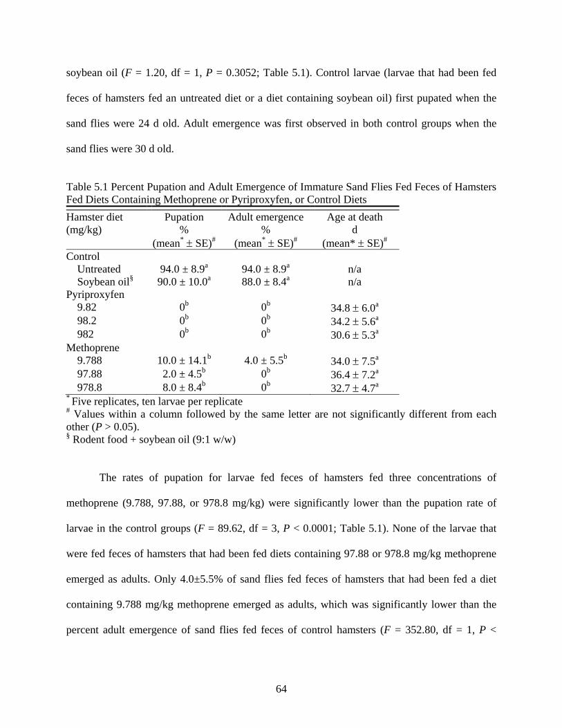
64
soybean oil (F = 1.20, df = 1, P = 0.3052; Table 5.1). Control larvae (larvae that had been fed
feces of hamsters fed an untreated diet or a diet containing soybean oil) first pupated when the
sand flies were 24 d old. Adult emergence was first observed in both control groups when the
sand flies were 30 d old.
Table 5.1 Percent Pupation and Adult Emergence of Immature Sand Flies Fed Feces of Hamsters Fed Diets Containing Methoprene or Pyriproxyfen, or Control Diets Hamster diet (mg/kg)
Pupation
% (mean* ± SE)#
Adult emergence%
(mean* ± SE)#
Age at death d
(mean* ± SE)#
Control Untreated 94.0 ± 8.9a 94.0 ± 8.9a n/a Soybean oil§ 90.0 ± 10.0a 88.0 ± 8.4a n/a Pyriproxyfen 9.82 0b 0b 34.8 ± 6.0a 98.2 0b 0b 34.2 ± 5.6a 982 0b 0b 30.6 ± 5.3a Methoprene 9.788 10.0 ± 14.1b 4.0 ± 5.5b 34.0 ± 7.5a 97.88 2.0 ± 4.5b 0b 36.4 ± 7.2a 978.8 8.0 ± 8.4b 0b 32.7 ± 4.7a * Five replicates, ten larvae per replicate # Values within a column followed by the same letter are not significantly different from each other (P > 0.05). § Rodent food + soybean oil (9:1 w/w)
The rates of pupation for larvae fed feces of hamsters fed three concentrations of
methoprene (9.788, 97.88, or 978.8 mg/kg) were significantly lower than the pupation rate of
larvae in the control groups (F = 89.62, df = 3, P < 0.0001; Table 5.1). None of the larvae that
were fed feces of hamsters that had been fed diets containing 97.88 or 978.8 mg/kg methoprene
emerged as adults. Only 4.0±5.5% of sand flies fed feces of hamsters that had been fed a diet
containing 9.788 mg/kg methoprene emerged as adults, which was significantly lower than the
percent adult emergence of sand flies fed feces of control hamsters (F = 352.80, df = 1, P <

65
0.0001; Table 5.1). The age at death of sand flies fed feces of methoprene-treated hamsters as
larvae was over 32 d (Table 5.1).
In the pyriproxyfen larval bioassay, 100% mortality was observed during the larval stage
for sand flies fed feces of hamsters fed diets containing 9.82, 98.2 or 982.0 mg/kg pyriproxyfen.
The mean age of the larvae at death was over 30 d (Table 5.1).
Fig. 5.1 A Pupa-Form Larva That Had Been Fed Feces of Hamsters Fed a Diet Containing 9.82 mg/kg Pyriproxyfen as a Larva (A), and a Normal Pupa That Had Been Fed Feces of Untreated Hamsters as a Larva (B)
The majority of the sand fly larvae that were fed feces of hamsters offered diets
containing methoprene or pyriproxyfen died as late 4th instar larvae. The larvae in these groups
developed at a normal rate (the same rate as control larvae). Like the control larvae, the larvae in
the treatment groups eventually ceased feeding and cleared their guts as late 4th instar larvae.
However, rather than progressing to the pupal stage, most of the sand fly larvae in the treatment
groups remained as late 4th instar larvae for up to 19 d before eventually dying. Some of the
larvae that were fed feces of methoprene- or pyriproxyfen-treated hamsters did transform from
4th instar larvae into pupa-form larvae before dying (Fig. 5.1). These larvae developed normally
as 2nd, 3rd, and 4th instar larvae, but became an intermediate form between larva and pupa after

66
they ceased feeding and cleared their guts. Pupa-form larvae survived for several days, but
eventually died without becoming pupae.
5.4 Discussion
The food intake of the hamsters in this study was not affected by the methoprene or
pyriproxyfen treatments at any of the concentrations tested. This finding suggests that the treated
diets were palatable to hamsters. Furthermore, significantly more food was consumed by
hamsters when it contained soybean oil, independent of insecticide treatment. The bait
preferences are known for the rodents involved in many of the sand fly/rodent associations found
in Old World ZCL foci, and insecticide-treated baits could be developed for use in field trials in
these scenarios. In Southwest Asia Rhombomys opimus and Meriones spp. are readily baited with
oats, and in Sub-Saharan Africa five rodent genera known be reservoirs of L. major (Mastomys,
Taterillus, Aethomys, Tatera, and Arvicanthis) have been successfully captured in traps baited
with corn flour (Githure et al. 1986; Yaghoobi-Ershadi et al. 2000, 2005).
The results of this study suggest that both methoprene and pyriproxyfen remained
pharmacologically active after passing through the guts of hamsters, and that the compounds
were present at sufficiently high concentrations to affect the development and survival of
immature sand flies. As juvenile hormone analogues, both methoprene and pyriproxyfen were
expected to have the same effect on the development of immature sand flies. The development of
immature sand flies fed feces of hamsters fed diets containing methoprene or pyriproxyfen was
identical to that of control sand flies until the 4th larval instar. At this point, nearly all of the
surviving control larvae subsequently pupated. Larvae that had been fed feces of pyriproxyfen-
treated hamsters remained as 4th instar larvae or became pupa-form larvae, and all of these sand
flies eventually died before pupation. The development of sand fly larvae fed feces of
methoprene-treated hamsters was similar to that of larvae fed feces of pyriproxyfen-treated

67
hamsters. However, pupation of larvae fed feces of hamsters fed diets containing methoprene
was observed at all concentrations, and adult emergence was seen at the lowest concentration.
The finding that pyriproxyfen treatments fully prevented pupation and adult emergence at all
concentrations tested while similar concentrations of methoprene resulted in some pupation and
adult emergence is consistent with other studies that compared the effectiveness of methoprene
and pyriproxyfen against other insects. The LC50 for methoprene was more than 20 x higher than
pyriproxyfen in an evaluation of the relative toxicity of methoprene and pyriproxyfen in topsoil
against immature Ctenocephalides felis (Rajapakse et al. 2002). Similarly, pyriproxyfen was
found to be 21.5 x more toxic than methoprene to larvae of Aedes albopictus (Ali et al. 1995).
Against larvae of Culex quinquefasciatus and A. albopictus, methoprene provided significant but
incomplete inhibition of adult emergence, even at the highest concentrations tested (Nayar et al.
2002).
The results of this study add the juvenile hormone analogues methoprene and
pyriproxyfen to the list of insecticides that potentially can be used as rodent feed-throughs for the
control of phlebotomine sand flies in certain sand fly/rodent associations. The identification of
multiple insecticides that have been found to be effective as rodent feed-throughs against sand
fly larvae in the laboratory increases the likelihood that a suitable compound will be found for
use in field trials. However, future studies on the relative residual activity and environmental
persistence of the compounds will be required before field trials can be conducted.

68
CHAPTER 6. EVALUATION OF NOVALURON AS A RODENT FEED-THROUGH UNDER SIMULATED FIELD CONDITIONS FOR CONTROL OF SAND FLY LARVAE (DIPTERA: PSYCHODIDAE) 6.1 Introduction
Phlebotomine sand flies are major biting pests of man and are the vectors of the
protozoan parasites that cause leishmaniasis. Worldwide, there are an estimated 2 million new
cases of leishmaniasis annually, and 12 million people are currently believed to be infected
(WHO 2006). Throughout North Africa, the Middle East and SW Asia, Phlebotomus papatasi is
the primary vector of Leishmania major, the causative agent of zoonotic cutaneous leishmaniasis
(ZCL).
In spite of their importance, there are no effective control or preventive measures
currently available for sand flies in ZCL foci. In arid and semi-arid foci, P. papatasi exhibits a
close association with several burrowing rodent that serve as reservoirs of L. major. In ZCL foci
in the Old World, rodent burrows are considered to be the primary immature habitats for P.
papatasi, and sand fly larvae have been observed feeding on the feces of rodents. Therefore,
rodent feed-through insecticides are a potential means of controlling sand fly larvae.
Proof of concept for rodent feed-through control of larvae of P. papatasi has been
established in laboratory studies using the benzoylurea chitin synthesis inhibitor novaluron
(Mascari et al. 2007b). All diet concentrations of novaluron tested in a preliminary rodent feed-
through study (9.88, 98.8 and 988 mg/mg) were 100% effective in killing sand fly larvae that fed
on the feces of novaluron-treated rodents (Mascari et al. 2007b). However, additional laboratory
studies are required to determine if novaluron would be appropriate for field use. There were
three objectives of this study: 1) to determine the minimum concentration of novaluron mixed
with hamster feces that would prevent development of sand fly larvae and the minimum dose of
novaluron for hamsters that would be effective as a feed-through against sand fly larvae 2) to
69
determine the persistence of the larvicidal effect of novaluron in hamster feces held under
simulated field conditions 3) to determine the effectiveness of novaluron as a feed-through in
preventing the development of sand fly larvae when novaluron-treated food makes up only a
portion of a hamster’s daily diet.
6.2 Materials and Methods
6.2.1 Sand Flies
The sand flies used in these studies were from a laboratory colony of a Turkish strain of
P. papatasi established at Louisiana State University (Mascari et al. 2007b). The sand fly larvae
in the colony were reared using a larval diet composed of a composted and dried 1:1 mixture of
rabbit feces and rabbit chow (Young et al. 1981). Adult sand flies were provided 20% sucrose
solution ad libitum, and obtained blood meals from Syrian hamsters. The colony was maintained
in environmental chambers at 28 °C, 90% RH.
6.2.2 Hamsters
Syrian hamsters were housed individually in micro-isolator cages. The maintenance of
the hamsters and all experimental procedures followed Animal Care & Use Protocol No. 05-074,
which was approved by the Institutional Animal Care and Use Committee at Louisiana State
University, Baton Rouge, LA. Research involving the hamsters was conducted in compliance
with the Animal Welfare Act and other federal statutes and regulations relating to animals and
experiments involving animals and adheres to principles stated in the Guide for the Care and
Use of Laboratory Animals, NRC Publication, 1996 edition.
6.2.3 Experiment 1: Direct Treatment of Hamster Feces with Novaluron
A series of ten-fold dilutions of technical novaluron (98.8% a.i., Makhteshim Agan
Industries Ltd., Tel Aviv, Israel) was prepared in acetone. The feces of untreated hamsters were
collected and dried at room temperature for 7 d. The feces were crushed using a sterilized glass

70
mortar and pestle and treated with novaluron solutions (at a rate of 1 mL/1 g feces) yielding eight
concentrations ranging from 9.88x10⁻⁵ to 988 mg/kg novaluron. A solvent-control diet (made by
treating feces with acetone) also was prepared. The acetone was allowed to evaporate for 12 h,
and the feces were stored at -80 °C until used. Larval bioassays were conducted as described by
Mascari et al. (2007). A 200 mg portion of feces was placed on the plaster surface of each
bioassay vial. Ten 2nd instar (13±1-d old) larvae then were transferred to each bioassay vial and
held in an environmental chamber at 28 °C, 90% RH. Four bioassay vials were prepared for each
concentration of novaluron and for the solvent-control group.
Larvae were observed under magnification daily, and larval mortality (defined as the lack
of response to prodding with a blunt probe after 15 s) and the percentage of sand flies that
successfully emerged as adults were recorded. Larvae were observed for abnormal behavioral
and morphological characteristics. Evidence of feeding (the presence of frass in the vials and
dark material in the guts of larvae) also was monitored. The percent survival of sand flies and the
age of sand flies at death were compared with repeated measures ANOVA performed with the
GLM procedure (SAS Institute 2001). The Tukey multiple comparison procedure was used to
separate significantly different means.
6.2.4 Experiment 2: Minimum Effective Dose of Novaluron as a Feed-Through
Hamster diets were prepared by adding technical novaluron to a meal-form laboratory
rodent diet (5001 Rodent Diet, LabDiet®, PMI Nutrition International, Brentwood, MO).
Novaluron and the diet were thoroughly mixed to achieve six concentrations (9.88x10⁻⁵, 9.88x10-
4, 9.88x10⁻³, 9.88x10⁻², 9.88x10⁻¹, and 9.88 mg/kg). A control diet (untreated laboratory rodent
diet) also was prepared. Three hamsters were randomly assigned to each of the seven hamster
diet groups (six novaluron treatment groups and one control group).

71
At 12:00 h each day for nine days, the hamsters were provided with 15 g of their
respective diet. The uneaten portion of the food was collected the following day at 12:00 h, and
the daily food intake and daily doses of novaluron that were ingested by the hamsters were
calculated. The daily doses of novaluron for individual hamsters were compared within hamster
diet groups using repeated measures ANOVA, performed with the GLM procedure of SAS (SAS
Institute 2001). The Tukey multiple comparison procedure was used to separate significantly
different means. The feces voided by each hamster were collected daily for nine days. The feces
of each hamster were placed in uncovered containers, dried at room temperature for seven days,
and then stored at -80 °C until used.
Feces voided by the three hamsters in each diet group were pooled. Larval bioassays
were conducted as described above using hamster feces collected after 9 d of feeding as larval
diets. Six bioassay vials were prepared for each of the six larval diets (feces of hamsters fed a
diet containing five concentrations of novaluron or a control diet). The development and survival
of sand fly larvae were monitored and statistically analyzed as described above for Experiment 1.
6.2.5 Experiment 3: Aging Feces under Simulated Field Conditions
A meal-form hamster diet containing 988 mg/kg technical novaluron and an untreated
control hamster diet were prepared. Twelve hamsters were weighed and randomly assigned to
each of the two hamster diet groups (total of 24 hamsters). Hamsters were fed their respective
diets for 9 d as described above. The daily food intake and the daily doses of novaluron which
were ingested by the hamsters were calculated, and were statistically analyzed as described
above for Experiment 2.
A total of 12 g of feces voided by control or novaluron-treated hamsters after 9 d of
feeding on their respective diets (1 g of feces voided by each hamster) was placed in a 120 ml
specimen cup with a 2 cm thick basal layer of plaster of Paris. The specimen cups containing

72
feces were stored in an environmental chamber at 28 °C, 90% RH (conditions which simulated
the temperature and humidity within a rodent burrow; Kay and Whitford 1978). The specimen
cups were placed on filter papers that were kept saturated with distilled water in glass dishes.
Samples of the aged feces (2 g) were taken from the specimen cups at 30 d intervals for 150 d,
and the aged feces were stored at -80 °C until used in sand fly larval bioassays.
Aged hamster feces were fed to second instar sand flies as described above in Experiment
1. Six bioassay vials were used for each of the 12 larval diet groups (feces of control or
novaluron-treated hamsters aged for six time periods: 0, 30, 60, 90, 120, or 150 d). The
development and survival of sand fly larvae were monitored and statistically analyzed as
described above for Experiment 1.
6.2.6 Experiment 4: Partial Consumption of Novaluron-Treated Food by Hamsters
Meal-form hamster diets containing 0 or 988 mg/kg novaluron were prepared. Three
hamsters were randomly assigned to each of the following three groups: hamsters fed exclusively
a diet containing 988 mg/kg novaluron for nine days, hamsters fed exclusively an untreated diet,
or hamsters fed 1 g of diet containing 988 mg/kg novaluron for 3 h each day and then afterwards
provided with untreated diet. The hamsters were fed using these protocols for 9 d. The daily food
intake and daily doses of novaluron for each hamster were calculated as described above. The
feces voided by each hamster were collected daily for 9 d and were processed as described
above.
The feces collected from hamsters after feeding for 9 d were pooled by treatments,
crushed using a mortar and pestle, and used in sand fly larval bioassays. The larval bioassays
were conducted as described above. Six bioassay vials were used for each of the three larval diet
groups (feces of hamsters exclusively fed novaluron-treated or control diets, or feces of hamsters

73
fed novaluron-treated food as a portion of their diet). The development and survival of sand fly
larvae were monitored and statistically analyzed as described above.
6.3 Results
6.3.1 Experiment 1: Direct Treatment of Hamster Feces with Novaluron
Larvae in each of the larval diet groups were observed feeding, and frass was found in
each bioassay vial. The percent survival from 2nd instar to adult was 100% for larvae fed acetone
treated hamsters feces. At the concentrations of novaluron tested, the mean percent survival of
sand fly larvae fed novaluron-treated hamster feces ranged from 0 to 100% (Table 6.1). Mortality
of larvae fed feces containing as little as 9.88x10⁻¹ mg/kg novaluron was significantly different
from mortality of control larvae (F = 199.47, df = 8 P < 0.0001); mortality was 100% at 9.88
mg/kg and above (Table 6.1). The mean longevity of sand fly larvae that died after being fed
novaluron-treated feces ranged from 3.0±0.2 to 4.1± 0.3 d (Table 6.1).
Table 6.1 Mortality and Longevity of 2nd Instar Sand Flies Fed Hamster Feces Directly Treated with Novaluron Solutions
Concentration (mg/kg) of novaluron in larval diet
Mortality %
(mean* ± SE)#
Longevity d
(mean* ± SE) 0 0.0±0.0a n/a 9.88x10⁻⁵ 5.0±5.8a n/a 9.88x10⁻⁴ 2.5±5.0a n/a 9.88x10⁻³ 0.0±0.0a n/a 9.88x10⁻² 0.0±0.0a n/a 9.88x10⁻¹ 35.0±19.1b 4.0±0.4 9.88 100.0±0.0c 4.1±0.3 98.8 100.0±0.0c 3.0±0.3 988 100.0±0.0c 3.0±0.3 * Six replicates, ten larvae per replicate # Values within a column followed by the same letter are not significantly different from each other, P> 0.05 n/a not applicable

74
6.3.2 Experiment 2: Minimum Effective Dosage of Novaluron as a Feed-Through
The mean body weight of the 12 hamsters in this study was 143.3±3.0 g, and the body
weights of hamsters assigned to different diet groups were not significantly different (F = 0.24,
df = 5, P = 0.9358). The mean daily food intake of the 12 hamsters in this study was 7.2±1.4 g,
and the amount of food eaten by hamsters in different diet groups was not significantly different
(F = 0.28, df = 5, P = 0.9256). The mean daily doses of novaluron for hamsters ranged from
4.8x10⁻⁵ to 5.1x10⁻¹ (Table 6.2).
Table 6.2 Means (±SE) of Body Weight, Food Intake, and Daily Dosages of Novaluron for Syrian Hamsters
Concentration (mg/kg) of novaluron in diet
Body weight g
(mean ± SE) #
Food intake g/d
(mean* ± SE) #
Daily dose mg/kg
(mean* ± SE)#
0 142.3±3.2a 7.3±1.5a n/a 9.88x10⁻⁴ 144.4±3.5a 6.9±1.6a 4.8x10⁻⁵±1.1x10⁻⁵ 9.88x10⁻³ 144.0±1.0a 7.2±1.4a 5.0x10⁻⁴±9.4±10-5 9.88x10⁻² 143.0±4.6a 7.2±1.1a 5.0x10⁻³±7.8x10⁻⁴ 9.88x10⁻¹ 144.0±3.6a 7.1±1.4a 4.9x10⁻²±9.7x10⁻³ 9.88 142.0±3.6a 7.3±1.1a 5.1x10⁻¹±8.4x10⁻² * Nine replicates, three hamsters per replicate # Values in a column followed by the same letter are not significantly different from each other, P>0.05 n/a not applicable
Evidence of feeding was observed for larvae in each of the larval diet groups. The percent
survival from 2nd instar to adult was 90.0±8.2% for larvae fed feces of untreated hamsters (Table
6.3). At the tested concentrations of novaluron fed to hamsters, the mean percent survival of sand
fly larvae fed feces of novaluron-treated hamsters ranged from 0.0 to 95.0% (Table 6.3).
Mortality of larvae fed feces of hamsters fed a diet containing as little as 9.88x10⁻¹ mg/kg
novaluron was significantly different from mortality of larvae fed feces of hamsters fed an

75
untreated diet (F = 188.61, df = 5, P < 0.0001; Table 6.3). The mean longevity of sand fly larvae
that died after being fed feces of novaluron-treated hamsters ranged from 4.2 to 5.6 (Table 6.3).
Table 6.3 Mortality and Longevity of 2nd Instar Sand Flies Fed Feces of Hamsters Fed Diets Containing Novaluron
Hamster diet novaluron concentration
(mg/kg)
Mortality %
(mean* ± SE)#
Longevity d
(mean* ± SE) 0 10.0±8.2a n/a 9.88x10⁻⁴ 7.5±9.6a n/a 9.88x10⁻³ 12.5±9.6a n/a 9.88x10⁻² 5.0±5.0a n/a 9.88x10⁻¹ 100.0±0.0b 5.6±0.69.88 100.0±0.0b 4.2±0.6* Six replicates, ten larvae per replicate # Values within a column followed by the same letter are not significantly different from each other, P> 0.05 n/a not applicable
6.3.3 Experiment 3: Aging Feces under Simulated Field Conditions
The mean body weight of the 36 hamsters was 150.8±10.2 g, and the mean body weights
of hamsters in the two hamster diet groups were not significantly different (F = 0.16, df = 1, P =
0.85; Table 6.4). The mean daily food intake for hamsters fed diets containing 988 mg/kg
novaluron or acetone alone were not significantly different (F = 1.65, df = 1, P = 0.19; Table
6.4). The estimated mean daily dosage of novaluron was 63.1±10.0 mg/kg body weight.
Table 6.4 Means (±SE) of Body Weight, Food Intake, and Daily Dosages of Novaluron for Syrian Hamsters
Hamster diet group Body weight g
(mean* ± SE) #
Food intake g/d
(mean* ± SE) #
Daily dose mg/kg
(mean* ± SE)
Control (0 mg/kg) 151.0±11.7a 9.5±1.7a n/a Novaluron (988 mg/kg) 151.8±8.6a 9.7±1.6a 63.1±10.0 * Nine replicates, three hamsters per replicate # Values in a column with the same letter are not significantly different from each other, P>0.05 n/a not applicable

76
Larvae in each of the larval diet groups were observed feeding, and frass was found in
each bioassay vial. The mean percent adult emergence for sand flies fed feces of untreated
hamsters that had been aged for any of the time periods (0 to 150 d) was over 90%. There were
no significant differences between percent adult emergence at different aging periods (F =
230.34, df = 11, P < 0.0001; Fig. 6.1).
Aging period (d)0 30 60 90 120 150
Adu
lt em
erge
nce
(%)
0
20
40
60
80
100
Control Novaluron
A A A A AA
B BBC
BCBC
C
Figure 6.1 Percent Adult Emergence of Sand Flies Fed Feces of Control or Novaluron-Treated Hamsters; Feces Were Aged under Simulated Field Conditions (28 °C, 90% RH) for up to 150 d
The mean percent adult emergence for sand flies fed feces of novaluron-treated hamsters
was significantly different from sand flies fed feces of control hamsters when the feces were
aged for any of the time periods (Fig. 6.1). All larvae that had been fed feces of novaluron-
treated hamsters aged for 0 or 30 d died before adult emergence. The mean percentage of larvae
that emerged as adults after being fed feces of novaluron-treated hamsters that had been aged for

77
60 (3.3±5.2%), 90 (10.0±8.9%), or 120 d (6.7±8.2%) was significantly lower than for larvae fed
feces of control hamsters (Fig. 6.1). The mean percent adult emergence for larvae fed feces of
novaluron-treated hamsters aged for 150 d was 16.7±12.1%, which was significantly different
from both larvae fed feces of control hamsters and larvae fed feces of novaluron-treated hamsters
aged for 0 or 30 d (Fig. 6.1).
6.3.3 Experiment 4: Partial Consumption of Novaluron-Treated Food by Hamsters
The mean daily food intake of the hamsters was 7.3±0.8 g. The mean daily food intake of
hamsters fed novaluron-treated food as a portion of their daily diet was significantly different
from the food intake of hamsters fed exclusively untreated or novaluron-treated diets (F = 6.30,
df = 2, P = 0.0029; Table 6.5). The mean body weight of hamsters in this study was 136.0±13.0
g, and the mean body weights of hamsters in the three hamster diet groups were not significantly
different (F = 0.40, df = 2, P = 0.6846; Table 6.5).
Hamster diet containing 988 mg/kg novaluron constituted 14.8±1.9% of the total daily
food intake of hamsters in the partial feeding group (Table 6.5). The mean daily dose of
novaluron for hamsters in the different diet groups are reported in Table 6.5.
Table 6.5 Means (±SE) of Body Weight, Food Intake, and Daily Doses of Novaluron for Syrian Hamsters Offered Food Containing Novaluron as All, Part, or None of Their Diet
Concentration (mg/kg)
Body weight g
(mean* ± SE)#
Food intake g/d
(mean* ± SE)#
Portion of diet§ %
(mean* ± SE)
Daily dose mg/kg
(mean* ± SE)
0 141.3±20.5a 7.4±0.8a 0 0 988 (all of diet) 131.0±8.9a 7.6±0.7a 100 57.4±6.0 988 (part of diet) 135.7±9.9a 6.9±0.9b 14.8±1.9 7.3±0.4 * Nine replicates, three hamsters per replicate # Values in a column followed by the same letter are not significantly different from each other, P>0.05 § Portion of diet that was novaluron-treated food

78
Larvae in each of the larval diet groups were observed feeding, and frass was found in
each bioassay vial. The mean percent survival from 2nd instar to adult for the sand flies in the
untreated hamster feces larval diet group was 96.7±3.3%. Sand fly larvae that were fed feces
from hamsters that had consumed diets containing novaluron died before pupation (Table 6.6).
Larvae that had been fed either feces from hamsters that exclusively had been fed a diet
containing novaluron or both a diet containing 988 mg/kg novaluron and untreated hamster food
were ataxic and ceased feeding, and none pupated. The mean longevity of sand fly larvae fed
feces from hamsters that exclusively had been fed a diet containing 988 mg/kg novaluron and
feces from hamsters were fed both diets containing novaluron and untreated diets was not
significantly different (F = 0.95, df = 1, P = 0.3317; Table 6.6).
Table 6.6 Mortality and Longevity of 2nd Instar Sand Flies Fed Feces of Hamsters Fed Diets Containing Novaluron. Hamsters Were Fed Novaluron-Treated Food as All, Part, or None of Their Daily Diet
Hamster diet group Mortality
% (mean* ± SE)#
Longevity d
(mean* ± SE)# Control 3.3 ± 5.2a n/a Novaluron-treated diet 100.0b 3.6 ± 1.2a Partial consumption 100.0b 3.8 ± 1.6a * Six replicates, ten larvae per replicate # Values within a column followed by the same letter are not significantly different from each other, P> 0.05 n/a not applicable
6.4 Discussion
In experiments 1 and 2, complete control of sand fly larvae was observed when the larvae
were fed either hamster feces containing 9.88 mg/kg novaluron or feces of hamsters fed a diet
containing 9.88x10⁻¹ mg/kg novaluron. The concentrations that were 100% effective against sand
fly larvae in experiments 1 and 2 were different by an order of magnitude, but diet and fecal
concentrations of novaluron cannot be equated. Laboratory studies on the metabolism and

79
excretion of novaluron in rats have shown that 95.4% of novaluron is eliminated in feces for up
to 168 h (FAO 2005). If hamsters excrete novaluron at a rate similar to rats, an increase in the
concentration of novaluron in the feces of hamsters over the 9-d period of this study would be
expected. In experiment 2, the mean amount of food consumed by a hamster each day (7.2±1.4
g) was higher than the amount of feces voided by a hamster (approximately 2 g). Since the
majority of novaluron is eliminated in feces, the concentration of novaluron in the feces of a
hamster should be higher than the concentration of novaluron in a hamster’s diet. The purpose of
these experiments was to measure the effect of the treatments on the survival of sand flies fed
feces of novaluron-treated hamsters, but the rate at which novaluron is eliminated from hamsters
could be an important variable to measure in future studies.
When feces of novaluron-treated hamsters were held under simulated field conditions (28
°C, 90% RH) for up to 30 d, all larvae that consumed these feces died before pupation. A
significant reduction in treated larval survival relative to control was observed when the feces
were aged for up to 150 d. Based on the findings of experiment 1, we can conclude that feces of
hamsters fed a diet containing 988 mg/kg that had been aged for 30 d contained at least 9.88
mg/kg novaluron (a concentration that caused complete larval mortality).
Studies on the fate of novaluron in soil have shown that between 32 and 49% of the
compound can be present in soil after 127-195 d (FAO 2005). Therefore, baits containing 316
mg/kg novaluron (32% of 988 mg/kg novaluron) could be expected to be 100% effective as a
rodent feed-through for larval sand fly control. However, novaluron may have been degraded at a
faster rate in hamster feces than in soil due to the rapid proliferation of fungi. While we
approximated the temperature and humidity of a rodent burrow in experiment 3, in an actual field
setting novaluron in feces could degrade at an even faster rate than we observed in this study.
80
Therefore, the rate at which novaluron in feces is degraded under different simulated field
conditions could be another important topic for future studies.
Novaluron-treated diet made up approximately 15% of the food consumed daily by
hamsters in experiment 4, and the feces of these hamsters was equally as effective against sand
fly larvae as feces of hamsters exclusively fed a novaluron-treated diet. The results of experiment
4 suggest that when novaluron is eliminated by orally dosed hamsters, it is uniformly distributed
in the feces. This is an important observation because artificial baits for wildlife do not fully
supplant naturally available food sources. The results of experiment 4 indicate that novaluron
would be effective under circumstances where baits make up only a small portion of the diet of
the target rodents in a field setting.
While target rodents in field trials of feed-through control measures for sand fly larvae
cannot be expected to exclusively consume novaluron-treated baits, a key component of potential
field work would be to identify baits that are readily consumed by target rodents in different foci.
The amount of food consumed by study hamsters was not affected by novaluron treatments at
any of the concentrations tested in experiments 2, and 3 (significant differences were only
observed in experiment 4 when hamsters were fed novaluron-treated food as a portion of their
diet, and this observation may have been a result of the frequent changing of food which
disrupted the hamsters’ feeding). This observation is consistent with findings of Mascari et al
(2007b), in which the authors reported that the quantity of food consumed by hamsters was not
affected by concentrations of novaluron as high as 988 mg/kg. It is not known whether the food
intake of the different rodents that could be targeted in field trails would be similarly unaffected
by novaluron treatments. However, the grain-based baits that would be prepared for field trials
could contain a palatability agent, such as a vegetable oil. Palatability agents are commonly used

81
in conjunction with rodenticides in order to increase bait uptake by target animals and may mask
any potential odor or taste of novaluron.
The important characteristics of an insecticide used in a rodent bait for control of sand fly
larvae would be A) that it is excreted in feces of bait-fed rodents rather than metabolized, B) that
it persists in the environment, and C) that it is effective in preventing the development and
survival of sand fly larvae when the bait makes up only a portion of a target rodent’s diet. In this
study, significant control of sand fly larvae was observed when they were fed feces of novaluron-
treated hamsters that had been aged for up to 150 d or feces of novaluron-treated hamsters when
only 15% of their daily diet was novaluron-treated food. Therefore, novaluron is a good
candidate for further evaluation as a rodent feed-through insecticide against sand fly larvae.
Since the results of this study suggest that novaluron could be effective as a rodent feed-through
insecticide in a field setting, the next step would be to evaluate the effects of novaluron-treated
baits on sand fly populations in different rodent/sand fly associations.
82
CHAPTER 7. EFFECT OF ORAL IVERMECTIN TREATMENT OF RODENTS ON SURVIVAL OF SAND FLY (DIPTERA: PSYCHODIDAE) LARVAE FED ON THE RODENT FECES AND FEMALE SAND FLIES FED ON THE RODENTS 7.1 Introduction
Sand flies are hematophagous Diptera of the subfamily Phlebotominae, and, with the
exception of New Zealand and some Pacific islands, are found in most parts of both the New
World and Old World between 50 °N and 40 °S. Sand flies are often significant biting pests of
man; their bites can cause acute dermatitis and delayed-type hypersensitivity reactions. Sand
flies also are vectors of medically important viruses, bacteria, and protozoa. The protozoa of at
least 20 Leishmania spp. are transmitted by sand flies and are the causative agents of human
leishmaniasis (WHO 2008). Leishmaniasis is a zoonotic disease with a broad clinical spectrum
that is estimated to affect as many as 12 million people, and 2 million new cases of leishmaniasis
are believed to occur annually (WHO 2006).
In the Old World, Leishmania major is the causative agent of zoonotic cutaneous
leishmaniasis (ZCL), which has an enzootic transmission cycle among populations of locally
abundant, burrow-dwelling rodents. Man becomes infected with L. major by the bite of
infectious sand flies (Phlebotomus papatasi in Southwestern Asia, the Middle East, and North
Africa; Phlebotomus duboscqi in Sub-Saharan Africa) in settlements located near areas of
intense transmission or as a result of movement into enzootic areas, such as during military
operations or during suburban expansion (Faulde et al. 2008; Traore et al. 2001).
No effective preventive or control measures are currently available for Old World ZCL.
Personal protective measures (including the use of repellents, bednets, and insecticide treated
materials) provide inconsistent and incomplete protection against sand fly bites and infection
with L. major (Jumaian 1998). Despite the close association of rodent burrows and vector

83
species, direct treatment of burrows with insecticides has not been shown to have a significant
effect on sand fly populations (Karapati’an et al. 1983).
Proof of concept for ivermectin as a rodent feed-through insecticide for control of larval
sand flies has been established; the feces of hamsters fed a a diet containing 20 mg/kg ivermectin
were shown to be 100% effective against sand fly larvae (Mascari et al. 2008). While the
majority of the drug is rapidly eliminated in the feces, ivermectin reaches detectable levels in the
blood of orally dosed mammals (Pound et al. 2004; Campbell et al. 1983). Several field studies
have demonstrated significant reductions in survival of mosquitoes that obtained bloodmeals
from ivermectin-treated hosts (Tesh 1990; Cartel 1991; Foley 2000, Bockarie 1999). The toxicity
of ivermectin has been demonstrated for phlebotomine sand flies fed ivermectin-treated blood
through an artificial membrane (Kassem et al. 2001). Therefore, targeting rodents with an
ivermectin feed-through also could affect post-bloodmeal survival of sand flies.
The primary objective of this research was to determine whether the post-bloodmeal
survival of adult sand flies would be affected by feeding their rodent hosts a diet containing
ivermectin. In this study the insecticidal effect of ivermectin treatments against bloodfed sand
flies was monitored for 14 d after rodents were withdrawn from their ivermectin-treated diets,
and sand fly larval bioassays were conducted using feces voided by ivermectin-treated rodents
over this same time period.
7.2 Materials and Methods
7.2.1 Sand Flies
The sand flies used in these experiments were from a laboratory colony of a Turkish
strain of P. papatasi established at Louisiana State University (Mascari et al. 2007b). The sand
fly larvae in the colony were reared using a larval diet made of a composted and dried 1:1
mixture of rabbit feces and rabbit chow (Young et al. 1981). Adult sand flies were provided with

84
20% sucrose solution ad libitum, and they obtained bloodmeals from Syrian hamsters. The
colony was maintained in environmental chambers at 28 °C, 90% RH.
7.2.2 Hamsters
Syrian hamsters were housed individually in micro-isolator cages. The maintenance of
the hamsters and all experimental procedures followed Animal Care & Use Protocol No. 05-074,
which was approved by the Institutional Animal Care and Use Committee at Louisiana State
University, Baton Rouge, LA. Research involving the hamsters was conducted in compliance
with the Animal Welfare Act and other federal statutes and regulations relating to animals and
experiments involving animals and adheres to principles stated in the Guide for the Care and
Use of Laboratory Animals, NRC Publication, 1996 edition.
7.2.3 Hamster Treatments
Ivermectin (Merck & Co., Inc., Whitehouse Station, NJ) was added to a meal-form
laboratory rodent diet (5001 Rodent Diet, LabDiet®, PMI Nutrition International, Brentwood,
MO). A diet containing 20 mg/kg ivermectin was prepared, and a control diet (untreated meal-
form rodent diet) also was prepared.
Three hamsters were assigned randomly to each of the two diet groups (ivermectin-
treated diet or control diet). At 18:00 h each day for nine days, the hamsters were provided with
25 g of their respective diets. The uneaten portion of the food was collected the following day at
18:00 h, and the daily food intake for each hamster was calculated. The daily doses of ivermectin
that were ingested by the hamsters also were calculated. Feces voided by each hamster were
collected daily for 9 d during feeding. Feces were air-dried at room temperature for 7 d, and then
stored at -80 °C until used in larval bioassays. After being fed their respective diets for 9 d, all
hamsters were withdrawn from their meal-form diets and provided with an untreated pellet diet

85
(5001 Rodent Diet, LabDiet®, PMI Nutrition International, Brentwood, MO). Feces then were
collected after 3, 7, and 14 d.
7.2.4 Adult Bloodfeeding Assays
Adult sand flies were allowed to feed on hamsters 0, 3, 7, and 14 d after they were
withdrawn from untreated or ivermectin-treated meal-form diet. Bloodfeeding bioassays were
conducted in clear polycarbonate boxes with a cloth sleeve to allow hamsters and sand flies to be
introduced into the container. Thirty 2 to 4-d old nulliparous female sand flies were transferred
using a mouth aspirator into each bioassay container.
Hamsters were chemically immobilized with an anesthetic mixture of ketamine HCl (100
mg/kg body weight) and xylazine HCl (10 mg/kg body weight) administered via intra-peritoneal
(IP) injection. The depth of anesthesia was considered sufficient when hamsters were no longer
ambulatory, and had lost their righting reflex (defined as the ability of hamsters to regain sternal
recumbency) and superficial pain response (determined by pinching the skin over the anterior
surface of the feet). Once immobilized, ophthalmic ointment was placed in the hamsters’ eyes to
protect them against potential sand fly bites.
A single immobilized hamster was placed in each bioassay container. The sand flies were
allowed to feed for 50 min before the hamsters were removed from the containers (Fig. 7.1).
When the hamsters were recovered from anesthesia, they were returned to their micro-isolator
cages. The number of engorged sand flies was recorded immediately, and they were provided
with a piece of filter paper saturated with 20% sucrose solution. The sand flies were kept in the
bioassay container for 24 h to allow them to form a peritrophic membrane around the bloodmeal;
past experience has shown that moving sand flies by mouth aspirator before the peritrophic
membrane is formed may increase mortality rates. After 24 h the mortality was recorded, and
live sand flies were transferred using a mouth aspirator to a 150 mL glass jar with a plaster of

86
Paris base and a fine mesh lid. The sand flies were provided with 20% sucrose solution on a
cotton pad. Mortality (defined as lack of movement during 30 s observation) was recorded again
after 24 h (48 h post-bloodmeal).
Figure 7.1 Sand Flies Taking a Bloodmeal from a Chemically Immobilized Ivermectin-Treated Syrian Hamster
Differences in mean survivorship of sand flies that had taken bloodmeals from
ivermectin-treated or control hamsters that had been withdrawn from their meal-form diets for
different time periods were compared after 24 and 48 h using repeated measures ANOVA (SAS
Institute 2001). Treatment means were separated by Tukey’s honestly significant difference
(HSD) multiple comparison test.
The viability (successful hatching) of eggs deposited by bloodfed sand flies on the plaster
surface of the jars also was recorded. Sand flies were allowed to lay eggs until 12 d post-
bloodmeal (sand flies from this colony typically lay eggs 5 d post-bloodmeal, and eggs begin to

87
hatch 7 d after being deposited). Eggs were observed for signs of hatching up to 14 d after the
first eggs were deposited. After the eggs hatched, larvae were counted and removed from the jars
to allow an accurate count of newly hatched eggs each day. The mean number of eggs per jar and
the mean percent of eggs that hatched were calculated and compared using Student’s t-test (SAS
Institute 2001).
7.2.5 Larval Feed-Through Bioassays
Feces voided by hamsters 0, 3, 7, and 14 d after being withdrawn from an untreated or
ivermectin-treated meal-form diet were collected. Feces of the three hamsters in each diet group
were pooled and then crushed using a sterilized glass mortar and pestle.
Larval bioassays were conducted as described by Mascari et al. (2007a). A 200 mg
sample of crushed hamster feces was transferred to the plaster surface of each bioassay vial. Ten
2nd instar (13±1-d old) larvae were transferred to each bioassay vial (six vials per treatment) and
held in an environmental chamber at 28 °C, 90% RH, 14:10 (L:D) photoperiod. Larvae were fed
feces of control or ivermectin-treated hamsters collected at each of the time periods (0, 3, 7, and
14 d after being returned to untreated, pellet diet).
The larvae were observed under magnification daily. Larval mortality (defined as the lack
of response to prodding with a blunt probe after 15 s) was recorded, and the larvae were
observed for abnormal behavioral and morphological characteristics. Evidence of feeding (the
presence of frass in the vials and dark material in the guts of larvae) also was monitored.
The percent survival of immature sand flies to adult emergence after being fed their
respective diets was compared using repeated measures ANOVA performed with the GLM
procedure (SAS Institute 2001). The Tukey multiple comparison procedure was used to separate
significantly different means.

88
7.3 Results
7.3.1 Hamster Treatments
The mean body weight of the six hamsters in this study was 137.3±18.6 g, and the body
weights of hamsters assigned to different diet groups were not significantly different (F = 0.2, df
= 1, P = 0.6816). The mean daily food intake of the hamsters was 7.4±1.4 g. The amount of food
consumed by ivermectin-treated hamsters was significantly greater than for control hamsters (F
= 5.31, df = 1, P = 0.0253; Table 7.1).
Table 7.1 Means (±SE) of Body Weight, Food Intake, and Daily Dosages of Ivermectin for Syrian Hamsters
Hamster diet group Body weight g
(mean ± SE) #
Food intake g/d
(mean* ± SE) #
Daily dosage mg/kg
(mean* ± SE)
Control 141.0±21.7a 7.0±1.4a 0 Ivermectin (20 mg/kg) 133.7±18.9a 7.9±1.2b 1.0±0.3 * Nine replicates, three hamsters per replicate # Values in a column followed by the same letter are not significantly different from each other, P>0.05 7.3.2 Adult Bloodfeeding Assays
All sand flies that were exposed to a hamster for 50 min successfully took a bloodmeal.
All sand flies survived for at least 48 h after taking bloodmeals from hamsters that had been
withdrawn from an untreated meal-form diet for 0, 3, 7, or 14 d (Table 7.2; Table 7.3). The 24 h
post-bloodmeal mortality of sand flies that had taken bloodmeals from hamsters withdrawn from
an ivermectin-treated diet for 0, 3, or 7 d was greater than 50% (Table 7.2). The 48 h survival
was 0% for sand flies that took a bloodmeal from hamsters withdrawn from an ivermectin-
treated diet for 0, 3, or 7 d (Table 7.3). The 24 and 48 h post-bloodmeal survival was 100% for
sand flies fed on hamsters withdrawn from an ivermectin-treated diet for 14 d.

89
Table 7.2 Post-Bloodmeal (24 h) Survival of Sand Flies Fed on Ivermectin-Treated Hamsters
Hamster diet
Survival (at 24 h) %
(mean* ± SE) # 0 DAT§ 3 DAT§ 7 DAT§ 14 DAT§
Control 0 ppm 100.0±0.0a 100.0±0.0a 100.0±0.0a 100.0±0.0a
Ivermectin 20 ppm 42.2±8.3b 13.3±14.5b 46.7± 18.6b 100.0±0.0a
* 3 hamsters, 30 sand flies per hamster # Values in a column followed by the same letter are not significantly different from each other, P>0.05 § Days after hamsters were withdrawn from an untreated or ivermectin-treated meal-form diet Table 7.3 Post-Bloodmeal (48 h) Survival of Sand Flies Fed on Ivermectin-Treated Hamsters
Hamster diet
Survival (at 48 h) %
(mean* ± SE) # 0 DAT§ 3 DAT§ 7 DAT§ 14 DAT§
Control 0 ppm 100.0±0.0a 100.0±0.0a 100.0±0.0a 100.0±0.0a
Ivermectin 20 ppm 0.0±0.0b 0.0±0.0b 0.0±0.0b 100.0±0.0a
* 3 hamsters, 30 sand flies per hamster # Values in a column followed by the same letter are not significantly different from each other, P>0.05 § Days after hamsters were withdrawn from an untreated or ivermectin-treated meal-form diet
The mean number of eggs deposited by sand flies that took bloodmeals from untreated or
ivermectin-treated hamsters 14 d after withdrawal from their respective meal-form diets was not
significantly different (t = 0.17, df = 4, P = 0.8725; Table 7.4). Additionally, the mean
percentage of eggs that hatched after being deposited by sand flies that took bloodmeals from
untreated or ivermectin-treated hamsters 14 d after withdrawal from their respective meal-form
diets also was not significantly different (t = 0.28, df = 4, P = 0.7913; Table 7.4).

90
Table 7.4 Mean Number and Viability of Eggs Deposited by Sand Flies That Had Taken Bloodmeals from Hamsters 14 d after Being Withdrawn from an Untreated or Ivermectin-Treated Diets
Hamster diet No. eggs Percent hatch
mean* ± SE# Range mean* ± SE# range Control 0 mg/kg 831.3±142.0a 689-974 71.9±6.1a 64.9-76.0 Ivermectin 20 mg/kg 808.3±184.6a 690-1021 74.4±13.4a 59.5-85.6 * 3 repetitions, 30 bloodfed sand flies per repetition # Values in a column followed by the same letter are not significantly different from each other, P>0.05
7.3.3 Larval Feed-Through Bioassays
Feces voided by hamsters on the same day they were withdrawn from an ivermectin-treated diet
(0 d) were fed to sand fly larvae and yielded 100% larval mortality. The percent survival
No. days after withdrawal from untreated or ivermectin-treated diets0 d 3 d 7 d 14 d
Adu
lt em
erge
nce
(%)
0
20
40
60
80
100
Control Ivermectin
A
B
A A A A
C
BC
Figure 7.2 Percent Adult Emergence (Mean ± SE) of Sand Flies Fed as 2nd Instars the Feces of Untreated or Ivermectin-Treated Hamsters; Feces Used in This Bioassay Were Voided by Hamsters 0, 3, 7, or 14 d after the Hamsters Were Withdrawn from Their Respective Diets

91
of sand fly larvae fed feces of hamsters withdrawn from an ivermectin-treated diet for 3 and 7 d
(28.3±14.7% and 13.3±12.1%, respectively) was significantly different from the survival of
larvae fed feces of untreated hamsters collected at the same time periods (85.0±13.8% and
93.3±8.2%, respectively; F = 96.60, df = 7, P < 0.0001). The mean percent survival was not
significantly different between larvae fed feces voided by hamsters 14 d after they were
withdrawn from an ivermectin-treated or untreated diet (91.7±11.7% and 96.7±8.2%,
respectively).
7.4 Discussion
Ivermectin treatment of hamsters was 100% effective against bloodfeeding sand flies for
up to 7 d after hamsters were withdrawn from ivermectin-treated diets. This is consistent with
other studies showing the effects of ivermectin against a broad range of nematodes and
arthropods. In humans, ivermectin is commonly used in mass drug administrations (a single dose
of 0.2 mg/kg body weight) to clear microfilaria of Wuchereria bancrofti and Onchocerca
volvulus. During these mass drug administrations, ivermectin also has been shown to remain
active against mosquitoes feeding on treated humans for long periods of time after treatment.
Post-bloodmeal survival of Anopheles farauti mosquitoes fed on ivermectin-treated humans was
significantly lower than control for up to 44 d post-treatment (Foley et al. 2000). Remarkably, a
significant reduction in post-bloodmeal survival of Aedes polynesiensis mosquitoes fed on
ivermectin-treated humans was reported for up to 6 months post-treatment (Cartel et al. 2001).
The survival of ticks also has been shown to be affected by treating hosts with ivermectin.
Significant mortality was observed for the ticks Ornithodoros moubata and Boophilus microplus
that had fed on ivermectin-treated cattle, and the effect persisted for up to 21 d post-treatment
(Centurier and Barth 1980, Nolan et al. 1981). However, the post-treatment duration of the
insecticidal effects of ivermectin on bloodfeeding sand flies was less than 14 d. The hamsters in

92
this study received a mean daily dose of 1.0±0.3 mg/kg ivermectin for 9 consecutive days, which
is substantially higher than the 0.2 mg/kg administered once orally or by injection to humans and
other animals. The shorter duration of effect against sand flies suggests that ivermectin may be
rapidly eliminated by hamsters or that sand flies are less susceptible to the serum concentrations
of ivermectin that may be present in hosts more than 1 wk after treatment (Chiu and Lu 1989).
The results of this study also can be used to direct the frequency with which ivermectin-
treated baits should be delivered in a field setting. Since the effects of oral treatment of rodents
on sand fly adults and larvae persisted for at least 1 wk, treatment of target rodents with
ivermectin (or access of target rodents to ivermectin-treated baits) does not need to be
continuous, but should not be withdrawn for more than 1 wk.
Fecundity was not significantly different for sand flies that took a bloodmeal from an
untreated or ivermectin-treated hamster 14 d post-treatment. A previous study reported that sand
flies that had ingested a sub-lethal dose of ivermectin in a bloodmeal produced around 2/3 the
number of eggs as control sand flies (Kassem et al. 2001). It is possible that the results of this
study did not detect a similar effect of ivermectin on sand fly fecundity because a sub-lethal
serum concentration of ivermectin may have occurred in hamsters between 7 and 14 d post-
treatment. The percent hatch of eggs of sand flies that ingested sub-lethal doses of ivermectin
was not lower than control sand flies in the study conducted by Kassem et al. (2001), nor was it
lower for sand flies that took a bloodmeal from an ivermectin-treated hamster in this study.
Feces of ivermectin-treated hamsters were 100% effective against sand fly larvae when
collected immediately after hamsters were withdrawn from ivermectin-treated diets (0 d), which
is consistent with the results of Mascari et al. (2008). The results of this study also are consistent
with the elimination profile of ivermectin in other rodents. In rats, only about 2% of ivermectin is
eliminated via urine of orally dosed rats; the remainder is eliminated in feces (Chiu and Lu

93
1989). The rate at which ivermectin is eliminated in feces also has been described using a rat
model; 57.4 to 58.4% of the administered drug is eliminated 1 d after administration. These
figures increase to between 83.0% and 91.7% five days after administration (WHO Food
Additives Series 27: 696. Ivermectin). Despite the rapid elimination of ivermectin in feces, the
high dose of ivermectin administered to the hamsters in this study led to the significant effects
against sand fly larvae that were observed up to 7 d after hamsters were withdrawn from an
ivermectin-treated diet.
This study confirms that ivermectin-treated rodent baits developed as a feed-through to
control sand fly larvae also could have a collateral effect on bloodfeeding adult sand flies, thus
affecting a larger portion of the sand fly population than would be estimated due to larvicide
activity alone. While ivermectin treatment of rodent hosts of L. major may reduce the overall
population of sand flies, this approach would have its greatest impact on the most
epidemiologically important subset of the sand fly population: females that have taken a
bloodmeal from potentially infected rodents. In this study sand flies that have taken a bloodmeal
from an ivermectin-treated hamster died within 2 d. The extrinsic incubation period of L. major
in P. papatasi is approximately 6 d, and female P. papatasi sand flies take bloodmeals every 5 to
7 d. Therefore, targeting rodents in ZCL foci with ivermectin-treated baits could, in effect, turn
L. major-infected hosts into dead-end hosts in the transmission cycle by killing sand flies before
the extrinsic incubation period for L. major can be completed. Furthermore, since female sand
flies that take bloodmeals from ivermectin-treated rodents would not feed again, the ivermectin
treatments would eliminate any infectious sand flies, serving as a dead-end in transmission of L.
major parasites for 5 to 7 d.
Sand fly species that feed on the feces of rodents as larvae could be controlled using feed-
through insecticides, and for those species that do not feed on rodent feces, treating rodent hosts

94
with a systemic insecticide could be an effective control measure. Whereas domesticated animals
that serve as reservoirs for Leishmania infantum, such as dogs, can be topically treated with
insecticides or fitted with insecticide treated collars to prevent sand fly bites and transmission to
humans, this approach is not practical for wild mammals (Halbig et al. 2000, Mencke et al.
2003). In fact, with the exception of foci of visceral leishmaniasis in which dogs serve as the
primary reservoir, there are no effective control measures for sand flies. In these situations,
reservoirs of Leishmania spp. could be targeted with baits containing a systemic insecticide to
control sand flies and potentially reduce the incidence of human infection.
Additional studies (such as on the persistence of ivermectin in feces over time under
simulated field conditions and the effectiveness of ivermectin-treated baits when they make up
only a portion of a target rodent’s daily diet) will be required before conducting a field
evaluation of ivermectin-treated rodent baits to control sand flies. Results of these studies would
determine whether ivermectin would be effective as a stand-alone feed-through and systemic
insecticide, or whether it would be more suitable as a systemic insecticide used in conjunction
with a potentially more effective feed-through insecticide. Additional insecticides also should be
evaluated for potential use as systemic or dual systemic/feed-through agents against
phlebotomine sand flies.

95
CHAPTER 8. EVALUATION OF RHODAMINE B AS AN ORALLY DELIVERED BIOMARKER FOR RODENTS AND A FEED-THROUGH TRANS-STADIAL BIOMARKER FOR PHLEBOTOMINE SAND FLIES (DIPTERA: PSYCHODIDAE) 8.1 Introduction
Phlebotomine sand flies are major biting pests of man and are the vectors of the
protozoan parasites that cause leishmaniasis. Worldwide, there are an estimated 2 million new
cases of leishmaniasis annually, and 12 million people are currently believed to be infected
(WHO 2006). Throughout North Africa, the Middle East and SW Asia, Phlebotomus papatasi is
the primary vector of Leishmania major, the causative agent of zoonotic cutaneous leishmaniasis
(ZCL).
In arid and semi-arid foci, P. papatasi exhibits a close association with several burrowing
rodent reservoirs of L. major. Sand fly larvae are found in habitats that provide darkness, a moist
substratum, organic matter for food, and protection from unfavorable weather conditions (for
example temperature and precipitation). In ZCL foci in the Old World, rodent burrows are
considered to be the primary immature habitats for P. papatasi.
While larvicides are commonly used to control mosquitoes and many other flies of
medical and veterinary importance, there is no current use of larvicides for phlebotomine sand
fly control. There are hundreds of species of phlebotomine sand fly found in a diverse range of
habitats around the world, and the larval habitats remain entirely unknown for many species.
Identifying and locating the larval habitats of sand flies is challenging due to the difficulty of
sampling for sand fly larvae, which can involve the processing of large quantities of soil with
little or no result (Feliciangeli 2004). For the few species for which the larval habitats are known
to be within rodent burrows, delivering insecticides to the precise larval habitats can be difficult
to achieve, and attempts have largely been unsuccessful (Karapati’an et al. 1983). Nevertheless,

96
some insecticides (Bacillus thuringiensis israelensis and Bacillus sphaericus) have been shown
to be effective against immature sand flies in the laboratory (Wahba 2000, Wahba et al. 1999).
Larvae of several species of phlebotomine sand flies have been recovered from soil taken
from rodent burrows including P. papatasi, P. langeroni, P. martini, P. duboscqi, and P.
chinensis (Mutinga et al. 1986, Doha et al. 1990, Artemiev et al. 1972, Morsy et al. 1993,
Nicolescu and Bilbie 1980). In Old World ZCL foci, sand fly larvae also have been observed
feeding on the feces of rodents (WHO 1968). Because of this fact, rodent feed-through
insecticides are a potential means of controlling sand fly larvae, and this approach has recently
been evaluated in laboratory studies using chitin synthesis inhibitors (diflubenzuron and
novaluron), juvenile hormone analogs (methoprene and pyriproxyfen), and ivermectin (Mascari
et al. 2007a, Mascari et al. 2007b, Mascari et al. 2008). The results of these studies constituted
proof of concept for feed-through rodent baits for the control of sand fly larvae.
Prior to or simultaneous with field evaluations of feed-through control of sand flies in the
different sand fly/rodent associations that exist, establishing whether the larvae of different
species of sand fly feed on the feces of rodents must be demonstrated. While sand fly larvae have
been recovered from rodent burrows and have been observed feeding on the feces of rodents,
larval sampling is an impractical method to demonstrate the larval diet of sand flies. However,
there are currently no alternative methods available to directly demonstrate if the larval diet of
phlebotomine sand flies is largely or exclusively rodent feces.
The primary objective of this study was to develop a method to identify adult sand flies
that had fed as larvae on the feces of bait-fed rodents. Numerous techniques exist for marking
insects including tagging, painting, mutilation, dusting with fluorescent powder, dyeing, marking
with radio-isotopes or trace elements. Of these, the use of dyes, radio-isotopes, and trace-
elements are most appropriate for use in mark-capture studies (as opposed to mark-release-

97
recapture studies) because the insects can be self-marked in nature by contact with or ingestion
of the markers. The use of a dye was selected for this study because of cost, safety, and the
ability to detect small quantities of certain dyes at low concentrations using a fluorescence
microscope or a spectrofluorometer. Rhodamine B (a xanthene dye with fluorescent properties)
was chosen as a potential feed-through dye because of its reported low mammalian toxicity,
because it is eliminated in the feces of orally dosed mammals, and because rhodamine B is an
efficient fluorophore (has a high quantum yield) allowing its detection at low concentrations.
Currently, rhodamine B is used as a biomarker to determine the diet of the nematodes
Trichostrongylus colubriformis and Nippostrongylus braziliensis that parasitize the intestines of
mammals (Bansemir and Sukhdeo 2001, Bottjer and Bone 1984). Rhodamine B also was shown
to function as a trans-stadial marker for sawflies; larvae were fed a diet containing rhodamine B
and the dye was detected in larvae, pupae, and adults (Heron 1968). The primary objective of
this research was to develop a fluorescent tracer technique using rhodamine B as a rodent feed-
through to identify adult sand flies that had fed on the feces of rhodamine-B treated hamsters.
We also made observations on rhodamine B marking of bait-fed rodents and the female flies that
fed upon them.
8.2 Materials and Methods
8.2.1 Sand Flies
A laboratory colony of a Turkish strain of P. papatasi was established at Louisiana State
University (Mascari et al. 2007). The sand flies in the colony were reared using a larval diet
consisting of a dried and decomposed 1:1 mixture of rabbit feces and rabbit chow (Young et al.
1981). The colony was maintained in environmental chambers at 28 °C, 90% RH, 14:10 (L:D)
photoperiod.

98
8.2.2 Syrian Hamsters
Twelve Syrian hamsters were housed individually in micro-isolator cages. The
maintenance of the hamsters and the experimental procedures of this research followed Animal
Care & Use Protocol No. 05-074 which was approved by the Institutional Animal Care and Use
Committee at Louisiana State University, Baton Rouge, LA. Research involving the hamsters
was conducted in compliance with the Animal Welfare Act and other federal statutes and
regulations relating to animals and experiments involving animals and adheres to principles
stated in the Guide for the Care and Use of Laboratory Animals, NRC Publication, 1996 edition.
8.2.3 Feed-Through
Rhodamine B (Sigma-Aldrich, St. Louis, MO) was added to a meal-form laboratory
rodent diet (5001 Rodent Diet, LabDiet®, PMI Nutrition International, Brentwood, MO)
containing soybean oil (100 g/kg) as a palatability and sticking agent. Three concentrations of
rhodamine B in rodent diet were prepared (50, 500, and 5,000 mg/kg); a control rodent diet also
was prepared (a powdered rodent diet containing 100 g/kg soybean oil).
Three hamsters were assigned randomly to each of the four diet groups (three diets
containing rhodamine B, and one control diet). At 12:00 h each day for nine days, the hamsters
were provided with 25 g of their respective diets. The uneaten portion of the food was collected
the following day at 12:00 h, and the daily food intake for each hamster was calculated. The
daily doses of rhodamine B also were calculated for each hamster (in mg/kg body weight). Feces
voided by each hamster were collected daily for 9 d during feeding. Feces were air-dried in
darkness at room temperature for 7 d, and then stored at -80 °C until being examined for the
presence of rhodamine B or used in larval bioassays.
After being fed their respective diets for 9 d, the hamsters were returned to an untreated
pellet diet (5001 Rodent Diet, LabDiet®, PMI Nutrition International, Brentwood, MO). The

99
hamsters were examined weekly for 8 wk under white light using the overhead lighting of the
vivarium, and the presence and location of markings (pink color) on the hamsters was recorded.
8.2.4 Bioassay
Feces voided after nine days of feeding in the rhodamine B feed-through experiment
were collected, pooled by hamster diet group, and crushed using a glass mortar and pestle. Four
larval diets were used in sand fly larval bioassays: feces of hamsters fed 0, 50, 500, or 5,000
mg/kg rhodamine B. The larval bioassays were conducted according to the methods described by
Mascari et al. (2007a). Portions of the larval diets (0.2 g) were transferred to the plaster surfaces
of each bioassay vial. Ten 2nd instar larvae (13±1-d old) were transferred to each bioassay vial
and were held in darkness in an environmental chamber at 28 °C, 90% RH. Six bioassay vials
were used for each of the four larval diet groups.
The larvae were observed under magnification daily in subdued lighting, and larval
mortality (defined as the lack of response to prodding with a blunt probe after 15 s) was
recorded. Evidence of feeding, the presence of frass in the vials, dark material in the guts of
larvae, and the visual presence of rhodamine B in larvae also was monitored. Sand flies were
killed by freezing within 1 d of emergence and were stored in darkness at -80 °C. The percent
adult emergence of sand flies in each larval diet group was compared using repeated measures
ANOVA performed with the GLM procedure (SAS Institute 2001). The Tukey multiple
comparison procedure was used to separate significantly different means.
8.2.5 Blood-Feeding
Hamsters were fed diets containing 0 and 5,000 mg/kg rhodamine B for 9 d. On the ninth
day, the hamsters were chemically immobilized with an anesthetic mixture of ketamine HCl (100
mg/kg body weight) plus xylazine HCl (10 mg/kg body weight) administered via IP injection.
When the hamsters were immobilized, they were placed individually in clear, polycarbonate

100
cages (30.5 x 30.5 x 30.5 cm) containing 15 adult female sand flies (2 to 4-d old). The sand flies
were allowed to feed on the hamsters for 30 min before the hamsters were removed.
Immediately after feeding, five sand flies from each group were transferred to a 150 mL
jar lined with plaster of Paris and were provided with 20% sucrose solution ad libitum; the
remaining blood-fed sand flies were killed by freezing, transferred to 2 mL cryogenic vials, and
stored at -80 °C. After 5 d (after the sand flies had fully digested their bloodmeals), the sand flies
were transferred to 120 mL jars with a moistened plaster of Paris base, and the sand flies were
allowed to lay eggs. The oviposition jars were stored at 28 °C, 90% RH. After 3 d (8 d post-
bloodmeal), the adult sand flies were killed by freezing, transferred to 2 mL cryogenic vials, and
stored at -80 °C. Eggs were monitored daily, and upon hatching the larvae were fed a larval diet
ad libitum (rabbit feces-rabbit chow). As adult sand flies emerged, they were killed by freezing,
transferred to 2 mL cryogenic vials, and stored at -80 °C.
8.2.6 Fluorescence Microscopy
All adult sand flies that had been killed by freezing in the larval and bloodfeeding
bioassays were examined using fluorescence microscopy; three randomly selected fecal pellets
voided by each hamster also were examined using fluorescence microscopy. Individual
specimens (adult sand flies or hamster feces) were placed in the well of a glass concavity slide
and covered with a glass cover-slip to prevent air currents in the lab from moving specimens
during observation. The slides were placed on the stage of a fluorescence stereomicroscope
(Zeiss SteREO Lumar.V12, Zeiss, Göttingen, Germany) and observed using incandescent
illumination. Digital images were captured using Zeiss AxioVision (version 4.6) using a 200 ms
exposure time. The specimens then were observed using fluorescence microscopy using a
rhodamine filter cube (excitation wavelength 540 nm, emission wavelength 625 nm). Three
exposure times (1, 4, and 15 s) were used for capturing images during fluorescence microscopy

101
to identify the optimal exposure time for distinguishing fluorescence due to rhodamine B in
specimens experimentally exposed to the dye from potential auto-fluorescence in control
specimens. Specimens were considered positive for the presence of rhodamine B using
fluorescence microscopy if they appeared red and could be distinguished readily from the black
background.
8.3 Results
8.3.1 Feed-Through
The mean body weight of the hamsters was 133.8±5.3 g, and the mean body weights of
hamsters in the different hamster diet groups were not significantly different (F = 0.83, df = 3, P
= 0.4795; Table 8.1). The mean daily food intake of the hamsters was 10.8±1.2 and there was no
significant difference between hamsters in the different diet groups (F = 0.95, df = 3, P = 0.4600;
Table 8.1). The mean daily dose of rhodamine B for treated hamsters ranged from 4.0 to 401.8
mg/kg (Table 8.1).
Table 8.1 Means (±SE) of Body Weight, Food Intake, and Daily Dosages of Rhodamine B for Syrian Hamsters Fed Rhodamine B-Treated or Untreated Diets for 9 d Concentration (mg/kg) of Rhodamine B in diet
Body weight g
(mean* ± SE) #
Food intake g/d
(mean* ± SE) #
Daily dose mg/kg
(mean* ± SE)#
0 132.0±4.0a 10.7±1.4 a 0 50 131.3±5.0 a 10.6±1.2 a 4.0±0.5500 134.0±6.0 a 10.8±1.0 a 40.4±3.95,000 138.0±6.0 a 11.1±1.0 a 401.8±38.3
* Nine replicates, three hamsters per replicate # Values in a column followed by the same letter are not significantly different from each other, P>0.05
Hamsters fed diets containing 50, 500, or 5,000 mg/kg rhodamine B were marked by the
dye after feeding on their diets for 1 d. Bright pink coloration was present on the skin of the
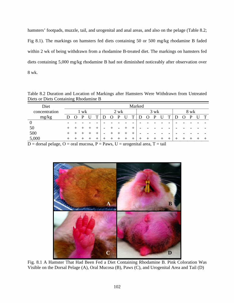
102
hamsters’ footpads, muzzle, tail, and urogenital and anal areas, and also on the pelage (Table 8.2;
Fig 8.1). The markings on hamsters fed diets containing 50 or 500 mg/kg rhodamine B faded
within 2 wk of being withdrawn from a rhodamine B-treated diet. The markings on hamsters fed
diets containing 5,000 mg/kg rhodamine B had not diminished noticeably after observation over
8 wk.
Table 8.2 Duration and Location of Markings after Hamsters Were Withdrawn from Untreated Diets or Diets Containing Rhodamine B
Diet concentration
mg/kg
Marked 1 wk 2 wk 3 wk 8 wk
D O P U T D O P U T D O P U T D O P U T0 - - - - - - - - - - - - - - - - - - - - 50 + + + + + - + - + + - - - - - - - - - - 500 + + + + + - + + + + - - - - - - - - - - 5,000 + + + + + + + + + + + + + + + + + + + +
D = dorsal pelage, O = oral mucosa, P = Paws, U = urogenital area, T = tail
Fig. 8.1 A Hamster That Had Been Fed a Diet Containing Rhodamine B. Pink Coloration Was Visible on the Dorsal Pelage (A), Oral Mucosa (B), Paws (C), and Urogenital Area and Tail (D)
A B
D C

103
Feces of hamsters fed a diet containing rhodamine B were pink, and the feces could be
distinguished from feces of control hamsters when they were viewed under incandescent
illumination (Fig. 8.2). An increasing intensity of the pink coloration in feces appeared to be
associated with increasing doses of rhodamine B. When examined using fluorescence
microscopy with a 1 s exposure time, feces of hamsters fed diets containing all three
concentrations of rhodamine B (50, 500, 5,000 mg/kg) appeared red, while feces of control
hamsters appeared as a black field (Fig. 8.2).
Fig. 8.2 Images of Feces of Four Hamsters Taken under Incandescent Lighting (A, B, C, and D), and Using Fluorescence Microscopy with a 1 sec Exposure Time (E, F, G, and H). The Feces Pictured Are from a Hamster Fed a Control Diet (A and E), or a Diet Containing 50 (B and F), 500 (C and G) or 5,000 mg/kg Rhodamine B (D and H)
8.3.2 Bioassay
The mean percent adult emergence in the larval bioassay was 97.5±5.3 d, and there was
no significant difference in percent emergence of sand flies in different larval diet groups (F =
0.18, df = 3, P = 0.9118; Table 8.3). The mean age of sand flies at adult emergence was 30.0±1.3
d, and there was no significant difference in age of emergence of sand flies in different larval diet
groups (F = 1.35, df = 3, P = 0.2592; Table 8.3).
A B C D
E F G H

104
Table 8.3 Results of the Rhodamine B Feed-Through Larval Bioassay (Percent Adult Emergence, Age at Adult Emergence, and Fluorescence of Adult Sand Flies That Were Fluorescent when Observed Using Fluorescence Microscopy). Second Instar Sand Flies Were Fed Feces of Hamsters That Had Been Fed a Diet Containing 0, 50, 500, or 5,000 mg/kg Rhodamine B Larval diet
mg/kg Adult emergence
% (mean* ± SE)#
Age at adult emergence d
(mean* ± SE)#
Marked
0 98.3 ± 4.1a 30.2 ± 1.4 a not marked 50 96.7 ± 5.2 a 30.0 ± 1.3 a inconsistent marking 500 98.3 ± 4.1 a 29.7 ± 1.3 a all marked 5,000 96.7 ± 8.2 a 30.1 ± 1.2 a all marked * Six replicates, ten larvae per replicate # Values within a column with the same letter are not significantly different, P> 0.05
Adults that as larvae were fed feces of rhodamine B-treated hamsters were positive for
the presence of rhodamine B (appeared red) when examined using fluorescence microscopy. An
exposure time of 15 s was used to detect fluorescence due to rhodamine B in sand flies. At this
exposure time, adult sand flies fed feces of untreated hamsters as larvae appeared as a black
field. All sand flies fed feces of hamsters that had been fed a diet containing 5,000 mg/kg
rhodamine B as larvae were fluorescent (n = 58); sand flies fed feces of hamsters fed a diet
containing 500 mg/kg rhodamine B exhibited a fainter fluorescence, but were all still discernable
from control flies (n = 59). Adult sand flies that had been fed as larvae the feces of hamsters fed
a diet containing 50 mg/kg rhodamine B were difficult or impossible to distinguish from control
sand flies. As indicated by fluorescence, rhodamine B was principally located in the thoraces and
femurs, but often also was present in the abdomen (Fig. 8.3; Fig. 8.4). Rhodamine B appeared to
be absent from the head, wings, and the legs below the femur.
8.3.3 Blood-Feeding
All of the sand flies exposed to anesthetized hamsters successfully obtained bloodmeals
(Fig. 8.5). The sand flies that were killed immediately after feeding on rhodamine B-treated

105
Fig. 8.3 Images of Two Female Sand Flies Taken under Incandescent Lighting (A and B), and Using Fluorescence Microscopy (C and D). The Sand Fly Pictured in the First Column (A and C) Had Been Fed as a Larva the Feces of a Hamster That Had Been Fed a Diet Containing 5,000 mg/kg Rhodamine B, and the Sand Fly Pictured in the Second Column (B and D) Had Been Fed as a Larva the Feces of a Hamster That Had Been Fed a Control Diet
Fig. 8.4 Images of Two Male Sand Flies Taken under Incandescent Lighting (A and B), and Using Fluorescence Microscopy (C and D). The Sand Fly Pictured in the First Column (A and C) Had Been Fed as a Larva the Feces of a Hamster That Had Been Fed a Diet Containing 5,000 mg/kg Rhodamine B, and the Sand Fly Pictured in the Second Column (B and D) Had Been Fed as a Larva the Feces of a Hamster That Had Been Fed a Control Diet
A B
C D
A B
C D

106
hamsters were positive for the presence of rhodamine B (appeared red) when examined using
fluorescence microscopy with an exposure time of 15 s. As indicated by fluorescence, rhodamine
B was located is in the thorax, femur, posterior tip of the abdomen, and the malpighian tubules.
Rhodamine B appeared to be absent from the head, the legs below the femur, and the bloodmeal
itself (Fig. 8.6). Sand flies that took bloodmeals from untreated hamsters appeared as a black
field using fluorescence microscopy with an exposure time of up to 15 s exposure time.
Fig. 8.5 A Female Sand Fly Taking a Bloodmeal from the Hind Foot of an Anesthetized, Rhodamine B-Treated Hamster
Sand flies that were killed 8 d after feeding on control or rhodamine B-treated hamsters
were not marked (were not considered positive for the presence of rhodamine B) when examined
with fluorescent microscopy using an exposure time up to 15 s. Similarly, the eggs, larvae, and
adult progeny of sand flies that took a bloodmeal from a control or rhodamine B-treated hamster

107
also were not marked when examined with fluorescence microscopy using a 15 sec exposure
time.
Fig. 8.6 Images of Two Bloodfed Female Sand Flies Taken under Incandescent Lighting (A and B), and Using Fluorescence Microscopy (C and D). The Sand Fly Pictured in the First Column (A and C) Had Taken a Bloodmeal from a Hamster That Had Been Fed a Diet Containing Rhodamine B, and the Sand Fly Pictured in the Second Column (B and D) Had Taken a Bloodmeal from a Hamster Fed a Control Diet
8.4 Discussion
To be an effective biomarker, the marker must be non-toxic and readily consumed by
target animals. In this study, there was no significant difference in the amount of food consumed
by hamsters at any of the diet concentrations of rhodamine B tested. Furthermore, rhodamine B
is a dye with low mammalian toxicity (LD50 887 mg/kg mouse oral), and has been used safely in
a wide variety of mammals (Fisher 1999). The finding that the food intake of hamsters was not
affected by rhodamine B treatments is consistent with results for other small mammals. For
example, there was no difference in the bait preference of ground squirrels targeted with
untreated baits or baits containing 2500 mg/kg rhodamine B (Sullens and Verts 1978). Similarly,
red-backed voles and field voles did not discriminate between untreated food or food containing
C D
B A

108
rhodamine B (Buckner 1968). The results of this study support the idea that rhodamine B does
not affect the palatability of baits for rodents, and suggest that effective baits containing
rhodamine B could be prepared for the various rodents present in different ZCL foci.
All hamsters that consumed diets containing rhodamine B in this study were visibly
marked. Hamsters fed a diet containing 50 or 500 mg/kg rhodamine B were marked for a period
of up to 2 wk, while hamsters fed a diet containing 5,000 mg/kg rhodamine B were marked for
more than 8 wk. Persistence of markings due to rhodamine B have been shown to vary between
different mammalian species. For example, coypu fed a diet containing 5,000 mg/kg rhodamine
B remained marked for up to 225 d, whereas mountain beavers fed a diet containing 1000 to
3400 mg/kg rhodamine B remained marked for only 2 wk (Fichet-Calvet 1999; Lindsey 1983).
This is the first report of rhodamine B being used as a biomarker for hamsters and adds support
for the use of rhodamine B as an orally delivered marker for mammals. The findings in this study
also demonstrate that the persistence of rhodamine B as a biomarker in hamsters is associated
with the dose of rhodamine B.
Numerous biomarkers have been used to monitor the ingestion of baits by mammals
including fat-soluble markers such as Dupont oil blue A, antimicrobial markers such as
tetracycline hydrochloride and sulfadimethoxine, and the cholecyctographic agent iophenoxic
acid (Southey et al. 2002; Creekmore et al. 2002). Unlike rhodamine B, none of these oral
biomarkers for mammals have been detected in the feces of orally dosed mammals. In rats, only
3-5% of orally dosed rhodamine B is excreted unchanged in feces (Webb and Hansen 1961).
Nevertheless, in this study feces of hamsters that consumed rhodamine B-treated diets were
fluorescent. This finding is consistent with studies using rhodamine B to mark other small
mammals. For example, feces of black-tailed jackrabbits and cottontail rabbits orally dosed with
rhodamine B also were reported to be visibly marked and fluorescent under UV illumination

109
(Evan and Griffith 1973). The results of this study suggest that examining feces for rhodamine B
could provide an alternative measure of bait uptake by target rodents that would be less invasive
and time consuming than examining the rodents themselves.
Sand fly larvae fed feces of hamsters that had been fed a diet containing 500 or 5,000
mg/kg rhodamine B were fluorescent as adults when examined using fluorescence microscopy.
The results of this study constitute the first report of a feed-through biomarker for larvae and the
subsequent adult insects for a medically important insect. In this study, fluorescence due to
rhodamine B was detected primarily in the thorax of sand flies. This finding indicates that a
marking system using rhodamine B would be compatible with the processing required to identify
sand flies. Sand flies are cleared before identification, and key taxonomic characteristics are
located in the head and abdomen (the cibarium of a sand fly is used to distinguish medically
important Phlebotomus spp. from Sergentomyia spp., and the spermatheca of female sand flies or
the external genitalia of male sand flies are necessary to differentiate species within Phlebotomus
spp.).
The results of this study suggest that fluorescence due to rhodamine B is transient in sand
flies that have taken a bloodmeal from rhodamine B-treated hamsters. Using fluorescence
microscopy, rhodamine B was observed in the malpighian tubules of recently engorged sand
flies. In insects, the malpighian tubules are the primary system responsible for excretion,
suggesting that rhodamine B is rapidly eliminated as the bloodmeal is processed. This hypothesis
is further supported by the absence of fluorescence due to rhodamine B in the bloodmeal itself.
A biomarker incorporated into a rodent bait to demonstrate the suitability of rodent feed-
through control of sand fly larvae in a rodent/sand fly association must: A) be palatable to
rodents, B) mark the rodents and their feces, C) be excreted unchanged in the feces of bait-fed
rodents (rather than metabolized), and D) be detectable in adult sand flies that have consumed

110
feces of bait-fed rodents as larvae. In this study hamsters readily consumed food containing high
concentrations of rhodamine B (up to 5,000 mg/kg), and markings due to rhodamine B were
detected on the hamsters and their feces. Adult male and female sand flies that had been fed
feces of rhodamine B-treated hamsters as larvae could be distinguished from control sand flies
using fluorescence microscopy. Therefore, rhodamine B is a good candidate for further
evaluation as a biomarker for rodents and sand flies in feed-through studies. Since the results of
this study suggest that rhodamine B could be an effective biomarker, the next step would be to
evaluate potential interactions between rhodamine B and feed-through insecticides, to evaluate
rhodamine B in additional rodent and sand fly models, and to develop a high-throughput and
quantitative technique, such as spectrofluorometry, for detecting rhodamine B in specimens.
Additional fluorescent dyes with non-overlapping excitation an emission wavelengths also could
be evaluated for potential use in conjunction with rhodamine B and feed-through or systemic
insecticides.
Using rhodamine B incorporated rodent baits in the field could allow the identification of
specific foci with sand fly/rodent associations that would be susceptible to control using feed-
through or systemic insecticides, through the detection of adult male and female sand flies that
had fed on the feces of baited rodents as larvae, and adult female sand flies that have taken a
bloodmeal from bait-fed rodents. The development of rhodamine B as a feed-through biomarker
also could have an enormous impact on future studies on sand flies, and could be the
breakthrough for several unanswered questions: What percentage of sand flies in an area are
associated with rodents? Which of the sand fly species feed on rodent feces as larvae? What
percentage of human-biting flies in an area could potentially be eliminated using rodent baits
containing insecticides? How far do sand flies disperse from their immature habitat? What

111
distance around human settlements must be targeted with insecticide-treated rodent baits to
create an appropriate barrier?
Using rhodamine B as a feed-through biomarker also could provide detection of changes
in sand fly populations due to larval control measures. Currently, monitoring the effects of larval
control of phlebotomine sand flies during field trials is a problematic. The ideal method of
demonstrating successful larval control would be to directly measure reductions in the larval
population through larval sampling, which is impractical. Alternatively, adult sampling
techniques (such as sticky paper traps and light traps) could be used, but there are inherent
problems with adult sampling to demonstrate larval control, including potential false negative
interpretations of results (such as not detecting control when it happens, which could happen
through immigration of adult sand flies into the study area) or false positive interpretations of
results (which would be attributing a reduction in adult populations to successful larval control,
when it actually is a natural seasonal population decline or differential changes in microclimates
of test sites).
In conclusion, this study is the first report of a fluorescent dye fed to larvae and then
detected in adults of a medically important insect. The value of this method is that it can mark
rodents and their feces to establish the consumption of treated-baits, mark adult female sand flies
that feed rodents for the duration of persistence of the dye in rodents, and mark adult male and
female sand flies that had fed on feces of bait-fed rodents as larvae. In specific rodent sand fly
associations, the differential marking of both male and female sand flies or only female sand flies
can be used to direct field control studies: a rodent bait containing a systemic insecticide would
be used at sites where only female sand flies that fed on rodents are marked, while if male and
female sand flies are marked, feed-through insecticides also could be used.

112
SUMMARY AND CONCLUSIONS
In North Africa, the Middle East, and Southwest Asia, the sand fly P. papatasi is the
vector of L. major, the causative agent of ZCL. All life stages of P. papatasi exhibit a close
association with the burrowing rodent reservoirs of L. major. Despite this close association,
currently there are no effective control measures available for the sand fly P. papatasi. However,
the primary larval habitats of P. papatasi in ZCL foci are rodent burrows, and sand fly larvae
have been observed feeding on the feces of rodents. Because of this fact, novel control methods
targeting sand fly larvae that feed on rodent feces (using rodent feed-through insecticides) and
adult sand flies that take bloodmeals from rodents (using a systemic insecticide) were evaluated
in this study. Furthermore, a novel biomarker system was evaluated that marked rodents and
their feces, adult female sand flies that took bloodmeals from treated rodents, and adult male and
female sand flies that had fed on feces of bait-fed rodents as larvae. This biomarker system could
be a valuable tool for monitoring host-targeted control trials for sand flies in the field.
The studies presented in Chapters 2 to 5 of this dissertation evaluated the development
and survival of P. papatasi larvae fed feces from Syrian hamsters that had been fed a diet
containing the chitin synthesis inhibitors diflubenzuron and novaluron, the juvenile hormone
analogs pyriproxyfen and methoprene, or the macrocyclic lactone ivermectin. In the studies on
diflubenzuron, novaluron, methoprene, and pyriproxyfen, the mean daily food intake of the
insecticide-treated hamsters was not significantly different from the food intake of control
hamsters at any of the concentrations tested (up to approximately 1000 mg/kg a.i.), but the mean
daily food intake of hamsters was significantly lower than control for hamsters fed a diet
containing more than 20 mg/kg ivermectin. Sand fly larvae (2nd instars) that had been fed feces
of hamsters fed diets containing approximately 10, 100, or 1000 mg/kg diflubenzuron,
novaluron, or pyriproxyfen, or 20 mg/kg ivermectin all died before adult emergence. Feces of

113
hamsters fed a diet containing up to 978.8 mg/kg methoprene caused significant but less than
100% mortality of immature sand flies. The results of the studies in Chapters 2 to 5 constitute
proof of concept for feed-through control of sand fly larvae using diflubenzuron, novaluron,
pyriproxyfen, and ivermectin.
The studies presented in Chapter 6 of this dissertation determined the minimum effective
dose of novaluron as a feed-through for control of sand fly larvae, and evaluated novaluron feed-
through under simulated field conditions. Complete control of sand fly larvae was observed
when they were fed hamster feces containing 9.88 mg/kg novaluron, or when they were fed the
feces of hamsters fed a diet containing 9.88x10⁻¹ mg/kg novaluron. Feces of hamsters fed a diet
containing 988 mg/mg novaluron were aged for up to 150 d at 28 °C and 90% RH. All larvae
that had been fed feces of novaluron-treated hamsters aged for 0 or 30 d died before adult
emergence, and the mortality of sand fly larvae fed feces aged up to 150 d was significantly
different from control. The last experiment presented in Chapter 6 evaluated the effectiveness of
novaluron as a feed-through in preventing the development of sand fly larvae when novaluron-
treated food made up only a portion of a hamster’s daily diet. All larvae that had been fed either
feces from hamsters that exclusively had been fed a diet containing novaluron or hamsters that
consumed novaluron-treated food as approximately 15% of their daily diet died before pupation.
In this study, significant control of sand fly larvae was observed when they were fed
feces of novaluron-treated hamsters that had been aged for up to 150 d or feces of novaluron-
treated hamsters when only a portion of their daily diet was novaluron-treated food. Therefore,
novaluron is a good candidate for further evaluation as a rodent feed-through insecticide against
sand fly larvae. Since the results of this study suggest that novaluron could be effective as a
rodent feed-through insecticide in a field setting, the next step would be to evaluate the effects of
novaluron-treated baits on sand fly populations in different rodent/sand fly associations.

114
The studies presented in Chapter 7 of this dissertation evaluated whether the post-
bloodmeal survival of adult female sand flies would be affected by feeding their rodent hosts a
diet containing ivermectin. The 48 h survival was 0% for sand flies that took a bloodmeal from
hamsters withdrawn from an ivermectin-treated diet for 0, 3, or 7 d, but was 100% for sand flies
fed on hamsters withdrawn from an ivermectin-treated diet for 14 d. Feces voided by ivermectin-
treated hamsters also were collected 0 to 14 d after the hamsters were withdrawn from their
ivermectin-treated diets, and the feces were fed to sand fly larvae. Mortality was 100% for larvae
fed feces voided 0 d after withdrawal, and was significantly higher than control larvae for up to 7
d after withdrawal. The mean percent survival of sand fly larvae fed feces of ivermectin-treated
hamsters 14 d after they were withdrawn from an ivermectin-treated diet was not significantly
different from control.
This study confirms that ivermectin-treated rodent baits developed as a feed-through to
control sand fly larvae also could have a collateral effect on bloodfeeding adult sand flies, thus
affecting a larger portion of the sand fly population than would be estimated due to larvicide
activity alone. While ivermectin treatment of rodent hosts of L. major may reduce the overall
population of sand flies, this approach would have its greatest impact on the most
epidemiologically important subset of the sand fly population: females that have taken a
bloodmeal from potentially infected rodents.
In the studies presented in Chapter 8 of this dissertation, hamsters were fed diets
containing rhodamine B, and the mean daily food intake of the rhodamine B-treated hamsters
was not significantly different from the food intake of control hamsters at any of the
concentrations tested (50, 500, or 5000 mg/kg rhodamine B). Hamsters fed diets containing
rhodamine B were marked by the dye after feeding on their diets for up to 8 weeks. Feces of
rhodamine B-treated hamsters also were marked when examined using fluorescence microscopy.
115
The development and survival of sand fly larvae fed feces of rhodamine B-treated hamsters was
not significantly different from control sand flies. Adult male and female sand flies that had were
fed as larvae the feces of rhodamine B-treated hamsters were fluorescent when examined using
fluorescent microscopy, and could be distinguished from control sand flies. Adult female sand
flies that took bloodmeals from rhodamine B-treated hamsters were fluorescent, but not when
they were examined 8 d after bloodfeeding.
Using rhodamine B incorporated rodent baits in the field could allow the identification of
specific foci with sand fly/rodent associations that would be susceptible to control using feed-
through or systemic insecticides, through the detection of adult male and female sand flies that
had fed on the feces of baited rodents as larvae, and adult female sand flies that have taken a
bloodmeal from bait-fed rodents. The development of rhodamine B as a feed-through biomarker
also could have an enormous impact on future studies on sand flies, and could be the
breakthrough for several unanswered questions about sand fly ecology.
116
REFERENCES Abramowitz, M. 2004. Drugs for parasitic infections. The Medical Letter. 46:e1-e12. Alexander, B. 2000. Sampling methods for phlebotomine sandflies. Medical and Veterinary Entomology. 14: 109-122. Alexander, B., Cadena, H., Usma, M.C., and Rojas, C.A. 1995. Laboratory and field evaluations of a repellent soap containing diethyl toluamide (DEET) and permethrin against phlebotomine sand flies (Diptera: Psychodidae) in Valle del Cauca, Colombia. American Journal of Tropical Medicine and Hygiene. 52: 169-173. Ali A., and Kok-Yokomi, M. L. 1990. Preliminary evaluations of methoprene, diflubenzuron, and chlorpyriphos against Psychoda alternata Say (Diptera: Psychodidae) in turf. Journal of the Florida Mosquito Control Association. 61: 4-8. Ali, A., Nayar, J.K. and Xue, R.D. 1995. Comparative toxicity of selected larvicides and insect growth regulators to a Florida laboratory population of Aedes albopictus. Journal of the American Mosquito Control Association, 11: 72-76. Alten, B., Caglar, S.S., Kaynas, S., Simsek, and F.M. 2003. Evaluation of protective efficacy of K-OTAB impregnated bednets for cutaneous leishmaniasis control in Southeast Anatolia-Turkey. Journal of vector Ecology. 28: 53-64. Alten, B., Caglar, S.S., Simsek, F.M., Kaynas, S., and Perich, M.J. 2003. Field evaluation of an area repellent system (Thermacell) against Phlebotomus papatasi (Diptera: Psychodidae) and Ochlreotatus caspius (Diptera: Culicidae) in Sanliurfa Province, Turkey. Journal of Medical Entomology. 40: 930-934. Al-Zahrani, M.A., Peters, W., and Evans, D.A. 1988. Phlebotomus sergenti, a vector of Leishmania tropica in Saudi Arabia. Transactions of the Royal Society of Tropical Medicine and Hygiene. 82: 416. Arias, J.R. and Freitas, R.A. 1982. On the vectors of cutaneous leishmaniasis in the central Amazon of Brasil. 4. Sand fly emergence from a ‘terra firme’ forest floor. Acta Amazonica. 12: 609–611. Artemiev, M.M., Flerova, O.A., and Belyaev, A.E. 1972. Quantitative evaluation of the productivity of breeding places of sandflies in the wild and in villages. Meditsinskaya Parazitologiya I Parazitarnye Bolezni. 41: 31–35. Ashford R.W., Desjeux, P., and De Raadt, P. 1991. Estimation of population risk of infection and number of cases of leishmaniasis. Parasitology Today. 8: 104-105. Asilian, A., Sadeghinia, A., Shariati, F., Imam Jome, M., and Ghoddusi, A. 2003. Efficacy of permethrin-impregnated uniforms in the prevention of cutaneous leishmaniasis in Iranian soldiers. Journal of Clinical Pharmacy and Therapeutics. 28: 175-178.

117
Bansemir, A.D. and Sukhdeo, M.V. 2001. Intestinal distribution of worms and host ingesta in Nippostrongylus brasiliensis. Journal of Parasitology. 87: 1470-1472. Basimike, M. and Mutinga, M.J. 1990. Temperature and moisture content of soils of termite mounds and animal burrows in relation to relative abundance of adult phlebotomine sandflies (Diptera: Psychodidae) in Marigat semiarid area, Baringo District, Kenya. Environmental Entomology. 19: 485-489. Basimike, M. and Mutinga, M.J. 1992. The relative abundance of Phlebotomus martini Parrot and Phlebotomus duboscqi Neveu-Lemaire (Diptera: Psychodidae) in animal burrows and termite mounds in Marigat Location, Baringo District, Kenya. Insect Science and its Application. 13: 173-176. Batieha, A., Saliba, E.K., Graham, R., Mohareb, E., Hijazi, Y., and Wijeyaratne, P. 2000. Seroprevalence of West Nile, Rift Valley, and sandfly arboviruses in Hashimiah, Jordan. Emerging Infectious Diseases. 6: 358–362. Beach, R., Young, D.G., and Killu, G. 1986. New phlebotomine sand-fly colonies II. Laboratory colonization of Phlebotomus duboscqi (Diptera: Psychodidae). Journal of Medical Entomology. 23: 114-115. Bern, C., Joshit, A.B., Jha, S.N., Das, M.L., Hightower, A., Thakur, G.D., and Bista, M.B. 2000. Factors associated with visceral leishmaniasis in Nepal: bed-net use is strongly protective. American Journal of Tropical Medicine and Hygiene. 64: 184-188. Bettini, S., Contini, C., Atzeni, M.C., and Tocco, G. 1986. Leishmaniasis in Sardinia. I. Observations on a larval breeding site of Phlebotomus perniciosus, Phlebotomus perfiliewi perfiliewi and Sergentomyia minuta (Diptera: Psychodidae) in the canine leishmaniasis focus of Soleminis (Cagliari). Annals of Tropical Medicine and Parasitology. 80: 307–315. Bockarie, M.J., Hii, J.L., Alexander, N.D., Bockarie, F., Dagoro, H., Kazura, J.W., and Alpers, M.P. 1999. Mass treatment with ivermectin for filariasis control in Papua New Guinea: impact on mosquito survival. Medical and Veterinary Entomology. 13: 120-123. Bottjer, K.P. and Bone, L.W. 1984. Ingestion of dye by the sexes of Trichostrongylus colubriformis. Journal of Parasitology. 70: 671-676. Buckner, C.H. 1968. Reactions of small mammals to vital dyes. Canadian Entomologist. 100: 476-477. Buescher, M.D., Rutledge, L.C., Wirtz, R.A., Glackin, K.B., and Moussa, M.A. 1982. Laboratory tests of repellents against Lutzomyia longipalpis (Diptera: Psychodidae). Journal of Medical Entomology. 19: 176-180. Cameron, M.M., Milligan, P.J., Llanos-Cuentas, A., and Davies, C.R. 1995. An association between phlebotomine sandflies and aphids in the Peruvian Andes. Medical and Veterinary Entomology. 9:127-132.
118
Campbell, W.C., Fisher, M.H., Stapley, E.O., Ablers-Schonberg, A., and Jacob, T.A. 1983. Ivermectin: a potent new antiparasitic agent. Science. 221:823-828. Campbell-Lendrum, D., Pinto, M.C., and Davies, C. 1999. Is Lutzomyia intermedia (Lutz & Neiva, 1912) more endophagic than Lutzomyia whitmani (Antunes & Coutinho, 1939) because it is more attracted to light? Memorias do Instituto Oswaldo Cruz. 94:21-22. Cartel, J.L., Sechan, Y., Spiegel, A., Nguyen, L., Barbazan, P., Martin, P.M., and Roux, J.F. 1991. Cumulative mortality rates in Aedes polynesiensis after feeding on polynesian Wuchereria bancrofti carriers treated with single doses of ivermectin, diethylcarbamazine and placebo. Tropical Medicine and Parasitology. 42: 343-345. Casanova, C. 2001. A soil emergence trap for collections of Phlebotomine sandflies. Memorias do Instituto Oswaldo Cruz. 96: 273–275. Centurier, C. and Barth, D. 1980. On the efficacy of ivermectin versus ticks (O. moubata, R. appendiculatus and A. variegatum) in cattle. Zentralblatt fur Bakteriologie, Parasitenkunde, Infektions-krankheiten und Hygiene. 58: 267-319. Chaniotis, B.N., Correa, M.A., Tesh, R.B., and Johnson, K.M.1974. Horizontal and vertical movements of phlebotomine sandflies in a Panamanian rain forest. Journal of Medical Entomology. 11:369-375. Chaniotis, B.N., Parsons, R.E., Harlan, H.J., and Correa, M.A. 1982. A pilot study to control phlebotomine sand flies (Diptera: Psychodidae) in a neotropical rain forest. Journal of Medical Entomology. 19: 1-5. Charrel, R.N., Gallian, P., Navarro-Mari, J.M., Nicoletti, L., Papa, A., Sánchez-Seco, M.P., Tenorio, A., and de Lamballerie, X. 2005. Emergence of Toscana virus in Europe. Emerging Infectious Diseases. 11:1657-663. Chastel, C., Bach-Hamba, D., Launay, H., Le Lay, G., Hellal, H., and Beaucornu, J.C. 1983. Infections à arbovirus en Tunisie : nouvelle enquête sérologique chez les petits mammifères sauvages. Bulletin de la Societe de Pathologie Exotique. 76: 21–33. Clarke, G.R., Stallknecht, D.E., and Howerth, E.W. 1996. Experimental infection of swine with a sandfly (Lutzomyia shannoni) isolate of vesicular stomatitis virus, New Jersey serotype. Journal of Veterinary Diagnostic Investigation. 8: 105-108. Cohen, D., Zaide, Y., Karasenty, E., Schwarz, M., LeDuc, J.W., Slepon, R. 1999. Prevalence of antibodies to West Nile fever, sandfly fever Sicilian, and sandfly fever Naples viruses in healthy adults in Israel. Public Health Review. 27: 217–230. Cook, C.W., and Gerhardt, R.R. 1977. Selective mortality of insects in manure from cattle fed Rabon® and Dimilin®. Env Entomol. 6: 589-590.

119
Courtenay, O., Gillingwater, K., Gomes, P.A., Garcez, L.M., and Davies, C.R. 2007. Deltamethrin-impreganted bednets reduce human landing rates of sandfly vector Lutzomyia longipalpis in Amazon households. Medical and Veterinary Entomology. 21: 168-176. Creekmore, T.E., Rocke, T.E., and Hurley, J. 2002. A baiting system for delivery of an oral plague vaccine to black-tailed prairie dogs. Journal of Wildlife Diseases. 38: 32-39. Cutler, G.C., Scott-Dupree, C.D., Tolman, J.H., and Harris, C.R. 2006. Toxicity of the insect growth regulator novaluron to the non-target predatory bug Podisus maculiventris (Heteroptera : Pentatomidae). Biological Control. 38: 196-204. David, J.R., Stamm, L.M., Bezerra, H.S., Souza, R.N., Killick-Kendrick, R., and Lima, J.W.O. 2001. Deltamethrin-impreganted dog collars have a potent anti-feeding and insecticidal effects on Lutzomyia longipalpis and Lutzomyia migonei. Memorias do Instituto Osawldo Cruz. 96: 839-847. de Barjac, H., Larget, .I, and Killick-Kendrick, R. 1981. Toxicite de Bacillus thuringiensis var. israelensis, serotype h14, pour les larves de phlebotomes vecteurs de leishmanioses. Bulletin de la Societe de Pathologie Exotique. 74: 485-489. De Queiroz, R.G., Vasconcelos, I.A.B., and Vasconcelos, A.W. 1994. Cutaneous leishmaniasis in Ceara State in northeastern Brazil: incrimination of Lutzomyia whitmani (Diptera: Psychodidae) as a vector of Leishmania braziliensis in Baturite´ Municipality. American Journal of Tropical Medicine and Hygiene. 50: 693–698. Deane, L. and Deane, M.P. 1957. Observacoes so bre abrigos e criadouros de flebotomos no Noroeste do Estado do Ceara. Revista Brasileira de Malariologia e Doencas Tropicais. 9: 225–246. Dedet, J.P. 1992. Leishmania et leishmanioses du continent American. Annales de l'Institute Pasteur. 4: 13-25. Dedet, J.P., Desjeux, P., and Derouin, F. 1982. Ecologie d’un foyer de leishmaniose cutanee dans la region de Thies (Senegal, Afrique de l’Ouest. 4. Infestation spontanee et biologie de Phlebotomus duboscqi Neveu-Lemaire 1906. Bulletin de la Societe de Pathologie Exotique. 75: 588–589. Dedet, J.P., Esterre, P. and Pradinaud, R. 1987. Individual clothing prophylaxis of cutaneous leishmaniasis in the Amazonian area. Transactions of the Royal Society of Tropical Medicine and Hygiene. 81: 748. Dedet, J.P., Gay, F., and Chatenay, G. 1989. Isolation of Leishmania species from wild mammals in French Guiana. Transactions of the Royal Society of Tropical Medicine and Hygiene. 83: 613-615. Dergacheva, T.I., and Zherikhina I.I. 1980. Changes in the population density and species composition of Phlebotomidae as a result of eradication of great gerbils in a focus of zoonotic

120
cutaneous leishmaniasis in the Karshi Steppe. Meditsinskaya Parazitolojiya I Parazitarnye Bolezni. 49: 50-55. Desjeux P. 2004. Leishmaniasis: current situation and new perspectives. Comparative Immunology, Microbiology, and Infectious Diseases. 27: 305-318. Desjeux, P. 1991. Information on the epidemiology and control of the leishmaniases by country and territory. WHO/LEISH/91.30. Geneva: World Health Organization. Dhanda, V., Rodrigues, F.M., and Ghosh, S.N. 1972. Isolation of Chandipura virus from sand flies in Aurangabad. Indian Journal of Medical Research. 58: 179–180. Dhiman, R.C. 1995. Effect of minor engineering intervention in the control of breeding of Phlebotomus papatasi (Scopoli) sandflies. Southeast Asian Journal of Tropical Medicine and Public Health. 26: 368-370. Dhiman, R.C. and Sharma, V.P. 1994. Evaluation of neem oil as a sandfly, Phlebotomus papatasi (Scopoli) repellent in an oriental sore endemic area in Rajasthan. Southeast Asian Journal of Tropical Medicine and Public Health. 25: 608-610. Dhiman, R.C., Shetty, P.S., and Dhanda, V. 1983. Breeding habitats of phlebotomine sand flies in Bihar, India. Indian Journal of Medical Research. 77: 29-32. Disney, R.H. 1966. A trap for Phlebotomine sandflies attracted to rats. Bulletin of Entomological Research. 56:445-451. Disney, R.H.L. 1968. Observations on a zoonosis: Leishmaniasis in British Honduras. Journal of Applied Ecology. 5: 1-59. Doha, S. and Shehata, M.G. 1992. Leishmania infantum MON-98 isolated from naturally infected Phlebotomus langeroni (Diptera: Psychodidae) in El Agamy, Egypt. Journal of Medical Entomology. 29: 891-893. Doha, S., Kamal, H., Shehata,M., Helmy, N., Kader,M.A., El Said, S., and El Sawaf, B.M. 1990. The breeding habitats of Phlebotomus sand flies (Diptera: Psychodidae) in El Agamy, Alexandria, Egypt. Journal of the Egyptian Society of Parasitology. 20: 747–752. Echevarria, J.M., de Ory, F., Guisasola, M.E., Sanchez-Seco, M.P., Tenorio, A., and Lozano, A. 2003. Acute meningitis due to Toscana virus infection among patients from both the Spanish Mediterranean region and the region of Madrid. Journal of Clinical Virology. 26: 79–84. Eduardo, E., Gomez, L., and Hashiguchi, Y. 1991. Monthly variation in natural infection of the sandfly Lutzomyia ayacuchensis with Leishmania mexicana in an endemic focus in the Ecuadorian Andes. Annals of Tropical Medicine and Parasitology. 85: 407–411. Eitrem, R., Vene, S., and Niklasson, B. 1990. Incidence of sand fly fever among Swedish United Nations soldiers on Cyprus during 1985. American Journal of Tropical Medicine and Hygiene. 43: 207–211.
121
Eliseev, L.N. 1980. Principles and methods of control of zoonotic cutaneous leishmaniasis. USSR Ministry of Health and WHO Seminar on Control of Leishmaniases: Moscow. Elnaiem, D.A. and Ward, R.D. 1991. Oviposition attractants and stimulants for the sandfly Lutzomyia longipalpis (Diptera: Psychodidae). Journal of Medical Entomology. 29: 5-12. Elnaiem, D.A., Elnahas, A.M., and Aboud, M.A. 1999. Protective efficacy of lambdacyhalothrin-impreganted bednets against Phlebotomus orientalis, the vector of visceral leishmaniasis in Sudan. Medical and Veterinary Entomology. 13: 310-314. Elnaiem, D.A., Hassan, M.M., Maingon, R., Nureldin, G.H., Mekawi, A.M., Miles, M., and Ward, R.D. 2001. The Egyptian mongoose, Herpestes ichneumon, is a possible reservoir host of visceral leishmaniasis in eastern Sudan. Parasitology. 122: 531-536. Esterre, P. 1986. Evaluation d'un programme de lutte contre la leishmaniose cutanee dans un village forestier de Guyane Francaise. Bulletin de l'Organization Mondiale de Sante. 64: 559-565. Evans, J. and Griffith R.E. 1973. A fluorescent tracer and mark- er for animal studies. Journal of Wildlife Management 37: 73-81. Faizulin, F.G. 1980. Experience on organization and implementation of control of zoonotic cutaneous leishmaniasis in Golodnaya Steppe of Uzbekskaya SSR. USSR Ministry of Health and WHO Seminar on Control of Leishmaniasis. Moscow. p. 17. (FAO) Food and Agriculture Organization of the United Nations. 1981. FAO Plant Production and Protection Paper 42. Pesticide residues in food - 1981. FAO, Geneva, Switzerland, 9-17. (FAO) Food and Agriculture Organization of the United Nations. 2005. JMPR – Pesticide residue data. FAO, Geneva, Switzerland, 797-871. Faulde, M., Schrader, J., Heyl, G., Amirih, M., and Hoerauf, A. 2008. Zoonotic cutaneous leishmaniasis outbreak in Mazar-e Sharif, northern Afghanistan: an epidemiological evaluation. International Journal of Medical Microbiology. 298: 543-550. Feliciangeli, M.D. 2004. Natural breeding places of phlebotomine sandflies. Medical and Veterinary Entomology. 18: 71-80. Feliciangeli, M.D. and Rabinovich, J. 1998. Abundance of Lutzomyia ovallesi but not Lu. gomezi (Diptera: Psychodidae) correlated with cutaneous leishmaniasis incidence in north-central Venezuela. Medical and Veterinary Entomology. 12:121-131. Fichet-Calvet, E. 1999: Persistence of a systemic labelling in fur and guard hairs by ingestion of Rhodamine B in Myocaster coypus (Rodentia). Mammalia 63: 241–244.

122
Fichet-Calvet, E., Jomâa, I., Ben Ismail, R., and Ashford, R.W. 2003. Leishmania major infection in the fat sand rat Psammomys obesus in Tunisia: interaction of host and parasite populations. Annals of Tropical Medicine and Parasitology. 97: 593-603. Floch, H. 1957. Sur la prophylaxie de la leishmaniose forestiere americaine. Rivista di Malariologia. 36: 243-253. Foley, D.H., Bryan, J.H., and Lawrence, G.W. 2000. The potential of ivermectin to control the malaria vector Anopheles farauti. Transactions of the Royal Society of Tropical Medicine and Hygiene. 94: 625-628. Fontenille, D., Traore-Lamizana, M., Trouillet, J., Leclerc, A., Mondo, M., Ba, Y., Digoutte, J.P., and Zeller, H.G. 1994. First isolations of arboviruses from phlebotomine sand flies in West Africa. American Journal of Tropical Medicine and Hygiene. 50: 570-574. Forattini, O.P. 1954. Algunas observacoes sobre a biologia dos flebotomos dos zones de leishmaniose visceral ora em estudo do Estado de Para. O’Hospital (Rio de Janeiro). 14: 3–6. Fossati, F.P. and Maroli, M. 1986. Laboratory tests of three repellents against Phlebotomus perniciosus (Diptera: Psychodidae). Transactions of the Royal Society of Tropical Medicine and Hygiene. 80: 771-773. Fryauff, D.J., Shoukry, M.A., Hanafi, H.A., Choi, Y.M., Kamel, K.E., and Schreck, C.E. 1996. Contact toxicity of permethrin-impreganted military uniforms to Culex pipiens (Diptera: Culicidae) and Phlebotomus papatasi (Diptera: Psychodidae): effects of laundering and time of exposure. Journal of the American Mosquito Control Association. 12: 84-90. Gaidamovich, S.Y., Baten, M.A., Klisenko, G.A., and Melnikova, Y.E. 1984. Serological studies on sandfly fevers in the Republic of Bangladesh. Acta Virologica. 28: 325–328. Gavgani, A.S., Hodjati, M.H., Mohite, H., and Davies, C.R. 2002. Effect of insecticide-impregnated dog collars on incidence of zoonotic visceral leishmaniasis in Iranian children: a matched-cluster randomised trial. The Lancet. 360: 374-379. Gebre-Michael, T., Pratlong, F., and Lane, R.P. 1993. Phlebotomus (Phlebotomus) duboscqi (Diptera: Psychodidae), naturally infected with Leishmania major in southern Ethiopia. Transactions of the Royal Society of Tropical Medicine and Hygiene. 87: 10-11. Gemetchu, T., Laskay, T., and Frommel, D. 1990. Phlebotomine sandflies (Diptera: Psychodidae, Phlebotominae) of Ochollo, southwestern Ethiopia: Species composition and natural infection of Phlebotomus pedifer with Leishmania aethiopica. Ethiopian Journal of Science. 13: 43-50. Ghosh, S.M. 1950. The control of Phlebotomus (Sand-flies) with DDT and BHC (Gammexane). Indian Journal of Malariology. 4: 175. Gillies, M.T. 1980. The role of carbon dioxide in host-finding by mosquitoes (Diptera: Culicidae): a review. Bulletin og Entomological Research. 70: 525-532.

123
Githure, J.I., Beach, R.F., and Lightner, L.K. 1984. The isolation of Leishmania major from rodents in Baringo District, Kenya. Transactions of the Royal Society of Tropical Medicine and Hygiene. 78: 283. Githure, J.I., Schnur, L.F., Le Blancq, S.M., and Hendricks, L.D. 1986. Characterization of Kenyan Leishmania spp. and identification of Mastomys natalensis, Taterillus emini and Aethomys kaiseri as new hosts of Leishmania major. Annals of Tropical Medicine and Parasitology: 80, 501-507. Grassi, B. 1907. Ricerche sui flebotomi. Memorie della Societa Italiana di Scienze Naturali. 14: 353–394. Gray, G.C., Johnson, A.A., Thornton, S.A., Smith, W.A., Knobloch, J., Kelley, P.W., Escudero, L.O., Huayda, M.A., Wignall, F.S. 1990. An epidemic of Oroyo fever in the Peruvian Andes. American Journal of Tropical Medicine and Hygiene. 42: 215-221. Grenot, C. 2001. Adaptations des petits vertebres aux conditions arides du Sahara. Bulletin de la societe de zoologie Francaise. 126: 129-167. Gunders, A.E., Foner, A., and Montilio, B. 1968. Identification of Leishmania species isolated from rodents in Israel. Nature. 219: 85-86. Halbig, P., Hodjati, M.H., Mazloumi-Gavgani, A.S., Mohite, H., Davies, C.R. 2000. Further evidence that deltamethrin-impregnated collars protect dogs from sandfly bites. Medical and Veterinary Entomology. 14: 223-226. Hanson, W.J. 1961. The breeding places of Phlebotomus in Panama (Diptera: Psychodidae). Annals of the Entomological Society of America. 54: 317–322. Hanson, W.J. 1968. The immature stages of the family Phlebotominae in Panama (Diptera: Psychodidae). PhD Thesis, University of Kansas, U.S.A. Hemmersbach-Miller, M., Parola, P., Charrel, R.N., Paul Durand, J., and Brouqui, P. 2004. Sandfly fever due to Toscana virus: an emerging infection in southern France. European Journal of Internal Medicine. 15:316–317. Heron, R.J. 1968. Vital dyes as markers for behavioral and population studies of the larch sawfly, Pristiphora erichsonii (Hymenopters: Tenthredinidae). Canadian Entomologist. 100: 470-475. Hertig, M. 1949. Phlebotomus and residual DDT in Greece and Italy. American Journal of Tropical Medicine and Hygiene. 29: 773-809. Hertig, M. and Fairchild, G.B. 1948. The control of Phlebotomus in Peru with DDT. American Journal of Tropical Medicine and Hygiene. 28: 207-230.

124
Hertig, M. and Fisher, R.A. 1945. Control of sandflies with DDT. Bulletin of the U.S. Army Medical Department. 88: 97-101. Hole, F. 1981. Effects of animals on soil. Geoderma. 25: 75-112. (IPCS) International Programme on Chemical Safety. 1994. PIM 292 - Ivermectin. Geneva, Switzerland: World Health Organization. Izri, A., Temmam, S., Moureau, G., Hamrioui, B., de Lamballerie, X., and Charrel, R.N.. 2008. Sandfly fever Sicilian virus, Algeria. Emerging Infectious Diseases. 14: 795-797. Jacusiel, F. 1947. Sandfly control with DDT residual spray; field experiments in Palestine. Bulletin of Entomological Research. 38: 479-488. Jalouk, L., Al Ahmed, M., Gradoni, L., and Maroli, M. 2007. Insecticide-treated bednets to prevent anthroponotic cutaneous leishmaniasis in Aleppo Governate, Syria: results from two trials. Transaction of the Royal Society of Tropical Medicine and Hygiene. 101: 360-367. Joshi, D., Gurbacharya, V.L., and Shrestha, S.L. 1986. Status of leishmaniasis in Nepal. Journal of the Institute for Medicine. 8: 7–12. Jukova, N.N. 1944. Employment of “K preparation” for sand fly control. Meditsinskaia Parazitologiia i Parazitarnye Bolezni. 13: 93. Jumaian, N., Kamhawi, S.A., Halalsheh, M., and Abdel-Hafez, S.K. 1998. Short report: outbreak of cutaneous leishmaniasis in a nonimmune population of soldiers in Wadi Araba, Jordan. American Journal of Tropical Medicine and Hygiene. 58: 160-162. Kalyanasundaram, M., Srinivasan, R., Subramanian, S., and Panicker, K.N.1994. Relative potency of DEPA as a repellent against the sandfly Phlebotomus papatasi. Medical and Veterinary Entomology. 8: 68-70. Kamesipour, A., Rafati, S., Davoudi, N., Maboudi, F., and Modabber, F. 2006. Leishmaniasis vaccine candidates for development: a global overview. Indian Journal of Medical Research.123: 423-438. Karabatsos, N. 1985. International catalogue of arboviruses including certain other viruses of vertebrates. American Society of Tropical Medicine and Hygiene, San Antionio, Texas. Karapet’ian A.B., Dzhabarov, L.N., Mamigonova, R.I., and Sabatov, E.A. 1983. Use of D-20 aerosol insecticide smoke pots in controlling burrow sandflies. Meditsinskaia parazitologiia i parazitarnye bolezni. 4: 78-81. Kassem, H.A., Tewfick, M.K., and El Sawaf, B.M. 2001. Evaluation of avermectins as sandfly control agents. Annals of Tropical Medicine and Parasitology. 95: 405-411.

125
Kay F.R., and Whitfield, W.G. 1978. The burrow environment of the banner-tailed kangaroo rat, Dipodmys spectabilis, in South-Central New Mexico. American Midland Naturalist. 99: 270-279. Kellina, O.I. 1981. Problem and current lines of investigations on the epidemiology of leishmaniasis and its control in the USSR. Bulletin de la Societe de Pathologie Exotique. 306-318. Kerr, S.F., McHugh, C.P., and Dronen, N.O. 1995. Leishmaniasis in Texas: prevalence and seasonal transmission of Leishmania mexicana in Neotoma micropus. American Journal of Tropical Medicine and Hygiene. 53: 73-77. Killick-Kendrick, R. 1987. Breeding places of Phlebotomus ariasi in the Cevennes focus of leishmaniasis in the South of France. Parassitologia. 29: 181-191. Killick-Kendrick, R., Killick-Kendrick, M., and Tang, Y. 1995. Anthroponotic cutaneous leishmaniasis in Afghanistan: The high susceptibility of Phlebotomus sergenti to Leishmania tropica. Transactions of the Royal Society of Tropical Medicine and Hygiene. 89: 477. Killick-Kendrick, R., Killick-Kendrick, M., Focheaux, C., Dereure, J., Puech, M.P., and Cadiergues, M.C. 1997. Protection of dogs from bites of phlebotomine sandflies by deltamethrin collars for control of canine leishmaniasis. Medical and Veterinary Entomology. 11: 105-111. Killick-Kendrick, R., Rioux, J.A., Bailly, M., Guy, M.W., Wilkes, T.J., Guy, F.M., Davidson, I., Knechtli, R., Ward, R.D., and Guilvard, E. 1984. Ecology of leishmaniasis in the south of France. 20. Dispersal of Phlebotomus ariasi Tonnoir, 1921 as a factor in the spread of visceral leishmaniasis in the Cévennes. Ann Parasitol Hum Comp. 59:555-572. Killick-Kendrick, R., Wilkes, T.J., Alexander, K, Bray, R.S., Rioux, J.A., Bailly, M., 1985. The distance of attraction of CDC light traps to phlebotomine sand flies. Annales de Parasitologie Humaine et Comparee. 60: 763-767. Klun, J.A., Khriman, A., and Debboun, M. 2006. Repellent and deterrent effects of SS220, Picaridin, and Deet suppress human blood feeding by Aedes aegypti, Anopheles stephensi, and Phlebotomus papatasi. Journal of Medical Entomology. 43: 34-39. Kroeger, A., Avila, E.V., and Morison, L. 2002. Insecticide impregnated curtains to control domestic transmission of cutaneous leishmaniasis in Venezuela: cluster randomised trial. British Medical Journal. 325: 810-813. Kunz S.E., Hogan, B.F., Wright, J.E. 1977. Inhibition of development in a field population of horn flies treated with diflubenzuron. J Econ Entomol. 70: 298-300. Lawyer, P.G., Mebrahtu, Y., and Ngumbi, P.M. 1991. Phlebotomus guggisbergi (Diptera: Psychodidae), a vector of Leishmania tropica in Kenya. American Journal of Tropical Medicine and Hygiene. 44: 290-298.

126
Le Pont, F., Padilla, J.M., Desjeux, P., Richard, A., and Mouchet, J. 1989. Impact of the spraying of deltamethrin in a focus of leishmaniasis in Bolivia. Annales de la Société Belge de Médecine Tropicale. 69: 223-232. Lewis, D.J. 1982. A taxonomic review of the genus Phlebotomus (Diptera: Psychodidae). Bulletin of the British Museum (Natural History) Entomology. 45: 121-209. Lindsey, G.D. 1983. Rhodamine B: A systemic fluorescent marker for studying mountain beavers Aplodontia rufa and other animals. Northwest Science 57: 16-21. Madsen, M., Overgaard Nielsen, B., Holter, P., Pedersen, O.C., Brochner Jespersen, J., Vagn Jensen, K.M., Nansen, P., and Gronvold, J. 1990. Treating cattle with ivermectin: effects on the fauna and decomposition of dung pats. Jouranl of Applied Ecology. 27:1-15. Majori, G., Maroli, M., and Sabatinelli, G. 1989. Efficacy of permethrin-impregnated curtains against endophilic phlebotomine sandflies in Burkina Faso. Medical and Veterinary Entomolgy. 3: 441-444. Maroli, M. and Majori, G. 1991. Permethrin-impregnated curtains against phlebotomine sandflies (Diptera: Psychodidae): Laboratory and field studies. Parassitologia. 33(Suppl): 399–404. Maroli, M., Gramiccia, M., and Gradoni, L. 1987. Natural infection of Phlebotomus perfiliewi with Leishmania infantum in a cutaneous leishmaniasis focus of the Abruzzi region, Italy. Transactions of the Royal Society of Tropical Medicine and Hygiene. 81: 596-598. Mascari, T.M., Mitchell, M.A., Rowton, E.D., and Foil, L.D. 2007a. Laboratory evaluation of diflubenzuron as a feed-through for the control of immature sand flies (Diptera: Psychodidae). Journal of Medical Entomology. 44:171-174. Mascari, T.M., Mitchell, M.A., Rowton, E.D., and Foil, L.D. 2007b. Evaluation of novaluron as a feed-through insecticide for control of immature sand flies (Diptera: Psychodidae). Journal of Medical Entomology. 44: 714-717. Mascari, T.M., Mitchell, M.A., Rowton, E.D. and Foil, L.D. 2008. Ivermectin as a rodent feed-through insecticide for control of immature sand flies (Diptera: Psychodidae). Journal of the American Mosquito Control Association 24: 323-326 Mavale, M.S., Fulmali, P.V., Geevarghese, G., Arankalle, V.A., Ghodke, Y.S., Kanojia, P.C., and Mishra, A.C. 2006. Venereal transmission of Chandipura virus by Phlebotomus papatasi (Scopoli). American Journal of Tropical Medicine and Hygiene. 75: 1151-1152. Mavale, M.S., Fulmali, P.V., Ghodke, Y.S., Mishra, A.C., Kanojia, P., and Geevarghese, G. Experimental transmission of Chandipura virus by Phlebotomus argentipes (diptera: psychodidae). American Journal Tropical Medicine and Hygiene. 76: 307-309.

127
Mayrink, W., Botelho, A.C.C., Magalhaes, P.A., Batista, S.M., Lima, A.O., Genaro, O. 2006. Imunoterapia, imunoquimioterapia e quimioterapia no tratamento da leishmaniose tegumentar americana. Revista da Sociedade Brasileira de Medicina Tropical. 39:14-21. McCarthy, M.C., Haberberger, R.L., Salib, A.W., Soliman, B.A., El-Tigani, A., Khalid, I.O. 1996. Evaluation of arthropod-borne viruses and other infectious disease pathogens as the causes of febrile illnesses in the Khartoum Province of Sudan. Journal of Medical Virology. 48: 141–146. McCombie-Young, T.C., Richmond, A.E., and Brendish, G.R. 1926. Sandflies and sandfly fever in the Peshawar District. Indian Journal of Medical Research. 13: 961–1021. McHugh, C.P., Grogl, M., and Kerr, S.F. 1990. Isolation of Leishmania mexicana from Neotoma micropus. Journal of Parasitology. 76: 741-742. McHugh, C.P., Ostrander, B.F., Raymond, R.W., and Kerr, S.F. 2001. Population dynamics of sand flies (diptera: psychodidae) at two foci of leishmaniasis in Texas. Journal of Medical Entomology. 38: 268-277. Mencke, N., Volf, P., Volfova, V., and Stanneck, D. 2003. Repellent efficacy of a combination containing imidacloprid and permethrin against sand flies (Phlebotomus papatasi) on dogs. Parasitology Research. 90: S108-S111. Mendoza-Montero, J., Gamez-Rueda, M.I., Navarro-Mari, J.M., de la Rosa-Fraile, M., and Oyonarte-Gomez, S. 1998. Infections due to sandfly fever virus serotype Toscana in Spain. Clinical Infectious Diseases. 27: 434–436. Miller R.W. 1974. TH6040 as a feed additive for control of the face fly and house fly. Journal of Economic Entomology. 67: 697. Miller R.W. 1975. Feeding TH6040 to chickens: effect on larval house flies in manure and determination of residues in eggs. Journal of Economic Entomology. 68: 181-182. Miller R.W., Corley, C., Oehler, D.D, and Pickens, L.G. 1976. Feeding TH 6040 to cattle: residues in tissues and milk and breakdown in manure. Journal of Agricultural and Food Chemistry. 24: 687-688. Miller, J.A., Kunz, S.E., Oehler, D.D., Miller, and R.W. 1981. Larvicidal activity of Merck MK-933, an avermectin, against the horn fly, stable fly, face fly, and house fly. Journal of Economic Entomology. 74:608-611. Montoya-Lerma, J. and Lane, R.P. 1996. Factors affecting host preference of Lutzomyia evansi (Diptera: Psychodidae) a vector of visceral leishmaniasis in Colombia. Bulletin of Entomological Research. 86: 43-50. Morsy, T., Aboul, R., Sarwat, M., Arafa, M., and El Gozamy, B. 1993. Some aspects of Phlebotomus papatasi (Scopoli) in greater Cairo, Egypt. Journal of the Egyptian Society of Parasitology. 23: 399-417.

128
Morsy, T.A., El Bahrawy, A.F., and El Dakhil, M.A. 2001. Ecto- and blood parasites affecting Meriones rex trapped in Najran, Saudi Arabia. Journal of the Egyptian Society of Parasitology. 31: 399-405. Morsy, T.A., Sabry, A.H., Rifaat, M.M., and Wahba, M.M. 1996. Psammomys obesus Cretzschmar, 1828 and zoonotic cutaneous leishmaniasis in Sinai Peninsula, Egypt. Journal of the Egyptian Society of Parasitology. 26: 375-381. Mulla, M.S., Thavara, U., Tawatsin, A., Chompoosri, J., Zaim, M., and Su, T. 2003. Laboratory and field evaluation of novaluron, a new acylurea insect growth regulator, against Aedes aegypti (Diptera: Culicidae). J. Vector Ecol. 28:241-254. Muller, G.C., Junnila, A., Kravchenko, V.D., Revay, E.E., Butler, J., and Schlein, Y. 2008. Indoor protection against mosquito and sand fly bites: a comparison between citronella, linalool, and geraniol candles. Journal of the American Mosquito Control Association. 24: 150-153. Mutero, C.M., Mutinga, M.J., Birley, M.H., Amimo, F.A., and Munyinyi, D.M. 1991. Description and performance of an updraft trap for sandflies. Tropical Medicine and Parasitology. 42: 407-412. Mutinga, M.J. and Kamau, C.C. 1986. Investigations of the epidemiology of Leishmaniasis in Kenya-II. The breeding-sites of phlebotomine sandflies in Marigat, Baringo District, Kenya. Insect Science and its Applications. 7: 37–44. Mutinga, M.J., Kamau, C.C, Kyai, F.M., and Omogo, D.M. 1989. Epidemiology of Leishmaniases in Kenya. V. Wider search for breeding habitats of phlebotomine sand flies in three kala-azar endemic foci. East African Medical Journal. 66: 173-182. Nadim A., Javadian, E., and Mohebali, M. 1997. The experience of leishmanization in the Islamic Republic of Iran. Eastern Mediterranean Health Journal. 3: 284-289. Nadim, A. and Amini, H. 1970. Seasonal variations of natural leptomonad infection in sandflies of rodent burrows. Tropical and Geographical Medicine. 22: 476-478. Nadzharov, A.Y. 1966. Leishmaniasis incidence and control in Azerbaijan SSR. Meditsinskaya Parazitologiya i Parazitarnye Bolezni, Moskva. 35: 463-470. Nadzharov, A.Y. and Gasan-Zade, G.B. 1980. Experience on eradication of foci of anthroponotic cutaneous leishmaniasis and visceral leishmaniasis in Azerbaijanskaya SSR. USSR Ministry of Health and WHO Seminar on Control of the LeishmaniasesL Moscow. Naucke, T.J., Lorentz, S., and Grunewald, H. 2006. Laboratory testing of the insect repellents IR3535 and DEET against Phlebotomus mascitti and P. duboscqi (Diptera: Psychodidae). International Journal of Medical Microbiology. 296: 230-232. Nayar, J.K., Ali, A., and Zaim, M. 2002. Effectiveness and residual activity comparison of granular formulations of insect growth regulators pyriproxyfen and s-methoprene against Florida

129
mosquitoes in laboratory and outdoor conditions. Journal of the American Mosquito Control Association, 18: 196-201 Neronov V.M., and Gunin, P.D. 1971. Structure of natural foci of zoonotic cutaneous leishmaniasis and its relationship to regional morphology. Bulletin of the World Health Organization. 44: 577-584. Nolan, J., Schnitzerling, H.J., and Bird, P. 1981. Evaluation of the potential of systemic slow release chemical treatments for control of the cattle tick (Boophilus microplus) using ivermectin. Australian Veterinary Journal. 57: 493-497. Palatnik-de-Sousa, C.B. 2008. Vaccines for leishmaniasis in the fore coming 25 years. Vaccine. 26: 1709-1724. Pandya, A.P. 1983. Impact of antimalaria house spraying on phlebotomid population in Surat district, Gujarat. Indian Journal of Medical Research. 78: 354-360. Papa, A., Konstantinou, G., Pavlidou, V., and Antoniadis, A. 2006. Sandfly fever virus outbreak in Cyprus. Clinics in Microbiology and Infection. 12: 192–194. Parvizi, P., Benlarbi, M., and Ready, P.D. 2003. Mitochondrial and Wolbachia markers for the sandfly Phlebotomus papatasi: little population differentiation between peridomestic sites and gerbil burrows in Isfahan province, Iran. Medical and Veterinary Entomology. 17: 351-362. Pener, H. and Wilamovski, A. 1996. Susceptibility of larvae of the sandfly Phlebotomus papatasi (Diptera:Psychodidae) to Bacillus sphaericus. Bulletin of Entomological Research. 86:173-175. Perfil’ev, P.P. 1968. Diptera. Phlebotomidae (Sandflies). Fauna of USSR, 3. Israeli Program for Scientific Translations, Jerusalem. Perich, M.J., Hoch, A.L., Rizzo, N., and Rowton, E.D. 1995. Insecticide barrier spraying for the control of sand fly vectors of cutaneous leishmaniasis in rural Guatemala. American Journal of Tropical Medicine and Hygiene. 52: 485-488. Petrishcheva, P.A. and Izyumskaya, N.G. 1941. (Abstract) Review of Applied Entomology (B). 34: 78. Peyrefitte, C.N., Devetakov, I., Pastorino, B., Villeneuve, L., Bessaud, M., Stolidi, P. 2005. Toscana virus and acute meningitis. Emerging Infectious Diseases. 11:778–780. Phillip, C.B., Paul, J.R., and Sabin, A.B. 1944. Dimethyl phthalate as a repellent in control of Phlebotomus (Pappataci or Sandfly) Fever. War Medicine. 6; 27-33. Phillips, A., Ward, R., Ryan, L., Molyneux, D.H., Lainson, R., and Shaw, J.J. 1986. Chemical analysis of compounds extracted from the tergal "spots" of Lutzomyia longipalpis from Brazil. Acta Trop. 43:271-276.

130
Pires, C.A. 1984. Phlebotomus of Portugal. I--Natural infestation of Phlebotomus ariasi Tonnoir, 1921 and Phlebotomus perniciosus Newstead, 1911, by Leishmania in the zoonotic focus of Arrábida (Portugal). Annales de Parasitologie Humaine et Comparee. 59: 521-524. Pound, J.M., Miller, J.A., and Oehler, D.D. 2004. Depletion rates of injected and ingested ivermectin from blood serum of penned white-tailed deer, Odocoileus virginianus (Zimmermann) (Artiodactyla: Cervidae). Journal of Medical Entomology. 41: 65-68. Rajapakse, C.N., Meola, R., and Readio, J. 2002. Comparative evaluation of juvenoids for control of cat fleas (Siphonaptera: Pulicidae) in topsoil. Journal of Medical Entomology, 39: 889-894. Reyburn, H., Ashford, R., Mohsen, M., Hewitt, S., and Rowland, M. 2000. A randomized controlled trial of insecticide-treated bednets and chaddars or top sheets, and residual spraying of interior rooms for the prevention of cutaneous leishmaniasis in Kabul, Afghanistan. Transaction of the Royal Society of Tropical Medicine and Hygiene. 94: 361-366. Rioux, J.A., Ashford, R.W., and Khiami, A. 1992. Ecoepidemiology of leishmaniases in Syria. 3. Leishmania major infection in Psammomys obesus provides clues to life history of the rodent and possible control measures. Annales de parasitologie humaine et comparée. 67: 163-165. Rioux, J.A., Killick-Kendrick, R., Leaney, A.J., Young, C.J., Turner, D.P., Lanotte, G., and Bailly, M. 1979. Ecology of leishmaniasis in the south of France. 11. Canine leishmaniasis: successful experimental transmission from dog to dog by the bite of Phlebotomus ariasi Tonnoir, 1921. Annales de Parasitologie Humaine et Comparee. 54: 401-407. Rioux, J.A., Petter, F., Akalay, O., Lanotte, G., Ouazzani, A., Seguignes, M., and Mohcine, A. 1982. Meriones shawi (Duvernoy, 1842) (Rodentia, Gerbillidae) a reservoir of Leishmania major, Yakimoff and Schokhor, 1914 (Kinetoplastida, Trypanosomatidae) in South Morocco. Comptes rendus des séances de l'Académie des sciences. Série III, Sciences de la vie. 294: 515-517. Robert, L.L., Perich, M.J., and Schlein, Y. 1997. Phlebotomine sand fly control using bait fed adults to carry the larvicide Bacillus sphaericus to the larval habitat. Journal of the American Mosquito Control Association. 13:140–144. Robert, L.L., Perich, M.J., Schlein, Y., and Jacobson, J.L. 1998. Bacillus sphaericus inhibits hatching of phlebotomine sand fly eggs. Journal of the American Mosquito Control Association. 14: 351-352. Rutledge, L.C. and Ellenwood, D.A. 1975. Production of phlebotomine sandflies on the open forest floor in Panama: the species complement. Environmental Entomology. 4: 71–77. Saliba, E.K., Disi, A.M., Ayed, R.E., Saleh, N., al-Younes, H., Oumeish, O., and al-Ouran, R. 1994. Rodents as reservoir hosts of cutaneous leishmaniasis in Jordan. Annals of Tropical Medicine and Parasitology. 88: 617-22.

131
Sang, D.K., Njeru, W.K., and Ashford, R.W. 1992. A possible animal reservoir for Leishmania tropica s.l. in Kenya. Annals of Tropical Medicine and Parasitology. 86: 311-312. Sanyal, R.K., Banerjee, D.P., Ghosh T.K., Ghose J.N., Misra, S.S., Roy, I.P. and Rao, C.K. 1979. A longitudinal review of kala-azar in Bihar. Journal of Communicable Diseases. 11: 149-169. Saravia, N.G. 2004. Can leishmaniasis still be considered a TDR category 1 disease? If moved, what is required? In: Report of the Scientific Working Group Meeting on Leishmaniasis. 2004 February 2-4; Geneva, Switzerland. Geneva, Switzerland: World Health Organization. p 130-137. SAS Institute. 2001. SAS system for Windows, release 8.2. SAS Institute, Cary, NC. Schlein, Y. and Warburg, A. 1986. Phytophagy and the feeding cycle of Phlebotomus papatasi (Diptera: Psychodidae) under experimental conditions. Journal of Medical Entomology. 23:11-15. Schlein, Y. and Yuval, B. 1987. Leishmaniasis in the Jordan Valley. IV. Attraction of Phlebotomus papatasi (Diptera: Psychodidae) to plants in the field. Journal of Medical Entomology. 24:87-90. Schlein, Y., Warburg, A, Schnur, L.F., LeBlancq, S.M., and Gunders, A.E. 1984. Leishmaniasis in Israel: reservoir hosts, sandfly vectors and leishmanial strains in the Negev, Central Arava and along the Dead Sea. Transactions of the Royal Society of Tropical Medicine and Hygiene. 78: 480-484. Schmidt, M.L. and Schmidt, J.R. 1969. Relative effectiveness of chemical repellents against Phlebotomus papatasi (Diptera: Psychodidae). Journal of Medical Entomology. 6: 79-80. Schreck, C.E., Kline, D.L., Chaniotis, B.N., Wilkinson, N., McGovern, T.P., and Weidhaas, D.E. 1982. Evaluation of personal protective methods against phlebotomine sand flies including vectors of leishmaniasis in Panama. American Journal of Tropical Medicine and Hygiene. 31: 1046-1053. Sergiev, V.P. 1978. The epidemiological effectiveness of suppressing an isolated population of the natural reservoir of zoonotic cutaneous leishmaniasis. World Health Organization: Geneva Seyedi Rashti, M.A., and Nadim, A. 1972. The use of Macfadyen’s technique for the collection of sand-fly larvae. Bulletin de la Socie´te´ de Pathologie Exotique. 63: 881–884. Seyedi-Rashti M.A., and Nadim, A. 1973. Attempt to control zoonotic cutaneous leishmaniasis in the Isfahan area, Iran. 9th International Congress on Tropical medicine and Malaria. p 135. Seyedi-Rashti, M.A., and Nadim, A. 1974. Attempts to control zoonotic cutaneous leishmaniasis in the Isfahan area, Iran. Iranian Journal of Public Health. 2: 199-203. Sharma, V.P., and Dhiman, R.C. 1993. Neem oil as a sand fly (Diptera: Psychodidae) repellent. Journal of the American Mosquito Control Association. 9: 364-366.

132
Shenbrot, G., Krasnov, B., Khokhlova, I., Demidova, T., and Fielden, L. 2002. Habitat-dependent differences in architecture and microclomate of the burrows of Sundevall's jird (Meriones crassus) (Rodentia: Gerbillinae) in the Negev Desert, Israel. Journal of Arid Environments. 51: 265-279. Sommer, C., and Steffansen, B. 1993. Changes with time after treatment in the concentrations of ivermectin in fresh cow dung and in cow pats aged in the field. Veterinary Parasitology. 48:67-73. Southey, A.K., Sleeman, D.P., and Gormley, E. 2002. Sulfadimethoxine and rhodamine B as oral biomarkers for European badgers (Meles meles). Journal of Wildlife Diseases. 38: 378-384. Srinivassan, R., Radjame, K., Panicker, K.N., and Dhanda. V. 1995. Response of gravid Phlebotomus papatasi females to an oviposition attractant/stimualnt associated with conspecific eggs. Indian Journal of Experimental Biology. 33: 757-760. Su, T., Mulla, M.S., and Zaim, M. 2003. Laboratory and field evaluations of novaluron, a new insect growth regulator (IGR), against Culex mosquitoes. Journal of the American Mosquito Control Association. 19: 408-418. Sullins G.L. and Verts, B.J. 1978. Baits and baiting techniques for control of Belding's ground squirrels. Journal of Wildlife Management. 42:890-896. Tesh, R.B. and Guzman, H. 1990. Mortality and infertility in adult mosquitoes after the ingestion of blood containing ivermectin. American Journal of Tropical Medicine and Hygiene. 43: 229-233. Tesh, R.B. and Papaevangelou, G. 1977. Effect of insecticide spraying for malaria control on the incidence of sandfly fever in Athens, Greece. American Journal of Tropical Medicine and Hygiene. 26: 163-166. Traoré, K.S., Sawadogo, N.O., Traoré, A., Ouedraogo, J.B., Traoré, K.L., and Guiguemdé, T.R. 2001. Preliminary study of cutaneous leishmaniasis in the town of Ouagadougou from 1996 to 1998. Bulletin de la Societe de Pathologie Exotique. 94: 52-55. Triplehorn, C.A. and Johnson, N. F. 2005. Borror and DeLong’s Introduction to the Study of Insects (7th Ed). Brooks / Thomson Cole USA Valenta, D.T., Tang, Y., and Anez, N. 1995. A new method to determine the distance at which phlebotomine sandflies are attracted to light under simulated field conditions. Boletin de la Direccion de Malariologia y Saneamiento Ambiental. 35(Suppl): 353-358. Valerio, L., and Maroli, and M. 2005. Valutazione dell’effetto repellente ed anti-feeding dell’ollio d’aglio (Allium sativum) nei confronti dei felbotomi (Diptera: Psychodidae). Annali dell'Istituto Superiore di Sanità. 41: 253-256.

133
Vieria, V.P., Ferreira, A.L., and Falqueto, A. 1999. Pesquisa do criadouros de flebotomineos no ambiente peridomiciliar, em area endemica de leishmaniose tegumentar (LT) no Estado do Espirito Santo. Revista da Sociadade Brasiliera de Medicine Tropical, 32(Suppl): 31-32. Vioukov, V.N. 1987. Control of Transmission. In The Leishmaniases in Biology and Medicine (Peters, W. and Killick-Kendrick, R. eds). Academic Press, London. Wahba, M.M. 2000. The influence of Bacillus sphaericus on the biology and histology of Phlebotomus papatasi. Journal of the Egyptian Society of Parasitology. 30: 315-323. Wahba, M.M., Labib, I.M., and el Hamshary, E.M. 1999. Bacillus thuringiensis var. israelensis as a microbial control agent against adult and immature stages of the sandfly, Phlebotomus papatasi under laboratory conditions. Journal of the Egyptian Society of Parasitology. 29: 587-597. Warburg, A., Montoya-Lerma, J., Jaramillo, C., Cruz-Ruiz, A.L., and Ostrovska, K. 1991. Leishmaniasis vector potential of Lutzomyia spp. in Colombian coffee plantations. Medical and Veterinary Entomology. 5: 9-16. Ward, R.D. and Morton, I.E. 1991. Pheromones in mate choice and sexual isolation between siblings of Lutzomyia longipalpis (Diptera:Psychodidae). Parassitologia. 33(Suppl): 527-533. Webb, J.M. and Hansen, W.H. 1961. Studies of the metabolism of rhodamine B. Toxicology and Applied Pharmacology 3: 86-95. (WHO) World health Organization. 1968. WHO Inter-Regional Traveling Seminar on Leishmaniasis. WHO. Geneva. (WHO) World Health Organization. 2007. Leishmaniasis. http://www.who.int/leishmaniasis/en/ Wright J.E. 1974. Insect growth regulators: laboratory and field evaluations of Thompson-Hayward TH-6040 against the house fly and the stable fly. Journal of Economic Entomology 67: 746-747. Yaghoobi-Ershadi M.R., Akhavan, A.A., Zahraei-Ramazani A.R., Javadian, E., and Motavalli-Ememi, M. 2000. Field trails for the control of zoonotic cutaneous leishmaniasis in Badrood, Iran. Annals of Saudi Medicine. 20: 386-389. Yaghoobi-Ershadi M.R., Zahraei-Ramazani, A.R, Akhavan, A.A., Jalali-Zand, A.R., Abdoli, and H., Nadim, A. 2005. Rodent control operations against zoonotic cutaneous leishmaniasis in rural Iran. Ann Saudi Med. 25: 309-312. Yaghoobi-Ershadi, M.R., Jafari, R., and Hanafi-Bojd, A.A. 2004. A new epidemic focus of zoonotic cutaneous leishmaniasis in central Iran. Annals of Saudi Medicine. 24: 98-101. Yaghoobi-Ershadi, M.R., Moosa-Kazemi S.H., Zahraei-Ramazani, A.R., Jalai-Zand, A.R., Akhavan, A.A., Arandian A.H., Abdoli, H., Houshmand, B., Nadim, A., and Hosseini, M. 2006. Evaluation of deltamethrin-impreganted bed nets and curtains for control of zoonotic cutaneous

134
leishmaniasis in a hyperendemic area of Iran. Bulletin de la Societe de Pathologie Exotique. 99: 43-48. Young D.G., Perkins, P.V., and Endriss, R.G. 1981. A larval diet for rearing phlebotomine sand flies (Diptera: Psychodidae). Journal of Medical Entomology. 18: 446. Young, D.G. 1972. Phlebotomine sand flies from Texas and Florida (Diptera: Psychodidae). Florida Entomologist. 55: 61-64. Young, D.G. and Arias, J.R. 1992. Phlebotomine sandflies in the Americas. Washington: Pan American Health Organization. Young, D.G., and Duncan, M.A., 1994. Guide to the Identification and Geographic Distribution of Lutzomyia Sand Flies in Mexico, the West Indies, Central and South America (Diptera: Psychodidae). Associate Publishers, Gainesville, FL, p. 881.

135
APPENDIX A. COPYRIGHT PERMISSION LETTERS
August 26, 2008 Thomas M Mascari, MSc DLSHTM 402 Life Sciences Building Louisiana State University Baton Rouge, LA 70803 Dear Mr. Mascari, The Entomological Society of America grants you permission to use the articles cited below as part of your doctoral dissertation at Louisiana State University, that is titled "Novel Methods for Control of Phlebotomine Sand Flies (Diptera:Psychodidae)." Mascari, T.M., Mitchell, M.A., Rowton, E.D., and Foil, L.D. 2007a. Laboratory evaluation of diflubenzuron as a feed-through for the control of immature sand flies (Diptera: Psychodidae). Journal of Medical Entomology. 44:171-174. Mascari, T.M., Mitchell, M.A., Rowton, E.D., and Foil, L.D. 2007b. Evaluation of novaluron as a feed-through insecticide for control of immature sand flies (Diptera: Psychodidae). Journal of Medical Entomology. 44: 714-717. Please provide proper acknowledgement. Sincerely, Alan Kahan Director of Communications Entomological Society of America 10001 Derekwood Lane, Suite 100 Lanham, MD 20706-4876 Phone: 301-731-4535 ext. 3020 Fax: 301-731-4538 [email protected] www.entsoc.org

136
Dear Thomas Mascari, The American Mosquito Control Association (AMCA) kindly grants you permission to use excerpts of the following article which was published in the Journal of the American Mosquito Control Association: Mascari, T.M., Mitchell, M.A., Rowton, E.D. and Foil, L.D. 2008. Ivermectin as a rodent feed-through insecticide for control of immature sand flies (Diptera: Psychodidae). Journal of the American Mosquito Control Association 24: 323-326 Please note that this material is copyrighted by the American Mosquito Control Association and all content is required to be properly cited. The Journal of the American Mosquito Control Association must be listed as the source of any information. Please do not hesitate to contact me with any questions or concerns. Thank you, Sarah Gazi AMCA Executive Director 15000 Commerce Parkway, Suite C Mount Laurel, NJ 08054 Ph: 856-439-9222 Fax: 856-439-0525

137
VITA
Thomas Michael Mascari was born to Walter and Kate Mascari in New Orleans,
Louisiana, in May 1979. Thomas completed a Bachelor of Arts at Louisiana State University,
majoring in Anthropology, in 2001. In 2002, he completed a Master of Science at the London
School of Hygiene and Tropical Medicine, under the late Professor Christopher Curtis. The title
of his masters project was “Baseline data collection on Wuchereria bancrofti infection
prevalence and intensity, the evaluation of LF diagnostic tools, and the measurement of
lymphoedema patient leg volumes in Newala District, Tanzania.” In 2003, he returned to
Louisiana State University and began work on a Doctor of Philosophy under Dr. Michael Perich.
Following Dr. Perich’s death later in 2003, Thomas continued his studies under Dr. Lane Foil.