phlebotomine sandflies and the spreading of leishmaniases and other diseases of public health
TRANSCRIPT

Medical and Veterinary Entomology (2012), doi: 10.1111/j.1365-2915.2012.01034.x
R E V I E W A R T I C L E
Phlebotomine sandflies and the spreadingof leishmaniases and other diseases of public healthconcern
M. M A R O L I1, M. D. F E L I C I A N G E L I2, L. B I C H A U D3, R. N. C H A R R E L3
and L. G R A D O N I1
1Unit of Vector-Borne Diseases and International Health, Istituto Superiore di Sanita, Rome, Italy, 2Centro Nacional de
Referencia de Flebotomos, BIOMED, Facultad de Ciencias de la Salud, Universidad de Carabobo, Maracay, Venezuela and3Unite des Virus Emergents, Faculte de Medecine, Universite de la Mediterranee—IRD, Marseille, France
Abstract. Phlebotomine sandflies transmit pathogens that affect humans and animalsworldwide. We review the roles of phlebotomines in the spreading of leishmaniases,sandfly fever, summer meningitis, vesicular stomatitis, Chandipura virus encephalitisand Carrion’s disease. Among over 800 species of sandfly recorded, 98 are proven orsuspected vectors of human leishmaniases; these include 42 Phlebotomus species inthe Old World and 56 Lutzomyia species in the New World (all: Diptera: Psychodidae).Based on incrimination criteria, we provide an updated list of proven or suspectedvector species by endemic country where data are available. Increases in sandflydiffusion and density resulting from increases in breeding sites and blood sources, andthe interruption of vector control activities contribute to the spreading of leishmaniasisin the settings of human migration, deforestation, urbanization and conflict. In addition,climatic changes can be expected to affect the density and dispersion of sandflies.Phlebovirus infections and diseases are present in large areas of the Old World,especially in the Mediterranean subregion, in which virus diversity has proven to behigher than initially suspected. Vesiculovirus diseases are important to livestock andhumans in the southeastern U.S.A. and Latin America, and represent emerging humanthreats in parts of India. Carrion’s disease, formerly restricted to regions of elevatedaltitude in Peru, Ecuador and Colombia, has shown recent expansion to non-endemicareas of the Amazon basin.
Key words. Carrion’s disease, Chandipura virus encephalitis, leishmaniasis, phle-botomine sandflies, sandfly fever, summer meningitis, vesicular stomatitis.
Introduction
Among the most important emerging and resurging vector-borne protozoal diseases, the leishmaniases are second onlyto malaria in terms of numbers of people affected [WorldHealth Organization (WHO), 2010]. With few exceptions,phlebotomine sandflies are the unique haematophagous insectsproven to transmit leishmaniases through the bite of infectedfemale that have previously fed on an infected mammal.Exceptions in leishmaniasis infection are rare and include:
Correspondence: Luigi Gradoni, Unit of Vector-Borne Diseases and International Health, Istituto Superiore di Sanita, Rome 00161, Italy.Tel.: + 39 06 4990 2309; Fax: + 39 06 4990 3561; E-mail: [email protected]
(a) venereal transmission (Symmers, 1960); (b) congenitaltransmission (Eltoum et al., 1992); (c) infection by bloodtransfusion (Bruce-Chwatt, 1972), and (d) needle transmis-sion among drug users (Alvar et al., 2008). Suggestionsthat leishmaniases may be transmitted by the bites ofhaematophagous arthropods other than phlebotomine sandflies(e.g. fleas, ticks) are not generally supported by convinc-ing experimental evidence (Killick-Kendrick, 1999; Dantas-Torres, 2011), although it should be noted that strong evidenceincriminating day-feeding midges (Diptera: Ceratopogonidae)
© 2012 The Authors
Medical and Veterinary Entomology © 2012 The Royal Entomological Society 1

2 M. Maroli et al.
as potential vectors of Leishmania spp. (Kinetoplastida:Trypanosomatidae) among red kangaroos (Macropus rufus) inAustralia has been presented (Dougall et al., 2011).
Sandflies are also known to be vectors of other humanpathogens, such as Bartonella spp. (Carrion’s disease), and anumber of viral agents causing sandfly fever, summer menin-gitis, vesicular stomatitis and Chandipura virus encephalitis(Depaquit et al., 2010).
Over the last decades, there has been significant resur-gence of several long-known, vector-borne diseases. Incidencesof malaria, leishmaniasis, dengue and plague have increasedin numerous foci, in some of which they were thought tohave been brought under effective control. In most instances,the appearance of new and the resurgence of old diseasesand pathogens can be associated with ecological and climaticchanges that have favoured an increase in vector densities.Irrigation, dam construction and other development projects,deforestation and urbanization have all resulted in changes invector population densities. Furthermore, the increase in humantravel has enabled the spread of infectious agents of human andanimal origin (e.g. pets) by introducing them into areas fromwhich they had been hitherto absent (Colwell et al., 2011).
Although the close relationships among climate conditions,phlebotomine sandfly seasonality and leishmaniasis are welldocumented, limited investigations have attempted to linkinter-annual fluctuations in the incidence of leishmaniasis toclimate cycles (WHO, 2010). Apart from predictive studieson the possible expansion of leishmaniasis endemic zones tocentral Europe (Fischer et al., 2011) and North America (Gon-zales et al., 2010), few field studies have been carried out onthe effects of longterm climate change on disease dissemina-tion. Epidemiological investigations in Sudan (Thomson et al.,1999) and Tunisia (Ben-Ahmed et al., 2009) have shown thatthe annual incidence of visceral leishmaniasis (VL) over acertain period was correlated with annual rainfall in previ-ous years. Epidemic waves of VL in northeastern Brazil havebeen associated with human migration to urban areas afterprolonged periods of aridity. In addition, inter-annual fluctu-ations in incidences of leishmaniasis in Bahia (Brazil), CostaRica and Colombia may be associated with El Nino southernoscillation indices (Franke et al., 2002; Cardenas et al., 2006;Chaves & Pascual, 2006). Finally, the recent northward spreadof leishmaniasis in Italy was correlated with a 30-year expan-sion of its vectors towards northern latitudes (Maroli et al.,2008).
In this paper, we review the roles of phlebotomine sandfliesin the transmission and spreading of leishmaniases, sandflyfever, summer meningitis, vesicular stomatitis, Chandipuravirus encephalitis and Carrion’s disease.
Phlebotomine sandfly taxonomy, distributionand biology
Taxonomy
The name ‘sandfly’ can be misleading, as it wrongly sug-gests to laypeople that they may be at risk of vector-bornedisease while on holiday on the beach. Actually, the English
denomination refers to the pale (sandy) colour of this insect.There is further confusion because in certain parts of the world,midges of the genus Culicoides (Diptera: Ceratopogonidae)and blackflies (Diptera: Simuliidae) are referred to by the samename. A distinction must therefore be made for the vectors ofthe leishmaniases and other diseases of public health concern,which are correctly termed ‘phlebotomine sandflies’ (Killick-Kendrick, 1999). To date, over 800 species have been estimatedto exist in different regions of the world. They are grouped inthe suborder Nematocera of the order Diptera, family Psy-chodidae, subfamily Phlebotominae. Phlebotomine sandfliesshare the family Psychodidae with the non-vector, non-bitingmoth flies (subfamily Psychodinae), often seen around showerdrains.
Currently, the classification of phlebotomine sandfliesremains controversial, cumbersome and far from being defini-tive. Based on the pioneering classification of Theodor (1948,1958), Lewis et al. (1977) proposed two genera for Old Worldspecies, Phlebotomus Rondani and Sergentomyia Franca, andthree for New World species, Lutzomyia Franca, BrumptomyiaFranca & Parrot, and Warileya, Hertig. The genus ChiniusLeng, 1987 is a distinct taxon used for some Chinese sand-fly species with primitive characters (Leng, 1987). These threegenera (Phlebotomus, Sergentomyia and Chinius) are widelyaccepted by modern Old World taxonomists. A few otherspecies have been or are about to be named, but so far these areof unknown medical importance (WHO, 2010). In the genusPhlebotomus, 11 subgenera, 96 species and 17 subspecies havebeen recognized by Lewis (1982).
For the Neotropical sandflies, most entomologists still fol-low the classification of Lewis et al. (1977), later reviewedby Young & Duncan (1994), who recognize the three generanamed above, Lutzomyia, Brumptomyia and Warileya, whichinclude 15 subgenera and 11 species groups. More recentrevisions have been proposed, but none has been univer-sally accepted. The most recent and comprehensive is that byGalati (2003), who recognized 464 species of Neotropical phle-botomine sandflies grouped into 23 genera, 20 subgenera, threespecies groups and 28 series (WHO, 2010).
Distribution
Phlebotomine sandflies are principally present in the warmzones of Asia, Africa, Australia, southern Europe and theAmericas (Killick-Kendrick, 1999). Their distribution extendsnorthwards to just above a latitude of 50◦ N in southwestCanada (Young et al., 1984) and just below this latitude innorthern France and Mongolia (Lewis, 1982). Their southern-most distribution ends at a latitude of 40◦ S, but they are absentfrom New Zealand and the Pacific islands (Lane, 1993). Theiraltitudinal distribution extends from below sea level (Dead Sea)(Lane, 1993) to 3300 m a.s.l. in Afghanistan (Artemiev, 1980).
Biology
Phlebotomine sandflies undergo complete metamorphosisthrough four developmental stages: egg; larva (four instars);
© 2012 The Authors
Medical and Veterinary Entomology © 2012 The Royal Entomological Society, Medical and Veterinary Entomology, doi: 10.1111/j.1365-2915.2012.01034.x

Phlebotomine sandflies and public health 3
pupa, and adult. The immature stages, unlike those ofmosquitoes, do not require standing water to complete theirdevelopment, although they need relatively moist and warmhabitats. The eggs are laid by adult females in a suitable habi-tat rich in organic content, such as animal excreta and soil,which provides the newly emerged larvae with shelter, nutri-tion and moisture. Eggs (0.3–0.5 mm in length) are initiallywhite or light grey in colour but often turn dark brown orblack within a few hours of oviposition. Egg hatching is highlytemperature-dependent and subsequent larval development isgenerally slow. Embryonic and larval development periodswere recently determined over a 1-year period for nine sandflyspecies belonging to six genera or subgenera, by the study of15 laboratory colonies. After the female has taken a blood-meal and completed oviposition, first-instar larvae emerge in12–19 days, pupae in 25–59 days, and adults in 35–69 days(Volf & Volfova, 2011).
Larvae are caterpillar-shaped with head capsules and smallleaf-like antennae. They have long caudal setae that can helpin their identification as sandfly larvae, although these arenot usually employed in taxonomy because they are rarelycollected in nature (Feliciangeli, 2004) (Fig. 1). The larvaemove very little distance from the oviposition site. Pupae aresimilar to small chrysalises in which the fourth-stage larvalexuvia are attached at one end to a solid substrate.
Adults are small and seldom exceed 3.5 mm in body length(Molyneux & Ashford, 1983). They are covered with densehairs and hold their wings in a characteristic ‘V’ shape overtheir backs when at rest (Fig. 2). They range in colour fromalmost white to almost black. The legs are very long anddelicate. Both males and females feed on sugary secretionsfrom plants or from honeydew produced by homopterousaphids (Hemiptera: Aphidoidea). Females require at least onebloodmeal in order to complete development of egg batches.Only a few phlebotomine sandfly species are able to produceviable eggs without a bloodmeal. Unlike mosquitoes, theirattack on the host is silent. Adults are mainly active in theevening, at night and in the early morning, although they canbite during the day if disturbed.
The flight speed of phlebotomines is considerably slowerthan that of mosquitoes and is <1 m/s (Killick-Kendrick
Fig. 1. Fourth-larval stage of the sandfly Phlebotomus perniciosus.
Fig. 2. Blood-fed female sandfly (Phlebotomus papatasi ). Note thedense hairs and characteristic V-shape in which the wing is held.
et al., 1986). They are unable to fly at wind speeds higherthan this rate, which is the main factor limiting the rangeof their dispersal. Their flight range is typically very short(about 300 m) and thus adult activities are usually restrictedto the vicinity of larval breeding sites. Evidence frommark–release–recapture studies indicates that forest speciesdisperse at shorter distances than peridomestic ones. Forexample, Phlebotomus ariasi may disperse over 2 km (Riouxet al., 1979; Killick-Kendrick et al., 1984). By contrast,Neotropical forest species seldom appear to disperse overdistances of >1 km (WHO, 2010). Mating (Fig. 3) occursat or near the host. The males congregate in leks on ornear the host and produce sex pheromones. Vibration of thewings by males can be important in encouraging females tomate (Oliveira et al., 2001). Resting sites are often near tolarval breeding sites and consist of cool, humid and darkmicro-habitats (Killick-Kendrick, 1999). The seasonal activityof adult sandflies is affected mainly by temperature andrainfall.
Fig. 3. Phlebotomus perniciosus mating before a blood meal taken bythe female in laboratory.
© 2012 The Authors
Medical and Veterinary Entomology © 2012 The Royal Entomological Society, Medical and Veterinary Entomology, doi: 10.1111/j.1365-2915.2012.01034.x

4 M. Maroli et al.
Leishmaniases
Phlebotomine sandfly species involved in Leishmania spp.transmission
Among the over 800 phlebotomine sandfly species estimatedto exist, only 98 species of Phlebotomus and Lutzomyiagenera are currently proven or suspected vectors of humanleishmaniases (Tables 1 and 2). The role of species belongingto the genus Sergentomyia in Leishmania spp. transmissionamong mammal hosts needs to be elucidated (Mukherjee et al.,1997; Senghor et al., 2011). Tables 1 and 2 are based on datacollated from Killick-Kendrick (1999) and the WHO (2010),integrated with information in the most recent literature andpersonal evaluations in cases of doubtful reports. Only coun-tries that have reported indisputable endemic human leishma-niases are listed. In the Old World, proven or probable vectorsaccount for a total of 42 species, of which 20 are implicatedin the transmission of Leishmania infantum, six in the trans-mission of Leishmania donovani, seven in the transmissionof Leishmania major, seven in the transmission of Leishmaniatropica and three in the transmission of Leishmania aethiopica.Each species appears to be involved in the transmission of oneLeishmania agent only, except Phlebotomus sergenti, whichhas been incriminated in the transmission of both L. tropicaand L. aethiopica in parts of Ethiopia (Gebre-Michael et al.,2004), and Phlebotomus alexandri, the role of which in thetransmission of both recognized species of the L. donovanicomplex (L. donovani s.s. and L. infantum) in parts of China,and probably in other countries, is still to be ascertained.Among the phlebotomine sandflies recorded in the New World,56 species, all of which belong to the genus Lutzomyia, areinvolved in the transmission of 15 Leishmania species, namely,L. infantum (= Leishmania chagasi ), Leishmania brazilien-sis, Leishmania guyanensis, Leishmania mexicana, Leishma-nia amazonensis, Leishmania panamensis, Leishmania peru-viana, Leishmania lainsoni, Leishmania shawi, Leishmanianaiffi, Leishmania garnhami, Leishmania pifanoi, Leishmanialindenbergi, Leishmania venezuelensis and Leishmania colom-biensis. By contrast with Phlebotomus spp., some Lutzomyiaspecies are probably able to transmit more than one Leishma-nia species; for example, Lu. migonei has been found to beinfected with four different parasite species (see below).
The incrimination of a species as a vector is based on a seriesof widely accepted criteria (Killick-Kendrick, 1990; WHO,2010): (a) the vector must feed on humans; (b) in zoonoticentities of leishmaniasis, the vector must also bite the reservoirhost(s); (c) the vector must be infected in nature with thesame Leishmania species as occurs in humans, and this mustbe ascertained by comparison of isolates using isoenzymes orDNA; (d) the vector must support the complete developmentof the parasite after the infecting bloodmeal has been digested,and (e) the vector must be able to transmit the parasite by biteto a susceptible host while taking a bloodmeal.
With regard to the ‘degree’ of incrimination (species canbe ‘proven’, ‘strongly suspected’ or ‘suspected’ vectors), itmust be admitted that even these reference standard criteriamay be subject to interpretation or may be extremely difficultto meet. An example refers to the fourth criterion concerning
the ability of a species to support parasite development: thedescription of permissive vectors by Myskova et al. (2007)indicates that no rule should be taken into account separatelyand isolated from an epidemiological context; these authorsdetected massive promastigote infections after blood digestionin 80% of Neotropical Lutzomyia longipalpis artificially fed onblood containing Old World wild-type L. major. Furthermore,meeting the fifth criterion by (laboratory) demonstrationof Leishmania spp. transmissibility to susceptible hosts isnotoriously difficult because sandflies must first be infected‘naturally’ through the ingestion of the appropriate stage andnumber of parasites (the best option being by biting on infectedreservoir hosts), and those that survive in laboratory conditionsafter blood digestion must be induced to feed again on anaıve susceptible host, a procedure that is quite difficult toaccomplish (Pozio et al., 1985).
Given these limitations, the present analysis of the literaturetakes into account the following minimal requirements forrobust vectorial incrimination: (a) epidemiological evidenceindicated by the overlapping of the geographical distributionsof the vector and the human disease; (b) evidence that thevector feeds on humans, and (c) evidence that the vectorsupports natural gut infections with promastigotes of the sameLeishmania species as occurs in humans. Evidence for speciesincrimination is further reinforced in endemic settings fromwhich the usual proven vectors are apparently absent, or inwhich species meeting the criteria listed here are the onlyhuman-biting phlebotomine sandflies. Accordingly, we haveproduced an updated list of proven vector species by endemiccountry where data are available, which are shown markedwith asterisks in Tables 1 and 2.
In the Old World, updated evidence suggests Phlebotomusargentipes as a possible vector of L. donovani cutaneousleishmaniasis (CL) in Sri Lanka (Senanayake et al., 2011)and confirms the vectorial role of Phlebotomus orientalis forL. donovani in Kenya (Ngumbi et al., 2010), Phlebotomussalehi for L. major (Davami et al., 2011) and members of thePhlebotomus major complex for L. infantum in Iran (Aziziet al., 2008; Emami & Yazdi, 2008). Furthermore, P. sergentihas been confirmed as the vector of CL caused by L. tropicas.l. in Algeria (Boubidi et al., 2011) and Tunisia (Tabbabiet al., 2011). With regard to the identity of the leishmanialagent Leishmania killicki, to which the latter two recordsactually refer, we find that Pratlong et al. (2009) provided clearevidence that this species belongs to the largely polymorphicL. tropica taxon. Finally, we were convinced by the evidenceprovided by Leng & Zhang (2001) and Zhang & Leng (2002)that Phlebotomus chinensis and Phlebotomus sichuanensis areindeed two separate species, both of which are involved inL. infantum transmission, although in different VL endemicareas of China.
In the New World, recent evidence incriminates Lutzomyiaforattinii and Lu. migonei as new potential vectors of VL.In the state of Mato Grosso, Brazil, Pita-Pereira et al. (2008)found Lu. forattinii together with the isomorphic Lutzomyiacruzi, which was macroscopically distinguishable by itsexternal coloration; both species were naturally infected byparasites identified as L. infantum by molecular methods.Lutzomyia migonei, recently suspected to be the VL vector
© 2012 The Authors
Medical and Veterinary Entomology © 2012 The Royal Entomological Society, Medical and Veterinary Entomology, doi: 10.1111/j.1365-2915.2012.01034.x
Phlebotomine sandflies and public health 5
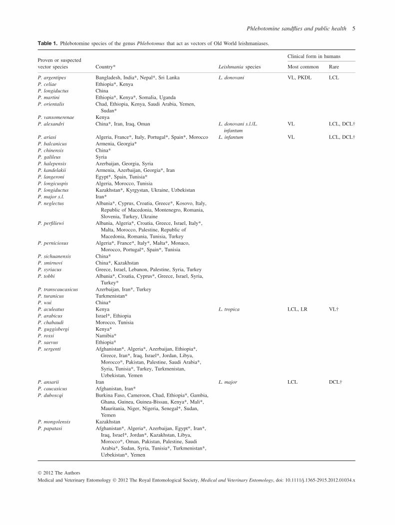
Table 1. Phlebotomine species of the genus Phlebotomus that act as vectors of Old World leishmaniases.
Clinical form in humansProven or suspectedvector species Country* Leishmania species Most common Rare
P. argentipes Bangladesh, India*, Nepal*, Sri Lanka L. donovani VL, PKDL LCLP. celiae Ethiopia*, KenyaP. longiductus ChinaP. martini Ethiopia*, Kenya*, Somalia, UgandaP. orientalis Chad, Ethiopia, Kenya, Saudi Arabia, Yemen,
Sudan*P. vansomerenae KenyaP. alexandri China*, Iran, Iraq, Oman L. donovani s.l./L.
infantumVL LCL, DCL†
P. ariasi Algeria, France*, Italy, Portugal*, Spain*, Morocco L. infantum VL LCL, DCL†P. balcanicus Armenia, Georgia*P. chinensis China*P. galileus SyriaP. halepensis Azerbaijan, Georgia, SyriaP. kandelakii Armenia, Azerbaijan, Georgia*, IranP. langeroni Egypt*, Spain, Tunisia*P. longicuspis Algeria, Morocco, TunisiaP. longiductus Kazakhstan*, Kyrgystan, Ukraine, UzbekistanP. major s.l. Iran*P. neglectus Albania*, Cyprus, Croatia, Greece*, Kosovo, Italy,
Republic of Macedonia, Montenegro, Romania,Slovenia, Turkey, Ukraine
P. perfiliewi Albania, Algeria*, Croatia, Greece, Israel, Italy*,Malta, Morocco, Palestine, Republic ofMacedonia, Romania, Tunisia, Turkey
P. perniciosus Algeria*, France*, Italy*, Malta*, Monaco,Morocco, Portugal*, Spain*, Tunisia
P. sichuanensis China*P. smirnovi China*, KazakhstanP. syriacus Greece, Israel, Lebanon, Palestine, Syria, TurkeyP. tobbi Albania*, Croatia, Cyprus*, Greece, Israel, Syria,
Turkey*P. transcaucasicus Azerbaijan, Iran*, TurkeyP. turanicus Turkmenistan*P. wui China*P. aculeatus Kenya L. tropica LCL, LR VL†P. arabicus Israel*, EthiopiaP. chabaudi Morocco, TunisiaP. guggisbergi Kenya*P. rossi Namibia*P. saevus Ethiopia*P. sergenti Afghanistan*, Algeria*, Azerbaijan, Ethiopia*,
Greece, Iran*, Iraq, Israel*, Jordan, Libya,Morocco*, Pakistan, Palestine, Saudi Arabia*,Syria, Tunisia*, Turkey, Turkmenistan,Uzbekistan, Yemen
P. ansarii Iran L. major LCL DCL†P. caucasicus Afghanistan, Iran*P. duboscqi Burkina Faso, Cameroon, Chad, Ethiopia*, Gambia,
Ghana, Guinea, Guinea-Bissau, Kenya*, Mali*,Mauritania, Niger, Nigeria, Senegal*, Sudan,Yemen
P. mongolensis KazakhstanP. papatasi Afghanistan*, Algeria*, Azerbaijan, Egypt*, Iran*,
Iraq, Israel*, Jordan*, Kazakhstan, Libya,Morocco*, Oman, Pakistan, Palestine, SaudiArabia*, Sudan, Syria, Tunisia*, Turkmenistan*,Uzbekistan*, Yemen
© 2012 The Authors
Medical and Veterinary Entomology © 2012 The Royal Entomological Society, Medical and Veterinary Entomology, doi: 10.1111/j.1365-2915.2012.01034.x
6 M. Maroli et al.
Table 1. Continued
Clinical form in humansProven or suspectedvector species Country* Leishmania species Most common Rare
P. salehi India*, Iran*, PakistanP. bergeroti Burkina Faso, Chad, Egypt, Iran, Mauritania,
Oman, YemenP. longipes Ethiopia* L. aethiopica LCL DCLP. pedifer Kenya*, Ethiopia*P. sergenti Ethiopia*
∗Countries in which the sandfly species is a proven vector (for criteria, see text). Elsewhere, a sandfly species is suspected to be a vector on thebasis of epidemiological evidence or because it is a proven vector elsewhere.†In cases of severe immunosuppression (e.g. HIV co-infection).DCL, disseminated or diffuse cutaneous leishmaniasis (the two forms are treated together in this review); LCL, localized cutaneous leishmaniasis;LR, leishmaniasis recidivans; PKDL, post-kala-azar dermal leishmaniasis; VL, visceral leishmaniasis.
in La Banda, Argentina (Salomon et al., 2010), has also beenindicated as a possible vector in Brazil (Pernabuco state)because L. infantum DNA has been detected in wild-caughtspecimens. This finding suggests that this species may beresponsible for the transmission of the disease in areas fromwhich the usual VL vector, Lu. longipalpis, is absent (deCarvalho et al., 2010). As far as the vectors of CL, weadd to previous lists the following information: (a) Lutzomyianuneztovari anglesi is a vector of L. amazonensis in Bolivia, asconfirmed by anthropophily, biochemical identification of wildisolates and successful experimental infection (Martinez et al.,1999); (b) Lutzomyia ayacuchensis was recently found in Perunaturally infected by promastigotes typed as L. guyanensis(Cordova et al., 2011); (c) Lutzomyia fischeri is included asa proven vector because of repeated observations in Brazilof natural promastigote infections identified as L. braziliensis,associated with anthropophily and a spatial distribution relatedto human CL (Margonari et al., 2010; Rocha et al., 2010;Pita-Pereira et al., 2011); (d) in Venezuela, Lu. migonei hasrecently been reported as a putative vector of L. guyanensisand L. mexicana (Feliciangeli et al., 2011), and past reportshave incriminated Lutzomyia gomezi as a proven vector ofL. braziliensis and Lutzomyia ovallesi as responsible for thetransmission of not only L. braziliensis, but also L. mexicana(Feliciangeli et al., 1994; Jorquera et al., 2005); (e) in theYucatan peninsula of Mexico, Lutzomyia cruciata, Lutzomyiapanamensis, Lutzomyia shannoni and Lutzomyia ylephiletorare considered to represent possible vectors of L. mexicanabecause recent investigations using molecular techniques havedetected natural infections (Pech-May et al., 2010).
Role of phlebotomine sandflies in the pathogenesisof leishmaniasis
As well as acting as Leishmania spp. vectors in the par-asite lifecycle, phlebotomine sandflies may also be directlyinvolved in the pathogenesis of leishmaniasis. During thefeeding process on the vertebrate host’s skin, the sali-vary gland content is injected into the haemorrhagic poolupon which a female sandfly feeds. Sandfly saliva containsa variety of pharmacologic agents, such as anticoagulants,
vasodilators, anti-platelet agents and immunomodulatory andanti-inflammatory molecules (Andrade et al., 2007). Probablyas a result of co-evolutionary mechanisms, the haemostaticand immune modifications of the feeding site play some partin Leishmania spp. transmission as they affect parasite estab-lishment. Since the early description of L. major infectionenhancement by the vasodilator and immunomodulator maxi-dilan, a 6.5-kDa peptide from Lu. longipalpis saliva (an unnat-ural parasite–vector combination) (Titus & Ribeiro, 1988),several studies have been performed mainly in Lu. longipalpisand Phlebotomus papatasi and, more recently, in Lutzomyiaintermedia. The immunomodulatory effects, including theenhancement of parasite burden or protection from exacerbateddisease, have been reviewed by Rohousova & Volf (2006). Innaıve hosts (human ex vivo cells and in vivo murine models),Lu. longipalpis saliva or maxidilan alone promotes downregu-lation of tumour necrosis factor-α (TNF-α) secretion, therebyaffecting blood coagulation, major histocompatibility complexmolecule expression, cytotoxicity against infected cells, andneutrophil migration, while upregulating secretion of inter-leukin 6 (IL-6), a type 2 T helper cell (Th2) cytokine promot-ing humoral responses. It also suppresses T cell proliferationin vitro and delayed-type hypersensitivity (DTH) in vivo, andexerts anti-complement activity. Phlebotomus papatasi salivahas effects similar to those of Lu. longipalpis saliva in termsof decreasing TNF-α synthesis, increasing IL-6 productionand inhibiting lymphoproliferation, but specifically downreg-ulates nitric oxide synthesis by macrophages and upregulatesthe Th2 cytokine IL-4, a strong suppressor of protective Th1responses. Such sandfly species-specific responses have beenrecently highlighted by the demonstration that Lu. intermediaenhances rather than decreases TNF-α production (Menezeset al., 2008).
When human and canine hosts are repeatedly exposedto bites of uninfected phlebotomine sandflies, they developspecific immunoglobulin G (IgG) antibodies against salivaryproteins: this occurs less frequently and mostly within a rangeof 43–45 kDa proteins for Lu. longipalpis (Gomes et al., 2002;Hostomska et al., 2008) and more consistently to a 35-kDaprotein for P. papatasi saliva (Rohousova et al., 2005). Caninehosts exposed to Phlebotomus perniciosus bites develop IgG1
© 2012 The Authors
Medical and Veterinary Entomology © 2012 The Royal Entomological Society, Medical and Veterinary Entomology, doi: 10.1111/j.1365-2915.2012.01034.x

Phlebotomine sandflies and public health 7
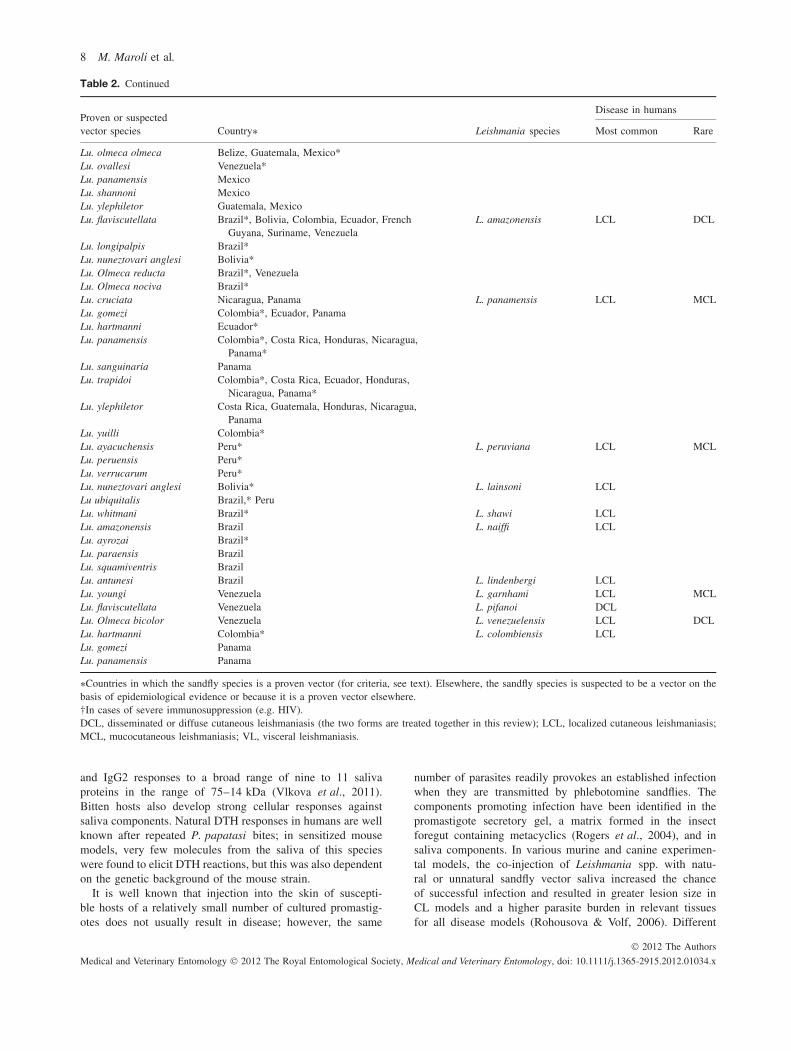
Table 2. Phlebotomine species of the genus Lutzomyia that act as vectors of New World leishmaniases.
Disease in humansProven or suspectedvector species Country∗ Leishmania species Most common Rare
Lu. almerioi Brazil* L. infantum VL LCL, DCL†Lu. cruzi Brazil*Lu. evansi Colombia*, Costa Rica, Mexico, Nicaragua,
Venezuela*Lu. forattinii BrazilLu. longipalpis Argentina*, Bolivia*, Brazil*, Colombia*, Costa
Rica, El Salvador, Guatemala, Honduras*,Mexico, Nicaragua, Paraguay, Venezuela*
Lu. migonei Argentina, BrazilLu. pseudolongipalpis VenezuelaLu. sallesi BrazilLu. ayrozai Bolivia L. braziliensis LCL MCL, DCL†Lu. c. carrerai Bolivia*Lu. columbiana ColombiaLu. complexa Brazil*Lu. cruciata MexicoLu. edwardsi BrazilLu. fischeri Brazil*Lu. gomezi Venezuela*Lu. intermedia Brazil, French Guyana, ParaguayLu. llanosmartinsi Bolivia*Lu. migonei Argentina, Brazil*, ParaguayLu. neivai Argentina, Brazil*Lu. nuneztovari anglesi Bolivia*Lu. ovallesi Belize, Colombia, Guatemala*, Honduras, Mexico,
Nicaragua, Panama, Venezuela*Lu. panamensis Guatemala*, Honduras, Nicaragua, Panama,
Venezuela*Lu. paraensis BrazilLu. pescei PeruLu. pessoai BrazilLu. pia ColombiaLu. shawi Bolivia*Lu. spinicrassa Colombia*, Venezuela*Lu. tejadai PeruLu. townsendi ColombiaLu. trinidadensis VenezuelaLu. ylephiletor Guatemala*, HondurasLu. youngi Costa Rica, Venezuela, ColombiaLu. yucumensis Bolivia*Lu. wellcomei Brazil*, French GuyanaLu. whitmani Argentina, Brazil*, ParaguayLu. anduzei Brazil*, French Guyana, Guyana, Suriname,
VenezuelaL. guyanensis LCL MCL
Lu. ayacuchensis Peru*Lu. longiflocosa Colombia*Lu. migonei VenezuelaLu. shawi Bolivia*Lu. umbratilis Brazil*, Colombia*, French Guyana, Guyana,
Suriname, VenezuelaLu. whitmani Brazil*Lu. anthophora U.S.A. L. mexicana LCL MCL, DCL†Lu. ayacuchensis Ecuador*Lu. columbiana ColombiaLu. cruciata Mexico, NicaraguaLu. diabolica U.S.A.Lu. migonei Venezuela
© 2012 The Authors
Medical and Veterinary Entomology © 2012 The Royal Entomological Society, Medical and Veterinary Entomology, doi: 10.1111/j.1365-2915.2012.01034.x
8 M. Maroli et al.
Table 2. Continued
Disease in humansProven or suspectedvector species Country∗ Leishmania species Most common Rare
Lu. olmeca olmeca Belize, Guatemala, Mexico*Lu. ovallesi Venezuela*Lu. panamensis MexicoLu. shannoni MexicoLu. ylephiletor Guatemala, MexicoLu. flaviscutellata Brazil*, Bolivia, Colombia, Ecuador, French
Guyana, Suriname, VenezuelaL. amazonensis LCL DCL
Lu. longipalpis Brazil*Lu. nuneztovari anglesi Bolivia*Lu. Olmeca reducta Brazil*, VenezuelaLu. Olmeca nociva Brazil*Lu. cruciata Nicaragua, Panama L. panamensis LCL MCLLu. gomezi Colombia*, Ecuador, PanamaLu. hartmanni Ecuador*Lu. panamensis Colombia*, Costa Rica, Honduras, Nicaragua,
Panama*Lu. sanguinaria PanamaLu. trapidoi Colombia*, Costa Rica, Ecuador, Honduras,
Nicaragua, Panama*Lu. ylephiletor Costa Rica, Guatemala, Honduras, Nicaragua,
PanamaLu. yuilli Colombia*Lu. ayacuchensis Peru* L. peruviana LCL MCLLu. peruensis Peru*Lu. verrucarum Peru*Lu. nuneztovari anglesi Bolivia* L. lainsoni LCLLu ubiquitalis Brazil,* PeruLu. whitmani Brazil* L. shawi LCLLu. amazonensis Brazil L. naiffi LCLLu. ayrozai Brazil*Lu. paraensis BrazilLu. squamiventris BrazilLu. antunesi Brazil L. lindenbergi LCLLu. youngi Venezuela L. garnhami LCL MCLLu. flaviscutellata Venezuela L. pifanoi DCLLu. Olmeca bicolor Venezuela L. venezuelensis LCL DCLLu. hartmanni Colombia* L. colombiensis LCLLu. gomezi PanamaLu. panamensis Panama
∗Countries in which the sandfly species is a proven vector (for criteria, see text). Elsewhere, the sandfly species is suspected to be a vector on thebasis of epidemiological evidence or because it is a proven vector elsewhere.†In cases of severe immunosuppression (e.g. HIV).DCL, disseminated or diffuse cutaneous leishmaniasis (the two forms are treated together in this review); LCL, localized cutaneous leishmaniasis;MCL, mucocutaneous leishmaniasis; VL, visceral leishmaniasis.
and IgG2 responses to a broad range of nine to 11 salivaproteins in the range of 75–14 kDa (Vlkova et al., 2011).Bitten hosts also develop strong cellular responses againstsaliva components. Natural DTH responses in humans are wellknown after repeated P. papatasi bites; in sensitized mousemodels, very few molecules from the saliva of this specieswere found to elicit DTH reactions, but this was also dependenton the genetic background of the mouse strain.
It is well known that injection into the skin of suscepti-ble hosts of a relatively small number of cultured promastig-otes does not usually result in disease; however, the same
number of parasites readily provokes an established infectionwhen they are transmitted by phlebotomine sandflies. Thecomponents promoting infection have been identified in thepromastigote secretory gel, a matrix formed in the insectforegut containing metacyclics (Rogers et al., 2004), and insaliva components. In various murine and canine experimen-tal models, the co-injection of Leishmania spp. with natu-ral or unnatural sandfly vector saliva increased the chanceof successful infection and resulted in greater lesion size inCL models and a higher parasite burden in relevant tissuesfor all disease models (Rohousova & Volf, 2006). Different
© 2012 The Authors
Medical and Veterinary Entomology © 2012 The Royal Entomological Society, Medical and Veterinary Entomology, doi: 10.1111/j.1365-2915.2012.01034.x
Phlebotomine sandflies and public health 9
combinations of saliva origin and parasite species havebeen tested, such as Lu. longipalpis/L. major (unnatural), andLu. longipalpis/L. infantum, P. papatasi /L. major and P. per-niciosus/L. infantum (natural). Among the mechanisms pro-posed to explain these effects, the functional alteration ofantigen-presenting cells and the IL-4-driven development ofTh2 immune responses are the most plausible.
Apparently a paradox, the immunity elicited by sandflysaliva in some experimental models appears to be protectiveagainst Leishmania spp. infection (Kamhawi, 2000). Salivarygland lysates/sonicates, purified salivary proteins or bitesby uninfected P. papatasi or Lu. longipalpis reduced theseverity of CL in treated mice challenged with L. major orL. amazonensis. Analogously, treatment of hamsters with DNAplasmids coding for Lu. longipalpis salivary proteins protectedthe animals against fatal VL following L. infantum challenge(Gomes et al., 2008). In combination, these findings suggestthat vaccination against vector antigens may represent a novelmethod for controlling leishmaniasis. This protective effect,however, was not shown in a Lu. intermedia/L. braziliensischallenge in a murine model reported by Andrade et al.(2007). Given both the large diversity of vector–parasitenatural associations and the fact that phlebotomine sandflyspecies differ in salivary antigens [and that differences maybe present within a species, as recently shown by Rohousovaet al. (2012) in three Phlebotomus species], any protectiveeffects may also be extremely specific, which lowers the likelyfeasibility of worldwide vector-based vaccines. Furthermore,recently Rohousova et al. (2011) used an experimental murinemodel involving Phlebotomus duboscqi exposure with anL. major challenge to show that short-term exposure to bitesshortly before challenge is indeed protective, whereas bothlongterm exposures or a long delay prior to challenge aftershort-term exposure are not. This explains the persistence ofsevere Leishmania spp. infections in endemic areas in whichindividuals are repeatedly exposed to bites and/or sandflyactivity is seasonal. Nevertheless, the protective effects ofsaliva may play an important role in the dynamics of clinico-epidemiological patterns (e.g. in the patterns of asymptomaticvs. symptomatic infections) found in endemic settings.
Current spreading of leishmaniases
Overview
About 20 named Leishmania species and subspecies arepathogenic for humans, in whom leishmaniases have diverseclinical manifestations. Visceral leishmaniasis, caused byL. donovani in the Old World and L. infantum in both theOld and New Worlds, is the most severe form, with an esti-mated yearly incidence of 500 000 cases (Desjeux, 1996).Several species of Leishmania cause tegumentary diseasesin which the clinical spectrum varies from localized, dis-seminated or diffuse CL (treated together in this review),to mucocutaneous leishmaniasis and pathological sequelaefollowing L. donovani VL (post-Kala-azar dermal leishmania-sis) or L. tropica CL (leishmaniasis recidivans). Disfigurement,social and psychological stigma are severe consequences of
the diseases, for which the estimated yearly incidence is1–1.5 million cases (Desjeux, 1996).
Each parasite species circulates in natural foci of infec-tion where susceptible phlebotomine species and mammalscoexist. Nosogeographical entities of leishmaniasis can becategorized in two main epidemiological groups of, respec-tively, zoonotic (representing the large majority of such enti-ties) and anthroponotic leishmaniases. Traditionally, Old andNew World nosogeographical entities have been consideredseparately because they involve different parasites (with theexception of L. infantum), vectors and ecosystems. However,common risk factors for human leishmaniases are found world-wide and include poverty and poor housing, conflict, humanmigration, deforestation and urbanization.
Old World leishmaniases
Autochthonous cases of human leishmaniases are currentlyreported from 80 countries, by contrast with the 66 countriesrecorded by Desjeux (2001). However, despite the endemicnature of disease in these countries, the Leishmania speciesinvolved were not determined in nine countries and thephlebotomine vector species, either proven or suspected, werenot identified in 16.
Zoonotic leishmaniases. Three well-recognized Old WorldLeishmania species have a zoonotic nature. Zoonotic VLcaused by L. infantum is widespread in countries of theMediterranean basin and central Asia. Cutaneous leishmaniasisinfections by this parasite are also found within the sameendemic range, where they are usually sporadic, although insome foci they may show hyperendemic patterns (Corradetti,1952; Svobodova et al., 2009). Several vector species areinvolved, most of which belong to the subgenus Phlebotomus(Larroussius). Dogs are the main domestic reservoirs, andfoxes, jackals and wolves represent sylvatic rservoirs (Fig. 4).The classical zoonotic CL is caused by L. major, a parasitewidely distributed in arid and savannah areas in which severalrodent species act as reservoir hosts. Proven vectors belongto the subgenus Phlebotomus (Phlebotomus) and P. papatasiis the principal vector over a wide geographical range thatextends from northern Africa to India (Fig. 5). A third zoonoticagent of CL, L. aethiopica, is limited to the highlands ofEthiopia and Kenya. It is a classical parasite of the Hyracoidea(e.g. Procavia capensis) transmitted by the Larroussius speciesPhlebotomus longipes and Phlebotomus pedifer.
Anthroponotic leishmaniases. Two species, L. donovani andL. tropica, have exclusively or predominantly anthroponotictransmission patterns that result in several thousands ofhuman cases. However, the presence of mammal reservoirshas been indicated in several endemic settings, suggestingan ancient type of parasite transmission, such as in easternSudan for L. donovani (Dereure et al., 2003) and in northwestAfrica (Maghreb) (Dereure et al., 1991; Boubidi et al., 2011;
© 2012 The Authors
Medical and Veterinary Entomology © 2012 The Royal Entomological Society, Medical and Veterinary Entomology, doi: 10.1111/j.1365-2915.2012.01034.x

10 M. Maroli et al.
(A) (B)
Fig. 4. Zoonotic visceral leishmaniasis, Leishmania infantum. (A) Periurban biotope where the Old World sandfly Phlebotomus (Larroussius)neglectus acts as the main vector (Albania). (B) An infected dog with severe clinical signs of disease.
(A) (B)
Fig. 5. Old World zoonotic cutaneous leishmaniasis, Leishmania major (Libya). (A) Rodent reservoir burrows (Psammomys obesus), breeding sitefor the vector Phlebotomus (Phlebotomus) papatasi. (B) Human lesion.
Jaouadi et al., 2011), northern Israel (Jacobson et al., 2003;Svobodova et al., 2006) and Iran (Mohebali et al., 2005) forL. tropica s.l. In the Asian continent, anthroponotic VL causedby L. donovani is restricted to northeast India, Bangladeshand Nepal, where P. argentipes is the sole vector. In EastAfrica (Kenya, Ethiopia, Somalia, Sudan and Uganda) andthe Arabian peninsula, the distribution of L. donovani isassociated with those of P. orientalis and/or Phlebotomusmartini. Anthroponotic CL caused by L. tropica is highlyprevalent in semi-arid subtropical regions extending fromthe southeast of Turkey to the northwest of India. In well-established foci, CL is transmitted person-to-person throughP. (Paraphlebotomus) sergenti in urban settings. Large-scaleurban migrations influence L. tropica transmission patterns andinfections may occur in outbreaks that last for some years(Fig. 6). In addition, small and discontinuous foci are alsofound in northern Africa, Israel, Greece and Saudi Arabia, aswell as in sub-Saharan Africa in Kenya and Ethiopia (Hailu
et al., 2006), where L. tropica can be transmitted by otherphlebotomine sandfly species (Table 1).
Causes of the spreading of Old World leishmaniases arelargely diverse, although they are mostly associated withhuman social, behavioural and individual factors, such asmassive migrations, conflict, man-made environmental changesand immunosuppressive conditions. As such, the contributionof phlebotomine sandfly dynamics to the spread of diseasemay vary greatly according to the various epidemiologicalsettings in which it occurs. For instance, epidemics of humanimmunodeficiency virus (HIV) and the increasing use ofimmunosuppressive therapies have substantially contributed toan increase in numbers of VL cases from southern Europeto India (Alvar et al., 2008), whereas changes in sandflypopulations can be assumed to have no specific role inthese events. Civilians and soldiers have been substantiallyaffected by leishmaniases in recent conflicts in Sudan,Iraq, Afghanistan and, recently, Libya (Amro et al., 2012).Furthermore, urbanization and the domestication of zoonotic
© 2012 The Authors
Medical and Veterinary Entomology © 2012 The Royal Entomological Society, Medical and Veterinary Entomology, doi: 10.1111/j.1365-2915.2012.01034.x
Phlebotomine sandflies and public health 11
(A) (B)
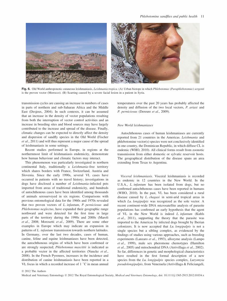
Fig. 6. Old World anthroponotic cutaneous leishmaniasis, Leishmania tropica. (A) Urban biotope in which Phlebotomus (Paraphlebotomus) sergentiis the proven vector (Morocco). (B) Scarring caused by a severe facial lesion in a patient in Syria.
transmission cycles are causing an increase in numbers of casesin parts of northern and sub-Saharan Africa and the MiddleEast (Desjeux, 2004). In such contexts, it can be assumedthat an increase in the density of vector populations resultingfrom both the interruption of vector control activities and anincrease in breeding sites and blood sources may have largelycontributed to the increase and spread of the disease. Finally,climatic changes can be expected to directly affect the densityand dispersion of sandfly species in the Old World (Fischeret al., 2011) and will thus represent a major cause of the spreadof leishmaniasis in some settings.
Recent studies performed in Europe, in regions at thenorthernmost limit of leishmaniasis endemicity, demonstratehow human behaviour and climatic factors may interact.
This phenomenon was particularly investigated in northerncontinental Italy, traditionally a Leishmania-free territorywhich shares borders with France, Switzerland, Austria andSlovenia. Since the early 1990s, several VL cases haveoccurred in patients with no travel history; investigations indogs have disclosed a number of Leishmania-infected petsimported from areas of traditional endemicity, and hundredsof autochthonous cases have been identified among thousandsof animals serosurveyed in six regions. Comparisons withprevious entomological data for the 1960s and 1970s revealedthat two proven vectors of L. infantum, P. perniciosus andPhlebotomus neglectus, have expanded their geographic rangenorthward and were detected for the first time in largeparts of the territory during the 1990s and 2000s (Maroliet al., 2008; Morosetti et al., 2009). There are some otherexamples in Europe which may indicate an expansion inpatterns of L. infantum transmission towards northern latitudes.In Germany, over the last two decades, cases of human,canine, feline and equine leishmaniases have been detected,the autochthonous origins of which have been confirmed orare strongly suspected; Phlebotomus mascittii is indicated asa probable vector in the affected territories (Naucke et al.,2008). In the French Pyrenees, increases in the incidence anddistribution of canine leishmaniasis have been reported in aVL focus in which a recorded increase of 1 ◦C in mean annual
temperatures over the past 20 years has probably affected thedensity and diffusion of the two local vectors, P. ariasi andP. perniciosus (Dereure et al., 2009).
New World leishmaniases
Autochthonous cases of human leishmaniases are currentlyreported from 21 countries in the Americas. Leishmania andphlebotomine vector(s) species were not conclusively identifiedin one country, the Dominican Republic, in which diffuse CL isendemic (WHO, 2010). All clinical forms result from zoonotictransmission from either domestic or sylvatic reservoir hosts.The geographical distribution of the disease spans an areaextending from Texas to Argentina.
Visceral leishmaniasis. Visceral leishmaniasis is recordedas endemic in 12 countries in the New World. In theU.S.A., L. infantum has been isolated from dogs, but noconfirmed autochthonous cases have been reported in humans(WHO, 2010). In the past, VL has been considered a ruraldisease caused by L. chagasi in semi-arid tropical areas inwhich Lu. longipalpis was recognized as the sole vector. Arecent continent-wide DNA microsatellite analysis of parasitepopulations has confirmed an early hypothesis that the agentof VL in the New World is indeed L. infantum (Kuhlset al., 2011), supporting the theory that the parasite wasimported to the Americas by infected dogs brought by Iberiancolonizers. It is now accepted that Lu. longipalpis is not asingle species but a sibling complex, as evidenced by thefindings of studies using various approaches, such as breedingexperiments (Lanzaro et al., 1993), allozyme analysis (Lampoet al., 1999), male sex pheromone chemotypes (Hamiltonet al., 2005) and mitochondrial DNA (Arrivillaga et al., 2002).So far, differences in genetic and morphological characteristicshave resulted in the first formal description of a newspecies from the Lu. longipalpis species complex, Lutzomyiapseudolongipalpis, from Venezuela (Arrivillaga & Feliciangeli,
© 2012 The Authors
Medical and Veterinary Entomology © 2012 The Royal Entomological Society, Medical and Veterinary Entomology, doi: 10.1111/j.1365-2915.2012.01034.x
12 M. Maroli et al.
2001). A sibling clade of Lu. longipalpis that is geneticallywell separated has been found to predominate in areas that arehyperendemic for VL in the north of northeastern Brazil (Wattset al., 2005).
Most of the VL cases in the New World occur in Brazil.During the 1990s and 2000s, an epidemic in which 3000–4000cases were reported annually was attributed to urbanizationprocesses in the states of Piauí, Maranhao, Bahia, Ceara,Para, Rio Grande do Norte and Roraima. The urbanizationresulted in the disorderly proliferation of crowded slumswith poor sanitary conditions; the presence in such areas ofLu. longipalpis led to the onset and establishment of domesticand peridomestic cycles of the disease (Costa, 2008; Maia-Elkhoury et al., 2008; Rangel & Vilela, 2008). Although lessmassively, urban VL has also occurred in other countries, suchas Venezuela (Aguilar et al., 1998; Zerpa et al., 2002) andParaguay (Canese, 2000).
The recent geographical expansion of VL in South Americais indicated by the first appearance of a human VL focus inArgentina in 2006, at the northeastern border with Paraguayand Brazil; the disease was associated to L. infantum-infecteddogs and Lu. longipalpis sandflies (Acardi et al., 2010).The spatial distribution of Lu. longipalpis appears to beheterogeneous in Argentina, in which vectors are concentratedin limited patches of high abundance characterized by highertree coverage and poor urban services (Fernandez et al., 2010).In a focus of low VL endemicity from which Lu. longipalpiswas apparently absent, Lu. migonei has been suspected asthe vector (Salomon et al., 2010). This finding, along withseveral reports from other countries that incriminated VLvectors other than Lu. longipalpis (Table 2), call attentionto a possible widespread adaptation of L. infantum to otherepidemiologically relevant Lutzomyia species.
Cutaneous leishmaniasis. Prior to the 1960s, CL wasprimarily confined to forested areas. That the condition iswidely known as ‘ulcera de los chicleros’ (ulcer developedin gatherers of ‘chicle’, a gummy latex from the forest treeManilkara zapota) in Mexico and is designated ‘guerrilla’ssore’ in Venezuela and Colombia reflects the historically closecontact of humans with a sylvatic environment that maintainsseveral species of phlebotomine vectors among wild species ofmammalian reservoirs. Hence, hunting, lumbering and miningactivities have been associated with the disease. Since the1960s, transmission has increasingly spread to peridomesticareas. Massive migration from the high plateau to low tropicalareas in the Andean region, intensive deforestation and theestablishment of new settlements have greatly contributed tothe increase in numbers of cases (Desjeux, 2001; Aagaard-Hansen et al., 2010). The impact of environmental changeson the behaviour of vectors has been exhaustively reviewedby Rangel & Lainson (2009) in Brazil, and by Gonzalezet al. (2011) in Mexico. Deforestation and the replacementof primary forest by monocultures (e.g. coffee plantations)has been found to be crucial to the establishment of someLutzomyia vectors in Venezuela and Colombia (Scorza, 1985;Alexander et al., 1995). Although deforestation was predictedto reduce human contact with wild reservoirs and sandflies,
and was hoped to lead to the local eradication of importantLeishmania species (e.g. L. braziliensis in parts of Braziland L. panamensis in Panama), human encroachment of wildhabitats has resulted in the intrusion of Lutzomyia speciesinto the domestic environment, thus radically changing theepidemiological profile. Examples of Lutzomyia species thathave participated in this invasion include Lu. nuneztovarianglesi, in Bolivia, Lutzomyia verrucarum and Lutzomyiaperuensis in Peru, and Lu. ovallesi and Lu. gomezi inVenezuela (Gomez et al., 1998; Campbell-Lendrum et al.,2001). In Brazil, it has long been observed that environmentalmodifications in Sao Paulo state occurred early in the 20thcentury allowed Lu. intermedia to play a predominant role inthe transmission of L. braziliensis (Tolezano, 1994). Similarly,Lutzomyia whitmani spread in most of the municipalities ofTocantins state in which leishmaniasis was endemic, in areashighly degraded by deforestation followed by migration andunplanned settlements (Vilela et al., 2011). In Argentina, theabundance of Lu. whitmani in the Iguazu Falls area was foundto be associated with economic and leisure activities in primaryand secondary forest that led to marked environmental changes(Salomon et al., 2009), and even moderate environmentalmodifications were reported to have an impact on Lutzomyianeivai distribution in Salta, in which CL is hyperendemic(Quintana et al., 2010).
Urbanization has greatly contributed to the emergenceand increase of CL in the New World. The disease hasbeen spreading in cities in Brazil, such as Belo Horizonte(Carvalho et al., 2010), Mexico (Sanchez-García et al., 2010),Venezuela (Bonfante-Garrido et al., 1984; Scorza & Rojas,1990), Colombia (Sandoval et al., 1998; Bejarano et al., 2002;Cortes & Fernandez, 2008), Bolivia (García et al., 2009) andArgentina (Salomon et al., 2008) (Fig. 7).
Viral diseases
Overview
Phlebotomine sandflies are involved in the transmission ofseveral viral agents, among which the most important aregrouped into the Phlebovirus genus (family Bunyaviridae),which includes the sandfly fever Sicilian and Toscana viruses,and the Vesiculovirus genus (family Rhabdoviridae), whichincludes vesicular stomatitis, and the Chandipura and Isfahanviruses.
The risk for infection with sandfly-transmitted phleboviruseshas been shown to pertain to very large areas of the Old World(southern Europe, Africa, the Middle East, central and westernAsia) in association with the presence of sandfly vectors (Teshet al., 1976). Recent investigations have indicated that virusdiversity in the Mediterranean basin is higher than initiallysuspected, and that populations living south and east of theMediterranean Sea have a high risk for infection during theirlifetime (Sanbonmatsu-Gamez et al., 2005; Papa et al., 2006;Konstantinou et al., 2007; Carhan et al., 2010; Bahri et al.,2011; Ergunay et al., 2011, 2012; Kocak Tufan et al., 2011).
The International Committee for Taxonomy of Virusescurrently recognizes several phleboviruses associated with
© 2012 The Authors
Medical and Veterinary Entomology © 2012 The Royal Entomological Society, Medical and Veterinary Entomology, doi: 10.1111/j.1365-2915.2012.01034.x

Phlebotomine sandflies and public health 13
(A) (B) (C)
Fig. 7. New World cutaneous leishmaniasis, Leishmania braziliensis. (A) A typical new urban settlement in Latin America (Venezuela). (B, C)Cutaneous leishmaniasis in human patients.
sandflies in the Old World (Plyusnin et al., 2011). Theseinclude two virus species: (a) sandfly fever Naples virus,which includes the Naples virus, Tehran virus, Karimabadvirus and Toscana virus, (b) and Salehabad virus, whichincludes the Salehabad and Arbia viruses. A further twovirus isolates (sandfly fever Sicilian virus and Corfou virus)are listed, but not included among the nine recognizedspecies of the Phlebovirus genus. In addition, recent fieldand clinical studies have provided increasing evidence thatthe number of known viruses in the genus Phlebovirusmay be substantially underestimated (Charrel et al., 2009;Collao et al., 2010; Moureau et al., 2010; Zhioua et al., 2010;Anagnostou et al., 2011).
Among the viral agents belonging to the genus Vesiculovirus,at least 28 infect invertebrates and vertebrates (Wunner et al.,1995). Those infecting humans and domestic animals, forwhich phlebotomine sandflies can be regarded as biologicalvectors, include the New Jersey, Indiana, Alagoas, Chalchaqui,Chandipura, Cocal, Isfahan and Piry viruses (Table 3). Othervesiculoviruses have not been adequately tested for infectivityor pathogenicity in domestic animals and humans (Letchworthet al., 1999). Vesicular stomatitis viruses causing stomatitisin humans and domestic livestock are largely endemic inthe New World, whereas Chandipura encephalitis virus andIsfahan virus are endemic in the Old World in some partsof India (Basak et al., 2007), Iran (Tesh et al., 1977b), andTurkmenistan and other central Asian republics (Gaidamovichet al., 1978). Although serological evidence for human infec-tions has been reported for Isfahan virus (associated withP. papatasi transmission), it has not been definitively linkedto human illness and has been found to be non-pathogenic inhorses, cattle and other ruminants (Marriott, 2005).
Sandfly fever
Sandfly fever, also known as Phlebotomus fever, pappatacifever or three-day fever, has been an important cause offebrile disease during military operations since at least theNapoleonic Wars (Oldfield et al., 1991). It has also historically
caused significant morbidity among non-native populations inMediterranean regions (Pick, 1886). An Austrian commissionin 1909 reported that the illness was caused by a filterable agentfound in the blood of infected soldiers and that the vector wasthe sandfly P. papatasi (Doerr et al., 1909). During World WarII (WWII), German troops based in the Mediterranean areasuffered from sandfly fever (Hallmann, 1943). Allied forcesstationed in the Mediterranean and Middle East reported tensof thousands of cases and attack rates of 3–10% (locally upto 80%) (Sabin, 1951; Hertig & Sabin, 1964). First reportsin Tunisian and Algerian regions, where the presence ofP. papatasi was entomologically established, date from April1943 (Sabin et al., 1944). In August 1943, after the Alliedlanding in southern Italy, sandfly fever accounted for at least25% of cases of fever of unknown origin. Outbreaks ofsandfly fever occurred repeatedly in the former U.S.S.R. in theperiod from 1945 to 1950, predominantly in Crimea, Romania,Moldavia and the central Asian republics. Epidemics related tothe activity of P. papatasi were reported in northern Africa,southern Europe, the Middle East and central Asia (Sabin,1951; Hertig & Sabin, 1964).
The observation of two or more attacks in the sameindividual resulted in the early suggestion that sandflyfever might be caused by distinct viruses (Livschitz, 1937).However, it was almost impossible to distinguish theseclinically. Sabin (1951) confirmed the existence of more thanone strain of sandfly fever virus. Serum samples collected fromsoldiers after the landing of the Allied troops in southern Italyduring WWII allowed the isolation of two different viruses,respectively named the Naples and Sicilian viruses. Volunteersinoculated with Naples virus developed the typical symptoms,but were not subsequently protected against infection with theSicilian strain (Sabin, 1955).
Naples virus. The Naples virus was first isolated from afebrile patient in Italy in 1944 (Sabin, 1955). Additionalrecoveries have been made in Egypt, India, Iran, Pakistan,Serbia and the former Soviet Union (Gaidamovich et al., 1974;Goverdhan et al., 1976). In 1976, a founding study extended
© 2012 The Authors
Medical and Veterinary Entomology © 2012 The Royal Entomological Society, Medical and Veterinary Entomology, doi: 10.1111/j.1365-2915.2012.01034.x
14 M. Maroli et al.
Table 3. Most common vesiculoviruses infecting domestic animals and humans (from Letchworth et al., 1999).
First isolation
Virus name Place and year Host References
Indiana Indiana, U.S.A., 1925 Bovine Cotton (1926)New Jersey New Jersey, U.S.A., 1926 Equine Cotton (1927)Cocal Trinidad, Brazil, 1964 Insect, rodents Jonkers et al. (1964)Alagoas Brazil, 1964 Equine, bovine, human Federer et al. (1967)Chandipura India, 1965 Human Bhatt & Rodrigues (1967)Piry Brazil, 1973 Opossum Theiler & Downs (1973)Isfahan Iran, 1975 Human Tesh et al. (1977b)Chalcaqui Argentina, 1982 Insects Calisher et al. (1987)
the known distribution of Naples virus to include Bangladesh,Ethiopia, Greece, Iraq, Morocco, Saudi Arabia, Sudan, theTerritory of the Afars and Issas (now Djibouti), Turkey andformer Yugoslavia (Tesh et al., 1976). Antibodies to the Naplesvirus have been found in residents of Turkmenia, Tajikistan,Uzbekistan, Azerbaijan and Moldavia (Gaidamovich et al.,1978). Seroepidemiological studies conducted in areas aroundthe Mediterranean indicate that Naples virus infections havedecreased during the last 30 years (Tesh & Papaevangelou,1977). Although the renewed interest in phlebotomine sandfly-transmitted phleboviruses has produced numerous studiesduring the last decade, Naples virus has not been isolatedor detected by polymerase chain reaction (PCR) since 1987(Feinsod et al., 1987). This may suggest that the Naples virusbecame progressively extinct.
Sicilian virus. The prototype strain of Sicilian virus wasisolated from humans in 1943 (Sabin, 1955). Other isolateswere subsequently obtained in Egypt, India, Iran, Pakistan andAfghanistan (R. Taylor & J. Casals, personal communication,2005; Goverdhan et al., 1976; Tesh et al., 1977a). Sicilian virusis also present in Bangladesh, Greece, Cyprus, Iraq, Morocco,Saudi Arabia, Somalia, Sudan, Tunisia, Turkey, the southernEuropean and central Asian republics of the former U.S.S.R.(Turkmenia, Tajikistan, Uzbekistan, Azerbaijan and Moldavia),former Yugoslavia, France and Portugal (C. Hannoun, per-sonal communication, 2005; Filipe, 1974; Tesh et al., 1976;Gaidamovich et al., 1978; Eitrem et al., 1991).
Epidemiological data are of specific interest in the diagnosisbecause of the seasonality of vector activity and its intrinsicepidemic nature. Cases of sandfly fever begin to appear inApril and gradually build to a peak in September. Thus, theepidemiological pattern of the disease mirrors the lifecycle ofP. papatasi. The viruses that cause sandfly fever (Naples andSicilian viruses) have a wide geographical distribution, whichparallels that of P. papatasi, which was dominant during thefirst half of the 20th century. DDT-based spraying campaigns(1940–1960) to control the insect vectors of typhus and toeradicate malaria and dengue fever (Dunlap, 1981), as well asthe easy access of farmers to agricultural insecticide (WHO,1979), reduced numbers of P. papatasi in regions in whichinsecticides were sprayed. Later reports of virus isolation andserologic studies indicate that phleboviruses are still present inthe Mediterranean coastal regions of Europe and North Africa,
the Nile valley, most of southwest Asia, areas adjacent tothe Black and Caspian Seas, and in central Asia includingBangladesh (Tesh et al., 1976).
The clinical pictures corresponding to infections with theNaples and Sicilian viruses are virtually identical. After anincubation period of 3–6 days, sandfly fever is characterizedby the sudden onset of fever, headache, retro-orbital pain, pho-tophobia, generalized aching, malaise and chills. The face canbe suffused, with injection of the conjunctivas and scleras, andphotophobia is accompanied by intense ocular pain on move-ment of the eyes. At times, a faint pink erythema is present overthe shoulders and thorax, and the spleen is palpable in a smallpercentage of patients. The duration of fever is 2–4 days in85% of cases, but may extend to 11 days in extreme cases. Leu-copoenia is present in most cases at admission to hospital andthe lowest counts are recorded in the immediate post-febrileperiod. The virus is present in the blood of patients 24 h beforethe onset of fever and during the first 24 h thereafter. The Sicil-ian and Naples viruses are not recovered from the cerebrospinalfluid and, by contrast with Toscana virus, have not been asso-ciated with neurological manifestations. No mortality has beenrecorded in thousands of clinically observed cases. Complica-tions have not been noted but convalescence is occasionallyprolonged for weeks (Sabin, 1955; Bartelloni et al., 1976).
Summer meningitis caused by Toscana virus
Toscana virus was first isolated from P. perniciosus andPhlebotomus perfiliewi collected in Italy in 1971 (Veraniet al., 1982). The first evidence for the human pathogenicityand neurotropism of Toscana virus was reported more than10 years after the discovery of the virus (Charrel et al., 2005).Because of the transient viraemic condition in human patients,it was suggested that this phlebovirus might cast the vectorsthemselves in the role of reservoirs because male sandflies werefound to be infected in nature and transovarial transmissionwas demonstrated in the laboratory (Ciufolini et al., 1985,1989; Tesh & Modi, 1987). In addition, venereal transmissionfrom infected P. pernicious males to uninfected females wasdemonstrated (Maroli et al., 1993). However, the progressivedecrease in viral infection rates observed from generationto generation in sandfly colonies suggested that this viruscould not be maintained indefinitely by vertical or venerealtransmission. Consequently, the existence of a reservoir was
© 2012 The Authors
Medical and Veterinary Entomology © 2012 The Royal Entomological Society, Medical and Veterinary Entomology, doi: 10.1111/j.1365-2915.2012.01034.x

Phlebotomine sandflies and public health 15
considered. Serological studies showed no evidence of viralcirculation among domestic or wild animals, although aToscana virus strain was isolated from the brain of a bat,Pipistrellus kuhli (Verani et al., 1988).
The first large Italian study showed that Toscana virus was aprominent cause of summer meningitis in central Italy (Nico-letti et al., 1991). Until recently, its known distribution waslimited to Italy and Portugal (Charrel et al., 2005). Morerecently, as indicated by virus isolation or serological surveys,the geographical distribution of the virus has been extendedto include France, Spain, Slovenia, Greece, Cyprus, Elba andTurkey (Charrel et al., 2005; Santos et al., 2007; Sondereggeret al., 2009; Gabriel et al., 2010; Kay et al., 2010; Ergunayet al., 2011, 2012). Some studies have reported the presence ofToscana virus based on serological evidence using immunoflu-orescence assays (IFAs) or enzyme-linked immunosorbentassays (ELISAs) conducted in Tunisia, Kosovo and Greece(Papa et al., 2010; Bahri et al., 2011; Venturi et al., 2011).Although they may indicate the actual presence of Toscanavirus in these regions, antigenic cross-reactivity between somephleboviruses can be misleading, and possibly reflects the pres-ence of phleboviruses other than Toscana virus but more or lessclosely related to it. Therefore, these studies should be takenas providing preliminary data that merit further confirmationthrough virus isolation or PCR detection and sequencing con-firmation. This is particularly relevant because of the recentreport of novel viruses that are closely related to but distinctfrom Toscana virus in Tunisia (Punique virus), France (Mas-silia virus) and Spain (Granada virus), the antibodies for whichare seen to react against Toscana virus antigens by IFA andELISA.
New phleboviruses
Recently, virological and molecular evidence for the pres-ence of a phlebovirus closely related to but distinct fromSicilian virus was reported in Algeria (Moureau et al., 2010),Tunisia (Zhioua et al., 2010) and Turkey (Carhan et al., 2010;Kocak Tufan et al., 2011). Adria virus (a relative of Arbiavirus) was detected, but not isolated, in phlebotomine sandfliescollected in Albania and subsequently in a human case (Papaet al., 2010; Anagnostou et al., 2011). Massilia virus was iso-lated from P. perniciosus in southeast France (Charrel et al.,2009). Granada virus was isolated from sandflies (unidentified)in Spain (Collao et al., 2010). Punique virus was isolated innorthern Tunisia from P. perniciosus and Phlebotomus longi-cuspis in 2008 (Zhioua et al., 2010). The latter three virusesare closely related to but distinct from other members of thesandfly fever Naples virus species. To date, there are no datato support the suggestion that they cause disease in humans.
It is, therefore, a matter of priority to address the pub-lic health impacts of these newly described phlebovirusesvia seroprevalence studies and virological investigations ofclinical cases of fever of unknown origin and infectionsof the central nervous system. Interestingly, both CL andVL are also endemic in most of the regions in whichsandfly-associated phleboviruses occur (Tesh et al., 1976).Very recently, the epidemiological link between human
leishmaniasis and phleboviral infections, which has beenassumed for a long time, was statistically established in south-east France between L. infantum and Toscana virus (Bichaudet al., 2011).
Moreover, recent studies indicate that in relation to pre-viously accepted parameters: (a) the geographic distributionof sandfly-associated phleboviruses is much larger; (b) thenumber of phleboviruses infecting sandflies is higher; (c) thenumber of sandfly species involved in transmission may bemore important, and (d) the relationship between sandfly-bornephleboviruses and Leishmania parasites is tighter. In light ofthese revisions, it is pivotal to reinforce research programmesthat aim to achieve a better understanding of interactionsamong sandfly-borne phleboviruses, Leishmania parasites andsandflies.
Vesicular stomatitis disease
Three vesiculoviruses, vesicular stomatitis virus (VSV)-Alagoas, VSV-Indiana and VSV-New Jersey, cause vesicularstomatitis disease in humans and domestic livestock. Featurescommon to these three VSVs include the inability of naturallyinfected vertebrates to produce a sustained, high-titre viraemia,and the capacity for transovarial transmission in arthropodhosts.
Infections in humans. Human VSV is endemic in Mexico,Central America, northern South America and eastern Brazil,as well as in limited areas of the southeastern U.S.A.(Letchworth et al., 1999). Most human infections appear tobe subclinical. When symptomatic, the disease in humans isa severe, but uniformly non-fatal, influenza-like illness. Inpatients with clinical manifestations, the initial symptom ishigh fever that is often biphasic. Subsequent symptoms are flu-like and include severe malaise, headaches, myalgia, arthralgia,retrosternal pain, eye aches and nausea. Vesicle formation onthe oral mucosa, lips and nose is possible, but rare (Pattersonet al., 1958). In most rural areas in which VSV is active,residents do not have easy access to medical care and areunlikely to seek attention for such relatively minor complaints,and thus their aetiology is never determined. Therefore, thetrue incidence of clinical illness caused by infection with theseviruses is unknown, and they may be much more widespreadthan is indicated by the relatively few viral isolations obtainedfrom sick persons and from the limited serosurveys that havebeen undertaken (Comer & Tesh, 1991). Accidental infectionswith some VSVs in laboratory personnel have usually produceda mild, self-limiting, flu-like illness characterized by fever,mya1gia, headache and malaise of 3–5 days in duration(Comer & Tesh, 1991; Acha & Szyfres, 2003). Interestingly,VSV has been engineered to target cancer cells or to stimulateimmunity against diseases such as acquired immune deficiencysyndrome (AIDS) or influenza (Lichty et al., 2004).
Infection in domestic animals. Vesicular stomatitis virus dis-ease is an important infection of cattle, horses and pigs. Clin-ical disease presents severe vesiculation and/or ulceration of
© 2012 The Authors
Medical and Veterinary Entomology © 2012 The Royal Entomological Society, Medical and Veterinary Entomology, doi: 10.1111/j.1365-2915.2012.01034.x

16 M. Maroli et al.
the dorsal surface of the tongue, oral tissues, feet and teats,and results in substantial loss of productivity. This is of greatpractical importance because attack rates in dairies can be ashigh as 96% (Ellis & Kendall, 1964) and the economic conse-quences are in the range of US$100–250 per cow (Letchworthet al., 1999). Except for its appearance in horses, the diseaseis clinically indistinguishable from foot-and-mouth disease. Itoccurs seasonally every year in the southeastern U.S.A., south-ern Mexico, throughout Central America, in northern SouthAmerica, and in eastern Brazil. In the U.S.A. the diseasehas two different patterns of occurrence: in the southeast-ern states (Georgia, Alabama, and North and South Carolina)yearly occurrences of clinical cases in livestock were reportedfrom the early 1900s to the mid-1970s, since when viralactivity in the region has been focal and limited to isolatedwildlife populations. By contrast, in the southwestern states(New Mexico, Arizona, Utah and Colorado), VSV outbreakshave occurred sporadically at approximately 10-year intervals,with the last cycle of activity occurring during 1995–1998(Rodríguez, 2002).
Biological vectors. Strong evidence supports the role of bit-ing arthropods as vectors of vesiculoviruses and indeed thelatter may actually be well-adapted insect viruses that inciden-tally infect mammals. Among arthropods, midges [Culicoidesspp. (Ceratopogonidae)] (Perez de Leon et al., 2006), black-flies (Simuliidae) (Mead et al., 2004), mosquitoes [Aedes spp.(Diptera: Culicidae)] and other dipteran insects have beenimplicated in VSV transmission (Stallknecht et al., 1999; deMattos et al., 2001; Rodríguez et al., 2002; Krauss et al., 2003;Lichty et al., 2004). However, phlebotomine sandflies seem tobe the only vectors to have been confirmed biologically. Thestrongest evidence of their involvement in VSV transmissionincludes: (a) the isolation of viruses from wild-caught malesand females; (b) the demonstration of infection by an oralroute, replication, and transmission by bite; (c) temporal and/orspatial associations between infected phlebotomine sandfliesand infected vertebrates, and (d) the demonstration of transo-varial virus transmission (Comer & Tesh, 1991). It is highlyprobable that phlebotomine sandflies are the only biologicalvectors because they have been found infected in the absenceof clinical cases in humans or domestic animals. For example,the VSV-Indiana serotype has been isolated repeatedly frompools of Lutzomyia spp. and from three pools of unfed femaleLutzomyia trapidoi in Panama, in an area with no diseasecases (Comer & Tesh, 1991). By contrast, blackflies, midges,mosquitoes and other non-haematophagous insects have onlybeen found to be infected during epidemics and probably serveas mechanical vectors (Letchworth et al., 1999). Three speciesof Lutzomyia sandflies have been associated with VSV trans-mission, namely Lu. trapidoi, Lu. ylephiletor and Lu. shannoni(Comer & Tesh, 1991).
Chandipura virus encephalitis
Another vesiculovirus, Chandipura virus, has recentlyemerged as a human pathogen associated with several out-breaks of severe encephalitis in different parts of India.
Although the virus closely resembles the prototype vesiculo-virus, VSV, it can be readily distinguished by its ability toinfect humans (Basak et al., 2007). Chandipura virus was firstisolated in 1965 from two patients suffering from febrile illnessin the village of Chandipura in India (Bhatt & Rodrigues,1967). Although Chandipura virus was later identified as thecause of mild dengue-like symptoms in human patients, andwas also isolated from an encephalopathic child in 1980(Rodrigues et al., 1983), the first evidence for its associationwith severe human epidemics was obtained only in 2003, whenthe virus was identified as the cause of an outbreak of acuteencephalitis in children, in which the fatality rate was high (183deaths in 329 cases, 55.6%), in Andhra Pradesh, India (Raoet al., 2004). In 2004, a second outbreak with a fatality rateof >75% was reported in the eastern state of Gujarat (Chadhaet al., 2005). More recently, another outbreak of Chandipuravirus-associated encephalitis in children aged < 15 years (78cases, 43.6% fatality rate) was reported in the district of Nagpurin Maharashtra (Gurav et al., 2010).
Chandipura virus was reported to have been isolated frompools of wild-caught Phlebotomus spp. sandflies (Dhandaet al., 1972). Recently, Chandipura virus was detected insandfly specimens belonging to the genus Sergentomyia(Geevarghese et al., 2005). Nevertheless, P. papatasi is themost suspected vector because it is a dominant anthropophagicand domiciliary species prevalent in several parts of India.Vertical transmission of Chandipura virus in P. papatasi hasbeen established, and vertically infected males can transferthe virus to females by venereal (horizontal) transmission(Tesh & Modi, 1983). The virus has also been isolated fromphlebotomine sandflies in Senegal (Fontenille et al., 1994) andfrom a hedgehog (Atelerix spiculus) in Nigeria, suggesting abroader geographic distribution.
Bartonellosis
Bartonella bacilliformis, a motile, aerobic and Gram-negativebacterium, lives within cells of the human reticuloendothelialsystem and attaches to erythrocytes. In humans, it causes adisease known as Carrion’s disease, which has two clinicallydistinct phases: an acute or haematic phase, known as ‘Oroyafever’, and an eruptive or tissue phase, known as ‘Peruvianwart’ or ‘verruga peruana’. Any infected person can experienceeither one or both phases, which can occur once or more thanonce during a lifetime.
That the two phases of this condition represented differentmanifestations of the same disease was unknown until evidenceprovided in the late 1800s by Daniel Alcides Carrion, aPeruvian medical student who inoculated himself with materialtaken from a ‘verruga’ lesion of a chronic patient whileattempting to describe the evolution of the disease (Schultz,1968). After 3 weeks, Carrion developed classic symptoms ofthe acute disease phase, thus establishing a common aetiologyfor these two syndromes. He died from bartonellosis on 5October 1885 and was recognized as a martyr of Peruvianmedicine.
The most common clinical features of the acute phaseof bartonellosis are muscle and joint pain, fever, headache,
© 2012 The Authors
Medical and Veterinary Entomology © 2012 The Royal Entomological Society, Medical and Veterinary Entomology, doi: 10.1111/j.1365-2915.2012.01034.x

Phlebotomine sandflies and public health 17
delirium and, ultimately, coma. It results in death in up to40% of untreated patients, but mortality can reach around 90%when opportunistic infection with Salmonella spp. occurs. Thechronic phase is characterized by an eruptive phase, in whichpatients develop a cutaneous rash produced by a proliferationof endothelial cells. Humans are the only known reservoir forB. bacilliformis and asymptomatic bacteraemia occurs in 0.5%of persons in endemic regions.
Carrion’s disease is restricted to central Peru, Ecuadorand southwestern Colombia. In the past, most reported casesoccurred in regions of altitude ranging from 500 m to 3200 ma.s.l. However, recent epidemics have been reported in previ-ously non-endemic heights of the Amazon basin, which sug-gests that the endemic range of the disease is expanding. More-over, the El Nino-related phenomenon of 1997–1998 resultedin an up to four-fold increase in B. bacilliformis infections insome geographic regions (Chinga-Alayo et al., 2004).
The only known vectors of B. bacilliformis are phle-botomine sandflies of the genus Lutzomyia (Alexander, 1995).Townsend (1913, 1914) was the first to suggest that sand-flies might act as vectors of B. bacilliformis, based on evi-dence derived from the presence of sandflies in the areas inwhich the disease occurred, their biting habits and knowl-edge of the infectious nature of the disease. Transmission ofB. bacilliformis was first documented by the intradermal injec-tion of infected, homogenized Lu. verrucarum into monkeys(Noguchi et al., 1929) and later by successful transmissionby wild Lu. verrucarum induced to feed on macaques (Her-tig, 1942). Other studies (Shannon, 1929) provided furtherevidence that Lu. verrucarum is the most probable vector ofB. bacilliformis in the Rimac Valley of Peru, although otherhuman-biting phlebotomine species in the area, such as Lut-zomyia noguchii and Lu. peruensis, may also be involved.Lutzomyia verrucarum appears to be absent from Ecuador anda competent vector is still to be identified (Alexander et al.,1992; Young & Duncan, 1994). The most likely vector inColombia is Lutzomyia columbiana, which is closely relatedto Lu. verrucarum. It is highly anthropophilic and is found inall areas of Colombia in which bartonellosis outbreaks haveoccurred (Gamarra, 1964).
Based on the data reported on Bartonella foci in Ecuadorand Colombia, it appears that the epidemiology of bartonel-losis is far from being elucidated. Indeed, the disease is notrestricted to elevations greater than 800 m, and also occurs inareas from which Lu. verrucarum is absent. Outbreaks con-tinue to be recorded in areas in which B. bacilliformis, butnot Lu. verrucarum, is endemic, which implies that other Lut-zomyia sandflies or even other arthropods may serve as vec-tors. Ellis et al. (1999) demonstrated by PCR analysis that 1%of 104 wild-caught Lu. peruensis harboured B. bacilliformis.Furthermore, DNA from a potentially novel Bartonella sp.resembling Bartonella grahamii (96% similarity) was identi-fied in another Lu. peruensis sample in that study (Ellis et al.,1999). In Ecuador, an increasing number of atypical cases withmono-phase verrucous cutaneous disease have been recorded inrecent times. Studies have confirmed the presence of sporadic,atypical bartonellosis in residents of the lowlands and observa-tions suggest that bartonellosis is significantly under-reportedas a result of the existence of mild clinical disease, possibly
associated with less virulent bacterial strains, which are nowdisseminating or re-emerging in previously disease-free areas(Amano et al., 1997).
Conclusions
The burden of leishmaniases
Among the phlebotomine sandfly-borne diseases, leishmani-ases are the most widespread. Leishmaniases and other tropicalinfectious diseases, such as Chagas’ disease and sleeping sick-ness, are generally regarded as neglected diseases because ofthe lack of effective, affordable and easy-to-use drug treat-ments. As most affected patients live in developing countries,the pharmaceutical industry has traditionally ignored these dis-eases. Particularly, VL affects the poorest segments of popula-tions in the poorest countries, which find it difficult to accessappropriate diagnosis and treatment. There is limited invest-ment in the development of new drugs for VL and the mosteffective treatments are often unavailable or unaffordable forpatients in endemic areas. In most VL-affected areas, the maindrugs for therapy remain pentavalent antimony salts, which aredrugs developed in the 1940s that require 3–6 weeks of dailyintramuscular or intravenous administration, and which carry arisk for potentially serious side-effects. In some areas VL diag-nosis is often established on clinical assessment or non-specificlaboratory assays. Because most affected populations are poorand rural, the VL burden may not be perceived as a nationalissue by health care policymakers, and the treatment and con-trol of the disease are not usually accorded high priority. Publicinvestment in treatment and control would decrease the dis-ease burden of VL and help to alleviate poverty. The burdenof tegumentary forms of leishmaniasis is even higher in termsof incidence. Despite their relatively benign nature, these dis-eases are epidemiologically unstable and result in unpredictablefluctuations in numbers of cases; hence, major epidemics arefrequent. Drug treatment is difficult and, although these dis-eases have severe social and psychological consequences, theyare even more neglected than VL by national health authorities.
Highlights and challenges in phlebotomine research
In April 2011 Professor Robert Killick-Kendrick gave aspeech at the 7th International Symposium on PhlebotomineSandflies (Killick-Kendrick, 2011). Several issues concerningnewly identified facets of Leishmania spp. transmission andsandfly biology were mentioned, including: the influence ofsaliva on the course of Leishmania spp. infection; pheromonesand mating of sibling species of Lu. longipalpis; evidence forcryptic species that do not have the same vector competence;new hypotheses on the mechanism of Leishmania spp.transmission by bite; observations on dispersal and flight speed,and the sources of sugars taken in nature and their possibleinfluence on Leishmania spp. development in the vector. Withregard to other phlebotomine-associated pathogens, importantopen issues refer to the putative role of vertebrates as reservoirsof phleboviruses and/or other modes of virus that overwinter
© 2012 The Authors
Medical and Veterinary Entomology © 2012 The Royal Entomological Society, Medical and Veterinary Entomology, doi: 10.1111/j.1365-2915.2012.01034.x

18 M. Maroli et al.
in temperate zones, and the existence of other potential vectorsof B. bacilliformis.
The spread of laboratory sandfly colonies over the pastdecades has shed light on several aspects of the biologyof these insects. However, laboratory conditions are soartificial that laboratory-based observations may bear littlerelevance to what happens in nature. Nevertheless, it shouldbe noted that recent laboratory studies on the interactionbetween Leishmania species and sandflies have permitted theclassification of sandfly species into ‘specific’ or ‘permissive’vectors (Myskova et al., 2007) and, although this conceptmight need to be refined further, at present it provides auseful classification system for future studies on Leishmaniaspecies–sandfly associations and the molecules involvedin parasite attachment to the sandfly midgut. The broadvectorial competence of permissive sandflies also has importantepidemiological consequences because it may permit thesuccessful adaptation of Leishmania species to new vectorspecies (Volf & Myskova, 2007).
Regrettably, field biology research worldwide is limitedto the work of relatively few groups of entomologistsexperienced in phlebotomine research. If this trend continues,aspects of sandfly behaviour that might be relevant to targetcontrol may remain unknown or neglected. In addition,studies on geospatial and analytical models applied to socialand environmental variables relevant to Leishmania spp.transmission ecology are still quite limited.
Specific control measures against sandflies have been shownto be promising in pilot control programmes (Alexander &Maroli, 2003), but these are difficult to sustain. Whatever themeasure, its application should be based on good knowledgeof the disease. As Killick-Kendrick (2010), pointed out, if amother doesn’t think the disease is carried by a biting fly, shewill see no reason to ask her children to sleep under a bednet.Educational health programmes seem to have been neglectedand, when they have been implemented, they have been poorlyevaluated.
Acknowledgements
This article is dedicated to the memory of Dr Robert Killick-Kendrick (b. 20 June 1929, Hampton, U.K.; d. 22 October2011, Sumene, France). He was a protagonist in the establish-ment of laboratory colonies of phlebotomine sandflies, whichled to fundamental studies on Leishmania species–vector inter-actions. His contributions to this area of research are partic-ularly remarkable for studies performed with Professor. J.-A. Rioux in the Cevennes, France, which included the firstexperimental infection of dogs with the Leishmania infantumvector Phlebotomus ariasi, the elucidation of sandfly flightranges by means of capture–tagging–recapture operations, andthe description of cyclical promastigote transformation dur-ing L. infantum intestinal migration in the competent vector.Also remarkable were studies on pyrethroid-impregnated col-lars, performed with his wife Mireille, which have been highlysuccessful in pilot trials in the prevention of both human andcanine leishmaniases.
This work was partially funded by EU grant FP7-261504EDENext and is catalogued by the EDENext Steering Com-mittee as EDENext022 (http://www.edenext.eu). The contentsof this publication are the responsibility of the authors and donot necessarily reflect the views of the European Commission.MDF was supported by a grant from the EU (INCO-CT-2005-015407).
References
Aagaard-Hansen, J., Nombela, N. & Alvar, J. (2010) Populationmovement: a key factor in the epidemiology of neglected trop-ical diseases. Tropical Medicine and International Health, 15,1281–1288.
Acardi, S.A., Liotta, D.J., Santini, M.S., Romagosa, C.M. & Salomon,O.D. (2010) Detection of Leishmania infantum in naturally infectedLutzomyia longipalpis (Diptera: Psychodidae: Phlebotominae) andCanis familiaris in Misiones, Argentina: the first report of aPCR-RFLP and sequencing-based confirmation assay. Memorias doInstituto Oswaldo Cruz, 105, 796–799.
Acha, P.N. & Szyfres, B. (2003) Vesicular stomatitis. Zoonoses andCommunicable Diseases Common to Man and Animals, 3rd edn,pp. 347–355. Pan American Health Organization, Washington, DC.
Aguilar, C.M., Fernandez, E., Fernandez, R. et al. (1998) Urbanvisceral leishmaniasis in Venezuela. Memorias do Instituto OswaldoCruz, 93, 15–16.
Alexander, B. (1995) A review of bartonellosis in Ecuador andColombia. American Journal of Tropical Medicine and Hygiene, 52,354–359.
Alexander, B. & Maroli, M. (2003) Control of phlebotomine sandflies.Medical and Veterinary Entomology, 17, 1–18.
Alexander, J.B., Takaoka, H., Eshita, Y., Gomez, E.A. & Hashiguchi,Y. (1992) New records of phlebotomine sandflies (Diptera: Psy-chodidae) from Ecuador. Memorias do Instituto Oswaldo Cruz, 87,123–130.
Alexander, B., Usma, M.C., Cadena, H. et al. (1995) Phlebotominesandflies associated with a focus of cutaneous leishmaniasis inValle del Cauca, Colombia. Medical and Veterinary Entomology,9, 273–278.
Alvar, J., Aparicio, P., Aseffa, A. et al. (2008) The relationshipbetween leishmaniasis and AIDS: the second 10 years. ClinicalMicrobiology Reviews, 21, 334–359.
Amano, Y., Rumbea, J., Knobloch, J., Olson, J. & Kron, M. (1997)Bartonellosis in Ecuador: serosurvey and current status of cutaneousverrucous disease. American Journal of Tropical Medicine andHygiene, 57, 174–179.
Amro, A., Gashout, A., Al-Dwibe, H. et al. (2012) First molecularepidemiological study of cutaneous leishmaniasis and potential riskfactors associated with the armed conflict in Libya. PloS NeglectedTropical Diseases, 6, e1700.
Anagnostou, V., Parlados, G., Athanasiou-Metaxa, M. & Papa, A.(2011) Novel phlebovirus in a febrile child, Greece. EmergingInfectious Diseases, 17, 940–941.
Andrade, B.B., de Oliveira, C.I., Brodskyn, C.I., Barral, A. & Barral-Netto, M. (2007) Role of sandfly saliva in human and experimentalleishmaniasis: current insights. Scandinavian Journal of Immunol-ogy, 66, 122–127.
Arrivillaga, J.C. & Feliciangeli, M.D. (2001) Lutzomyia pseudolongi-palpis: the first new species within the longipalpis (Diptera: Psycho-didae: Phlebotominae) complex from La Rinconada, Curarigua, LaraState, Venezuela. Journal of Medical Entomology, 38, 783–790.
© 2012 The Authors
Medical and Veterinary Entomology © 2012 The Royal Entomological Society, Medical and Veterinary Entomology, doi: 10.1111/j.1365-2915.2012.01034.x

Phlebotomine sandflies and public health 19
Arrivillaga, J.C., Norris, D.E., Feliciangeli, M.D. & Lanzaro, G.C.(2002) Phylogeography of the Neotropical sandfly Lutzomyialongipalpis inferred from mitochondrial DNA sequences. InfectionGenetics and Evolution, 2, 83–95.
Artemiev, M.M. (1980) A revision of sandflies of the subgenusAdlerius (Diptera, Phlebotominae, Phlebotomus). ZoologicheskyZhurnal, 59, 1177–1192. [In Russian.]
Azizi, K., Rassi, Y., Javadian, E., Motazedian, M.H., Asgari, Q. &Yaghoobi-Ershadi, M.R. (2008) First detection of Leishmania infan-tum in Phlebotomus (Larroussius) major (Diptera: Psychodidae)from Iran. Journal of Medical Entomology, 45, 726–731.
Bahri, O., Fazaa, O., Ben Alaya-Bouafif, N., Bouloy, M., Triki, H. &Bouattour, A. (2011) Role of Toscana virus in meningo-encephalitisin Tunisia. Pathologie Biologique (Paris), 59, e125–e127.
Bartelloni, P.J. & Tesh, R.B. (1976) Clinical and serologic responsesof volunteers infected with phlebotomus fever virus (Sicilian type).American Journal of Tropical Medicine and Hygiene, 25, 456–462.
Basak, S., Mondal, A., Polley, S., Mukhopadhyay, S. & Chattopad-hyay, D. (2007) Reviewing Chandipura: a vesiculovirus in humanepidemics. Bioscience Reports, 27, 275–298.
Bejarano, E.E., Uribe, S., Rojas, W. & Dario Velez, I. (2002) Phle-botomine sandflies (Diptera: Psychodidae) associated with theappearance of urban leishmaniasis in the city of Sincelejo, Colombia.Memorias do Instituto Oswaldo Cruz, 97, 645–647.
Ben-Ahmed, K., Aoun, K., Jeddi, F., Ghrab, J., El-Aroui, M.A. &Bouratbine, A. (2009) Visceral leishmaniasis in Tunisia: spatialdistribution and association with climatic factors. American Journalof Tropical Medicine and Hygiene, 81, 40–45.
Bhatt, P.N. & Rodrigues, F.M. (1967) Chandipura: a new arbovirusisolated in India from patients with febrile illness. Indian Journal ofMedical Research, 55, 1295–1305.
Bichaud, L., Souris, M., Mary, C. et al. (2011) Epidemiologic rela-tionship between Toscana virus infection and Leishmania infantumdue to common exposure to Phlebotomus perniciosus sandfly vector.PLoS Neglected Tropical Diseases, 5, e1328.
Bonfante-Garrido, R., Barroeta, S., Mejía de Alejos, M.A., Melendez,E., Arredondo, C., Urdaneta, R. & Urdaneta, I. (1984) Leishmania-sis cutanea urbana en Barquisimeto, Venezuela. Boletín de la OficinaSanitaria Panamericana, 97, 105–110.
Boubidi, S.C., Benallal, K., Boudrissa, A. et al. (2011) Phlebotomussergenti (Parrot, 1917) identified as Leishmania killicki host inGhardaıa, south Algeria. Microbes and Infection, 13, 691–696.
Bruce-Chwatt, L.J. (1972) Blood transfusion and tropical disease.Tropical Diseases Bulletin, 69, 825–862.
Calisher, C.H., Monath, T.P., Sabattini, M.S., Mitchell, C.J., Lazuick,J.S., Tesh, R.B. & Cropp, C.B. (1987) A newly recognizedvesiculovirus, Calchaqui virus, and subtypes of Melao and Maguariviruses from Argentina, with serologic evidence for infections ofhumans and horses. American Journal of Tropical Medicine andHygiene, 36, 114–119.
Campbell-Lendrum, D., Dujardin, J., Martinez, E. et al. (2001) Domes-tic and peridomestic transmission of American cutaneous leish-maniasis: changing epidemiological patterns present new controlopportunities. Memorias do Instituto Oswaldo Cruz, 96, 159–162.
Canese, A. (2000) Leishmaniosis visceral canina en el area metropoli-tana de la ‘Gran Asuncion’, Paraguay. Medicina (Buenos Aires), 60(Suppl. 3), 65.
Cardenas, R., Sandoval, C.M., Rodríguez-Morales, A.J. & Franco-Paredes, C. (2006) Impact of climate variability in the occurrenceof leishmaniasis in northeastern Colombia. American Journal ofTropical Medicine and Hygiene, 75, 273–277.
Carhan, A., Uyar, Y., Ozkaya, E. et al. (2010) Characterization of asandfly fever Sicilian virus isolated during a sandfly fever epidemicin Turkey. Journal of Clinical Virology, 48, 264–269.
Carvalho, G.M., Gontijo, C.M., Falcao, A.L. & Andrade Filho, J.D.(2010) Study of phlebotomine sandflies (Diptera: Psychodidae)collected in a Leishmania-endemic area of the metropolitan regionof Belo Horizonte, Brazil. Journal of Medical Entomology, 47,972–976.
de Carvalho, M.R., Valenca, H.F., da Silva, F.J. et al. (2010) NaturalLeishmania infantum infection in Migonemyia migonei (Franca,1920) (Diptera: Psychodidae: Phlebotominae) the putative vectorof visceral leishmaniasis in Pernambuco State, Brazil. Acta Tropica,116, 108–110.
Chadha, M.S., Arankalle, V.A., Jadi, R.S., Joshi, M.V., Thakare, J.P.,Mahadev, P.V. & Mishra, A.C. (2005) An outbreak of Chandipuravirus encephalitis in the eastern districts of Gujarat state, India.American Journal of Tropical Medicine and Hygiene, 73, 566–570.
Charrel, R.N., Gallian, P., Navarro-Mari, J.M. et al. (2005) Emer-gence of Toscana virus in Europe. Emerging Infectious Diseases,11, 1657–1663.
Charrel, R.N., Moureau, G., Temmam, S. et al. (2009) Massilia virus,a novel Phlebovirus (Bunyaviridae) isolated from sandflies in theMediterranean. Vector Borne Zoonotic Diseases, 9, 519–530.
Chaves, L.F. & Pascual, M. (2006) Climate cycles and forecastsof cutaneous leishmaniasis, a non-stationary vector-borne disease.PLoS Medicine, 3, e295.
Chinga-Alayo, E., Huarcaya, E., Nasarre, C., del Aguila, R. & Llanos-Cuentas, A. (2004) The influence of climate on the epidemiology ofbartonellosis in Ancash, Peru. Transactions of the Royal Society ofTropical Medicine and Hygiene, 98, 116–124.
Ciufolini, M.G., Maroli, M. & Verani, P. (1985) Growth of twophleboviruses after experimental infection of their suspected sandflyvector, Phlebotomus perniciosus (Diptera: Psychodidae). AmericanJournal of Tropical Medicine and Hygiene, 34, 174–179.
Ciufolini, M.G., Maroli, M., Guandalini, E., Marchi, A. & Verani, P.(1989) Experimental studies on the maintenance of Toscana andArbia viruses (Bunyaviridae: Phlebovirus). American Journal ofTropical Medicine and Hygiene, 40, 669–675.
Collao, X., Palacios, G., de Ory, F. et al. (2010) Granada virus:a natural phlebovirus reassortant of the sandfly fever Naplesserocomplex with low seroprevalence in humans. American Journalof Tropical Medicine and Hygiene, 83, 760–765.
Colwell, D.D., Dantas-Torres, F. & Otranto, D. (2011) Vector-borneparasitic zoonoses: emerging scenarios and new perspectives.Veterinary Parasitology, 182, 14–21.
Comer, J.A. & Tesh, R.B. (1991) Phlebotomine sandflies as vec-tors of vesiculoviruses: a review. Parassitologia, 33 (Suppl. 1),143–150.
Cordova, O., Vargas, F., Hashiguchi, Y., Kato, H. & Gomez, E. (2011)Identificacion de species de Leishmania en pacientes y flebotominosen areas de transmision en una region de Peru. Revista Peruana deMedicina Experimental y Salud Publica, 28, 446–453.
Corradetti, A. (1952) The epidemiology and control of oriental sore inAbruzzo, Italy. American Journal of Tropical Medicine and Hygiene,1, 618–622.
Cortes, L.A. & Fernandez, J.J. (2008) Species of Lutzomyia involvedin an urban focus of visceral and cutaneous leishmaniasis in ElCarmen de Bolívar, Bolívar, Colombia. Biomedica, 28, 433–440.
Costa, C.H. (2008) Characterization and speculations on the urbaniza-tion of visceral leishmaniasis in Brazil. Cadernos de Saude Publica,24, 2959–2963.
© 2012 The Authors
Medical and Veterinary Entomology © 2012 The Royal Entomological Society, Medical and Veterinary Entomology, doi: 10.1111/j.1365-2915.2012.01034.x

20 M. Maroli et al.
Cotton, W.E. (1926) Vesicular stomatitis and its relation to thediagnosis of foot-and-mouth disease. Journal of the AmericanVeterinary Medical Association, 69, 313–332.
Cotton, W.E. (1927) Vesicular stomatitis. Veterinary Medicine, 22,169–175.
Dantas-Torres, F. (2011) Ticks as vectors of Leishmania parasites.Trends in Parasitology, 27, 155–159.
Davami, M.H., Motazedian, M.H., Kalantari, M., Asgari, Q., Bad-zohre, A. & Mohammadpour, I. (2011) First microscopical andmolecular-based characterization of Leishmania major within nat-urally infected Phlebotomus salehi (Diptera: Psychodidae) in Farsprovince, southern Iran. Annals of Tropical Medicine and Parasitol-ogy, 105, 485–491.
Depaquit, J., Grandadam, M., Fouque, F., Andry, P. & Peyrefitte, C.(2010) Arthropod-borne viruses transmitted by Phlebotomine sand-flies in Europe: a review. Eurosurveillance, 15, 19507.
Dereure, J., Rioux, J.A., Gallego, M., Perieres, J., Pratlong, F.,Mahjou, J. & Saddiki, H. (1991) Leishmania tropica in Morocco:infection in dogs. Transactions of the Royal Society of TropicalMedicine and Hygiene, 85, 595.
Dereure, J., El-Safi, S.H., Bucheton, B. et al. (2003) Visceral leishma-niasis in eastern Sudan: parasite identification in human and dogs;host–parasite relationships. Microbes and Infection, 5, 1103–1108.
Dereure, J., Vanwambeke, S.O., Male, P., Martinez, S., Pratlong,F., Balard, Y. & Dedet, J.P. (2009) The potential effects of globalwarming on changes in canine leishmaniasis in a focus outside theclassical area of the disease in southern France. Vector Borne andZoonotic Diseases, 9, 687–694.
Desjeux, P. (1996) Leishmaniasis. Public health aspects and control.Clinics in Dermatology, 14, 417–423.
Desjeux, P. (2001) The increase in risk factors for leishmaniasisworldwide. Transactions of the Royal Society of Tropical Medicineand Hygiene, 95, 239–243.
Desjeux, P. (2004) Leishmaniasis: current situation and new perspec-tives. Comparative Immunology, Microbiology and Infectious Dis-eases, 27, 305–318.
Dhanda, V., Rodrigues, F.M. & Ghosh, S.N. (1972) Isolation ofChandipura virus from sandflies in Aurangabad. Indian Journal ofMedical Research, 58, 179–180.
Doerr, R., Franz, K. & Taussig, S. (1909) Das Pappatacifieber, einepidemisches drei-tage-Fieber im Adriatischen Kusten gebieteOesterreich-Ungarns. Wiener Klinische Wochenschrift, 22, 609–610.
Dougall, A.M., Alexander, B., Holt, D.C. et al. (2011) Evidenceincriminating midges (Diptera: Ceratopogonidae) as potential vec-tors of Leishmania in Australia. International Journal for Parasitol-ogy, 41, 571–579.
Dunlap, T.R. (1981) DDT: Scientists, Citizens, and Public Policy.Princeton University Press, Princeton, NJ.
Eitrem, R., Stylianou, M. & Niklasson, B. (1991) High prevalencerates of antibody to three sandfly fever viruses (Sicilian, Naples,and Toscana) among Cypriots. Epidemiology and Infection, 107,685–691.
Ellis, E.M. & Kendall, H.E. (1964) The public health and economiceffects of vesicular stomatitis in a herd of dairy cattle. Journal ofthe American Veterinary Medical Association, 144, 377–380.
Ellis, B.A., Rotz, L.D., Leake, J.A.D. et al. (1999) An outbreak ofacute bartonellosis (Oroya fever) in the Urubamba region of Peru,1998. American Journal of Tropical Medicine and Hygiene, 61,344–349.
Eltoum, I.A., Zijlstra, E.E., Ali, M.S., Ghalib, H.W., Satti, M.M.,Eltoum, B. & el-Hassan, A.M. (1992) Congenital kala-azar and
leishmaniasis in the placenta. American Journal of TropicalMedicine and Hygiene, 46, 57–62.
Emami, M.M. & Yazdi, M. (2008) Entomological survey of phle-botomine sandflies (Diptera: Psychodidae) in a focus of visceralleishmaniasis in central Iran. Journal of Vector Borne Diseases, 45,38–43.
Ergunay, K., Whitehouse, C.A. & Ozkul, A. (2011) Current statusof human arboviral diseases in Turkey. Vector Borne ZoonoticDiseases, 11, 731–741.
Ergunay, K., Aydogan, S., Ilhami Ozcebe, O. et al. (2012) Toscanavirus (TOSV) exposure is confirmed in blood donors from central,north and south/southeast Anatolia, Turkey. Zoonoses and PublicHealth, 59, 148–154.
Federer, K.E., Burrows, R. & Brooksby, J.B. (1967) Vesicular stom-atitis virus—the relationship between some strains of the Indianaserotype. Research in Veterinary Science, 8, 103–117.
Feinsod, F.M., Ksiazek, T.G., Scott, R.M., Soliman, A.K., Farrag, I.H.,Ennis, W.H., Peters, C.J., El Said, S. & Darwish, M.A. (1987) Sandfly fever-Naples infection in Egypt. American Journal of TropicalMedicine and Hygiene, 37, 193–196.
Feliciangeli, M.D. (2004) Natural breeding places of phlebotominesandflies. Medical and Veterinary Entomology, 18, 71–80.
Feliciangeli, M.D., Rodriguez, N., Bravo, A., Arias, F. & Guzman,B. (1994) Vectors of cutaneous leishmaniasis in north-centralVenezuela. Medical and Veterinary Entomology, 8, 317–324.
Feliciangeli, M.D., Villegas, P., Torrellas, A. et al. (2011) Molecularepidemiology reveals that Lutzomyia migonei and dogs (Canisfamiliaris) maintain at least two transmission cycles of cutaneousleishmaniasis in Venezuela. Females in the Verrucarum groupare also involved in the CL transmission in the study area. 7thInternational Symposium on Phlebotomine Sandflies , 25–30 April2011, Kuadasi. Abstract Book, pp. 74–75.
Fernandez, M.S., Salomon, O.D., Cavia, R., Perez, A.A., Acardi, S.A.& Guccione, J.D. (2010) Lutzomyia longipalpis spatial distributionand association with environmental variables in an urban focusof visceral leishmaniasis, Misiones, Argentina. Acta Tropica, 114,81–87.
Filipe, A.R. (1974) Serological survey for antibodies to arboviruses inthe human population of Portugal. Transactions of the Royal Societyfor Tropical Medicine and Hygiene, 68, 311–314.
Fischer, D., Moeller, P., Thomas, S.M., Naucke, T.J. & Beierkuhn-lein, C. (2011) Combining climatic projections and dispersal ability:a method for estimating the responses of sandfly vector species toclimate change. PLoS Neglected Tropical Diseases, 5, e1407.
Fontenille, D., Traore-Lamizana, M., Trouillet, J. et al. (1994) Firstisolations of arboviruses from phlebotomine sandflies in WestAfrica. American Journal of Tropical Medicine and Hygiene, 50,570–574.
Franke, C.R., Ziller, M., Staubach, C. & Latif, M. (2002) Impact ofthe El Nino/Southern Oscillation on visceral leishmaniasis, Brazil.Emerging Infectious Diseases, 8, 914–917.
Gabriel, M., Resch, C., Gunther, S. & Schmidt-Chanasit, J. (2010)Toscana virus infection imported from Elba into Switzerland.Emerging Infectious Diseases, 16, 1034–1036.
Gaidamovich, S.Y., Kurakhmedova, S.A. & Melnikova, E.E. (1974)Aetiology of Phlebotomus fever in Ashkhabad studied in retrospect.Acta Virologica, 18, 508–511.
Gaidamovich, S.I., Obukhova, V.R., Sveshnikova, N.A., Chered-nichenko, I.N. & Kostiukov, M.A. (1978) Natural foci of virusesborne by Phlebotomus papatasi in the U.S.S.R. according to a sero-logic study of the population. Voprosy Virusologii, 5, 556–560.
© 2012 The Authors
Medical and Veterinary Entomology © 2012 The Royal Entomological Society, Medical and Veterinary Entomology, doi: 10.1111/j.1365-2915.2012.01034.x

Phlebotomine sandflies and public health 21
Galati, E.A.B. (2003) Classificacao de phlebotominae. Flebotomíneosdo Brasil (ed. by E. F. Rangel & R. Lainson), pp. 23–51. FIOCRUZ, Rio de Janeiro, RJ.
Gamarra, A. (1964) Geografia medica de la verruga peruana. Verrugaperuana en la zona fronteriza de Canchis-Chinchipe. Revista deSanidad de Policia, 24, 50–53.
García, A.L., Parrado, R., Rojas, E., Delgado, R., Dujardin, J.C. &Reithinger, R. (2009) Leishmaniases in Bolivia: comprehensivereview and current status. American Journal of Tropical Medicineand Hygiene, 80, 704–711.
Gebre-Michael, T., Balkew, M., Ali, A., Ludovisi, A. & Gramic-cia, M. (2004) The isolation of Leishmania tropica and L. aethiopicafrom Phlebotomus (Paraphlebotomus) species (Diptera: Psychodi-dae) in the Awash Valley, northeastern Ethiopia. Transactions of theRoyal Society of Tropical Medicine and Hygiene, 98, 64–70.
Geevarghese, G., Arankalle, V.A., Jadi, R., Kanojia, P.C., Joshi, M.V.& Mishra, A.C. (2005) Detection of Chandipura virus from sandfliesin the genus Sergentomyia (Diptera: Phlebotomidae) at KarimnagarDistrict, Andhra Pradesh, India. Journal of Medical Entomology, 42,495–496.
Gomes, R.B., Brodskyn, C., de Oliveira, C.I. et al. (2002) Serocon-version against Lutzomyia longipalpis saliva concurrent with thedevelopment of anti-Leishmania chagasi delayed-type hypersensi-tivity. Journal of Infectious Diseases, 186, 1530–1534.
Gomes, R., Teixeira, C., Teixeira, M.J. et al. (2008) Immunity to asalivary protein of a sandfly vector protects against the fatal outcomeof visceral leishmaniasis in a hamster model. Proceedings of theNational Academy of Sciences of the United States of America, 105,7845–7850.
Gomez, B., Sanchez, E. & Feliciangeli, M.D. (1998) Man–vectorcontact of phlebotomine sandflies (Diptera: Psychodidae) in north-central Venezuela as assessed by bloodmeal identification usingdot-ELISA. Journal of the American Mosquito Control Association,14, 28–32.
Gonzales, C., Wang, O., Strutz, S.E., Gonzales-Salazar, C. & Sanchez-Cordero Sarkar, S. (2010) Climate change and risk of leishmaniasisin North America: predictions from ecological niche models of vec-tor and reservoir species. PLoS Neglected Tropical Diseases, 4, e585.
Gonzalez, C., Rebollar-Tellez, E.A., Ibanez-Bernal, S., Becker-Fauser,I., Martínez-Meyer, E., Peterson, A.T. & Sanchez-Cordero, V.(2011) Current knowledge of Leishmania vectors in Mexico: howgeographic distribution of species relate to transmission areas. Amer-ican Journal of Tropical Medicine and Hygiene, 85, 839–846.
Goverdhan, M.K., Dhanda, V., Modi, G.B., Bhatt, P.N., Bhagwat,R.B., Dandawate, C.N. & Pavri, K.M. (1976) Isolation of Phle-botomus (sandfly) fever virus from sandflies and humans duringthe same season in Aurangabad district, Maharashtra state, India.Indian Journal of Medical Research, 64, 57–63.
Gurav, Y.K., Tandale, B.V., Jadi, R.S. et al. (2010) Chandipura virusencephalitis outbreak among children in Nagpur division, Maharash-tra, 2007. Indian Journal of Medical Research, 132, 395–399.
Hailu, A., Di Muccio, T., Abebe, T., Hunegnaw, M., Kager, P.A. &Gramiccia, M. (2006) Isolation of Leishmania tropica from anEthiopian cutaneous leishmaniasis patient. Transactions of the RoyalSociety of Tropical Medicine and Hygiene, 100, 53–58.
Hallmann, (1943) Beitrag zum Pappatacifieber 1941 auf der Balkan-halbinsel. Deutsche Tropenmedizinische Zeitschrift, 43, 64–68.
Hamilton, J.G., Maingon, R.D., Alexander, B., Ward, R.D. & Brazil,R.P. (2005) Analysis of the sex pheromone extract of individualmale Lutzomyia longipalpis sandflies from six regions in Brazil.Medical and Veterinary Entomology, 19, 480–488.
Hertig, M. (1942) Phlebotomus and Carrion’s disease. AmericanJournal of Tropical Medicine, 22 (Suppl.), 1–8.
Hertig, M. & Sabin, A.B. (1964) Sandfly fever (pappataci, phleboto-mus, three-day fever). Preventive Medicine in World War II, Vol. 7:Communicable Diseases (ed. by E. C. Hoff), pp. 109–174. Officeof the Surgeon General, U.S. Department of the Army, Washington,DC.
Hostomska, J., Rohousova, I., Volfova, V., Stanneck, D., Mencke, N.& Volf, P. (2008) Kinetics of canine antibody response to saliva ofthe sandfly Lutzomyia longipalpis. Vector Borne Zoonotic Diseases,8, 443–450.
Jacobson, R.L., Eisenberger, C.L., Svobodova, M. et al. (2003) Out-break of cutaneous leishmaniasis in northern Israel. Journal of Infec-tious Diseases, 188, 1065–1073.
Jaouadi, K., Haouas, N., Chaara, D. et al. (2011) First detection ofLeishmania killicki (Kinetoplastida, Trypanosomatidae) in Cten-odactylus gundi (Rodentia, Ctenodactylidae), a possible reservoir ofhuman cutaneous leishmaniasis in Tunisia. Parasites and Vectors, 4,159.
Jonkers, A.H., Shope, R.E., Aitken, T.H.G. & Spence, L. (1964) Cocalvirus, a new agent in Trinidad related to vesicular stomatitisvirus, type Indiana. American Journal of Veterinary Research, 25,236–241.
Jorquera, A., Gonzalez, R., Marchan-Marcano, E., Oviedo, M. &Matos, M. (2005) Multiplex-PCR for detection of natural Leishma-nia infection in Lutzomyia spp. captured in an endemic region forcutaneous leishmaniasis in state of Sucre, Venezuela. Memorias doInstituto Oswaldo Cruz, 100, 45–48.
Kamhawi, S. (2000) The biological and immunomodulatory propertiesof sandfly saliva and its role in the establishment of Leishmaniainfections. Microbes and Infection, 2, 1765–1773.
Kay, M.K., Gibney, K.B., Riedo, F.X., Kosoy, O.L., Lanciotti, R.S.& Lambert, A.J. (2010) Toscana virus infection in an Americantraveller returning from Sicily, 2009. Emerging Infectious Diseases,16, 1498–1500.
Killick-Kendrick, R. (1990) Phlebotomine vectors of the leishmani-ases: a review. Medical and Veterinary Entomology, 4, 1–24.
Killick-Kendrick, R. (1999) The biology and control of phlebotominesandflies. Clinics in Dermatology, 17, 279–289.
Killick-Kendrick, R. (2010) Education is key to controlling visceralleishmaniasis. Bulletin of the World Health Organization, 88,11–12.
Killick-Kendrick, R. (2011) Twenty years of ISOPS: sandfly scienceand reminiscences. 7th International Symposium on PhlebotomineSandflies , 25–30 April 2011, Kuadasi. Abstract Book, pp. 19–20.
Killick-Kendrick, R., Rioux, J.A., Bailly, M. et al. (1984) Ecology ofleishmaniasis in the south of France. 20. Dispersal of Phlebotomusariasi Tonnoir, 1921 as a factor in the spread of visceralleishmaniasis in the Cevennes. Annales de Parasitologie Humaineet Comparee, 59, 555–572.
Killick-Kendrick, R., Wilkes, T.J., Bailly, M., Bailly, I. & Righton,L.A. (1986) Preliminary field observations on the flight speed of aphlebotomine sandfly. Transactions of the Royal Society of TropicalMedicine and Hygiene, 80, 138–142.
Kocak Tufan, Z., Weidmann, M., Bulut, C., Kinikli, S., Hufert, F.T.,Dobler, G. & Demiroz, A.P. (2011) Clinical and laboratory findingsof a sandfly fever Turkey virus outbreak in Ankara. Journal ofInfection, 63, 375–381.
Konstantinou, G.N., Papa, A. & Antoniadis, A. (2007) Sandfly-feveroutbreak in Cyprus: are phleboviruses still a health problem? TravelMedicine and Infectious Diseases, 5, 239–242.
© 2012 The Authors
Medical and Veterinary Entomology © 2012 The Royal Entomological Society, Medical and Veterinary Entomology, doi: 10.1111/j.1365-2915.2012.01034.x
22 M. Maroli et al.
Krauss, H., Schiefer, H.G., Weber, A. et al. (2003) Viral zoonoses.Zoonoses: Infectious Disease Transmissible from Animals toHumans, 3rd edn, pp. 119–121. ASM Press, Washington, DC.
Kuhls, K., Alam, M.Z., Cupolillo, E. et al. (2011) Comparativemicrosatellite typing of New World Leishmania infantum revealslow heterogeneity among populations and its recent Old World ori-gin. PLoS Neglected Tropical Diseases, 5, e1155.
Lampo, M., Torgerson, D., Marquez, L.M., Rinaldi, M., García, C.Z.& Arab, A. (1999) Occurrence of sibling species of Lutzomyialongipalpis (Diptera: Psychodidae) in Venezuela: first evidence fromreproductively isolated sympatric populations. American Journal ofTropical Medicine and Hygiene, 61, 1004–1009.
Lane, R.P. (1993) Sandflies (Phlebotominae). Medical Insects andArachnids (ed. by R. P. Lane & R. W. Crosskey), pp. 78–119.Chapman & Hall, London.
Lanzaro, G.C., Ostrovska, K., Herrero, M.V., Lawyer, P.G. & War-burg, A. (1993) Lutzomyia longipalpis is a species complex: geneticdivergence and interspecific hybrid sterility among three popula-tions. American Journal of Tropical Medicine and Hygiene, 48,839–847.
Leng, Y.J. (1987) A preliminary survey of phlebotomine sandfliesin limestone caves of Sichuan and Guizhou Provinces, south-west China, and description and discussion of a primitive newgenus Chinius. Annals of Tropical Medicine and Parasitology, 81,311–317.
Leng, Y.J. & Zhang, L.M. (2001) Chinese phlebotomine sandflies ofsubgenus Adlerius nitzulescu, 1931 (Diptera: Psychodidae) and theidentity of Phlebotomus sichuanensis Leng & Yin, 1983. Part I.Taxonomical study and geographical distribution. Parasite, 8, 3–9.
Letchworth, G.J., Rodriguez, L.L., Del, C. & Barrera, J. (1999) Vesic-ular stomatitis. Veterinary Journal, 157, 239–260.
Lewis, D.J. (1982) A taxonomic review of the genus Phlebotomus(Diptera: Psychodidae). Bulletin of the British Museum (NaturalHistory) (Entomology), 45, 121–209.
Lewis, D.J., Young, D.G., Fairchild, G.B. & Minter, D.M. (1977)Proposals for a stable classification of the Phlebotomine sandflies(Diptera: Psychodidae). Systematic Entomology, 2, 319–332.
Lichty, B.D., Power, A.T., Stojdl, D.F. & Bell, J.C. (2004) Vesicularstomatitis virus: re-inventing the bullet. Trends in MolecularMedicine, 10, 210–216.
Livschitz, J.M. (1937) Studies on Pappataci fever. Medical Parasitol-ogy and Parasitic Diseases (Moscow), 6, 938–943.
Maia-Elkhoury, A.N., Alves, W.A., Sousa-Gomes, M.L., Sena, J.M.& Luna, E.A. (2008) Visceral leishmaniasis in Brazil: trends andchallenges. Cadernos de Saude Publica, 24, 2941–2947.
Margonari, C., Soares, R.P., Andrade-Filho, J.D. et al. (2010) Phle-botomine sandflies (Diptera: Psychodidae) and Leishmania infectionin Gafanhoto Park, Divinopolis, Brazil. Journal of Medical Entomol-ogy, 47, 1212–1219.
Maroli, M., Ciufolini, M.G. & Verani, P. (1993) Vertical transmissionof Toscana virus in Phlebotomus perniciosus via the secondgonotrophic cycle. Medical and Veterinary Entomology, 7, 283–286.
Maroli, M., Rossi, L., Baldelli, R. et al. (2008) The northward spreadof leishmaniasis in Italy: evidence from retrospective and ongoingstudies on the canine reservoir and phlebotomine vectors. TropicalMedicine and International Health, 13, 256–264.
Marriott, A.C. (2005) Complete genome sequences of Chandipura andIsfahan vesiculoviruses. Archives of Virology, 150, 671–680.
Martinez, E., Le Pont, F., Torrez, M., Telleria, J., Vargas, F., Dujardin,J.C. & Dujardin, J.P. (1999) Lutzomyia nuneztovari anglesi (Le Pont& Desjeux, 1984) as a vector of Leishmania amazonensis in a
sub-Andean leishmaniasis focus of Bolivia. American Journal ofTropical Medicine and Hygiene, 61, 846–849.
de Mattos, C.A., de Mattos, C.C. & Rupprecht, C.E. (2001) Rhab-doviruses. Virology, 4th edn (ed. by D. M. Knipe, P. M. Howley,D. E. Griffin, R. M. Lamb, M. A. Martin, B. Roizman & S. E.Straus), pp. 1245–1278. Lippincott Williams & Wilkins, Philadel-phia, PA.
Mead, D.G., Howerth, E.W., Murphy, M.D., Gray, E.W., Noblet, R.& Stallknecht, D.E. (2004) Blackfly involvement in the epidemictransmission of vesicular stomatitis New Jersey virus (Rhabdoviri-dae: Vesiculovirus). Vector Borne Zoonotic Diseases, 4, 351–359.
Menezes, M.J., Costa, D.J., Clarencio, J. et al. (2008) Immunomodu-lation of human monocytes following exposure to Lutzomyia inter-media saliva. BMC Immunology, 9, 12.
Mohebali, M., Hajjaran, H., Hamzavi, Y. et al. (2005) Epidemiologi-cal aspects of canine visceral leishmaniasis in the Islamic Republicof Iran. Veterinary Parasitology, 129, 243–251.
Molyneux, D.H. & Ashford, R.W. (1983) The Biology of Trypanosomaand Leishmania, Parasites of Man and Domestic Animals. Taylor &Francis, London.
Morosetti, G., Bongiorno, G., Beran, B. et al. (2009) Risk assessmentfor canine leishmaniasis spreading in the north of Italy. GeospatialHealth, 4, 115–127.
Moureau, G., Bichaud, L., Salez, N. et al. (2010) Molecular andserological evidence for the presence of novel phleboviruses insandflies from northern Algeria. Open Virology Journal, 4, 15–21.
Mukherjee, S., Quamarul Hassan, M., Ghosh, A., Ghosh, K.N., Bhat-tacharya, A. & Adhya, S. (1997) Leishmania DNA in Phlebotomusand Sergentomyia species during a kala-azar epidemic. AmericanJournal of Tropical Medicine and Hygiene, 57, 423–425.
Myskova, J., Svobodova, M., Beverley, S.M. & Volf, P. (2007) Alipophosphoglycan-independent development of Leishmania in per-missive sandflies. Microbes and Infection, 9, 317–324.
Naucke, T.J., Menn, B., Massberg, D. & Lorentz, S. (2008) Sandfliesand leishmaniasis in Germany. Parasitology Research, 103, 65–68.
Ngumbi, P.M., Kaburi, J.C., Anjili, C.O. & Haas, F. (2010) Phleboto-mus (Larroussius) orientalis (Diptera: Psychodidae) as a probablesecondary vector of visceral leishmaniasis in Kenya. Journal of Vec-tor Borne Diseases, 47, 58–60.
Nicoletti, L., Verani, P., Caciolli, S. et al. (1991) Central nervoussystem involvement during infection by Phlebovirus Toscana ofresidents in natural foci in central Italy (1977–1988). AmericanJournal of Tropical Medicine and Hygiene, 45, 429–434.
Noguchi, H., Shannon, R.C., Tilden, E.B. & Tyler, J.R. (1929) Theaetiology of Oroya fever. XIV. The insect vectors of Carrion’sdisease. Journal of Experimental Medicine, 49, 993–1008.
Oldfield, E.C. III, Wallace, M.R., Hyams, K.C., Yousif, A.A., Lewis,D.E. & Bourgeois, A.L. (1991) Endemic infectious diseases of theMiddle East. Review of Infectious Diseases, 13 (Suppl. 3), 199–217.
Oliveira, S.G., Bottecchia, M., Bauzer, L.G.S.R., Souza, N.A., Ward,R.D., Kyriacou, C.P. & Peixoto, A.A. (2001) Courtship song genesand speciation in sandflies. Memorias do Instituto Oswaldo Cruz,96, 403–405.
Papa, A., Konstantinou, G., Pavlidou, V. & Antoniadis, A. (2006)Sandfly fever virus outbreak in Cyprus. Clinical Microbiology andInfection, 12, 192–194.
Papa, A., Velo, E. & Bino, S. (2010) A novel phlebovirus in Albaniansandflies. Clinical Microbiology and Infection, 17, 582–587.
Patterson, W.C., Mott, L.O. & Jenney, E.W. (1958) Study of vesicularstomatitis in man. Journal of the American Veterinary MedicalAssociation, 133, 57–66.
© 2012 The Authors
Medical and Veterinary Entomology © 2012 The Royal Entomological Society, Medical and Veterinary Entomology, doi: 10.1111/j.1365-2915.2012.01034.x

Phlebotomine sandflies and public health 23
Pech-May, A., Escobedo-Ortegon, F.J., Berzunza-Cruz, M. & Rebollar-Tellez, E.A. (2010) Incrimination of four sandfly species previouslyunrecognized as vectors of Leishmania parasites in Mexico. Medicaland Veterinary Entomology, 24, 150–161.
Perez de Leon, A.A. & Tabachnick, W.J. (2006) Transmission ofvesicular stomatitis New Jersey virus to cattle by the bitingmidge Culicoides sonorensis (Diptera: Ceratopogonidae). Journalof Medical Entomology, 43, 323–329.
Pick, A. (1886) Zur Pathologie und Therapie einer eigenthumlichenendemischen Krankheitsform. Wiener Medizinische Wochenschrift,33, 1141–1145.
Pita-Pereira, D., Cardoso, M.A., Alves, C.R., Brazil, R.P. & Britto, C.(2008) Detection of natural infection in Lutzomyia cruzi andLutzomyia forattinii (Diptera: Psychodidae: Phlebotominae) byLeishmania infantum chagasi in an endemic area of visceralleishmaniasis in Brazil using a PCR multiplex assay. Acta Tropica,107, 66–69.
Pita-Pereira, D., Souza, G.D., Pereira, T. de A., Zwetsch, A., Britto,C. & Rangel, E.F. (2011) Lutzomyia (Pintomyia) fischeri (Diptera:Psychodidae: Phlebotominae), a probable vector of American cuta-neous leishmaniasis: detection of natural infection by Leishmania(Viannia) DNA in specimens from the municipality of Porto Ale-gre (RS), Brazil, using multiplex PCR assay. Acta Tropica, 120,273–275.
Plyusnin, A., Beaty, B.J., Elliott, R.M. et al. (2011) Genus Phle-bovirus. Virus Taxonomy. Ninth Report of the International Commit-tee on Taxonomy of Viruses (ed. by A. M. Q. King, M. J. Adams,E. B. Carstens & E. J. Lefkowitz), pp. 735–737. Academic Press,San Diego, CA.
Pozio, E., Maroli, M., Gradoni, L. & Gramiccia, M. (1985) Laboratorytransmission of Leishmania infantum to Rattus rattus by the bite ofexperimentally infected Phlebotomus perniciosus. Transactions ofthe Royal Society of Tropical Medicine and Hygiene, 79, 524–526.
Pratlong, F., Dereure, J., Ravel, C. et al. (2009) Geographical distribu-tion and epidemiological features of Old World cutaneous leishma-niasis foci, based on the isoenzyme analysis of 1048 strains. TropicalMedicine and International Health, 14, 1071–1085.
Quintana, M.G., Salomon, O.D. & De Grosso, M.S. (2010) Distribu-tion of phlebotomine sandflies (Diptera: Psychodidae) in a primaryforest–crop interface, Salta, Argentina. Journal of Medical Entomol-ogy, 47, 1003–1010.
Rangel, E.F. & Lainson, R. (2009) Proven and putative vectors ofAmerican cutaneous leishmaniasis in Brazil: aspects of their biologyand vectorial competence. Memorias do Instituto Oswaldo Cruz,104, 937–954.
Rangel, E.F. & Vilela, M.L. (2008) Lutzomyia longipalpis (Diptera,Psychodidae, Phlebotominae) and urbanization of visceral leishma-niasis in Brazil. Cadernos de Saude Publica, 24, 2948–2952.
Rao, B.L., Basu, A., Wairagkar, N.S. et al. (2004) A large outbreakof acute encephalitis with high fatality rate in children in AndhraPradesh, India, in 2003, associated with Chandipura virus. Lancet,364, 869–874.
Rioux, J.A., Killick-Kendrick, R., Leaney, A.J., Turner, D.P., Bailly,M. & Young, C.J. (1979) Ecologie des Leishmanioses dans le sudde la France—12. Dispersion horizontale de Phlebotomus ariasiTonnoir, 1921. Esperience preliminaires. Annales de ParasitologieHumane et Comparee, 54, 673–682.
Rocha, L.S., Falqueto, A., dos Santos, C.B., Ferreira, A.L., da Graca,G.C., Grimaldi, G. Jr & Cupolillo, E. (2010) Survey of naturalinfection by Leishmania in sandfly species collected in southeastern
Brazil. Transactions of the Royal Society of Tropical Medicine andHygiene, 104, 461–466.
Rodrigues, J.J., Singh, P.B., Dave, D.S., Prasan, R., Ayachit, V.,Shaikh, B.H. & Pavri, K.M. (1983) Isolation of Chandipura virusfrom the blood in acute encephalopathy syndrome. Indian Journalof Medical Research, 77, 303–307.
Rodríguez, L.L. (2002) Emergence and re-emergence of vesicularstomatitis in the United States. Virus Research, 10, 211–219.
Rogers, M.E., Ilg, T., Nikolaev, A.V., Ferguson, M.A.J. & Bates, P.A.(2004) Transmission of cutaneous leishmaniasis by sandflies isenhanced by regurgitation of fPPG. Nature, 430, 463–467.
Rohousova, I. & Volf, P. (2006) Sandfly saliva: effects on host immuneresponse and Leishmania transmission. Folia Parasitologica, 53,161–171.
Rohousova, I., Ozensoy, S., Ozbel, Y. & Volf, P. (2005) Detection ofspecies-specific antibody response of humans and mice bitten bysandflies. Parasitology, 130, 493–499.
Rohousova, I., Hostomoska, J., Vlkova, M., Kobets, T., Lipoldova, M.& Volf, P. (2011) The protective effect against Leishmania infec-tions conferred by sandfly bites is limited to short-term exposure.International Journal for Parasitology, 41, 481–485.
Rohousova, I., Volfova, V., Nova, S. & Volf, P. (2012) Individualvariability of salivary gland proteins in three Phlebotomus species.Acta Tropica, 122, 80–86.
Sabin, A.B. (1951) Experimental studies on Phlebotomus (pappataci,sandfly) fever during World War II. Archiv fur die GesamteVirusforschung, 4, 367–410.
Sabin, A.B. (1955) Recent advances in our knowledge of dengue andsandfly fever. American Journal of Tropical Medicine and Hygiene,4, 198–207.
Sabin, A. B., Philip, C. B. & Paul, J. R. (1944) Phlebotomus (pappatacior sandfly) fever. A disease of military importance: summary ofexisting knowledge and preliminary report of original investigations.Journal of the American Medical Association, 125, 603–606.
Salomon, O.D., Quintana, M.G. & Zaidenberg, M. (2008) Urbandistribution of Phlebotominae in a cutaneous leishmaniasis focus,Argentina. Memorias do Instituto Oswaldo Cruz, 103, 282–287.
Salomon, O.D., Acardi, S.A., Liotta, D.J. et al. (2009) Epidemiolog-ical aspects of cutaneous leishmaniasis in the Iguazu Falls area ofArgentina. Acta Tropica, 109, 5–11.
Salomon, O.D., Quintana, M.G., Bezzi, G., Moran, M.L., Betbeder, E.& Valdez, D.V. (2010) Lutzomyia migonei as putative vector ofvisceral leishmaniasis in La Banda, Argentina. Acta Tropica, 113,84–87.
Sanbonmatsu-Gamez, S., Perez-Ruizn, M., Collaon, X. et al. (2005)Toscana virus in Spain. Emerging Infectious Diseases, 11,1701–1707.
Sanchez-García, L., Berzunza-Cruz, M., Becker-Fauser, I. & Rebollar-Tellez, E.A. (2010) Sandflies naturally infected by Leishmania(L.) mexicana in the peri-urban area of Chetumal city, QuintanaRoo, Mexico. Transactions of the Royal Society of Tropical Medicineand Hygiene, 104, 406–411.
Sandoval, C.M., Angulo, V.M., Gutierrez, R., Munoz, G. & Ferro, C.(1998) Especies de Lutzomyia (Diptera: Psychodidae) posiblesvectores de leishmaniasis en la ciudad de Bucaramanga, Santander,Colombia. Biomedica, 18, 161–168.
Santos, L., Simoes, J., Costa, R., Martins, S. & Lecour, H. (2007)Toscana virus meningitis in Portugal, 2002–2005. Eurosurveillance,12, E3–E4.
Schultz, M.G. (1968) Daniel Carrion’s experiment. New EnglandJournal of Medicine, 278, 1323–1326.
© 2012 The Authors
Medical and Veterinary Entomology © 2012 The Royal Entomological Society, Medical and Veterinary Entomology, doi: 10.1111/j.1365-2915.2012.01034.x

24 M. Maroli et al.
Scorza, J.V. (1985) El papel del cafeto en la endemicidad de laleishmaniasis cutanea en Venezuela. Boletín de la Direccion deMalariología y Saneamiento Ambiental, 25, 82–88.
Scorza, J.V. & Rojas, E. (1990) La leishmaniasis tegumentaria vene-zolana: problematica contemporanea en el estado Trujillo. Boletín dela Direccion de Malariología y Saneamiento Ambiental, 30, 14–16.
Senanayake, S.A.S.C., Abeyewickreme, W., Abeysinghe, R.R., Dot-son, E.M. & Karunaweera, N.D. (2011) Phlebotomus argentipes:possible vector of leishmaniasis in Sri Lanka. Proceedings of theAnnual Research Symposium—Medicine, University of Colombo,Colombo (Sri Lanka), Abstract OP1.
Senghor, M.W., Niang, A.A., Depaquit, J. et al. (2011) Canine leish-maniasis caused by Leishmania infantum transmitted by Ser-gentomyia species (Diptera: Psychodidae) in Senegal: ecological,parasitological and molecular evidences. 7th International Sym-posium on Phlebotomine Sandflies , 25–30 April 2011, Kuadasi,Abstract Book, pp. 47–48.
Shannon, R.C. (1929) Entomological investigations in connection withCarrion’s disease. American Journal of Hygiene, 10, 78–110.
Sonderegger, B., Hachler, H., Dobler, G. & Frei, M. (2009) Importedaseptic meningitis due to Toscana virus acquired on the island ofElba, Italy, August 2008. Eurosurveillance, 14, 19079.
Stallknecht, D.E., Howerth, E.W., Reeves, C.L. & Seal, B.S. (1999)Potential for contact and mechanical vector transmission of vesicularstomatitis virus New Jersey in pigs. American Journal of VeterinaryResearch, 60, 43–48.
Svobodova, M., Votypka, J., Peckova, J. et al. (2006) Distinct trans-mission cycles of Leishmania tropica in two adjacent foci, northernIsrael. Emerging Infectious Diseases, 12, 1860–1868.
Svobodova, M., Alten, B., Zídkova, L. et al. (2009) Cutaneousleishmaniasis caused by Leishmania infantum transmitted byPhlebotomus tobbi. International Journal for Parasitology, 39,251–256.
Symmers, W.S. (1960) Leishmaniasis acquired by contagion. A caseof marital infection in Britain. Lancet, 7116, 127–132.
Tabbabi, A., Bousslimi, N., Rhim, A., Aoun, K. & Bouratbine, A.(2011) First report on natural infection of Phlebotomus sergenti withLeishmania promastigotes in the cutaneous leishmaniasis focus insoutheastern Tunisia. American Journal of Tropical Medicine andHygiene, 85, 646–647.
Tesh, R.B. & Modi, G.B. (1983) Growth and transovarial transmissionof Chandipura virus (Rhabdoviridae: Vesiculovirus) in Phlebotomuspapatasi. American Journal of Tropical Medicine and Hygiene, 32,621–623.
Tesh, R.B. & Modi, G.B. (1987) Maintenance of Toscana virusin Phlebotomus perniciosus by vertical transmission. AmericanJournal of Tropical Medicine and Hygiene, 36, 189–193.
Tesh, R.B. & Papaevangelou, G. (1977) Effect of insecticide sprayingfor malaria control on the incidence of sandfly fever in Athens,Greece. American Journal of Tropical Medicine and Hygiene, 26,163–166.
Tesh, R.B., Saidi, S., Gajdamovic, S.J., Rodhain, F. & Vesenjak-Hirjan, J. (1976) Serological studies on the epidemiology of sandflyfever in the Old World. Bulletin of the World Health Organization,54, 663–674.
Tesh, R., Saidi, S., Javadian, E. & Nadim, A. (1977a) Studies on theepidemiology of sandfly fever in Iran. I. Virus isolates obtained fromPhlebotomus. American Journal of Tropical Medicine and Hygiene,26, 282–287.
Tesh, R., Saidi, S., Javadian, E., Loh, P. & Nadim, A. (1977b) Isfahanvirus, a new vesiculovirus infecting humans, gerbils, and sandflies
in Iran. American Journal of Tropical Medicine and Hygiene, 26,299–306.
Theiler, M. & Downs, W.G. (1973) The Arthropod-Borne Virusesof Vertebrates. An Account of the Rockefeller Foundation VirusProgram 1951–1970. Yale University Press, New Haven, CT.
Theodor, O. (1948) Classification of the Old World species ofthe subfamily phlebotominae (Diptera, Psychodidae). Bulletin ofEntomological Research, 39, 85–115.
Theodor, O. (1958) Psychodidae—Phlebotominae. Die Fliegen derPalaearktischen Region (ed. by E. Lindner), pp. 1–55. Schweizer-bart’sche Verlagsbuchhandlung (Nagele u. Obermiller), Stuttgart.
Thomson, M.C., Elnaiemm, D.A., Ashford, R.W. & Connor, S.J.(1999) Towards a kala-azar risk map for Sudan: mapping the poten-tial distribution of Phlebotomus orientalis using digital data of envi-ronmental variables. Tropical Medicine and International Health, 4,105–113.
Titus, R.G. & Ribeiro, J.M. (1988) Salivary gland lysates fromthe sandfly Lutzomyia longipalpis enhance Leishmania infectivity.Science, 239, 1306–1308.
Tolezano, J.E. (1994) Ecoepidemiological aspects of American cuta-neous leishmaniasis in the State of Sao Paulo, Brazil. Memorias doInstituto Oswaldo Cruz, 89, 427–434.
Townsend, C.H.T. (1913) Preliminary characterization of the vectorof verruga, Phlebotomus verrucarum sp. nov. Insecutor InscitiaeMenstruus, 1, 107–109.
Townsend, C.H.T. (1914) Resumen de las labores en le Peru sobreel Phlebotomus verrucarum y su agencia en la trasmision de laverruga. Anales de Zoología Aplicada (Lima), 1, 44–64.
Venturi, G., Marchi, A., Fiorentini, C. et al. (2011) Prevalence ofantibodies to phleboviruses and flaviviruses in Peja, Kosovo.Clinical Microbiology and Infection, 17, 1180–1182.
Verani, P., Ciufolini, M.G., Nicoletti, L. et al. (1982) Ecological andepidemiological studies of Toscana virus, an arbovirus isolated fromPhlebotomus. Annali dell’Istituto Superiore di Sanita, 18, 397–399.
Verani, P., Ciufolini, M.G., Caciolli, S. et al. (1988) Ecology ofviruses isolated from sandflies in Italy and characterization of a newPhlebovirus (Arbia virus). American Journal of Tropical Medicineand Hygiene, 38, 433–439.
Vilela, M.L., Azevedo, C.G., Carvalho, B.M. & Rangel, E.F. (2011)Phlebotomine fauna (Diptera: Psychodidae) and putative vectorsof leishmaniases in impacted area by hydroelectric plant, state ofTocantins, Brazil. PLoS One, 6, e27721.
Vlkova, M., Rohousova, I., Drahota, J. et al. (2011) Canine antibodyresponse to Phlebotomus perniciosus bites negatively correlateswith the risk of Leishmania infantum transmission. PloS NeglectedTropical Diseases, 5, e1344.
Volf, P. & Myskova, J. (2007) Sandflies and Leishmania: specificversus permissive vectors. Trends in Parasitology, 23, 91–92.
Volf, P. & Volfova, V. (2011) Establishment and maintenance ofsandfly colonies. Journal of Vector Ecology, 36 (Suppl. 1), 1–9.
Watts, P.C., Hamilton, J.G.C., Ward, R.D. et al. (2005) Male sexpheromones and the phylogeographic structure of the Lutzomyialongipalpis species complex (Diptera: Psychodidae) from Brazil andVenezuela. American Journal of Tropical Medicine and Hygiene, 73,734–743.
World Health Organization (1979) Environmental Health Criteria 9:DDT and its Derivatives. WHO, Geneva.
World Health Organization (2010) Control of the leishmaniasis. Reportof a Meeting of the WHO Expert Committee on the Control ofLeishmaniases , 22–26 March 2010, WHO Technical Report Series.WHO, Geneva.
© 2012 The Authors
Medical and Veterinary Entomology © 2012 The Royal Entomological Society, Medical and Veterinary Entomology, doi: 10.1111/j.1365-2915.2012.01034.x

Phlebotomine sandflies and public health 25
Wunner, W.H., Calisher, C.H., Dietzgen, R.G. et al.(1995) Rhab-doviridae. Archives of Virology, 140 (Suppl. 10), 275–288.
Young, D.G. & Duncan, M.A. (1994) Guide to the identificationand geographic distribution of Lutzomyia sandflies in Mexico,the West Indies, Central and South America (Diptera: Psycho-didae). Memoirs of the American Entomological Institute, 54,1–881.
Young, D.G. & Perkins, P.V. (1984) Phlebotomine sandflies of NorthAmerica (Diptera: Psychodidae). Journal of the American MosquitoControl Association, 44, 263–304.
Zerpa, O., Ulrich, M., Benitez, M. et al. (2002) Epidemiologicaland immunological aspects of human visceral leishmaniasis on
Margarita Island, Venezuela. Memorias do Instituto Oswaldo Cruz,97, 1079–1083.
Zhang, L.M. & Leng, Y.J. (2002) Chinese phlebotomine sandflies ofthe subgenus Adlerius Nitzulescu, 1931 (Diptera: Psychodidae) andthe identity of P. sichuanensis Leng & Yin, 1983. II. Genotypingand numerical analysis. Parasite, 9, 287–291.
Zhioua, E., Moureau, G., Chelbi, I. et al. (2010) Punique virus, a novelphlebovirus, related to sandfly fever Naples virus, isolated fromsandflies collected in Tunisia. Journal of General Virology, 91,1275–1283.
Accepted 25 May 2012
© 2012 The Authors
Medical and Veterinary Entomology © 2012 The Royal Entomological Society, Medical and Veterinary Entomology, doi: 10.1111/j.1365-2915.2012.01034.x