novel variants in ap4b1 cause spastic tetraplegia
TRANSCRIPT

CASE REPORT Open Access
Novel variants in AP4B1 cause spastictetraplegia, moderate psychomotordevelopment delay and febrile seizures in aChinese patient: a case reportWen-Cong Ruan1, Jia Wang2, Yong-Lin Yu1, Yue-Ping Che1, Li Ding1, Chen-Xi Li1, Xiao-Dong Wang2* andHai-Feng Li1*
Abstract
Introduction: The AP4B1 gene encodes a subunit of adaptor protein complex-4 (AP4), a component of intracellulartransportation of proteins which plays important roles in neurons. Bi-allelic mutations in AP4B1 cause autosomalrecessive spastic paraplegia-47(SPG47).
Case presentation: Here we present a Chinese patient with spastic tetraplegia, moderate psychomotor developmentdelay and febrile seizures plus. Brain MRIs showed dilated supratentorial ventricle, thin posterior and splenium part ofcorpus callosum. The patient had little progress through medical treatments and rehabilitating regimens. Whole exomesequencing identified novel compound heterozygous truncating variants c.1207C > T (p.Gln403*) and c.52_53delAC(p.Cys18Glnfs*7) in AP4B1 gene. Causal mutations in AP4B1 have been reported in 29 individuals from 22 families so far,most of which are homozygous mutations.
Conclusions: Our study enriched the genetic and phenotypic spectrum of SPG47. Early discovery, diagnosis andproper treatment on the conditions generally increase chances of improvement on the quality of life for patients.
Keywords: Spastic tetraplegia, Sequencing, Mutation, Rehabilitation
IntroductionHereditary spastic paraplegias (HSPs) are a group ofclinically and genetically heterogeneous neurologicaldisorders with the features of progressive weakness andspasticity of lower limbs. Autosomal recessive HSPs areusually accompanied by other abnormalities such asseizures, intellectual disability, peripheral neuropathy,and/or extrapyramidal involvement [1]. The AP4B1 geneencodes a subunit of adaptor protein complex-4(AP4),which is a component of intracellular transportation
proteins [2, 3]. Four subunits of AP4 (AP4M1, AP4E1,AP4S1, and AP4B1) have been associated with similarautosomal recessive-HSP characterized by progressivespastic paraplegia and severe mental retardation withpoor or absent speech development. These HSPs arecollectively called “AP-4 deficiency syndrome” [4]. Bi-allelic mutations in AP4B1 cause autosomal recessivespastic paraplegia-47 (SPG47, MIM: 614066). Disease-causing mutations in AP4B1 have been reported in 29individuals from 22 families. Most of these mutationsare homozygous (23/29) [5–8].Here we report novel compound heterozygous truncat-
ing variants in AP4B1 in a nine years-old Chinese boy withclinical features including spastic tetraplegia, moderate
© The Author(s). 2020 Open Access This article is licensed under a Creative Commons Attribution 4.0 International License,which permits use, sharing, adaptation, distribution and reproduction in any medium or format, as long as you giveappropriate credit to the original author(s) and the source, provide a link to the Creative Commons licence, and indicate ifchanges were made. The images or other third party material in this article are included in the article's Creative Commonslicence, unless indicated otherwise in a credit line to the material. If material is not included in the article's Creative Commonslicence and your intended use is not permitted by statutory regulation or exceeds the permitted use, you will need to obtainpermission directly from the copyright holder. To view a copy of this licence, visit http://creativecommons.org/licenses/by/4.0/.The Creative Commons Public Domain Dedication waiver (http://creativecommons.org/publicdomain/zero/1.0/) applies to thedata made available in this article, unless otherwise stated in a credit line to the data.
* Correspondence: [email protected]; [email protected] Gene, LLC, Beijing 100080, China1Department of Rehabilitation, The Children’s Hospital, Zhejiang UniversitySchool of Medicine, Zhejiang 310052, China
Ruan et al. BMC Medical Genetics (2020) 21:51 https://doi.org/10.1186/s12881-020-0988-3

psychomotor development delay, and febrile seizures plus(FS). The conditions were improved through several yearsof rehabilitation.
Case presentationClinical presentationPatient is a 9-year-old boy born to non-consanguineoushealthy parents. He was born at 40 weeks of gestation bynatural delivery. The birth weight was 3.4 kg. Apgarscores were 10–10 − 10. There was no special medicalhistory during pregnancy, and no perinatal complica-tions were noticed. He was able to hold up his headfirmly at 3 months, roll over at 6 months, sit uprightlyon his own at 10 months, stand unaidedly at 20 months,and walk well at 2-year-old. He began to speak a fewwords at 2 years old, such as “baba”, “mama”.
He was admitted to the hospital at the age of 9months forsitting unstably without assistance. The psychomotor devel-opmental delay was noticed. Long-term local rehabilitationstarted immediately. During the time, he had a seizure trig-gered by fever. The conditions, including upward rolling ofthe eyes, lips cyanosis, tonic stiffening of the upper limbsand lacking of consciousness, lasted for almost 10 mins. Hewas diagnosed as febrile seizures plus (FS+) by his physicianin local hospital. The patient was treated with oral adminis-tration of topiramate tablets for 3months (dosage un-known). However, he suffered recurrent febrile seizures(2~3 times /year, lasting 2 to 10min each time) with thesame manifestations. At the age of 6, he was given 0.1ml/kg(Bid) of oral solution of levetiracetam (100mg/ml). One yearlater, seizures occurred again, but had been controlled withadjusted dosage to12.5mg/kg (Bid) of levetiracetam tablets.
Fig. 1 MRI scan results of patient presented unstable walk with lisping at seven-year-old and was referred to a pediatric neurologist at the 7-year-old (201702). a Brain MRIs showed dilated supratentorial ventricle. b Thin posterior part and splenium of corpus callosum. c Total spine MRimaging suggested 1, L4, 5 recessive spina bifida. d Cervical and thoracic spinal cord scan was normal. e Lumbosacral segment of spinal cordscan was normal. f Pelvic radiograph was normal
Ruan et al. BMC Medical Genetics (2020) 21:51 Page 2 of 6
The patient presented unstable walk with lisping atseven-year-old. He was referred to a pediatric neurolo-gist. His physical examination results were described asfollowing: he could communicate and express his needsin simple words with a lisp; he could stand and walkwithout support, jump on both feet, and stand on onefoot for a while. However, he could neither jump on onefoot nor run. He appeared lumbar lordosis, pelvic tilt,hip flexion, foot valgus, insufficient camptodactyly ofankle when walking alone, bilateral babinski sign (+),and ankle clonus (+).Brain MRIs showed dilated supratentorial ventricle
(Fig. 1a), thin posterior and splenium part of corpuscallosum (Fig. 1b). Total spine MR imaging suggested1, L4, 5 recessive spina bifida (Fig. 1c). Cervical, thor-acic, and lumbosacral segment of spinal cord scan, pelvicradiograph, and electromyography were all normal (Fig.1d-f). Recessive spina bifida was the result of full (orlateral) splicing of the spine, and tethered spinal cordsyndrome was excluded. No apparent changes were foundfrom laboratory tests of blood routine, blood biochemistry,blood genetic metabolic mass spectrometry and thyroidfunctional indices.
Molecular studiesWe performed Whole Exome Sequencing on the patient inorder to identify the disease-causing variants. The exomeswere captured from peripheral blood DNA using AgilentSureSelectV6 kit and sequenced by Illumina HiSeq4000(Paired-end). Data processing, alignment (using a Burrows-Wheeler algorithm, BWA-mem) and variant calling wereperformed using Genome Analysis Tool Kit (GATK v4)
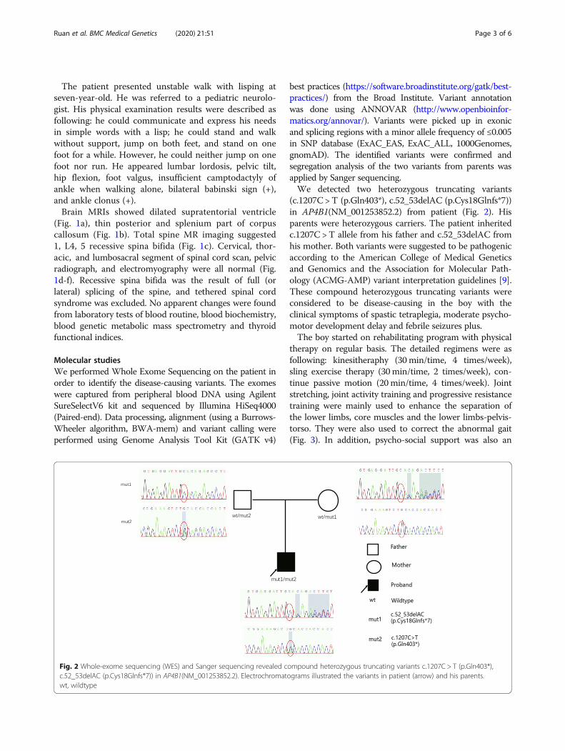
best practices (https://software.broadinstitute.org/gatk/best-practices/) from the Broad Institute. Variant annotationwas done using ANNOVAR (http://www.openbioinfor-matics.org/annovar/). Variants were picked up in exonicand splicing regions with a minor allele frequency of ≤0.005in SNP database (ExAC_EAS, ExAC_ALL, 1000Genomes,gnomAD). The identified variants were confirmed andsegregation analysis of the two variants from parents wasapplied by Sanger sequencing.We detected two heterozygous truncating variants
(c.1207C > T (p.Gln403*), c.52_53delAC (p.Cys18Glnfs*7))in AP4B1(NM_001253852.2) from patient (Fig. 2). Hisparents were heterozygous carriers. The patient inheritedc.1207C > T allele from his father and c.52_53delAC fromhis mother. Both variants were suggested to be pathogenicaccording to the American College of Medical Geneticsand Genomics and the Association for Molecular Path-ology (ACMG-AMP) variant interpretation guidelines [9].These compound heterozygous truncating variants wereconsidered to be disease-causing in the boy with theclinical symptoms of spastic tetraplegia, moderate psycho-motor development delay and febrile seizures plus.The boy started on rehabilitating program with physical
therapy on regular basis. The detailed regimens were asfollowing: kinesitheraphy (30min/time, 4 times/week),sling exercise therapy (30min/time, 2 times/week), con-tinue passive motion (20min/time, 4 times/week). Jointstretching, joint activity training and progressive resistancetraining were mainly used to enhance the separation ofthe lower limbs, core muscles and the lower limbs-pelvis-torso. They were also used to correct the abnormal gait(Fig. 3). In addition, psycho-social support was also an
Fig. 2 Whole-exome sequencing (WES) and Sanger sequencing revealed compound heterozygous truncating variants c.1207C > T (p.Gln403*),c.52_53delAC (p.Cys18Glnfs*7)) in AP4B1(NM_001253852.2). Electrochromatograms illustrated the variants in patient (arrow) and his parents.wt, wildtype
Ruan et al. BMC Medical Genetics (2020) 21:51 Page 3 of 6

important part of the treatment. He showed significantimprovement after 2 years of program judging by theangle changes of dorsiflexion, popliteal fossa, and adduc-tors as well as the evaluation data on Gross Motor Func-tion Measure (GMFM) (Table 1). An improvement onactive and passive foot dorsiflexion angle, popliteal fossaangle and adductor angle reflected the progress of limbfunction in patient (Table 1). The increase in GMFMscore suggested that the patient’s gross motor functionhas not regressed (in theory), but progressed continuouslyover time, proving the effectiveness of treatment.
Discussion and conclusionsWe reported a patient with spastic tetraplegia, moderatepsychomotor development delay and febrile seizures plus-.A paternal heterozygous nonsense variant c.1207C >T(p.Gln403*) and a maternal heterozygous frameshift variantc.52_53delAC (p.Cys18Glnfs*7) which resulted in the
introduction of a premature termination codon in two dif-ferent alleles were identified in AP4B1 gene. Ebrahimi-Fakhari et al [5] reported the clinical and geneticcharacterization of nineteen probands with AP4B1-associ-ated SPG47 including early developmental delay and intel-lectual disability(100%), delayed motor development(100%),neonatal or infantile hypotonia(100%), delayed speech de-velopment(94%), progression to spastic diplegia (89%), lossof independent walking (88%), short stature (57%), thin cor-pus callosum(73%), delayed myelination or white matterloss(67%), ventriculomegaly(40%). Only half of the patientshad epilepsy (47%), especially febrile seizures (3/19). Symp-toms of the patient we reported here were consistent withthe clinical characterizations of patients reported previ-ously. Accogli et al. [6] reported another SPG47-relatedchild who was admitted to the hospital at the age of 14months. The child had an afebrile generalized tonic-clonicepilepticus status which required resuscitation. Our patient,who had been diagnosed as febrile seizures plus, hadafebrile seizures more than 2 times before developingtypical febrile seizures. He also continued febrile seizuresbeyond the age of 6 years. This condition was a relativelyrare feature reported before.AP4 is a heterotetrameric adaptor protein which com-
posed of two large subunits, beta-4 (AP4B1) andepsilon-4 (AP4E1), one medium protein, mu-4 (AP4M1),and one small protein, sigma-4 (AP4S1)(10). Besides ofAP4B1, mutations on other three subunits can also causeautosomal recessive-HSPs. Homozygous or compoundheterozygous mutations in AP4M1 result in autosomalrecessive spastic paraplegia-50 (SPG50, MIM:612936) ischaracterized by neonatal hypotonia that progresses tohypertonia and spasticity and severe mental retardationwith poor or absent speech development [10, 11]. AP4E1and AP4S1 are related to SPG51 (MIM:613744) andSPG52 (MIM:614067) respectively with the similar
Fig. 3 Photos of physical therapy of the patient. a Unilateral lower limb resistance standing. b Autonomous outreach training on slings.c Autonomous Achilles tendon stretching training
Table 1 Examination of rehabilitation effects
201705b 201709 201801 201906
Dorsiflexion Angle(active)
left −10° 0° 0° 25°
Right -10° 0° 0° 15°
Dorsiflexion Angle(passive)
left −5° 15° 15° 45°
Right −5° 15° 15° 25°
Popliteal fossa Angle
left 90° 90° 97° 100°
Right 85° 90° 95° 100°
Adductors Angle 80° 90° 90° 105°
GMFMa 82.13 84.96 86.45 90.58a GMFM Gross Motor Function Measureb Date for physical test
Ruan et al. BMC Medical Genetics (2020) 21:51 Page 4 of 6

symptoms [4, 12, 13]. Hardies et al. [14] reported twosisters, born of unrelated Caucasian parents, whoshowed clinical features including developmental delay,febrile seizures, and spastic paraplegia caused by AP4S1.The older sister had five brief generalized febrile seizuresbetween 5months and 5 years of age whose manifest-ation was similar to our patient.Totally, twenty-two mutants in AP4B1 have been reported
including the ones from our patient (Supp Table 1). Homo-zygous mutations c.304C >T (p.Arg102*) and c.664delC(p.Leu222Cysfs*31) are the most frequently detected variantsfrom consanguineous families. The allele counts are 10 foreach of two mutations from 30 patients (Fig. 4). The vastmajority of pathogenic variants identified so far are truncat-ing variants (allele counts ratio is 52/60) which can often beassumed to disrupt gene function by leading to a completeabsence of the gene product by nonsense-mediated decay ofan altered transcript or lack of transcription (e.g. nonsense,frameshift, canonical splice site).The patient has little progress through medications
and rehabilitations. His seizures are well controlled byadjusted medication of anti-epileptic drug, 12.5 mg/kg(Bid) of levetiracetam tablets. He remained seizure-freefor more than 2 years. No apparent regression had beenseen in patient’s motor development by physical therapy,including joint stretching, joint activity training and pro-gressive resistance training. His speech and languagedevelopment had been severely delayed for a long time.He could speak a few words (“baba”, “mama”) until 2-year-old. We thought it might have a relationship withseizures to some extent, therefore, we paid more atten-tion to anti-epileptic treatment clinically. For languageand speech impairment, the hospital’s teaching and par-ent intervention methods were used due to the need toprotect the children’s other daily activities. Our depart-ment keeps on providing follow-up care at home regu-larly and adjusting the guidance program according tothe situation of patient. He is supported and cared byrelatives, friends as well as the whole society. In
conclusion, in this report we identified two novel patho-genic variants from a Chinese patient with clinical featuresof hereditary spastic paraplegias, including spastic tetra-plegia, moderate psychomotor development delay and fe-brile seizures plus. Our findings expanded the knowledgeof genotypic and phenotypic heterogeneity and similarityof HSPs. Early discovery, diagnosis and proper treatmenton the conditions generally increase chances of improve-ment on the quality of life for patients.
Supplementary informationSupplementary information accompanies this paper at https://doi.org/10.1186/s12881-020-0988-3.
Additional file 1: Supplemental Table1. Mutants in AP4B1 genecollected in research papers.
AbbreviationsACMG-AMP: American College of Medical Genetics and Genomics and theAssociation for Molecular Pathology; FS: Febrile seizures; GMFM: Gross MotorFunction Measure; HSPs: Hereditary spastic paraplegias; SPG47: Autosomalrecessive spastic paraplegia-47
AcknowledgementsWe thank the patient and his parents for participating in this study.
Authors’ contributionsWCR, HFL proposed the meaning and concept of the study and designedthe plan for the case. WCR, YLY, YPC, LD, CXL made contributions to datacollection and analysis. WCR, JW, HFL, XDW drafted and revised themanuscript. All of the authors read and approved the final manuscript to bepublished and agreed to be responsible for the accuracy of the data anddetails.
FundingThis work was supported by The National Key Research and DevelopmentProgram of China of the 13th Five-Year Plan (No.2016YFC1306205); the pro-vincial key disciplines of Zhejiang traditional Chinese medcine (combinationof traditional Chinese and Western medicine) (No.2017-XK-A41); Techno-logical Research Program of Zhejiang (2015C33178) made contributions tothe design of the study, data collection and analysis.
Availability of data and materialsThe datasets generated during the current study are available in NCBI SRA,under the accession number “SRR11117837”.
Fig. 4 Lollipop graph shows mutations in AP4B1 gene reported in literatures. Red triangles indicate the two variants identified in our patient
Ruan et al. BMC Medical Genetics (2020) 21:51 Page 5 of 6

Ethics approval and consent to participateWritten informed consent was obtained from both the patient’s legalguardian (his parents) to participate in this study. This study was approvedby the human ethics committees of The Children’s Hospital, ZhejiangUniversity School of Medicine.
Consent for publicationWritten informed consent was obtained from both the patient’s legalguardian (his parents) for the publication of the details and geneticsequencing of the case report.
Competing interestsThe authors declare that they have no competing interests.
Received: 5 November 2019 Accepted: 28 February 2020
References1. Klebe S, Stevanin G, Depienne C. Clinical and genetic heterogeneity in
hereditary spastic paraplegias: from SPG1 to SPG72 and still counting. RevNeurol (Paris). 2015;171(6–7):505–30. Available from. https://doi.org/10.1016/j.neurol.2015.02.017.
2. Dell’Angelica EC, Mullins C, Bonifacino JS. AP-4, a novel protein complexrelated to clathrin adaptors. J Biol Chem. 1999;12(11):7278–85.
3. Hirst J, Bright NA, Rous B, Robinson MS. Characterization of a fourthadaptor-related protein complex. Mol Biol Cell. 1999;10(8):2787–802.
4. Abou Jamra R, Philippe O, Raas-Rothschild A, Eck SH, Graf E, Buchert R, et al.Adaptor protein complex 4 deficiency causes severe autosomal-recessiveintellectual disability, progressive spastic paraplegia, shy character, and shortstature. Am J Hum Genet. 2011;88(6):788–95.
5. Ebrahimi-Fakhari D, Cheng C, Dies K, Diplock A, Pier DB, Ryan CS, et al.Clinical and genetic characterization of AP4B1-associated SPG47. Am J MedGenet Part A. 2018;176(2):311–8.
6. Accogli A, Hamdan FF, Poulin C, Nassif C, Rouleau GA, Michaud JL, et al. Anovel homozygous AP4B1 mutation in two brothers with AP-4 deficiencysyndrome and ocular anomalies. Am J Med Genet Part A. 2018;176(4):985–91.
7. Hebbar M, Shukla A, Nampoothiri S, Bielas S, Girisha KM. Locus and allelicheterogeneity in five families with hereditary spastic paraplegia. J HumGenet [Internet]. 2019;64(1):17–21. Available from:. https://doi.org/10.1038/s10038-018-0523-y.
8. Helbig KL, Farwell Hagman KD, Shinde DN, Mroske C, Powis Z, Li S, et al.Diagnostic exome sequencing provides a molecular diagnosis for a significantproportion of patients with epilepsy. Genet Med. 2016;18(9):898–905.
9. Richards CS, Bale S, Bellissimo DB, Das S, Grody WW, Hegde MR, et al. ACMGrecommendations for standards for interpretation and reporting ofsequence variations: Revisions 2007. Genet Med. 2008;10(4):294–300.
10. Tüysüz B, Bilguvar K, Koçer N, Yalçinkaya C, Çaǧlayan O, Gül E, et al.Autosomal recessive spastic tetraplegia caused by AP4M1 and AP4B1 genemutation: expansion of the facial and neuroimaging features. Am J MedGenet Part A. 2014;164(7):1677–85.
11. Verkerk AJMH, Schot R, Dumee B, Schellekens K, Swagemakers S, Bertoli-Avella AM, et al. Mutation in the AP4M1 gene provides a model forNeuroaxonal injury in cerebral palsy. Am J Hum Genet. 2009;85(1):40–52.
12. Moreno-De-Luca A, Helmers SL, Mao H, Burns TG, Melton AMA, Schmidt KR,et al. Adaptor protein complex-4 (AP-4) deficiency causes a novelautosomal recessive cerebral palsy syndrome with microcephaly andintellectual disability. J Med Genet. 2011;48(2):141–4.
13. Najmabadi H, Hu H, Garshasbi M, Zemojtel T, Abedini SS, Chen W, et al.Deep sequencing reveals 50 novel genes for recessive cognitive disorders.Nature. 2011;478(7367):57–63.
14. Hardies K, May P, Djémié T, Tarta-Arsene O, Deconinck T, Craiu D, et al.Recessive loss-of-function mutations in AP4S1 cause mild fever-sensitiveseizures, developmental delay and spastic paraplegia through loss of AP-4complex assembly. Hum Mol Genet. 2015;24(8):2218–27.
Publisher’s NoteSpringer Nature remains neutral with regard to jurisdictional claims inpublished maps and institutional affiliations.
Ruan et al. BMC Medical Genetics (2020) 21:51 Page 6 of 6