nursing assessment in pediatric hematology/ · pdf filenursing assessment in pediatric...
TRANSCRIPT

Nursing Assessment in Pediatric Hematology/Oncology
Wendy Landier RN MSN CPNP CPON®
© APON 2003

OBJECTIVES
• Recognize abnormal laboratory results and associated nursing implications.
• Discuss the special aspects of the role of the nurse in the care of the child or adolescent with cancer and their families.
• Implement practices designed to improve the quality of life for patients and families affected by childhood cancer.
© APON 2003

Childhood Cancer: Symptom Onset
• May be rapid or insidious
• Diagnosis often delayed
• Symptoms often vague
© APON 2003

Childhood Cancer: Common Chief Complaints
• Pallor, bleeding
• Fatigue
• Persistent fever
• Headache, visual changes
• Lymphadenopathy
• Bone pain, joint pain, limp
• Abdominal mass
• Cough, respiratory difficulties
© APON 2003

The Diagnostic Workup: “Waiting and Not Knowing”
• Uncertainty regarding diagnosis and prognosis
• Worry and preoccupation with anticipated outcome
• Intensity/agony of this period often unrecognized
• Nurses can listen, debrief, and offer support
Clarke-Steffen, 1993
© APON 2003

Clinical Manifestations of Cancer
• Changes in blood cell production, due to: – bone marrow infiltration by tumor
– chronic disease
• Mass, resulting in: – compression of organs
– compression of vital structures
• Tumor byproducts, causing alterations in: – electrolytes
– hormones, metabolism
– immunologic response © APON 2003
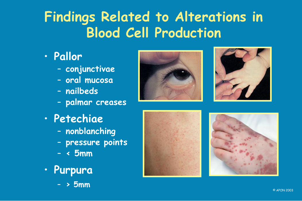
Findings Related to Alterations in Blood Cell Production
• Pallor – conjunctivae – oral mucosa – nailbeds – palmar creases
• Petechiae – nonblanching – pressure points – < 5mm
• Purpura – > 5mm
© APON 2003
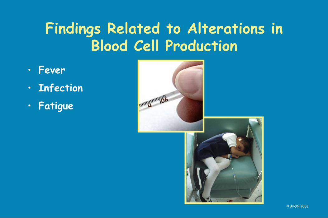
Findings Related to Alterations in Blood Cell Production
• Fever
• Infection
• Fatigue
© APON 2003

The Mediastinum : What is It?
Space between:
– sternum and spine
– suprasternal notch and diaphragm
– parietal pleura
© APON 2003

The Mediastinum: What’s Inside?
• Thymus
• Thyroid
• Esophagus
• Lymph nodes
• Trachea & bronchi
• Heart & pericardium
• Great vessels
• Nerves
http://www.bartleby.com/107/Images/small/image968.jpg
© APON 2003

Causes of Mediastinal Mass
• Leukemia, lymphoma
• Neuroblastoma
• Other tumors
• Infections (e.g., TB)
© APON 2003

Findings Related to Mediastinal Mass: Respiratory Compromise
• Cough
• Wheezing
• Tracheal/bronchial compression
• Respiratory distress/arrest
© APON 2003
Mediastinal Mass
Never force a child with a mediastinal mass to lie
down—this could result in respiratory arrest!
Never sedate a child with a mediastinal mass unless you are prepared to intubate!
© APON 2003

Findings Related to Mediastinal Mass: Superior Vena Cava Syndrome
• Facial swelling and/or distended neck veins
• Compression of great vessels/vital structures
• May be misdiagnosed as an allergic reaction
© APON 2003

Findings Related to Masses: Lymphadenopathy
• Abnormal enlargement of lymph nodes – > 1 cm
– often firm, matted
• Location of enlarged nodes – regional versus generalized
- epitrochlear (elbow) & supraclavicular nodes usually pathologic
© APON 2003
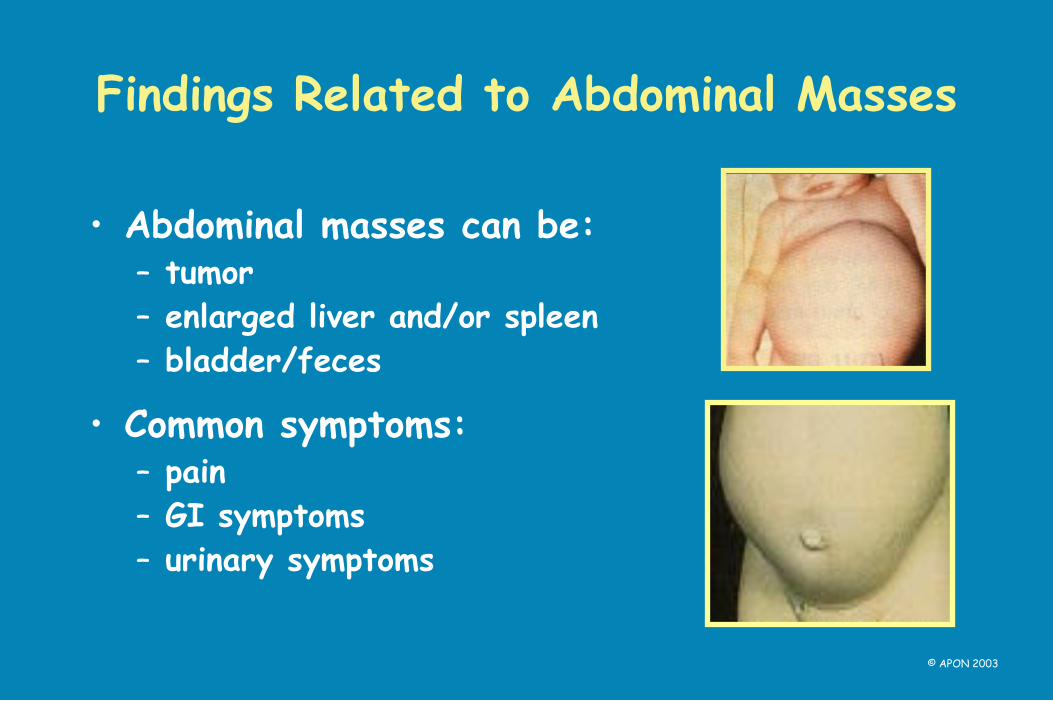
Findings Related to Abdominal Masses
• Abdominal masses can be: – tumor
– enlarged liver and/or spleen
– bladder/feces
• Common symptoms: – pain
– GI symptoms
– urinary symptoms
© APON 2003

Findings Related to Musculoskeletal Masses
• Constitutional symptoms – fatigue
– fever
– weight loss
• Pain (onset/timing/location)
• Gait problems
© APON 2003

Findings Related to CNS Tumors
• Morning headaches
• Vomiting
• Hemiparesis
• Cranial nerve palsies
• Diplopia, nystagmus, strabismus
• Ataxic gait
• Decreased coordination
• Seizures
© APON 2003
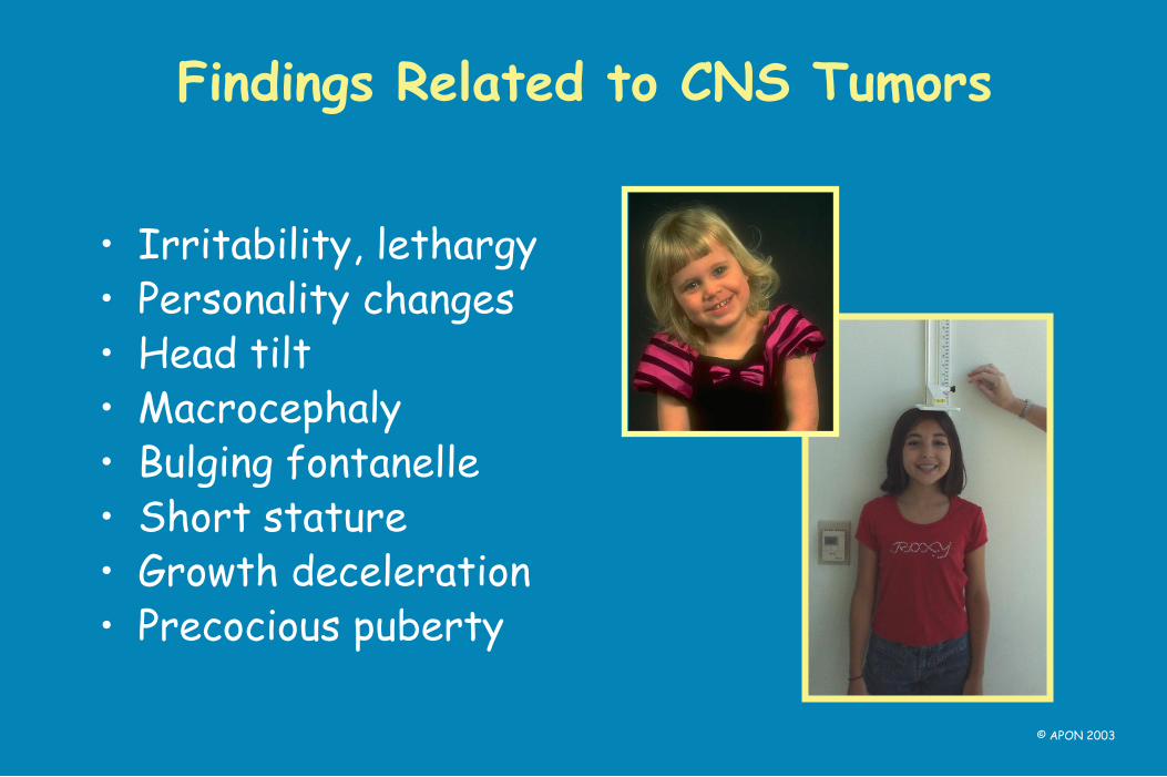
Findings Related to CNS Tumors
• Irritability, lethargy • Personality changes • Head tilt • Macrocephaly • Bulging fontanelle • Short stature • Growth deceleration • Precocious puberty
© APON 2003

Assessing Lab Values
http://ehpnet1.niehs.nih.gov/docs/2002/110-4/ss.html
© APON 2003

How to Read a CBC
© APON 2003

How to Read a CBC
• Measure of formed elements: – red blood cells
– white blood cells
– platelets
• Additional information: – hemoglobin
– hematocrit
– RBC size/shape
– WBC differential
• Always check norms for age!!
© APON 2003
Plasma – 55% of whole blood
Erythrocytes – 45% of whole blood
Buffy coat (leukocytes and platelets) - <1%

Red Blood Cell Assessment
• RBC count – total # of RBCs in each ml of
blood
• Hemoglobin – iron-rich protein found inside
RBCs, measured in gm/dl – indicator of O2-carrying
capacity
• Hematocrit – % of RBC’s by volume
© APON 2003

Red Blood Cell Assessment: RBC, Hgb, Hct
• Normal
• Low (anemia)
• High (polycythemia)
© APON 2003

Red Blood Cell Assessment: Size & Color
• MCV (mean cell volume)
– RBC size
• MCH (mean cell hemoglobin)
MCHC (mean cell hemoglobin concentration)
– RBC hemoglobin content (color)
• RDW (red cell distribution width)
– variation in RBC size
© APON 2003

Red Blood Cell Assessment: Size & Color
• MCV (RBC size): – normal (normocytic) – low (microcytic) – high (macrocytic)
• MCH, MCHC (RBC color): – normal (normochromic) – low (hypochromic)
• RDW (RBC size variation): – normal – high (wide variation in RBC size)
© APON 2003

Platelet Count
• Platelets – plug holes in damaged
blood vessels
– prevent bleeding
© APON 2003

Platelet Count Assessment
• Normal
• Low (thrombocytopenia)
• High (thrombocytosis)
© APON 2003

White Blood Count
• White blood cells:
– fight infection
– make antibodies
– several subtypes of WBCs make up the “differential count”
© APON 2003
Lymphocyte
Neutrophil
Platelet

White Blood Count Assessment
• Normal
• Low (leukopenia)
• High (leukocytosis)
© APON 2003

WBC Differential
• Assesses percentage of each different subtype of WBC in blood
• Reported as % of total cells counted
• % of all types reported should add up to 100
© APON 2003
Eosinophil
Neutrophil
Band
Lymphocyte
Monocyte

WBC Differential: Types of Cells
• Neutrophils (infection-fighters) – segs or polys (mature) – bands or stabs (young)
• Lymphocytes (immunity)
• Monocytes (phagocytosis)
• Eosinophils (allergy, parasites)
• Basophils (hypersensitivity)
• Blasts (very immature) (Blasts should ALWAYS be considered
ABNORMAL unless proven otherwise)
© APON 2003

Evaluating the Neutrophil Count
• Neutrophil count increases with: – bacterial infections
• increased % of neutrophils
• increased % of immature neutrophils (bands/stabs)
• “shift to the left”
– glucocorticoid therapy, stress, epinephrine
• Neutrophil count decreases with: – viral infections, certain drugs
– diseases involving the bone marrow
– congenital and acquired neutropenias
– hypersplenism
© APON 2003

Evaluating the Neutrophil Count
• Patients with low neutrophil counts are at high risk of developing bacterial infections.
• The lower the neutrophil count, and the longer it stays low, the higher the risk of infection.
• Patients with very low neutrophil counts may not be able to mount a response (show an increase in WBC) in the presence of infection.
© APON 2003

Calculating the Absolute Neutrophil Count (ANC)
ANC = % segs (polys) + % bands (stabs) x total WBC (in 1000s)
33 (% segs)
1 (% bands)
34% x 3,600 (WBC in 1000s)
= 1224
+
(ANC)
© APON 2003

Absolute Neutrophil Count (ANC)
• >1500 = normal
• <1000 = impaired ability to fight infection
• <500 = at risk for serious infection
© APON 2003
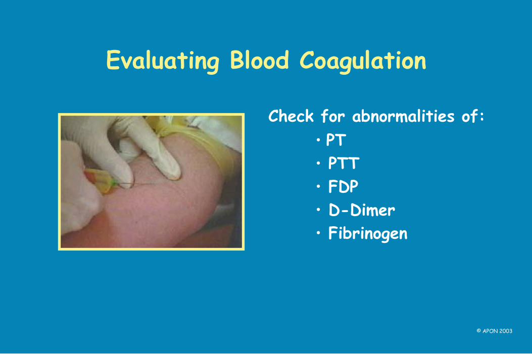
Evaluating Blood Coagulation
• PT
• PTT
• FDP
• D-Dimer
• Fibrinogen
Check for abnormalities of:
© APON 2003

Evaluating Kidney Function
• U/A
• BUN
• serum creatinine
• creatinine clearance
• GFR
Check for abnormalities of:
© APON 2003

Evaluating Liver Function
• bilirubin
• ALT
• AST
• alkaline phosphatase
• prothrombin time
Check for abnormalities of:
© APON 2003

Evaluating Cultures
• Check culture reports on all of your patients
• Report positive cultures IMMEDIATELY
• Patient therapy may change based on results
© APON 2003

Evaluating Vital Signs
• Know norms for age
• Measure precisely using correct technique
• Always evaluate every set of vital signs on each patient (whether or not your take them yourself)!!!
© APON 2003

Evaluating Vital Signs: Temperature
• No rectal temps for oncology patients!
• Fever is an emergency:
– in neutropenic patients
– in patients with central lines or other implanted apparatus (e.g., shunts)
– in immunodeficient or asplenic patients (e.g, Wiskott-Aldrich, sickle cell)
© APON 2003

Evaluating Vital Signs: Fever
• Shaking chills – may occur before onset of fever
– also considered an emergency
• Check capillary refill – normal = brisk (immediate)
– report if delayed ( > 2 seconds)
• Notify MD/NP/PA immediately – urgent evaluation/intervention required
© APON 2003

Evaluating Vital Signs: Tachycardia
• Anxiety
• Anemia
• Hypovolemia
• Shock
• Fever
• Pain
Potential causes:
© APON 2003

Evaluating Vital Signs: Tachypnea
• Potential causes – anxiety – hypoxia – fever – pain – respiratory
compromise
• Evaluate: – retractions
– nasal flaring
– color (dusky, cyanotic)
– breath sounds
© APON 2003

Evaluating Vital Signs: Hypotension
• Hypotension is an emergency - report immediately!
• Potential causes:
– septic shock (can be rapidly fatal!!)
– hypovolemia (dehydration, bleeding)
© APON 2003

Evaluating Vital Signs: Hypertension
• Requires prompt assessment and intervention
• Potential etiology: – steroids
– renal
– increased intracranial pressure - report!
may require prn or routine medication
© APON 2003

Evaluating Vital Signs: Pain
• Assess pain with all vital signs
• Use age-appropriate assessment tool
• Potential causes: – disease – treatment (e.g., mucositis, surgery) – infection
• Pain requires intervention!
© APON 2003

Nursing Assessment
• Thorough physical assessment
• Special attention to: – mouth
– skin
– perianal area
© APON 2003

Evaluating Neutropenic Patients
• Usual signs of infection may be absent: – erythema – warmth – pus/drainage – rales
• Pain/tachypnea/fever may be only signs of infection
• Fever or shaking chills require immediate intervention!
© APON 2003
Photos courtesy of Dr. W. Hughes

BIBLIOGRAPHY
Ablin, A. R. (1997). Supportive Care of Children with Cancer (2nd Ed.) Baltimore: Johns Hopkins University Press.
Baggott C. R., Kelly K. P., Fochtman, D. & Foley, G. V. (2002). Nursing
Care of Children and Adolescents with Cancer (3rd Ed.) Philadelphia: W. B. Saunders.
Clarke-Steffen L. (1993). Waiting and not knowing: the diagnosis of
cancer in a child. Journal of Pediatric Oncology Nursing, 10(4):146-53.
Fernbach, D.J. & Vietti, T.J. (1991). Clinical Pediatric Oncology (4th Ed.) St. Louis: Mosby-Yearbook.
© APON 2003
BIBLIOGRAPHY
Hockenberry-Eaton, M. J. (Ed.) (1998). Essentials of Pediatric Oncology Nursing: A Core Curriculum. Glenview IL: Association of Pediatric Oncology Nurses.
Pizzo, P. A. & Poplack, D. G. (2002). Principles and Practice of
Pediatric Oncology (4th Ed.) Philadelphia: Lipincott Williams & Wilkins.
Pui,C-H, (Ed.) (1999). Childhood Leukemias. NYC: Cambridge University Press.
Zitelli, B. J. & Davis, H. W. (Eds.). (1997)Atlas of Pediatric Physical
Diagnosis. St. Louis: Mosby.
© APON 2003