nutrition, environment, and autoimmunity sabrina schmitz epob 4800 spring 2004
TRANSCRIPT

Nutrition, Environment, and Autoimmunity
Sabrina Schmitz
EPOB 4800
Spring 2004

OUTLINE
• REVIEW: IMMUNE SYSTEM
• “SELF” VS. “NON-SELF” DISTINCTION
• DEVELOPMENT OF AUTOIMMUNITY
• AUTOIMMUNE DISEASE MODELS
• ENVIRONMENT AND NUTRUTION
• CONCLUSION

ReviewLymphocyte Differentiation

Review
• T cells: lymphocytes with antigen-specific receptors; play a major role in initiation and suppression of cell-mediated and humoral immunity– Cytotoxic T cells
– Helper T cells

Review
• B cells– Produce
immunoglobulins (Ig):• B cell receptors: bound
to B cell surface; allow antigen recognition (subsequent signaling or proliferation)
• Secreted antibodies: unbound: “opsonize” pathogens.“Eat Me”

Lymphocyte Communication

Cytokines and Chemokines• Cytokines: locally acting tissue hormones
responsible for growth, differentiation, function, inhibition and apoptosis of cells.– Designations: type I, II; pro-inflammatory.– Ex. Interleukins, Interferons
• Chemokines: chemo-attractant cytokines—very large and very diverse group of cytokines– Capable of inducing directional migration of activated
leukocytes– mediate acute and chronic inflammation– Also regulate: cell-to-cell adhesion, angiogenesis,
embryogenesis, etc.– Ex. Leukotrienes, platelet activating factor, transforming
growth factor beta superfamily members, etc.

Apoptosis
• Apoptosis is a genetically-regulated programmed cell death “program” in which the dying cell actively executes the decomposition and packaging of its internal contents in response to a variety of stimuli. It varies from necrosis in many ways, but most importantly, by its failure to induce an inflammatory response

Apoptotic pathways

Apoptotic pathways
CD95

Apoptotic pathways

Apoptotic pathways


Development of autoimmunity
*Dysregulation of apoptosis
*Distinction between “self” and “non-self” blurred

Distinction between “self” vs. “non-self”
• Immune system must be able to distinguish between pathogenic cells and host cells
• T and B cells have antigen-specific receptors– Autoreactive TCRs and BCRs must be deleted
from repertoire.– Other lymphocytes take cues from T and B
cells

T and B cell receptor diversification
• The TCR and BCR repertoire of an individual must technically be able to identify every foreign antigen.
• Several genetic recombination mechanisms are involved in diversification.
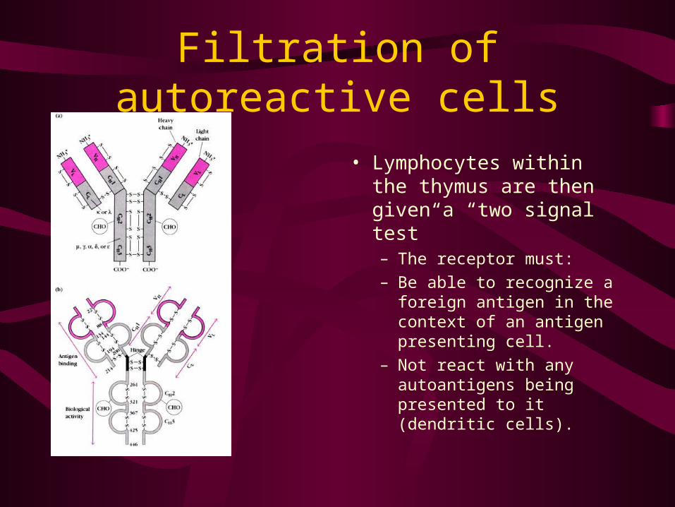
Filtration of autoreactive cells
• Lymphocytes within the thymus are then given a “two signal test”– The receptor must:
– Be able to recognize a foreign antigen in the context of an antigen presenting cell.
– Not react with any autoantigens being presented to it (dendritic cells).

Antigen presentation
HLA: human leukocyte antigen; various cell-surface proteins coded for by genes on the MHC; often necessary to activate major immune responses as a secondary safety mechanism

Autoimmunity and autoimmune diseases

Definitions
• Autoimmunity: state in which the immune system recognizes and removes host cells.
• Autoimmune Disease: state in which autoreactive lymphocytes are allowed to proliferate and do harmful damage.

Development of autoimmunity
• Three requirements– Genetic:
• the individual must be able to produce HLAs capable of expressing autoantigens as well as TCRs and BCRs capable of recognizing them.
• Additional genetic predispositions: faulty transcrption/translation enzymes, or tissue weakness.
– Developmental• The tolerizing mechanisms must overlook the autoreactive
lymphocytes (anergy, silencing)
– Environment• An environmental trigger of some sort must initiate the reaction
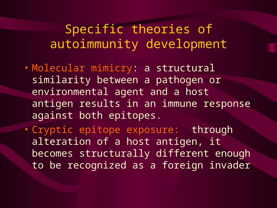
Specific theories of autoimmunity development
• Molecular mimicry: a structural similarity between a pathogen or environmental agent and a host antigen results in an immune response against both epitopes.
• Cryptic epitope exposure: through alteration of a host antigen, it becomes structurally different enough to be recognized as a foreign invader

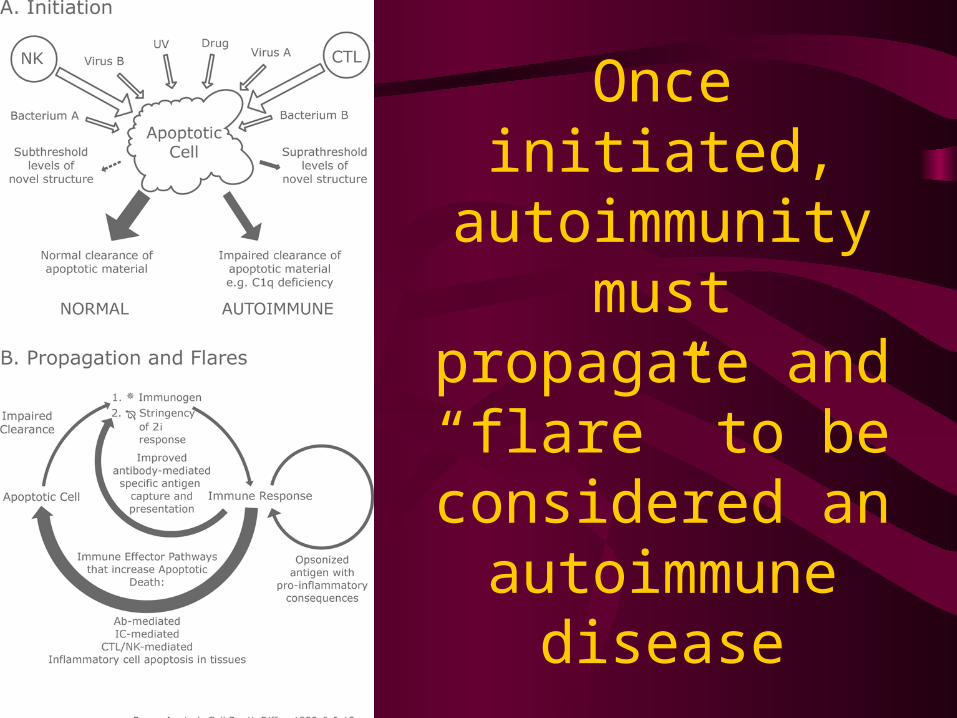
Once initiated, autoimmunity must
propagate and “flare” to be
considered an autoimmune
disease

Once initiated, autoimmunity must
propagate and “flare” to be
considered an autoimmune
disease

Once initiated, autoimmunity must
propagate and “flare” to be
considered an autoimmune
disease

Autoimmune disease models
Prevalence of various autoimmune diseases

Autoimmune diseases associated with targeted cell destruction
• Tissue-specific autoimmune diseases
• Also, symptom-specific.
Table***: Autoimmune diseases associated with targeted cell destruction (Kalden, 2003)
DISEASE CELL KILLED
Diabetes (IDDM) pancreatic β cell
Multiple Sclerosis (MS)
oligodendrocyte
Hashimoto’s thyroiditis
thyrocyte
Sjögren’s syndrome acinus and ductal cells
Polymyositis myocyte
Primary biliary cirrhosis
bile duct cells
Ex. Multiple Sclerosis• a chronic demyelinating disease with observed onset usually in early
adulthood and ultimately resulting in patches of hardened tissue in tissues • Damage to the central nervous system in MS is T cell mediated, and
usually waxes and wanes throughout the lives of afflicted individuals. • Although no single unifying cause can be identified for MS development in all
patients, the role of viral molecular mimicry is becoming increasingly emphasized in research.
• Because both MS patients and healthy individuals express myelin basic protein (MBP) in the thymus, T cells must be rendered tolerant to the dominant epitopes of the protein. This is strong evidence for molecular mimicry and exposure of cryptic epitopes in the development of MS (Paul, 1999).
• The most popular theory: “only during episodes of peripheral T-cell activation by crossreactive viral epitopes do these normally quiescent lymphocytes acquire the capacity to traverse the blood-brain barrier and initiate the immune response against myelin in the central nervous system” (Paul, 1999).

Other disorders involving CNS autoimmunity
• MS, Alzheimer’s Disease (AD), Parkinson’s Disease (PD), amyotrophic lateral sclerosis (ALS), and to a certain extent, mental disorders (schizophrenia, bipolar disorder (BPD), attention deficit and hyperactivity disorder (ADHD), and depression) can be categorized as diseases associated with target cell death.
• Recent research has begun looking at the etiologies neuropsychiatric disorders from a common mechanistic perspective: CNS autoimmunity and/or neuronal apoptosis. Specific points of interest:
– Nerve growth factor (NGF) is a trophic factor, responsible for localized inhibition and proliferation of neurons during the development of the nervous system. In the absence of this neurotrophin, both naïve and differentiated neurons underwent programmed cell death. Thus, neurotrophin provides possible treatment options preventing neuronal apoptosis.
– Adhesion molecules are capable of similar local neuronal control, highlighting the importance of local factors in the development and maintenance of a healthy nervous system

Autoimmunity associated with abnormal processing of apoptotic cells: systemic
lupus erythematosus • Systemic lupus erythematosus (SLE) is defined as “an
inflammatory connective tissue disease of unknown cause that occurs chiefly in women and that is characterized especially by fever, skin rash, and arthritis, often by acute hemolytic anemia, by small hemorrhages in the skin and mucous membranes, by inflammation of the pericardium, and in serious cases by involvement of the kidneys and central nervous system” (Merriam, 2003).
• SLE is diagnosed based on specific clinical findings and polyclonal B cell immunity (Fitzpatrick, 2001).
• Multiple induction and propagation factors including genetic predisposition, drugs, chemicals, food, and infectious agents contribute to the complex etiopathogenesis of SLE.

Traditional 4-stage model of SLE
1. Susceptibility phase: 10 fold increase in heritability between di- and mono-zygotic twins.
1. MHC II alleles strongly correlated to lesion type
2. Genetic defects in cytokine production also observed.
2. Induction: anti-dsDNA T cells allowed to proliferate
1. cryptic determinant exposure and UV exposure suspected.

SLE continued…
• 3. Expansion phase: typical antigen-driven immune response– Epitope spreading: inflammatory processes
mediate exposure/recognition of other autoantigens.

SLE continued…
• 4. Tissue damage:. Much of the damage can be attributed to the presence of antigen-antibody complexes, which are either deposited in the affected tissue (lesions generally occur due to complex deposition in the various cutaneous layers) or are circulating throughout the body (often resulting in nephritis or other serious systemic complications). – Lupus band test– The fundamental lupus lesion is “fibrinoid degeneration of
connective tissue and walls of the blood vessels associated with an inflammatory infiltrate of lymphocytes and plasma cells” (Fitzpatrick, 2001).

Autoimmune disease associated with enhanced cell survival/proliferation
Table ***: Autoimmune diseases associated with enhanced cell survival/proliferation (Kalden, 2003)
DISEASE CELLS/TISSUES
Rheumatoid arthritis (RA)
pannus
Scleroderma fibroblasts
Autoimmune lympho-proliferative syndrome (ALPS)/ Canale-Smith syndrome (CSS)cells of the immune system
cells of the immune system
Thyrotoxicosis (Graves’ disease)
thyrocyte

Ex. Scleroderma
• Scleroderma is defined as “a usually slowly progressive disease marked by the deposition of fibrous connective tissue in the skin and often in internal organs and structures, by hand and food pain upon exposure to cold, and by tightening and thickening of the skin—also called “dermatosclerosis” (Merriam-Webster, 2002).
Scleroderma continued…
• Scleroderma is very environment-sensitive.
• Observations that led to scleroderma as a particularly environment-sensitive disease primarily originate in epidemiological data revealing “occupational clusters” of extreme scleroderma prevalence.

“Atypical” autoimmune disease: celiac disease (CD)
• CD is a complex disease characterized by a wide spectrum of lesions in the intestinal mucosa that can ultimately lead to atrophy of the villi.– Permanent intolerance to gluten triggers the
production of anti-gliadin antibodies.– “atypical” because IgA antibodies generated
often disappear (and villi return to normal) after gluten is taken out of the diet.
Additional complications
• Epitope spreading: other systems and tissues put at risk
• Additional health complications– Renal abnormalities due to antigen-antibody
complex depositon– Neurological side-effects: depression,
neuropathy, fatigue.

Environmental factors in development of autoimmunity
and autoimmune disease

Hess, 2002
“Environmental chemicals and autoimmune disease: cause and
effect”

Hess, 2002: Proposed mechanisms for chemical immunomodulation
• 1. Chemical would ordinarily elicit antigen-specific immune response– In some individuals: polyclonal B cell
activation—autoantibody production.

Hess, 2002: Proposed mechanisms for chemical immunomodulation
• 2. The agent may directly exert toxic effect on cells of immune system or other cells– Impairment of the immune response
• Ex. Slow phagocytosis of apoptotic celld
– Release of intracellular constituents• Ex. dsDNA: autoantibodies formed--SLE

Hess, 2002: Proposed mechanisms for chemical immunomodulation
3. Molecular mimicry: ross-reactivity due to host/agent structural similarities
4. Agents could directly interact with regulatory factors that modify gene activity:
1. Impairment of T cell DNA methylationtranscription/translation of autoreactive TCRsautoimmunity

Hess, 2002: Proposed mechanisms for chemical immunomodulation
6. Agents might induce free radical production indirect initiation of inflammatory response.

Hess, 2002: Environmental immunomodulants
1. Aromatic amines and hydrazines1. Over 70 drugs or medications drug-related
lupus (DRL) most common autoimmune process observed
2. Ex. Minoxidil (in rogaine), penicillin, streptomycin, sulfa drugs, tetracyclines, ibuprofen, gold salts, estrogens, enalapril

Hess, 2002: Environmental immunomodulants
2. Hydrazines: widely prevalent in agriculture and industry
1. Numerous commercial applications: synthesis of plastics, anti-corrosives, rubber products, herbicides, photographic supplies, perservatives, textiles, dyes and pharmaceuticals.
2. Also naturally present in tobacco, tobacco smoke, mushrooms and penicillium.1. Smokers have increased risk of SLE

Hess, 2002: Environmental immunomodulants
3. Tartrazine aka FD&C yellow #51. A yellow dye present in thousands of foods and
drugs
2. Reported association with development of:1. Asthma
2. Urticaria
3. Angioedema
4. Rhinorhea
5. Allergic reactions
6. SLE (phototoxic potentials)

Hess, 2002: Environmental immunomodulants
4. Aromatic amines: present in permanent hair coloring solutions; can be absorbed through the scalp.
1. Ex. Paraphenylenediamine: associated with cases of connective tissue disease-like symptoms1. Scleroderma-like lesions
2. Evidence for and against association with SLE.

Hess, 2002: Environmental immunomodulants
5. Silica: occupational “hot spots”1. Exposure to silica dust includes: mining, quarrying,
tunneling, stone cutting, monumental masonry, use of abrasives or abrasive blasting; certain glass-manufacturing occupations; boiler scaling, chemistry laboratories.
2. Certain art mediums: porcelain, pottery, and brick work.
3. Very “immunoactive”:1. Potent, non-specific adjuvant effect (possibly responsible for
autoantibodies widely observed in workers’ connective tissue.
2. Ability to activate microvascular endothelial cells, peripheral blood mononuclear cells, and dermatofibroblasts (SLE common)
3. Genetic link: research has linked prevalence of the TNF-alpha(2) allele.

Hess, 2002: Environmental immunomodulants
6. Vinyl chloride: former ingredient of hairspray; occupational exposure (esp. associated with plastics.
1. Associated with occupational acro-osteolysis, scleroderma-like skin changes; miscellaneous bone abnormalities.1. HLA-DR5 genetic association; specifically linked to
haplotypes: AI, B8 DR3.
2. May bind to nucleotidespossible formation of highly oxidized metabolites further reactivity.
Hess, 2002: Environmental immunomodulants
7. Organic solvents1. Exposure via inhalation is a key risk factor
1. Widely used: paints, varnish, thinners, waxes (floor and shoe polish), inks, adhesives, antifreeze mixtures, motor fuel, pharmaceutical products and preservatives
2. Polyhalogenated hydrocarbons are especially dangerous
3. Some unconfirmed studies of immunologic abnormalities in workers of computer manufacturing plants
2. Clinical observations: connective tissue diseases; especially scleroderma

Hess, 2002: Environmental immunomodulants
8. Chlorinated hydrocarbons1. Widespread usage limited in late 1950s, after an
accidental exposure to hexachlorobenzene (HCB) resulted in porphyria development in ~4000 people.
2. Today used mostly for removing grease from metal parts; found in many adhesive spot removers, carpet cleaners, and paint strippers
3. Scleroderma and eosinophilic fasciitis are the most commonly observed etiologies.
Hess, 2002: Environmental immunomodulants
9. Epoxy resins: plastisizer1. Autoimmune reactions observed: morphea (type
of scleroderma) and DRL.
2. Is chemically similar to several drugs (procainamide, d-penicillamine, chlorpromazine and isoniazid)
Hess, 2002: Environmental immunomodulants
9. Heavy metals1. Chronic exposure to metals can induce immune complex
deposition in kidneys2. Gold: previously used to treat RA; renal disease very
common side-effect; lymphocyte abnormalities also common.3. Cadmium: also renal involvement; presence of ANAs also
detected4. Mercury: Very common environmental contaminant (esp. via
bioaccumulation)1. Reaches water supply via medical/scientific waste discharge;
fluorescent lights, batteries, etc.2. Observed health effects: morphological and physiological
disruption of lymphocytes; modification of self-proteins through mercuric-thiol interactions
3. Environmental trigger of scleroderma; SLE and asthma also observed.

Soh et al.(2003)
• Naturally occuring isoquinoline alkaloids.– Foodstuffs: cheese, broiled sardine, broiled beef; yolk
and white of broiled eggs, milk, soy sauce, bananas, port wine, beer, whisky
– Salsolinol and tetrahydropapaveroline (THP) have been detected in human fluids and tissues, including regions of the brain (esp. in PD patients).
• Formed endogenously by the condensation of dopamine with acetaldehyde and dopaldehyde.
• Have both pro- and anti-oxidant properties• Demonstrated to inhibit mitochondrial respiration, cause DNA
damage (chromosomal aberrations) and apoptosis (via MAPK and JNK pathway disruption) of neurons.

Nakao et al. (2003)
• Observed the effects of aspartame (metabolized to formaldehyde) on thymocyte morphology– Results: a levels upwards of
100 microM of methanol in the bloodstream produced shrunken thymocytes with hypodiploid DNS.
– Aspartame has been deemed safe in human studies, however, other researchers have reported blood levels of as high as 625 microM from test subjects.

Nutritional factors effecting autoimmunity
Individual factors
1. Genistein: 1. phytoestrogen; protects diverse types of cells from damage
from toxic stimuli2. Zeng et al. (2003):
1. subjected hippocampal neurons to beta-amyloid protein (A-beta)1. A-beta: a major component of senile plaques in the CNS of AD patients;
associated with elevation of intracellular free Ca2+, elevated ROS levels, and caspase-3 activationapoptosis characterized by decreased cell viability and DNA condensation/fragmentation.
2. Results: ROS, Ca2+, and apoptosis were all reduced significantly3. Support the suggested role of phytoestrogens in the upregulation of brain-
derived neurotrophic factor mRNA; and stimulation of striatal dopamine release.

Individual nutrtional factors continued…2. Antioxidants
1. The usual suspects: carotenoids, tocopherols...
2. Glutathione peroxidase (PHGPx): unique selenoenzyme that directly reduces phospholipid hydroperoxides
1. Shidoji et al. (2002) and others confirm key role in preventing excess cardiolipin oxidation.
Mitochondria plays a central role in apoptosis mediated by oxidations

Individual nutrtional factors continued…3. PUFAs
1. Low n-3:n-6 ratios in industrialized contries; n-3s inhibit pro-inflammatory eicosanoid and cytokine production by peripheral tissues (prevent the dysregulation of apoptotic homeostasis).

Individual nutrtional factors continued…PUFAs…
1. Shapiro (2002)1. Evaluated previous research: patients suffering from chronic
illness often suffer major depression (and visa versa); connection believed to result from shared pathophysiological processes.1. Risk factors: decreased n-3 intake, abnormal
metabolism of lipid and phospholipid mediators, increased production of pro-inflammatory cytokines, dysregulation and dysfunction of the HPA axis, and immune dysfunction.
2. N-3 PUFAs believed to provide anti-depressant effects based on abilities to inhibit PKC; pharmaceuticals that inhibit PKC significantly reduce pain, but are dangerous b/c of excessive PKC inhibition.
2. Analyzed heart rate variability (HRV) as a measurement of sympatho-vagal balance. Results: increased n-3 consumption produced increased adrenergic stimulation.

Nutritional factors continued…
• Probiotics:– Healthy gut microflora population very important for good
digestion and pathogenic defense.– Disruption of normal microflora population (via antibiotics,
stress—decreased gut pH, artificial foodstuffs, etc.) often results in inflammation malabsorptionmalnutrition (esp. antioxidants, etc.) further inflammation and health problems.
– Similarly…Bauer (2001): Emphasized the importance of lactobacilli in hydrogen-peroxide production in the vaginaonly induces apoptosis in abnormal cells (anti-tumor effect).
• Dysregulation of this balance (by HPV or other environmental/pathogenic agents) allows abnormal cells to avoid apoptosis.

Whole-food vegan diet
McCarty (2001)

Vegan….
• Whole-food vegan diets down regulate systemic IGF-I (lymphocyte growth factor)
• Diet rich in n-3 fatty acids down regulates prolactin production (also LGF), shown to increase severity of pre-existing autoimmune disease.– Also propose using dopamine-agonists to
suppress prolactin secretion

Vegan…
• McCarty’s reasoning based on the total lack of autoimmune disease among sub-Saharan black Africans as long as they followed their traditional quasi-vegan lifestyles
• Epidemiological studies correlate animal product and/or saturated fat consumption with IDDM and MS.

Vegan…
• McCarty also points out:– Favorable impact of low latitude and high
altitude on autoimmune diseases; likely because of increased vitamin D production.
– Androgens, which appear to upregulate thymocyte apoptosis, may also be a good option for treating autoimmune disease in womenmen enjoy relative protection from autoimmune diseases.

Summary
• The large number of distinct cell types, chemical signaling molecules, physical signaling interactions, and cell life/death pathways makes it difficult to make generalizations about factors contributing to autoimmunity.
• However, the role of environmental contaminants and decreased nutrition is highly supported.
• Thus, the development of autoimmunity and progression of autoimmune disease must be approached from a basic perspective: a healthy lifestyle.

QUESTIONS?