o steopathic manipulative medicine for upper extremity pain in adolescent athletes
DESCRIPTION
Anne Marie C Zeller, MSc , DO Family Medicine Resident: Year 2 Undergraduate Osteopathic Manipulative Medicine Fellow- Graduated Chief Faculty : Michael P. Rowane, DO, MS, FAAFP, FAAO. O steopathic Manipulative Medicine for Upper Extremity Pain in Adolescent Athletes. - PowerPoint PPT PresentationTRANSCRIPT
Osteopathic Manipulative Medicine for Upper Extremity Pain in Adolescent Athletes
Anne Marie C Zeller, MSc, DO
Family Medicine Resident: Year 2Undergraduate Osteopathic Manipulative Medicine Fellow- Graduated Chief
Faculty:Michael P. Rowane, DO, MS, FAAFP, FAAO

“I have no desire to be a cat, which walks so lightly that it never creates a disturbance.”
-A. T. Still
Objectives
Discuss common causes and diagnoses in regards to adolescent shoulder and elbow pain
Discuss basic tenets of examination of shoulder, elbow, and wrist
High-yield and efficient osteopathic manipulative medicine treatments for shoulder, elbow, and wrist
Practice , Practice, Practice!
Pediatric Population
MUST consider the maturation of the physis or growth plates
Weakness at the physis and decreased resistance to shear and tensile forces compared to the surrounding ligaments, tendons, and muscles, PREDISPOSE this population to injury.
Mechanism of Injury of Shoulder Pain
• Repetitive micro trauma or overuse mechanisms:1. Acceleration: Athletes uses optimum load to generate
force• Example: racquet and pitching sports
2. Dynamic force: arm is moving against sustained resistance• Example: swimming
3. Static force: action of the shoulder muscles when then are held in a constant position with isometric contraction• Example: dancer or gymnast
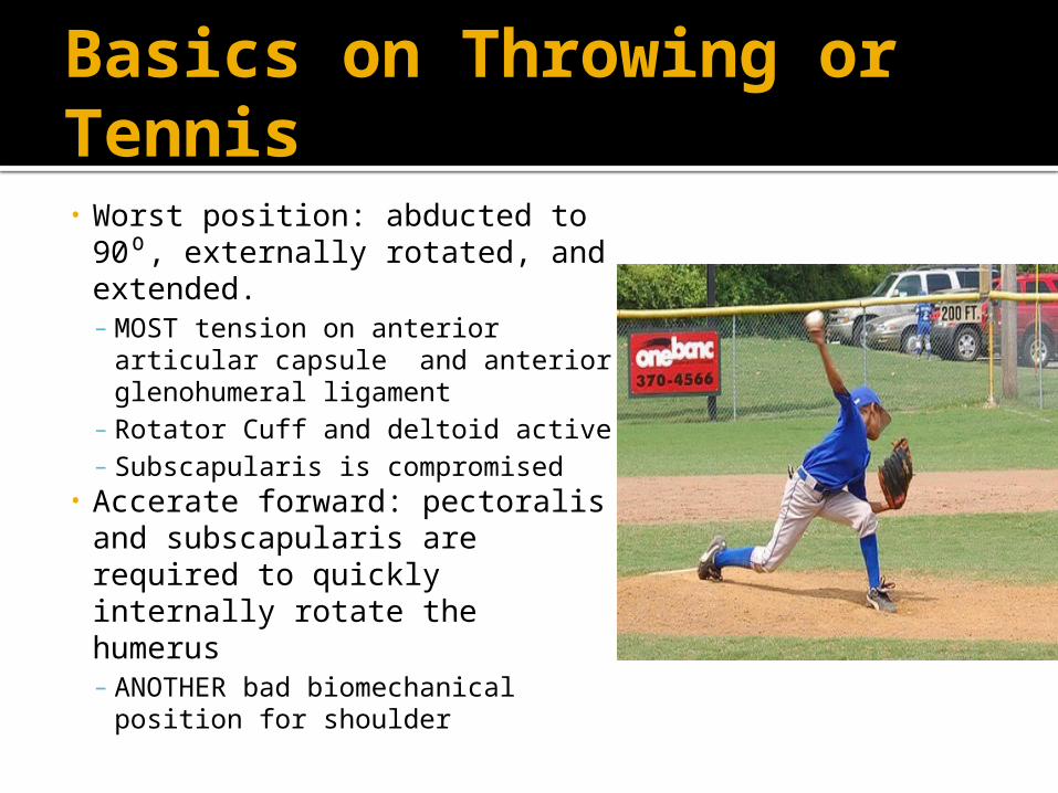
Basics on Throwing or Tennis
• Worst position: abducted to 90 , ⁰externally rotated, and extended.– MOST tension on anterior articular
capsule and anterior glenohumeral ligament
– Rotator Cuff and deltoid active– Subscapularis is compromised
• Accerate forward: pectoralis and subscapularis are required to quickly internally rotate the humerus– ANOTHER bad biomechanical
position for shoulder
Articular Units of Shoulder Complex Covered Today
1) Glenohumeral Joint2) Sternoclavicular and Acromioclavicular Joints3) Scapulothoracic Joint
Remember: Shoulder Pain is NOT JUST Rotator Cuff! Shoulder involves Ribs, Thoracics, Lumbars, Cervicals, Cranial bones Innominates, and Sacrum
Most Common Adolescent Athlete Shoulder Injuries
Epidemiology, Pathology and OMM treatment
“Doctor, shouldn’t you leave treating cervical
dysfunctions to the OB/GYN physicians?”-Anonymous Lawyer
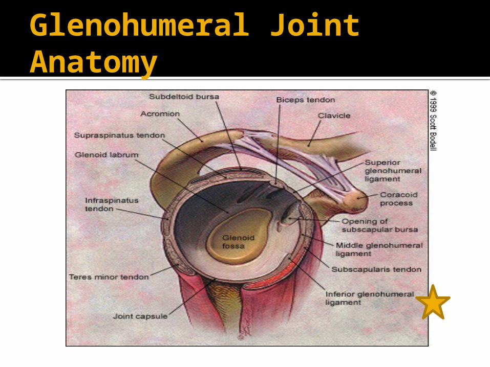
Glenohumeral Joint Anatomy
Epidemiology of in the Glenohumeral Joint Injuries
• Traumatic events makes up 86% of Glenohumeral instability in adolescent athletes 16 and older.
• Skeletally mature athletes with GH instability = surgery due to 80-90% recurrence rate
• Skeletally immature athletes = EXTREMELY careful in evaluating because of the high chance of fracture of proximal humerus.
Anterior Dislocation
• 90% of traumatic dislocation• Mechanism of Injury: high energy injury of a fall on an
outstretched hand while shoulder in abduction and external rotation
• S/S: “dead arm”- transient loss of sensation or numbness in involved extremity (axillary nerve), obvious deformity, pt hold arm internally rotated, + anterior apprehension test
• Diagnosis: Pt history, physical exam, x-rays• Treatment: Primary- closed reduction of dislocation,
Secondary- surgery due to recurrence rate with conservative treatment .8
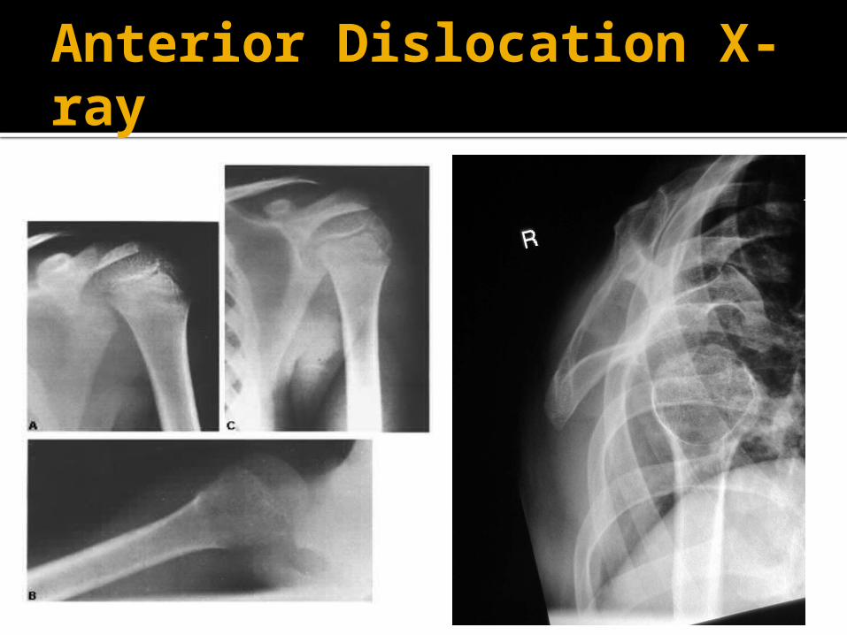
Anterior Dislocation X-ray
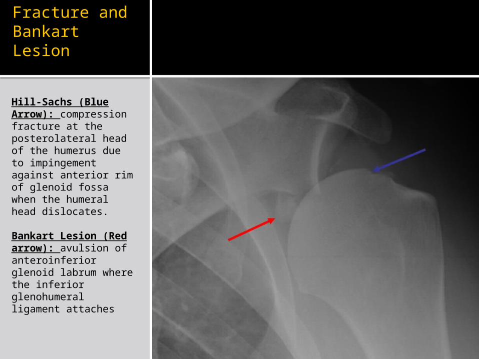
Hill-Sachs Fracture and Bankart Lesion
Hill-Sachs (Blue Arrow): compression fracture at the posterolateral head of the humerus due to impingement against anterior rim of glenoid fossa when the humeral head dislocates.
Bankart Lesion (Red arrow): avulsion of anteroinferior glenoid labrum where the inferior glenohumeral ligament attaches
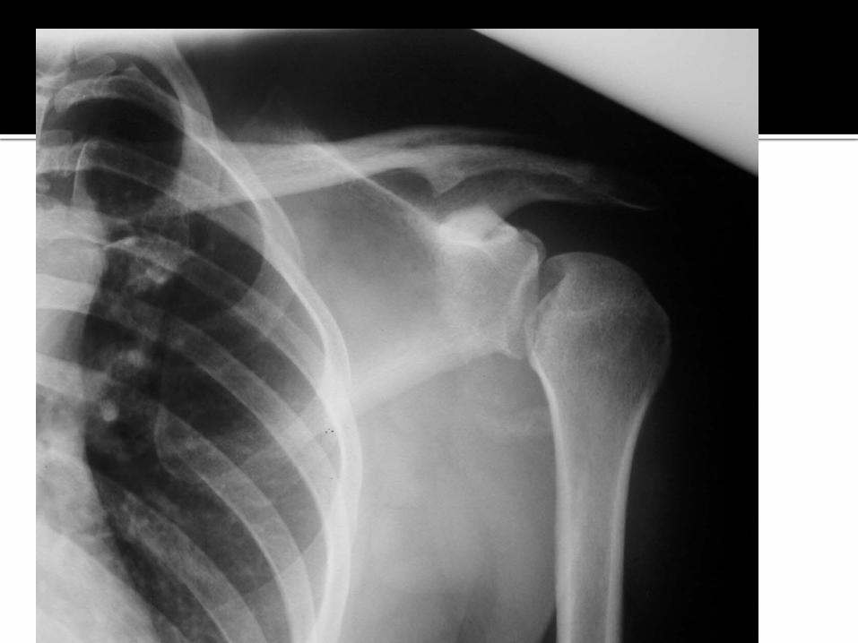
Posterior Dislocation
• < 5% of traumatic shoulder dislocations• MOI: Fall on an outstretched hand with shoulder in
adduction and internal rotation or direct anterior trauma. – Example: Offensive Linemen: forward flexed and internally
rotation of shoulder for blocking• S/S: May not have deformity, + posterior apprehension
test, complain of shoulder pain and have limited external rotation with <90 shoulder flexion⁰
• Treatment: rotator cuff rehab is most successful after closed reduction
Atraumatic Instability
• Majority are bilateral, multidirectional• Hypermobility (generalized joint laxity) of joints from
sports that weaken rotator cuff from overhead motions– Examples: gymnastics and swimming
• S/S: nonspecific shoulder pain, feeling of shoulder dislocation with overhead activities, hyperextension of other joints of UE, + apprehension signs, + sulcus sign, strength deficits in rotator cuff muscles and scapular stabilizers (serratus anterior, pectoralis, and latissimus dorsi)
• Treatment: conservative rehab with strengthening NOT stretching exercises
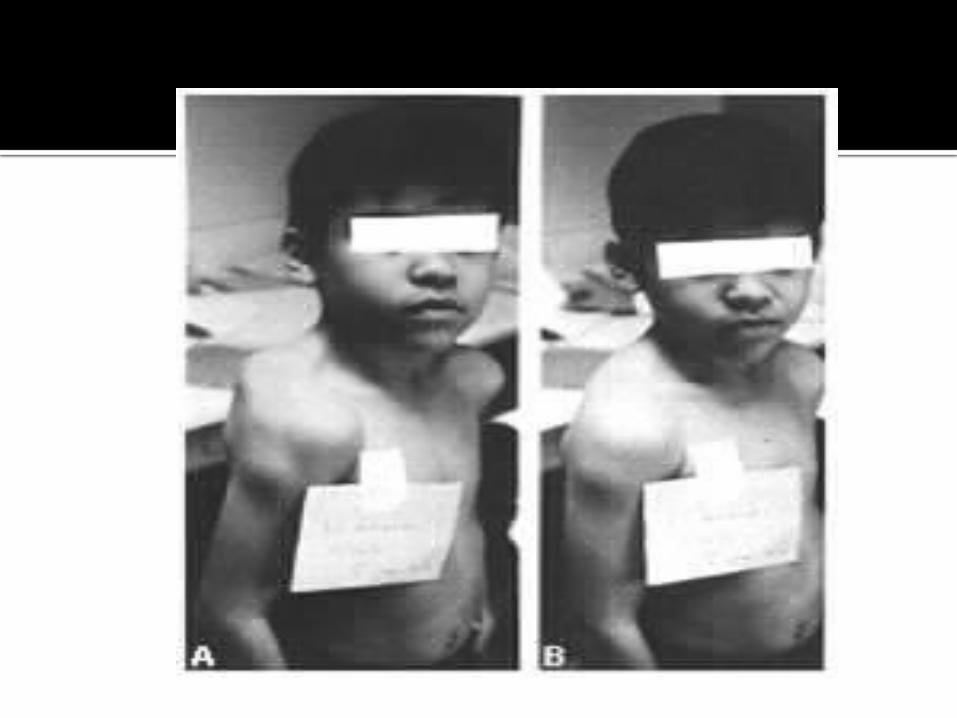
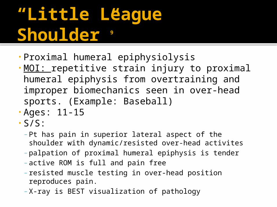
“Little League Shoulder”9
• Proximal humeral epiphysiolysis• MOI: repetitive strain injury to proximal humeral
epiphysis from overtraining and improper biomechanics seen in over-head sports. (Example: Baseball)
• Ages: 11-15• S/S: – Pt has pain in superior lateral aspect of the shoulder with
dynamic/resisted over-head activites– palpation of proximal humeral epiphysis is tender– active ROM is full and pain free– resisted muscle testing in over-head position reproduces pain.– X-ray is BEST visualization of pathology
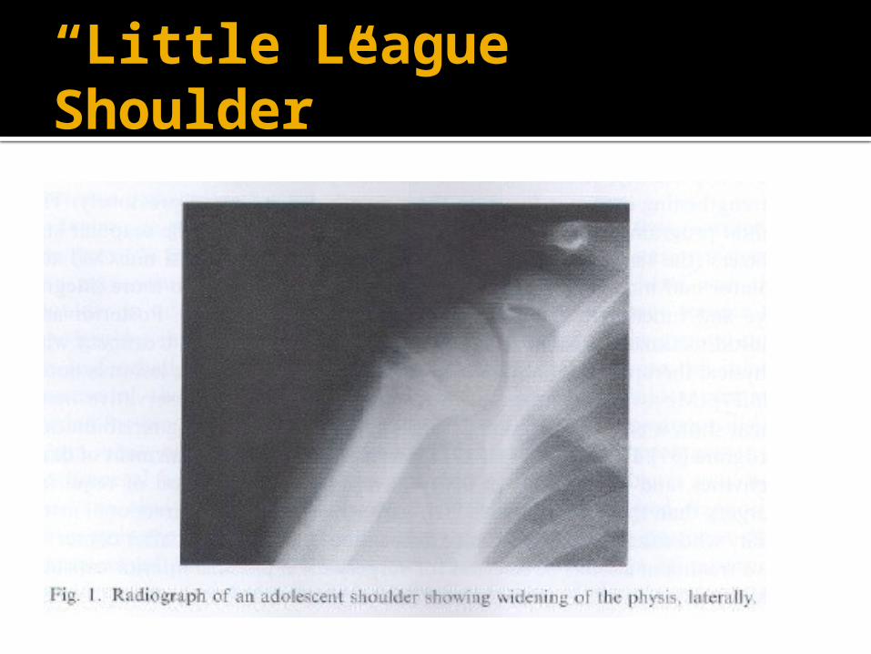
“Little League Shoulder”
Osteopathic Manipulative Medicine for Shoulder Pain/Dysfunction in Adolescent Athlete
Osteopathic Manipulative Medicine for Shoulder Pain in Adolescent Athlete
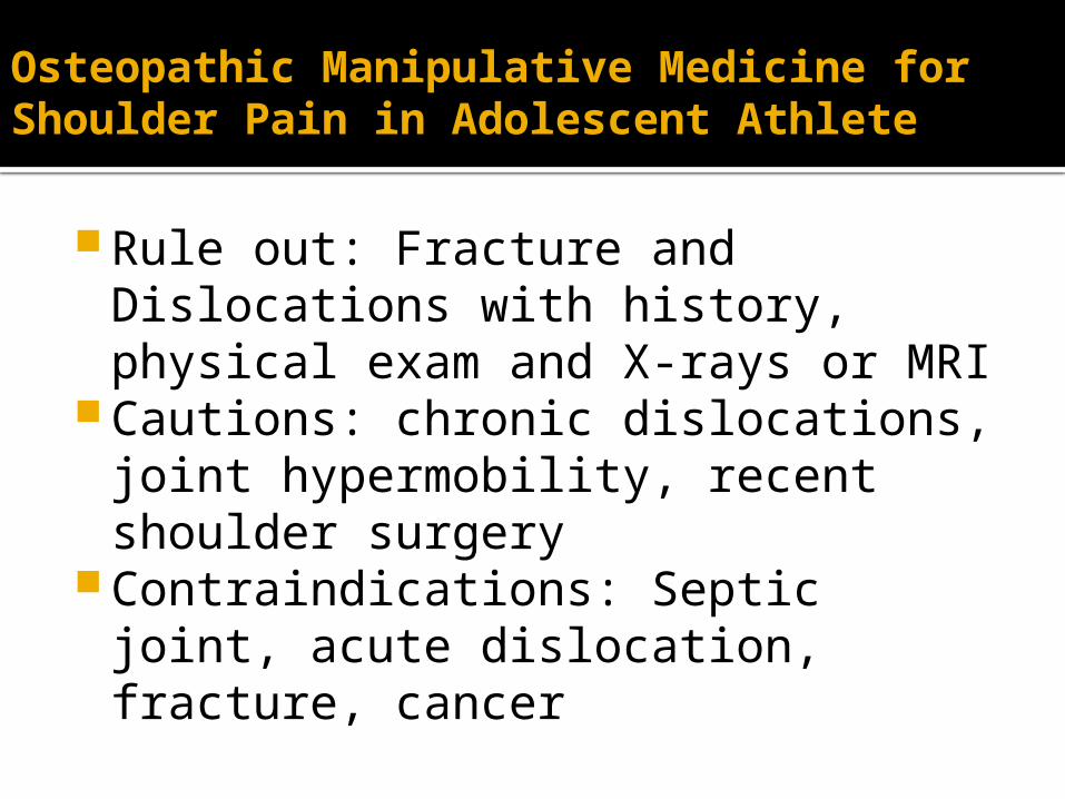
Rule out: Fracture and Dislocations with history, physical exam and X-rays or MRI
Cautions: chronic dislocations, joint hypermobility, recent shoulder surgery
Contraindications: Septic joint, acute dislocation, fracture, cancer
BLT Humerus/Rotator Cuff- Seated
BLT Humerus/Rotator Cuff- Seated
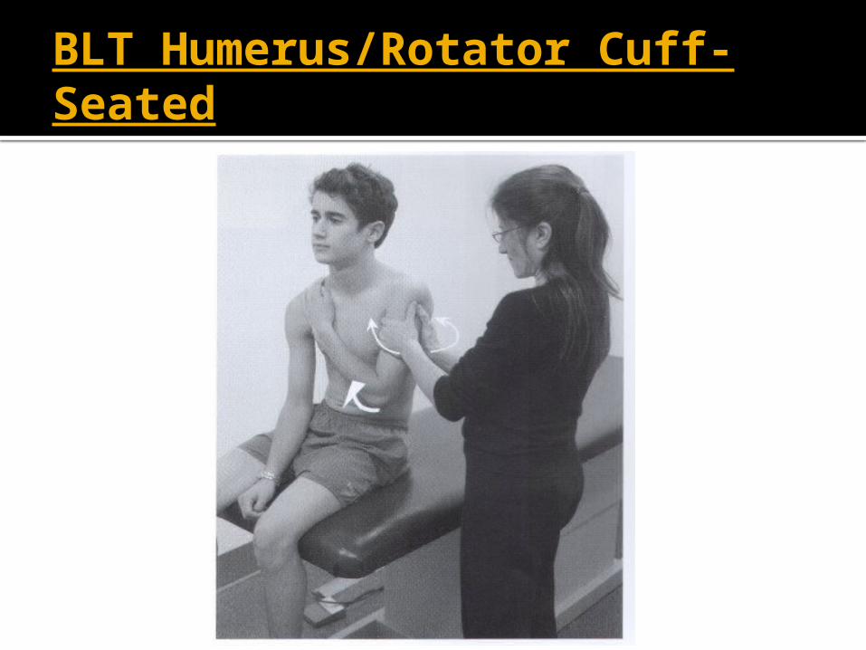
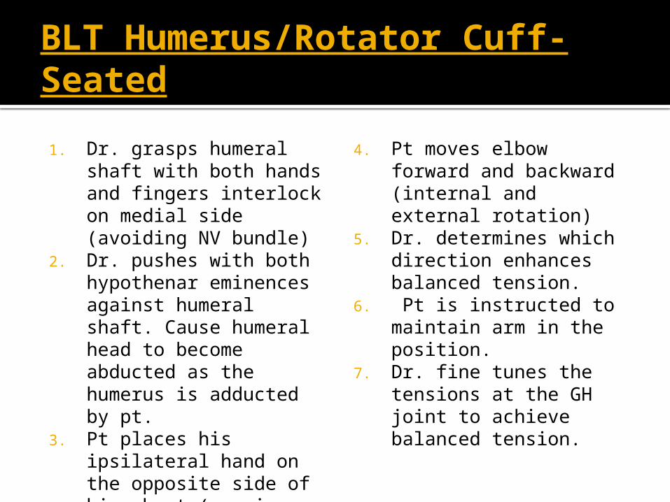
1. Dr. grasps humeral shaft with both hands and fingers interlock on medial side (avoiding NV bundle)
2. Dr. pushes with both hypothenar eminences against humeral shaft. Cause humeral head to become abducted as the humerus is adducted by pt.
3. Pt places his ipsilateral hand on the opposite side of his chest (causing internal rotation and adduction)
4. Pt moves elbow forward and backward (internal and external rotation)
5. Dr. determines which direction enhances balanced tension.
6. Pt is instructed to maintain arm in the position.
7. Dr. fine tunes the tensions at the GH joint to achieve balanced tension.
Direct Myofascial Release Technique-Anterior Cervical Fascia
Direct Myofascial Release Technique-Anterior Cervical Fascia
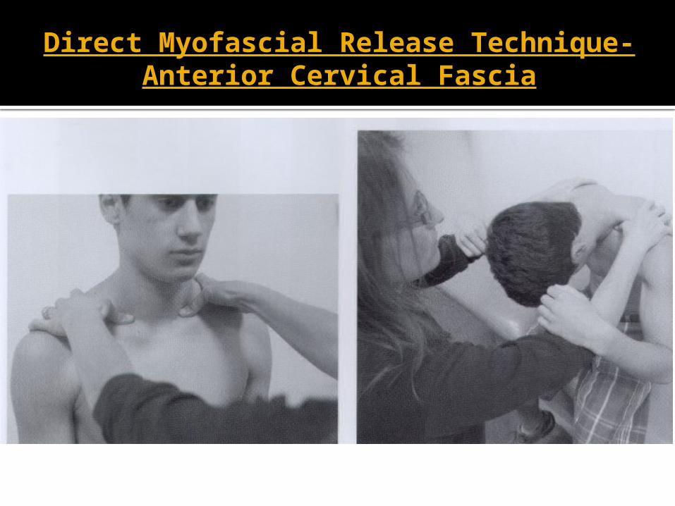
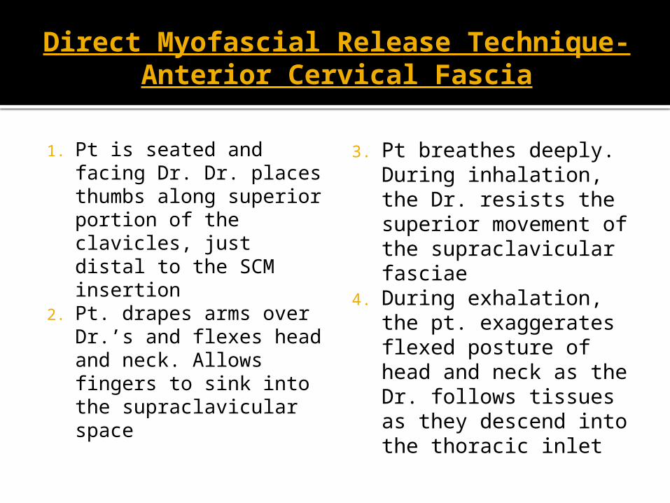
1. Pt is seated and facing Dr. Dr. places thumbs along superior portion of the clavicles, just distal to the SCM insertion
2. Pt. drapes arms over Dr.’s and flexes head and neck. Allows fingers to sink into the supraclavicular space
3. Pt breathes deeply. During inhalation, the Dr. resists the superior movement of the supraclavicular fasciae
4. During exhalation, the pt. exaggerates flexed posture of head and neck as the Dr. follows tissues as they descend into the thoracic inlet
Anatomic Mechanism of OMM Clavicle treatment
According to Sutherland model, the claviopectoral fascia has a similar role to the interosseous membranes of the forearm and lower leg in that it guides and limits movement of the bone.
What is the Scapulothoraic Joint?
Serratus anterior, rhomboid and teres major are viewed as the functional ligaments of the joint.
BLT treatment presented addresses Serratus anterior, subscapularis, rhomboid, latissimus dorsi, teres major and lower trapezius muscles.
BLT Scapulothoracic Joint Seated
BLT Scapulothoracic Joint Seated
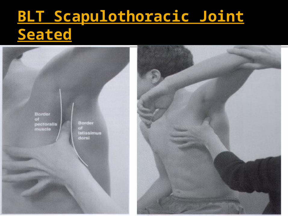
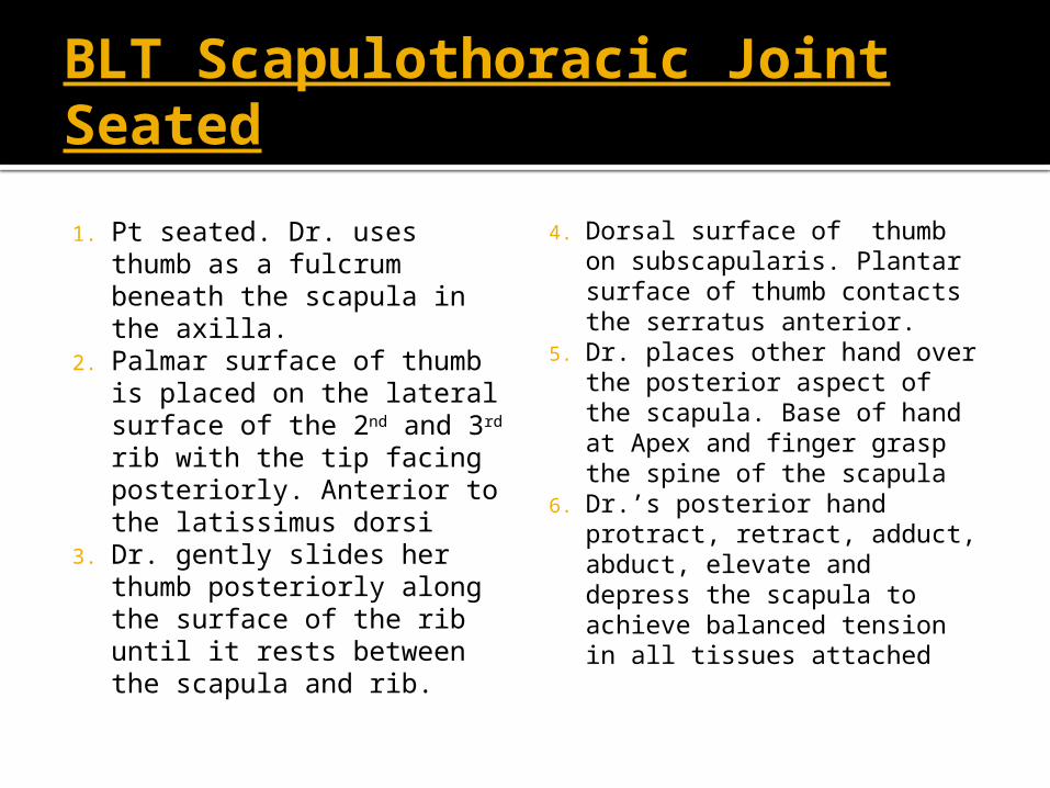
1. Pt seated. Dr. uses thumb as a fulcrum beneath the scapula in the axilla.
2. Palmar surface of thumb is placed on the lateral surface of the 2nd and 3rd rib with the tip facing posteriorly. Anterior to the latissimus dorsi
3. Dr. gently slides her thumb posteriorly along the surface of the rib until it rests between the scapula and rib.
4. Dorsal surface of thumb on subscapularis. Plantar surface of thumb contacts the serratus anterior.
5. Dr. places other hand over the posterior aspect of the scapula. Base of hand at Apex and finger grasp the spine of the scapula
6. Dr.’s posterior hand protract, retract, adduct, abduct, elevate and depress the scapula to achieve balanced tension in all tissues attached
Topics not addressed but are influential in shoulder pain/dysfunction treatment
OMM Treatment of Ribs, Cranial bones, Cervical Vertebrae, Thoracic Vertebrae, Lumbar Vertebrae, Innominates, Sacrum with S/CS, ME, Indirect Myofascial, Still, or FPR.
Extensive information on Throwing and other sport mechanisms in the shoulder and its contributions to shoulder injury and pain
Osteopathic Manipulative Medicine for Elbow Pain/Dysfunction in Adolescent Athlete
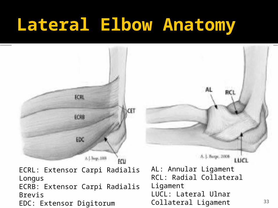
Lateral Elbow Anatomy
33
ECRL: Extensor Carpi Radialis LongusECRB: Extensor Carpi Radialis BrevisEDC: Extensor Digitorum CommunisECU:Extensor Carpi UlnarisCET: Common Extensor Tendon
AL: Annular LigamentRCL: Radial Collateral LigamentLUCL: Lateral Ulnar Collateral Ligament
Most common Mechanism of Injury in Lateral Elbow Pain
Precipitated by activities that require repetitive wrist extension, radial deviation and forearm supination
Examples: Hammering, painting, tennis backhand
34
Common Presentation
Patient typically reports an insidious onset but will often relate a history of overuse without trauma.
Pain with gripping objects (“coffee cup sign) and shaking hands (“politician’s sign”)
Numbness or tingling: Suggest radicular symptoms
35
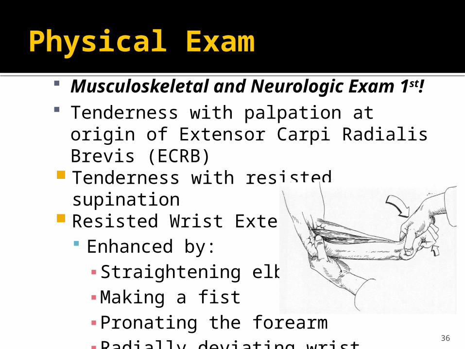
Physical Exam Musculoskeletal and Neurologic Exam 1st! Tenderness with palpation at origin of Extensor
Carpi Radialis Brevis (ECRB) Tenderness with resisted supination Resisted Wrist Extension Test
Enhanced by:▪ Straightening elbow▪ Making a fist▪ Pronating the forearm▪ Radially deviating wrist
36
Physical Exam
Middle Finger Test Resist the extension of the proximal
interphalangeal joint of 3rd digit Stresses the extensor digitorum and ECRB Positive if pain is over the lateral epicondyle.
37
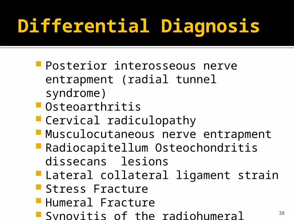
Differential Diagnosis
Posterior interosseous nerve entrapment (radial tunnel syndrome)
Osteoarthritis Cervical radiculopathy Musculocutaneous nerve entrapment Radiocapitellum Osteochondritis dissecans lesions Lateral collateral ligament strain Stress Fracture Humeral Fracture Synovitis of the radiohumeral joint
38
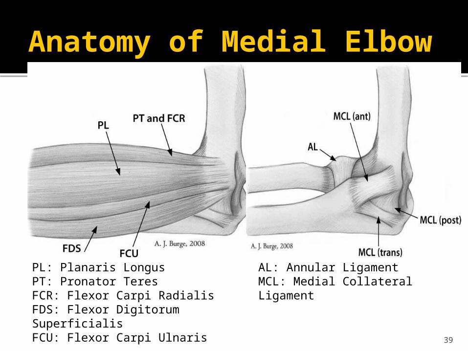
Anatomy of Medial Elbow
39
PL: Planaris LongusPT: Pronator TeresFCR: Flexor Carpi RadialisFDS: Flexor Digitorum SuperficialisFCU: Flexor Carpi Ulnaris
AL: Annular LigamentMCL: Medial Collateral Ligament
Mechanism of Injury Medial Elbow Pain
Forceful and/or continuous flexion and pronation at the wrist
Activities requiring a large amount of stabilization applied by the wrist
Common Activities Examples: Racquet sports Swimming Swinging a Golf Club Throwing Computer Keyboard Playing Piano
Certain occupations Examples▪ Carpenters▪ Plumbers▪ Meat cutter
40
Common Presentation in Medial Elbow Pain
Pain and tenderness along medial elbow extending into forearm
Difficulty gripping without pain Decreased wrist strength Tightness/stiffness when stretching elbow and
wrist
41
Physical Exam: Medial Elbow Pain
Testing for Valgus Stability in Extension: MCL Anterior Capsule Bony articulations
42
Differential Diagnosis of Medial Arm Pain
Fracture Osteochondritis dissecans Osteoarthrosis MCL injury Little League elbow- increased valgus angle in
adolescent throwing athletes Flexor-Pronator Strain Ulnar neuropathy (neuritis, entrapment) Pediatric- avulsion fracture
43
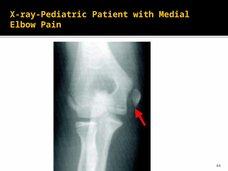
X-ray-Pediatric Patient with Medial Elbow Pain
44
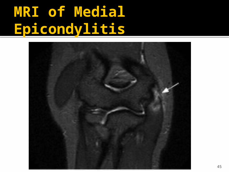
MRI of Medial Epicondylitis
45
Osteopathic Manipulative Medicine for Elbow Pain
Osteopathic Manipulative Medicine Considerations in Elbow Pain
Diagnose and treat Somatic Dysfunctions in: Cervical spine, Thoracic spine, Ribs , Scapula, and Clavicle
To reduce and/or correct somato-somatic reflexes and some of the myofascial pain referrals
To improve the venous and lymphatic drainage
Osteopathic Manual Medicine
OMT Techniques Presented Address: Radial Head Humero-Radial Joint Humero-Ulnar Joint Distal Radio-Ulnar Joint Carpal Joints
Diagnosing Somatic Dysfunction of the Elbow
Patient seated with elbows flexed at 900 and forearms at 00 of pronation and supination (thumbs up).
Then check for supination or pronation restrictions. The radial head moves posteriorly with pronation and
anteriorly with supination. Therefore a pronated forearm (with restricted
supination) will have a posterior radial head somatic dysfunction.
Supinated forearm (with restricted pronation) will have an anterior radial head somatic dysfunction.
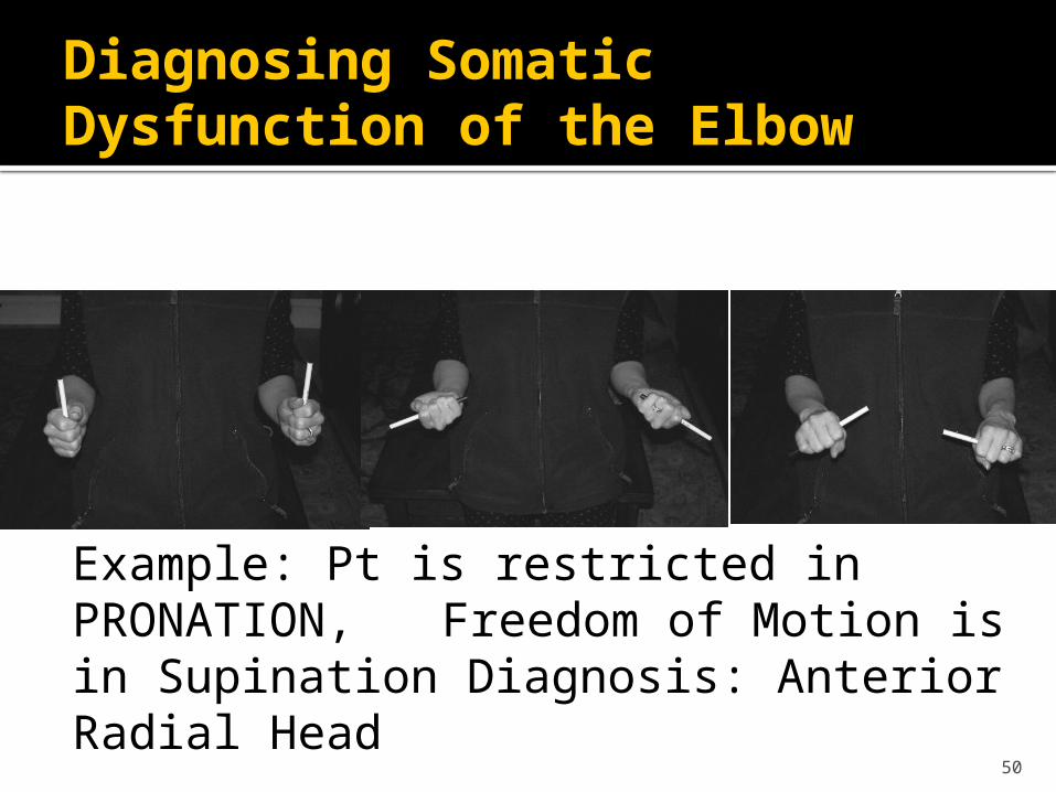
Diagnosing Somatic Dysfunction of the Elbow
50
Example: Pt is restricted in PRONATION, Freedom of Motion is in
Supination Diagnosis: Anterior Radial Head
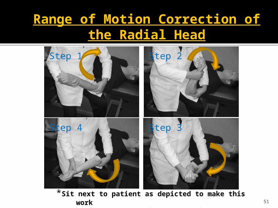
Range of Motion Correction of the Radial Head
51
Step 1 Step 2
Step 4 Step 3
*Sit next to patient as depicted to make this work
* Start in full Pronation and end in full Supination
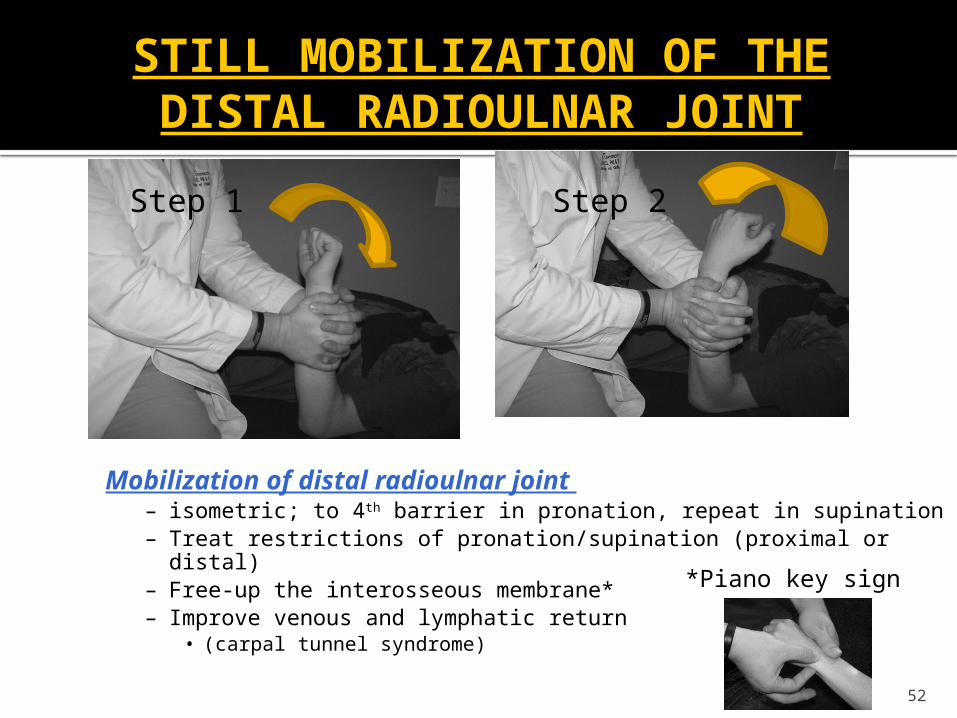
STILL MOBILIZATION OF THE DISTAL RADIOULNAR JOINT
52
Step 1 Step 2
Mobilization of distal radioulnar joint – isometric; to 4th barrier in pronation, repeat in supination– Treat restrictions of pronation/supination (proximal or distal)– Free-up the interosseous membrane*– Improve venous and lymphatic return
• (carpal tunnel syndrome)
*Piano key sign
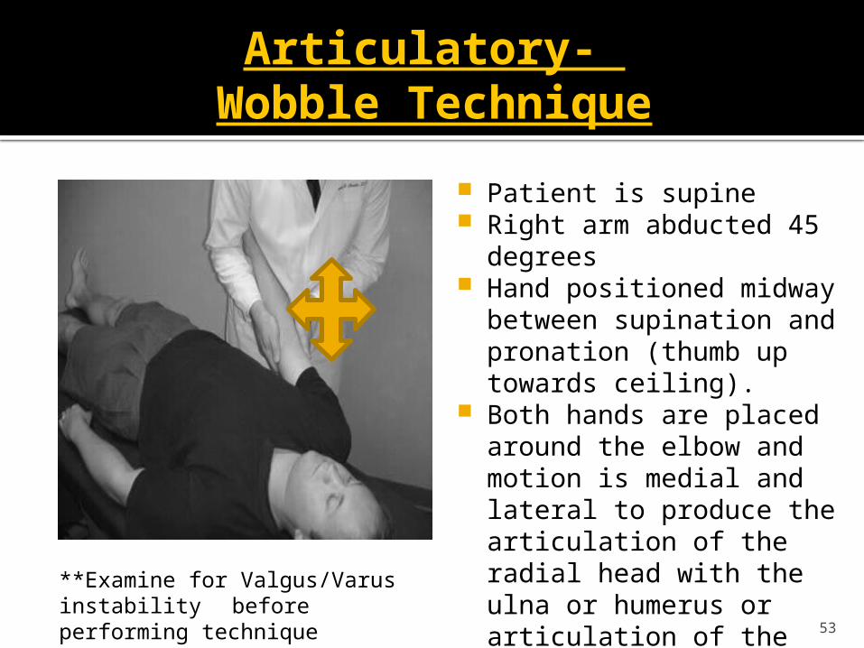
Articulatory- Wobble Technique
Patient is supine Right arm abducted 45 degrees Hand positioned midway between
supination and pronation (thumb up towards ceiling).
Both hands are placed around the elbow and motion is medial and lateral to produce the articulation of the radial head with the ulna or humerus or articulation of the humeroulnar joint.
53
**Examine for Valgus/Varus instability before performing technique
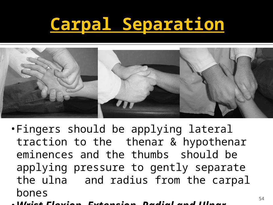
Carpal Separation
54
• Fingers should be applying lateral traction to the thenar & hypothenar eminences and the thumbs should be applying pressure to gently separate the ulna and radius from the carpal bones• Wrist Flexion, Extension, Radial and Ulnar Deviation
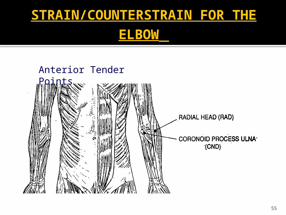
STRAIN/COUNTERSTRAIN FOR THE
ELBOW
55
Anterior Tender Points
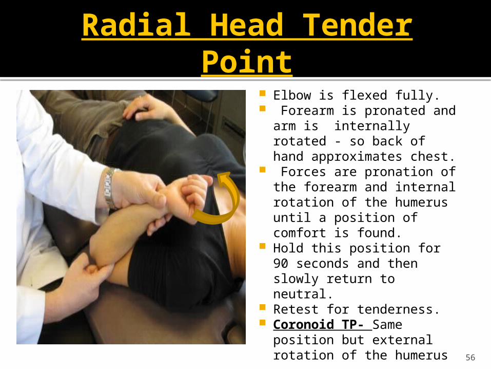
Radial Head Tender Point
Elbow is flexed fully. Forearm is pronated and arm
is internally rotated - so back of hand approximates chest.
Forces are pronation of the forearm and internal rotation of the humerus until a position of comfort is found.
Hold this position for 90 seconds and then slowly return to neutral.
Retest for tenderness. Coronoid TP- Same position
but external rotation of the humerus
56
Conclusion
ALWAYS perform a musculoskeletal and neurologic exam FIRST
OMT learned today for shoulder and elbow Glenohumeral Joint Sternoclavicular and Acromioclavicular joint Scapulothoraic joint Radial Head Humero-Radial Joint Humero-Ulnar Joint Distal Radio-Ulnar Joint Carpal Joints
References
1) Wojtys E. et al. “Sports injuries in the immature athlete.” Orthop Clin North Am 1987; 18 (4): 689-708.
2) Ogata et al.early development and ossification of the human clavicle—an embryologic study.1990, Vol. 61, No. 4 , Pages 330-334
3) Gardner E.”The embryology of the clavicle.” Clin Orthop 1968;58:94) Carreiro, Jane D.O. Pediatric Manual Medicine. (2009). Churchill Livingstone.5) BRIAN L. MAHAFFEY, M.D.PATRICK A. SMITH, M.D. “Shoulder Instability in Young
Athletes.” American Family Physician6) Lawton RL et al. “Pediatric shoulder instability: presentation, findings, treatment, and outcomes.”
J Pediatric Orthop 2002.; 2252-61.7) Good CR et al. “Traumatic shoulder dislocation in the adolescent athlete: advances in surgical
treatment.” Curr Opin Pediatr 2005; 17:25-9.8) Jakobsen BW et al. “Primary repair versus conservative treatment of first-time traumatic anterior
dislocation of the shoulder: a randomized study with 10-year follow-up.” Arthroscopy 2007; 23 (2): 118-23.
9) Krabak et al. “Shoulder and Elbow Injuries in the Adolescent Athlete.” Phys Med Rehabil Clin N Am. 19 (2008) 271-285.
10) American Osteopathic Association. Foundations in Osteopathic Medicine. (2003)
References
11. Young et.al (2011) “Lateral Epicondylitis.” 5-minutle Sports Medicine Consult. Lippincott Williams & Wilkins.
12. Zeisig E. et al.(2006) Extensor origin vascularity related to pain in patients with Tennis elbow. Knee Surg Sports Traumatol Arthrosc.14(7):659.
13. Walz D, et al (2010). Epicondylitis: Pathogenesis, Imaging, and Treatment. Radiographics. 30: 167-184.
14. Gruchow (1979). “Epidemiologic Study of Tennis Elbow. Incidence, recurrence, and effectiveness of prevention strategies”. American Journal of Sports Medicine. 7(4): 234-238.
15. Young et.al (2011) “Medial Epicondylitis.” 5-minutle Sports Medicine Consult. Lippincott Williams & Wilkins.
16. Smidt, N. et al (2002). “Corticosteroid injections, physiotherapy, or a wait-and-see policy for lateral epicondylitis: a randomized controlled trial.” Lancet. 359: 657-662.
59
References
17. Bisset L, et al. (2005) “A systematic review and meta-analysis of clinical trials on physical interventions for lateral epicondylalgia. British Journal of Sports Medicine. 39: 411-422.
18. Grewal R. (2009) “Functional outcome of arthorscopic extensor carpi radialis brevis tendon release in chronic lateral epicondylitis.” Journal of Hand Surgery. 34: 849-857.
19. Des Moines University OMM Department. “Treatment of Elbow Somatic Dysfunctions Laboratory Handout.” Updated 2010.
20. Figueroa J. Professional collaboration with AOA Lateral and Medial Epicondylitis Lecture.
21. Lewis D. Upper Extremity IV Lab and Lecture. Spring 2011. Des Moines University.
22. Simons DG, Travell JG, Simons LS. Myofascial Pain and Dysfunction: The Trigger Point Manual. Volume 1. Upper Half of Body. 2nd Ed. Baltimore, Williams & Wilkins, 1999, pp. 485-907 60
APPENDIX: SUPPLEMENTAL OMM Techniques
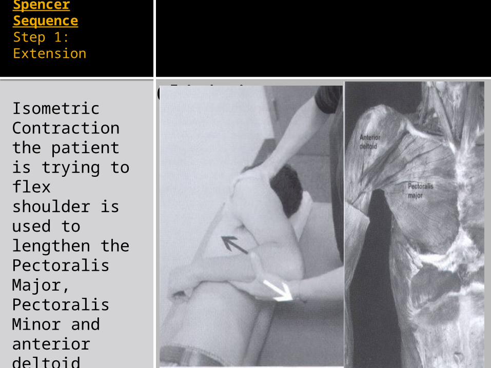
Spencer SequenceStep 1: Extension
Click icon to add pictureIsometric Contraction the patient is trying to flex shoulder is used to lengthen the Pectoralis Major, Pectoralis Minor and anterior deltoid
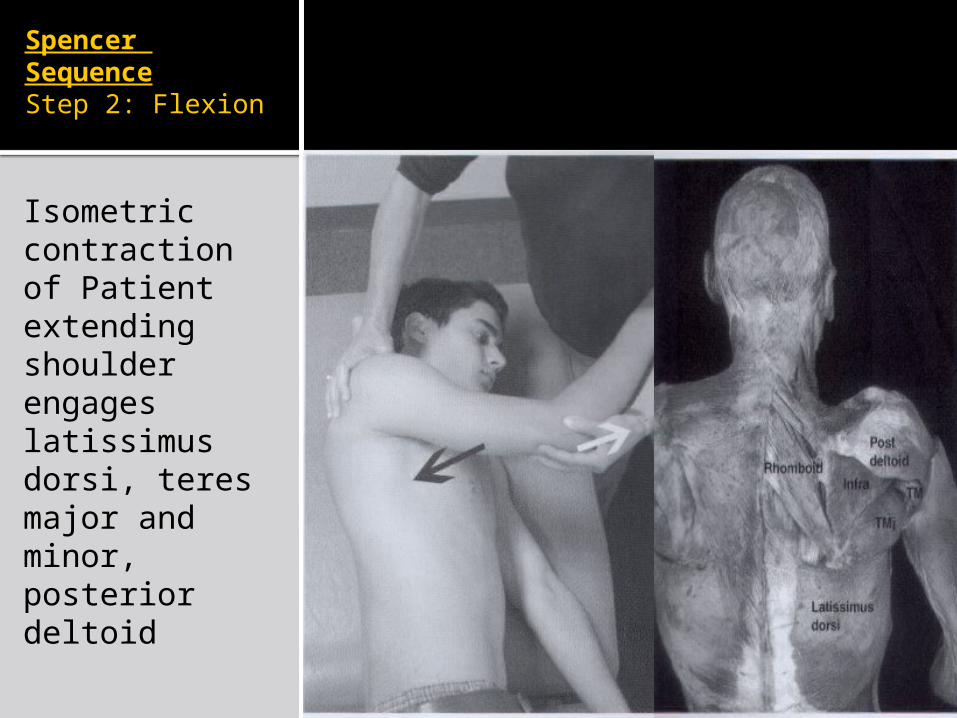
Spencer SequenceStep 2: Flexion
Click icon to add pictureIsometric contraction of Patient extending shoulder engages latissimus dorsi, teres major and minor, posterior deltoid
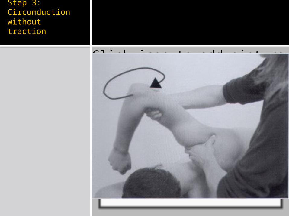
Spencer SequenceStep 3: Circumduction without traction
Click icon to add picture
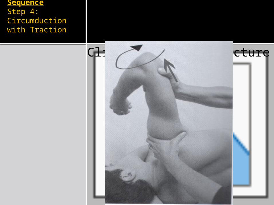
Spencer SequenceStep 4: Circumduction with Traction
Click icon to add picture
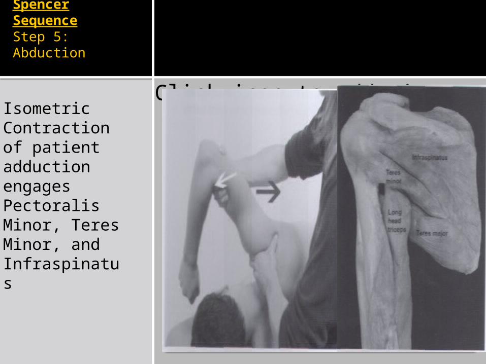
Spencer SequenceStep 5: Abduction
Click icon to add pictureIsometric Contraction of patient adduction engages Pectoralis Minor, Teres Minor, and Infraspinatus
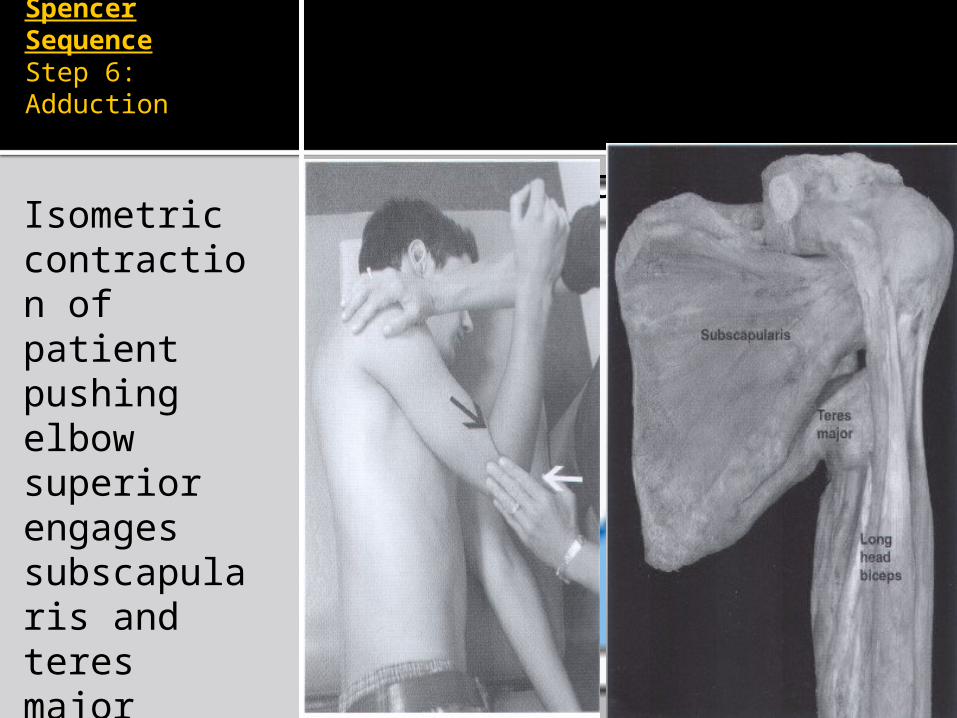
Spencer SequenceStep 6: Adduction
Click icon to add pictureIsometric contraction of patient pushing elbow superior engages subscapularis and teres major
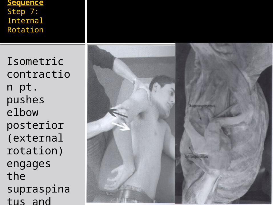
Spencer SequenceStep 7: Internal Rotation
Click icon to add pictureIsometric contraction pt. pushes elbow posterior (external rotation) engages the supraspinatus and infraspinatus muscles
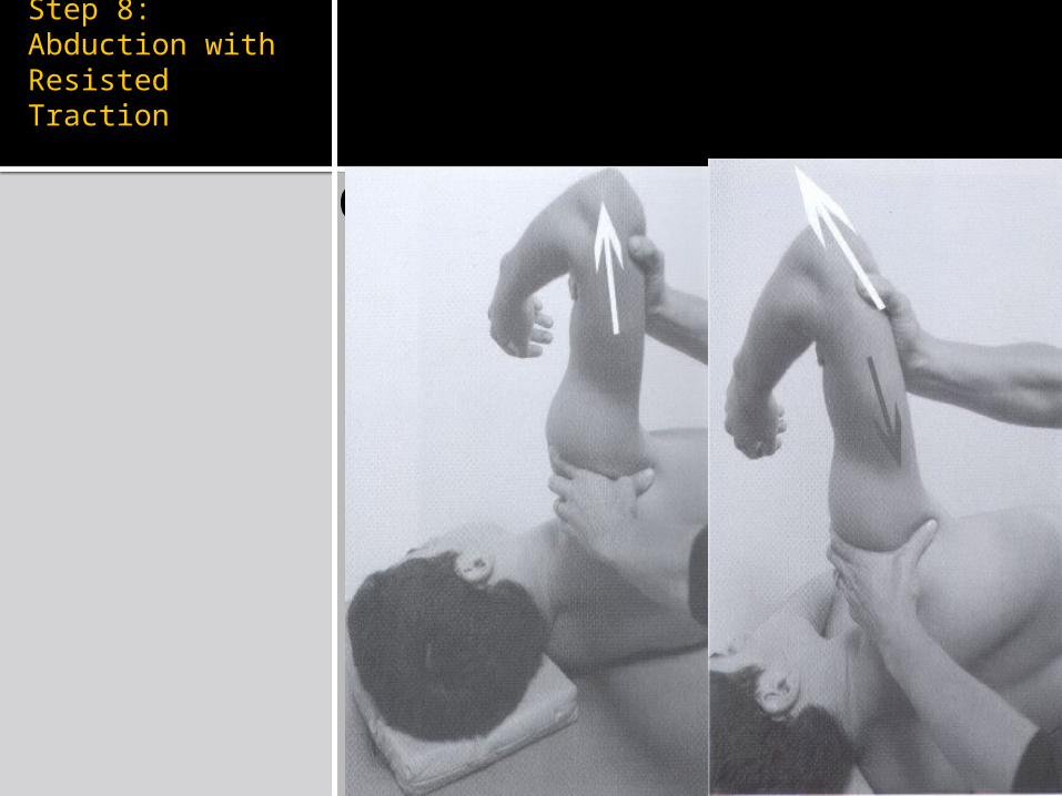
Spencer SequenceStep 8: Abduction with Resisted Traction
Click icon to add picture
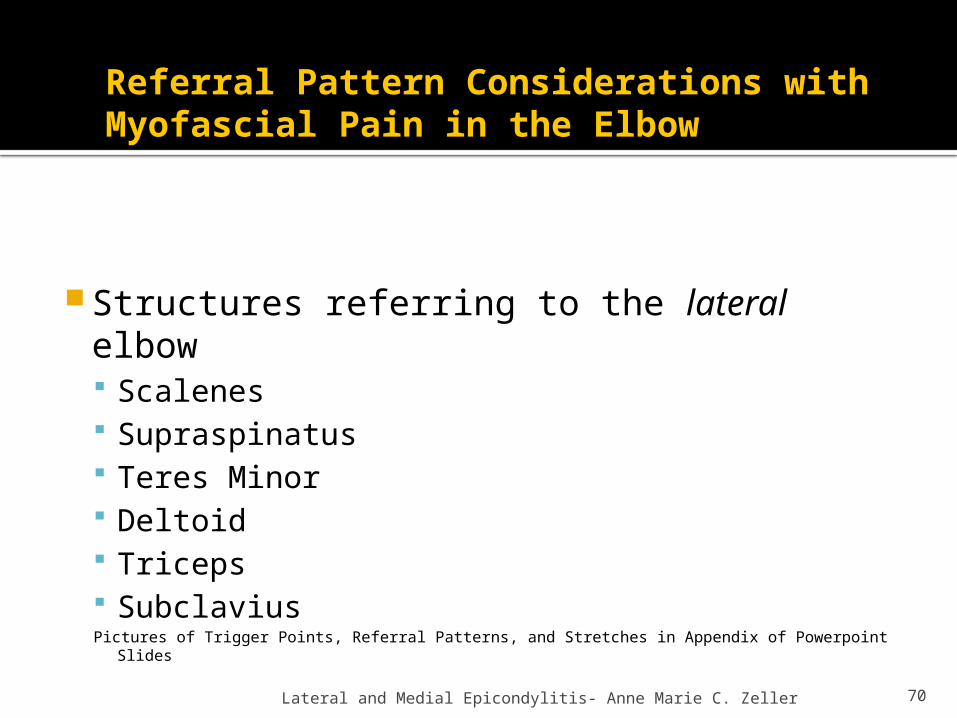
Referral Pattern Considerations with Myofascial Pain in the Elbow
Structures referring to the lateral elbow Scalenes Supraspinatus Teres Minor Deltoid Triceps SubclaviusPictures of Trigger Points, Referral Patterns, and Stretches in Appendix of Powerpoint Slides
Lateral and Medial Epicondylitis- Anne Marie C. Zeller 70
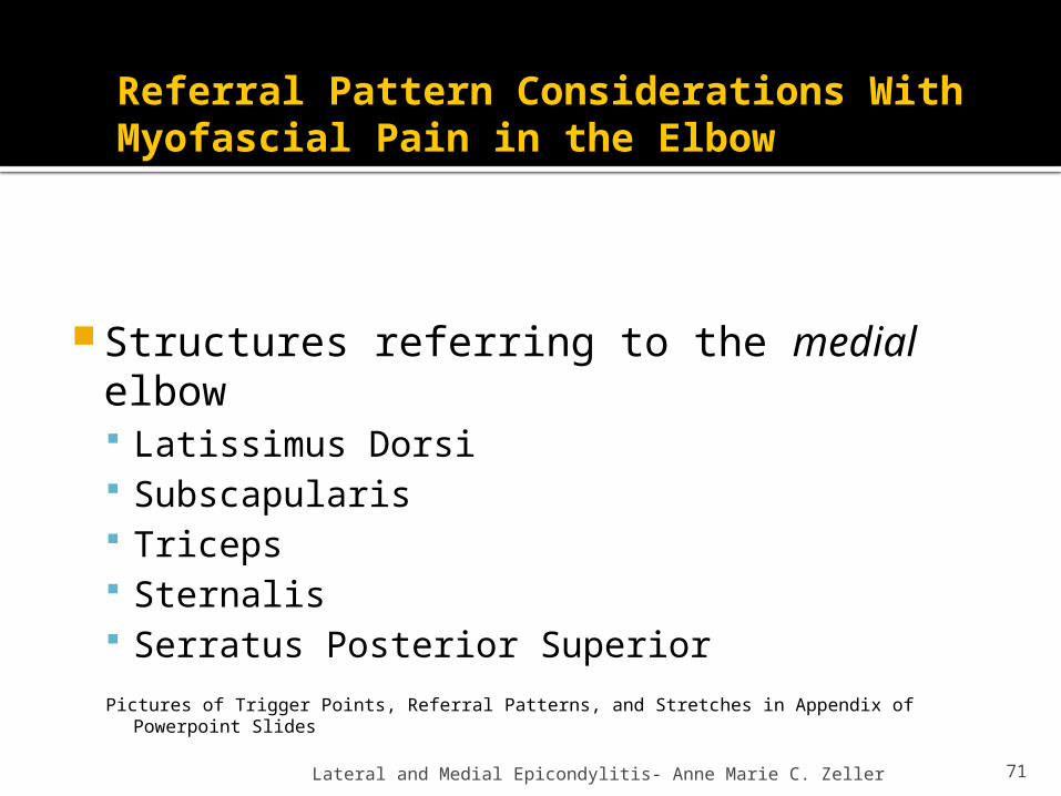
Referral Pattern Considerations With Myofascial Pain in the Elbow
Structures referring to the medial elbow Latissimus Dorsi Subscapularis Triceps Sternalis Serratus Posterior Superior
Pictures of Trigger Points, Referral Patterns, and Stretches in Appendix of Powerpoint Slides
Lateral and Medial Epicondylitis- Anne Marie C. Zeller 71
Treatment of Myofascial Pain
Identify the trigger points: Taut band Tender to palpation Recognition of Pain Referral of pain (“triggers pain somewhere else”)
Treat by stretching May use spray and stretch
Treat by needling Dry needle or infiltrate trigger point with lidocaine
Lateral and Medial Epicondylitis- Anne Marie C. Zeller 72

Myofascial Triggerpoints for Lateral Elbow Pain
73
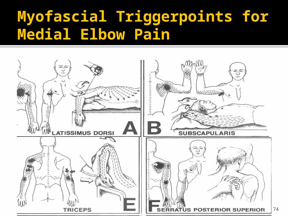
Myofascial Triggerpoints for Medial Elbow Pain
74