obstructive sleep apnea
TRANSCRIPT


Obstructive Sleep Apnea
(OSA)Dr.Muhammad Waseem
Resident Medical Officer
CDU,BVH,Bahawalpur

“That, if then had worked after long sleep,
will make me sleep again”
The Tempest
Shakespeare

SLEEP PHYSIOLOGY
Sleep is a period of bodily rest with
reduced awareness of the environment .
Two phases of sleep
REM
NREM
NREM and REM sleep bouts alternate with
each other throughout night (average
cycle length is 90 minutes)

REM SLEEP
Rapid eye movements
Generalized hypotonia of muscles
Irregular rate and depth of respiration
Marked suppression of hypothalamic
Regulation of homeostasis

NON REM SLEEP
Normal muscle tone
Regular respiration
Four stages of NREM sleep based on EEG
Stage 1-small amplitude high frequency (alpha)
waves resembling awake state
Stage 4-large amplitude and lowest frequency
waves(delta) approaching REM

STAGES OF SLEEP

SLEEP WAVES

CONTROL OF
SLEEP
VLPO=
Ventrolateral
Preoptic Nucleus
ORX=Orexin

CONTROL
OF SLEEP
FUNCTIONS OF SLEEP
“Restoration of body” –as metabolic and energy
demands are reduced.
NREM -replenishes cerebral glycogen stores
REM –restoration of depleted noradrenergic
neurons
Consolidation of memory and improved learning !!
CHANGES IN RESPIRATORY SYSTEM
DURING NREM SLEEP
Depresses activity of respiratory pump muscles
Markedly depresses activity of airway dilator
muscles----- upper airway obstruction
Resultant decreased ventilation causes PaCO2 to
rise by 5-6 mmHg
Fall in PaO2 in sleep does not affect healthy
individuals.
Causes significant hypoxemia in COPD patients who
may require supplemental oxygen during sleep not
during waking hours.

CHANGES IN RESPIRATORY SYSTEM DURING NREM SLEEP
CO2 Apnea threshold increased during NREM sleep .
Hypoxic patients who may be hypocapnic during NREM
there are increased chances of having CSA
UA obstruction due to reduced tone of dilator muscles may
cause OSA during stage 1,2 of NREM as there is reduced
CO2 sensitivity of central chemo receptors ( change in
membrane properties of neurons )
Reduced activity of respiratory motor neurons due to
withdrawal of excitatory effects of wakefulness on these neurons.
Contribute to the hypoventilation that occurs during sleep

CHANGES IN RESPIRATORY SYSTEM DURING REM SLEEP
Profound atonia of all muscles
Irregular respiration results but average ventilation
changes a little compared to wakefulness
Increased upper airway obstruction due to
hypotonia of dilator pharyngeal muscles
Considerable suppression and disorganized
activity of diaphragm
OSA episodes and oxygen desaturation are
longer and more severe than NREM

CARDIOVASCULAR EFFECTS OF REM
Marked fluctuations in sympathetic outflow
Bidirectional changes in HR and BP
Sinus bradycardia, sinus arrest and adverse
cardiac events such as arrhythmias, Ac MI ,
sudden death may occur in patients with CAD

KEY MUSCLES
affecting hyoid: geniohyoid, sternohyoid(XII)
affecting tongue: genioglossus(XII)
affecting palate: tensor palatine,levator palatini(V)
inputs from respiratory centres (ventral medulla)
Most importnat output is negative intrapharyngeal
pressure during inspiration the resulting contraction
keeps pharynx open during inspiration


INTRODUCTION
OSA is a sleep disorder that involves cessation
or significant decrease in airflow in the
presence of breathing effort.
It is the most common type of sleep-
disordered breathing (SDB) and is
characterized by recurrent episodes of upper
airway (UA) collapse during sleep

DEFINITIONS OF RESPIRATORY EVENTS
Sleep Disordered Breathing: repeated
episodes of apnoea/hypopnea during sleep
associated with sleep fragmentations, arousals
& desaturation.
Apnea: is defined by the American Academy
of Sleep Medicine (AASM) as the cessation of
airflow for at least 10 seconds.

Hypopnea: A decrement in airflow of 50% or more
associated with a 4% fall in oxygen saturation
and/or EEG arousal.
Respiratory effort–related arousal (RERA):
Is an event characterized by increasing respiratory
effort for 10 seconds or longer leading to an
arousal from sleep but one that does not fulfil the
criteria for a hypopnea or apnea.

CLASSIFICATION OF APNEAS
Obstructive events are characterized by continued
thoracoabdominal effort in the setting of partial or
complete airflow cessation.
Central events by lack of thoracoabdominal effort in
this setting.
Mixed events have both obstructive and central
features. They generally begin without
thoracoabdominal effort and end with several
thoracoabdominal efforts in breathing

No of apnea plus hypopnea per hour of sleep.
1. It is standard metric used to quantitate the
severity of sleep apnea.
2. An AHI greater than 5 to 10 events per hour
is indicative of OSA.
THE APNEA/HYPOPNEA INDEX
(AHI)

SLEEP-RELATED BREATHING DISORDER
CONTINUUM
obstructive sleep apnea should be considered as a continuum
of disease, i.e., a spectrum of abnormalities from snoring to
obesity-hypoventilation syndrome

PATHOPHYSIOLOGY
Transmural pressure is the difference between
intraluminal pressure and the pressure exerted
by surrounding tissue. Mismatch results in
decrease in calibre of UA
Increase pressure from surrounding tissues
(anatomical)
Decrease in tone of pharyngeal muscles
(neuromuscular)

PHARYNGEAL AIRWAY
Patency depends on balance of forces that tend
to collapse pharynx(negative intraluminal pressure
and extraluminal pressure) and the contraction of
dilator muscles
Transmural press(Ptm)=P(lumen) –P(tissue)
PCritical–at which airflow ceases completely
Normal: –8 cm H2O OSA: > 0 cm H2O


ANATOMIC FACTORS
Enlarged tonsils (children)
volume of the tongue
soft tissue
lateral pharyngeal walls
length of the soft palate
abnormal positioning of the maxilla and
mandible


NEUROMUSCULAR FACTORS
Decreased Neuromuscular activity in the
UA, including reflex activity
Reduced motor output to upper airway
muscles


RISK
FACTORS FOR
OBSTRUCTIVE SLEEP APNEA

EPIDEMIOLOGY
Prevalence in US is 2-4% for women and 4-
9% for men.
SDB remains undiagnosed in approximately
93% of affected women and 82% of
affected men.

HISTORY
Nocturnal Symptoms
Snoring, usually loud, habitual and bothersome to others
Witnessed apnea, which often interrupts snoring and ends with a snort
Gasping and choking sensations that arouse the patient from sleep
Nocturia
Insomnia
Restless sleep, with patients often experiencing frequent arousals and tossing or turning during the night

Daytime Symptoms
Non-restorative sleep (“waking up as tired as when they
went to bed”)
Morning headache, dry or sore throat
Excessive daytime sleepiness (EDS) that usually begins
during quiet activities (reading, watching television); as
the severity worsens, patients begin to feel sleepy
during activities that generally require alertness (school,
work, driving).
Daytime fatigue/tiredness
Cognitive deficits; memory and intellectual impairment
(short-term memory, concentration)

Daytime symptoms(cont.)
Decreased vigilance
Morning confusion
Personality and mood changes, including depression and anxiety
Sexual dysfunction, including impotence and decreased libido
Gastroesophageal reflux
Hypertension
Depression


CONDITION IN WHICH SLEEP APNEA
SHOULD BE SUSPECTED

EXCESSIVE DAYTIME SLEEPINESS
(EDS)
One of the most common and difficult symptom
Reduces quality of life, impairs daytime
performance and causes neurocognitive deficits
(e.g. memory deficits).
Assessed using the Epworth Sleepiness Scale (ESS).
This questionnaire is used to help determine how
frequently the patient is likely to doze off in 8
frequently encountered situations

EPWORTH SLEEPINESS SCALE (ESS)
QUESTIONNAIRE
In contrast to just feeling tired, how likely are you to doze off or fall asleep in the following situations?
1. Sitting and reading
2. Watching TV
3. Sitting inactive a public place (i.e. a theater or a meeting)
4. As a passenger in a car for an hour without break
5. Lying down to rest in the afternoon when circumstances permit
6. Sitting and talking to someone
7. Sitting quietly after lunch without alcohol
8. In a car, while stopping for a few minutes in traffic

0 = Would Never Doze
1 = Slight Chance Of Dozing
2 = Moderate Chance Of Dozing
3 = High Chance Of Dozing
ESS > 10 is considered abnormal.
ESS score of 16 is associated with A greater propensity to fall asleep on the multiple sleep latency test (MSLT) and correlates with MSLT
ESS is useful for evaluating responses to treatment; the ESS score should decrease with effective treatment.

COMMON
CAUSES OF
PERSISTANT
DAYTIME
SLEEPING
TENDENCY

PHYSICAL EXAMINATION
Obesity – Body mass index (BMI) greater than 30 kg/m2
Large neck circumference – Greater than 17 inches in men and 15 inches in women
Abnormal (increased) Mallampati score
Narrowing of the lateral airway walls
Enlarged tonsils
Retrognathia or micrognathia
High-arched hard palate

Systemic Hypertension, Present In
Approximately 50% Of Patients With
OSA
Congestive Heart Failure (CHF)
Pulmonary Hypertension
Stroke
Metabolic Syndrome
Type 2 Diabetes Mellitus

CARDIOVASCULAR DISEASE IN
OBSTRUCTIVE SLEEP APNEA
Excessive Vagal stimulation causes
bradycardia
Bradycardia and hypoxia can provoke
serious cardiac rhythm disturbances i.e.
premature beats ,asystole, ventricular
tachycardia , cardiac arrest.

PROGRESSION OF OSA
Severity of apnea known to worsen over time
Roughly AHI doubles every decade
Wisconsin sleep study AHI increased 2.6 to 5.1 /h
over 8 years
Accelerated by obesity: 10% weight gain caused
32% increase in AHI
Vibration induced trauma and edema of soft
tissues of upper airways and pharyngeal muscle
dysfunction responsible for progression
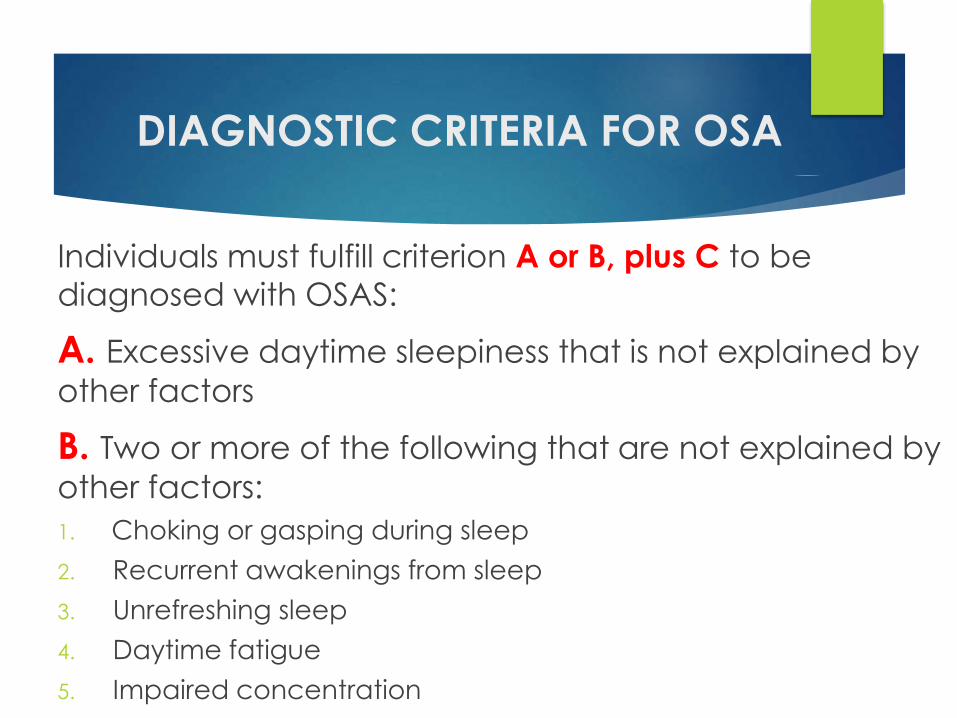
DIAGNOSTIC CRITERIA FOR OSA
Individuals must fulfill criterion A or B, plus C to be
diagnosed with OSAS:
A. Excessive daytime sleepiness that is not explained by
other factors
B. Two or more of the following that are not explained by
other factors:
1. Choking or gasping during sleep
2. Recurrent awakenings from sleep
3. Unrefreshing sleep
4. Daytime fatigue
5. Impaired concentration

C. Overnight monitoring demonstrates 5 to 10
or more obstructed breathing events per hour
during sleep
Or
Greater than 30 events per 6 hours of sleep.
These events may include any combination of
obstructive apnea, hypopnea, or respiratory
effort–related arousals.

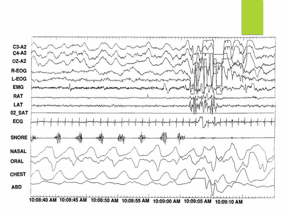
WORK UP
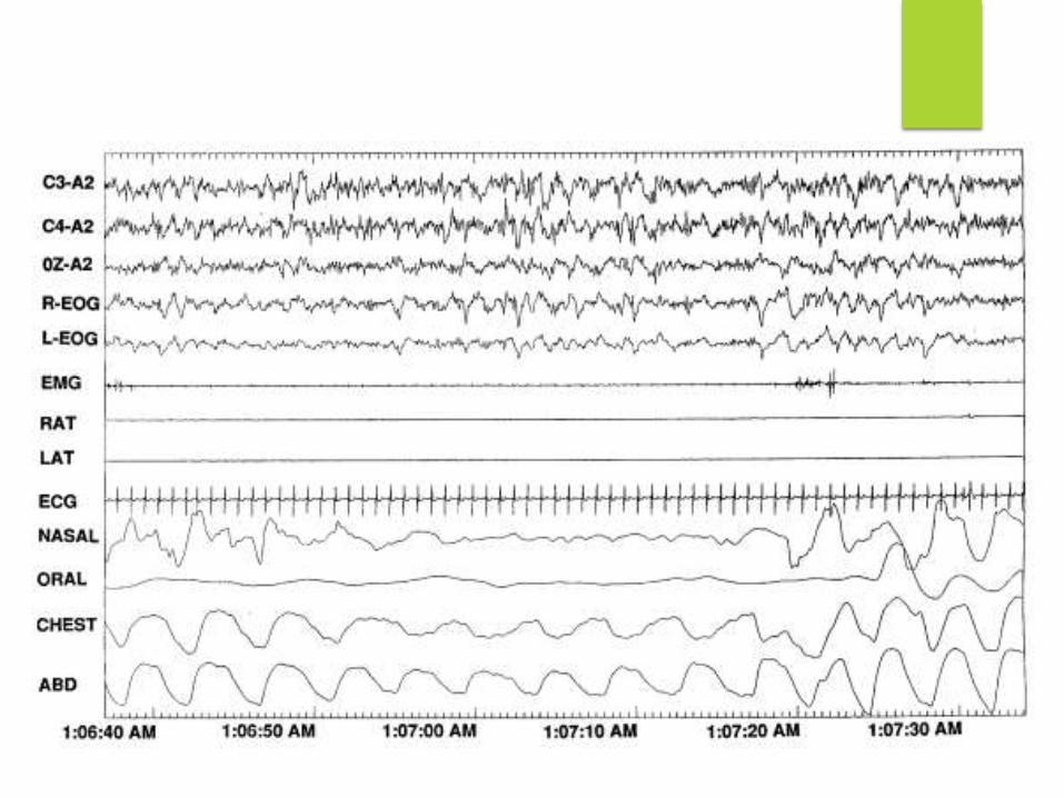
An overnight sleep study, or polysomnography:
In-laboratory measurement of sleep architecture
and electroencephalographic (EEG) arousals,
eye movements (EOG), chin movements,
airflow, respiratory effort, oximetry,
electrocardiographic (ECG) tracings, body
position, snoring, and leg movements
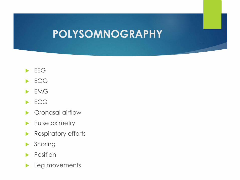
POLYSOMNOGRAPHY
EEG
EOG
EMG
ECG
Oronasal airflow
Pulse oximetry
Respiratory efforts
Snoring
Position
Leg movements

AASM Classification

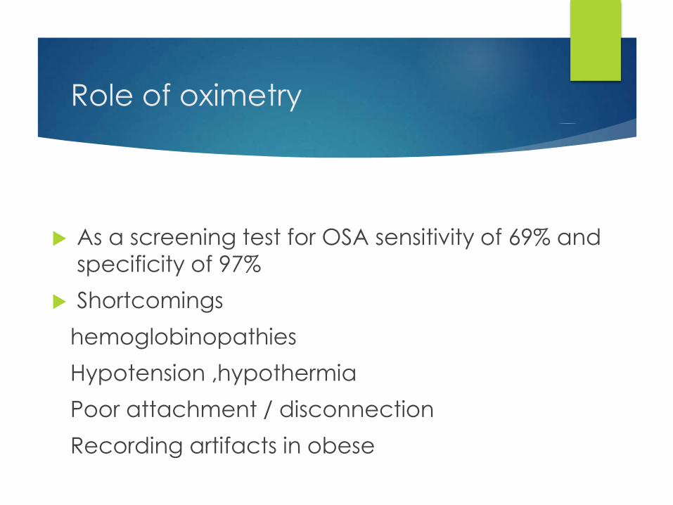
Role of oximetry
As a screening test for OSA sensitivity of 69% and
specificity of 97%
Shortcomings
hemoglobinopathies
Hypotension ,hypothermia
Poor attachment / disconnection
Recording artifacts in obese

Routine Laboratory Tests Usually Are Not
Helpful In Obstructive Sleep Apnea
(OSA) Unless A Specific Indication Is
Present.
Routine Radiographic Imaging Of The
UA Is Not Performed.
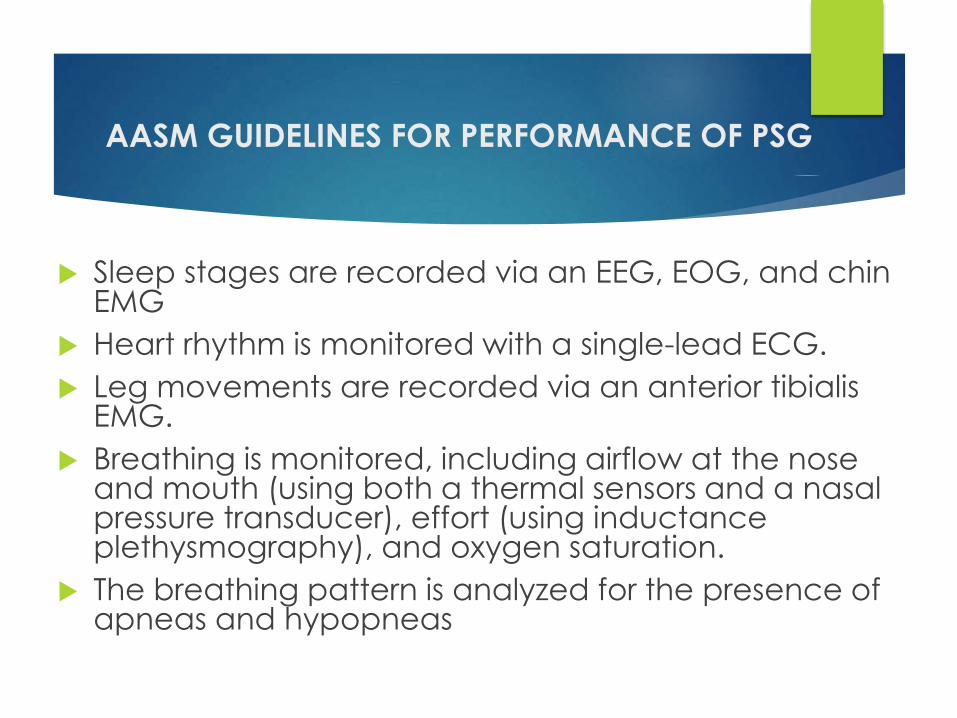
AASM GUIDELINES FOR PERFORMANCE OF PSG
Sleep stages are recorded via an EEG, EOG, and chin EMG
Heart rhythm is monitored with a single-lead ECG.
Leg movements are recorded via an anterior tibialisEMG.
Breathing is monitored, including airflow at the nose and mouth (using both a thermal sensors and a nasal pressure transducer), effort (using inductance plethysmography), and oxygen saturation.
The breathing pattern is analyzed for the presence of apneas and hypopneas


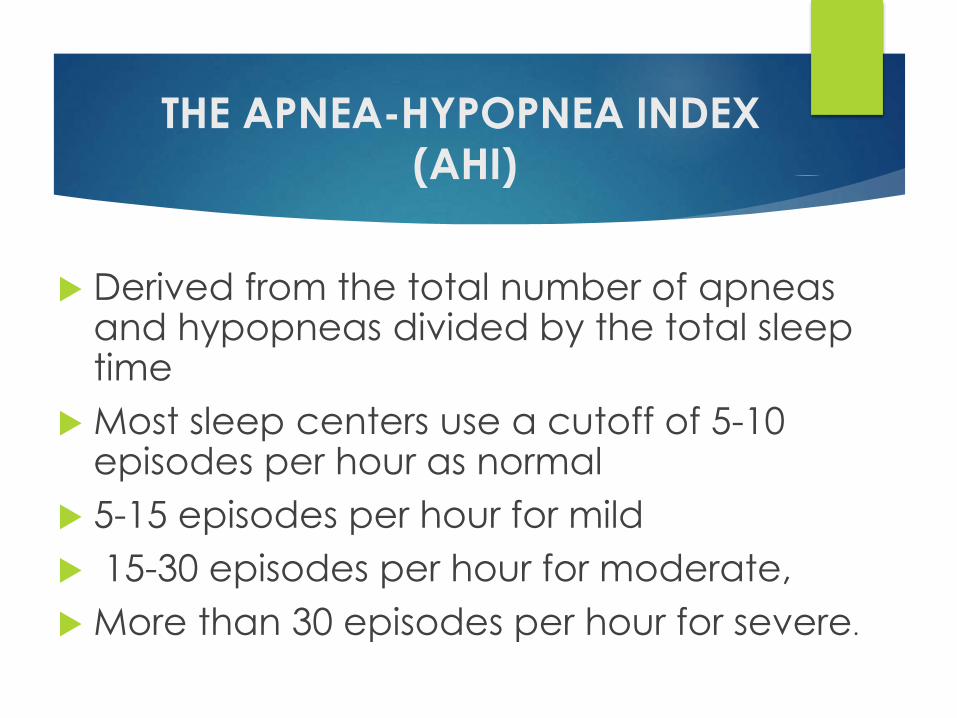
THE APNEA-HYPOPNEA INDEX (AHI)
Derived from the total number of apneasand hypopneas divided by the total sleep time
Most sleep centers use a cutoff of 5-10 episodes per hour as normal
5-15 episodes per hour for mild
15-30 episodes per hour for moderate,
More than 30 episodes per hour for severe.

SPLIT-NIGHT PSG
Patients with high clinical probability and symptoms of OSA should undergo a split-night PSG study.
The final portion of the study is used for titrating the continuous positive airway pressure (CPAP) device.
First part –diagnosis of OSA
Second part –CPAP titration
Useful in patients of OSA with obvious symptoms and AHI > 40/h over first 2 hours
Not recommended for mild to moderate OSA without daytime sleepines


HOME TESTING
3 levels of portable monitors are :
1. LEVEL 2 a portable monitor with the same parameters as a full attended PSG (includes EEG)
2. LEVEL 3 with at least 4 channels, including flow, effort, oximetry and heart rate
3. LEVEL 4 with fewer than 4 channels, often oximetry with flow or oximetry alone.
Level 3 monitors are best used to confirm the diagnosis of OSA

MULTIPLE SLEEP LATENCY TEST
[MSLT ]
Objective measurement of excessive daytime
sleepiness (EDS) consists of 4-5 naps of 20-minute
duration every 2 hours during the day.
The latency to sleep onset for each nap is
averaged to determine the daytime sleep latency.
Normal daytime sleep latency is greater than 10-15
minutes.
OSAHS is generally associated with latencies of less
than 10 minutes.

MSLT is generally used to confirm the
diagnosis of narcolepsy in patients in whom
narcolepsy is a consideration.
Narcoleptic patients have rapid eye
movement sleep on at least 2 of the 4-5 naps
during the day


TREATMENT
Mild apnea have a wider variety of options.
Moderate-to-severe apnea should be treated with
nasal continuous positive airway pressure (CPAP).


CONSERVATIVE NONSURGICAL
TREATMENT
Weight loss- 1% reduction in weight is
associated with 3% reduction in AHI events
Avoidance of alcohol for 4-6 hours prior to
bedtime
Sleeping on one’s side rather than on the
stomach or back
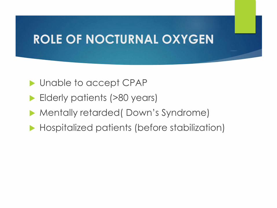
ROLE OF NOCTURNAL OXYGEN
Unable to accept CPAP
Elderly patients (>80 years)
Mentally retarded( Down’s Syndrome)
Hospitalized patients (before stabilization)

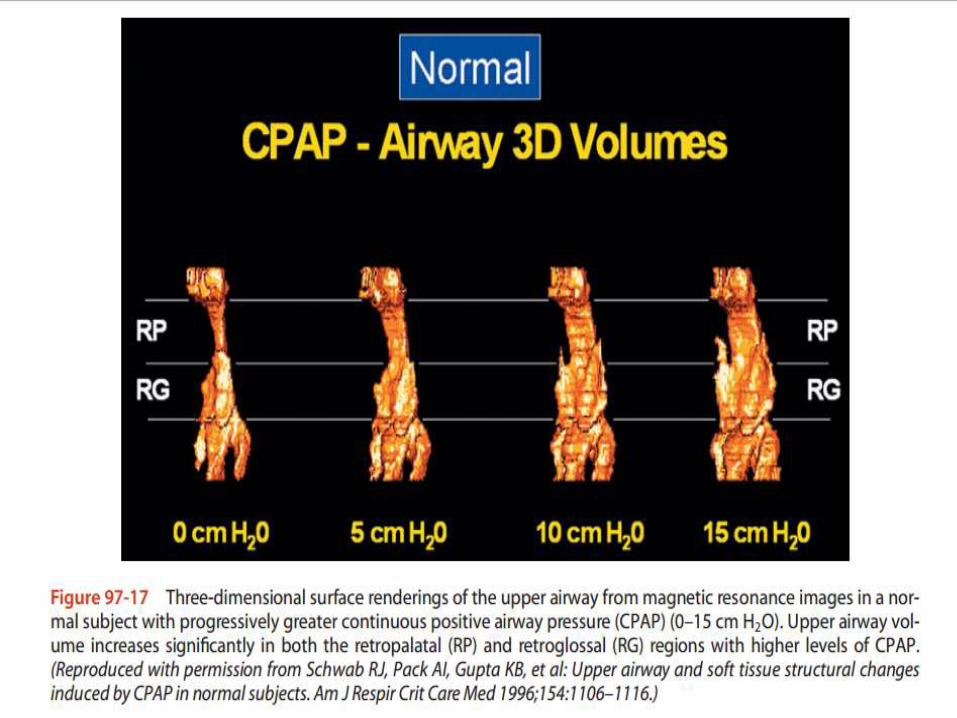
NASAL CPAP THERAPY
The most effective treatment for OSA
Increases the caliber of the airway in the
retropalatal and retroglossal regions
It increases the lateral dimensions of the UA and
thins the lateral pharyngeal walls
Maintain UA patency during sleep, preventing the
soft tissues from collapsing


GUIDELINES FOR USE OF CPAP
All patients with an apnea-hypopnea index (AHI)
greater than 30 regardless of symptoms.
For patients with an AHI of 5-30, CPAP is indicated
if the patient has one of the following: excessive
daytime sleepiness (EDS), hypertension, or
cardiovascular disease.


OTHER MODALITIES
1. Bi-level Therapy
2. Oral Appliances
3. Surgical Correction of the Upper Airway
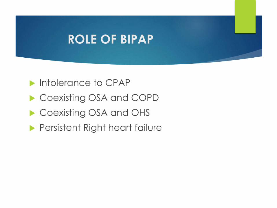
ROLE OF BIPAP
Intolerance to CPAP
Coexisting OSA and COPD
Coexisting OSA and OHS
Persistent Right heart failure

ROLE OF ORAL APPLIANCES
Two types of devices
–Tongue advancing device
–Mandibular repositioning device
Improve airway patency by enlarging the airway
and improving the muscle tone
–Devices are not as effective as CPAP
–Useful for patients with simple snoring
–OSA cases who do not tolerate or fail CPAP


PHARMACOLOGIC THERAPY
Modafinil(CNS stimulant) approved by FDA for use in
patients who have residual daytime sleepiness despite
optimal use of CPAP. Caffiene,nicotine,cannabinoids are
other examples of CNS stimulants
SSRI’s (paroxetine and fluoxetine) shown to increase
genioglossal muscle activity and decrease REM sleep
(apneas are more common in REM),although no
reduction in AHI index
Protriptyline
Respiratory Stimulants (acetazolamide,theophylline,doxapram

OBSTRUCTIVE SLEEP APNEA IN
SPECIAL POPULATIONS
Children
Prevalence-2% , equal in boys and girls
Adenotonsillar hypertrophy is the major etiology
EDS is not a common symptom but problems with
school work
Tonsillectomy is the major treatment.
Pregnancy
IUGR is associated with pregnant women with
untreated OSAHS.

UPPER AIRWAY RESISTANCE
SYNDROME
Abnormal respiratory effort, nasal airflow limitation, minimal or no oxygen desaturation (greater than 90 percent oxygen saturation), and frequent sleep arousals in the absence of obstructive apneas
Untreated diagnosed UARS patients over a 4-year period were found to have increased symptoms of daytime fatigue, insomnia, depression, increased sleep disturbance
First-line treatment is CPAP

OBESITY HYPOVENTILATION SYNDROME
OR THE PICKWICKIAN SYNDROME
Defined by morbid obesity (body mass index greater
than 40 kg/m2) and chronic hypoventilation with
hypercapnia (PaCO2 greater than 45 mmHg) during
wakefulness.
Characteristic findings observed include awake
resting hypoxemia, hyper somnolence, signs of cor
pulmonale (right-sided heart failure and lower
extremity edema) and nocturnal hypoventilation.
The diagnosis of OHS requires a demonstration of at
least a 10 mmHg increment in PaCO2 during sleep.

The TREATMENT STRATEGY for OHS weight loss (which
improves pulmonary function, central ventilatory
drive and concomitant OSA)
However, it should not be used as the only strategy
as it is difficult to achieve and maintain.
Nocturnal non-invasive ventilation(NIPPV), the
treatment of choice, has been demonstrated to
correct daytime and nighttime hypoxemia and
hypercapnia, ameliorates sleep fragmentation,
allows for respiratory muscle rest, reduces
pulmonary artery pressures and improves right
ventricular function.


OSA RELATED SYNDROMES
Syndrome Z :
syndrome X(metabolic syndrome) + OSA.
Overlap Syndrome :
chronic obstructive pulmonary disease +OSA

“The secret is here in the present. If
you pay attention to the present, you
can improve upon it. And, if you
improve on the present, what comes
later will also be better."
“The alchemist” by PAULO COEHLO

Dedicated to Dr Sami
Dogar
