ocr a2 f214 hormones · web view2015/05/04 · ocr a2 f214 hormones specification: define the terms...
TRANSCRIPT

OCR A2 F214 HORMONES
Specification:
1. Define the terms endocrine gland, exocrine gland, hormone and target tissue
2. Explain the meaning of the terms first messenger and second messenger, with reference to adrenaline and cyclic AMP (cAMP)
3. Describe the functions of the adrenal gland
4. Describe, with the aid of diagrams and photographs, the histology of the pancreas and outline its role as an endocrine and exocrine gland
5. Explain how blood glucose concentration is regulated, with reference to insulin, glucagon and the liver
6. Outline how insulin secretion is controlled with reference to potassium channels and calcium channels in beta cells .Compare and contrast the causes of Type 1 (insulin dependent) and Type 2 (non-insulin dependent) diabetes mellitus
7. Discuss the use of insulin produced by genetically modified bacteria and the potential use of stem cells, to treat diabetes mellitus
.8. Outline the hormonal and nervous mechanisms involved in the control of
heart rate in humansDefinitions
TERM MEANINGEndocrine gland Glands that produce and secrete
hormones into blood plasma
Endocrine glands are ductless glands
Exocrine gland Glands that secrete substances into a duct that transports the secretions to a local area where the secretions are used
eg salivary glands secreting saliva into the mouth, the pancreas secretes digestive enzymes into the small intestine
Hormone Small molecules, secreted by endocrine glands into blood plasma
Hormones are transported to and affect the activity of specific target tissues
Hormones may be polypeptides or proteins (insulin), steroids (testosterone) or amines (adrenaline)
Endocrine glands secrete low concentrations of hormone very rapidly when stimulated
1

Hormones have a very short life in the body. Some are broken down in the liver. Adrenaline lasts for 1-3 minutes
Target tissue Target tissue consists of target cells with specific receptor molecules within their plasma membranes, to which specific complementary hormone molecules bind. The hormone and receptor molecules have complementary shapes
For example, insulin binds to specific receptor molecules in the plasma membranes of liver and muscle cells, the main insulin target cells
Indicate the exocrine and endocrine glands in the diagram below
First and Second Messengers
When an endocrine gland is stimulated by a change in its environment, a hormone is released. The hormone is the first messenger
The hormone binds to a specific hormone receptor molecule in the plasma membrane of the target cell.
2

This binding of the hormone activates the synthesis of another molecule inside the target cell called the second messenger.
The second messenger stimulates a sequence of events in the cell, involving activation of enzymes. This sequence of events is sometimes described as a cascade of events
The Action of Adrenaline to Illustrate the First and Second Messengers
Adrenaline is secreted by the adrenal medulla region of the adrenal glands (positioned just above the kidneys) in response to stress, excitement or danger.
The liver is one target organ for adrenaline
Adrenaline is a soluble amine and unable to enter its target cells
1. Adrenaline is the first messenger that binds to specific adrenaline receptors in the plasma membrane of liver cells
2. This binding is possible because adrenaline molecules have a specific shape that is complementary to the receptor molecules
3

3. Binding of adrenaline to receptor activates a G-protein (in the plasma membrane)
4. Activated G-protein activates adenyl cyclase (an enzyme within the membrane – see diagram below)
5. Adenyl cyclase catalyses the conversion of ATP to cyclic AMP (cAMP). Cyclic AMP is the second messenger
6. cAMP causes further events to occur in the liver cell involving the activation of other enzymes. This enzyme activation may involve phosphorylating the enzyme or altering its 3D structure. The second messenger system is a multi-step process described as a cascade effect
7. The final reaction is the hydrolysis of glycogen to glucose:
glycogen + water glucose
The enzyme glycogen phosphorylase catalyses this hydrolysis of glycogen to glucose (glycogenolysis)
How the Multi-step Process enables a small number of adrenaline molecules to cause the release of many glucose molecules from liver cells
1. One adrenaline molecule causes the synthesis of many cAMP molecules
2. This multiplying effect occurs at every later step in the pathway because cAMP temporarily binds to an enzyme and is recycled to activate other enzymes
3. The multiplying effect is often described as a cascade of events
Functions of the Adrenal Gland
The two adrenal glands lie just above the two kidneys
Each adrenal gland has a cortex (outer region) and medulla (inner region)
The Adrenal Cortex
Produces some steroid hormones
Mineralocorticoids such as aldosterone that controls sodium and potassium concentrations in blood plasma. Target organ is the kidney
. Glucocorticoids such as cortisol that stimulates the synthesis of glucose
from amino acids in the liver
The Adrenal Medulla
The adrenal medulla secretes adrenaline in response to stress, excitement and danger
Adrenaline is a catecholamine made from amino acids
4

Most cells have adrenaline receptor molecules. There are many adrenaline target tissues and the effects of adrenaline are widespread and different in different target tissues
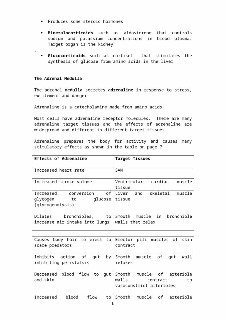
Adrenaline prepares the body for activity and causes many stimulatory effects as shown in the table on page 7
Effects of Adrenaline Target Tissues
Increased heart rate SAN
Increased stroke volume Ventricular cardiac muscle tissue
Increased conversion of glycogen to glucose (glycogenolysis)
Liver and skeletal muscle tissue
Dilates bronchioles, to increase air intake into lungs
Smooth muscle in bronchiole walls that relax
Causes body hair to erect to scare predators
Erector pili muscles of skin contract
Inhibits action of gut by inhibiting peristalsis
Smooth muscle of gut wall relaxes
Decreased blood flow to gut and skin Smooth muscle of arteriole walls contract to vasoconstrict arterioles
Increased blood flow to muscles and brain
Smooth muscle of arteriole walls relax to vasodilate arterioles
Why Adrenaline has Different Effects on Different Target Tissues
Different target tissues may have different types of adrenaline receptors
The concentrations of cAMP may be higher or lower in different cells
The second messenger may be different in different target cells
The second messenger may activate different enzymes
The Pancreas
Location of the Pancreas in the Body
5
The pancreas is a small organ lying below the stomach
The pancreas has both exocrine and endocrine functions
Exocrine Functions of the Pancreas
The pancreas secretes pancreatic juice into the pancreatic duct that empties the juice into the first section of the small intestine -the duodenum
The release of pancreatic juice is triggered by nervous stimulation
Pancreatic juice contains:
Lipase – an enzyme that hydrolyses the digestion of lipids to fatty acids and glycerol
Amylase – an enzyme that hydrolyses starch to maltose
Trypsin – an enzyme that hydrolyses proteins to polypeptides
Sodium hydrogen carbonate – making the fluid alkaline (pH 8.0) to neutralise the acidic contents of the duodenum (the chyme that has just left the stomach)
Most of the pancreatic cells synthesise these hydrolytic enzymes
Endocrine Functions of the Pancreas
The pancreas has tissues within the Islets of Langerhans that produce and secrete two hormones directly into blood plasma.
The islets of Langerhans contain two types of cells:
The alpha cells (α-cells) produce and secrete the hormone glucagon into blood plasma
The beta cells (β-cells) produce and secrete the hormone insulin into blood plasma
Both α-cells and β-cells detect the blood plasma concentrations of glucose and respond accordingly
6

Check also the photomicrograph picture of pancreatic tissue on page 24,
Name cells (a) and (b) in the diagrams above
7

Control of Blood Plasma Glucose Concentration
Normal blood plasma glucose concentration is 90mg 100cm-3 (also expressed between 4-6 mmoldm-3)
Blood plasma glucose concentration is carefully controlled by negative feedback mechanisms
Define negative feedback: …………………………………………………
…………………………………………………………………………………
…………………………………………………………………………………
Why is it Important to control Blood Plasma Glucose Concentration?
Glucose is the main respiratory substrate
Glucose affects the water potential of the blood plasma and deviations in this water potential will cause damage to cells
If blood plasma glucose concentration is above the normal level for too long (hyperglycaemia) plasma water potential will be lower than that in cells. Blood cells and cells in the tissues will lose water by osmosis and shrink
If blood plasma glucose concentration is lower than the normal level for too long (hypoglycaemia) plasma water potential will be higher than that in cells. Blood cells and cells in the tissues will gain water by osmosis and burst
Response if Blood Glucose Concentration Increases
1. Increase in blood plasma glucose concentration above a threshold level is detected by β cells in the Islets of Langerhans
2. β-cells secrete insulin into the blood plasma
3. The target/effector cells are the hepatocytes (liver cells) and muscle cells that have specific receptor molecules for insulin in their plasma membranes
4. Insulin binds to the insulin receptor molecules in the plasma membranes of the target cells
5. This binding of insulin to hepatocyte receptors, activates a series of enzyme-controlled reactions in the cells as follows:
More protein channels for glucose transport are placed in the membrane via vesicles from the Golgi apparatus
Therefore more glucose enters the cell via the glucose channels
More glucose is converted to glycogen for storage (glycogenesis)
More glucose is used in respiration of the target cells
6. As more glucose enters the cell, blood plasma glucose concentration decreases and the secretion of insulin from β-cells decreases
8

Response if Blood Plasma Glucose Concentration Decreases
1. Decrease in plasma glucose concentration below the threshold level is detected by the α-cells in the Islets of Langerhans
2. α-cells secrete glucagon into blood plasma
3. The target cells are the hepatocytes (liver cells) and muscle cells that have specific receptor molecules for glucagon in their plasma membranes
4. Glucagon binds to the glucagon receptor molecules in the plasma membranes of the target cells (hepatocytes)
5. This binding of glucagon to cell receptors, activates adenyl cyclase in the plasma membranes. Adenyl cyclase catalyses the conversion of ATP to cyclic AMP inside the hepatocytes
6. cAMP activates a series of enzyme-controlled reactions in the cells as follows:
increased hydrolysis of glycogen to glucose (glycogenolysis)
increased synthesis of glucose from amino acids and lipids (gluconeogenesis)
use of more fatty acids in respiration
7. Glucose leaves the hepatocytes through glucose channels by facilitated diffusion
8. The overall effect is to increase blood plasma glucose concentrations and this leads to a reduction in secretion of glucagon from α-cells
Note:
The secretion of insulin into blood plasma inhibits the secretion of glucagon
The secretion of glucagon into blood plasma inhibits the secretion of insulin
9

Some Important Definitions to Learn:
Glycogenesis: conversion of glucose to glycogen
Which hormone stimulates glycogenesis? ..........................................
Glycogenolysis: hydrolysis of glycogen to glucose
Which hormones stimulate glycogenolysis? ………………………….
………………………………………………………………………………
Gluconeogenesis: synthesis of glucose from amino acids and lipids
Which hormone stimulates gluconeogenesis? ……………………….
Control of Insulin Secretion from β-cells
The mechanism of insulin secretion is complex and not fully understood but the main processes are indicated below
1. The plasma membrane of a β-cell has K+ channels that are normally open allowing K+ ions to diffuse out of the cell. This diffusion of K+ ions maintains a membrane potential difference of -70mV (more negative inside the cell) – the membrane is polarised
2. When glucose levels outside the β-cells increase, more glucose diffuses into the cells via glucose transporter proteins
3. Inside the cell, more glucose is phosphorylated (first stages of glycolysis) and metabolised in glycolysis in the cell cytoplasm to produce more ATP
4. Increased levels of ATP in the β-cells cause closure of K+ channels
5. K+ ions cannot diffuse out of the cell and the membrane potential reduces to -30mV. The membrane is depolarised
6. Ca 2+ channels in the membrane are normally closed. As the membrane potential changes, the Ca 2+ channels open and Ca 2+ ions diffuse into the cell down an electrochemical gradient
7. The Ca 2+ ions cause the vesicles containing insulin, to move to the plasma membrane, fuse with the membrane and release insulin by exocytosis
Diabetes Mellitus
Diabetes mellitus is a disease in which the body can no longer control its blood plasma glucose concentrations.
After a meal, blood plasma glucose concentrations reach very high levels – the patient becomes hyperglycaemic.
After exercise or fasting, the diabetic becomes hypoglycaemic as blood plasma glucose concentrations fall too low
Symptoms of Diabetes
10

Symptom Explanation
Glucose in urine High blood plasma glucose concentration leads to high glucose concentrations in the glomerular filtrate in the kidneys. Not all of the glucose is reabsorbed back into the blood plasma and excess glucose is lost in urine
Excretion of large volumes of urine
The presence of glucose in the nephron filtrate in the collecting ducts reduces the filtrates water potential and makes the water potential gradient between filtrate and blood plasma less steep. Therefore, not enough water is reabsorbed back into the blood plasma. More water is lost in the urine
Excessive thirst The plasma water potential remains too low because insufficient water is reabsorbed back into the plasma from the kidney nephron filtrate. The low plasma water potential stimulates the sensation of thirst
Tiredness and fatigue Less glucose is taken up by body cells. There is less glucose in cells as a substrate for aerobic respiration and less ATP produced as an energy source. The glycogen energy stores in cells are lower since less glucose is converted to glycogen in cells
Type 1 Diabetes
Starts in childhood and called juvenile-onset diabetes
Pancreas cannot secrete enough insulin
Often called insulin-dependent diabetes since patients require daily insulin therapy (injections)
Cannot be treated by dietary changes alone (such as reducing carbohydrate intake)
May be due to an autoimmune response in which the patients own immune system attacks the β-cells and destroys them
Risk Factors for Type 1 Diabetes
Probably a genetic link
An autoimmune disease that may be triggered by a viral infection
Type 2 Diabetes
Begins later in life – often starts in middle age (50 years +)
The pancreas does secrete insulin but the liver and other target organs do not respond to insulin. There is probably a reduction in the specific insulin receptors made by target cells
Often called non-insulin-dependent diabetes since insulin therapy has no effect on the symptoms
Risk Factors for Type 2 Diabetes
Obesity
A diet high in sugars, particularly refined sugars
Being of Asian or Afro-Caribbean origin
11

Family history
Treatment of Diabetes
There is no cure for diabetes
Type 1 diabetes is treated by insulin injection. The patient must check their blood glucose concentration regularly with a simple sensor. Urine can be checked for glucose using a dipstick
Type 2 diabetes is treated by diet and exercise
Long Term Complications of Diabetes
Complications are more likely if the condition is not well controlled.
The complications result from cell damage because the blood plasma water potential is not controlled within narrow limits on an hour to hour and day to day basis.
Blood cells and cells in the tissues may lyse or crenate if the external fluid water potential is persistently too high (causing cell lysis) or too low (causing cell crenation)
Damage is likely to occur in the following organs and tissues:
The eye – retinal damage may occur leading to blindness
The kidney – leading to renal failure and the need for dialysis or kidney transplants
The cardiovascular system – damage to blood vessels is more likely leading to atherosclerosis
Sources of Insulin
1. Traditionally, insulin from pig pancreas was used but this is now in short supply
2. GM (genetically modified) insulin is now used. Escherichia coli bacteria are genetically modified by incorporating the human insulin gene into the E.coli genome
3. In the future, it is hoped that stem cells can be used to produce β-cells. Research has been carried out with human embryonic stem cells but problems have been encountered. Pancreatic stem cells have been found in mouse pancreas and these stem cells may be used to produce mouse β cells. If similar stem cells can be found in the human pancreas, stem cell therapy could be used as a treatment for diabetics.
The advantages of using stem cell therapy are:
It has the potential to cure the condition (insulin treatment only manages the condition)
The treatment is permanent and has a long term effect. Insulin treatment requires regular repeated treatment throughout life
Advantages of using Insulin from Genetically Modified Bacteria
12

The insulin produced is human, therefore no chance of an allergic reaction to the product
It is cheaper to produce than to extract from animals
Production can be matched to demand and therefore the supply is dependable
Less ethical objections since there is no cruelty to pigs
No religious objections compared with pig insulin
Can be used by vegetarians
The product quality is standardised and reliable
Comparing Control by Nerves and Control by HormonesFeature Control by nerves Control by Hormones
Pathways Along neurones In blood plasma
Form of information Nerve impulses Chemicals
Speed Fast Slow
Target area Cell or tissue at the end of the neurone
Whole tissues or organs
Duration of effect Short term Short term or long term (growth)
Type of response Muscle contraction or secretion by glands
Many different responses
Control of Heart Rate in Hum ans
Some Useful DefinitionsHeart Rate is the number of heart beats per minute or the number of cardiac cycles per minuteStroke Volume is the volume of blood pumped through the heart in one cardiac cycle. The units are cm3 or dm3
Cardiac Output is the volume of blood pumped through the heart in one minute. It is calculated by the following equation: Cardiac Output = Heart Rate x Stroke Volume
Heart rate control Cardiac muscle is myogenic – it is self- stimulating and does not need any
nervous impulses from the central nervous system to maintain a steady state heart rate
The SAN is the pacemaker that initiates the waves of excitation to bring about atrial and ventricular contractions. Under resting conditions, the SAN controls the heart rate at about 60-80 beats per minute
However, the cardiac muscle does need nervous impulses from the brain to increase or decrease the heart rate from the steady state level
13

The heart is supplied by autonomic nerves from the cardiovascular control centre in the medulla oblongata of the brain
The nervous impulses along these nerves innervate (connect to) the SAN and can alter the frequency of the waves of excitation from the SAN
Impulses transmitted from the medulla oblongata along the accelerator nerve (sympathetic nerve) cause an increase in heart rate. Impulses transmitted from the medulla oblongata along the vagus nerve (parasympathetic nerve) cause a decrease in heart rate
Adrenaline also causes an increase in heart rate and an increase in stroke volume
Label the autonomic nerves on the diagram above:
1. The accelerator nerve
2. The vagus nerveFactors Leading to an Increase in Heart Rate
1. Increased concentration of CO2 in blood plasma resulting from increased muscle contraction. Blood pH decreases and this is detected by chemoreceptors in the carotid arteries, aorta and medulla oblongata. Sensory impulses are transmitted from these receptors to the medulla oblongata. The response is impulses passed along the acceleratory nerve to the SAN
2. During exercise, limb movement is detected by stretch receptors in the skeletal muscles. Sensory impulses along sensory neurones are passed to the cardiovascular control centre in the medulla oblongata. The response is impulses passed along the acceleratory nerve to the SAN
Factors Leading to a Decrease in Heart Rate
Blood pressure is monitored by baroreceptors in the walls of the carotid artery and the aorta. If blood pressure in these vessels increases, sensory
14

impulses to the cardiovascular centre result in motor impulses along the vagus nerve to the SAN leading to a decrease in heart rate
15