ocular trauma dr jo dalgleish facem medical education eastern health
TRANSCRIPT

Ocular Ocular TraumaTrauma
Dr Jo DalgleishDr Jo DalgleishFACEMFACEM
Medical Education Medical Education
Eastern HealthEastern Health

Ocular TraumaOcular Trauma►Trauma HistoryTrauma History
►History of the injuryHistory of the injury►Details of traumaDetails of trauma
►Pre injury visionPre injury vision►Previous ocular injuriesPrevious ocular injuries►Medical historyMedical history►Current medicationsCurrent medications►AllergiesAllergies
Ocular TraumaOcular Trauma►Trauma ExaminationTrauma Examination
►Visual AcuityVisual Acuity►May need topical anaesthesiaMay need topical anaesthesia
►Pupil testingPupil testing►Eye movementEye movement►Visual fieldsVisual fields►Palpation eyelids and orbital marginsPalpation eyelids and orbital margins►Sensation testing Sensation testing
Forehead, cheekForehead, cheek

Ocular TraumaOcular Trauma►Trauma ExaminationTrauma Examination
►Slit lampSlit lamp►Including fluorescein stainingIncluding fluorescein staining
►Seidel TestSeidel Test►Applanation tonometryApplanation tonometry►Dilated fundus examDilated fundus exam►Ancillary TestsAncillary Tests
►Color visionColor vision►GonioscopyGonioscopy►Imaging studiesImaging studies

Ocular TraumaOcular Trauma► Non penetratingNon penetrating
► AbrasionsAbrasions► Lacerations (partial thickness)Lacerations (partial thickness)► Chemical injuriesChemical injuries► RadiationRadiation
► PenetratingPenetrating► BluntBlunt
► Subconjunctival haemorrhageSubconjunctival haemorrhage► HyphemaHyphema► Iris damageIris damage► Cataracts & lens dislocationsCataracts & lens dislocations► Retinal tears and detachmentsRetinal tears and detachments► Orbital fracturesOrbital fractures► Retro bulbar haemorrhageRetro bulbar haemorrhage

Ocular TraumaOcular Trauma►Corneal & Conjunctival AbrasionsCorneal & Conjunctival Abrasions
►SymptomsSymptoms►PainPain►PhotophobiaPhotophobia►Foreign body sensationForeign body sensation►Epiphoria (tearing)Epiphoria (tearing)►History of scratching the eyeHistory of scratching the eye
Ocular TraumaOcular Trauma►Corneal & Conjunctival AbrasionsCorneal & Conjunctival Abrasions
►SignsSigns►Epithelial staining defect with Epithelial staining defect with fluoresceinfluorescein
►Conjunctiva injectionConjunctiva injection►Swollen eyelidSwollen eyelid►Mild anterior chamber reactionMild anterior chamber reaction►Mild subconjunctival haemorrhageMild subconjunctival haemorrhage►Negative Seidels testNegative Seidels test

Ocular TraumaOcular Trauma►Corneal & Conjunctival AbrasionsCorneal & Conjunctival Abrasions
Radiation InjuriesRadiation Injuries

Ocular TraumaOcular Trauma►Corneal & Conjunctival AbrasionsCorneal & Conjunctival Abrasions

Ocular TraumaOcular Trauma►Corneal & Conjunctival AbrasionsCorneal & Conjunctival Abrasions

Ocular TraumaOcular Trauma►Corneal & Conjunctival AbrasionsCorneal & Conjunctival Abrasions
►ExaminationExamination Visual acuityVisual acuity Slit lamp examinationSlit lamp examination
►Measure size of abrasionMeasure size of abrasion►Evaluate for anterior chamber reactionEvaluate for anterior chamber reaction
Seidels testSeidels test Evert lidsEvert lids
►Check for foreign bodiesCheck for foreign bodies

Ocular TraumaOcular Trauma► Corneal & Conjunctival AbrasionsCorneal & Conjunctival Abrasions
►ManagementManagement Non contact lens wearerNon contact lens wearer
► CycloplegicCycloplegic► Antibiotic ointmentAntibiotic ointment► Patch optionalPatch optional
A patch is not applied when the abrasion is at A patch is not applied when the abrasion is at significant risk of infection (eg scratches from significant risk of infection (eg scratches from tree branches or nails)tree branches or nails)
Contact lens wearerContact lens wearer► CycloplegicCycloplegic► Tobramycin dropsTobramycin drops► Never patchNever patch

Ocular TraumaOcular Trauma► Corneal & Conjunctival AbrasionsCorneal & Conjunctival Abrasions
Follow-upFollow-up► Non contact wearer / small noncentral abrasionNon contact wearer / small noncentral abrasion
► Topical antibiotic 4 daysTopical antibiotic 4 days► Return if symptoms persist or worsenReturn if symptoms persist or worsen
► Non contact wearer / central or large abrasionNon contact wearer / central or large abrasion► Daily or 2Daily or 2ndnd daily review to ensure defect healing daily review to ensure defect healing► Topical antibiotics until healedTopical antibiotics until healed► May continue cycloplegicsMay continue cycloplegics
► Contact lens wearerContact lens wearer► Daily review until defect healedDaily review until defect healed► Topical tobramycin for additional 2 days after Topical tobramycin for additional 2 days after healedhealed
► Resume contact-lens use after 3-4 days form fully Resume contact-lens use after 3-4 days form fully healed and after lens checked by specialist.healed and after lens checked by specialist.
► If at any time a corneal infiltrate is detected If at any time a corneal infiltrate is detected immediate referral required.immediate referral required.

Ocular TraumaOcular Trauma►Corneal & Conjunctival AbrasionsCorneal & Conjunctival Abrasions

Ocular TraumaOcular Trauma►Corneal & Conjunctival AbrasionsCorneal & Conjunctival Abrasions

Ocular TraumaOcular Trauma►Chemical BurnChemical Burn
►Injuries with chemicals require IMMEDIATE Injuries with chemicals require IMMEDIATE treatment before history and examinationtreatment before history and examination
►Copious Irrigation with saline, hartmanns Copious Irrigation with saline, hartmanns or wateror water
►Topical local anaesthetic drops prior to Topical local anaesthetic drops prior to irrigationirrigation
►IV tubing is a good delivery systemIV tubing is a good delivery system►Evert lids to remove particulate matterEvert lids to remove particulate matter►Check pH ( wait 5 minutes after irrigation)Check pH ( wait 5 minutes after irrigation)►URGENT referralURGENT referral

Ocular TraumaOcular Trauma►Chemical BurnChemical Burn
►Acidic agents generally cause less damageAcidic agents generally cause less damage►Grade and prognosis of burn determined by Grade and prognosis of burn determined by amount of corneal damage and limbal amount of corneal damage and limbal ischaemiaischaemia
►Limbal ischaemia is extremely importantLimbal ischaemia is extremely important►Demonstrates level of damageDemonstrates level of damage►Indicates ability of corneal stem cells to Indicates ability of corneal stem cells to regenerate damaged cornearegenerate damaged cornea
►Whiter eyes more alarming than red eyesWhiter eyes more alarming than red eyes

Ocular TraumaOcular Trauma► Chemical BurnsChemical Burns
GradeGrade PrognosisPrognosis Limbal Limbal IschaemiaIschaemia
Corneal InvolvementCorneal Involvement
II GoodGood NoneNone Epithelial Epithelial damagedamage
IIII GoodGood < 1/3< 1/3 Haze (Haze (but iris details but iris details
visiblevisible))
IIIIII GuardedGuarded 1/3 to 1/3 to 1/21/2
Total Total epithelial epithelial loss (loss (with haze with haze
obscuring iris detailsobscuring iris details))
IVIV PoorPoor > 1/2> 1/2 Cornea OpaqueCornea Opaque

Ocular TraumaOcular Trauma► Chemical BurnChemical Burn
►Mild to ModerateMild to Moderate Corneal epithelial Corneal epithelial
defectsdefects Focal epithelial lossFocal epithelial loss Sloughing of epitheliumSloughing of epithelium No significant No significant
perilimbal ischaemiaperilimbal ischaemia Focal conjunctival Focal conjunctival
chemosischemosis Hyperemia, haemorrhageHyperemia, haemorrhage Eyelid oedemaEyelid oedema Mild anterior chamber Mild anterior chamber
reactionreaction Superficial burns to Superficial burns to
periocular skinperiocular skin
► Mild to moderate Mild to moderate chemical injurychemical injury

Ocular TraumaOcular Trauma►Chemical BurnChemical Burn
►Moderate to severeModerate to severe

Ocular TraumaOcular Trauma► Chemical BurnChemical Burn
► Moderate to severeModerate to severe Pronounced chemosisPronounced chemosis Perilimbal blanchingPerilimbal blanching Corneal oedemaCorneal oedema Corneal opacificationCorneal opacification Little / no view of Little / no view of Mod to severe A/C Mod to severe A/C
reactionreaction Increased IOPIncreased IOP Deep partial to full Deep partial to full
thickness burns to thickness burns to periorbital skinperiorbital skin
Local necrotic Local necrotic retinopathyretinopathy
► Penetration alkali thru scleraPenetration alkali thru sclera
fluorescein uptake maybe fluorescein uptake maybe slow may need repeat slow may need repeat applicationapplication
If entire epithelium If entire epithelium sloughed off no uptakesloughed off no uptake
► Severe chemical Severe chemical injuryinjury

Ocular TraumaOcular TraumaTreatmentTreatment
► Mild to Moderate Mild to Moderate chemical injurychemical injury
► After irrigationAfter irrigation► Topical antibiotics 2-Topical antibiotics 2-4/244/24
► Consider cycloplegicsConsider cycloplegics► Avoid Avoid
phenylephrinephenylephrine► Patch for 24 hoursPatch for 24 hours► Oral analgesiaOral analgesia► Acetazolamide if IOP Acetazolamide if IOP elevatedelevated
► Artificial tearsArtificial tears► Consider high dose vit Consider high dose vit CC
► Follow-up daily until Follow-up daily until corneal defect healedcorneal defect healed
► Watch for Watch for ulceration and ulceration and infectioninfection
► Moderate to Severe Moderate to Severe chemical injurychemical injury
► After irrigationAfter irrigation► Admission for IOP Admission for IOP monitoring and corneal monitoring and corneal healinghealing
► Debride necrotic tissueDebride necrotic tissue► Topical antibiotic qidTopical antibiotic qid► Cycloplegic qidCycloplegic qid► Topical steroid 4-9x/dayTopical steroid 4-9x/day► PatchPatch► Antiglaucoma RxAntiglaucoma Rx► Lysis of conjunctival Lysis of conjunctival adhesionsadhesions
► Consider soft contact Consider soft contact lens or collagen shieldlens or collagen shield
► Collagenase inhibitors if Collagenase inhibitors if corneal melt (+/- glue)corneal melt (+/- glue)
► Corneal transplantCorneal transplant

Ocular TraumaOcular Trauma►Corneal Foreign BodyCorneal Foreign Body
►SymptomsSymptoms►Foreign body sensationForeign body sensation►EpiphoriaEpiphoria►Blurred visionBlurred vision►Photophobia (resolves with local)Photophobia (resolves with local)►History of foreign body to the eyeHistory of foreign body to the eye►If history of high velocity or force If history of high velocity or force consider intraocular F/Bconsider intraocular F/B

Ocular TraumaOcular Trauma► Corneal Foreign BodyCorneal Foreign Body
► SignsSigns Corneal foreign bodyCorneal foreign body Rust ringRust ring Conjunctival injectionConjunctival injection Eyelid oedemaEyelid oedema Mild A/C reactionMild A/C reaction Slit lampSlit lamp
► Locate FBLocate FB► Evert lidsEvert lids► Negative seidels testNegative seidels test► Measure defectMeasure defect
Refer for dilated eye examinationRefer for dilated eye examination► If suspect intraocular FBIf suspect intraocular FB► Decreased visual acuityDecreased visual acuity► Corneal oedemaCorneal oedema► Irregular pupilIrregular pupil► Loss red reflexLoss red reflex

Ocular TraumaOcular Trauma►Corneal Foreign BodyCorneal Foreign Body

Ocular TraumaOcular Trauma►Corneal Foreign BodyCorneal Foreign Body

Ocular TraumaOcular Trauma►Corneal Foreign BodyCorneal Foreign Body

Ocular TraumaOcular Trauma►Corneal Foreign BodyCorneal Foreign Body

Ocular TraumaOcular Trauma►Corneal Foreign BodyCorneal Foreign Body
TreatmentTreatment Apply LAApply LA Remove FBRemove FB
►Cotton bud, needleCotton bud, needle Remove rust ringRemove rust ring
►Needle or burrNeedle or burr►Leave if deep, over visual axisLeave if deep, over visual axis
Measure size of defectMeasure size of defect CycloplegicCycloplegic Topical antibioticTopical antibiotic Consider patch 24hrsConsider patch 24hrs

Ocular TraumaOcular Trauma►Corneal Foreign BodyCorneal Foreign Body
Follow-upFollow-up Small < 1-2mm, non central, cleanSmall < 1-2mm, non central, clean
►3-4 days topical antibiotic3-4 days topical antibiotic Central or large defect, residual rust Central or large defect, residual rust ring, infiltratering, infiltrate►Review 24 hours Review 24 hours ►Topical antibioticsTopical antibiotics►Leave rust ring 2-3 days and treat with Leave rust ring 2-3 days and treat with antibiotics before removalantibiotics before removal
►Refer if concernedRefer if concerned

Ocular TraumaOcular Trauma►Conjunctival LacerationsConjunctival Lacerations
►Conjunctiva torn and edges rolledConjunctiva torn and edges rolled►May see exposed white scleraMay see exposed white sclera►Conjunctival haemorrhages may be presentConjunctival haemorrhages may be present►Determine likelihood of intraocular or Determine likelihood of intraocular or intraorbital FB or globe ruptureintraorbital FB or globe rupture
►Careful examination to rule out scleral Careful examination to rule out scleral laceration or subconjunctival FBlaceration or subconjunctival FB
►Most lacerations heal without Most lacerations heal without intervention (if >1.5cm consider suture)intervention (if >1.5cm consider suture)
►Antibiotic ointment Antibiotic ointment
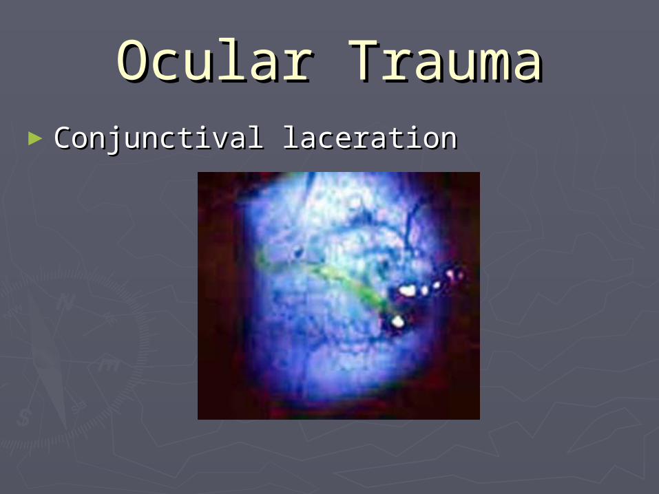
Ocular TraumaOcular Trauma►Conjunctival lacerationConjunctival laceration

Ocular TraumaOcular Trauma►Corneal LacerationsCorneal Lacerations
►History of cutting or tearing corneaHistory of cutting or tearing cornea►Seidels test crucial in distinguishing Seidels test crucial in distinguishing partial from full thickness lacerationspartial from full thickness lacerations
►Mild partial thickness lacerations Mild partial thickness lacerations managed as corneal abrasions including managed as corneal abrasions including close follow-upclose follow-up
►Careful examination of A/C and IOPCareful examination of A/C and IOP►Urgent referral if suspect full thicknessUrgent referral if suspect full thickness
►Pad eyePad eye►Avoid topical dropsAvoid topical drops

Ocular TraumaOcular Trauma►Corneal lacerationsCorneal lacerations
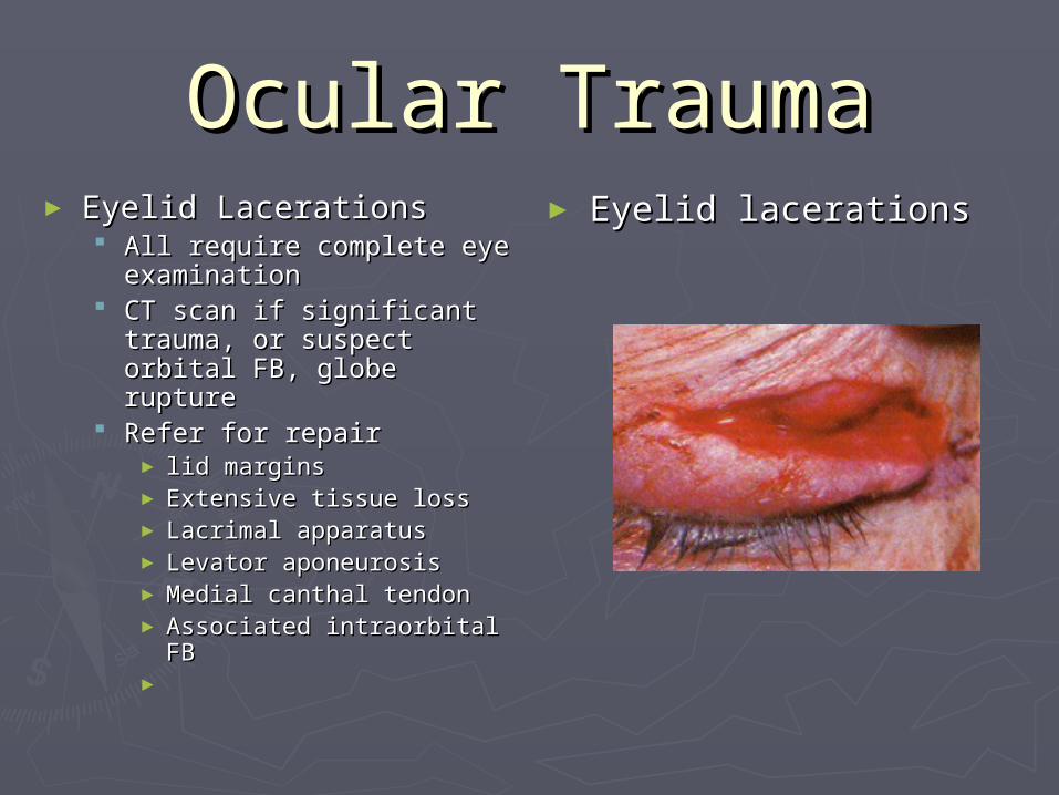
Ocular TraumaOcular Trauma► Eyelid LacerationsEyelid Lacerations
All require complete All require complete eye examinationeye examination
CT scan if significant CT scan if significant trauma, or suspect trauma, or suspect orbital FB, globe orbital FB, globe rupturerupture
Refer for repairRefer for repair► lid marginslid margins► Extensive tissue lossExtensive tissue loss► Lacrimal apparatusLacrimal apparatus► Levator aponeurosisLevator aponeurosis► Medial canthal tendonMedial canthal tendon► Associated intraorbital Associated intraorbital FBFB
►
► Eyelid lacerationsEyelid lacerations

Ocular TraumaOcular Trauma►HyphemaHyphema
SymptomsSymptoms►PainPain►Blurred visionBlurred vision►History of traumaHistory of trauma
SignsSigns►Blood in anterior chamber (layer +/or Blood in anterior chamber (layer +/or clot)clot)
►Reduced visual acuityReduced visual acuity

Ocular TraumaOcular Trauma►HyphemaHyphema

Ocular TraumaOcular Trauma►HyphemaHyphema

Ocular TraumaOcular Trauma► HyphemaHyphema
ManagementManagement►Assess for associated injuriesAssess for associated injuries►Hospitalize if > 1/3 anterior chamberHospitalize if > 1/3 anterior chamber►Bed rest Bed rest ►Elevate head 30 degreesElevate head 30 degrees►Shield both eyesShield both eyes►Avoid all aspirin and NSAIDSAvoid all aspirin and NSAIDS►Consider Amicar ( aminocaproic acid)Consider Amicar ( aminocaproic acid)►Atropine drops qidAtropine drops qid►AnalgesiaAnalgesia►AntiemeticsAntiemetics►Rx for IOPRx for IOP

Ocular TraumaOcular Trauma► HyphemaHyphema
Follow-upFollow-up►Check visual acuity, IOP & Slit lamp exam bidCheck visual acuity, IOP & Slit lamp exam bid►Look for increased IOP, new bleeding & corneal Look for increased IOP, new bleeding & corneal stainingstaining
►Add topical steroids if fibrinous A/C reaction or Add topical steroids if fibrinous A/C reaction or worseningworsening
►Surgical evacuation of hyphemaSurgical evacuation of hyphema►Refrain from strenuous activity > 2/52Refrain from strenuous activity > 2/52►O/P O/P
► 2-3/7 after discharge2-3/7 after discharge► 3-4 weeks for gonioscopy and dilated eye exam3-4 weeks for gonioscopy and dilated eye exam► Then 6/12 to 12/12 as prone to acute and chronic Then 6/12 to 12/12 as prone to acute and chronic glaucoma, cataracts & retinal tearsglaucoma, cataracts & retinal tears

Ocular TraumaOcular Trauma► Commotio RetinaeCommotio Retinae
SymptomsSymptoms► Decreased vision or asymptomaticDecreased vision or asymptomatic► Recent ocular trauma ( usually blunt)Recent ocular trauma ( usually blunt)
SignsSigns► Confluent area retinal whiteningConfluent area retinal whitening
DDxDDx► Retinal detachmentRetinal detachment► Branch retinal artery occlusionBranch retinal artery occlusion
Work-upWork-up► Complete opthalmic examination ( including dilated Complete opthalmic examination ( including dilated fundus)fundus)
TreatmentTreatment► Usually noneUsually none
Follow-upFollow-up► Repeat dilated exam at 1-2/52Repeat dilated exam at 1-2/52► Return sooner if decreased vision, flashes, floaters Return sooner if decreased vision, flashes, floaters etcetc

Ocular TraumaOcular Trauma►Commotio RetinaeCommotio Retinae

Ocular TraumaOcular Trauma► Intraocular Foreign Intraocular Foreign bodybody Consider in all high Consider in all high velocity ocular injuriesvelocity ocular injuries
Self sealing lacerationSelf sealing laceration Iris tearIris tear Irregular pupilIrregular pupil Lens opacityLens opacity Shallow A/CShallow A/C Inflammatory reaction Inflammatory reaction Low IOPLow IOP CT scan of orbitCT scan of orbit Endopthalmitis 48% casesEndopthalmitis 48% cases
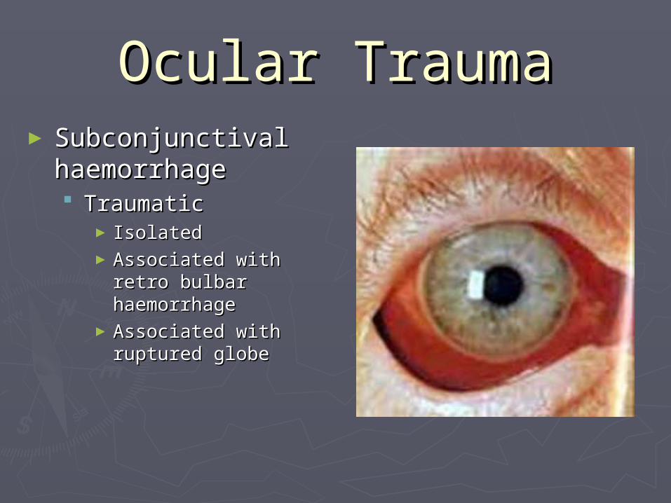
Ocular TraumaOcular Trauma► Subconjunctival Subconjunctival haemorrhagehaemorrhage TraumaticTraumatic
►IsolatedIsolated►Associated with Associated with retro bulbar retro bulbar haemorrhagehaemorrhage
►Associated with Associated with ruptured globeruptured globe
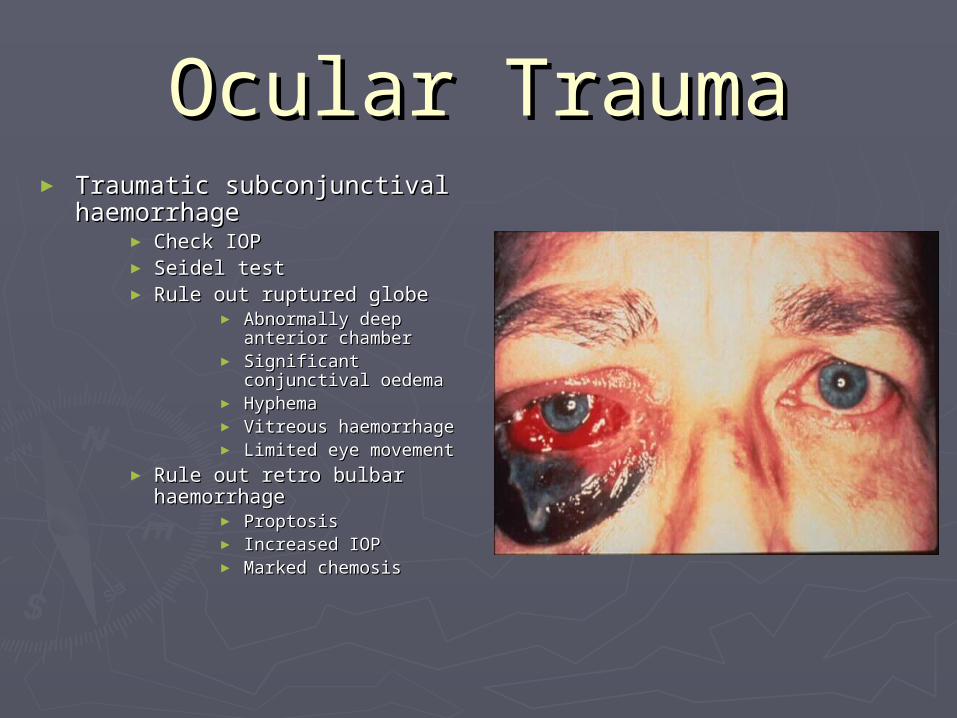
Ocular TraumaOcular Trauma► Traumatic subconjunctival Traumatic subconjunctival
haemorrhagehaemorrhage► Check IOPCheck IOP► Seidel testSeidel test► Rule out ruptured globeRule out ruptured globe
► Abnormally deep Abnormally deep anterior chamberanterior chamber
► Significant Significant conjunctival oedemaconjunctival oedema
► HyphemaHyphema► Vitreous haemorrhageVitreous haemorrhage► Limited eye movementLimited eye movement
► Rule out retro bulbar Rule out retro bulbar haemorrhagehaemorrhage
► ProptosisProptosis► Increased IOPIncreased IOP► Marked chemosisMarked chemosis

Ocular TraumaOcular Trauma►Ruptured GlobeRuptured Globe

Ocular TraumaOcular Trauma►Penetrating Eye InjuryPenetrating Eye Injury

Ocular TraumaOcular Trauma► Penetrating Eye InjuriesPenetrating Eye Injuries
SymptomsSymptoms► Suggested by historySuggested by history► Decreased visionDecreased vision► painpain
SignsSigns► Decreased visual acuityDecreased visual acuity► Periorbital haematoma & Periorbital haematoma &
lacerationslacerations► Full thickness laceration Full thickness laceration
of sclera or corneaof sclera or cornea► Subconjunctival Subconjunctival
haemorrhagehaemorrhage► Pupil distortionPupil distortion► Visible uveal tissueVisible uveal tissue► CataractCataract► Loss red reflexLoss red reflex► Low IOPLow IOP► Subluxed lensSubluxed lens► Commotio retinaeCommotio retinae

Ocular TraumaOcular Trauma►Penetrating Eye InjuriesPenetrating Eye Injuries

Ocular TraumaOcular Trauma

Ocular TraumaOcular Trauma► Penetrating Eye Penetrating Eye InjuriesInjuries Ruptured globeRuptured globe
►Severe conjunctival Severe conjunctival oedema & haemorrhageoedema & haemorrhage
►Abnormally deep Abnormally deep anterior chamberanterior chamber
►HyphemaHyphema►Limitation of eye Limitation of eye movementmovement
►Intraocular contents Intraocular contents outside the globeoutside the globe

Ocular TraumaOcular Trauma► Penetrating Eye InjuriesPenetrating Eye Injuries
TreatmentTreatment►Once the diagnosis of ruptured globe or Once the diagnosis of ruptured globe or penetrating injury is made defer ALL further penetrating injury is made defer ALL further examination until time of surgical repairexamination until time of surgical repair
►Avoid placing any pressure on the globe and Avoid placing any pressure on the globe and risking extrusion of intraocular contents.risking extrusion of intraocular contents.
► Protect eye with shieldProtect eye with shield► Nil by mouthNil by mouth► Systemic antibioticsSystemic antibiotics► AntiemeticAntiemetic► Tetanus prophylaxisTetanus prophylaxis► SedationSedation► Strict bed restStrict bed rest► CT scan orbit and brain ( +/- B scan)CT scan orbit and brain ( +/- B scan)► Arrange urgent referral and transferArrange urgent referral and transfer

Ocular TraumaOcular Trauma► HyphemaHyphema
► MicrohyphemaMicrohyphema Small hyphema with Small hyphema with
suspended red cells only suspended red cells only (no layered clot)(no layered clot)
Graded 1+ to 4+ depending Graded 1+ to 4+ depending on quantity cellson quantity cells
May settle and form May settle and form hyphemahyphema
Can cause Increased IOP Can cause Increased IOP and 2and 2ndnd haemorrhage haemorrhage
TreatmentTreatment► Cease anticoagulants Cease anticoagulants
aspirin and NSAIDSaspirin and NSAIDS► Bed rest with 30 Bed rest with 30
degrees head degrees head elevation 4/7elevation 4/7
► Topical cycloplegic Topical cycloplegic +/- steroid+/- steroid
► Review 1-2/7 or Review 1-2/7 or sooner if vision sooner if vision changeschanges
► Daily review if IOP Daily review if IOP increasedincreased
► Gonioscopy and Gonioscopy and dilated eye dilated eye examination >2/52examination >2/52
► MicrohyphemaMicrohyphema

Ocular TraumaOcular Trauma► Lens SubluxationLens Subluxation
►Partial disruption Partial disruption of zonular fibresof zonular fibres
►Lens remains Lens remains partially in partially in pupillary aperturepupillary aperture
►CausesCauses► Acquired myopiaAcquired myopia► AstigmatismAstigmatism► diplopiadiplopia
►Observe if Observe if asymptomaticasymptomatic
►Surgical removal Surgical removal

Ocular TraumaOcular Trauma► Lens DislocationLens Dislocation
►Complete disruption Complete disruption of zonular fibresof zonular fibres
►Lens displaced out of Lens displaced out of pupillary aperturepupillary aperture
►May be in anterior May be in anterior chamber or posteriorchamber or posterior
►Lensectomy required Lensectomy required if capsule is damagedif capsule is damaged
►May precipitate AACG May precipitate AACG myopia, astigmatism myopia, astigmatism or diplopia.or diplopia.

Ocular TraumaOcular Trauma► Lens DislocationLens Dislocation
Anterior chamberAnterior chamber► Dilate pupilDilate pupil► Pt supinePt supine► Indent cornea Indent cornea ► Constrict pupil once Constrict pupil once repositionedrepositioned
► Refer for laser Refer for laser iridectomyiridectomy
► Surgical removalSurgical removal CataractCataract Reduction failsReduction fails Recurrent dislocationsRecurrent dislocations
VitreousVitreous► Capsule intactCapsule intact
Asymptomatic, no Asymptomatic, no inflammation, observeinflammation, observe
► Capsule rupturedCapsule ruptured Symptomatic, inflammedSymptomatic, inflammed Surgical removal of lensSurgical removal of lens

Ocular TraumaOcular Trauma► Traumatic CataractTraumatic Cataract
►May not be apparent May not be apparent for years after for years after traumatrauma
►Petalliform cataract Petalliform cataract with compact star-with compact star-shaped opacity most shaped opacity most commonly foundcommonly found
►Management is same Management is same as for age related as for age related cataractscataracts
►Increased risk Increased risk dehiscence during dehiscence during extractionextraction

Ocular TraumaOcular Trauma► Retinal tear / Retinal tear / detachmentdetachment
► flashes, floaters, flashes, floaters, curtain across visioncurtain across vision
► Peripheral +/or Peripheral +/or central losscentral loss
► Elevation retina with Elevation retina with a flap tear or breaka flap tear or break
► Decreased IOPDecreased IOP► Afferent pupil defectAfferent pupil defect► Macula-on RD urgent Macula-on RD urgent referralreferral
► Macula-off RD less Macula-off RD less urgenturgent

Ocular TraumaOcular Trauma► Orbital Blow-out Orbital Blow-out
fracturefracture► SymptomsSymptoms
PainPain► Especially with Especially with
attempted vertical attempted vertical eye movementeye movement
Local tendernessLocal tenderness Binocular double visionBinocular double vision Eyelid swellingEyelid swelling
► SignsSigns Restricted eye movementRestricted eye movement
► Especially in Especially in upward and / or upward and / or lateral gaze lateral gaze
Orbital Subcutaneous Orbital Subcutaneous emphysemaemphysema
Infraorbital nerve hyper Infraorbital nerve hyper or paraesthesiaor paraesthesia
EnophthalmosEnophthalmos PtosisPtosis Associated globe Associated globe
injuriesinjuries

Ocular TraumaOcular Trauma►Orbital fracturesOrbital fractures

Ocular TraumaOcular Trauma► Orbital fracturesOrbital fractures
Medial WallMedial Wall►Ethmoidal fractureEthmoidal fracture►Eyelid swelling Eyelid swelling after blow noseafter blow nose
►Lateral Lateral displacement of displacement of medial canthus & medial canthus & narrowing of narrowing of palpebral aperturepalpebral aperture
►CT scan with axial CT scan with axial viewsviews

Ocular TraumaOcular Trauma► Orbital fracturesOrbital fractures
Trap door fractureTrap door fracture►Relatively small Relatively small floor #floor #
►Significant muscle Significant muscle entrapmententrapment
►Common in paediatric Common in paediatric populationpopulation
►Needs prompt surgeryNeeds prompt surgery►Intense pain, nausea Intense pain, nausea & vomiting& vomiting
►Coronal CTCoronal CT

Ocular TraumaOcular Trauma► Orbital fracturesOrbital fractures
Tripod fractureTripod fracture►Lateral wallLateral wall► AkaAka zygomatic complex zygomatic complex fracturefracture
►Involves zygoma Involves zygoma disruption at disruption at zygomaticofrontal, zygomaticofrontal, temporal and temporal and maxillary sinusesmaxillary sinuses
►Flattening of malar Flattening of malar region of faceregion of face
►Inferior Inferior displacement of displacement of lateral canthuslateral canthus

Ocular TraumaOcular Trauma► Orbital fracturesOrbital fractures
Orbital Roof Orbital Roof fracturefracture►Life threatening Life threatening injuryinjury
►Fracture along Fracture along orbital surface of orbital surface of the frontal bonethe frontal bone
►Potential Potential communication communication between orbit and between orbit and anterior cranial anterior cranial fossafossa

Ocular TraumaOcular Trauma► Orbital fracturesOrbital fractures
Apex or Optic Apex or Optic canal #canal #►RareRare►Occurs with severe Occurs with severe traumatrauma
►May cause optic May cause optic neuropathy or neuropathy or transection of transection of optic nerveoptic nerve
►Axial CT scanAxial CT scan

Ocular TraumaOcular Trauma► Orbital fracturesOrbital fractures
► ManagementManagement Nasal decongestantsNasal decongestants AnalgesiaAnalgesia Broad spectrum antibioticsBroad spectrum antibiotics Instruct patient NOT to blow noseInstruct patient NOT to blow nose Surgical repair 10-14/7Surgical repair 10-14/7
► persisting diplopia when looking straight or with persisting diplopia when looking straight or with readingreading
► Cosmetically unacceptable enopthalmosCosmetically unacceptable enopthalmos► Large fractureLarge fracture
Review at 1/52 and 2/52 post traumaReview at 1/52 and 2/52 post trauma► Persisting diplopia or enophthalmosPersisting diplopia or enophthalmos
Monitor for associated ocular injuriesMonitor for associated ocular injuries► Orbital cellulitisOrbital cellulitis► Angle recession glaucomaAngle recession glaucoma► Retinal detachmentRetinal detachment

Ocular TraumaOcular Trauma► Retro bulbar HaemorrhageRetro bulbar Haemorrhage
SymptomsSymptoms► PainPain► Decreased visionDecreased vision
SignsSigns► Proptosis (with resistance to Proptosis (with resistance to
retropulsion)retropulsion)► Diffuse subconjunctival Diffuse subconjunctival
hemorrhage ( no posterior hemorrhage ( no posterior margin)margin)
► Elevated IOPElevated IOP► Eyelid oedemaEyelid oedema► Afferent pupil defectAfferent pupil defect► ChemosisChemosis► Reduced ocular movementReduced ocular movement► Loss color visionLoss color vision► CrepitusCrepitus► Infraorbital paraesthesiaInfraorbital paraesthesia
TreatmentTreatment► Reduce IOPReduce IOP► Lateral canthotomyLateral canthotomy► Orbital decompression surgeryOrbital decompression surgery