omm research review and omm for the shoulder researchmurray.pdf · omm research review and ......
TRANSCRIPT

OMM Research Review and OMT For The Shoulder
Trish Murray, D.O.


www.OMMEducation.com

Research Review
• “Modeled Osteopathic Manipulative Treatments: A Review of their in Vitro Effects on Fibroblast Tissue Preparations”; Manal Zein-Hammoud, PhD and Paul Standley, PhD; The Journal of the American Osteopathic Association; August 2015, vol. 115, Number 8, pp. 490-501.

10 years of work
• cellular and molecular mechanisms by which MFR and CS work on fibroblasts
• the mechanisms underlying clinical outcomes of OMT modalities
In vitro modeling 2 common OMT modalities:
Myofascial Release and Counterstrain
• fibroblast= the principle cell type of the fasciae
• synthesizes, organizes and remodels collagen.
• target for normal and abnormal biomechanical stimuli.
• These in vitro studies have shown that fibroblasts respond to strain by secreting proinflammatory and anti-inflammatory cytokines, undergoing hyperplasia as well as altering cell shape and alignment

• “One thing common to both deleterious repetitive motion strain (RMS) and curative OMT is biomechanical stimulation. This connection catalyzed our laboratory to investigate how various biomechanical strains modeling both RMSs and various OMT modalities affect cellular physiology and gene activation and suppression.”

Purpose of OMT
• to impart biomechanical stimuli to tissues to bring about change in the cellular function.
3 In Vitro Models
1. 2-dimensional fibroblast matrix
2. 3-dimensional fibroblast matrix dubbed a
“bioengineered tendon(BET)”
3. Co-culture that allows interaction between fibroblasts and myoblasts to create a modeled myofascial junction.

Stretching and Compressing Fibroblasts
• TO BEGIN an analysis of potential cell and tissue effects that may explain patients’ improvements after OMT.
(Isn’t this AWESOME!)

Earliest Studies
Effect that acyclic in vitro biophysical strain has on:
• cellular shape
• proliferation on fibroblasts
• nitric oxide (NO) production
• interleukin 1 (IL-1)
• interleukin 6 (IL-6)
Earliest Studies Findings
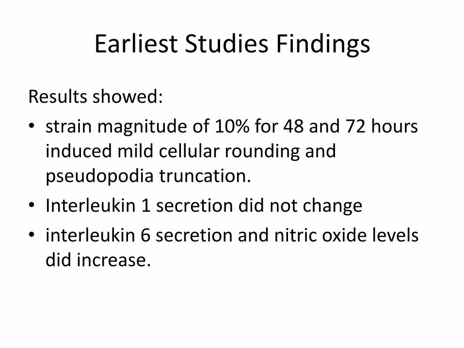
Results showed:
• strain magnitude of 10% for 48 and 72 hours induced mild cellular rounding and pseudopodia truncation.
• Interleukin 1 secretion did not change
• interleukin 6 secretion and nitric oxide levels did increase.

Earliest Studies Findings
Strain magnitude of 30%:
• caused reduced cell viability
• cell membrane decomposition
• pseudopodia loss.
**biomechanical strain has profound effects on proliferation, apoptosis, and cytokine production.

Later Studies
• combined modeled repetitive motion strain (RMS) and modeled counterstrain (CS) and myofascial release (MFR)
• investigated potential changes in human fibroblast proliferation and interleukin secretion.

Later Studies Findings
• Modeled RMS produced a delayed inflammatory response and reduction in cellular proliferation.
• Modeled CS and MFR reversed these effects.

Later Studies
• Fibroblast hyperplasia, hypertrophy, and secretion of cytokines and growth factors were examined with different MFR magnitudes (3%, 6%, 9% and 12%) and duration (0.5, 1,2,3,4, and 5 minutes):

Later Studies Findings
• Bioengineered tendon weight was increased with greater-magnitude (12%) treatment
(suggesting collagen is up-regulated by MFR)
Greater MFR magnitude also led to a statistically significant increase in:
• IL-1B,
• monocyte chemoattractant cytokine
• T-cell expressed and secreted chemotactic cytokine

• Alternatively, holding the strain magnitude constant at 6% and varying the duration of MFR:
• increased secretions of angiogenin
• IL-3
• IL-8,
• growth colony-stimulating factor
• thymus activation-regulated chemokine
• only in the 5-minute MFR group.

Conclusion
• changing magnitude and duration of MFR appears to enhance the secretion of a unique subset of cytokines and growth factors, possibly affecting physiologic responses that may potentially regulate inflammation and wound healing responses.

Most Recent Studies on Muscle Differentiation
• In fibroblasts and fibroblast-myoblast cocultures:
• “ we hypothesized that RMS would reduce muscle repair by inhibiting muscle differentiation, whereas MFR would reverse this effect.”
Muscle Differentiation Study Findings
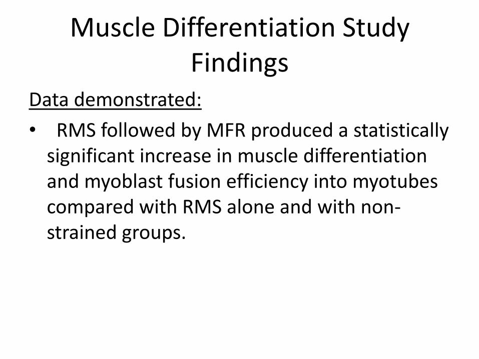
Data demonstrated:
• RMS followed by MFR produced a statistically significant increase in muscle differentiation and myoblast fusion efficiency into myotubes compared with RMS alone and with non-strained groups.

Wound Healing Studies
• studied the effect of various biomechanical strain patterns on fibroblast scratch wound closure
• Results showed that RMS alone caused reduced wound closure rates compared with the control group
• RMS was followed by 6% MFR reversed this finding.

Wound Healing Studies
• 3-dimensional Bioengineered Tendons (BETs): MFR was applied at 6% beyond resting length and held for 0, 1, 2, 3, 4, and 5 minutes and then released to baseline. Also, magnitudes were set at 0%, 3%, 6%, 9%, and 12% beyond resting length, held for 90 seconds and then released to baseline. The BETs width, wound area, and wound shape were measured and quantified.

Wound Healing Study Findings
• 12% strain resulted in larger wound area than nonstrained BETs at all time points.
• Statistically significant reduction in wound area was observed with 3% strain by 24 hours and 48 hours after strain compared with no strain, 6% strain, and 9% strain.
• Fixing the magnitude at 6% for 5 minutes resulted in a statistically significant decrease in wound area compared with no strain.

Conclusions for Dr. Standley’s Studies
• Biomechanical strain absolutely has an effect on cell physiology: Mechanotransduction
• Biomechanical strain can have a negative or a positive effect on cytokine secretion, cell proliferation, muscle differentiation and wound healing.


The Shoulder
• Most mobile joint in the body
• Multiarticular
• Scapulothoracic “joint” (physiologic)
• 3 “true” joints
– Glenohumeral
– Acromioclavicular
– Sternoclavicular

Abduction
• 0-60 degrees – glenohumeral
• 60-150 degrees – scapulothoracic
• 150-180 degrees – leaning to contralateral side

• If scapular position is disturbed, undue strain is placed on the three joints of the shoulder girdle. Due to this “strain pattern” the 21 muscles of the shoulder girdle and upper extremity will be functionally compromised and may reflect shoulder, elbow and/or wrist pathophysiology. Low back problems with associated latissimus dorsi contracture may also be an underlying cause of shoulder disability.

Glenohumeral Joint
• Ball-and-socket joint
• Shallow, unstable
• Most stable at 90 degrees abduction (close-pack position)
• Cavity is deepened by glenoid labrum

Acromioclavicular Joint
• Multiple ligamentous connections

Rotator Cuff=Musculotendinous
• Provides stability to glenohumeral joint
• Internal rotation – Subscapularis
• External rotation – Infraspinatus
– Teres minor
• Abduction – Supraspinatus


History
Listen to their story
Past Medical and Social History
Helps you assess overall wellness/protoplasm

Areas of Concern
• Cervical and Thoracic Spine
• Rib Cage
• Scapulothoracic joint
• AC joint
• Rotator Cuff
• Glenoid labrum
• Sternoclavicular joint

Physical Exam of Shoulder
1. General Posture
2. Range of Motion: “Scratch Test “
Flexion, Extension, Abduction, Adduction, External Rotation, Internal Rotation.
**Passive Adduction with pressure= AC joint
3. Strength/DTR’s
Physical Exam
4. Rotator Cuff:
Abduction= Supraspinatus
External Rotation= Infraspinatus and
Teres minor
Internal Rotation= Subscapularis
5. SLAP Tear/Joint Capsule
6. Special Tests: Thoracic Outlet Syndrome:
Addson’s Test/Hyperextension Test/Military Posture Test

SLAP tear or SLAP lesion
• injury to the glenoid labrum (fibrocartilaginous rim attached around the margin of the glenoid cavity).
• SLAP is an acronym for "superior labral tear from anterior to posterior".

OMT for the Shoulder
1. Must treat Thoracic Spine, Thoracic Outlet , Cervical Spine and Ribs.
(Still Technique)

OMT for the Shoulder
2. Scapula: Must release Adhesions and Optimize Motion
Counterstrain: Levator Scapulae and
“The Treasure Chest”

OMT for the Shoulder
3. Mobilize the entire shoulder: MFR sitting up
7 Stages of Spencer

Summary
1. Reviewed Dr. Standley’s in vitro work on fibroblasts.
2. Effective Evaluation and Treatment of the shoulder must involve the neck, thorax, ribs and scapula.
3. OMMEducation.com: CME credits in OMM from the comfort of your own home/office.
Weekly blog on OMM and Functional Medicine

Help Patients and Make Money
tmurraywellness.com
Patient Educational Materials:
1. Put a DENT in Your Pain
2. Put a DENT in Your Cholesterol
Diet/Exercise/Nutrition/Treatment
Joint Venture Marketing