omt of the thoracic spine eric j. milie d.o. internal medicine lecture series 1/5/04
TRANSCRIPT
OMT of the Thoracic Spine
Eric J. Milie D.O.
Internal Medicine Lecture Series
1/5/04
Objectives:
The physician will become comfortable with the osteopathic manipulative treatments demonstrated during the lecture
The osteopathic physician will be able to name thoracic spine and rib dysfunction
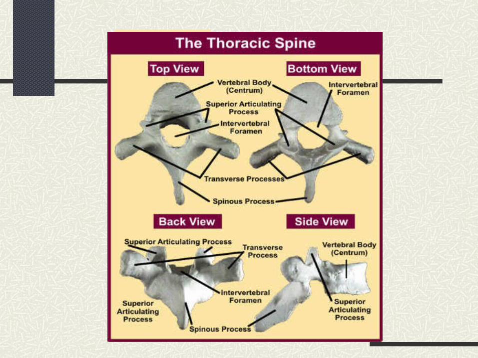
The osteopathic physician will understand the complex anatomy of the thoracic spine and thoracic inlet/outlet
Evidence Based Medicine
DR Noll, et al. Benefits of osteopathic manipulative treatment for hospitalized elderly patients with pneumonia. JAOA • Vol 100 • No 12 • December 2000 • 776-782
Showed that hospitalized pt’s treated with OMT required significantly shorter courses of intravenous antibiotics and also had a significantly shorter hospital stayAndersson GB, Lucente T, Davis AM, Kappler RE, Lipton JA, Leurgans S. A comparison of osteopathic spinal manipulation with standard care for patients with low back pain. N Engl J Med.1999; 341:1426 –1431Patients treated with OMT required less analgesics than patients treated with “standard medical therapy” alone, and were more satisfied with their care
Evidence Based Medicine cont.
Hoehler FK, Tobis JS, Buerger AA. Spinal manipulation for low back pain. JAMA.1981; 245:1835 –1838
Showed that patients treated with OMT received equal benefit compared to patients receiving analgesics and soft tissue massage
Licciardone JC, Stoll ST, Fulda KG, Russo DP, Siu J, WinnW, et al. Osteopathic manipulative treatment for chronic low back pain: a randomized controlled trial. Spine.2003; 28:1355 –1362
Patients with chronic back pain who received osteopathic manipulation reported greater improvements in back pain, greater satisfaction with back care throughout the trial, better physical functioning and mental health at 1 month, and fewer cotreatments at 6 months compared to the control group
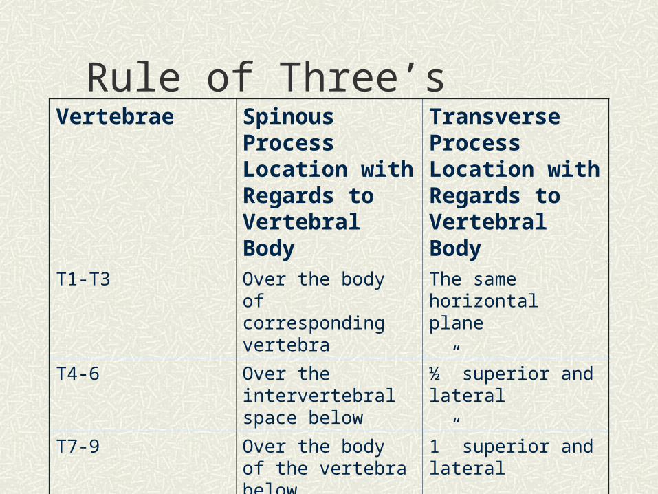
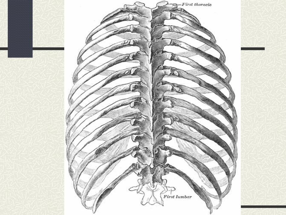
Rule of Three’sVertebrae Spinous Process
Location with Regards to Vertebral Body
Transverse Process Location with Regards to Vertebral Body
T1-T3 Over the body of corresponding vertebra
The same horizontal plane
T4-6 Over the intervertebral space below
½” superior and lateral
T7-9 Over the body of the vertebra below
1” superior and lateral
T10-12 Over the body of the corresponding vertebra
The same horizontal plane
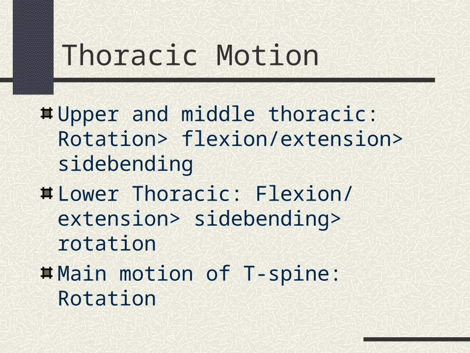
Thoracic Motion
Upper and middle thoracic: Rotation> flexion/extension> sidebending
Lower Thoracic: Flexion/ extension> sidebending> rotation
Main motion of T-spine: Rotation
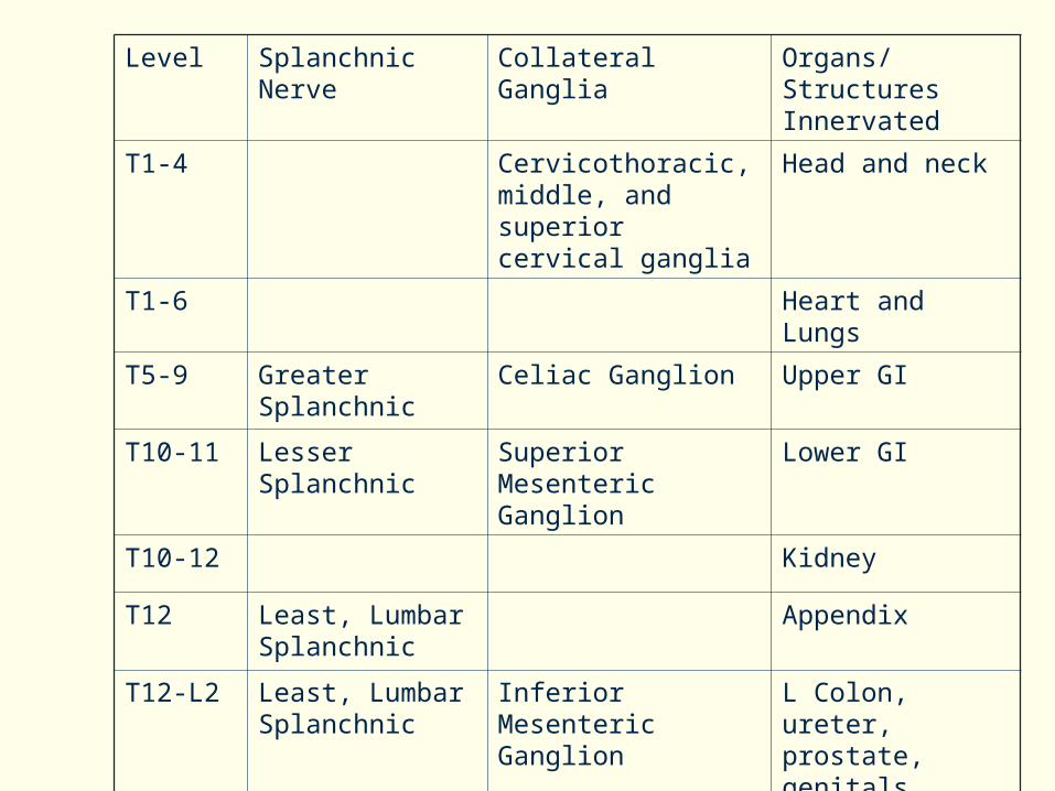
Level Splanchnic Nerve Collateral Ganglia Organs/ Structures Innervated
T1-4 Cervicothoracic, middle, and superior cervical ganglia
Head and neck
T1-6 Heart and Lungs
T5-9 Greater Splanchnic Celiac Ganglion Upper GI
T10-11 Lesser Splanchnic Superior Mesenteric Ganglion
Lower GI
T10-12 Kidney
T12 Least, Lumbar Splanchnic
Appendix
T12-L2 Least, Lumbar Splanchnic
Inferior Mesenteric Ganglion
L Colon, ureter, prostate, genitals, uterus
T2-8 Arms
T11-L2 Legs
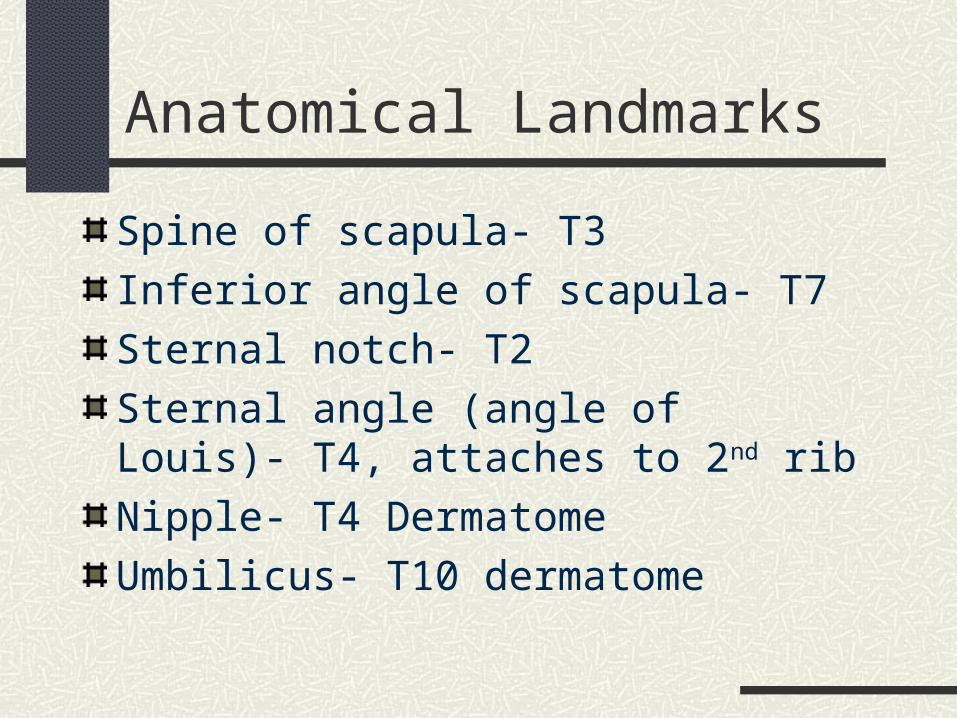
Anatomical Landmarks
Spine of scapula- T3
Inferior angle of scapula- T7
Sternal notch- T2
Sternal angle (angle of Louis)- T4, attaches to 2nd rib
Nipple- T4 Dermatome
Umbilicus- T10 dermatome
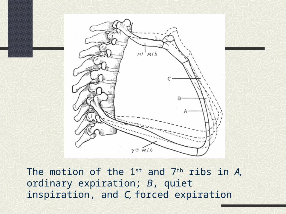
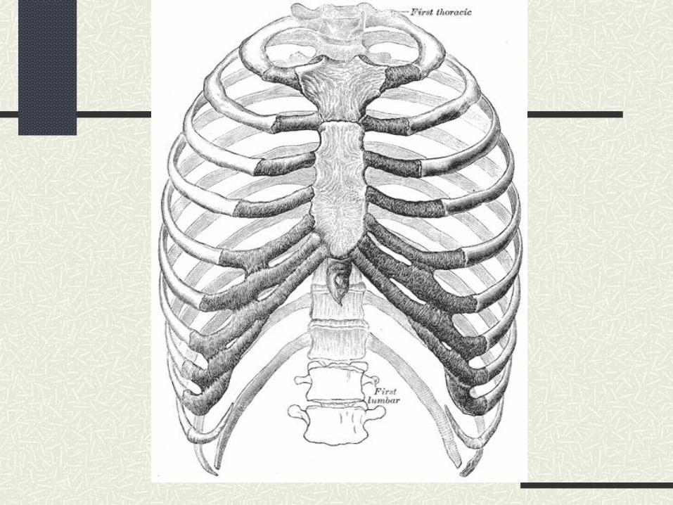
The motion of the 1st and 7th ribs in A, ordinary expiration; B, quiet inspiration, and C, forced expiration
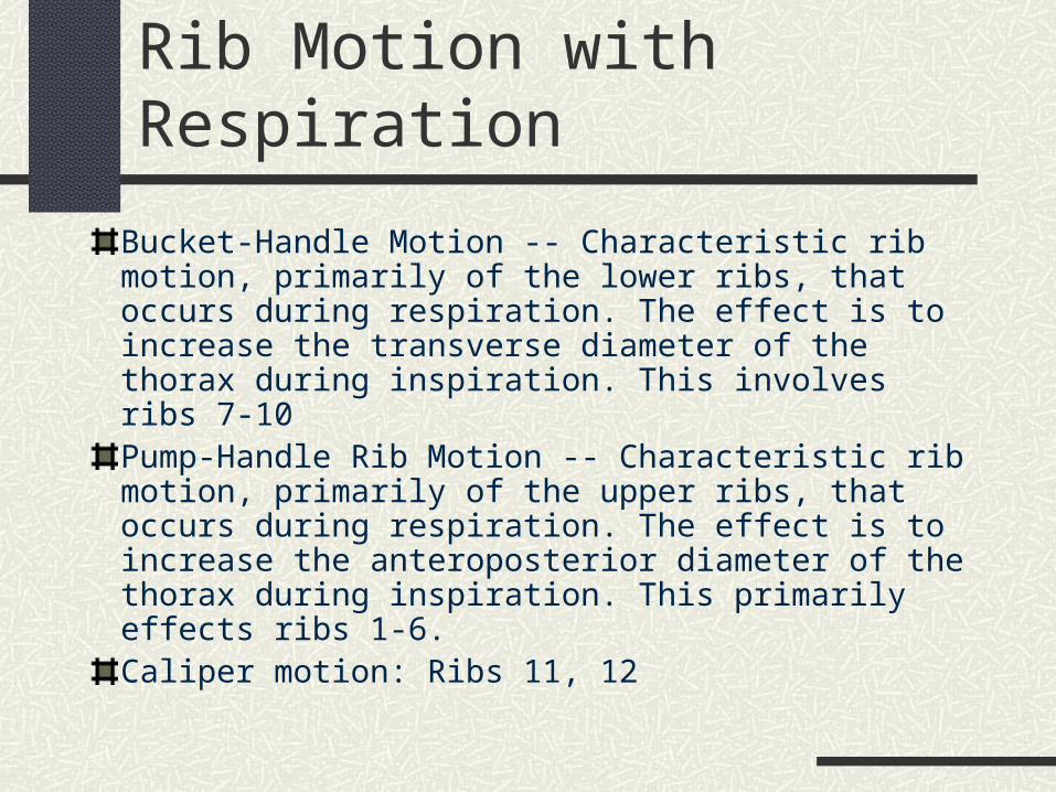
Rib Motion with Respiration
Bucket-Handle Motion -- Characteristic rib motion, primarily of the lower ribs, that occurs during respiration. The effect is to increase the transverse diameter of the thorax during inspiration. This involves ribs 7-10Pump-Handle Rib Motion -- Characteristic rib motion, primarily of the upper ribs, that occurs during respiration. The effect is to increase the anteroposterior diameter of the thorax during inspiration. This primarily effects ribs 1-6.Caliper motion: Ribs 11, 12
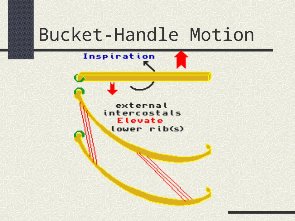
Bucket-Handle Motion

Pump-Handle Motion
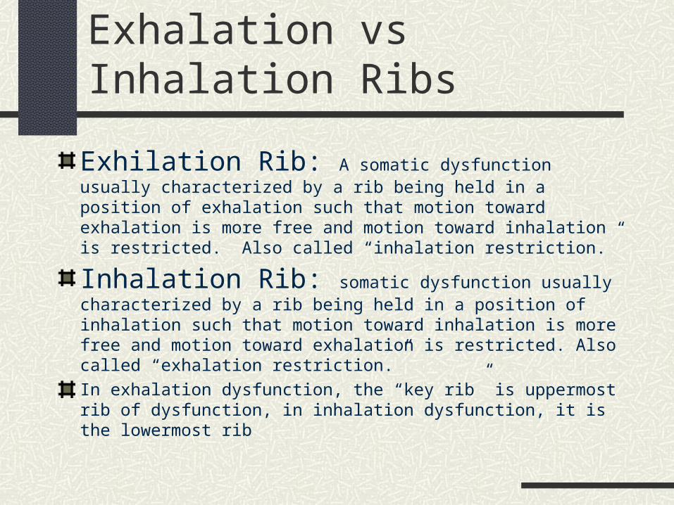
Exhalation vs Inhalation Ribs
Exhilation Rib: A somatic dysfunction usually characterized by a rib being held in a position of exhalation such that motion toward exhalation is more free and motion toward inhalation is restricted. Also called “inhalation restriction.”
Inhalation Rib: somatic dysfunction usually characterized by a rib being held in a position of inhalation such that motion toward inhalation is more free and motion toward exhalation is restricted. Also called “exhalation restriction.”
In exhalation dysfunction, the “key rib” is uppermost rib of dysfunction, in inhalation dysfunction, it is the lowermost rib
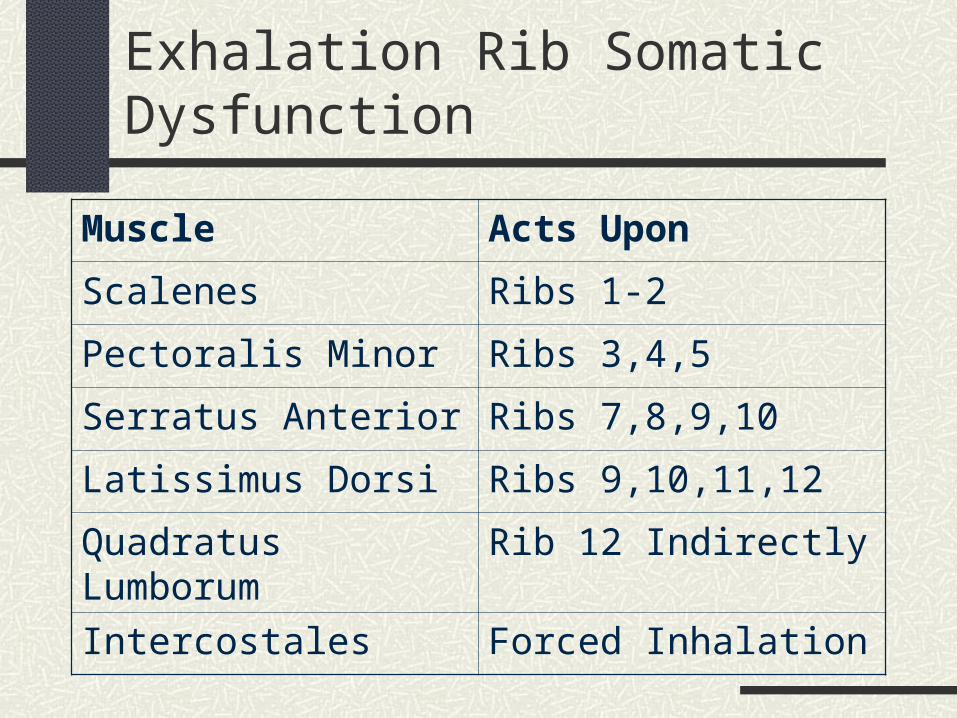
Exhalation Rib Somatic Dysfunction
Muscle Acts Upon
Scalenes Ribs 1-2
Pectoralis Minor Ribs 3,4,5
Serratus Anterior Ribs 7,8,9,10
Latissimus Dorsi Ribs 9,10,11,12
Quadratus Lumborum Rib 12 Indirectly
Intercostales Forced Inhalation
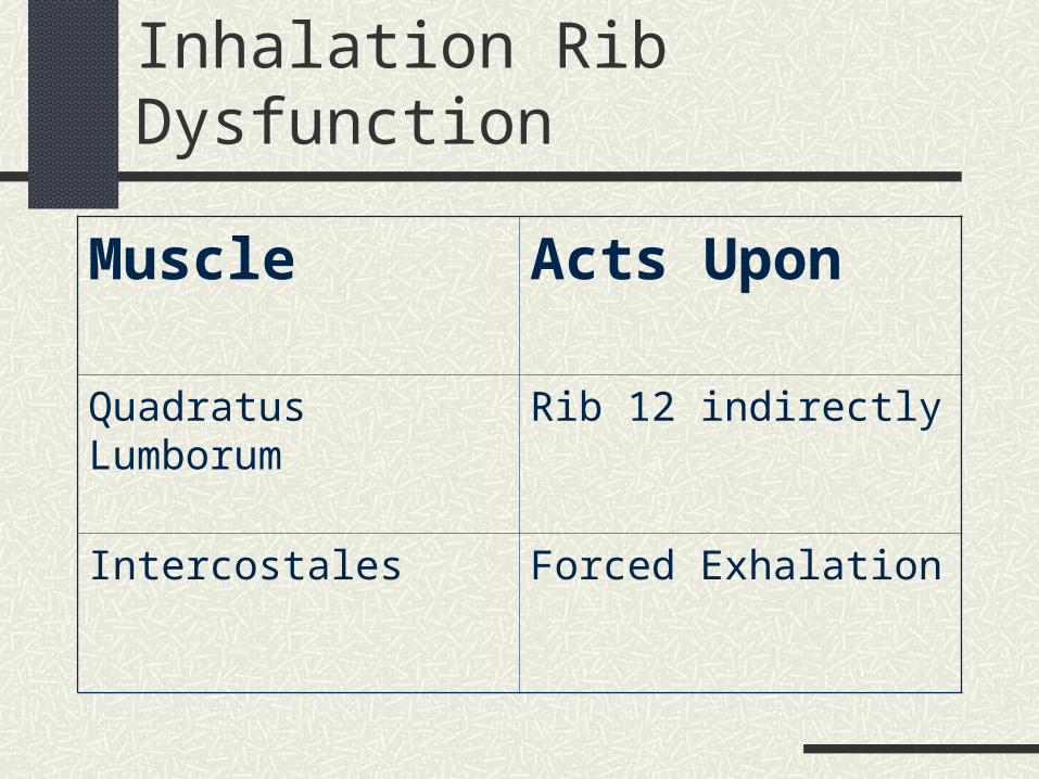
Inhalation Rib Dysfunction
Muscle Acts Upon
Quadratus Lumborum Rib 12 indirectly
Intercostales Forced Exhalation
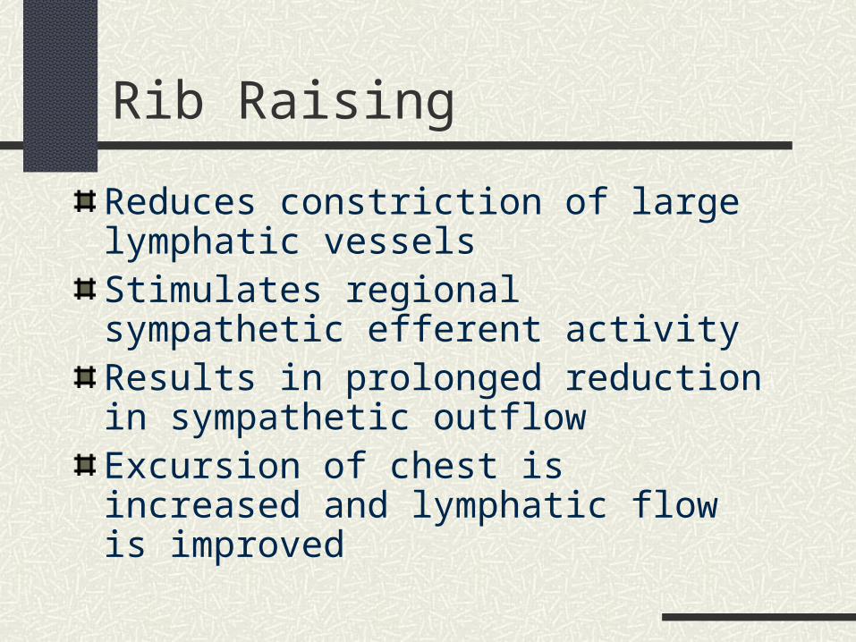
Rib Raising
Reduces constriction of large lymphatic vesselsStimulates regional sympathetic efferent activityResults in prolonged reduction in sympathetic outflowExcursion of chest is increased and lymphatic flow is improved
Rib Raising cont.
Patient Position: Supine
Physician Position: Standing or seated at the patient’s side

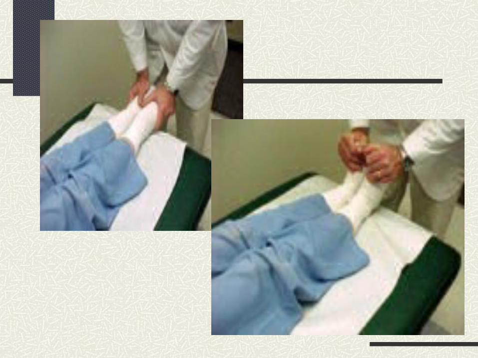
Rib Raising Procedure
Palms placed under patient’s thorax, so that pads of fingers at rib angles
Flex fingers, apply traction to the rib angle
While applying traction, bend knees/ lower trunk to raise ribs (lever/fulcrum action) Do not bend wrists
Move hands so that subsequent ribs treated
Treat opposite side of rib cage in same manner
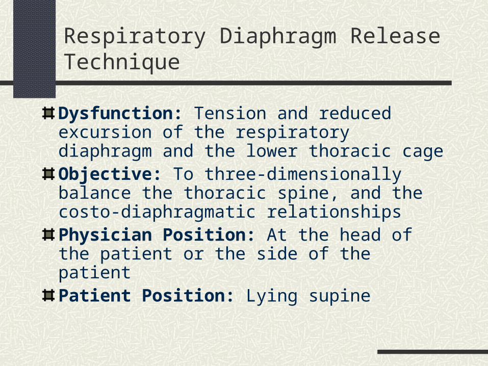
Respiratory Diaphragm Release Technique
Dysfunction: Tension and reduced excursion of the respiratory diaphragm and the lower thoracic cageObjective: To three-dimensionally balance the thoracic spine, and the costo-diaphragmatic relationshipsPhysician Position: At the head of the patient or the side of the patientPatient Position: Lying supine
Respiratory Diaphragm Release Procedure
Patient seated or supine
Physician standing behind patient or at patient’s side
Physician’s hands around thoracic cage, fingers under the costal margin
Check motion by gently rotating thoracic tissues
Treatment Phase: Hold thoracic tissues in the direction in which they move freely. Allow fascia to unwind, until it settles into a rhythmic vertical motion
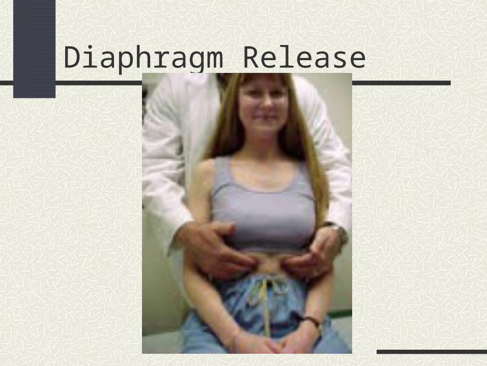
Diaphragm Release
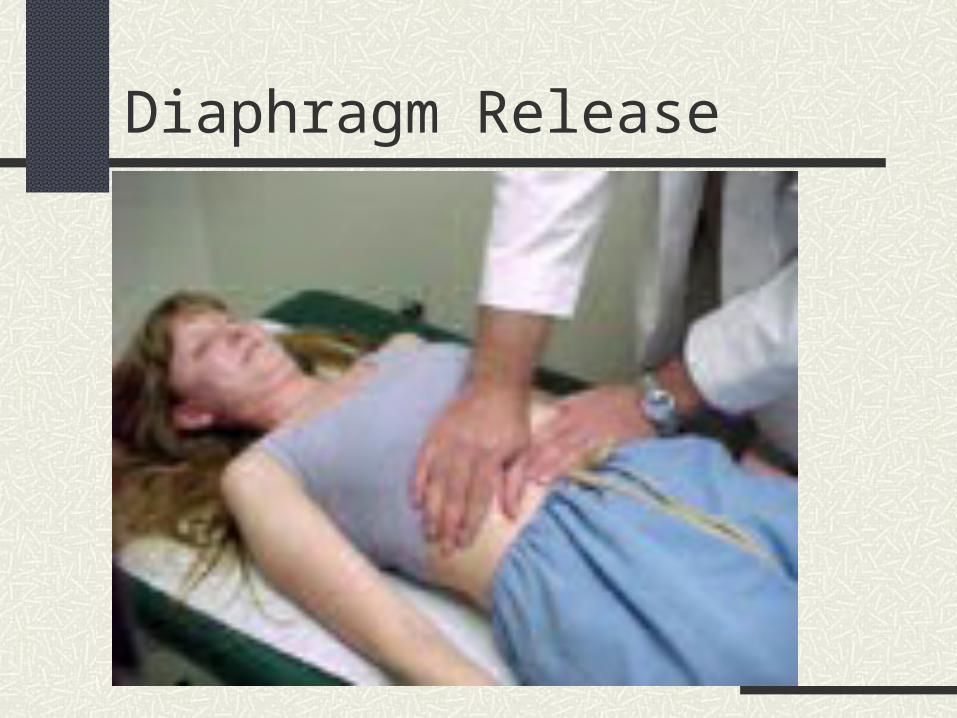
Diaphragm Release
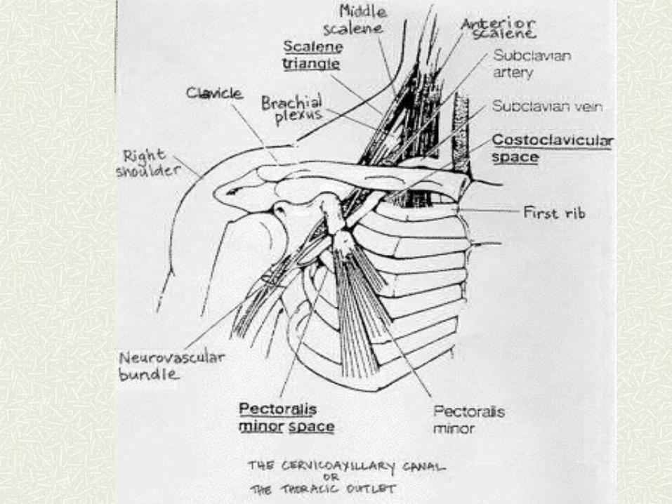
Thoracic Outlet
Roughly 4cm area boundaried anteriorly by the manubrium of the sternum, laterally by the 1st rib and its costal cartilage, and posteriorly by the body of T1
Covered by Sibson’s fascia, surrounded by the scalenes, SCM, and the trapezium
Subclavian vein and artery, brachial plexus, and lymphatics run through these structures
Thoracic Outlet Release
Purpose: To relax soft tissue restrictions and enhance lymphatic drainage from the head and neck
Physician Position: Sitting at head of table or side of patient
Patient Position: Lying supine
Thoracic Outlet Release technique
1. Place one hand posterior to the thoracic inlet (transversely) at the level of the first and second ribs.
2. Place the other hand at the same level on the anterior chest wall. 3. The area is motion tested for myofascial restrictions. These motions
include a side-to-side movement, a rotational or twisting movement, a superior or inferior movement, or an angular movement.
4. The area is treated directly (barrier engagement) or indirectly (position of fascial ease). Having the patient take three deep breaths, can facilitate a release.
5. The physician waits for a release and the area is re-evaluated. 6. Modification: Both hands can also be placed on the anterior thorax.
With this position, the thumbs contact the trapezius muscle and posterior upper two ribs. The fingers lie on the anterior chest wall and spread out.
Lymphatic Pumps
Dysfunction: Lymphatic Stasis
Objective: Improve lymphatic flow by altering intrathoracic pressure
Technique: Several different variations, including thoracic and pedal pump
Pedal Pump
“Fun With Alex” Lots of good action in this video. Alex is behind the wheel of a Ford F150. There is pumping, driving, revving in sneakers, white socks and barefeet. Alex is a hottie and knows how to pump it
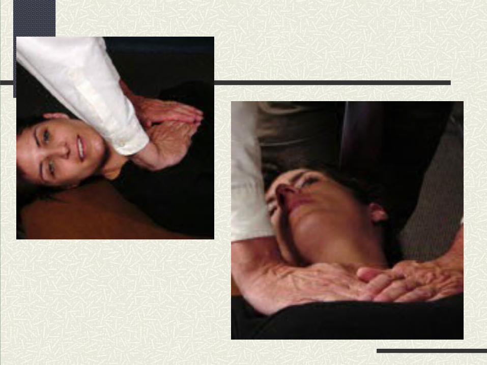
Lymphatic Pump Technique: Thoracic
1. Place your hands on the patient's anterior chest wall over the patient's pectoralis major muscles. The heels of your hands should lie on ribs 2-4.
2. With your elbows straight, have the patient breathe in through their open mouth and exhale passively. As the patient exhales, follow the exhalation motion downward and maintain the end point. This applies a compressive force.
3. With each following breath, slightly resist inhalation and maintain your pressure at the end position of exhalation.
4. One third of the way through the fourth or fifth inhalation, briskly remove your hands from the chest wall, as a rush of air will enter the patient's lungs.
Pedal Pump
A venous and lymphatic drainage technique applied through the lower extremities
Rhythmic plantar and dorsiflexion of the lower extremities
Physician at foot of patient, patient supine
Rate 30-45 cycles/minute
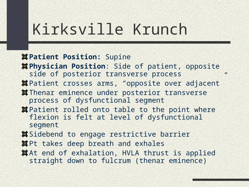
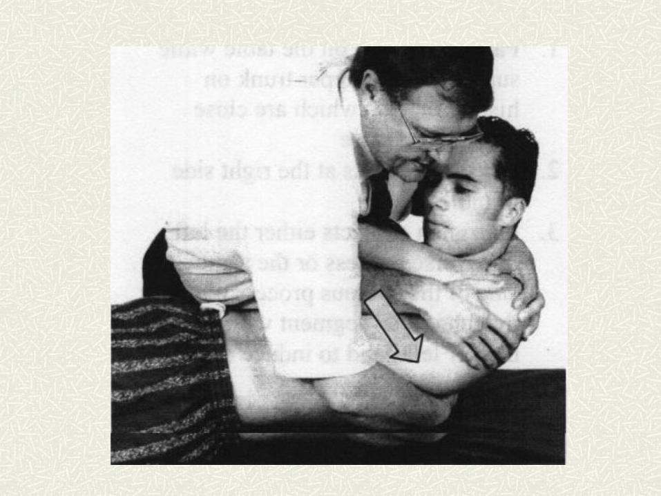
Kirksville Krunch
Patient Position: SupinePhysician Position: Side of patient, opposite side of posterior transverse processPatient crosses arms, “opposite over adjacent”Thenar eminence under posterior transverse process of dysfunctional segmentPatient rolled onto table to the point where flexion is felt at level of dysfunctional segmentSidebend to engage restrictive barrierPt takes deep breath and exhalesAt end of exhalation, HVLA thrust is applied straight down to fulcrum (thenar eminence)
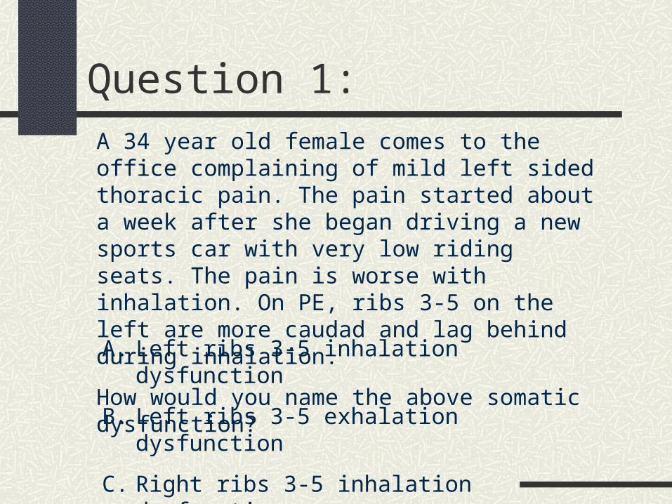
Question 1:A 34 year old female comes to the office complaining of mild left sided thoracic pain. The pain started about a week after she began driving a new sports car with very low riding seats. The pain is worse with inhalation. On PE, ribs 3-5 on the left are more caudad and lag behind during inhalation.
How would you name the above somatic dysfunction?
A. Left ribs 3-5 inhalation dysfunction
B. Left ribs 3-5 exhalation dysfunction
C. Right ribs 3-5 inhalation dysfunction
D. Right ribs 3-5 exhalation dysfunction
Question 2
In the previous case, which rib would be the “key rib” through which to direct treatment?
A. Rib 2
B. Rib 3
C. Rib 4
D. Rib 5
E. Rib 6
Question 3
If you choose to treat the previous patient with the “Kirksville Krunch” HVLA, in which direction would the thrust be applied?
A. Cephalad
B. Caudad
C. Straight down towards your fulcrum
D. HVLA contraindicated in the prior patient because she is a female of child bearing age
"Randy Works the Cavalier" Socks & BarefootRandy is from the midwest and worked his Cavalier in socks and barefoot. There is pumping and driving in this video with some cool angles.