one-lung ventilation and oxygenation...
TRANSCRIPT

One-lung ventilation and
oxygenation improvement
Prof. FJ Belda MD, PhD.
University of Valencia
Anesthesia and Critical Care Department
Hospital Clínico Universitario Valencia, Spain
mail: [email protected]

Summary
Indications
Respiratory physiology during OLV
Objectives during OLV
Ventilatory settings to improve oxygenation
Ventilatory mode
Tidal volume
Recruitment maneuvers and PEEP

Introduction
One-lung ventilation,
separation of the two lungs through the airway
• OLV provides:
– Improved exposure of surgical field
– Protection of healthy lung from infected/bleeding one
• OLV causes:
– Difficult airway and ventilatory setting
– Significant physiologic change
– Easily development of hypoxemia

Indication for OLV in real life
Avoid spillage or contamination of one lung from the other during surgery
Infection, Massive hemorrhage
Control of the distribution of ventilation
Surgical opening of a major conducting airway
Bronchopleural / cutaneous fistula
Tracheobronchial tree disruption
Life-threatening hypoxemia due to unilateral lung disease
Surgical exposure (high-low priority)
Thoracic aortic aneurysm, Pneumonectomy, giant unilateral lung cyst or bulla
Upper lobectomy, Mediastinal exposure, Thoracoscopy
Middle and lower lobectomies and subsegmental resections
Esophageal surgery, Thoracic spine procedures, Minimal invasive cardiac surgery .
Vs
ECCO2R
ECMO

Robertshaw DLT
Two lumen + cuffs
trachea and mainstem bronchus
Method of isolation: DLT
Right-sided
or left-sided
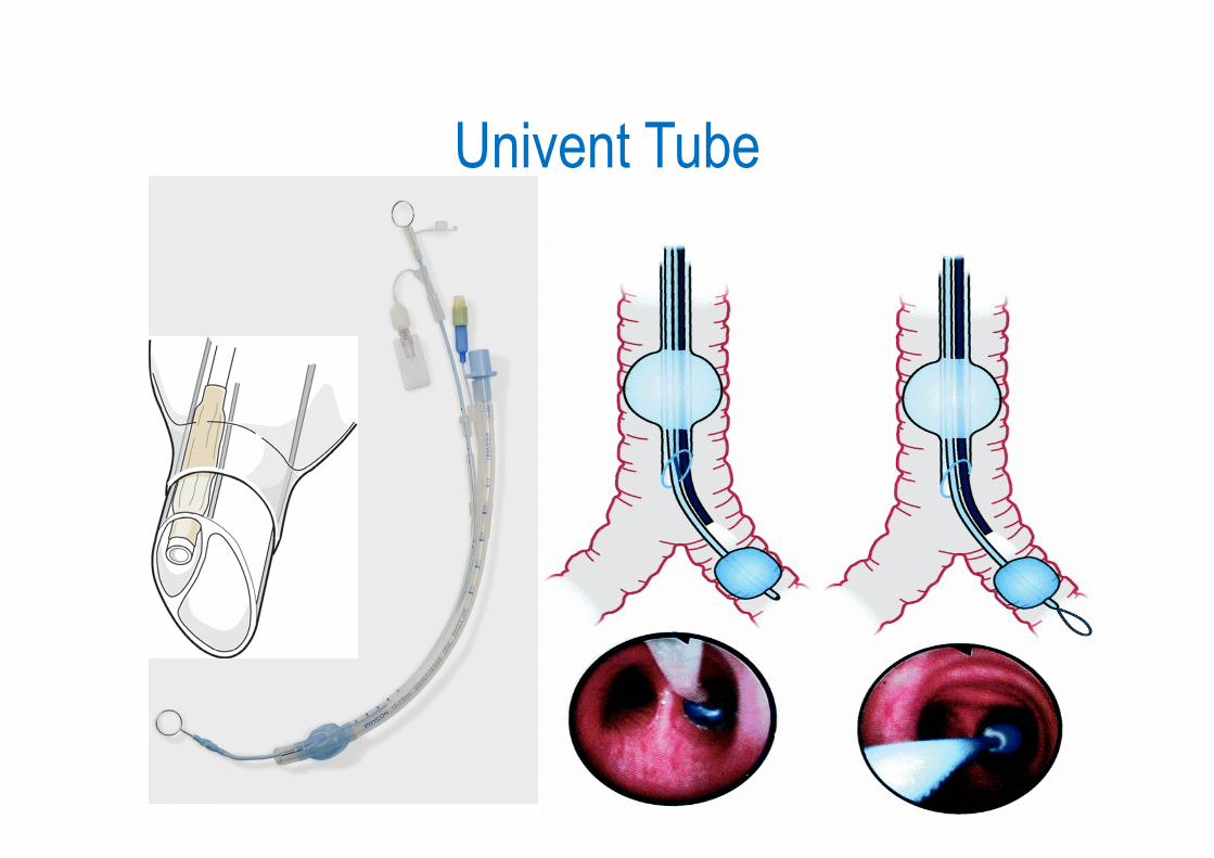
Univent Tube

Arndt endobronchial blockerWire guided Endobronchial Blocker (WEB)
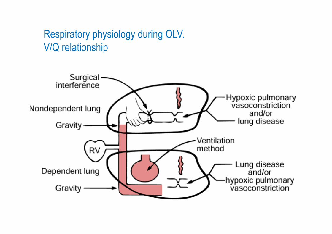
Respiratory physiology during OLV.
V/Q relationship

Two-lung ventilation and OLV
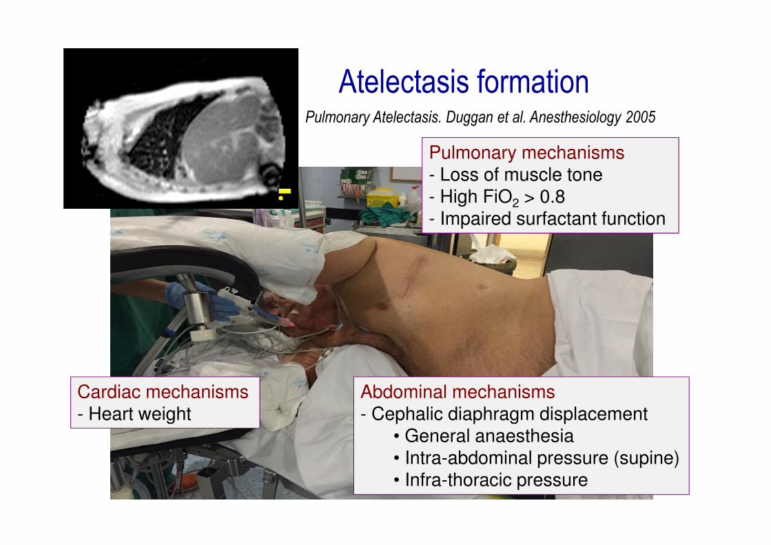
Pulmonary Atelectasis. Duggan et al. Anesthesiology 2005
Abdominal mechanisms- Cephalic diaphragm displacement
• General anaesthesia• Intra-abdominal pressure (supine)• Infra-thoracic pressure
Cardiac mechanisms- Heart weight
Pulmonary mechanisms- Loss of muscle tone- High FiO2 > 0.8- Impaired surfactant function
Atelectasis formation
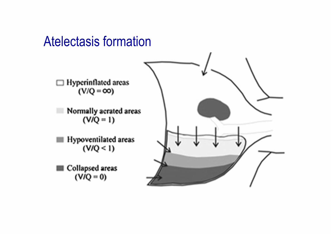
Atelectasis formation

Increases:Shunt/Dead Space
Airway resistance
Decreases:Lung volume
Complicance
ARDS-like condition
Respiratory mechanics
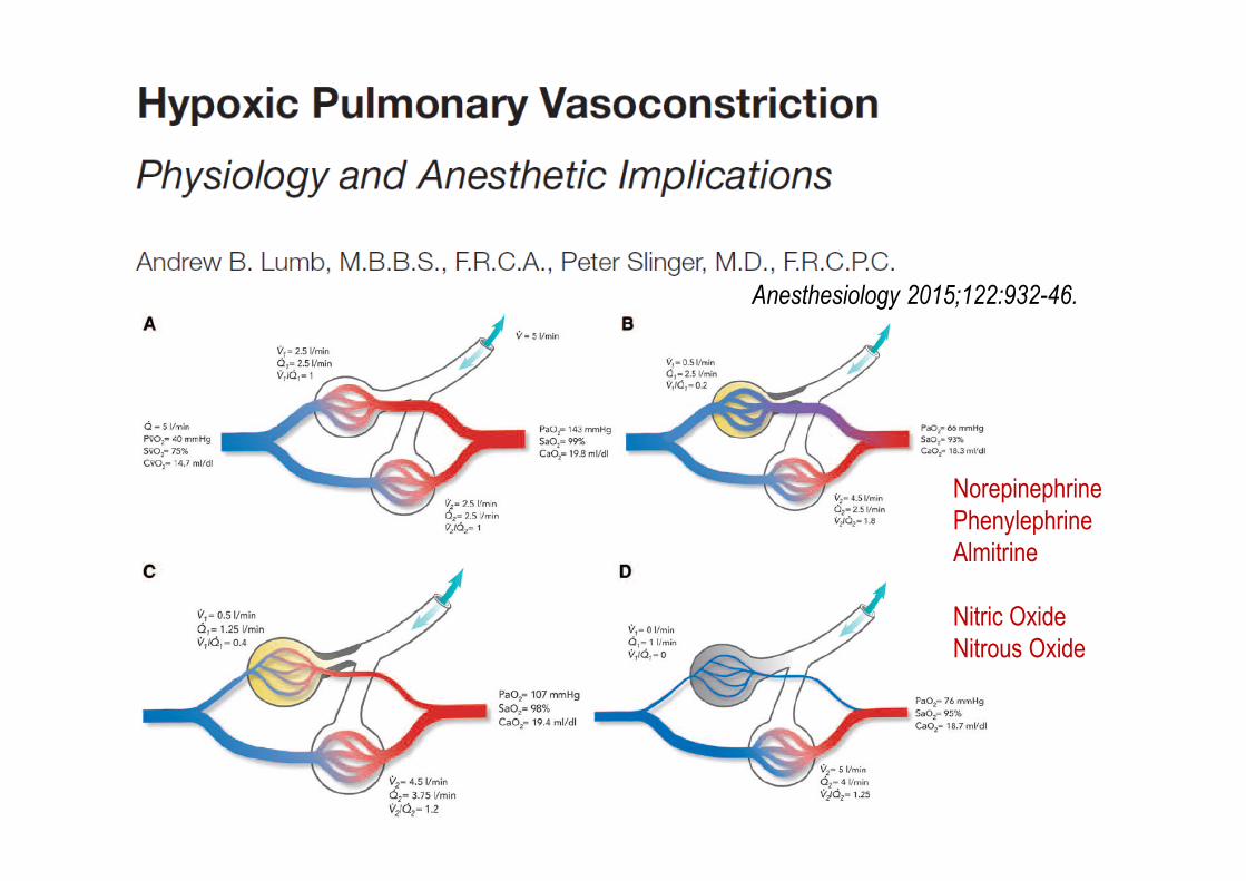
Anesthesiology 2015;122:932-46.
Norepinephrine
Phenylephrine
Almitrine
Nitric Oxide
Nitrous Oxide
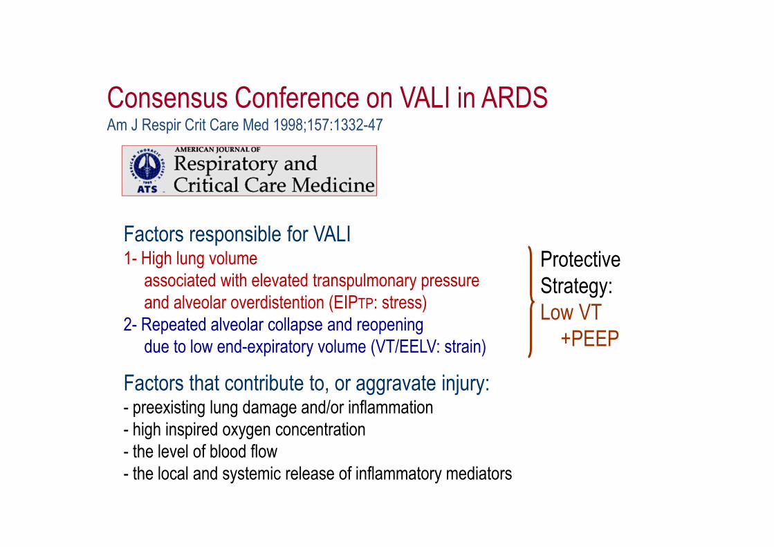
Factors responsible for VALI1- High lung volume
associated with elevated transpulmonary pressure
and alveolar overdistention (EIPTP: stress)
2- Repeated alveolar collapse and reopening
due to low end-expiratory volume (VT/EELV: strain)
Factors that contribute to, or aggravate injury:- preexisting lung damage and/or inflammation
- high inspired oxygen concentration
- the level of blood flow
- the local and systemic release of inflammatory mediators
Consensus Conference on VALI in ARDSAm J Respir Crit Care Med 1998;157:1332-47
Protective
Strategy:
Low VT
+PEEP

Inspiration
VILI: Volutrauma
Expiration

VILI: Atelectrauma

Setting Optimal Ventilation in
Thoracic Surgery
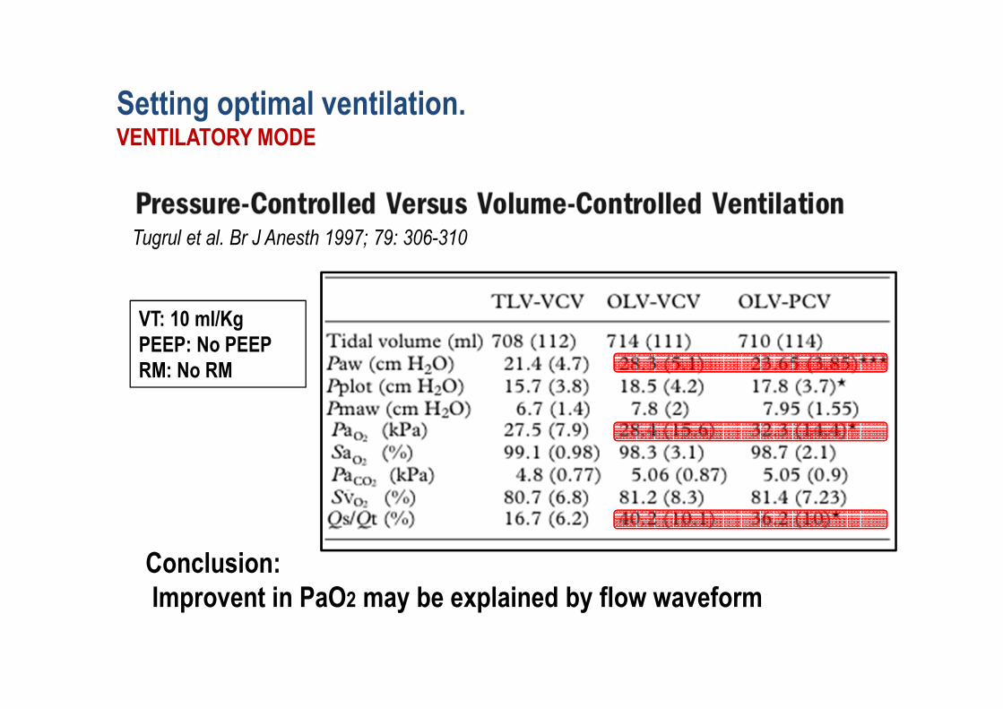
Tugrul et al. Br J Anesth 1997; 79: 306-310
VT: 10 ml/Kg
PEEP: No PEEP
RM: No RM
Conclusion:
Improvent in PaO2 may be explained by flow waveform
Setting optimal ventilation. VENTILATORY MODE

Unzueta et al. Anesth Analg 2007; 104:1029-33 VT: 9 ml/Kg
PEEP: No PEEP
RM: No RM
Conclusion:
The PCV does not lead to improvement in PaO2
Setting optimal ventilation. VENTILATORY MODE

Montes et al. J Cardiothorac surg 2010; 5:99
VT: 6 ml/Kg
PEEP: 5 cmH2O
RM: No RM
Conclusion:
The PCV does not lead to improvement in PaO2
Setting optimal ventilation. VENTILATORY MODE
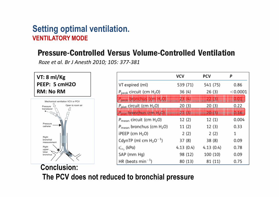
VT: 8 ml/Kg
PEEP: 5 cmH2O
RM: No RM
Roze et al. Br J Anesth 2010; 105: 377-381
Setting optimal ventilation. VENTILATORY MODE
Conclusion:
The PCV does not reduced to bronchial pressure

No differences in:
• Gas exchange
• Ventilator-induced lung injury (dPAlv)
Setting optimal ventilation. VENTILATORY MODE

VALI in thoracic surgery
Setting optimal ventilation. TIDAL VOLUME
2006;103:268-270
2006;103:271-273
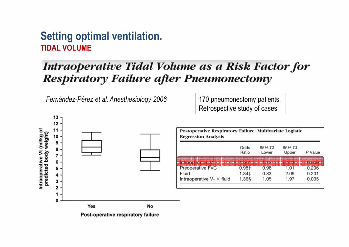
Fernández-Pérez et al. Anesthesiology 2006 170 pneumonectomy patients.
Retrospective study of cases
Setting optimal ventilation. TIDAL VOLUME

Michelet et al. Anesthesiology 2006
52 Esophaguectomy patients.
Prospective
VT: 6 vs 9ml/Kg
PEEP:5 vs 0 cmH2O
RM: No RM
Conclusion:
• Reduces inflammation
• Reduces ELWI
• Reduces ICU LOS
Setting optimal ventilation. TIDAL VOLUME

Kozian et al. Anesthesiology 2011
Setting optimal ventilation. TIDAL VOLUME

Always related to predicted body weight
NO DISCUSSION: Low Tidal Volume
Setting optimal ventilation. TIDAL VOLUME

Tusman et al. Anesth Analg 2004
Unzueta et al. Br J Anaesth 2012
Ferrando et al. Anesth Analg 2014
Cinnella et al. Acta Anesthesiol Scan 2008
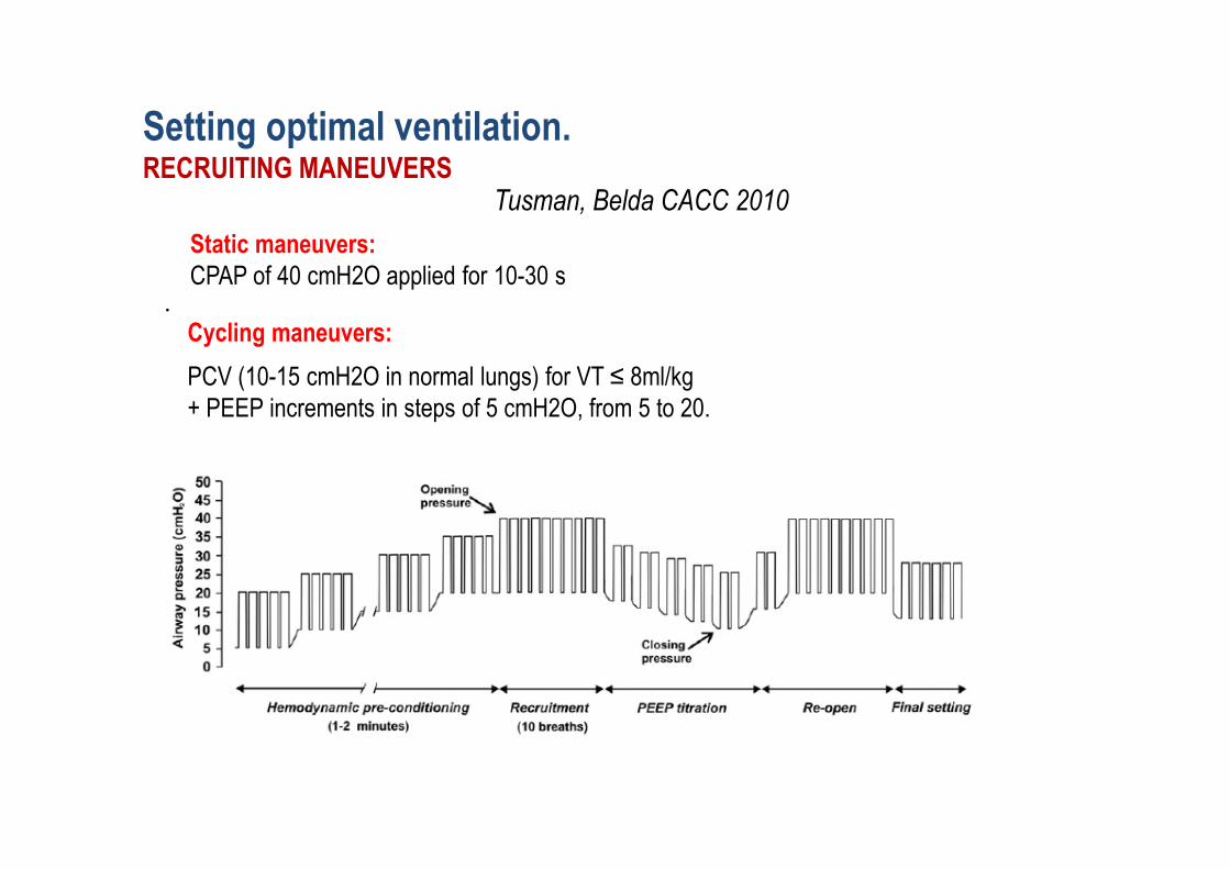
Setting optimal ventilation. RECRUITING MANEUVERS
•
Static maneuvers:
CPAP of 40 cmH2O applied for 10-30 s
Cycling maneuvers:
PCV (10-15 cmH2O in normal lungs) for VT ≤ 8ml/kg
+ PEEP increments in steps of 5 cmH2O, from 5 to 20.
Tusman, Belda CACC 2010
Setting optimal ventilation. RECRUITING MANEUVERS

•
Stepwise increases in PEEP
Time to adapt haemodynamics
Help to diagnose and treat an unrecognized
hypovolaemic state.
Lower pulmonary tissue stress
Increments in pressure and volume spread progressively
within more and more ‘recruited’ tissue
PEEP titration phase
helps to detect the level of PEEP
Setting optimal ventilation. RECRUITING MANEUVERS

Should always be used
Improves ventilatory efficiency
Decreases VILI
Arbitrarily set at 5 cmH2O
- Slinger et al. Anesthesiology 2001
- Fujirawa et al. J Clinic Anesth 2001
- Valenza et al. Eur J Anesthesiol 2004 (10cmH2O)
- Tusman et al. Anesth Analg 2004
- Michelet et al Br J Anesth 2005
- Cinnella et al. Acta Anesthesiol Scan 2008
- Ren et al. An Intensive Care 2008
- Park et al. Eur J Anesthesiol 2011
- Unzueta el al. Br J Anesth 2012 (8 cmH20)
Setting optimal ventilation. PEEP

Ferrando et al. Anesth Analg 2014
Setting optimal ventilation. PEEP

0
5
10
15
20
25
30
35
40
1 15 29 43 57 71 85 99 113 127 141 155 169 183 197 211 225 239 253 267
Dynamic RM + PEEP titration in VCV during OLV
PEEP
Crs
VCO2,br
Unpublished data
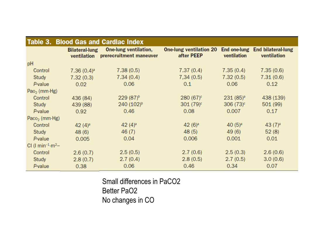
Small differences in PaCO2
Better PaO2
No changes in CO

RM + PEEP: No changes in Compliance (compared to baseline)
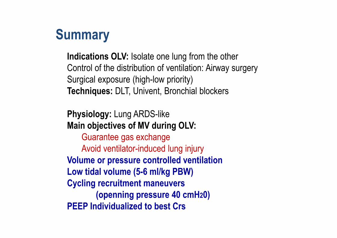
Indications OLV: Isolate one lung from the other
Control of the distribution of ventilation: Airway surgery
Surgical exposure (high-low priority)
Techniques: DLT, Univent, Bronchial blockers
Physiology: Lung ARDS-like
Main objectives of MV during OLV:
Guarantee gas exchange
Avoid ventilator-induced lung injury
Volume or pressure controlled ventilation
Low tidal volume (5-6 ml/kg PBW)
Cycling recruitment maneuvers
(openning pressure 40 cmH20)
PEEP Individualized to best Crs
Summary