orthofix calcaneal external...
TRANSCRIPT

OPERATIVE TECHNIQUE
Orthofix CalcanealExternal Fixator
INTRODUCTION
INDICATIONS
FEATURES AND BENEFITS
EQUIPMENT REQUIRED
PATIENT POSITIONING
APPLICATION OF CALCANEALTRACTION
OPERATIVE TECHNIQUE
Pin Position
Pin Placement
Fragment Manipulation
POST-OPERATIVE MANAGEMENT
1
1
2
3
4
4
4
5
6
8
8
Orthofix wishes to thankthe following surgeons for their contributionto the development of the technique:
GREG GRABOWSKI D.P.M.BRUNO MAGNAN M.D.

References1.Magnan B et al. G.I.O.T., 2006; 32 (suppl.1), S322-S326. 2.Magnan B et al. J Bone Joint Surg, 2006; 88 B, 1474-93.Cotton FJ, Wilson LT. Fractures of the os calcis. Boston MedicalJournal, 1908; 159, 559-565.4.Howard JL, Buckley R, McCormack R, Pate G, Leighton R,Petrie D, Galpin R. Complications following management ofdisplaced intra-articular calcaneal fractures: a prospective randomizedtrial comparing open reduction internal fixation with nonoperativemanagement. Journal of Orthopaedic Trauma, 2003; 4, 241-249.5.Rajkumar P, Henderson AA. Surgical treatment of displacedintra-articular fractures of the os-calcis. Foot and Ankle, 2003; 9, 3-6.6.Sanders R, Fortin P, DiPasquale T, Wall ing A. Operativetreatment in 120 displaced intraarticular calcaneal fractures. Resultsusing a prognostic computed tomography scan classification. ClinicalOrthopaedics and Related Researches, 1993; 290, 87-95.7.Randle JA, Kreder HJ, Stephen D, Williams J, Jaglal S, Hu R.Should calcaneal fractures be treated surgically? A meta-analysis.Clinical Orthopaedics and Related Researches, 2000; 377, 217-27.8.Richardson ML, Van Vu M, Vincent LM, Sangeorzan BJ,Benirschke SK. CT measurement of the calcaneal varus angle in thenormal and fractured hindfoot. Journal of Computer AssistedTomography, 1992; 16, 261-264.9.Rosenberg ZS, Feldman F, Singson RD.Intra-articular calcaneal fractures: computed tomographic analysis.Skeletal Radiology, 1987; 16,105-113. 10.Magnan B, Montanari M, Bragantini A, Bartolozzi P. Asystem of prognostic evaluation of CT imaging of heel fractures: theScore Analysis Verona (SAVE). Foot Disease, 1995; 1, 19-25.11.Csizy M, Buckley R, Tough S, Leighton R, Smith J, McCormackR, Pate G, Petrie D, Galpin R. Displaced intra-articular calcanealfractures: variables predicting late subtalar fusion. Journal ofOrthopaedic Trauma,. 2003; 2, 106-112.12.Sanders R.: Displaced intra-articular fractures of the calcaneus.Journal of Bone and Joint Surgery American Volume, 2000; 82, 225-250.13.Coughlin MJ. Calcaneal fractures in industrial patients. FootAnkle International, 2000; 21 (11): 896-905.14.Myerson M, Quil l GE Jr. Late complications of fractures of thecalcaneus. Journal of Bone and Joint Surgery American Volume, 1993;3, 331-341.15.Paley D, Hall H. Intra-articular fractures of the calcaneus. Acritical analysis of results and prognostic factors. Journal of Bone andJoint Surgery American Volume, 1993; 3, 342-354.16.Borell i J Jr, Torzil l i P. Effect of impact load on articularcartilage: development of an intra-articular fracture model. Journal ofOrthopaedic Trauma, 1997; 11, 319.
INDICATIONS
1) Articular Fractures of the Calcaneus- Sander’s CT classification I, II, III, IV- Rowe’s X-ray classification IV
2) Oblique or Coronal Calcaneal Body Fractures not involving the Subtalar joint
- Rowe’s X-ray classification III
INTRODUCTION
As reported in the literature 1, 2 calcaneal fractures involvingthe joints are disabling injuries which can jeopardize workand day-to-day activities3.The anatomy and biomechanics of the hindfoot account forthe difficulties in treating this type of fracture, since thecalcaneus is constantly subjected to compression forces,operates as a sesamoid bone in the Achilles-calcaneal-plantarsystem and has articular surfaces that form two complexjoints: Chopart’s and subtalar joint.Despite several comparative studies between the conservativeand surgical methods, the management of intra-articularcalcaneal fractures remains controversial.There are a number of reasons for this:
- difficulty in obtaining reduction using conservative methods, often leading to considerable sequelae, such as pain, hindfoot deformity, impingement, and disturbed gait;
- difficulty in reduction and fixation, even with an open surgical approach;
- the high risk of major complications related to open surgery4
Although open reduction and internal fixation is currentlyconsidered the treatment of choice for Sanders types II, IIIand IV fractures5,6, uncertainty remains about the comparativefinal results of surgical and conservative treatment7, sinceneither method provides good results without the risk ofconsiderable early and delayed complications.The main goal for treatment of articular displaced heelfractures should be the restoration of the three dimensionalstructure of the os calcis, emphasising correct alignment inthe coronal and axial planes and the height of the calcanealbody8,9, rather than anatomical reconstruction of thecongruency of the subtalar articular fragments6,10,8. The use ofexternal fixation to treat displaced articular fractures of theheel appears suitable for obtaining such goals, and istherefore as good rationale for the technique, which allowsstable fixation and a reduced risk of major complications.Anatomical restoration of the subtalar joint facet is verydifficult to obtain, particularly with percutaneous reductionand external fixation using two single pins stabilizing thearticular fragments, as demostrated by CT evaluation atfollow up. Some degree of stiffness and degenerative arthritisof the sub-talar joint following a displaced articular fracture11
is usually unavoidable whatever the chosen treatment12,13,14,15,due to the severe damage to the articular cartilage whichalmost always sustains high energy axial load16. The results ofour series indicate that neither the subtalar mobility nor theclinical outcome seem to be significantly affected byobtaining congruency of the subtalar facet, when comparedwith the results reported with open forms of treatment,which specifically aim to reduce the articular fragmentsanatomically, but correlate more with the results followingearly post-operative mobilization and the restoration ofBoehler’s angle12,10.
Percutaneous reduction and external fixation proved to be areliable technique for obtaining stable reconstruction offractures of the os calcis. The clinical results appear to becomparable to those obtained with open reduction andinternal fixation.The added advantages of minimally invasive procedures areconsiderably shortened operating and hospitalization time,and reduced risk of complications related to surgicalexposure.
1

2
Left
FEATURES AND BENEFITS
130°
25°
For right or left foot Swivelling Clamps to allowfor angled placement of the pins
Compression-distraction units to allow for fragment reduction
15 mm
35 mm
12mm
Right
35°35°

3
EQUIPMENT REQUIRED
Calcaneal Fixator Sterile Kit (99-M1450)
1) Calcaneal Fixator M1452) 3 mm Allen Key 100123) T-Wrench T M2104) Driver M2115) Pin 70/15 M3216) Pin 70/25 M315
COMPRISES7) Pin 100/30 M3178) Calcaneal Template Kit M224
Consisting of:6 Wires6 Wire Guides6 Pin Guides
�
�
�
�
�
�
�

4
OPERATIVE TECHNIQUE
Percutaneous Reduction
Through a percutaneous supra-lateral approach of theexternal part of the sinus tarsi, used by most authors,insert a small bone lever to lift and rotate thedepressed articular talamic fragments to reconstructthe articular surface and the Böhler's angle.
Alternatively, a direct plantar approach through thecalcaneal body or a para-Achilles postero-lateralapproach directly to the subtalar joint may be used.
APPLICATION OF CALCANEALTRACTION
Some authors prefer a supine position in order toapply a calcaneal traction using a Kirschner wire orSteinman Pin and reduce pre-operatively the varus orvalgus deformity of the posterior part of thecalcaneus.In case of reduction with a mini-invasive surgicalapproach, calcaneal traction is not necessary.
PATIENT POSITIONING
The patient is placed in lateral decubitus ontheuninjured side with the C-arm (fluoroscopy)positionedto allow lateral and axial view of the hidfootwhile notinterfering with the surgical field.Atourniquet can be applied at the base of the limbandinflated if necessary, very rare situation with mini-invasive and/or percutaneous approaches forthereduction of articular talamic fragments.

5
Pin Position
The fixator is positioned according to the fracturepattern. Once reduction of the talamic surface hasbeen achieved, the first two pins should always bepositioned on the talamic fragments, in subchondralbone, using the fixator as a template. It is possible topreliminary fix the fragments with wires, wire guidesand pin guides of the Calcaneal Template Kit.Once correct reduction has been achieved, remove thewires and wire guides, insert the pins and remove thepin guides. The remaining pins are inserted so thatthey create a counterbalance and maintain reductionof the articular surface in height. The position of thesecond and third clamp depends upon the integrity orfragmentation of the bone grip site and the fracturepattern.
It is advisable to apply pins as follows:å at the level of the anterior calcaneal apophysis in
Tongue Type fractures, according to Essex-Lopresticlassification, in which the articular fragmentshould be lifted and rotated with a force which ismore favourable if exerted from the anterior partof the calcaneus;
ç at the level of the posterior apophysis in JointDepression Type fractures, according to the Essex-Lopresti classification, in which the articularfargment sould be completely lifted vertically.
é From both (anterior and posterior) sides in higlycomminuted fractures of the entire calcaneal body,or when a greater stability is needed. It may be necessary to place one or two pins of the anterior clamp in the cuboid in case of severe comminution of the anterior calcaneal apophysis.
å
ç
é

6
Pin Placement
Insert the pin guides into the clamps and tighten theclamp covers to keep them parallel.Drill the first wire through the wire guide in theanterior pin position of the subtalar or third armclamp, just below the subchrondral bone of thesubtalar joint capturing the sustentacular fragment.
Gentle traction can be applied to the pin to closeany fracture gap. The pin can also be used as joy stickto correct any “step off” between fracture fragments.
Using the same procedure, insert the second pin in thetalamic fragment and the wires in the anterior andposterior fragments. Once the positions of the wireshave been confirmed with fluoroscopy, the first pin isinserted through the empty guide in the clamp, eithermanually using the T-wrench or with the drill using thedriver.

7
At this stage, when applying the pins at the level ofthe posterior or anterior calcaneal apophysis, it ispossible to correct the varus-valgus deformity of thecalcaneal body, by inserting the pin at an anglethrough the clamp that acts as a template. This angle can be captured bythe pin clamp’s swivel head.
Using the pins again as a joy stick, it is possible toachieve correct alignment of the calcaneal body withthe articular fragment already fixed with the firstclamp.
The remaining pins are then inserted and the pinguides removed. With all pins in proper alignment, the external fixator is then locked onto the pins,paying careful attention to allow for any swelling that may occurred.
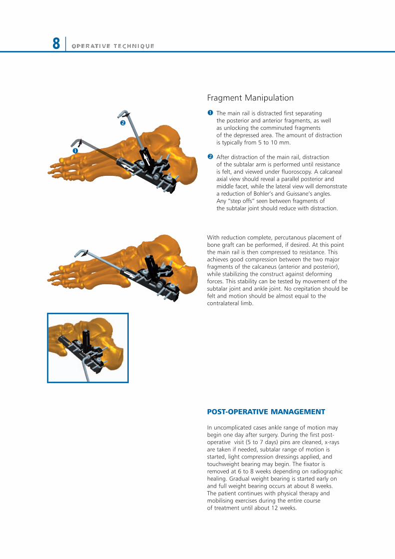
Fragment Manipulation
å The main rail is distracted first separatingthe posterior and anterior fragments, as wellas unlocking the comminuted fragmentsof the depressed area. The amount of distractionis typically from 5 to 10 mm.
ç After distraction of the main rail, distractionof the subtalar arm is performed until resistance is felt, and viewed under fluoroscopy. A calcanealaxial view should reveal a parallel posterior andmiddle facet, while the lateral view will demonstrate a reduction of Bohler’s and Guissane’s angles.Any “step offs” seen between fragments ofthe subtalar joint should reduce with distraction.
With reduction complete, percutanous placement ofbone graft can be performed, if desired. At this pointthe main rail is then compressed to resistance. Thisachieves good compression between the two majorfragments of the calcaneus (anterior and posterior),while stabilizing the construct against deformingforces. This stability can be tested by movement of thesubtalar joint and ankle joint. No crepitation should befelt and motion should be almost equal to thecontralateral limb.
POST-OPERATIVE MANAGEMENT
In uncomplicated cases ankle range of motion maybegin one day after surgery. During the first post-operative visit (5 to 7 days) pins are cleaned, x-raysare taken if needed, subtalar range of motion isstarted, light compression dressings applied, andtouchweight bearing may begin. The fixator isremoved at 6 to 8 weeks depending on radiographichealing. Gradual weight bearing is started early onand full weight bearing occurs at about 8 weeks.The patient continues with physical therapy andmobilising exercises during the entire course of treatment until about 12 weeks.i
å
ç
8
9
Bibliography
Barnard L, Odegardd JK. Conservative approach in thetreatment of fractures of the calcaneous. Journal of Bone andJoint Surgery, American Volume, 1955; 37-A, 1231-1236.Benirschke SK, Kramer PA. Wound healing complications inclosed and open calcaneal fractures. Journal of OrthopaedicTrauma, 2004; 18, 1-6.Castell i F. The minimally invasive osteosynthesis of intra-articular calcaneous fractures: early results. Abs. to 7thEFORT CONGRESS, June 2005. P2-504.Conn HR: The treatment of fractures of the os calcis. Journal ofBone and Joint Surgery American Volume, 1935; 17, 392-405.Dooley P, Buckley R, Tough S, McCormack B, Pate G,Leighton R, Petrie D, Galpin B. Bilateral calcaneal fractures:operative versus nonoperative treatment. Foot and AnkleInternational, 2004; 2, 47-52.Heffernan G, Khan F, Awan N, Riordain CO, Corrigan J. Acomparison of outcome scores in os calcis fractures. Irish Journalof Medical Science, 2000; 169(2), 127-128.Kenwright J.: Fractures of the calcaneum, Journal of Bone and JointSurgery British Volume,1993; 75(2), 176-177.Lim EV, Leung JP. Complications of intraarticular calcanearfractures. Clinical Orthopaedics and Related Researches, 2001;391, 7-16.Loucks C, Buckley R. Bohler's angle: correlation with outcomein displaced intra-articular calcaneal fractures. Journal ofOrthopaedic Trauma, 1999; 8, 554-558.Magnan B, Caudana R, Campacci A, Barzoi A,Molinaroli F, Zonta L, Martell i S. Follow-up clinico eradiografico mediante T.C. delle fratture di calcagno trattate conmini FEA. Chirurgia del piede, 1992; 16, 145-150.Morrey BF, Wiedeman GP Jr. Complications and long-termresults of ankle arthrodesis following trauma. Journal of Bone andJoint Surgery American Volume, 1980; 5, 777-784.Nogarin L, Magnan B, Bragantini A, Rebeccato A,Montanari M, Schiavon R. Trattamento a cielo chiuso degliaffossamenti talamici con mini-fissatori esterni. Fratture delcalcagno. Progressi in medicina e chirurgia del piede. Bologna.Aulo Gaggi editore. 1994.Paley D, Fischgrund J. Open reduction and circular externalfixation of intraarticular calcaneal fractures. Clinical Orthopaedicsand Related Researches,1993; 290, 125-131. Pajenda G., Chatwami S., Loidl G., Osterman R., VecseiV. Percutaneous screw fixation of the displaced calcanealfractures. Book of Abstract SICOT/SIROT 2002, p 308.Rammelt S, Zwipp H. Calcaneus fractures: facts, controversiesand recent developments. Injury, 2004; 5, 443-461. Sanders R. Hansen S.T. Jr, Mc Reynolds I.S. Trauma to thecalcaneus and its tendon: fractures of the calcaneous. In: JahssM.H.: Disorders of the foot and ankle. 2nd edition, Philadelphia,W.B. Saunders Co.,1991.Talarico LM, Vito GR, Zyryanov SY. Management ofdisplaced intraarticular displaced fractures by using externalfixation, minimally invasive open reduction, and early weight-bearing. Journal of Foot and Ankle Surgery, 2004; 43(1), 43-50.
Thornes BS, Collins AL, Timlin M, Corrigan J. Outcome ofcalcaneal fractures treated operatively and non-operatively. Theeffect of litigation on outcomes. Irish Journal of Medical Science,2002; 3, 155-157.Yong Soo Choi, Ki Soo Kim. Minimally invasive openreduction and cross screws fixation for Sanders tipe II fractures ofthe calcaneous. Book of Abstract SICOT/SIROT 2002, p 2020.Yong Soo Choi, Ki Soo Kim, Hyeon Gyu Boem. Biomechanicalstability of cross screws fixation fixation for Sanders type IIcalcaneal fractures. Book of Abstract SICOT/SIROT 2002, p 2021.Zwipp H, Tscherne H, Thermann H, Weber T. Osteosynthesis ofdisplaced intraarticular fractures of the calcaneus. Results in 123cases. Clinical Orthopaedics and Related Researches , 1993; 290,76-86.

Your Distributor is:
www.or thof i x . comEF-0701-OPT-E0 C 08/10
Deformity Correct ion I Trauma I Pediat r i cs I Bone Growth St imulat ion
Manufactured by: ORTHOFIX SrlVia Delle Nazioni 937012 Bussolengo (Verona)Italy
Telephone +39-0456719000Fax +39-0456719380
0123