orthopedics journal - rush university medical center · pdf file2016 rush orthopedics journal...
TRANSCRIPT

PLEASE NOTE: All physicians featured in this publication are on the medical faculty of Rush University Medical Center. Many of the physicians featured are in the private practice Midwest Orthopaedics at Rush and, as independent practitioners, are not agents or employees of Rush University Medical Center.
Rush is a not-for-profit health care, education and research enterprise comprising Rush University Medical Center, Rush University, Rush Oak Park Hospital and Rush Health.
M-4135 8/16
2016 RU
SH O
RTH
OPED
ICS JO
UR
NA
L
Orthopedics Journal2016 Rush
87802_Ortho_Cover_a2.indd 1-3 8/3/16 3:36 PM

BEST PRACTICE. For orthopedic specialists at Rush, good medicine and good teaching
are inextricably linked. In this issue, 4 of our faculty discuss the many facets of orthopedic
education at Rush—and why both teaching and learning are lifelong pursuits (see page 62).

Faculty Editors
Editor in Chief David Fardon, MD
Deputy Editors Brett Levine, MD, MS; Shane J. Nho, MD, MS; Robert W. Wysocki, MD; Adam Yanke, MD
Associate Editors Rachel M. Frank, MD; David M. Levy, MD; Andrew J. Riff, MD
Editor Emeritus Steven Gitelis, MD
2 Chairman’s Letter
3 Orthopedic Faculty and Fellows
8 Research Faculty
10 Department of Orthopedic Surgery Residents
Articles
11 Computer Navigation–Assisted Musculoskeletal Tumor Surgery: Update and Presentation of 4 Clinical Cases Mick P. Kelly, MD; Matthew W. Colman, MD
16 Chemokine Receptor Antagonists Can Inhibit Macrophage Migration Peng Shi, DDS, PhD; Ana V. Chee, PhD; David K. Liu, MS; Justin L. Zheng, BS; Ding Chen, MD; Zemin Li, MD; Chundo Oh, PhD; Di Chen, MD, PhD; Gunnar B. J. Andersson, MD, PhD; Howard S. An, MD
21 Anterior Cruciate Ligament Reconstruction: Optimizing Femoral Tunnel Position With Flexible Curved Reamers Brian Forsythe, MD; Michael J. Collins, BS; Thomas A. Arns, BS; Michael Khair, MD; Nikhil N. Verma, MD; Brian J. Cole, MD, MBA; Bernard R. Bach Jr, MD; Nozomu Inoue, MD, PhD
26 Limb Lengthening: A New Technology Monica Kogan, MD
28 Regrowth of Symptomatic Cam Deformity After Hip Arthroscopy and Femoral Osteochondroplasty Gregory L. Cvetanovich, MD; Brandon J. Erickson, MD; Randy Mascarenhas, MD; Simon X. Lee, MPH; Shane J. Nho, MD, MS
31 Adolescent Idiopathic Scoliosis: A Pilot Study With Differential Rod Bending Philip K. Louie, MD; Christopher J. DeWald, MD
37 Chronic Type III Acromioclavicular Separations: Modified Weaver-Dunn Reconstruction Failures Randy Mascarenhas, MD, FRCSC; Pav Sumner, MBBS, FRACS; Bryan M. Saltzman, MD; Myrna Dyck, BN, MSc; Anthony A. Romeo, MD; Peter B. MacDonald, MD, FRCSC
42 Update on Superior Labrum Anterior to Posterior Tears and Biceps Tendon Tears: Conclusions Based on Translational Research Performed at the Department of Orthopedic Surgery at Rush Brian J. Cole, MD, MBA; Robert J. Thorsness, MD; Nikhil N. Verma, MD; Anthony A. Romeo, MD
46 Minimally Invasive Transforaminal Lumbar Interbody Fusion: One Surgeon’s Learning Curve Dustin H. Massel, BS; Benjamin C. Mayo, BA; Sreeharsha V. Nandyala, MD; Steve Fineberg, MD; Kern Singh, MD
52 History of Lumbar Disc Science and Surgery Nicholas Brown, MD; Gunnar B. J. Andersson, MD, PhD; David Fardon, MD
56 Publications (2015)
62 Higher Learning An introduction to orthopedic education at Rush
63 Bedside Manner Taking surgical education beyond the operating room
Edward J. Goldberg, MD
65 A Cut Above Training the next generation of orthopedic surgeons
Simon Lee, MD
67 Down to the Core Preparing residents to practice at the highest level Brett Levine, MD, MS
69 Connecting the Dots Teaching young clinicians how research can transform patient care Anthony A. Romeo, MD
To view the 2016 Rush Orthopedics Journal online or to view past issues of the journal, please visit www.rush.edu/orthopedicsjournal.

2016 Rush Orthopedics Journal2
The theme of this year’s Rush Orthopedics Journal is education, and were I not confined to a single page, I could write volumes on the topic.
Education is a core mission of the Department of Orthopedic Surgery at Rush, just as education is a foundational pillar of Rush itself. Rush Medical College was the first medical school in Chicago, and the early faculty became nationally recognized for patient care, research, and teaching. After suspending its educational program from 1942 to 1969, the college reactivated its charter and merged with what was then Presbyterian-St. Luke’s Medical Center, because the leaders envisioned a learning environment where practitioners would take a leading role in the education of students.
The practitioner-teacher model is alive and well at Rush today, and it’s evident in all of our department’s educational endeavors. Our faculty members are committed to training and mentoring students, residents, and fellows, providing them with the knowledge and skills to become successful and productive members of the health care community.
We are also producing the next generation of translational researchers in orthopedics—through our highly competitive orthopedic residency and fellowship programs, and through graduate programs at Rush University. Rush offers both master’s and Doctor of Philosophy degrees in integrated biomedical sciences, which train students to find innovative
solutions to critical biomedical problems and, in conjunction with our faculty, to bring advances from the laboratory to the patient.
Enabling trainees and graduate students to apply the principles they are learning to solve real-world health issues improves both the educational experience and patient care. It also gives our clinicians and basic scientists opportunities to share their expertise with the men and women who represent the future of orthopedics.
Four of my colleagues, Edward J. Goldberg, MD, Brett Levine, MD, MS, Shane J. Nho, MD, MS, and Anthony A. Romeo, MD, were kind enough to share their personal experiences as teachers and mentors in this year’s interview feature (see p 62). I hope you’ll take a few moments to read their stories, which highlight the breadth and quality of orthopedic education at Rush.
Joshua J. Jacobs, MD
The William A. Hark, MD/Susanne G. Swift Professor of Orthopedic Surgery
Chairman, Department of Orthopedic Surgery
Rush University Medical Center
Chairman’s Letter
“Our faculty members are committed to training and mentoring students, residents, and fellows, providing them with the knowledge and skills to
become successful and productive members of the health care community.”

Orthopedic Faculty and Fellows (2015) 3
Orthopedic Faculty and Fellows ADULT RECONSTRUCTIVE SURGERY
Craig J. Della Valle, MD
Director, Division of Adult Reconstructive Surgery
Director, Section of Research
Professor, Department of Orthopedic Surgery
Associate director, Orthopedic Surgery Residency Program
Richard A. Berger, MD
Director, Section of Minimally Invasive Surgery
Assistant professor, Department of Orthopedic Surgery
Brett Levine, MD, MS
Associate professor, Department of Orthopedic Surgery
Wayne G. Paprosky, MD
Professor, Department of Orthopedic Surgery
Aaron Rosenberg, MD
Professor, Department of Orthopedic Surgery
Director, Adult Reconstructive Orthopedic Surgery Fellowship Program
Jorge O. Galante, MD, DMSc
The Grainger Director of the Rush Arthritis and Orthopedics Institute
Professor, Department of Orthopedic Surgery
FELLOWS
Paul Courtney, MD Medical school – Georgetown University School of Medicine,
Washington, DCResidency – Hospital of the University of Pennsylvania
Ismar Dizdarevic, MD Medical school – Sidney Kimmel Medical College–Jefferson
University, PhiladelphiaResidency – Mt. Sinai West, New York
Nicholas Frisch, MD, MBA Medical school – Tufts University School of Medicine, BostonResidency – Henry Ford Hospital, Detroit
Matthew Knedel, MD Medical school – Keck School of Medicine of USC, Los Angeles Residency – University of California San Francisco, Fresno
Christopher Melnic, MD Medical school – Tufts University School of Medicine, BostonResidency – Hospital of the University of Pennsylvania
Duy Phan, MD Medical school – Virginia Commonwealth University School of MedicineResidency – University of California Irvine Medical Center
Michael Ruffolo, MD Medical school – West Virginia University School of MedicineResidency – Carolinas Medical Center, Charlotte
Tad L. Gerlinger, MD
Associate professor, Department of Orthopedic Surgery
Scott M. Sporer, MD, MS
Director, Section of Quality and Outcomes
Associate professor, Department of Orthopedic Surgery
Nicholas T. Ting, MD
Assistant professor, Department of Orthopedic Surgery
Joshua J. Jacobs, MD
The William A. Hark, MD/Susanne G. Swift Professor of Orthopedic Surgery
Chairman, Department of Orthopedic Surgery

2016 Rush Orthopedics Journal4
ELBOW, WRIST, AND HAND SURGERY
Mark S. Cohen, MD
Director, Section of Hand and Elbow Surgery
Professor, Department of Orthopedic Surgery
John J. Fernandez, MD
Assistant professor, Department of Orthopedic Surgery
Robert W. Wysocki, MD
Assistant professor, Department of Orthopedic Surgery
HAND, UPPER EXTREMITY, AND MICROVASCULAR FELLOW
Nicholas Newsum, MD Medical school – University of Toledo College of Medicine and Life Sciences, OhioResidency – University of Florida Shands Hospital, Gainesville
FELLOW
Emily Vafek, MD Medical school – Robert Wood Johnson Medical School, New Brunswick, New JerseyResidency – Wake Forest Baptist Health, Winston-Salem, North Carolina
FOOT AND ANKLE SURGERY
ONCOLOGY ONCOLOGY AND SPINE SURGERY
George Holmes Jr, MD
Director, Section of Foot and Ankle Surgery
Associate Professor, Department of Orthopedic Surgery
Steven Gitelis, MD
Director, Section of Orthopedic Oncology
Rush University Professor of Orthopedic Oncology
Professor, Department of Orthopedic Surgery
Kamran S. Hamid, MD, MPH
Assistant professor, Department of Orthopedic Surgery
Simon Lee, MD
Assistant professor, Department of Orthopedic Surgery
Johnny L. Lin, MD
Assistant professor, Department of Orthopedic Surgery
Matthew W. Colman, MD
Assistant professor, Department of Orthopedic Surgery

Orthopedic Faculty and Fellows (2015) 5
PEDIATRIC ORTHOPEDIC SURGERY
Monica Kogan, MD
Director, Section of Pediatric Orthopedic Surgery
Assistant professor, Department of Orthopedic Surgery
Director, Orthopedic Surgery Residency Program
SPINE SURGERY
Frank M. Phillips, MD
Director, Division of Spine Surgery
Director, Section of Minimally Invasive Spine Surgery
Professor, Department of Orthopedic Surgery
Christopher DeWald, MD
Assistant professor, Department of Orthopedic Surgery
Gunnar B. J. Andersson, MD, PhD
The Ronald L. DeWald, MD, Endowed Chair in Spinal Deformities
Professor and chairman emeritus, Department of Orthopedic Surgery
David Fardon, MD
Assistant professor, Department of Orthopedic Surgery
Kim W. Hammerberg, MD
Assistant professor, Department of Orthopedic Surgery
Kern Singh, MD
Professor, Department of Orthopedic Surgery
Edward J. Goldberg, MD
Assistant professor, Department of Orthopedic Surgery
FELLOWS
Justin Paul, MD Medical school – Weill Cornell Medical College, Ithaca, New YorkResidency – NYU Langone Medical Center/Hospital for Joint Diseases, New York
Comron Saifi, MD Medical school – Columbia University College of Physicians and Surgeons, New YorkResidency – Columbia University Medical Center, New York
Arya Varthi, MD Medical school – University of Connecticut School of Medicine, FarmingtonResidency – Yale University, New Haven, Connecticut
Howard S. An, MD
The Morton International Chair of Orthopedic Surgery
Professor, Department of Orthopedic Surgery
Director, Spine Surgery Fellowship Program

2016 Rush Orthopedics Journal6
SPORTS MEDICINE, SURGERY
Adam Yanke, MD
Assistant professor, Department of Orthopedic Surgery
Bernard R. Bach Jr, MD
The Claude N. Lambert, MD/Helen S. Thomson Professor of Orthopedic Surgery
Professor, Department of Orthopedic Surgery
Brian J. Cole, MD, MBA
Director, Rush Cartilage Restoration Center
Associate chairman for academic affairs and professor, Department of Orthopedic Surgery
Charles A. Bush-Joseph, MD
Professor, Department of Orthopedic Surgery
Brian Forsythe, MD
Assistant professor, Department of Orthopedic Surgery
Shane J. Nho, MD, MS
Director, Section of Young Adult Hip Surgery
Assistant professor, Department of Orthopedic Surgery
Gregory Nicholson, MD
Associate professor, Department of Orthopedic Surgery
Anthony A. Romeo, MD
Director, Section of Shoulder and Elbow Surgery
Professor, Department of Orthopedic Surgery
FELLOWS
Rachel M. Frank, MD Medical school – Northwestern University Feinberg School of
Medicine, ChicagoResidency – Rush University Medical Center, Chicago
Nicole Friel, MD Medical school – Rush Medical College, ChicagoResidency – University of Pittsburgh Medical Center
Drew Lansdown, MD Medical school – Pritzker School of Medicine, University of Chicago Residency – University of California San Francisco
Andrew J. Riff, MD Medical school – Georgetown University School of Medicine,
Washington, DCResidency – Rush University Medical Center, Chicago
Brian Waterman, MD Medical school – Eastern Virginia Medical School, NorfolkResidency – William Beaumont Army Medical Center, El Paso, Texas
SHOULDER SURGERY FELLOW
David Savin, MD Medical school – David Geffen School of Medicine at UCLA,
Los AngelesResidency – University of Illinois at Chicago Hospital
Nikhil N. Verma, MD
Director, Division of Sports Medicine
Director, Section of Clinical Research
Associate professor, Department of Orthopedic Surgery
Director, Sports Medicine Fellowship Program
87802_Ortho_Body_a3.indd 6 8/4/16 11:02 AM

Orthopedic Faculty and Fellows (2015) 7
SPORTS MEDICINE, PRIMARY CARE
ORTHOPEDIC TRAUMATOLOGY
ORTHOPEDIC PHYSICAL MEDICINE AND REHABILITATION
Kathleen M. Weber, MD
Director, Primary Care/Sports Medicine Program
Assistant professor, Department of Orthopedic Surgery
Joel Williams, MD
Assistant professor, Department of Orthopedic Surgery
April M. Fetzer, DO
Assistant professor, Department of Orthopedic Surgery and Department of Physical Medicine and Rehabilitation
Joshua Blomgren, DO
Assistant professor, Department of Orthopedic Surgery and Department of Family Medicine
Jeffrey M. Mjaanes, MD
Assistant professor, Department of Orthopedic Surgery and Department of Pediatrics
Leda A. Ghannad, MD
Assistant professor, Department of Orthopedic Surgery and Department of Physical Medicine and Rehabilitation
Julia Bruene, MD
Assistant professor, Department of Orthopedic Surgery and Department of Family Medicine
David S. Cheng, MD
Assistant professor, Department of Orthopedic Surgery and Department of Physical Medicine and Rehabilitation
FELLOWJohn Nickless Jr, MD Medical school – Chicago Medical School Residency – Advocate Christ Medical Center, Oak Lawn, Illinois
Jeremy Alland, MD
Assistant professor, Department of Orthopedic Surgery and Department of Family Medicine

2016 Rush Orthopedics Journal8
BIOMATERIALS LABORATORY
Nadim J. Hallab, PhD
Director, Section of Biomaterials and Biomaterials Laboratory
Professor, Department of Orthopedic Surgery
Deborah J. Hall
Instructor, Department of Orthopedic Surgery
Anastasia Skipor, MS
Instructor, Department of Orthopedic Surgery
Thomas M. Turner, DVM
Associate professor, Department of Orthopedic Surgery
THE JOAN AND PAUL RUBSCHLAGER MOTION ANALYSIS LABORATORY
Markus A. Wimmer, PhD
Director, the Joan and Paul Rubschlager Motion Analysis Laboratory
Director, the Joan and Paul Rubschlager Tribology Laboratory
Associate chairman for research and professor, Department of Orthopedic Surgery
SECTION OF MOLECULAR MEDICINE
Tibor T. Glant, MD, PhD
Director, Section of Molecular Medicine
The Jorge O. Galante, MD, DMSc, Chair in Orthopaedic Surgery
Professor, Department of Orthopedic Surgery
Katalin Mikecz, MD, PhD
Professor, Department of Orthopedic Surgery
Not pictured:Tibor A. Rauch, PhD, associate professor, Department of Orthopedic Surgery
Research Faculty THE ROBBINS AND JACOBS FAMILY BIOCOMPATIBILITY AND IMPLANT PATHOLOGY LABORATORY
Robert M. Urban
Director, the Robbins and Jacobs Family Biocompatibility and Implant Pathology Laboratory
Associate professor, Department of Orthopedic Surgery
Robin Pourzal, PhD
Assistant professor, Department of Orthopedic Surgery

Orthopedic Faculty and Fellows (2015) 9
SECTION OF ORTHOPEDIC ONCOLOGY
Carl Maki, PhD
Associate professor, Department of Anatomy and Cell Biology
Qiping Zheng, PhD
Assistant professor, Department of Anatomy and Cell Biology
SPINE RESEARCH LABORATORY
SPINE BIOMECHANICS
SPINE BIOMECHANICS; CAD/COMPUTER ANALYSIS
Nozomu Inoue, MD, PhD
Professor, Department of Orthopedic Surgery
Raghu N. Natarajan, PhD
Professor, Department of Orthopedic Surgery
Alejandro A. Espinoza Orías, PhD
Assistant professor, Department of Orthopedic Surgery
THE JOAN AND PAUL RUBSCHLAGER TRIBOLOGY LABORATORY
Markus A. Wimmer, PhD
Director, the Joan and Paul Rubschlager Tribology Laboratory
Director, the Joan and Paul Rubschlager Motion Analysis Laboratory
Associate chairman for research and professor, Department of Orthopedic Surgery
Alfons Fischer, PhD
Visiting professor, Department of Orthopedic Surgery
Hannah J. Lundberg, PhD
Assistant professor, Department of Orthopedic Surgery
Mathew T. Mathew, PhD
Visiting professor, Department of Orthopedic Surgery
Not pictured:Joachim Kunze, PhD, visiting instructor, Department of Orthopedic SurgeryMichel Laurent, PhD, scientist, Department of Orthopedic Surgery
2016 Rush Orthopedics Journal10
Department of Orthopedic Surgery Residents
CLASS OF 2016
Nicholas M. Brown, MDMedical school – Columbia University College of Physicians
and Surgeons
Rachel M. Frank, MDMedical school – Northwestern University Feinberg School
of Medicine
Bryan D. Haughom, MDMedical school – University of California, San Francisco, School
of Medicine
Michael D. Hellman, MDMedical school – Jefferson Medical College of Thomas Jefferson
University
Andrew J. Riff, MDMedical school – Georgetown University School of Medicine
CLASS OF 2017
Gregory L. Cvetanovich, MDMedical school – Harvard Medical School
Brandon J. Erickson, MDMedical school – Tufts University School of Medicine
Yale A. Fillingham, MDMedical school – Rush Medical College
David M. Levy, MDMedical school – Columbia University College of Physicians
and Surgeons
Nathan G. Wetters, MDMedical school – University of Illinois College of Medicine
at Rockford
CLASS OF 2018
Bonnie P. Gregory, MDMedical school – University of Louisville School of Medicine
Molly C. Meadows, MDMedical school – Columbia University College of Physicians
and Surgeons
Bryan M. Saltzman, MDMedical school – Rush Medical College
Robert A. Sershon, MDMedical school – Rush Medical College
Matthew W. Tetreault, MDMedical school – University of Pittsburgh School of Medicine
CLASS OF 2019
Joshua Bell, MDMedical school – Medical College of Georgia at Georgia Regents
University
Kevin Campbell, MDMedical school – University of Wisconsin School of Medicine
and Public Health
Philip Louie, MD Medical school – University of Washington School of Medicine
Timothy Luchetti, MDMedical school – Columbia University College of Physicians
and Surgeons
Allison Rao, MDMedical school – Stanford University School of Medicine
CLASS OF 2020
Brian A. Basques, MDMedical school – Yale University School of Medicine
Daniel D. Bohl, MD, MPHMedical school – Yale University School of Medicine
Islam Elboghdady, MDMedical school – Rush Medical College
Charles Hannon, MDMedical school – Georgetown University School of Medicine
Mick Kelly, MDMedical school – University of Wisconsin School of Medicine
and Public Health
CLASS OF 2021
Junyoung Ahn, MDMedical school – University of Texas Southwestern Medical School
Nitin Goyal, MDMedical school – Northwestern University Feinberg School of Medicine
Ian MacLean, MDMedical school – University of Virginia School of Medicine
Arash Sayari, MDMedical school – University of Miami Leonard M. Miller School
of Medicine
David Zhu, MDMedical school – Yale School of Medicine
Computer Navigation–Assisted Musculoskeletal Tumor Surgery 11
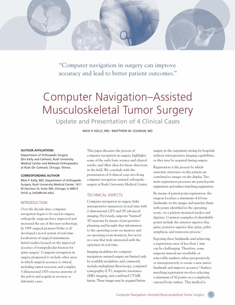
“Computer navigation in surgery can improve accuracy and lead to better patient outcomes.”
Computer Navigation–Assisted Musculoskeletal Tumor Surgery
Update and Presentation of 4 Clinical Cases MICK P. KELLY, MD / MATTHEW W. COLMAN, MD
AUTHOR AFFILIATIONS
Department of Orthopedic Surgery (Drs Kelly and Colman), Rush University Medical Center and Midwest Orthopaedics at Rush (Dr Colman), Chicago, Illinois.
CORRESPONDING AUTHOR
Mick P. Kelly, MD, Department of Orthopedic Surgery, Rush University Medical Center, 1611 W Harrison St, Suite 300, Chicago, IL 60612 ([email protected]).
INTRODUCTION
Over the decade since computer navigation began to be used in surgery, orthopedic surgeons have improved and increased the use of this new technology. In 1995 surgical pioneers Nolte et al1 developed a novel system of real-time localization of surgical instruments. Initial studies focused on the improved accuracy of transpedicular fixation for spine surgery.2 Computer navigation in surgery progressed to include other areas in which surgical accuracy is critical, including tumor resection and complex 3-dimensional (3D) osseous anatomy of the pelvis and scapula in revision or deformity cases.
This paper discusses the process of computer navigation in surgery, highlights some of the early basic science and clinical results, and offers ideas for future directions in the field. We conclude with the presentation of 4 clinical cases involving computer navigation–assisted orthopedic surgery at Rush University Medical Center.
TECHNICAL ASPECTS
Computer navigation in surgery links intraoperative maneuvers in real time with 2-dimensional (2D) and 3D advanced imaging. Previously, surgeons “learned” 3D anatomy by means of preoperative planning and brought that information to the operating room via memory and on operating room monitors, but never in a way that truly interacted with the operation in real time.
Imaging modalities for computer navigation–assisted surgery are limited only by available modalities and commonly include multiplanar fluoroscopy, computed tomography (CT), magnetic resonance (MR) imaging, and combined CT-MR fusion. These images may be acquired before
surgery in the outpatient setting for hospitals without intraoperative imaging capabilities, or they may be acquired during surgery.
Registration is the process by which anatomic structures on the patient are correlated to images on the display. Two main registration processes are paired-point registration and surface-matching registration.
By means of paired-point registration, the surgeon localizes a minimum of 4 bony landmarks on the images and matches them with points identified in the operating room, via a patient-mounted tracker and digitizer. Common examples of identifiable points include the anterior superior iliac spine, posterior superior iliac spine, pubic symphysis, and transverse process.3
Exposing these landmarks and achieving a registration error of less than 1 mm can be challenging. Therefore, some surgeons instead use resorbable or removable markers either preoperatively or intraoperatively to create a new native landmark and improve accuracy.4 Surface-matching registration involves selecting a minimum of 50 points on a completely exposed bony surface. This method is
2016 Rush Orthopedics Journal12
less favorable in tumor surgery because anatomic access may be limited and extended exposure may increase the risk of contamination.4 Finally, the navigation software determines the registration error, or the difference between the preoperative images and the real-time patient anatomy. Achievable registration error values can be well under 1 mm but should be less than 2 mm, depending on the application.3,4
ACCURACY AND OUTCOMES
Obtaining clean margins in pelvic tumor resection can be challenging because of tumor size and the proximity of neurovascular, gastrointestinal, and genitourinary organs. One study in which the investigators used foam-based bone models called Sawbones (Pacific Research Laboratories, Vashon, Washington) quantified the increased accuracy of pelvic osteotomy cuts by using a navigated osteotome and oscillating saw. Specifically, of the 24 bone cuts, the mean [SD] entry and exit cuts in the navigated group were 1.4 [1.0] and 1.9 [1.2] mm from the planned cuts, compared with 2.8 [4.9] and 3.5 [4.6] mm, respectively, in the nonnavigated group (P < .01). This
accuracy was reproduced in cadavers with 16 bone cuts with a mean of 1.5 [0.9] and 2.1 [1.5] mm from planned entry and exit sites, respectively.5
An additional Sawbones study, in which the investigators evaluated differences in the cutting accuracy of navigation–assisted simulated pelvic tumor resection versus nonnavigated resection in 10 senior and 13 junior surgeons, produced similar results.6 All surgeons attempted targeted multiplanar resection with a 10-mm margin from the simulated tumor. The location of the cut planes compared with the target planes was significantly improved by using navigation, averaging 2.8 mm (95% confidence interval [CI], 2.4-3.2 mm), compared with 11.2 mm (95% CI, 9.0- 13.3 mm) for the traditional group (P < .001). No difference was observed with stratification by senior and junior surgeons. Senior surgeons averaged 3.2 mm (95% CI, 2.7-3.8 mm) for navigated cuts versus 10.8 mm (95% CI, 8.0-13.6 mm) for freehand cuts (P < .001). In the 13 junior surgeons, the average location from the targeted plane was 2.4 mm (95% CI, 1.8-3.1 mm) versus 11.4 mm (95% CI, 7.9-15.0 mm) for
navigation and freehand techniques, respectively (P < .001). There were no intralesional resections in the navigation group versus 5 (22%) of 23 total resections in the freehand group. The majority of the intralesional resections were performed by junior residents.6
In a 2013 prospective study, the intralesional resection rate of primary bone tumors decreased after including computer navigation. At a single institution, the authors reported an intralesional resection rate of 29% in 539 primary bone tumors treated with a traditional technique. After the application of computer navigation–assisted surgery in April 2010, the authors decreased the rate to 8.7% in 23 primary bone tumors of the pelvis or sacrum.3 After a mean follow-up of 13.1 months, the local recurrence rate was 13%, decreased from the 27% that was reported with the traditional technique. The authors think that the increased accuracy preserved sacral nerve roots in 13 patients, permitted resection of otherwise inoperable advanced rectal cancer in 4 patients, and obviated hindquarter amputation in 3 patients. Although we cannot draw definitive conclusions from this small, nonrandomized
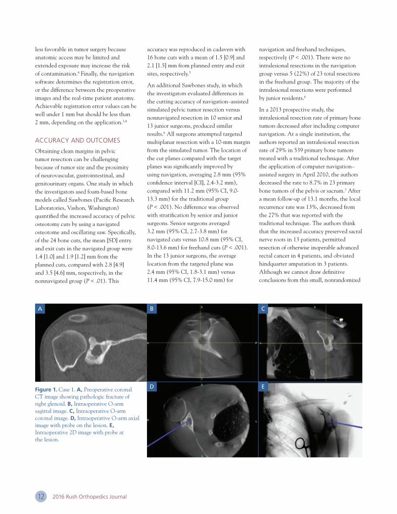
Figure 1. Case 1. A, Preoperative coronal CT image showing pathologic fracture of right glenoid. B, Intraoperative O-arm sagittal image. C, Intraoperative O-arm coronal image. D, Intraoperative O-arm axial image with probe on the lesion. E, Intraoperative 2D image with probe at the lesion.
A B
D
C
E

Computer Navigation–Assisted Musculoskeletal Tumor Surgery 13
case series, the early results are promising that the application of this new technology will lead to better clinical outcomes for patients with complex disease.
Long-term results are limited because of the recent adoption of this technology. Cho et al4 retrospectively reviewed the clinical course of 18 patients with computer navigation–assisted resection of stage IIB (Musculoskeletal Tumor Society staging system7) bone tumors after a minimum 3-year follow-up (mean, 48.2 months; range, 22-79). They reported free resection margins in each of the patients, and the local recurrence rate was 11% (n = 2).4 Both of the local recurrence cases were pelvic ring malignancies that recurred in the soft tissue, not at the osteotomy site. One of them recurred at 9 months, and this patient died 20 months postoperatively. The other recurrence was found 12 months postoperatively, and the patient remained disease free 18 months after resection of the local recurrence.
CASE STUDIES
Case 1
The patient, a 39-year-old man, had a cystic lesion and pathologic fracture of the glenoid of the right shoulder (Figure 1). With use of live computerized navigation system (O-arm Surgical Imaging System with StealthStation Navigation; Medtronic Sofamor Danek USA, Memphis, Tennessee), we localized the posterior glenoid lesion and used a high-speed drill and curettes to remove the tumor from the bone meticulously in all planes. We filled the cystic cavity with bioceramic cement (PRO-DENSE; Wright Medical Technology, Arlington, Tennessee).
Case 2
The patient, a 17-year-old boy, had a high-grade surface osteosarcoma of the left ilium (Figure 2). The medullary space was not involved by tumor. We thus performed a highly precise computer-navigated radical resection of the tumor, preserving the inner
table of the ilium, medial sciatic buttress, and anterior sacroiliac joint, thereby obviating reconstruction and preserving function.
Case 3
The patient, a 73-year-old man, had a massive sacrococcygeal chordoma with presacral extension and invasion of the rectum (Figures 3 through 5). In the first stage of the procedure, we performed a navigated anterior lumbosacral osteotomy through S2 with O-arm Surgical Imaging System with StealthStation Navigation. During the second stage, we performed a navigated completion of the anterior osteotomy through S2, acquiring a negative margin, and en bloc resection of the sacral tumor.
Case 4
The patient, a 16-year-old girl, had a debilitating S1 radiculitis and lumbosacral pain from a large cavitary aneurysmal bone cyst of the sacrum and L5 vertebra (Figures 6 and 7). We performed a transpedicular
Figure 2. Case 2. A, 3D model of the left ilium surface osteosarcoma. B, Axial T2-weighted MR image. C, Axial CT image. Three-dimensional model courtesy of Mayo Clinic Anatomic Modeling Laboratory.
B C
A

2016 Rush Orthopedics Journal14
corpectomy of the S1 and S2 bodies via extended intralesional curettage, followed by a combination of local bone autograft, allograft, and bioceramic cement (PRO-DENSE; Wright Medical Technology, Arlington, Tennessee) reconstruction of the corpectomy defect. The navigation provided visualization of the cavitary defect within L5, S1, and S2 and ensured that we had reached the extent of it in all angles.
DISCUSSION
Two of the disadvantages of computer navigation–assisted surgery are increased operative time and cost. Jeys et al3 in a 2013 case series reported a mean operating time of 260 minutes (range, 131-512 minutes). We assumed that the initial learning curve of navigated surgery increases operating room time. Radiation exposure is also increased because some applications require preoperative CT scans in addition to increased radiation exposure intraoperatively for the patient and surgical staff. This new technology also carries an increase in health care costs. It is difficult to assess how long it takes to recuperate these costs, if ever, because of unknown long-term clinical benefits. As we become increasingly aware of health care costs, we must support future research to understand the cost effectiveness of this new technology better.
FUTURE DIRECTIONS
Surgeons have already demonstrated successful use of robotic technology in the resection of paraspinal tumors.8 New haptic robot-assisted techniques for primary bone tumors maximize the accuracy of robots but allow surgeons to maintain tactile control over the cutting instruments.9 Three-dimensional modeling and printing
A
C
B
D
Figure 5. Case 3. Intraoperative images demonstrating the use of navigation for localization of osteotomy start point. Sagittal (A), coronal (B), and axial (C) intraoperative O-arm views. D, Intra-operative 2D image with live navigational probe in place.
Figure 4. Case 3. Intraoperative fluoroscopic images in sagittal (A, D) and axial (B, C) views demonstrate the navigation tracker on an S1 spinous process, presacral tumor extent, and navigation instruments.
A
C
B
D
A B
Figure 3. Case 3. Preoperative A, axial and B, sagittal T2-weighted MR images demonstrating perirectal and presacral, respectively, tumor extent in a 73-year-old man with massive sacrococcygeal chordoma.

Computer Navigation–Assisted Musculoskeletal Tumor Surgery 15
allow surgeons to conceptualize tumor structure, plan bone resection cuts, and develop patient-specific implants. Authors of a recent case report describe using CT navigation and 3D modeling to plan a complex pelvic chondrosarcoma resection combined with a successful 1-stage implantation of a 3D-printed titanium scaffold, plate, and acetabular cup fit specifically for the patient.10 These new advances continue to push the boundary for permitting resection of complex tumors with preservation of function.
CONCLUSIONS
Computer navigation in surgery can improve accuracy and lead to better patient outcomes. Currently, we use this technology with patients at Rush University Medical Center. We will continue to explore new areas of advancement and evaluate long-term outcomes and applications for this exciting technology.
References and financial disclosures are available online at www.rush.edu/orthopedicsjournal.
Figure 7. Case 4. Intraoperative axial multiplanar fluoroscopic image of the sacrum after bioceramic cementation of the cystic defect.
A B
Figure 6. Case 4. Axial (A) and sagittal (B) T2-weighted MR images demonstrating sacral aneurysmal bone cyst in a 16-year-old girl.

2016 Rush Orthopedics Journal16
AUTHOR AFFILIATIONS
Department of Orthopedic Surgery (Drs Shi, Chee, Andersson, and An; Messrs Liu and Zheng) and Department of Biochemistry (Drs Oh and Di Chen), Rush University Medical Center; and Midwest Orthopaedics at Rush (Drs Andersson and An), Chicago, Illinois; Department of Orthopedic Surgery (Dr Ding Chen), Second Xiangya Hospital, Central South University, Hunan; and Department of Spine Surgery (Dr Li), First Affiliated Hospital of Sun Yat-Sen, Guangzhou, People’s Republic of China.
CORRESPONDING AUTHOR
Howard S. An, MD, Department of Orthopedic Surgery, Rush University Medical Center, 1611 W Harrison St, Suite 300, Chicago, IL 60612 ([email protected]).
INTRODUCTION
More than 26 million Americans between the ages of 20 and 64 years experience back pain frequently.1 The costs for treatment of all spine-related conditions are estimated at $193.9 billion per year in the United States.2 The degree of symptom relief after spine treatments is unpredictable mainly because the exact cause of low back pain is poorly understood. Fortunately, cumulative
work by many researchers has begun to elucidate the molecular events inherent in disc degeneration and inflammation that may lead to discogenic low back pain. Patients may achieve better relief from low back pain and associated diseases by means of targeted biological therapies.
The intervertebral disc (IVD) has been recognized as one of the main sources of low back pain.3,4 The IVD has a unique structure, composed of a tough outer ring, the annulus fibrosus (AF), and a gelatinous inner core, the nucleus pulposus (NP). Environmental and genetic factors and aging play significant roles in disc degeneration. Disc degeneration is associated with the progressive loss of the proteoglycan content of the IVD, decreased matrix synthesis, higher concentrations of proteolytic enzymes and increased levels of proinflammatory cytokines. Leakage of these proteases and cytokines may spread through annular fissures creating a microenvironment that is more conducive to nerve and blood vessel ingrowth into the IVDs.5
Infiltration of macrophages and neutrophils into peripheral tissues is an important mechanism to remove tissues that have
been damaged or infected. In diseases characterized by chronic inflammation, such as rheumatoid arthritis, immune cells can cause structural damage and morbidity to the joints. Results from numerous immunohistological studies have shown that macrophages are detected in herniated disc tissues.6-9 When recruited to the herniated disc, macrophages may play a role in resorption of damaged tissue10,11 and also contribute to disc inflammation.9,12-14 In nonherniated cadaver disc tissues, Nerlich et al15 found a correlation between disc degeneration and the presence of macrophages. In their study, clusters of differentiation (CD)68-positive cells were detected in disc tissues with degeneration (Thompson Grade II to V) and not in those of fetuses, infants, and adolescents (Thompson Grade I). In patients with discogenic back pain and no disc herniation, Peng et al16 reported CD68-positive staining correlated with discogram-positive discs. These study results suggest that macrophage presence in the disc may be associated with not only herniated discs but also disc degeneration and back pain.
Chemokine Receptor Antagonists Can Inhibit Macrophage Migration PENG SHI, DDS, PHD / ANA V. CHEE, PHD / DAVID K. LIU, MS / JUSTIN L. ZHENG, BS / DING CHEN, MD / ZEMIN LI, MD
CHUNDO OH, PHD / DI CHEN, MD, PHD / GUNNAR B. J. ANDERSSON, MD, PHD / HOWARD S. AN, MD
“…these studies suggest that small molecule antagonists against CCR1 and CCR2 may be used as potential therapeutic
agents to inhibit macrophage migration into the disc.”

17
Inflammatory chemokines, small cytokines that play a role in the chemotaxis of immune cells, are expressed by disc cells. Regulated on activation, normal T cell expressed and secreted, also known as chemokine (C-C motif) ligand (CCL)5, was detected at higher levels in discogram-positive painful IVD tissues than in nonpainful IVD tissues.17 Also, ribonucleic acid (RNA) levels of macrophage inflammatory protein-1α, also known as CCL3, can be upregulated by cultured NP cells after treatment with interleukin (IL)-1 or tumor necrosis factor (TNF) α.18 To decrease immune cell migration and further destruction of tissues, investigators have targeted chemokines (CCL2, CCL3, and CCL5) and chemokine receptors ([CCRs]: CCR1, CCR2, and CCR5) for inhibition. Neutralizing antibodies or small molecule antagonists against these targets and others have been tested in inhibiting macrophage migration in vitro and preventing disease pathogenesis in vivo with a range of results
from not effective to successful.19 After decades of clinical trials with different inhibitors against a variety of receptors and ligands, a small molecule antagonist against CCR1, CCX354-C, was found to be safe, tolerable, and clinically active in reducing inflammation in patients with rheumatoid arthritis.20-23 We hypothesize that chemokines expressed by degenerative discs may play a role in recruiting macrophages. Inflammatory macrophages may initiate an immune response in the disc that may later resolve itself or worsen into chronic inflammation that eventually can lead to discogenic back pain. Inhibiting macrophage migration into the disc with antagonists against CCR1 and CCR2 may prevent the initiation of inflammation and subsequent back pain. In this study, we investigated which chemokines may be important for macrophage migration induced by human disc cells and tested which chemokine receptor antagonists can block this process.
MATERIALS AND METHODS
Cell Culture
We procured human spine segments (n = 6) from the Gift of Hope Organ Donor and Tissue Network of Illinois. We dissected IVDs and separated AFs and NPs. We isolated cells by means of sequential enzymatic digestion of disc tissue and plated them in 6-well plates. We cultured NP and AF cells in a monolayer with Dulbecco’s modified Eagle medium F-12 (Corning Life Sciences, Tewksbury, Massachusetts) supplemented with 20% fetal bovine serum until they reached confluency. Before treatment, cells were cultured for 24 hours in starvation media: Dulbecco’s modified Eagle medium F-12 supplemented with 1% insulin, human transferrin, and selenous acid; L-glutamine; gentamicin; and ascorbic acid. We then treated the cells with human recombinant IL-1 (R&D Systems, Minneapolis, Minnesota) (10 ng/mL) in starvation media for 24 hours. We collected cell pellets for RNA analysis and conditioned media for protein analysis and migration assays. We maintained human acute monocytic leukemia cell line THP-1 (American Type Culture Collection, Rockville, Maryland) as directed by the manufacturer in Roswell Park Memorial Institute 1640 medium (Life Technologies, Carlsbad, California) completed with 10% fetal bovine serum, 100 U/mL penicillin, and 100 µg/mL streptomycin at 37°C and 5% CO2.
Isolation of Total RNA and Measurement of Messenger RNA Levels With Real-time Polymerase Chain Reaction
We isolated total RNA from NP and AF cells by using an RNeasy Mini Kit (Qiagen, Valencia, California), and we measured messenger RNA (mRNA) levels of CCL2, CCL3, and CCL5 with real-time polymerase chain reaction (PCR) by using TaqMan Gene Expression Assays (Applied Biosystems, Foster City, California). Briefly, we reverse transcribed 0.5 µg of total RNA into complementary deoxyribonucleic acid (cDNA) with random primers by using a High Capacity RNA-to-cDNA Kit (Applied Biosystems).
Chemokine Receptor Antagonists Can Inhibit Macrophage Migration
A
B
Figure 1. Chemokine gene and protein expression are upregulated by interleukin-1 (IL-1) in nucleus pulposus (NP) and annulus fibrosus (AF) cells. We isolated human AF and NP cells from donor spine samples and cultured them in monolayers. To induce an inflammatory and degenerative phenotype, we treated the cells with IL-1 for 24 hours. We measured protein and messenger ribonucleic acid (mRNA) levels of chemokines chemokine (C-C motif) ligand (CCL)2, CCL3, and CCL5 by using assays and real-time polymerase chain reaction. Compared with those of the control treated cells, both A, mRNA and B, protein levels of CCL2, CCL3, and CCL5 were upregulated when treated with IL-1. Error bars represent standard error of the mean.Abbreviation: GAPDH, glyceraldehyde-3-phosphate dehydrogenase.

2016 Rush Orthopedics Journal18
We performed amplification with TaqMan PCR Master Mix (Applied Biosystems) and a spectrofluorometric thermal cycler (7300 Real-Time PCR System; Applied Biosystems). To standardize mRNA levels, we amplified glyceraldehyde-3-phosphate dehydrogenase as an internal control.
Multiplex Protein Assay (Quantitative Immunoassay)
We used multiplex sandwich immunoassays built on Luminex xMAP Technology (Luminex, Austin, Texas) to measure protein levels in the conditioned media of human NP and AF treated with IL-1 and untreated controls. We generated assay plates to detect the different analytes of interest: CCL2, CCL3, and CCL5. We processed samples in tandem with a broad range of standards for each protein, as provided by the manufacturer
(EMD Millipore, Billerica, Massachusetts). We processed assays through a Luminex 100 System (Luminex) to measure the mean fluorescent intensity for each protein simultaneously in each sample. We quantified the concentrations of each protein analyzed in these assays by using a standard curve recommended and validated by the manufacturer.
Migration and Inhibition Assay
We assayed migration of THP-1 cells by using Transwell plates with 3-µm pores (Corning Life Sciences) in the presence of conditioned media of the NP and AF cells. We coated the Transwell filter inserts with 33 µg/mL human fibronectin (Sigma-Aldrich, St. Louis, Missouri) and incubated them for 2 hours. We placed the THP-1 cells (5 × 10⁵) in the upper chambers of each insert. We placed conditioned media
from IL-1 or control treated AF and NP cells in the lower chambers. We used negative (starvation media alone) and positive controls (CCL2/monocyte chemoattractant protein [MCP]-1 100 ng/mL; Invitrogen, Carlsbad, California) in each assay. After 6 hours of incubation at 37°C and 5% CO2, the inserts were removed, and, using a hemocytometer, we counted the numbers of total cells that had migrated to the lower chambers. We performed inhibition assays as previously except that we preincubated the THP-1 cells at 3 concentrations (10, 50, and 100 µM) of antagonists against CCR1 and CCR2 (kindly provided by ChemoCentryx, Mountain View, California) for 10 minutes at room temperature before placing them in the upper chamber. We calculated migration rates relative to the positive control (CCL2/MCP-1 100 ng/mL). We calculated inhibition of migration relative to the conditioned media without inhibitor.
Surface Expression of Chemokine Receptors
We first incubated THP-1 cells (1 × 10⁶) with human immunoglobulin (Ig)G (R&D Systems) to block nonspecific binding sites and then incubated them with conjugated antibodies (human CCR1 Alexa Fluor 488 conjugated antibody [R&D Systems], human CCR2 allophycocyanin-conjugated antibody [BioLegend, San Diego, California], or their isotype controls [mouse IgG2B Alexa Fluor 488 isotype control (R&D Systems), mouse IgG2a, κ allophycocyanin isotype control (BioLegend)]) 5 µL each in flow cytometry staining buffer (R&D Systems). We washed cells 3 times in staining buffer, fixed with them with 1% paraformaldehyde, and then analyzed them by using flow cytometry.
RESULTS
Chemokine Gene and Protein Expression Upregulated by IL-1 in NP and AF Cells
Using real-time PCR, we analyzed mRNA levels of CCL2, CCL3, and CCL5 in human NP and AF cells after IL-1 treatment (Figure 1A). Compared with untreated controls, AF and NP cells treated with IL-1 expressed higher levels of CCL2 mRNA
Figure 2. Human monocyte migration is induced by annulus fibrosus (AF) and nucleus pulposus (NP) cells. We used an in vitro migration assay to determine whether monocyte migration could be induced by AF and NP cells. As shown in A, we placed a human monocytic cell line (THP-1) in the upper chamber and conditioned media from interleukin-1 (IL-1) or control treated cells in the lower chamber. After 6 hours, we determined the number of THP-1 cells that migrated to the lower chamber. The upper panel shows a schematic of the experiment. B shows that the conditioned media from the AF and NP cells treated with IL-1 were able to induce migration of THP-1 cells compared with those from the untreated controls (*P < .05). Error bars represent standard error of the mean.Abbreviations: CCL, chemokine (C-C motif) ligand; MCP, monocyte chemoattractant protein.
A
B

19
(37- and 51-fold), CCL3 mRNA (35- and 326-fold), and CCL5 mRNA (543- and 655-fold), respectively. The increases in mRNA levels were also observed at the protein levels. Using the Luminex multiplex assay, we measured the protein levels of these chemokines in the conditioned media (Figure 1B). Compared with the untreated controls, AF and NP cells treated with IL-1 produced higher amounts of chemokines, which were released into the conditioned media: CCL2 (193- and 178-fold), CCL3 (from undetectable to 447 and 831 pg/mL), and CCL5 (410- and 187-fold), respectively.
Human Monocyte Migration Induced by AF and NP Cells
We used an in vitro migration assay to determine whether monocyte migration could be induced by AF and NP cells. We placed THP-1 cells on the upper chamber
and conditioned media from the IL-1 or control treated cells in the lower chamber. After 6 hours, we determined the number of THP-1 cells that had migrated to the lower chamber (Figure 2). We calculated the migration rate relative to the positive control (CCL2/MCP-1 100 ng/mL). Conditioned media from untreated cells induced a low migration rate of 13% (NP) and 11% (AF) of the positive control, and those from cells treated with IL-1 were able to induce a higher migration of THP-1 cells of 65% (NP) and 72% (AF) of the positive control.
Chemokine Receptor Antagonists Inhibiting Migration Induced by Disc Cells
To test whether CCR1 and CCR2 antagonists can block migration of human macrophages induced by disc cells, we
incubated antagonists at 3 concentrations (10, 50, and 100 µM) with THP-1 cells for 10 minutes before the migration assay. CCR2 antagonists inhibited THP-1 migration induced by disc cells at all 3 concentrations and were more effective than were CCR1 antagonists (Figure 3). CCR1 did not inhibit migration at 10 µM but was able to inhibit 24% and 60% at 50 and 100 µM, respectively.
Chemokine Receptor Surface Expression on THP-1 Cells
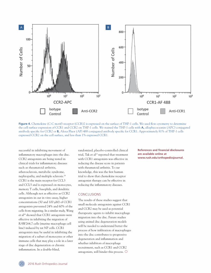
Because surface expression of the chemokine receptors on migrating cells would be important to determine the effectiveness of the antagonists, we analyzed CCR1 and CCR2 surface expression on THP-1 cells by using flow cytometry. After subtracting the number detected in the isotype controls, we found that more than 81% of THP-1 cells expressed CCR2 on the cell surface (Figure 4A), and less than 1% expressed CCR1 (Figure 4B).
DISCUSSION
These studies show that human NP and AF cells can be induced to express chemokines that attract macrophages and other immune cells into the disc. The 3 chemokines that we analyzed—CCL2, CCL3, and CCL5—were upregulated at both the RNA and protein levels. In the conditioned media of cells treated with IL-1, CCL2 protein levels accumulated at 10- to 100-fold higher levels than did CCL3 and CCL5. These data may suggest that CCL2 may be expressed early in inflammation and be important in recruiting the first set of immune cells to the injured or degenerated disc.
In our migration inhibition assays, we found that CCR2 antagonists were more effective than CCR1 antagonists in inhibiting THP-1 cell migration induced by NP and AF cells. CCR2 is the main receptor for CCL2 and is highly expressed in human peripheral blood monocytes. Our flow cytometry data helped confirm that the majority of THP-1 cells expressed CCR2 at the cell surface. These in vitro data suggest that CCR2 antagonists may be
Chemokine Receptor Antagonists Can Inhibit Macrophage Migration
Figure 3. Chemokine (C-C motif) receptor (CCR)1 and CCR2 antagonists can block migration of monocytes induced by disc cells. A, To test whether CCR1 and CCR2 antagonists can block migration of monocytes induced by disc cells, we coincubated antagonists with THP-1 cells (a human monocytic cell line) for 10 minutes and then placed them in the upper chamber for the migration assay. We placed the conditioned media from annulus fibrosus (AF) and nucleus pulposus (NP) cells treated with interleukin-1 (IL-1) in the lower chamber. We performed the migration assay by using different concentrations of the inhibitors. B, CCR1 antagonists inhibited migration of THP-1 cells significantly at concentrations between 50 and 100 µM, whereas CCR2 antagonists significantly inhibited migration at all concentrations tested (10, 50, and 100 µM). Error bars represent standard error of the mean.
A
B
2016 Rush Orthopedics Journal20
successful in inhibiting movement of inflammatory macrophages into the disc. CCR2 antagonists are being tested in clinical trials for inflammatory diseases such as rheumatoid arthritis, atherosclerosis, metabolic syndrome, nephropathy, and multiple sclerosis.24 CCR1 is the main receptor for CCL3 and CCL5 and is expressed on monocytes, memory T cells, basophils, and dendritic cells. Although not as effective as CCR2 antagonists in our in vitro assay, higher concentrations (50 and 100 µM) of CCR1 antagonists prevented 24% and 60% of the cells from migrating. In a similar study, Wang et al18 showed that CCR1 antagonists were effective in inhibiting the migration of RAW264.7 cells (murine macrophage cell line) induced by rat NP cells. CCR1 antagonists may be useful in inhibiting the migration of a subset of monocytes or other immune cells that may play a role in a later stage of disc degeneration or chronic inflammation. In a double-blind,
randomized, placebo-controlled clinical trial, Tak et al23 reported that treatment with CCR1 antagonists was effective in reducing the disease score in patients with rheumatoid arthritis. To our knowledge, this was the first human trial to show that chemokine receptor antagonist therapy can be effective in reducing the inflammatory diseases.
CONCLUSIONS
The results of these studies suggest that small molecule antagonists against CCR1 and CCR2 may be used as potential therapeutic agents to inhibit macrophage migration into the disc. Future studies using animal disc degeneration models will be needed to understand better the process of how infiltration of macrophages into the disc contributes to progressive degeneration and inflammation and whether inhibitors of macrophage recruitment, such as CCR1 and CCR2 antagonists, will hinder this process.
References and financial disclosures are available online at www.rush.edu/orthopedicsjournal.
Figure 4. Chemokine (C-C motif) receptor (CCR)2 is expressed on the surface of THP-1 cells. We used flow cytometry to determine the cell surface expression of CCR1 and CCR2 on THP-1 cells. We stained the THP-1 cells with A, allophycocyanin (APC) conjugated antibody specific for CCR2 or B, Alexa Fluor (AF) 488 conjugated antibody specific for CCR1. Approximately 81% of THP-1 cells expressed CCR2 on the cell surface, and less than 1% expressed CCR1.
A B

21Anterior Cruciate Ligament Reconstruction
AUTHOR AFFILIATIONS
Department of Orthopedic Surgery (Drs Forsythe, Khair, Inoue, Bach, Verma, Cole; Messrs Collins, Arns), Rush University Medical Center; and Midwest Orthopaedics at Rush (Drs Forsythe, Bach, Verma, Cole), Chicago, Illinois.
CORRESPONDING AUTHOR
Brian Forsythe, MD, Department of Orthopedic Surgery, Rush University Medical Center and Midwest Orthopaedics at Rush, 1611 W Harrison St, Suite 300, Chicago, IL 60612 ([email protected]).
INTRODUCTION
Injuries to the anterior cruciate ligament (ACL) are among the most common to the sports medicine orthopedist, so it has been the subject of an ever-increasing number of studies. Despite the amount of research devoted to anatomic ACL reconstruction, there is still considerable controversy surrounding the best way to prepare the femoral tunnel. Study results suggest that ACL reconstruction success depends on graft placement within the anatomic insertions of the native ACL footprint.1-5
Research has shown it is difficult to place a femoral tunnel in a way that reproduces native ACL insertional anatomy through a transtibial technique.6 In addition, this method can produce unacceptable vertical tunnels high in the notch.7 Transtibial techniques have been modified to decrease these risks and create a more anatomic femoral tunnel. However, there are still limitations in regard to graft placement and tibial tunnel positioning even with these modifications.8,9
Many surgeons use an anteromedial approach with flexible rods, first described by Cain and Clancy,10 to drill the femoral tunnel. Flexible rods limit the need for hyperflexion during tunnel placement as well as to allow tunnel placement in a more anterior and inferior site on the lateral condyle than is possible with conventional rods. Flexible reamers outperform rigid reamers when femoral tunnels are drilled through an anteromedial portal.11,12 Flexible instrumentation allows the creation of longer tunnels that are further away from the posterior cortex. In addition, tunnel placement with a rigid reamer with the knee in hyperflexion risks the creation
of a horizontal tunnel with elevated tunnel acuity.13 Conversely, having the knee flexed to 90° leads to a short tunnel that may breach the posterior cortex.
There is still controversy, however, regarding optimal knee flexion with use of flexible and rigid reamers through an anteromedial portal. In this study, we used a novel 3-dimensional (3D) technology to evaluate how knee flexion angle affects femoral tunnel dimensions as well as to determine the optimal flexion angle to drill when using curved and rigid guides through an anteromedial portal. We hypothesized that femoral tunnels drilled with curved guides rather than straight guides would create longer tunnels with greater distance to the posterior femoral cortex.
MATERIALS AND METHODS
Creation of 3D Computed Tomographic Knee Models at Various Flexion Angles
In this cadaveric study, we obtained 6 fresh frozen knees (4 male and 2 female) from screened individuals with no prior history
Anterior Cruciate Ligament ReconstructionOptimizing Femoral Tunnel Position With Flexible Curved Reamers
BRIAN FORSYTHE, MD / MICHAEL J. COLLINS, BS / THOMAS A. ARNS, BS / MICHAEL KHAIR, MD / NIKHIL N. VERMA, MD
BRIAN J. COLE, MD, MBA / BERNARD R. BACH JR, MD / NOZOMU INOUE, MD, PHD
“Three-dimensional imaging software can be used to model femoral tunnel placement accurately by using a standardized curved guide.”

2016 Rush Orthopedics Journal22
of arthritis, cancer, surgery, or any ligamentous knee injury. The mean age for the collected knees was 47 years (range, 26-59). After we collected the knees, we acquired computed tomography (CT) images (Volume Zoom; Siemens, Malvern, Pennsylvania) of each knee in the coronal, axial, and sagittal planes; we used 0.625-mm contiguous sections (20-cm field of view, 512 × 512 matrices) at various angles to gather cross- sectional images of the knee joint at specific flexion points. We flexed each knee by using an external fixation device and then scanned them at 90°, 110°, 125°, and maximum flexion (140° in 2 specimens, 135° in 2 specimens, and 125° in 2 specimens). We then used CT scans at the various flexion angles to create 3D knee models at each of the 4 flexion points under investigation. We imported the CT images of the knees at various flexion angles in Digital Imaging and Communications in Medicine format and segmented them by using 3D reconstruction software (Mimics; Materialise, Leuven, Belgium) to create 3D knee models for each flexion angle. We further converted the 3D CT models to point-cloud models.
Creation of 3D ACL Tunnel Models
Using custom computer-aided design software, we created a 3D model of a curved guide with a 42° bend (4.83 mm
in outer diameter; Smith & Nephew, Memphis, Tennessee) and a straight guide with the same outer diameter and identical dimensions for use during ACL reconstruction surgery (Figure 1A, B). We set a pivot point at the tip of each guide shaft (Figure 1B, C). A single surgeon (B.F.) identified the insertion points of the ACL tunnel guide for a single bundle reconstruction by using the midpoint of the footprint and topographical landmarks in the 3D CT
femur models (the insertion point) (Figure 1D). We placed the pivot point of each guide shaft model at the insertion point (Figure 2A). Then we rotated the guide shaft model toward medial and inferior directions about the pivot point until any portion of the guide shaft hit the medial condyle and medial plateau with 2 mm of clearance according to cartilage thickness (Figure 2B). We kept the curved guide shaft’s angle of inclination constant at 5°.
Figure 1. Curved guide and entry point A, Point-cloud model of the curved guide. B, C, 3D model of the curved guide and entry point (red). D, Lateral femoral condyle and entry point (red).
Figure 2. A, 3D representation of virtual placement of the curved guide. Red point: Entry point of the guide wire. B, Tunnel length and shortest distances from the guide wire on the 3D point-cloud model. The curved guide is automatically rotated around the entry point until the guide contacts the medial condyle and medial tibia plateau.
10% 20% 30% 40% 50% 60% 70% 80% 90%
Dis
tan
ce t
o C
ort
ex (
mm
)
Curved Guides
16
14
12
10
8
6
4
2
0
Figure 3. Least Distance Data Along a Tunnel Drilled With Curved Guides at Various Flexion Angles
Percentage Tunnel Length

23Anterior Cruciate Ligament Reconstruction
After we determined the guide’s position, we inserted a virtual straight guide wire into the lateral condyle in the direction of the straight portion at the tip of the curved guide shaft or the direction of the straight guide shaft. We extended the virtual guide wire until it reached the lateral wall or posterior wall of the lateral condyle (the exit point). We defined the length of the ACL tunnel as the distance between the insertion point and the exit point. We
calculated the distance from the guide wire to the posterior wall of the lateral condyle as the least distance in a plane perpendicular to the guide wire. We calculated the least distances to the posterior wall at 100 points along the guide wire as a function of the distance from the insertion point (0% at the insertion point and 100% at the exit point).
We created the 3D virtual guide shafts and ACL tunnel and performed 3D
measurements of the tunnel length and least distance to the posterior wall of the lateral condyle by using software. We used a custom-written Visual C++ program and Microsoft Foundation Class programming environment (Microsoft, Redmond, Washington).
Statistical Analysis
We recorded all data in a Microsoft Excel spreadsheet (Microsoft). We performed data analysis with IBM SPSS Statistics version 22 (IBM SPSS, Armonk, New York). We compared average tunnel lengths and the average least distance between the straight and curved guides by using a paired t test with a significance level less than .05.
RESULTS
Table 1 shows the average least distance data for the middle third of a 10-mm femoral tunnel drilled with curved and straight reamers. As the degree of knee flexion increases, the average least distance increases for both curved and straight reamers. Curved reamers consistently and significantly outperformed straight reamers in regard to least distance with the knee at 90° and 110° of flexion (P < .05). At
Table 1. Average Least Distance Along the Middle Third of a 10-mm Tunnel Created With Curved and Straight Guides
AVERAGE LEAST DISTANCE (mm)
Flexion Angle Curved Straight P Value
90° −0.191 ± 1.67 −0.595 ± 1.61 .0118
110° 1.306 ± 1.71 −0.086 ± 1.76 .0028
125° 4.233 ± 1.56 3.930 ± 1.91 .5886
Maximum 7.941 ± 2.10 8.319 ± 2.05 .8053
Data are given as mean ± standard deviation.Bolded numbers indicate statistically significant differences between curved and straight with paired t test (P < .05).
Figure 4. Least Distance Data Along a Tunnel Drilled With Straight Guides at Various Flexion Angles
10% 20% 30% 40% 50% 60% 70% 80% 90%
Dis
tan
ce t
o C
ort
ex (
mm
)
Straight Guides
16
14
12
10
8
6
4
2
0
Percentage Tunnel Length

2016 Rush Orthopedics Journal24
90° of flexion, results were significant, though both curved (−0.191 mm) and straight (−0.595 mm) reamers breached the posterior cortex.
Tables 2 and 3 show the average tunnel length for 8- and 10-mm tunnels virtually drilled at each flexion angle with curved and straight reamers. For both 8- and 10-mm straight and curved guides, increasing the knee flexion angle increased tunnel length. For 8-mm tunnels, tunnel length was significantly longer at 90° and 110° of flexion when we used a curved guide (P < .05). We could not properly analyze the 10-mm tunnels at 90° because of multiple specimens breaching the posterior cortex. We noted posterior breach at both 90° and 110° of flexion when using straight guides to drill both 8- and 10-mm tunnels. Using curved guides, we noted posterior wall cutout in 2 specimens at 90° when drilling a 10-mm tunnel. Using an 8-mm curved guide, we noted no incidence of posterior wall cutout.
Figures 3 and 4 show the least distance data for curved and straight guides, showing the distance to the posterior cortex for each flexion angle as a function of tunnel length. Increasing flexion angle consistently created tunnels with a greater distance from the posterior cortex (P < .05).
Tunnels created using curved guides had a significantly greater distance to the posterior cortex at 10% and between 40% and 70% of tunnel length at 90° of flexion (P < .05). At 110°, curved guides significantly outperformed straight guides for the first 80% of tunnel length (P < .05). At 125° and maximum flexion, we noted no significant differences.
DISCUSSION
Three-dimensional imaging software can be used to model femoral tunnel placement accurately by using a standardized curved guide through the anteromedial portal with knee flexion angle as the sole variable. Using this approach, the least distance data for both curved and straight guides suggest that drilling the femoral tunnel at 90° of flexion will put the posterior cortex greatly at risk, whereas greater flexion angles eliminate this danger. Curved guides increased the distance to the femoral cortex while preserving adequate tunnel length. The average distance to the posterior cortex along a tunnel drilled using curved and straight guides (Figures 3 and 4) shows that increasing knee flexion increases the distance to the posterior cortex. We created virtual tunnels of 8 and 10 mm to simulate tunnels created during ACL reconstruction using
soft-tissue and bone-tendon-bone grafts. Analyzing tunnel data (Tables 2 and 3) showed that at 90° and 110° of knee flexion, 8- and 10-mm femoral tunnels cannot be drilled reliably with straight guides without the risk of breaching the posterior femoral cortex; 10-mm tunnels drilled with a curved guide at 90° risk breaching the posterior femoral cortex as well.
The use of the anteromedial portal for drilling the femoral tunnel has become increasingly more common since the advent of flexible instrumentation. Anteromedial drilling creates a more anatomic tunnel, although it has some drawbacks, including the need for hyperflexion, short tunnels, and difficulty maintaining visualization while drilling.12,14-17 In a cadaveric study, Bedi et al8 analyzed the obliquity and length of femoral tunnels created through transtibial versus anteromedial portal drilling. The authors concluded that anteromedial drilling allows for increased obliquity; however, there is an increased risk of critically short femoral tunnels (<25 mm). In our study, femoral tunnel length was consistently greater than 25 mm with a knee flexion angle greater than 90°. In addition, flexible guides produced longer tunnels up to 125° of flexion.
Table 2. Average Tunnel Length Along the Anterior Edge, Center, and Posterior Edge of 8-mm Virtual Tunnels
Data are given as mean ± standard deviation.Bolded numbers indicate statistically significant differences between curved and straight with paired t test (P < .05).aIndicates posterior wall cutout in specimens (2 at 90° and 110° with straight guides).
AVERAGE TUNNEL LENGTH (mm)
8-MM CURVED GUIDE 8-MM STRAIGHT GUIDE
Flexion Angle Anterior Center Posterior Anterior Center Posterior
90° 28.5 ± 1.6 20.8 ± 3.7 12.4 ± 4.3 28.3 ± 0.7 18.8 ± 3.7 12.0 ± 4.5a
110° 35.2 ± 0.7 32.0 ± 0.9 28.0 ± 1.2 34.3 ± 1.9 30.6 ± 1.7 24.6 ± 4.4a
125° 35.7 ± 0.8 33.4 ± 2.3 32.0 ± 2.1 35.1 ± 1.2 32.6 ± 3.8 31.0 ± 4.1
Maximum 35.4 ± 4.0 35.9 ± 0.6 35.3 ± 1.3 37.3 ± 1.5 36.3 ± 0.6 35.5 ± 1.1

25
Results from a study in which the investigators analyzed the effect of knee flexion on femoral tunnel length during double-bundle ACL reconstruction showed that lesser degrees of knee flexion produced shorter tunnel lengths with use of straight guides.13 Our analysis of single-bundle ACL reconstruction produced similar results. With both curved and straight guides, increasing knee flexion produced significantly longer tunnel lengths.
Investigators in multiple studies have analyzed the risk of posterior femoral cortex breach when drilling the femoral tunnel. Steiner and Smart12 analyzed the use of flexible and rigid systems for femoral tunnel drilling from the anteromedial portal versus the transtibial approach at 110° of flexion. They discovered flexible pins exited significantly further from the posterior cortex than did rigid pins. In a separate study of tunnels drilled at 120° with a rigid system, findings showed that 75% of tunnels experienced posterior cortex compromise at an average of 21.3 mm.8 Decreasing knee flexion is a risk factor for posterior wall compromise. Our study results help confirm that increasing knee flexion significantly increases the distance to the posterior cortex, with maximum flexion allowing for the greatest distance.
In addition, flexible systems allowed for significantly greater tunnel length than did rigid systems. However, at 90° of flexion, neither rigid nor flexible systems could be used without compromising the femoral cortex or creating a critically short tunnel. In addition, at 110° of flexion, the posterior cortex was compromised when drilling a 10-mm tunnel with a straight guide.
There are some limitations to the present study. First, we set the thickness of articular cartilage at 2 mm. In a patient with the potential for thicker cartilage, this variable may alter positioning of the guide pin as it passes adjacent to the medial condyle. Second, we evaluated only the bony morphology without taking into account the soft tissue. Therefore, the entry point may be slightly more medial in a surgical setting when taking into account soft tissue.
CONCLUSIONS
The use of 42° flexible guides and reamers resulted in greater distance of the tunnel to the femoral cortex while preserving adequate tunnel length. For creating long femoral tunnels without the risk of breaching the posterior cortex, the optimal knee flexion angle is 110° or greater. Surgeons using flexible reamers should be
aware that knee flexion to at least 110° optimizes ACL femoral tunnel dimensions. In addition, surgeons using straight reamers should flex the knee to at least 125° to optimize femoral tunnel dimensions.
References and financial disclosures are available online at www.rush.edu/orthopedicsjournal.
Anterior Cruciate Ligament Reconstruction
Data are given as mean ± standard deviation.Bolded numbers indicate statistically significant differences between curved and straight with paired t test (P < .05).aIndicates posterior wall cutout in specimens (2 at 90° with curved and straight guides and 3 at 110° with straight guide).
Table 3. Average Tunnel Length Along the Anterior Edge, Center, and Posterior Edge of 10-mm Virtual Tunnels
AVERAGE TUNNEL LENGTH (mm)
10-MM CURVED GUIDE 10-MM STRAIGHT GUIDE
Flexion Angle Anterior Center Posterior Anterior Center Posterior
90° 30.2 ± 1.5 20.8 ± 3.7 12.7 ± 5.5a 29.4 ± 1.1 18.8 ± 3.3 8.9 ± 5.1a
110° 35.8 ± 0.9 32.2 ± 1.0 26.3 ± 4.1 35.2 ± 1.1 30.6 ± 1.6 25.9 ± 2.0a
125° 36.4 ± 1.5 34.8 ± 1.2 32.8 ± 2.0 35.6 ± 1.2 32.6 ± 3.8 30.5 ± 3.9
Maximum 35.4 ± 4.6 35.9 ± 0.6 35.4 ± 1.8 37.5 ± 1.8 36.3 ± 0.6 35.3 ± 1.5

2016 Rush Orthopedics Journal26
AUTHOR AFFILIATION
Department of Orthopedic Surgery, Rush University Medical Center; and Midwest Orthopaedics at Rush, Chicago, Illinois.
CORRESPONDING AUTHOR
Monica Kogan, MD, Department of Orthopedic Surgery, Rush University Medical Center and Midwest Orthopaedics at Rush, 1611 W Harrison St, Suite 300, Chicago, IL 60612 ([email protected]).
INTRODUCTION
For more than 50 years, clinicians have used distraction osteogenesis with great success to lengthen extremities.1,2 Techniques such as unilateral external fixation, multiplanar external fixation, lengthening over a nail, and external fixator lengthening followed by intramedullary (IM) nailing are all techniques for lengthening a limb.
Issues associated with external fixators, including pin tract infections, refractures, soft-tissue tethering, and joint stiffness are all well documented.3,4 The use of an IM device with an external fixator (lengthening over a nail) is a hybrid technique that avoids the complications of fracture and joint stiffness seen when lengthening with only an external fixator. Once the surgeon using
this technique achieves the desired length, he or she removes the fixator and locks the IM nail distally, which provides stability while the consolidate matures. This type of technique, however, is technically demanding and associated with complications, such as pin tract infections, failure to distract, fracture, and hardware failure.3
The ideal implant is an IM device that allows for stabilization during the lengthening process. The Intramedullary Skeletal Kinetic Distractor nail (ISKD; Orthofix International, Verona, Italy), an IM nail used in the past, was an internal device with a ratchet mechanism that lengthened the bone. The clinician programmed the desired length into the device before placement, and specific exercises were performed by the patient a few times per day to allow for 1 mm of lengthening daily. Common complications were devices that lengthened too fast because of the leg turning during activities such as sleeping, or the ratchet device not working properly, resulting in the leg not lengthening. With the ISKD, if the patient experienced complications such as delay in healing of the consolidate or excessive pain, there was no way to stop, slow, or
modify the lengthening process. In addition to those issues, the exercise of turning the leg to lengthen the bone was often extremely painful for the patients. The ISKD device was taken off of the market in 2012.
PRECICE SYSTEM
In 2011, Ellipse Technologies (Aliso Viejo, California) introduced the PRECICE system, a telescoping rod with an internal magnet that lengthens via a handheld external remote controller (ERC). The clinician programs the desired length into the ERC, and the patient performs the lengthening 3 times per day to produce 1 mm of length per day. The PRECICE system originally was approved to be lengthened by a medical professional, and patients were required to go to the physician’s office 3 times per day, including weekends. In 2013, the US Food and Drug Administration approved patients and caregivers performing the lengthening procedure at home.
With the PRECICE system, the clinician is able to fine-tune the correction to include further lengthening over what initially was programmed. Compression, or shortening, also can be performed if the limb is
Limb LengtheningA New Technology
MONICA KOGAN, MD
“This technology has revolutionized the method by which orthopedic surgeons can address limb-length inequalities.”

27Limb Lengthening
overcorrected. Because lengthening occurs only with the use of the ERC, the rate of lengthening can be adjusted if issues arise. If there is a delay in the consolidate or if the patient is having excessive pain, the lengthening can be slowed down Furthermore, unlike with the ISKD, the actual process of lengthening the device is painless.
The clinician can place the femoral PRECICE nails antegrade through the greater trochanter or piriformis fossa, or retrograde in skeletally mature patients. In patients with open growth plates, such as the 15-year-old boy shown in Figures 1 through 3, a greater trochanteric starting point is required. Tibial and humeral nails also exist. Tibial PRECICE nails can be placed only in skeletally mature patients, once the physis is closed, similar to placement of a standard tibial nail. Limiting factors for the use of the nail include canal diameter and the initial length of the bone to be lengthened. Since the introduction of the PRECICE system, the diameter and length options of the nails have improved, but the options are not as numerous as with a standard IM nail.
Deformities may need to be corrected before the lengthening procedure. Contraindications to using the PRECICE system are a body mass index (BMI) greater than 30 and a soft-tissue envelope greater than 8 cm around the bone. The magnet is not strong enough to work through soft-tissue sleeves larger than this, and the rod may be overly stressed with a BMI greater than 30. We have required patients with BMIs greater than 30 to lose weight before the procedure.
At Midwest Orthopaedics at Rush, we have performed 16 lower extremity lengthening procedures to date in patients ranging in age from 10 to 32 years. Indications have included limb-length inequalities secondary to trauma, congenitally short femur, proximal femoral focal deficiency, tibia hemimelia, and limb-length inequalities of unknown cause.
This technology has revolutionized the method by which orthopedic surgeons can address limb-length inequalities, but it is not without risks. Complications seen with placement of an IM nail in an extremity—
such as the risk of pulmonary embolism, fat embolism, or delayed healing of the consolidate—are still present. We are not, however, seeing the issues associated with external fixation. The device is also not as strong as a standard IM nail; therefore, to avoid rod breakage, the patient must not bear weight on the extremity until the consolidate is completely mature.
CONCLUSIONS
Patients who previously had undergone lengthening with an external fixator and later underwent lengthening with the PRECICE system have commented that they wished that the technology had been available earlier. The PRECICE system offers a new option for orthopedic surgeons to treat limb-length inequalities—one that does not cause pain during the lengthening process or have issues similar to those of external lengthening devices.
References and financial disclosures are available online at www.rush.edu/orthopedicsjournal.
Figure 1. Initial postoperative anteroposterior radiograph in a 15-year-old boy with a congenitally short femur, showing a corticoctomy. The patient had a small amount of valgus tilt of the femur, which was corrected with the placement of a straight rod.
Figure 2. Initial postoperative lateral radiograph of the femur of the same patient.
Figure 3. Anteroposterior radiograph obtained in the same patient 21 days after surgery. The magnet is visible at the level of the corticotomy, and bone bridging is visible as well.

2016 Rush Orthopedics Journal28
AUTHOR AFFILIATIONS
Department of Orthopedic Surgery (Drs Cvetanovich and Erickson) and Hip Preservation Center, Division of Sports Medicine, Department of Orthopedic Surgery (Dr Nho), Rush University Medical Center and Midwest Orthopaedics at Rush (Dr Nho), Chicago, Illinois; Department of Orthopedic Surgery, University of Texas Health Science Center at Houston (Dr Mascarenhas), Texas; Department of Orthopedic Surgery, University of Michigan (Mr Lee), Ann Arbor, Michigan.
CORRESPONDING AUTHOR
Gregory L. Cvetanovich, MD, Department of Orthopedic Surgery, Rush University Medical Center, 1611 W Harrison St, Chicago, IL, 60612 ([email protected]).
INTRODUCTION
Femoroacetabular impingement (FAI) is a common etiology of hip pain in young patients and may be a precipitating factor in development of hip osteoarthritis.1 FAI is anatomically divided into cam deformity, pincer deformity, and a combination of cam and pincer deformity, any of which can lead to chondral and labral injury.1 A cam deformity involves
an aspherical femoral head rotating in the acetabulum with excess bone at the femoral head-neck junction, whereas a pincer deformity involves acetabular overcoverage. Although treatment of FAI originally was described via open hip surgical dislocation, hip arthroscopy for the treatment of FAI has grown in popularity in recent years and now has become a well-established treatment for FAI.2 The goal of arthroscopic treatment of FAI is to remove the excess bone on the femoral head/neck junction (cam deformity), acetabulum (pincer deformity), or both (combined FAI), thereby restoring normal kinematics to the hip joint. Although long-term outcome data are sparse, reported outcomes of hip arthroscopy for FAI are generally good, and complication rates are low.2-4 Studies of patients who have had poor results from revision hip arthroscopy after primary hip arthroscopy most commonly identify underresection of FAI deformities as the cause of failure.5,6 To our knowledge, regrowth of a symptomatic cam deformity after prior femoral osteochondroplasty for FAI has not yet been reported.
The present study reports the incidence of regrowth of symptomatic cam lesions in patients who previously underwent femoral osteochondroplasty for femoroacetabular impingement by a single surgeon at a major hip arthroscopy center.
MATERIALS AND METHODS
Between August 2009 and July 2013, we retrospectively reviewed 665 consecutive hip arthroscopy procedures performed by the senior author in order to identify patients who had symptomatic regrowth of cam lesions after prior arthroscopic femoral osteochondroplasty for femoroacetabular impingement. We defined cam regrowth as a period of improved symptoms after arthroscopic hip surgery for FAI with radiographic evidence of a resected cam deformity followed by recurrence of pain leading to the observation of regrowth of a cam deformity on imaging.
We identified 1 case of symptomatic regrowth of a cam deformity after femoral osteochondroplasty for FAI during the study period.
Regrowth of Symptomatic Cam Deformity After Hip Arthroscopy and
Femoral Osteochondroplasty GREGORY L. CVETANOVICH, MD / BRANDON J. ERICKSON, MD / RANDY MASCARENHAS, MD / SIMON X. LEE, MPH / SHANE J. NHO, MD, MS
“Of the cohort of 665 hip arthroscopies in our study, only 1 patient experienced symptomatic regrowth of
a cam deformity after prior hip arthroscopy.”

29Regrowth of Symptomatic Cam Deformity After Hip Arthroscopy and Femoral Osteochondroplasty
CASE REPORT
A 24-year-old competitive hockey goalie presented with left hip pain. The pain began during a hockey game; was localized to the left groin; and was exacerbated by rotational movement, stair climbing, and putting on socks and shoes. Past medical
history and past surgical history were otherwise unremarkable. Examination of the left hip revealed mild tenderness at palpation of the adductors and hip flexors and limited left hip internal rotation to 15°. Provocative testing of the left hip revealed a positive impingement test,
positive psoas impingement test, negative subspine impingement test, and absence of a circumduction clunk. The left hip painful arc was from 12 to 3 o’clock. We obtained plain radiographs, which revealed a left hip lateral center edge angle of 38.5°, an alpha angle of 64°, preserved joint space, and positive crossover sign (Figure 1A, 1B). Magnetic resonance imaging (MRI) and a computed tomography scan helped confirm a cam deformity and anterosuperior left labral tear (Figure 1C, 1D). The patient underwent cortisone injection in the left hip, which provided complete pain relief for 2 months.
Because of a recurrence of symptoms 2 months after injection, the patient underwent left hip arthroscopy, labral repair, acetabular rim trimming, and femoral osteochondroplasty. He reported complete pain relief and was able to return to recreational ice hockey 6 months postoperatively. Arthroscopic images, intraoperative fluoroscopic images, and postoperative radiographs obtained 2 weeks after surgery revealed normal cartilage and complete resection of the cam deformity, with a postoperative lateral center edge angle of 37.5° and an alpha angle of 46.1° (Figure 2A, 2B). Two and a half years after the initial surgery, the patient returned with complaints of recurrent left hip pain located in the groin that was exacerbated by ice hockey and distance running. Left hip internal rotation was 10° (compared with 25° at 6 months after the index procedure). There was a positive impingement test with a painful arc from 1 to 3 o’clock. Plain radiographs revealed regrowth of a cam deformity (Figure 3A, 3B) with a lateral center edge angle of 38°, preservation of the joint space, and an alpha angle of 69.5°. MRI helped confirm left hip cam deformity regrowth (Figure 3C, 3D).
DISCUSSION
Hip arthroscopy with femoral and acetabular osteochondroplasty has become a well-established treatment for FAI, with good results and a relatively low complication rate.2 Nevertheless, some patients will have recurrent or persistent
Figure 1. Preoperative anteroposterior (A) and Dunn lateral (B) radiographs, axial magnetic resonance image (C), and axial computed tomographic scan (D) reveal a cam deformity and an anterosuperior labral tear, as well as a lateral center edge angle of 38.5°, an alpha angle of 64°, and positive crossover sign.
Figure 2. Postoperative anteroposterior (A) and Dunn lateral (B) radiographs reveal resolution of the patient’s cam deformity, as well as a lateral center edge angle of 37.5° and an alpha angle of 46.1°.
A
A
C
B
B
D

2016 Rush Orthopedics Journal30
hip pain and ultimately undergo revision hip arthroscopy.5-8 There are multiple causes for revision hip arthroscopy, but, to our knowledge, regrowth of a symptomatic cam deformity after adequate resection at the initial surgery has not been reported previously.
Patients who have persistent pain after initial hip arthroscopy generally choose to undergo revision hip arthroscopy.6 Hip disease identified in reported series of revision hip arthroscopy include labral disease, chondral injury, adhesions, instability, and previously unaddressed or undertreated bony lesions.5-8 Of note, a common cause of persistent pain after hip arthroscopy is insufficient treatment of the patient’s FAI. In some cases, a patient actually has combined FAI, and either the cam or the pincer lesion remains undiagnosed and unaddressed at the initial operation. In other cases, the surgeon performs an inadequate resection.5
Inadequate resection could be due to lack of dynamic hip motion intraoperatively. In order to access the entire cam deformity, the surgeon must bring the hip through a range of motion from complete extension to almost 90° of flexion. The most difficult areas of the cam deformity to access are the far lateral and far medial aspects of the deformity because of the need for neutral extension and internal rotation to full flexion and external rotation, respectively.
We report a novel indication for revision hip arthroscopy for FAI: regrowth of a symptomatic cam deformity. Of the cohort of 665 hip arthroscopies in our study, only 1 patient experienced symptomatic regrowth of cam deformity after prior hip arthroscopy for FAI. The senior author properly treated the patient’s cam deformity at the initial operation with adequate removal of bone, as evidenced by postoperative radiographs. After surgery, the patient had a period of decreased
pain and returned to full activity, further demonstrating the adequate resection. However, this period of symptomatic improvement was followed by recurrence of pain leading to the diagnosis of regrowth of his cam lesions at imaging.
In our study, we assess symptomatic regrowth. We do not obtain postoperative radiographs routinely. Asymptomatic cam lesions can go undetected, as shown by a 2010 MRI study in which the investigators found cam lesions in 14% of symptomatic volunteers.9 Investigators in future studies should assess the incidence of cam lesions and osteoarthritic changes in patients who have no pain after arthroscopic surgery. We know of only 1 small study of cam regrowth, in which Gupta et al found no cam regrowth at 28 months in 47 patients.10
The etiology of regrowth of cam deformity in this case is unclear. The initial cause of FAI has not been proved and may include multiple factors, including a hereditary component, which appears mostly after physeal closure, and a repetitive microtraumatic component secondary to increased activity level.11 On the basis of this mechanism for initial FAI development, a hypothesis is that cam regrowth could occur via persistence of repetitive microtrauma to the hip. Future studies could further elucidate the initial mechanism of FAI development, as well as the mechanism and risk factors for regrowth of cam deformities after hip arthroscopy to treat FAI.
CONCLUSIONS
Regrowth of symptomatic cam deformity is an unusual cause of recurrent hip pain after hip arthroscopy with adequate femoral osteochondroplasty. The true rate of asymptomatic regrowth of cam deformities after surgery is unknown, and this remains an area for future study.
References and financial disclosures are available online at www.rush.edu/orthopedicsjournal.
Figure 3. Anteroposterior (A) and Dunn lateral (B) hip radiographs obtained 2 and a half years after initial surgery reveal regrowth of cam deformity, as well as a lateral center edge angle of 38° and an alpha angle of 69.5°. Axial (C) and sagittal (D) magnetic resonance images also showed regrowth of the cam deformity.
A
C
B
D

31Adolescent Idiopathic Scoliosis
AUTHOR AFFILIATIONS
Department of Orthopedic Surgery (Drs Louie, DeWald), Rush University Medical Center; and Midwest Orthopaedics at Rush (Dr DeWald), Chicago, Illinois.
CORRESPONDING AUTHOR
Philip K. Louie, MD, Department of Orthopedic Surgery, Rush University Medical Center, 1611 W Harrison St, Suite 300, Chicago, IL 60612 ([email protected]).
INTRODUCTION
Adolescent idiopathic scoliosis (AIS), a complex 3-dimensional (3D) spinal deformity that can progress during the accelerated growth phase during puberty, affects 2% to 3% of the general population.1 Surgical treatment of AIS has evolved rapidly. Although the goals of preventing curve progression and addressing the physical appearance of the deformity have not changed, recent research has focused on obtaining a balanced spine in multiple planes.2-6
Traditionally, the focus of AIS surgery has been correction of the coronal deformity; however, the clinician also must address
the rib hump caused by abnormal kyphosis in the sagittal plane and rotation in the transverse (axial) plane. In the pediatric population, correcting the rib hump without rib resection (thoracoplasty) has been a growing area of research.4,5 In adults, addressing spinopelvic parameters and sagittal balance have emerged as foci of studies with results that show direct relationships between restoration of sagittal alignment and improved health-related quality of life (HRQoL) measures.2,7-10
Harrington11 introduced reduction by means of distraction and compression with internal fixation of the spine (Harrington rods and hooks) in the early 1960s. Investigators have tried sublaminar wires; cables; plates; springs; rods; rails; and many variations on hooks, screws, and other segmental anchoring systems over the years.12,13 Most recently, pedicle screws have shown fixation in all 3 columns of the spine, thereby improving the biomechanics of long constructs needed to treat the deformity. Pedicle screws provide maximal fixation by evenly distributing forces across each segment, while also achieving the
largest cross-sectional area of engagement across the osseous anatomy.3,14,15
Although posterior pedicle fixation is a popular treatment for AIS, study results have shown a loss of thoracic kyphosis with segmental pedicle screws compared with results with previous techniques.2,16-18 To address this shortfall, investigators have tried corrective strategies in the form of in situ rod bending, rod rotation, rod cantilever, direct vertebral rotation (DVR), and compression and distraction of rod segments.3,19,20 However, depending on the instrumentation and correction techniques used, thoracic hypokyphosis and lack of substantial improvement in the rib hump may persist after deformity correction.4,5,21
We present a systematic clinical approach and surgical technique to address the 3D deformity often seen in AIS. Through the use of surgical planning software, we measured radiographic parameters to develop calculated templates for analysis of best-fit models, appropriate sagittal plane correction consistent with HRQoL end points, and length of instrumented
Adolescent Idiopathic ScoliosisA Pilot Study With Differential Rod Bending
PHILIP K. LOUIE, MD / CHRISTOPHER J. DEWALD, MD
“The use of differential rod bending and metals with differing degrees of stiffness allowed for dual rod correction of scoliotic deformities.”

2016 Rush Orthopedics Journal32
constructs. The use of differential rod bending and metals with differing degrees of stiffness allowed for dual rod correction of scoliotic deformities. The purpose of this study was to evaluate the results of coronal, sagittal, and axial plane correction with our technique as compared with standard techniques. We also will look at concomitant changes in the untreated spine, including changes in the cervical and lumbar lordosis and pelvic parameters that influence HRQoL. In this article, we
will present 2 patients who have undergone surgical treatment with this technique.
MATERIALS AND METHODS
Patient Selection
This was a prospective, consecutive- patient, single-site, single-arm pilot study. Our goal was to evaluate the correction of deformity and changes in rod contour of preoperatively differentially bent rods, as guided by a computer-based software
program, to develop a template for a best-fit rod contour. In total, 17 patients (goal of 20) have been enrolled beginning in January 2013. Inclusion criteria include the following: age between 10 and 18 years; diagnosis of AIS; a preoperative Cobb angle22 main thoracic (MT) curve magnitude of at least 45°; and a Lenke classification23 curve type 1, 2, or 3. We excluded patients if they had undergone previous spine surgery.
CONCAVE METAL CONVEX METAL
CCr rail Ti rail, CCr rod, or Ti alloy rod
Ti rail CCr rod or Ti alloy rod
CCr rod Ti alloy rod or CP4 Ti rod
Ti alloy rod CP4 Ti rod
Table 1. Metal Pairings for Rail and Rod Placement
A stiffer concave metal pairs best with a softer convex metal. Abbreviations: CCr, cobalt chromium; CP4, commercially pure titanium, grade 4; Ti, titanium.
Figure 1. A, We placed a softer 5.5-mm titanium convex rod intraoperatively. B, We placed a similar 5.5-mm titanium alloy rod on the convexity of the deformity on a spine model. C, Next, we placed the stiffer 5.5-mm cobalt chromium cobalt rod intraoperatively. D, We placed a similar 5.5-mm cobalt chromium rod on the concavity of the deformity on a spine model.
A
C
B
D

33Adolescent Idiopathic Scoliosis
Preoperative Planning
We performed preoperative radiographic measurements by using Surgimap Spine version 2.05 or later (Nemaris, New York, New York). Quantitative assessments were as follows: curve magnitudes of the structural curve in the coronal plane (Cobb angles), sagittal curves, thoracic and lumbar modifiers (Lenke classification for AIS), coronal offset, number of levels to be fused, neutral and stable vertebrae, apical vertebra of the main thoracic curve, sagittal vertical axis (SVA), cervical lordosis, thoracic kyphosis (TK; T4-12), lumbar lordosis (LL; L1-S1), sacral slope (SS), pelvic tilt (PT), pelvic incidence (PI), and the best-fit radius curve in the sagittal plane encompassing the MT and thoracolumbar (TL) curves.
We also assessed rotation of the apical vertebra by using the Perdriolle method.24 On an anteroposterior (AP) radiograph, we aligned the edges of a nomogram directly with the innermost points of the lateral walls of the vertebral body, with a rotation
angle being read from a vertical line drawn through the convex pedicle.
Patients underwent supine push-prone or traction radiography in the coronal plane so we could determine the flexibility of each curve. We assessed quantitative comparison of the axial rotation of the apical vertebra between the standing AP radiograph and the push-prone radiograph to determine the score on a flexibility index.25
Surgical Technique
A single surgeon (C.J.D.) used the following technique. The surgeon exposed the midline of the posterior spine and used anchor placement of hooks and screws, osteotomies, and/or facetectomies on the basis of the spinal deformity’s rigidity as determined from the supine push-prone or traction radiographs. The surgeon selected a concave cobalt chromium (CCr) rod or rail, with rails being chosen for more rigid deformities (Table 1); the selection of the convex rod or rail then was determined by
stepping down to a less stiff rod or rail (ie, titanium [Ti]).
Next, the surgeon overbent the CCr concave rod or rail to approximately 20° greater than the resultant goal for thoracic kyphosis as determined by using the patient’s spinopelvic measurements (Figure 1). The surgeon used the method described by Cidambi et al3 to allow for the rod flattening that is expected to occur during correction on the basis of the deformity’s rigidity, rod or rail geometry, and the metal component (Ti versus CCr). The surgeon determined the level of target kyphosis mathematically by initially determining the PI.26
Initially, we placed an underbent, less stiff rod on the convex side of the deformity, obtaining partial correction of the coronal deformity. We then advanced Cricket reducers (K2M, Leesburg, Virginia) but did not tighten them fully. This action provides a cantilever push against the apex of the curve but allows rotation to occur around the convex rod. The convex rod acts as the axis of rotation, allowing the spinal deformity to derotate, thereby decreasing the rib hump deformity.
We then placed the overbent concave rod or rail, again by using Cricket reducers, slowly reducing the coronal deformity, while at the same time derotating the spinal deformity by pulling up the concave rotated side of the spine in a slowly advancing manner working from outside in toward the apex (Figure 2). To avoid point loading, we tightened the Crickets differentially, back and forth, creating a zipper effect to correct the spinal curvature slowly in all 3 planes.
Postoperative Evaluation
We conducted postoperative radiographic measurements on full-length (36-inch) AP and lateral radiographs obtained at 1, 3, 6, 12, and 24 months. Qualitative assessment included descriptions of radiolucencies, graft consolidation, and possible development of pseudarthrosis. Quantitatively, we measured the following parameters: Cobb angle of the proximal thoracic, MT, and LT curves; cervical
A CB
Figure 2. A, We then advanced the Crickets but did not tighten them fully. This action provides a cantilever push against the apex of the curve, with rotation occurring around the axis of this rod. B, Next, we placed the concave rod in standard fashion. C, We then advanced the Crickets working from outside in toward the apex. To avoid point loading, we tightened the Crickets differentially, back and forth, creating a zipper effect.

2016 Rush Orthopedics Journal34
lordosis; TK; LL; and SVA. At 1-month and end-point follow-up, we performed additional measurements of the SS, PT, rod contour (radius of curvature), coronal alignment, and radius curvature of the rods.
CASE 1
A 15-year-old girl presented with a Lenke 1BN curvature (Figure 3). Radiographs revealed spondylolysis at L5 with a concomitant grade 1 spondylolisthesis. Soft-tissue silhouettes revealed chest and abdominal asymmetry. Preoperatively, her right-sided MT curve was 43°. Additional assessment included a PI of 57°, LL of 77° (PI-LL mismatch of 20°), TK of 23°, and SS of 58°.
According to the surgical technique described, we performed a posterior spinal instrumented fusion with pedicle screws from T2 to T12. Postoperatively, the MT was 4°, a 91% correction. TK improved to 33°;
SVA, to 0.2 mm; LL, to 56°; and PI, to 56° (PI-LL mismatch of 0°). She appeared to be balanced coronally (0 mm). Soft-tissue silhouettes revealed chest and abdominal symmetry. Clinically, the preoperative rib hump was no longer present.
CASE 2
A 17-year-old girl presented with a Lenke 1A+ AIS right-sided MT curve of 52° (Figure 4). TK was 56°, LL was 83°, and PI was 56° (PI-LL mismatch of 27°). Clinically, the patient’s right shoulder was higher than her left at standing examination. We performed posterior spinal instrumented fusion from T4 to L1. Postoperatively, the MT was 6°, an 88% correction. TK was 29°, and the PI-LL mismatch was 1°. Clinically, her shoulders appeared to be level.
DISCUSSION
Investigators repeatedly have shown that adaptations of pedicle screw fixation for AIS deformity correction address the coronal curve with greater success than have previous methods. The use of pedicle screws permits fixation to all 3 columns of the spine.3,14,15 Pedicle fixation has been a popular adjunct to surgical treatment of AIS, but study results have shown a loss of TK with use of segmental pedicle screws compared with results with previous techniques.2,16-18 New techniques in recent years have improved derotation of thoracic scoliosis. Earlier instrumentation techniques, including Harrington rods and Luque rods, did not address the rotational component of spinal deformity adequately and left the patient in need of a rib resection or thoracoplasty to decrease the magnitude of the rib hump. Some newer pedicle
Figure 3. Case 1. A 15-year-old girl presented with a Lenke 1BN curvature. A, On the preoperative anteroposterior radiograph, her right-sided main thoracic (MT) curve was 43°. B, On the preoperative lateral radiograph, additional measurements included a pelvic incidence (PI) of 57°, lumbar lordosis (LL) of 77° (PI-LL mismatch of 20°), thoracic kyphosis (TK) of 23°, and sacral slope of 58°. C, On the postoperative anteroposterior radiograph, the MT was 4°, a 91% correction. D, On the postoperative lateral radiograph, TK was improved to 33°; sagittal vertical axis, to 0.2 mm; LL, to 56°; and PI, to 56°.
A CB D

35Adolescent Idiopathic Scoliosis
instrumentation systems can reduce the need for rib resection procedures.4,5 However, derotation procedures used with pedicle screw systems can be difficult and cumbersome and can flatten out the TK.3,16,17
Investigators in few studies have evaluated the clinical consequences of hypokyphosis in AIS. Thoracic hypokyphosis directly or indirectly influences junctional alignment, LL, pulmonary function, and the patient’s perception of deformity.27-30 Corrective strategies to address TK in patients with AIS have included in situ rod bending, rod rotation, rod cantilever, DVR, and compression and distraction of rod segments.3,19,20 However, thoracic hypokyphosis and inability to improve the rib hump significantly may persist after deformity correction.4,5,21
We used a combination of differential rod bending and different metals to perform a dual rod correction of the AIS deformity. In
both case examples, we achieved adequate corrections in the coronal, sagittal, and axial planes. We corrected the MT curve coronally from 43° to 4° (91%) in case 1 and 52° to 6° (88%) in case 2. With a specific focus on restoring anatomical TK, we corrected the PI-LL mismatch to less than 2° in both cases, successfully restoring sagittal balance. Similarly, for axial plane correction in both patients, we completely reduced the preoperative rib hump.
Specifically, sagittal balance is an extremely important aspect of the upright adult posture and adult spinal deformity, and its importance in the adolescent is becoming more obvious. Investigators have described the relationships among the overall sagittal balance, the spinopelvic measurements, and radiographic measurements in the lumbar spine (PI, PT, SS, and LL).3,10,16-
18,31 A greater understanding of spinal sagittal balance in AIS has increased the
surgical objective among scoliosis surgeons. Traditionally, the prevention of flatback syndrome (a common consequence of scoliosis surgery resulting in a poor sagittal profile) has been focused on spinal fusions that extend into the lumbar spine.10,31 However, little discussion has been focused on what the thoracic fusion’s sagittal profile effect would be on an unfused lumbar spine, as is often the scenario after posterior spinal fusion for AIS. As demonstrated in the 2 patients presented, we corrected the TK to allow for an improved LL relationship to the PI (PI-LL mismatch <10°). Most of the lumbar spine was not involved in the fusion construct, yet improvement in the TK allowed for improvement of the LL, resulting in improved sagittal balance, as described by Legaye et al.10
There is still much to learn regarding the role of spinopelvic parameters and the pathogenesis of the thoracic spine in
Figure 4. Case 2. A 17-year-old girl received a diagnosis of a Lenke 1A+ adolescent idiopathic scoliosis curvature. A, On the preoperative anteroposterior radiograph, her right-sided main thoracic (MT) curve was 52°. B, On the preoperative lateral radiograph, thoracic kyphosis (TK) was 56°, lumbar lordosis was 83°, and pelvic incidence was 56°. We performed a posterior spinal instrumented fusion from T4 to L1. C, On the postoperative anteroposterior radiograph, the MT was 6°, an 88% correction. D, On the postoperative lateral radiograph, her TK was 29°.
A CB D

2016 Rush Orthopedics Journal36
AIS. The relationship of the PI with the SS and PT has been well established.10,31 However, it is still unclear how these pelvic parameters directly relate to the curve type in AIS. Mac-Thiong et al16 evaluated lateral radiographs obtained in 160 patients with AIS and were unable to determine a direct relationship between the PI and TK. Although they observed significantly increased PI in patients with AIS compared with that in historical controls, they could determine no link between the sagittal spinopelvic morphology and AIS pathogenesis in the thoracic spine.
Abnormal sagittal pelvic morphology might alter loads applied to the spine and affect the progression of AIS. However, the effect of postoperative TK in patients with AIS affects the unfused LL. Newton et al17 found that a decrease in postoperative TK in Lenke type 1 curves significantly correlated with a decrease in LL. This finding suggests that preservation of TK may be critical in preventing iatrogenic loss of LL.
The rib hump traditionally has been a main concern for patients with a diagnosis of AIS. Investigators have tried thoracoplasty or DVR with use of all pedicle screw constructs, but these methods have not improved the rib hump greatly.4,5,32 With the use of en bloc DVR in 72 patients with AIS, Mattila et al5 were not able to improve the preoperative rib hump significantly 2 years postoperatively. Similarly, using DVR, Hwang et al4 were able to achieve an average of 54% rib prominence improvement in 148 patients with AIS at a minimum of 2 years postoperatively. Samdani et al32 observed similar rib deformity correction with the use of thoracoplasty and DVR in patients with a mild rib prominence (<9°), and patients with a large prominence required additional thoracoplasty. We observed complete resolution of the rib hump clinically, but we did not measure the finding with a scoliometer pre- or postoperatively. We currently are working on digitizing Perdriolle’s scale24 to apply as a standardized, objective measure of axial plane correction.
For our described method, the material
properties of the concave and convex rods were critical in performing the differential rod bending technique to achieve our goal of ideal correction in the coronal, axial, and sagittal planes. In 2009, Hayashi et al33 described techniques of differential bending of the rods and rails, but literature regarding the use of different rod materials is scarce. Initial attempts at derotations have resulted in a loss of kyphosis, which is associated with proximal junctional kyphosis and reduced LL.16-18
Both steel and Ti rods can flatten during insertion and rod derotation maneuvers. Using 2 steel rods, Cidambi et al3 found that overcorrecting the concave rod by approximately 20° resulted in a high degree of correction in the coronal and axial planes without loss of sagittal alignment. Given the findings of this study, we similarly overcontoured the concave rod by approximately 20° to achieve deformity correction. The described differential bending technique includes an initial cantilever push against the apex of the curve with a flattened convex softer rod that acts as the axis of rotation.
We then applied rotation round this axis (the convex rod) by using Cricket reducers on the concave stiffer rod. Thus, translation coupled with rotation causes the apex of the curve to rotate up to the concave rod, while simultaneously the stiffer concave rod flattens partially as correction is obtained and the softer rod flexes to match the stiffer concave rod into the ideal sagittal alignment.
For this study, we used a stiffer metal (CCr) for the concave rod, and we stepped down a grade for the convex rod (Ti). The Young modulus value for CCr (240 GPa) is more than 2 times the value for Ti (115 GPa).2,34 Thus, we attempted to underbend the convex (softer metal) rod and overbend the concave (stiffer metal) rod to achieve and maintain correction in all 3 planes.
Monazzam et al2 worked to identify factors, including types of metal rods used, that are important to restoring TK during instrumentation for AIS. They determined that the type of rod material was not
significant for thoracic hypokyphosis correction. However, they used the same metal for both rods in their constructs.
In our cases to date, we have observed TK correction with the use of different metals for the concave (CCr) and convex (Ti) rods. During the differential rod bending technique, the goal is that the softer convex metal rod matches the sagittal plane of the stiffer overbent concave rod. The concave rod is overbent to allow for the expected loss of degree of bending in the sagittal plane during correction, while the convex rod is underbent to act as an axis of rotation; the difference in metals allows the sagittal alignment to match between the 2 rods after correction of the coronal and rotational deformity.
CONCLUSIONS
We present a systematic clinical approach and subsequent surgical technique to address the 3D deformity often seen in AIS. Through the use of surgical planning software, we measured radiographic parameters to develop calculated templates for analysis of best-fit models by using spinopelvic measurements to determine appropriate sagittal plane correction consistent with HRQoL end points, as well as length of instrumented constructs. Differential overbending the CCr concave rod and underbending the Ti convex rod allows for the softer convex rod to match the sagittal plane of the stiffer concave rod while acting as the axis of rotation to achieve derotation of the rib hump deformity in AIS. Thus, we were able to correct the coronal, sagittal, and axial deformity successfully in patients with AIS.
References and financial disclosures are available online at www.rush.edu/orthopedicsjournal.

37Chronic Type III Acromioclavicular Separations
AUTHOR AFFILIATIONS
Department of Orthopedic Surgery (Dr Mascarenhas), University of Texas Health Science Center at Houston, Texas; Pan Am Clinic (Mr Sumner, Ms Dyck, Dr MacDonald), Section of Orthopedic Surgery, University of Manitoba, Winnipeg; Department of Orthopedic Surgery (Drs Saltzman, Romeo), Rush University Medical Center and Midwest Orthopaedics at Rush (Dr Romeo), Chicago, Illinois.
CORRESPONDING AUTHOR
Bryan M. Saltzman, MD, Department of Orthopedic Surgery, Rush University Medical Center, 1611 W Harrison St, Suite 300, Chicago, IL 60612 ([email protected]).
INTRODUCTION
A wide variety of surgical procedures have been recommended for the open treatment of acromioclavicular (AC) separations, with more than 100 different procedures reported in the literature.1 Weaver and Dunn2 first described the most common procedure in 1972. Several modified versions of this procedure have arisen since then and have become the standard for surgical care of AC dislocations—
namely, coracoclavicular (CC) and AC joint reconstruction by using soft-tissue grafts. The original procedure consisted of resection of the distal end of the clavicle with transfer of the coracoacromial (CA) ligament to the intramedullary shaft of the distal portion of the clavicle. The advantage of this technique is that it circumvents AC fixation, thus reducing the chance for the development of arthritic disease around the AC joint.3 Furthermore, the transferred CA ligament reconstitutes the damaged CC ligament complex. Subsequent modifications to this procedure have included the addition of screws or heavy sutures to protect the reconstructed CC ligament complex,4 as well as the more recent use of autogenous hamstring tendons as a replacement for CA ligament transfer.5
Although nonoperative treatment often is considered the standard of care for type I and II AC joint dislocations,2 the treatment of type III injuries remains controversial. This discrepancy is because unlike in type I and II injuries, in type III injuries there is concurrent rupture of the CC ligaments with loss of vertical stability and complete dislocation of the lateral
clavicle. Data on surgical management of this disease are increasing, but it remains unclear whether the risk for complications and postoperative outcomes seen with surgical intervention are superior to continued conservative management. This retrospective study serves to determine the success of a modified Weaver-Dunn procedure by using suture augmentation in restoring stability and function in patients for whom conservative treatment for type III AC separations had failed.
MATERIALS AND METHODS
We reviewed patient charts in 16 consecutive patients over a 3-year period who experienced traumatic grade III AC dislocations and for whom conservative management had failed. Failed conservative management consisted of continued pain despite a minimum of 3 months of physical therapy and a cortisone injection into the AC joint. The minimum follow-up was 2 years after surgery, and average follow-up in these 16 patients was 3.5 years after surgery. There was no incidence of obesity or other major medical comorbidities, and
Chronic Type III Acromioclavicular Separations
Modified Weaver-Dunn Reconstruction Failures RANDY MASCARENHAS, MD, FRCSC / PAV SUMNER, MBBS, FRACS / BRYAN M. SALTZMAN, MD / MYRNA DYCK, BN, MSC
ANTHONY A. ROMEO, MD / PETER B. MACDONALD, MD, FRCSC
“We propose that there may be a better way of stabilizing the distal portion of the clavicle and efforts should be made to explore further options…”

2016 Rush Orthopedics Journal38
no patients were smokers. No patients had prior injuries or surgeries in the affected shoulder before sustaining AC joint injury. We examined radiographs obtained at latest follow-up to assess stretching out of the reconstruction. In addition, we assessed patient pain levels with a visual analogue scale (VAS) pain scale and asked patients whether they felt the results of surgery were satisfactory or unsatisfactory
based on alleviation of prior symptoms and return to previous levels of functioning. We performed no power analysis or formal statistical comparisons with preoperative data measures in this purely observational study. The reporting of research on human subjects was within the accordance of the institution’s (University of Manitoba) ethical standards and institutional review board assessment.
The senior surgeon (P.B.M.) performed the modified Weaver-Dunn procedure in patients who were anesthetized and oriented in the supine position. The surgeon made the skin incision from the posterior border of the AC joint toward the coracoid process and detached the deltoid from the outer one-third of the clavicle to expose the acromion and distal portion of the clavicle. After exposing the acromion
Figure 1. Excision of 6 to 8 mm of the distal portion of the clavicle is performed at the beginning of the Weaver-Dunn procedure for chronic type III acromioclavicular joint separations.
Figure 2. The coracoacromial ligament is mobilized from the acromial end to its origin with passing of nonabsorbable, braided sutures through the free cut end.
Figure 3. Five braided monofilament sutures are passed around the coracoid process behind the coracoid attachment of the coracoacromial ligament.
Figure 4. Nonabsorbable sutures in the coracoacromial ligament are passed through the small drill holes at the cut end of the clavicle while held reduced, and then braided absorbable sutures are passed through the larger clavicle hole to hold the clavicle in a reduced position.
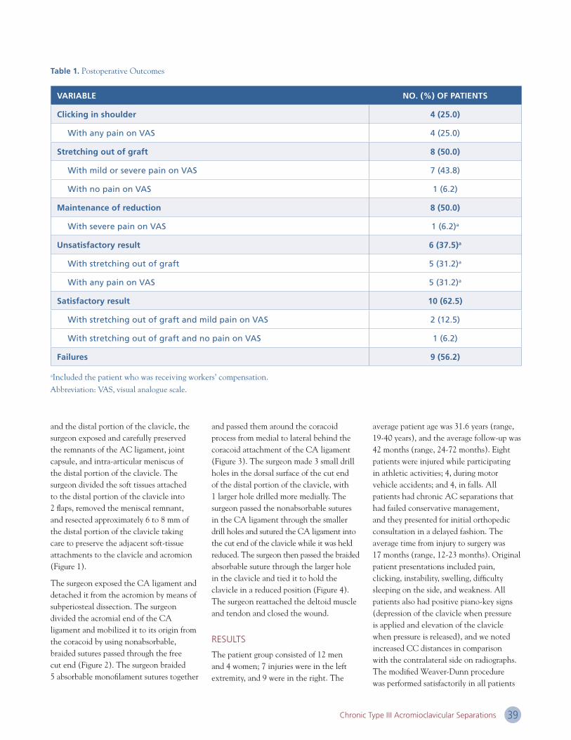
39Chronic Type III Acromioclavicular Separations
and the distal portion of the clavicle, the surgeon exposed and carefully preserved the remnants of the AC ligament, joint capsule, and intra-articular meniscus of the distal portion of the clavicle. The surgeon divided the soft tissues attached to the distal portion of the clavicle into 2 flaps, removed the meniscal remnant, and resected approximately 6 to 8 mm of the distal portion of the clavicle taking care to preserve the adjacent soft-tissue attachments to the clavicle and acromion (Figure 1).
The surgeon exposed the CA ligament and detached it from the acromion by means of subperiosteal dissection. The surgeon divided the acromial end of the CA ligament and mobilized it to its origin from the coracoid by using nonabsorbable, braided sutures passed through the free cut end (Figure 2). The surgeon braided 5 absorbable monofilament sutures together
and passed them around the coracoid process from medial to lateral behind the coracoid attachment of the CA ligament (Figure 3). The surgeon made 3 small drill holes in the dorsal surface of the cut end of the distal portion of the clavicle, with 1 larger hole drilled more medially. The surgeon passed the nonabsorbable sutures in the CA ligament through the smaller drill holes and sutured the CA ligament into the cut end of the clavicle while it was held reduced. The surgeon then passed the braided absorbable suture through the larger hole in the clavicle and tied it to hold the clavicle in a reduced position (Figure 4). The surgeon reattached the deltoid muscle and tendon and closed the wound.
RESULTS
The patient group consisted of 12 men and 4 women; 7 injuries were in the left extremity, and 9 were in the right. The
average patient age was 31.6 years (range, 19-40 years), and the average follow-up was 42 months (range, 24-72 months). Eight patients were injured while participating in athletic activities; 4, during motor vehicle accidents; and 4, in falls. All patients had chronic AC separations that had failed conservative management, and they presented for initial orthopedic consultation in a delayed fashion. The average time from injury to surgery was 17 months (range, 12-23 months). Original patient presentations included pain, clicking, instability, swelling, difficulty sleeping on the side, and weakness. All patients also had positive piano-key signs (depression of the clavicle when pressure is applied and elevation of the clavicle when pressure is released), and we noted increased CC distances in comparison with the contralateral side on radiographs. The modified Weaver-Dunn procedure was performed satisfactorily in all patients
Table 1. Postoperative Outcomes
VARIABLE NO. (%) OF PATIENTS
Clicking in shoulder 4 (25.0)
With any pain on VAS 4 (25.0)
Stretching out of graft 8 (50.0)
With mild or severe pain on VAS 7 (43.8)
With no pain on VAS 1 (6.2)
Maintenance of reduction 8 (50.0)
With severe pain on VAS 1 (6.2)a
Unsatisfactory result 6 (37.5)a
With stretching out of graft 5 (31.2)a
With any pain on VAS 5 (31.2)a
Satisfactory result 10 (62.5)
With stretching out of graft and mild pain on VAS 2 (12.5)
With stretching out of graft and no pain on VAS 1 (6.2)
Failures 9 (56.2)
aIncluded the patient who was receiving workers’ compensation.Abbreviation: VAS, visual analogue scale.

2016 Rush Orthopedics Journal40
with adequate postoperative reduction of the joint as defined by a lack of superior displacement of the distal portion of the clavicle in relation to the acromion (reduced AC joint).
In 8 cases (50.0%), there was stretching out of the reconstruction, defined as displacement of 1 complete width of the clavicle as measured by means of conventional radiography. In terms of VAS pain scores, 8 patients had no pain, 4 had mild pain, and 4 had severe pain; 1 of the 4 patients with severe pain was receiving workers’ compensation. Four patients (25.0%) had clicking in their shoulder, and 6 patients (37.5%) rated their results as unsatisfactory. On the basis of clinical and radiological results, there were 9 failures secondary to loss of reduction or pain (56.2%) and 7 successful cases (43.8%) defined by reduced AC joint, lack of pain as measured with the VAS, or shoulder clicking (Table 1).
DISCUSSION
AC separations originally were classified as grades I, II, and III in the 1960s.6,7 Rockwood8 modified this classification system in 1984 to allow for a more clear differentiation between AC injuries that required surgical treatment and those that warranted conservative treatment. This modification led to the inclusion of type IV, V, and VI separations. Type I and II injuries can be treated conservatively because they retain an intact deltotrapezial fascia and relatively functional CC ligament complex. However, type IV, V, and VI AC injuries generally require surgical intervention to correct the anatomical disruption caused by detached deltotrapezial fascia and complete destruction of the AC and CC ligament complexes.9
Unlike treatment for the other 5 types, the treatment of acute grade III AC separations remains controversial, especially concerning the indications for surgery. Investigators in 2 studies have reported good results with nonoperative management.10,11 Many studies in which the investigators have compared operative and nonoperative treatments have failed to
document superior results after surgery.12-16 Ceccarelli et al17 analyzed multiple randomized controlled trials and systematic reviews in which the investigators compared nonoperative with operative treatment for grade III AC injuries and found comparable results between the 2, with a higher incidence of complications in the surgical group. Hootman18 identified 24 studies of AC dislocation and reported that patients treated both surgically and conservatively reported similar overall satisfactory outcomes (88% surgical vs 87% conservative) and rates of little to no pain (93% vs 96%) but that conservative treatment led to a higher rate of normal to near-normal range of motion (86% vs 95%) and normal strength (87% vs 92%). Bannister et al12 also noted that patients with type III AC separations who were treated conservatively had an earlier return to sports and regained motion faster than did those treated operatively.
Similarly, MacDonald et al19 suggested that nonsurgical treatment of type III AC separations was superior compared with surgical management in restoring normal shoulder function in the first year after injury. Likewise, Tibone et al20 reported that strength and overall shoulder function were not affected significantly in patients treated conservatively for type III AC separations as opposed to those treated surgically. Cox21 reported that 86% of team physicians and 72% of residency chairs were treating type III AC separations nonoperatively. In a systematic review of the literature, Beitzel et al22 supported the notion of at least initial nonoperative management of grade III AC separations for a recommended 3 to 4 weeks before reevaluation of clinical symptoms; some patients will improve, and others will continue to have persistent pain and inability to return to sports or work and then may benefit from operative intervention.
Our findings are in line with those in the current literature in that they suggest that one-third of patients would have the surgery performed again, but two-thirds of patients either sustained graft elongation or had continued pain postoperatively.
This discrepancy highlights the need for investigators in future studies to explore different reconstruction techniques and to determine which patients may benefit most from surgical intervention.
Not all patients experience successful outcomes with conservative treatment. Some authors have reported unsatisfactory results in as many as 20% of patients treated conservatively (from “benign neglect” to closed treatment with a thoracobrachial cast and elastic clavicular strap) for grade III AC separations.11,23 These unsatisfactory results included instability, residual pain, weakness, cosmetic deformity, and loss of shoulder movement.24 Press et al25 reported that in comparison with patients treated operatively, patients with grade III separations who did not undergo surgery had inferior results in terms of time to resolution of pain; subjective impression of pain; functional limitations; range of motion; cosmesis; long-term satisfaction; and absolute values for peak torque, work, and power in tested motions. In addition, Dawe26 reported that in a series of 30 patients with grade III AC separations treated nonoperatively, almost 50% had sufficiently severe pain to give up contact sports or change jobs. Thus, even authors who advocate a nonoperative approach often recommend that surgical repair be considered for certain subgroups of patients such as those who are young and athletic or those who perform heavy labor.
The theory behind the modified Weaver-Dunn procedure maintains that initial stabilization of the distal portion of the clavicle by means of augmentation with a synthetic suture eventually may lead to stabilization by biological tissue. The weakness of the procedure lies in the fact that the biological tissue substitute (CA ligament) is not as strong as the original construct of the distal portion of the clavicle because 1 ligament is essentially replacing 3. Investigators in more recent biomechanical studies have shown the modified Weaver-Dunn technique with synthetic sutures to be the weakest of several AC reconstruction techniques,
41Chronic Type III Acromioclavicular Separations
especially when compared with anatomical reconstruction techniques with use of free tendon grafts.27-29 Results from several clinical and radiographic outcome studies have supported the findings found in the laboratory by showing the superiority of anatomical AC joint reconstruction with a soft-tissue graft as opposed to a synthetic ligament,30 synthetic loop augmentation,31 or tension band wiring.32 This series suggests that postoperative failure due to graft elongation is common after modified Weaver-Dunn reconstruction for chronic AC instability. This failure likely occurs with the dissolution of the polydioxanone suture and subsequent transfer of force to the biological tissue, which may not be strong enough to support the reconstruction.
Nevertheless, although stretching out was common, some of these patients remained satisfied despite many complaining of pain and clicking, which may be due to partial stability of the AC joint in addition to
the removal of mechanical impingement through excision of the distal portion of the clavicle. Ultimately, however, the radiographically visible failures often corresponded to clinical failures as well. We propose that there may be a better way of stabilizing the distal portion of the clavicle and efforts should be made to explore further options, such as a stronger biological tissue, perhaps autogenous hamstring tendon as a replacement for CA ligament transfer.5,33
This study has some limitations. Specifically, the lack of preoperative data with which to compare the postoperative findings limits an understanding of the interval levels of improvement or worsening in the postoperative course. Objective measurements of CC distance would have allowed for a quantifiable definition of displacement and reduction and a better description of stretching out as was defined in the postoperative outcomes.
In addition, the basis of our information was retrospective patient chart review and did not provide us with complete information relevant to the cases, such as patient hand dominance. Finally, although the objective of this observational study was met, we performed no statistical analyses to suggest further definitive conclusions with regard to the effectiveness of this procedure.
CONCLUSIONS
In this cohort, more than 50% of patients undergoing modified Weaver-Dunn AC reconstruction stretched out their reconstruction and had recurrent pain at an average 3.5 years of follow-up. Type III AC separations remain a difficult problem, and surgical treatment remains controversial.
References and financial disclosures are available online at www.rush.edu/orthopedicsjournal.

2016 Rush Orthopedics Journal42
AUTHOR AFFILIATIONS
Department of Orthopedic Surgery, Division of Sports Medicine (Drs Cole, Thorsness, Verma, Romeo), Rush University Medical Center; and Midwest Orthopaedics at Rush
(Drs Cole, Verma, Romeo), Chicago, Illinois.
CORRESPONDING AUTHOR
Brian J. Cole, MD, MBA, Rush University Medical Center and Midwest Orthopaedics at Rush, 1611 W Harrison St, Suite 300, Chicago, IL 60612 ([email protected]).
INTRODUCTION
Since they were first described by Andrews et al1 in 1985, injuries to the biceps-labral complex (BLC) increasingly have been recognized as a cause of shoulder pain. Specific lesions to the BLC, superior labrum anterior to posterior (SLAP) tears are notoriously difficult to diagnose and treat. SLAP tears frequently coincide with other shoulder disease, such as rotator cuff tears, biceps tendonitis, and subacromial impingement
syndrome. There has been a significant increase in the reported incidence and surgical treatment of SLAP tears.2
Most surgeons believe that SLAP tears arise from superior migration of the humeral head, biceps tension, or peel-back as a result of internal impingement.3 Since first classified by Snyder et al,4 modifications5 define 7 distinct pathologic variants, with the most common variant the type II SLAP tear (Figure 1). Our understanding of the role of the superior labrum in glenohumeral stability remains limited.
Diagnosing a SLAP tear on the basis of history and physical examination alone is a clinical challenge. Deep, posterior-superior shoulder pain is the most common symptom; however, the pain is often variable and nonspecific.6 Although SLAP tears can result from traction injury or a fall on an outstretched arm, overhead athletes often present with more insidious
symptoms or a SLAP prodrome.7-10 Physical examination tests used to diagnose SLAP tears include the O’Brien active compression test11 and the O’Driscoll dynamic shear test.12,13 Most tests for superior labral disease lack sensitivity, specificity, and accuracy.14-16 Magnetic resonance arthrography is the reference standard imaging modality for diagnosing SLAP lesions and is highly sensitive and specific (>95%); however, long head of the biceps tendon (LHBT) lesions within the bicipital tunnel are identified poorly with magnetic resonance imaging (Figure 2).17-21
We determine clinical management of SLAP tears according to the characteristics of the tear, the age and functional demands of the patient, and the presence of coexisting shoulder disease. When nonoperative treatment fails, surgical options include SLAP debridement, SLAP repair, biceps tenotomy, and biceps tenodesis (Figure 3).22-26 In the appropriate patient, we sometimes
Update on Superior Labrum Anterior to Posterior Tears and Biceps Tendon Tears
Conclusions Based on Translational Research Performed at the Department of Orthopedic Surgery at Rush
BRIAN J. COLE, MD, MBA / ROBERT J. THORSNESS, MD / NIKHIL N. VERMA, MD / ANTHONY A. ROMEO, MD
“…managing [these] tears depends on the characteristics of the tear, age and functional demands of the patient,
and concomitant disease such as biceps tendonitis.”

43Update on Superior Labrum Anterior to Posterior Tears and Biceps Tendon Tears
elect to perform SLAP repair in combination with biceps tenodesis. We present an evidence-based analysis that synthesizes basic science and clinical evidence of the role of the superior labrum in glenohumeral stability and the role of SLAP repair and biceps tenodesis in the management of symptomatic SLAP tears.
THE SUPERIOR LABRUM AND GLENOHUMERAL STABILITY
We do not yet know exactly how the superior labrum confers glenohumeral stability. Our limited understanding of its function is derived largely from cadaveric studies. In these studies, the glenohumeral joint with a simulated type II SLAP tear has demonstrated small but statistically significant increases in anterior-posterior and superior-inferior translation, suggesting a role of the superior labrum in maintaining glenohumeral stability.27-31 However, repair of SLAP lesions in simulated cadaveric models does not appear to restore normal glenohumeral stability regardless of anterior capsular laxity.27,30-32 Cadaveric models are limited by inconsistencies in capsulolabral anatomy and the size of simulated type II
SLAP lesions in different studies, which can affect the degree of glenohumeral translation.28-30,33 Furthermore, the effect of dynamic components of glenohumeral stability such as the rotator cuff and other in vivo variables cannot be evaluated effectively in cadaveric studies.
Strauss et al27 used computer-aided design to simulate anterior and posterior type II SLAP lesions in cadaver shoulders. They found increased glenohumeral translation in all planes and discovered that biceps tenodesis did not worsen abnormal glenohumeral translation further. Repair of posterior SLAP lesions along with biceps tenodesis restored abnormal glenohumeral translation with no significant difference from the baseline in any plane of motion.
BICEPS TENODESIS AS A TREATMENT FOR SYMPTOMATIC SLAP TEARS
Study investigators generally report good outcomes after SLAP repair, but these outcomes are substantially worse in patients older than 35 years, overhead athletes, and patients receiving workers’
compensation. These patients often complain of recalcitrant pain and stiffness postoperatively.34-36 Salvage options in these cases typically are limited to biceps tenodesis because study results suggest revision SLAP repair is associated with poor outcomes.23,37-39
New data suggest that primary tenodesis of the LHBT may be an effective alternative in specific patients, including those with concomitant biceps tendonitis or those who are manual laborers.40-42 Gupta et al42 retrospectively analyzed 28 patients (mean age, 44 years) with symptomatic SLAP tears and biceps tendonitis treated by means of primary open subpectoral biceps tenodesis; they demonstrated significant improvement in functional outcome scores and produced good patient satisfaction. Boileau et al40 prospectively followed 25 patients with type II SLAP tears treated with either SLAP repair or primary arthroscopic biceps tenodesis. There were significantly higher satisfaction scores and return to preinjury activity level in the biceps tenodesis group compared with results in the SLAP repair group. However, the group assignments were
Figure 1. Superior labrum anterior to posterior (SLAP) tear in an overhead athlete. A, View from a posterior portal demonstrates a normal glenoid (letter G) and biceps-superior labral complex (letter B). The anterior labrum is marked with the letter L. B, With a probe inserted from the anterior portal, the biceps-labral complex is elevated off the superior glenoid to demonstrate a type II SLAP tear (arrow).
A B

2016 Rush Orthopedics Journal44
not randomized, and the decision for SLAP repair was based solely on patient age, leading to substantial selection bias. In contrast, Ek et al41 retrospectively compared patients undergoing SLAP repair and biceps tenodesis for type II SLAP tears and found no significant difference in functional outcome scores, return to preinjury activity level, or complications despite a significant difference in age.
Deciding between SLAP repair and biceps tenodesis for type II SLAP tears includes consideration of patient age, occupation, activity level, concomitant disease, and workers’ compensation status.40,42-46 Although controversial, primary isolated biceps tenodesis is our choice in patients older than 40 years, patients receiving workers’ compensation, and especially patients with concomitant biceps tendonitis.
BICEPS TENODESIS WITH CONCOMITANT SLAP REPAIR
Although recent data may suggest that primary biceps tenodesis alone is an effective treatment for SLAP tears in
specific patients, we do not know whether concomitant SLAP repair in selected patients will improve results.40-42 At our institution, we sometimes elect combined biceps tenodesis and SLAP repair in patients with symptomatic SLAP tear and biceps tendonitis who may be affected by the potential microinstability present in the setting of a SLAP lesion that is not repaired.
We evaluated 86 patients with symptomatic SLAP lesions with or without biceps tendonitis who underwent SLAP repair alone (n = 45), biceps tenodesis alone (n = 23), or combined SLAP repair and biceps tenodesis (n = 18). There were no significant differences in rates of return to preoperative activity level among the groups. Patients who underwent combined tenodesis and SLAP repair demonstrated poorer functional outcome scores than did patients who underwent tenodesis or SLAP repair alone, even when we controlled for workers’ compensation status. Although this is a limited data set, it provides the first evidence demonstrating worse functional outcome scores with the combined procedure.47
BICEPS TENODESIS AND FAILED SLAP REPAIR
Failed SLAP repair results in continued shoulder pain and inability to return to the previous functional level,22,38 and results of revision SLAP repair are poor.37,48 Biceps tenodesis demonstrates good clinical outcomes after a failed SLAP repair.38-40,49 McCormick et al38 prospectively evaluated 42 patients (mean age, 39.2 years) who underwent biceps tenodesis for failed type II SLAP repairs. There was a significant improvement in functional outcome scores and postoperative shoulder range of motion at a mean follow-up of 3.6 years. In a retrospective cohort study, Gupta et al39 demonstrated significant improvements in clinical outcome scores in 11 patients (mean age, 40 years) who underwent open subpectoral biceps tenodesis for failed SLAP repair without complications or need for revision. Werner et al23 retrospectively evaluated 17 patients (mean age, 39 years) who underwent biceps tenodesis for failed SLAP repairs and also demonstrated significant improvements in functional outcome.
Figure 2. Coronal T2-weighted noncontrast material–enhanced magnetic resonance image demonstrating superior labrum anterior to posterior tear (arrow).
Figure 3. Intraoperative view from the posterior portal with patient in the beach chair position. Biceps tenotomy being performed before tenodesis in the setting of symptomatic type II superior labrum anterior to posterior lesion (arrow) in a patient with concomitant biceps tendonitis.
45Update on Superior Labrum Anterior to Posterior Tears and Biceps Tendon Tears
To our knowledge, there are no high-quality studies in which investigators have compared revision SLAP repair to biceps tenodesis for the management of failed SLAP repair.23,37-39,48 Future studies must foster our understanding of these injuries. At our institution, we routinely perform biceps tenodesis in the setting of failed SLAP repairs because we believe the results are more predictable.
SLAP MANAGEMENT IN THE OVERHEAD THROWING ATHLETE
Overhead athletes (especially pitchers) deserve special attention given their unique shoulder mechanics and high clinical expectations. The baseball pitch is the fastest described human motion, often exceeding 7000º per second, placing forces on the shoulder often exceeding 1000 N.50 SLAP tears might occur in pitchers from tension during the deceleration phase or peel-back during the late cocking phase of throwing, possibly exacerbated by microinstability or posterior capsular contracture in the setting of glenohumeral internal rotation deficit.1,51,52 Because some SLAP tears are asymptomatic, these lesions may be physiologic adaptations to overhead throwing kinematics, so surgeons should be cautious regarding SLAP repair in this setting.53
The most common failure of SLAP repair in overhead athletes is inability to return
to play because of continued pain, loss of range of motion, or changes in shoulder proprioception.54 In a 2010 systematic review, Gorantla et al35 pooled outcomes from studies in which the investigators evaluated return to preinjury level of play after SLAP repair and demonstrated a rate of 64%, with rates as low as 7% when overhead athletes specifically were evaluated.35,55 Nonoperative management also has poor return-to-play outcomes.56
Open subpectoral biceps tenodesis has demonstrated excellent clinical outcomes in patients with primary LHBT disease and also has demonstrated good outcomes after a failed SLAP repair.39,40,49 When compared with SLAP repair, biceps tenodesis has demonstrated equivocal clinical and return-to-play outcomes.40,41,57 However, results from a recent motion analysis study at our institution demonstrated that pitchers who underwent biceps tenodesis had more normalized physiologic pitching mechanics than did those who underwent SLAP repair.54 Overall, although biceps tenodesis may be an appropriate treatment for patients with SLAP tears and concomitant biceps tendonitis,42 further study is needed to determine the appropriate surgical management strategy for overhead athletes. Thus, surgeons should approach operative treatment cautiously and only after an initial trial of physical therapy.
CONCLUSIONS
The BLC is a relatively common source of disease in the painful shoulder; however, it does not have a clearly identified role in shoulder stability. Managing SLAP tears depends on the characteristics of the tear, age and functional demands of the patient, and concomitant disease such as biceps tendonitis. We recommend SLAP repair as the treatment of choice for type II SLAP lesions in young, active patients without associated biceps tendonitis who have a mechanical rationale explaining the cause of the tear. We recommend biceps tenodesis over SLAP repair when biceps tendonitis is present; for management of failed SLAP repairs; in relatively older, less active patients; and in patients receiving workers’ compensation. Although primary biceps tenodesis for management of SLAP tears in overhead athletes may be a viable alternative treatment, further study is needed to determine clinical outcomes. Investigators in future studies must evaluate outcomes comparing SLAP repair and biceps tenodesis for type II SLAP tears in different patient populations to refine the current decision-making algorithm for SLAP tears.
References and financial disclosures are available online at www.rush.edu/orthopedicsjournal.

2016 Rush Orthopedics Journal46
AUTHOR AFFILIATIONS
Department of Orthopedic Surgery (Drs Nandyala, Fineberg, Singh; Messrs Massel, Mayo), Rush University Medical Center; and Midwest Orthopaedics at Rush (Dr Singh), Chicago, Illinois.
CORRESPONDING AUTHOR
Kern Singh, MD, Department of Orthopedic Surgery, Rush University Medical Center and Midwest Orthopaedics at Rush, 1611 W Harrison St, Suite 300, Chicago, IL 60612 ([email protected]).
INTRODUCTION
As minimally invasive surgery (MIS) techniques become more popular, the need to understand factors that characterize proficiency in these procedures becomes paramount. Current study results support that mastery of the open technique is necessary to perform an MIS transforaminal lumbar interbody fusion (TLIF) successfully.1 However, investigators in few studies have attempted to characterize the learning curve for MIS TLIF. Proficiency measurements are characterized by plateaus in operative time, blood loss, accuracy of pedicle screw
placement, and intra- and postoperative complications.2 Most study investigators examining the surgical learning curve detail a single surgeon’s experience and use small patient populations.3 Conclusions drawn from these analyses agree that additional studies with larger patient samples are necessary to contribute meaningful data to the growing literature of the surgical learning curve.4,5
MIS TLIF is a viable alternative to the open procedure with less disruption of spinal anatomy.6-8 Clear indications or contraindications for the MIS approach may lie in the comfort and proficiency of the particular surgeon.9 Investigators in a limited number of studies have examined the learning curve for MIS TLIF and reported reduced operative time, visual analogue scale pain scores, estimated blood loss (EBL), and complication rates.10-13 In this study, we attempt to add to the growing literature of the surgical learning curve on the basis of 1 board-certified, fellowship-trained spine surgeon’s experience.
MATERIALS AND METHODS
After institutional review board approval (ORA 10101108), we retrospectively reviewed the senior author’s (K.S.) prospectively maintained surgical repository. Before the first MIS TLIF case of this series, the senior surgeon underwent the following training: an orthopedic surgery residency, spine surgery fellowship, 3 years of clinical practice using traditional open techniques, and self-taught MIS techniques on cadaveric specimens. We identified the first 100 patients undergoing MIS TLIF. We excluded patients by using the following criteria: revision cases, multilevel procedures, and use of alternative biologic agents. These exclusions resulted in 65 consecutive patients undergoing primary 1-level MIS TLIF. All patients underwent MIS TLIF for degenerative disc disease or grade I or II spondylolisthesis with stenosis. We divided the 65 patients into 2 equal, sequential cohorts: patients 1 to 33 and patients 34 to 65. All patients underwent at least 1 year of follow-up with computed tomography (CT) analysis. We compared
Minimally Invasive Transforaminal Lumbar Interbody Fusion
One Surgeon’s Learning Curve DUSTIN H. MASSEL, BS / BENJAMIN C. MAYO, BA / SREEHARSHA V. NANDYALA, MD / STEVE FINEBERG, MD / KERN SINGH, MD
“Surgical familiarity through increased operative experience with the minimally invasive surgery technique resulted in significant decreases in operative time, estimated blood loss, intravenous
fluid administration, and duration of anesthesia.”

47Minimally Invasive Transforaminal Lumbar Interbody Fusion
demographic characteristics, including age, sex, smoking status, operative diagnosis, and comorbidity burden, as assessed with the Charlson Comorbidity Index, between cohorts. Table 1 presents the baseline characteristics of both patient groups. We then analyzed complications, including any intraoperative, in-hospital, or postoperative event that required return to the operating room or additional intervention (eg, deep vein thrombosis). We obtained perioperative variables from the patient chart, as recorded by the surgical nursing staff, and anesthesia records.
Operative Technique
The surgeon used a unilateral approach with the Wiltse technique through a paramedian (4.5 cm lateral to the midline) skin incision. With fluoroscopic guidance and after incision of the skin and fascial layers, he developed a plane between the multifidus and longissimus muscles, whereby he enlarged the pathway to the spine by using sequential dilators. He used a high-speed burr to remove the facet and pars. After identifying the entirety of
the exiting and traversing nerve roots, he used a high-speed burr to complete the laminectomy. He collected a local bone graft, obtained from the laminectomy and facetectomy, in a bone trap. Under fluoroscopic guidance, he identified the interbody space. He used sequential end plate cutters to prepare the end plates. He filled an appropriately sized cage (Concorde; DePuy Spine, Raynham, Massachusetts) with either 4.2 mg (small kit) or 12 mg (large kit) of recombinant human (rh) bone morphogenetic protein (BMP)-2 (Infuse; Medtronic, Minneapolis, Minnesota), along with 5 mL of bone marrow aspirate from the cannulated pedicle and local bone graft. He used BMP as an off-label application. He also placed local bone anterior to the cage in the intervertebral space.
The surgeon then gently impacted the cage obliquely into the intervertebral space. He placed unilateral pedicle screws percutaneously over a guide wire. He placed a rod percutaneously through a separate stab incision and brought it into the gap between the screw heads and
locking nuts. He confirmed the rod’s course by using anteroposterior and lateral fluoroscopy. Once the screws were in place, he compressed them along the rod and tightened the nuts by using a torque wrench. He placed compression across the graft and closed the wound in layers. He performed the laminectomy, bilateral decompression of the spinal canal, and TLIF with a 21-mm nonexpandable tubular retractor. He did not perform posterolateral fusion and preserved midline muscular and ligamentous structures during the procedure.
Primary Analysis
We compared the first 33 patients with the second 32 patients according to perioperative outcomes measures: operative time (minutes), EBL (mL), intravenous (IV) fluids administered during the operation (mL), length of hospital stay (days), and anesthesia time from intubation to extubation (minutes). In addition, we compared intraoperative and postoperative complications (infection, implant failure, cage migration, and so on) between the 2 study groups. We also recorded adverse
Table 1. Baseline Characteristics
Abbreviation: CCI = Charlson Comorbidity Index.
CHARACTERISTIC PATIENT NUMBER P Value
1 to 33 34 to 65 (n = 33) (n = 32)
Age (years, mean [SD]) 59.3 [10.7] 54.2 [15.5] .260
Sex, No. (%) .267
Female 20 (60.6) 15 (46.9)
Male 13 (39.4) 17 (53.1)
Smoking status, No. (%) .224
Nonsmoker 27 (81.8) 21 (65.6)
Smoker 6 (18.2) 11 (34.4)
Diagnosis, No. (%) .260
Degenerative disc disease 3 (9.1) 6 (18.8)
Spondylolisthesis 30 (90.9) 26 (81.2)
Comorbidity burden (CCI, mean [SD]) 1.6 [0.9] 2.6 [0.6] .119
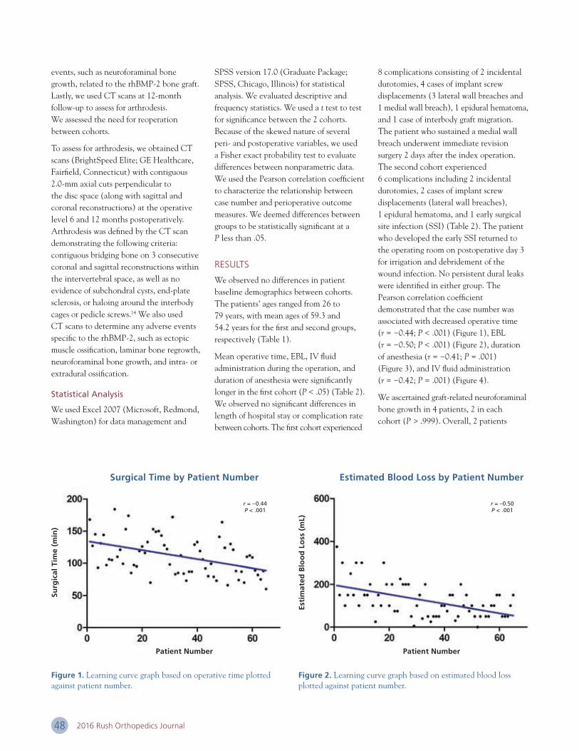
2016 Rush Orthopedics Journal48
events, such as neuroforaminal bone growth, related to the rhBMP-2 bone graft. Lastly, we used CT scans at 12-month follow-up to assess for arthrodesis. We assessed the need for reoperation between cohorts.
To assess for arthrodesis, we obtained CT scans (BrightSpeed Elite; GE Healthcare, Fairfield, Connecticut) with contiguous 2.0-mm axial cuts perpendicular to the disc space (along with sagittal and coronal reconstructions) at the operative level 6 and 12 months postoperatively. Arthrodesis was defined by the CT scan demonstrating the following criteria: contiguous bridging bone on 3 consecutive coronal and sagittal reconstructions within the intervertebral space, as well as no evidence of subchondral cysts, end-plate sclerosis, or haloing around the interbody cages or pedicle screws.14 We also used CT scans to determine any adverse events specific to the rhBMP-2, such as ectopic muscle ossification, laminar bone regrowth, neuroforaminal bone growth, and intra- or extradural ossification.
Statistical Analysis
We used Excel 2007 (Microsoft, Redmond, Washington) for data management and
SPSS version 17.0 (Graduate Package; SPSS, Chicago, Illinois) for statistical analysis. We evaluated descriptive and frequency statistics. We used a t test to test for significance between the 2 cohorts. Because of the skewed nature of several peri- and postoperative variables, we used a Fisher exact probability test to evaluate differences between nonparametric data. We used the Pearson correlation coefficient to characterize the relationship between case number and perioperative outcome measures. We deemed differences between groups to be statistically significant at a P less than .05.
RESULTS
We observed no differences in patient baseline demographics between cohorts. The patients’ ages ranged from 26 to 79 years, with mean ages of 59.3 and 54.2 years for the first and second groups, respectively (Table 1).
Mean operative time, EBL, IV fluid administration during the operation, and duration of anesthesia were significantly longer in the first cohort (P < .05) (Table 2). We observed no significant differences in length of hospital stay or complication rate between cohorts. The first cohort experienced
8 complications consisting of 2 incidental durotomies, 4 cases of implant screw displacements (3 lateral wall breaches and 1 medial wall breach), 1 epidural hematoma, and 1 case of interbody graft migration. The patient who sustained a medial wall breach underwent immediate revision surgery 2 days after the index operation. The second cohort experienced 6 complications including 2 incidental durotomies, 2 cases of implant screw displacements (lateral wall breaches), 1 epidural hematoma, and 1 early surgical site infection (SSI) (Table 2). The patient who developed the early SSI returned to the operating room on postoperative day 3 for irrigation and debridement of the wound infection. No persistent dural leaks were identified in either group. The Pearson correlation coefficient demonstrated that the case number was associated with decreased operative time (r = −0.44; P < .001) (Figure 1), EBL (r = −0.50; P < .001) (Figure 2), duration of anesthesia (r = −0.41; P = .001) (Figure 3), and IV fluid administration (r = −0.42; P = .001) (Figure 4).
We ascertained graft-related neuroforaminal bone growth in 4 patients, 2 in each cohort (P > .999). Overall, 2 patients
Surg
ical
Tim
e (m
in)
Esti
mat
ed B
loo
d L
oss
(m
L)
Patient Number Patient Number
Surgical Time by Patient Number Estimated Blood Loss by Patient Number
r = –0.44P < .001
Figure 1. Learning curve graph based on operative time plotted against patient number.
Figure 2. Learning curve graph based on estimated blood loss plotted against patient number.
r = –0.50P < .001

49Minimally Invasive Transforaminal Lumbar Interbody Fusion
in each cohort required reoperation to address surgical complications. In addition to the aforementioned reoperation cases, 2 patients in each cohort required reoperation (ventral laminoforaminotomy) for pseudarthrosis causing pain and disability. Lastly, 12-month CT scan results demonstrated a 93.9% and 93.8% arthrodesis rates for the first and second patient cohorts, respectively.
DISCUSSION
Perioperative characteristics associated with MIS TLIF primarily depend on the surgeon’s experience and level of comfort with the procedure. Attempts at an assessment of the learning curve are sparse in the surgical literature. Although investigators in several studies have examined the surgical learning curve for various orthopedic procedures, few have determined the steep surgical learning curve associated with MIS TLIF.10,15 Investigators in several studies assert that trends toward better clinical outcomes are observed with an increased number of cases.16 In an attempt to construct a learning curve model, we analyzed several peri- and postoperative variables associated with 1 surgeon’s experience with MIS TLIF procedures.
The results of the current study indicate that significant advances in intraoperative characteristics occur as a surgeon becomes more comfortable with the new MIS technique. We observed improvements in operative time, EBL, duration of anesthesia, and IV fluid administration between the first and second cohorts. The surgeon gains efficiency as he or she becomes more familiar with the surgical anatomy observed through the constrained operative window of the tubular retractor system. The current study is 1 of the largest learning curve analyses for 1-level MIS TLIF, and our results corroborate those of several smaller studies.
Neal and Rosner3 examined the learning curve of a single resident’s experience with 28 patients undergoing minimal access TLIF. Although there was a trend toward decreases in operative times with an increase in case number, the results did not differ significantly between the 2 cohorts (P = .25). Lee et al10 reported the learning curve associated with 60 1-level, 13 2-level, and 13 1-level plus adjacent level decompression MIS TLIFs for various spinal diseases. The authors noted a reduced operative time, EBL, and visual analogue scale pain scores between early and late cohorts. Lee et al11
demonstrated a reduced operative time, duration of fluoroscopy exposure, and patient-controlled analgesia use between the first and second halves of a 90-patient consecutive sample of patients undergoing 1-level MIS TLIF. The authors also observed a trend toward reduced EBL in the second cohort, although it was not statistically significant. In addition, Park et al12 demonstrated a reduced complication rate as the surgeon gained experience with the MIS TLIF technique. Although these results corroborated those observed in this study, the variability among diseases included and the operative requirements associated with multilevel techniques observed in these studies are several reasons for additional assessment of the learning curve in a more focused population.
The peri- and postoperative results of this study are similar to those of other studies on the MIS TLIF operative technique. Scheufler et al5 reported an average operative time of 104 minutes and an average total blood loss of 55 mL across 53 patients treated with MIS TLIF. In addition, the authors reported a single case of dural violation during spinal decompression and no cases of implant fracture or loosening, loss of correction, or interbody cage dislodgement or subsidence
Du
rati
on
of
An
esth
esia
(m
in)
IV F
luid
s D
uri
ng
Su
rger
y (m
L)
Patient Number Patient Number
Duration of Anesthesia by Patient Number IV Fluids During Surgery by Patient Number
r = –0.41P < .001
Figure 3. Learning curve graph based on duration of anesthesia plotted against patient number.
Figure 4. Learning curve graph based on intravenous (IV) fluid use plotted against patient number.
r = –0.42P < .001

2016 Rush Orthopedics Journal50
within the entire 16-month observation period.5 Beringer and Mobasser4 reported on a series of 8 patients undergoing unilateral pedicle screw MIS TLIF with a mean operative time of 160 minutes and a mean EBL of 100 mL. Postoperatively, there were no cases of radiculopathy or malpositioned screws. One patient required removal of pedicle screw instrumentation because of muscle spasms and pain.4
Investigators in various studies have reported on the surgical learning curve of other spinal surgeries. McLoughlin and Fourney17 assessed 52 patients undergoing MIS microdiscectomy with a tubular retractor system, demonstrating that by case 15, operative time had reached a steady state of 60 minutes. Hyde and Seits18 retrospectively reviewed extreme lateral interbody fusion results in 78 consecutive patients in terms of perioperative and follow-up outcome measures. The authors noted a trend toward reduced EBL and
operative time over the course of the operations; however, they observed no significant difference when they compared the first 10 patients with the last 10 patients. The authors concluded that there may have been a learning curve associated with the procedure, despite the lack of statistical significance. Because average operative time and EBL were low at the onset of the study, there was not much deviation from these numbers.
The occurrence of intra- and postoperative complications did not differ between the early and late cohorts in the current study. Similar intraoperative complication rates observed between the early and late cohorts exemplify the intrinsic difficulty of the MIS TLIF procedure. Postoperative complications in the first cohort included 4 cases of implant screw displacements (3 lateral wall breaches and 1 medial wall breach), 1 epidural hematoma, and 1 case of interbody graft migration. In the
second cohort, we observed 2 incidental durotomies, 2 cases of implant screw displacements (lateral wall breaches), 1 epidural hematoma, and 1 early SSI. Although these differences were not significant (P = .672), the potential for implant screw displacement is a common complication noted throughout a surgeon’s experience. Our analysis corroborates results from previous studies in which the investigators reported no observable differences in 12-month arthrodesis rates and reoperation rates between the early and later cohorts. Proper end-plate preparation, cage sizing, and the use of osteobiologic adjuvants may minimize the risk of pseudarthrosis.19
The senior surgeon has adopted several changes to his practice to address the complications demonstrated in this study. At the time of this study, he sometimes used a large BMP kit; however, he currently uses an extra small BMP kit. In addition,
Table 2. Perioperative Variables
VARIABLE PATIENT NUMBER P Value
aTime from cutting open the skin to sewing it back up.bStatistically significant.cFrom induction to extubation.d Complications included incidental durotomy (n = 4), implant screw displacement (n = 6; 5 lateral wall breaches and 1 medial wall breach), interbody graft migration (n = 1), early surgical site infection (n = 1), and epidural hematoma (n = 2).
Abbreviation: IV, intravenous.
1 to 33 34 to 65 (n = 33) (n = 32)
Operative timea (minutes) 124 98 <.001b
Anesthesia durationc (minutes) 182 154 <.001b
IV fluids during surgery (mL) 2089 1703 .004b
Estimated blood loss (mL) 176 85 <.001b
Length of hospital stay (days) 2.4 2.3 .643
Complications,d No. (%) 8 (24.2) 6 (18.8) .672
Neuroforaminal bone growth, No. (%) 2 (6.1) 2 (6.2) >.999
Reoperation, No. (%) 2 (6.1) 3 (9.4) .672
Arthrodesis, No. (%) 31 (93.9) 30 (93.8) .119

51Minimally Invasive Transforaminal Lumbar Interbody Fusion
the senior surgeon now uses contralateral fixation in patients in whom arthrodesis failed, those with dynamic or higher grade spondylolisthesis, or those with a large disc space. There is some variation in operative times in our analysis; Neal and Rosner3 speculated that as a surgeon gains familiarity with the narrower operative window, the surgeon may attempt more challenging cases, resulting in the variation observed. The more difficult cases also translate to slight increases in operative time; our analysis demonstrates this same effect with a small increase in operative time. We attribute this increase to surgeon familiarity with the MIS anatomy, resulting in more thorough decompression and end- plate preparation, 2 rate-limiting factors of the technique.
Finally, the generalizability of the learning curve involves several surgical factors, including the number and frequency of cases, surgeon training before technique implementation, prior or concurrent exposure to alternative MIS techniques, the use of navigation, mean patient size, disease severity, and various hospital factors. This study is a reflection of 1 surgeon’s progression in his experience
with MIS TLIF and, therefore, may not apply to all surgeons adopting MIS spine surgery.
There are several limitations to the current study. First, the surgical experience is limited to that of a single surgeon at a single academic institution. Future studies should attempt to correlate a surgical learning curve for multiple surgeons at multiple institutions. Second, the use of unilateral pedicle screw instrumentation and lack of posterolateral fusion is 1 of several strategies that can be used to perform the MIS TLIF technique. This variation in technique can lead to variability in reports of operative time and EBL and may increase the rate of clinically symptomatic pseudarthrosis. Lastly, our analysis is retrospective, which carries inherent bias. We attempted to reduce this bias by excluding revision surgeries and multiple-level fusions. Strength in our analysis lies in the objective measurement of blood loss via a suction canister that directly collects operative blood both directly and from the surgical sponges used in the operative field. We also used factors that were neutral in terms of surgeon bias, including recorded operative times
(recorded by the nursing staff), anesthetic times (recorded by the anesthesiologist), and intraoperative fluid parameters (indirect measure of operative time and blood loss).
CONCLUSIONS
On the basis of our study results, the learning curve for MIS TLIF appears to be objectively reproducible. Surgical familiarity through increased operative experience with the MIS technique resulted in significant decreases in operative time, EBL, IV fluid administration, and duration of anesthesia. We observed a slight increase in operative times during the surgical evolution that may be attributable to surgical familiarity with the operative anatomy, resulting in more thorough decompression and end-plate preparation. Lastly, our results indicate that the potential for intra- and postoperative complications, inherent in all surgeries, seems to be present regardless of a surgical learning curve.
References and financial disclosures are available online at www.rush.edu/orthopedicsjournal.

2016 Rush Orthopedics Journal52
AUTHOR AFFILIATIONS
Department of Orthopedic Surgery (Drs Brown, Andersson, Fardon), Rush University Medical Center; and Midwest Orthopaedics at Rush (Drs Andersson, Fardon), Chicago, Illinois.
CORRESPONDING AUTHOR
Nicholas Brown, MD, Department of Orthopedic Surgery, Rush University Medical Center, 1611 W Harrison St, Suite 300, Chicago, IL 60612 ([email protected]).
Religious, mystical, and superstitious beliefs highly influenced the treatment of spinal disorders in ancient Egypt and Babylonia. Nevertheless, the Ebers Papyrus and the Edwin Smith Papyrus (Figure 1) describe surgical techniques and associations between spinal injury and dysfunction.1 Some Egyptian mummies show signs of spinal surgery. However, it seems unlikely that operative treatments were common, especially given the following passage from the Babylonian Code of Hammurabi1:
…it is decreed that if a physician treats a patient with a metal knife for a severe wound and has caused the man to die—his hands shall be cut off.
Greek physicians began to take a more scientific approach to neurological symptoms. Hippocrates sometimes incorrectly distinguished ligaments from
nerves, but he attributed neurologic symptoms to spinal cord compression for which he prescribed rest, massage, heat, diet, and music.2,3
History of Lumbar Disc Science and Surgery
NICHOLAS BROWN, MD / GUNNAR B. J. ANDERSSON, MD, PHD / DAVID FARDON, MD
“Our understanding of the human intervertebral disc has progressed greatly and continues to evolve.”
Figure 1. The Edwin Smith Papyrus documents spinal care in ancient times.

53History of Lumbar Disc Science and Surgery
Roman physician Celius Aurelianus described spinal anatomy; detailed accounts of sciatica; and recommended treatments, including leaches, coals, and bloodletting.4 Arabic and Turkish texts also included signs and symptoms of sciatica and recommended a variety of treatments. Association of trauma and heavy lifting with radicular pain was a theme of many ancient texts.1
Domenico Cotugno (Figure 2) in his 1764 treatise, De Ischiade Nervosa Commentarius,5
first associated sciatic pain with the sciatic nerve. However, the term sciatica predates Cotugno and was used by Shakespeare (Figure 3) to curse politicians, as in his play Timon of Athens, act 4, scene 1: “Thou cold sciatica, cripple our senators, that their limbs may halt as lamely as their manners,” and again in Measure for Measure, act 1, scene 2: “How now, which of your hips has the most profound sciatica?”
Another 18th century physician, Giovanni Morgagni, known within Italy as the father of pathological anatomy, associated sites of spinal compression with the resultant disease. He thought compressive growths were tumors, although most likely he was describing tuberculosis of the spine (Pott disease).5 Rudolf Virchow, known in Germany as the father of modern pathology, and to his colleagues as the
pope of medicine (likely contested by Italians), was most famous for describing the Virchow triad of hypercoaguability. His contribution to spinal surgery was the first pathologic description of a herniated disc, eponymously named and mischaracterized as the Virchow tumor in 1857. French physician Charles Lasègue described the straight-leg raising test in 1864, although he did not explain why this maneuver elicited symptoms.6
During the late 19th and early 20th centuries, the first spinal surgeries were performed for disease of intervertebral discs. The first laminectomy was reported to have been performed in 1829.6 However, the first discectomy was likely in 1909 by German physician Fedor Krause for what he determined was an enchondroma. He performed this procedure on the basis of the advice of neurologist Hermann Oppenheim. The patient had immediate pain relief.5 Joel Goldthwait, who founded the Orthopaedic Service at the Massachusetts General Hospital in 1899, was, in 1911, probably the first physician to record an association between disc displacement and radicular symptoms. In 1927, Christian Georg Schmorl first used the term disc herniation, although he did not assign it clinical significance. His name is used to refer to intravertebral disc herniation—a Schmorl node.7
Resistance to the idea that material in the spinal canal originated from the intervertebral disc and caused nerve compression with radicular symptoms diminished during the next 2 decades. In 1927, Vittorio Putti published that sciatic nerve pain was due to foraminal nerve root irritation.8 Two years later, Walter Dandy published 2 cases in which he found “cartilaginous” fragments in the spinal canal.9 At the 1928 Surgical Academy of Paris, Théophile Alajouanine, the French neurologist who also described the Foix-Alajouanine vascular malformation of the spinal cord, presented a case of sciatica and postulated that the material was from the nucleus pulposus rather than from a tumor.10
He stated:
These nodules are neither tumors, chordomas, nor fibrochondromas and are distinctly different from chordomas. Basically, these are always related to the intervertebral disc. We have shown that these curious formations should be considered to result from herniation of the central pulp of the disc across the latter, the hernia produced either by trauma or by pathologic changes in the disc; in addition, the effects of these two causes can be combined.
Arguments that sciatica was from other sources, such as piriformis syndrome, and that the masses observed in the vertebral canal could be tumors persisted for a time. In 1930, Paul Bucy, MD, published his opinion that the masses were cartilaginous neoplasms.11 In 1931, Octave Crouzon, better known for the cranial disease named for him, reinforced Alajouanine’s findings that there were no physaliferous cells histologically, indicating they were not chordomas.12
Collaboration between neurosurgeon William Mixter, MD, and orthopedic surgeon Joseph Barr, MD, brought clinical relevance to the postulate that the cartilaginous material found in the spinal canal caused radicular symptoms and that this material originated from intervertebral discs, not cartilaginous tumors. In 1932, they performed the first surgery with a preoperative diagnosis of disc herniation. The procedure, a laminectomy from L2 to S1 performed in a 28-year-old man with radicular pain, decreased reflexes, and a positive straight-leg raising test, completely relieved his symptoms. On the basis of the temporal association between trauma and his symptoms, as well as histopathologic findings in the excised tissue being almost identical to disc material removed from cadavers at autopsy, Mixter and Barr concluded that this patient’s symptoms were due to a herniated disc. They presented their findings at the Massachusetts Medical Society and published in the New England Journal of Medicine in 1934. Their surgery involved wide exposure, bilateral paraspinal muscle dissection, laminectomy, and removal of disc fragments.13-15
Figure 2. Domenico Cotugno first associated sciatic pain with the sciatic nerve in 1764.

2016 Rush Orthopedics Journal54
The surgical technique remained relatively unchanged and gained popularity such that it was one of the most common orthopedic and neurosurgical procedures performed in the 1960s and 1970s. During this era of the disc, success from removing disc material that was causing clinically concordant radiculopathy encouraged surgeons to try widening the indications for the procedure. The expected consequence of less stringent indications is diminished quality of results in terms of percentage of happy patients.
The indications for disc surgery came to encompass the concept of discogenic pain, meaning pain arising from the disc that is not from nerve root compression and does not necessarily involve disc herniation. An early proponent of this theory was the Australian Harry Crock, MD, and the disease sometimes was called a Crock disc.16
The idea that simple, partial removal of nuclear material would succeed for such patients was not tenable, theoretically or empirically. For such internally deranged discs, more thorough removal of intradiscal contents and interbody fusion held out the promise of helping some patients who presented with such symptoms. The problem was—and continues to be—distinguishing patients who would benefit from such a procedure from those who would not. Discography grew out of the need to make that distinction, but the value of the procedure in predicting surgical outcome has remained controversial.
As with any disease that gains recognition within the medical community and popularity with the media, a number of alternative treatment options have flourished. One such treatment was
intradiscal injection of chymopapain, an enzyme derived from papaya that theoretically dissolved the nucleus without harming the annulus. Developed in 1963 by Chicago orthopedic surgeon Lyman Smith, MD, chemonucleolysis became a popular alternative to surgery.17 Although many patients experienced excellent results, there were reports of serious adverse effects of hemorrhage, pain, allergic reactions, and even paralysis.18 Smith was restricted temporarily from administering his own substance because he violated protocol by administering an injection a second time on a later date in a Chicago White Sox third baseman.19 Sale and distribution of chymopapain ceased in the United States in 2003, though it is still available elsewhere.20 There has been a renewed interest in chemonucleolysis with current research into developing equally effective but less toxic substances.
Improved methods of imaging paralleled the development of treatment modalities. The first medical use of X-rays was in 1896 by Wilhelm Roentgen. In 1918, Walter Dandy, MD, imaged the spinal canal by using injections of air. In 1922, French physicians Jean-Athanase Sicard and Jacques Forestier began using injections of lipiodol, a nonhydrosoluble fluid, to depict blockages in the spinal canal.21 As with many scientific advances, this one was discovered by accident. Sicard typically injected lipiodol into lumbar muscles for pain relief. A medical student was said to have accidently pushed the needle into the spinal canal; after berating the student, Sicard decided to X-ray the spine and discovered myelography. Another version states that it was Forestier himself who performed an errant injection, and the error was slightly less egregious because his intended location was the epidural space.22 Soon after the introduction of lipiodol as an adjuvant to imaging, the tilting table was invented, allowing inversion so contrast material traversed cranially through the spinal canal. However, lipiodol caused arachnoiditis, so removal of the agent was important but challenging. By the 1960s, nonionic water-soluble contrast
Figure 3. William Shakespeare used the term sciatica in the 17th century.

55History of Lumbar Disc Science and Surgery
materials became popular because they did not cause arachnoiditis and obviated the difficult extraction process.23
Discography, the injection of contrast material into the disc space, was first described by K. Lindblom in 194824 and refined when computed tomography came into medical use in the early 1970s.25 By the 1980s, magnetic resonance imaging superseded computed tomography as the preferred imaging modality to detect disc displacement, given its sensitivity, lack of ionizing radiation, and noninvasiveness.
Initial conservative treatment for disc-related symptoms has been substantiated by research results demonstrating that extruded discs can regress. In basic science studies, investigators have elucidated the molecular basis of pain generation.26 In addition, we now understand a wider spectrum of risk factors for herniation, and genetic factors have been found to play a large role.27 Imaging has advanced such that detailed depiction of the disc is possible preoperatively so that surgery is no longer exploratory and can be performed with microscopy or other magnification and limited incisions. Studies such as the Spine Patient Outcomes Research Trial published
by Weinstein et al28 have demonstrated the effectiveness of surgery in the treatment of disc disease for properly selected patients.
Our understanding of the human intervertebral disc has progressed greatly and continues to evolve. The physicians and basic scientists at Rush have made and continue to make important contributions to this progress. Howard An, MD, has made important advances related to disc disorders in both the basic science and clinical realms. He led a team of researchers—Koichi Masuda, MD; Gabriella Cs-Szabo, PhD; Yejia Zhang, MD, PhD; Ana Chee, PhD; Gunnar B. J. Andersson, MD, PhD; Hee-Jeong Im, PhD; and Eugene J-Ma Thonar, PhD—whose contributions have been acknowledged by the prestigious Kappa Delta Award for their project: Intervertebral Disc Repair or Regeneration by Growth Factor and/or Cytokine Inhibitor Protein Injection. Gunnar B. J. Andersson, MD, PhD, has been a longtime deputy editor for Spine, the journal that has published the most advanced knowledge regarding discs, and has made numerous contributions to the understanding of the biomechanics of the disc. One of the most basic elements,
agreement on the terminology used to identify various disc disorders, has been addressed by a multidisciplinary task force led by David Fardon, MD.29 Christopher DeWald, MD, carries on a family tradition of spine surgery at Rush, where his father Ronald DeWald, MD, was among the first to incorporate inclusion of the discs as a component of correcting spinal deformity. Edward J. Goldberg, MD, brought specialized training and techniques in cervical disc surgery to the Department of Orthopedic Surgery at Rush. Frank M. Phillips, MD, and Kern Singh, MD, are at the forefront of minimally invasive adaptations to disc surgery, and Matthew W. Colman, MD, is among the few surgical specialists in oncologic manifestations of disease that affects the spine. Physiatrists April M. Fetzer, DO, and David S. Cheng, MD, both add expertise in spinal injections and electrodiagnostic testing to the orthopedic spine team.
In addition to their own credits, the contributions of the orthopedic spine faculty members at Rush (Figure 4) include the work of the residents and fellows they have trained, who are practicing in spine care in various roles throughout the world. For patients who require an operation as part of their care, surgical procedures for treatment of disc disorders provide proven results, and our doctors continue to advance the field of lumbar disc science and surgery so that our patients and the patients of those we have trained receive the best in spinal care.
References and financial disclosures are available online at www.rush.edu/orthopedicsjournal.
Figure 4. The orthopedic spine team at Rush University Medical Center, from left to right: David S. Cheng, MD; Frank M. Phillips, MD; Kern Singh, MD; Edward J. Goldberg, MD; Gunnar B. J. Andersson, MD, PhD; April M. Fetzer, DO; Howard S. An, MD; and David Fardon, MD. Not pictured: Matthew W. Colman, MD; and Christopher DeWald, MD.

2016 Rush Orthopedics Journal56
Publications (2015)*
Abrams GD, Hart MA, Takami K, Bayne CO, Kelly BT, Espinoza Orías AA, Nho SJ. Biomechanical evaluation of capsulotomy, capsulectomy, and capsular repair on hip rotation. Arthroscopy. 2015;31(8):1511-1157.
Adams JE, King GJ, Steinmann SP, Cohen MS. Elbow arthroscopy: indications, techniques, outcomes, and complications. Instr Course Lect. 2015;64:215-224.
Ahn J, Bohl DD, Elboghdady I, Aboushaala K, Hassanzadeh H, Singh K. Postoperative narcotic consumption in workman’s compensation patients following a minimally invasive transforaminal lumbar interbody fusion. Spine (Phila Pa 1976). 2015;40(16):1284-1288.
Ahn J, Tabaraee E, Bohl DD, Aboushaala K, Singh K. Primary versus revision single-level minimally invasive lumbar discectomy: analysis of clinical outcomes and narcotic utilization. Spine (Phila Pa 1976). 2015;40(18):E1025-1030.
Ahn J, Tabaraee E, Bohl DD, Singh K. Minimally invasive posterior cervical foraminotomy. J Spinal Disord Tech. 2015;28(8):295-297.
Ahn J, Tabaraee E, Singh K. BMP-2-induced neuroforaminal bone growth in the setting of a minimally invasive transforaminal lumbar interbody fusion. J Spinal Disord Tech. 2015;28(5):186-188.
Ahn J, Tabaraee E, Singh K. Minimally invasive transforaminal lumbar interbody fusion. J Spinal Disord Tech. 2015;28(6):222-225.
Akeda K, Yamada T, Inoue N, Nishimura A, Sudo A. Risk factors for lumbar intervertebral disc height narrowing: a population-based longitudinal study in the elderly. BMC Musculoskelet Disord. 2015;16:344.
Bayne CO, Slikker W III, Ma J, Ruch DS, Leversedge FJ, Cohen MS, Wysocki RW. Clinical outcomes of the flexor carpi ulnaris turnover flap for posterior elbow soft-tissue defects. J Hand Surg Am. 2015;40(12):2358-2363.
Best C, Cheng D. Spinal schwannoma: an unusual case of lumbar radiculopathy. PM R. 2015;7(9):1011-1013.
Bhatia S, Lee S, Shewman E, Mather RC III, Salata MJ, Bush-Joseph CA, Nho SJ. Effects of acetabular rim trimming on hip joint contact pressures: how much is too much? Am J Sports Med. 2015;43(9):2138-2145.
Bhatia S, Saigal A, Frank RM, Bach BR Jr, Cole BJ, Romeo AA, Verma NN. Glenoid diameter is an inaccurate method for percent glenoid bone loss quantification: analysis and techniques for improved accuracy. Arthroscopy. 2015;31(4):608-614.e1.
Bogunovic L, Lee SX, Haro MS, Frank JM, Mather RC III, Bush-Joseph CA, Nho SJ. Application of the Goutallier/Fuchs Rotator Cuff Classification to the evaluation of hip abductor tendon tears and the clinical correlation with outcome after repair. Arthroscopy. 2015;31(11):2145-2151.
Bohl DD, Ahn J, Tabaraee E, Jain A, Grauer JN, Singh K. Urinary tract infection following posterior lumbar fusion procedures: an American College of Surgeons National Surgical Quality Improvement Program study. Spine (Phila Pa 1976). 2015;40(22):1785-1791.
Bohl DD, Webb ML, Lukasiewicz AM, Samuel AM, Basques BA, Ahn J, Singh K, Vaccaro AR, Grauer JN. Timing of complications after spinal fusion surgery. Spine (Phila Pa 1976). 2015;40(19):1527-1535.
Brown NM, Bell JA, Jung EK, Sporer SM, Paprosky WG, Levine BR. The use of trabecular metal cones in complex primary and revision total knee arthroplasty. J Arthroplasty. 2015;30(suppl 9):90-93.
Brown NM, Fardon D, Andersson G. Development of the modern total hip arthroplasty: part II. Rush Orthop J. 2015; 53-56.
Brown NM, Murray T, Sporer SM, Wetters N, Berger RA, Della Valle CJ. Extensor mechanism allograft reconstruction for extensor mechanism failure following total knee arthroplasty. J Bone Joint Surg Am. 2015;97(4):279-283.
Brown NM, Tetreault M, Cipriano CA, Della Valle CJ, Paprosky W, Sporer S. Modular tapered implants for severe femoral bone loss in THA: reliable osseointegration but frequent complications. Clin Orthop Relat Res. 2015;473(2):555-560.
Butt A, Hamlekhan A, Patel S, Royhman D, Sukotjo C, Mathew MT, Shokuhfar T, Takoudis C. A novel investigation of the formation of titanium oxide nanotubes on thermally formed oxide of Ti-6Al-4V. J Oral Implantol. 2015;41(5):523-531.
Campbell KA, Erickson BJ, Saltzman BM, Mascarenhas R, Bach BR Jr, Cole BJ, Verma NN. Is local viscosupplementation injection clinically superior to other therapies in the treatment of osteoarthritis of the knee: a systematic review of overlapping meta-analyses. Arthroscopy. 2015;31(10):2036-2045.e14.
Campbell KA, Saltzman BM, Mascarenhas R, Khair MM, Verma NN, Bach BR Jr, Cole BJ. Does intra-articular platelet-rich plasma injection provide clinically superior outcomes compared with other therapies in the treatment of knee osteoarthritis?: a systematic review of overlapping meta-analyses. Arthroscopy. 2015;31(11):2213-2221.
Chahal J, Van Thiel GS, Mather RC III, Lee S, Song SH, Davis AM, Salata M, Nho SJ. The patient acceptable symptomatic state for the modified Harris Hip Score and Hip Outcome Score among patients undergoing surgical treatment for femoroacetabular impingement. Am J Sports Med. 2015;43(8):1844-1849.
Chalmers PN, Botero HG, Meyer J, Frank JM, Sporer SM, Levine B. Computed tomography for the diagnosis of periprosthetic sepsis. Rush Orthop J. 2015; 48-52.
Chalmers PN, Mascarenhas R, Leroux T, Sayegh ET, Verma NN, Cole BJ, Romeo AA. Do arthroscopic and open stabilization techniques restore equivalent stability to the shoulder in the setting of anterior glenohumeral instability?: a systematic review of overlapping meta-analyses. Arthroscopy. 2015;31(2):355-363.
*This is a partial list of published works for the faculty members of the Department of Orthopedic Surgery at Rush in 2015. Works with electronic publication dates in 2015 and print publication dates in 2016 are not included in this list. Although only faculty members are cited, the department gratefully acknowledges the co-authorship of students, nurses, practitioners, therapists, residents, fellows, and colleagues at Rush.

Publications 57
Chalmers PN, Saltzman BM, Feldheim TF, Mascarenhas R, Mellano C, Cole BJ, Romeo AA, Nicholson GP. A comprehensive analysis of pectoralis major transfer for long thoracic nerve palsy. J Shoulder Elbow Surg. 2015;24(7):1028-1035.
Chalmers PN, Sgroi T, Riff AJ, Lesniak M, Sayegh ET, Verma NN, Cole BJ, Romeo AA. Correlates with history of injury in youth and adolescent pitchers. Arthroscopy. 2015;31(7):1349-1357.
Chalmers PN, Verma NN. How can we improve outcomes assessment? Orthopedics. 2015;38(10):594-596.
Chalmers PN, Walton D, Sporer SM, Levine BR. Evaluation of the role for synovial aspiration in the diagnosis of aseptic loosening after total knee arthroplasty. J Bone Joint Surg Am. 2015;97(19):1597-1603.
Cipriano C, Brockman L, Romancik J, Hartemayer R, Ording J, Ginder C, Krier J, Gitelis S, Kent P. The clinical significance of initial pulmonary micronodules in young sarcoma patients. J Pediatr Hematol Oncol. 2015;37(7):548-553.
Cipriano CA, Gray RR, Fernandez JJ. Hibernomas of the upper extremity: a case report and literature review. Hand (N Y). 2015;10(3):547-549.
Cipriano CA, Gruzinova IS, Frank RM, Gitelis S, Virkus WW. Frequent complications and severe bone loss associated with the repiphysis expandable distal femoral prosthesis. Clin Orthop Relat Res. 2015;473(3):831-838.
Clair AJ, Inneh IA, Iorio R, Berend KR, Della Valle CJ, Healy WL, Pelligrini VD. Can administrative data be used to analyze complications following total joint arthroplasty? J Arthroplasty. 2015;30(suppl 9):17-20.
Cochis A, Azzimonti B, Della Valle C, Chiesa R, Arciola CR, Rimondini L. Biofilm formation on titanium implants counteracted by grafting gallium and silver ions. J Biomed Mater Res A. 2015;103(3):1176-1187.
Cole BJ, Tilton AK, Frank, RM, Chubinskaya S, Wimmer MA, Verma NN, Yanke A. Collaborative clinical and basic research in arthritis treatment: a young woman benefits from 10 years of bench-to-bedside research at Rush. Rush Orthop J. 2015; 12-16.
Colman MW, Guss A, Bachus K, Spiker WR, Lawrence BD, Tabaraee E, Brodke DS. Anterior cage reconstruction improves stiffness and decreases cancellous subsidence in a spondylectomy model. Rush Orthop J. 2015; 40-42.
Cross JA, Cole BJ, Spatny KP, Sundman E, Romeo AA, Nicholson GP, Wagner B, Fortier LA. Leukocyte-reduced platelet-rich plasma normalizes matrix metabolism in torn human rotator cuff tendons. Am J Sports Med. 2015;43(12):2898-2906.
Cvetanovich GL, Chalmers PN, Bach BR Jr. Industry financial relationships in orthopaedic surgery: analysis of the Sunshine Act Open Payments Database and comparison with other surgical subspecialties. J Bone Joint Surg Am. 2015;97(15):1288-1295.
Cvetanovich GL, Chalmers PN, Streit JJ, Romeo AA, Nicholson GP. Patients undergoing total shoulder arthroplasty on the dominant extremity attain greater postoperative ROM. Clin Orthop Relat Res. 2015;473(10):3221-3225.
Cvetanovich GL, Fillingham YA, Harris JD, Erickson BJ, Verma NN, Bach BR Jr. Publication and level of evidence trends in the American Journal of Sports Medicine from 1996 to 2011. Am J Sports Med. 2015;43(1):220-225.
Cvetanovich GL, Harris JD, Erickson BJ, Bach BR Jr, Bush-Joseph CA, Nho SJ. Revision hip arthroscopy: a systematic review of diagnoses, operative findings, and outcomes. Arthroscopy. 2015;31(7):1382-1390.
Cvetanovich GL, Schairer WW, Haughom BD, Nicholson GP, Romeo AA. Does resident involvement have an impact on postoperative complications after total shoulder arthroplasty?: an analysis of 1382 cases. J Shoulder Elbow Surg. 2015;24(10):1567-1573.
Cvetanovich GL, Yanke AB, McCormick F, Bach BR Jr, Cole BJ. Trends in meniscal allograft transplantation in the United States, 2007 to 2011. Arthroscopy. 2015;31(6):1123-1127.
Della Valle CJ, Pellegrini VD Jr, Sporer SM, Woolson ST, Berry DJ. How do I get out of this jam?: early postoperative problems of primary total hip arthroplasty. Instr Course Lect. 2015;64:327-336.
Duan L, Perez RE, Davaadelger B, Dedkova EN, Blatter LA, Maki CG. p53-regulated autophagy is controlled by glycolysis and determines cell fate. Oncotarget. 2015;6(27):23135-23156.
Dubrovskaya Y, Tejada R, Bosco J III, Stachel A, Chen D, Feng M, Rosenberg A, Phillips M. Single high dose gentamicin for perioperative prophylaxis in orthopedic surgery: evaluation of nephrotoxicity. SAGE Open Med. 2015;3:2050312115612803.
Ellman MB, Sherman SL, Forsythe B, LaPrade RF, Cole BJ, Bach BR Jr. Return to play following anterior cruciate ligament reconstruction. J Am Acad Orthop Surg. 2015;23(5):283-296.
Erickson BJ, Frank RM, Harris JD, Mall N, Romeo AA. The influence of humeral head inclination in reverse total shoulder arthroplasty: a systematic review. J Shoulder Elbow Surg. 2015;24(6):988-993.
Erickson BJ, Harris JD, Chalmers PN, Bach BR Jr, Verma NN, Bush-Joseph CA, Romeo AA. Ulnar collateral ligament reconstruction: anatomy, indications, techniques, and outcomes. Sports Health. 2015;7(6):511-517.
Erickson BJ, Harris JD, Fillingham YA, Cvetanovich GL, Bush-Joseph C, Cole BJ, Bach BR Jr, Verma NN. Orthopedic practice patterns relating to anterior cruciate ligament reconstruction in elite athletes. Am J Orthop (Belle Mead NJ). 2015;44(12):E480-485.
Erickson BJ, Mascarenhas R, Sayegh ET, Saltzman B, Verma NN, Bush-Joseph CA, Cole BJ, Bach BR Jr. Does operative treatment of first-time patellar dislocations lead to increased patellofemoral stability?: a systematic review of overlapping meta-analyses. Arthroscopy. 2015;31(6):1207-1215.
Erickson BJ, Nwachukwu BU, Kiriakopoulos E, Frank RM, Levine B, Villarroel L, McCormick FM. In-hospital mortality risk for femoral neck fractures among patients receiving Medicare. Orthopedics. 2015;38(7):e593-596.
Erickson BJ, Nwachukwu BU, Rosas S, Schairer WW, McCormick FM, Bach BR Jr, Bush-Joseph CA, Romeo AA. Trends in medial ulnar collateral ligament reconstruction in the United States: a retrospective review of a large private-payer database from 2007 to 2011. Am J Sports Med. 2015;43(7):1770-1774.
Erickson BJ, Saltzman BM, Campbell KA, Fillingham YA, Harris JD, Gupta AK, Bach BR Jr. Rates of deep venous thrombosis and pulmonary embolus after anterior cruciate ligament reconstruction: a systematic review. Sports Health. 2015;7(3):261-266.

2016 Rush Orthopedics Journal58
Farkas GJ, Cvetanovich GL, Rajan KB, Espinoza Orías AA, Nho SJ. Impact of femoroacetabular impingement morphology on gait assessment in symptomatic patients. Sports Health. 2015;7(5):429-436.
Federer AE, Haughom BD, Levy DM, Riff AJ, Nho SJ. Blastomyces tenosynovitis of the foot and ankle: a case report and review of the literature. J Foot Ankle Surg. 2015;54(6):1183-1187.
Forsythe B, Frank RM, Ahmed M, Verma NN, Cole BJ, Romeo AA, Provencher MT, Nho SJ. Identification and treatment of existing copathology in anterior shoulder instability repair. Arthroscopy. 2015;31(1):154-166.
Frank JM, Harris JD, Erickson BJ, Slikker W III, Bush-Joseph CA, Salata MJ, Nho SJ. Prevalence of femoroacetabular impingement imaging findings in asymptomatic volunteers: a systematic review. Arthroscopy. 2015;31(6):1199-1204.
Frank JM, Slikker W III, Al-Shihabi L, Saltzman BM, Fernandez JJ. Total wrist fusion using an intramedullary rod technique with proximal row carpectomy. Tech Hand Up Extrem Surg. 2015;19(3):133-136.
Frank RM, Al-Shihabi L, Erickson B, Wysocki RW, Levine B. The effect of simulator training on safety and clinical performance of common upper extremity procedures. Rush Orthop J. 2015; 26-29.
Frank RM, Cole BJ. Meniscus transplantation. Curr Rev Musculoskelet Med. 2015;8(4):443-450.
Frank RM, Cross MB, Della Valle CJ. Periprosthetic joint infection: modern aspects of prevention, diagnosis, and treatment. J Knee Surg. 2015;28(2):105-112.
Frank RM, Mascarenhas R, Haro M, Verma NN, Cole BJ, Bush-Joseph CA, Bach BR Jr. Closure of patellar tendon defect in anterior cruciate ligament reconstruction with bone-patellar tendon-bone autograft: systematic review of randomized controlled trials. Arthroscopy. 2015;31(2):329-338.
Frank RM, Slikker W III, Lee SX, Lin J, Fernandez JJ, Wysocki RW, Cohen MS, Garras D, Lee S. Detection of traumatic arthrotomy using the saline load test. Rush Orthop J. 2015; 43-47.
Frank RM, Yanke A, Verma NN, Cole BJ. Immediate versus delayed meniscus allograft transplantation: letter to the editor. Am J Sports Med. 2015;43(5):NP8-9.
Friel NA, McNickle AG, DeFranco MJ, Wang F, Shewman EF, Verma NN, Cole BJ, Bach BR Jr, Chubinskaya S, Kramer SM, Wang VM. Effect of highly purified capsaicin on articular cartilage and rotator cuff tendon healing: an in vivo rabbit study. J Orthop Res. 2015;33(12):1854-1860.
Gielen E, O’Neill T, Pye S, Adams J, Ward K, Wu F, Laurent M, Claessens F, Boonen S, Vanderschueren D, Verschueren S. Bone turnover markers predict hip bone loss in elderly European men: results of the European Male Ageing Study (EMAS). Osteoporos Int. 2015;26(2):617-627.
Goldstein ZH, Yi PH, Haughom BD, Hellman MD, Levine BR. Bilateral extensor mechanism disruption after total knee arthroplasty in two morbidly obese patients. Orthopedics. 2015;38(5):e443-446.
Green GA, Pollack KM, D’Angelo J, Schickendantz MS, Caplinger R, Weber K, Valadka A, McAllister TW, Dick RW, Mandelbaum B, Curriero FC. Mild traumatic brain injury in major and minor league baseball players. Am J Sports Med. 2015;43(5):1118-1126.
Gregory JM, Klosterman EL, Thomas JM, Hammond J, Shewman EF, Wang VM, Verma NN, Romeo AA. Suture technique influences the biomechanical integrity of pectoralis major repairs. Orthopedics. 2015;38(9):e746-752.
Gross CE, Frank RM, Hsu AR, Diaz A, Gitelis S. External beam radiation therapy for orthopaedic pathology. J Am Acad Orthop Surg. 2015;23(4):243-252.
Gu SX, Li X, Hamilton JL, Chee A, Kc R, Chen D, An HS, Kim JS, Oh CD, Ma YZ, van Wijnen AJ, Im HJ. MicroRNA-146a reduces IL-1 dependent inflammatory responses in the intervertebral disc. Gene. 2015;555(2):80-87.
Gupta AK, Chalmers PN, Klosterman EL, Harris JD, Bach BR Jr, Verma NN, Cole BJ, Romeo AA. Subpectoral biceps tenodesis for bicipital tendonitis with SLAP tear. Orthopedics. 2015;38(1):e48-53.
Gupta AK, Harris JD, Erickson BJ, Abrams GD, Bruce B, McCormick F, Nicholson GP, Romeo AA. Surgical management of complex proximal humerus fractures-a systematic review of 92 studies including 4500 patients. J Orthop Trauma. 2015;29(1):54-59.
Harris JD, Hussey K, Wilson H, Pilz K, Gupta AK, Gomoll A, Cole BJ. Biological knee reconstruction for combined malalignment, meniscal deficiency, and articular cartilage disease. Arthroscopy. 2015;31(2):275-282.
Haughom BD, Erickson BJ, Hellman MD, Jacobs JJ. Do complication rates differ by gender after metal-on-metal hip resurfacing arthroplasty?: a systematic review. Clin Orthop Relat Res. 2015;473(8):2521-2529.
Haughom BD, Schairer WW, Hellman MD, Nwachukwu BU, Levine BR. An analysis of risk factors for short-term complication rates and increased length of stay following unicompartmental knee arthroplasty. HSS J. 2015;11(2):112-116.
Haughom BD, Schairer WW, Nwachukwu BU, Hellman MD, Levine BR. Does neuraxial anesthesia decrease transfusion rates following total hip arthroplasty? J Arthroplasty. 2015;30(suppl 9):116-120.
Haynes KR, Tseng HW, Kneissel M, Glant TT, Brown MA, Thomas GP. Treatment of a mouse model of ankylosing spondylitis with exogenous sclerostin has no effect on disease progression. BMC Musculoskelet Disord. 2015;16(1):368.
Hellman MD, Mascarenhas R, Gupta A, Fillingham Y, Haughom BD, Salata MJ, Nho SJ. The false-profile view may be used to identify cam morphology. Arthroscopy. 2015;31(9):1728-1732.
Kurakawa T, Kakutani K, Morita Y, Kato Y, Yurube T, Hirata H, Miyazaki S, Terashima Y, Maeno K, Takada T, Doita M, Kurosaka M, Inoue N, Masuda K, Nishida K. Functional impact of integrin α5b1 on the homeostasis of intervertebral discs: a study of mechanotransduction pathways using a novel dynamic loading organ culture system. Spine J. 2015;15(3):417-426.
Horowitz D, Gross CE, Frank RM, Hsu AR, Diaz A, Gitelis S. External beam radiation therapy for orthopaedic pathology. J Am Acad Orthop Surg. 2015;23(8):e9-e10.
Johnson AE, Gerlinger TL, Born CT. The American Academy of Orthopaedic Surgeons/Society of Military Orthopaedic Surgeons/Orthopaedic Trauma Associations/Pediatric Orthopaedic Association Disaster Response and Preparedness Course. J Orthop Trauma. 2015;29(suppl 10):S23-25.
Jung M, Kadam S, Xiong W, Rauch TA, Jin SG, Pfeifer GP. MIRA-seq for DNA methylation analysis of CpG islands. Epigenomics. 2015;7(5):695-706.

Publications 59
Khair MM, Tilton AK, Cole BJ. Emerging biologics in orthopedics. Am J Orthop (Belle Mead NJ). 2015;44(5):201.
Khan M, Della Valle CJ, Jacofsky DJ, Meneghini RM, Haddad FS. Early postoperative complications after total hip arthroplasty: current strategies for prevention and treatment. Instr Course Lect. 2015;64:337-346.
Kim JS, Ali MH, Wydra F, Li X, Hamilton JL, An HS, Cs-Szabo G, Andrews S, Moric M, Xiao G, Wang JH, Chen D, Cavanaugh JM, Im HJ. Characterization of degenerative human facet joints and facet joint capsular tissues. Osteoarthritis Cartilage. 2015;23(12):2242-2251.
Kling S, Karns MR, Gebhart J, Kosmas C, Robbin M, Nho SJ, Bedi A, Salata MJ. The effect of acetabular rim recession on anterior acetabular coverage: a cadaveric study using the false-profile radiograph. Am J Sports Med. 2015;43(4):957-964.
Kogan M, Frank RM. A picture is worth a thousand words: unconscious bias in the residency application process? Am J Orthop (Belle Mead NJ). 2015;44(9):E358-359.
Kroin E, Frank JM, Haughom B, Kogan M. Two cases of avascular necrosis after prophylactic pinning of the asymptomatic, contralateral femoral head for slipped capital femoral epiphysis: case report and review of the literature. J Pediatr Orthop. 2015;35(4):363-366.
Lee S, Frank RM, Harris J, Song SH, Bush-Joseph CA, Salata MJ, Nho SJ. Evaluation of sexual function before and after hip arthroscopic surgery for symptomatic femoroacetabular impingement. Am J Sports Med. 2015;43(8):1850-1856.
Lee S, Haro MS, Riff A, Bush-Joseph CA, Nho SJ. Arthroscopic technique for the treatment of pigmented villonodular synovitis of the hip. Arthrosc Tech. 2015;4(1):e41-46.
Lee S, Savin DD, Shah NR, Bronsnick D, Goldberg B. Scapular winging: evaluation and treatment: AAOS exhibit selection. J Bone Joint Surg Am. 2015;97(20):1708-1716.
Lee S, Wuerz TH, Shewman E, McCormick FM, Salata MJ, Philippon MJ, Nho SJ. Labral reconstruction with iliotibial band autografts and semitendinosus allografts improves hip joint contact area and contact pressure: an in vitro analysis. Am J Sports Med. 2015;43(1):98-104.
Leroux T, Chahal J, Wasserstein D, Verma NN, Romeo AA. A systematic review and meta-analysis comparing clinical outcomes after concurrent rotator cuff repair and long head biceps tenodesis or tenotomy. Sports Health. 2015;7(4):303-307.
Levine BR. CORR Insights: is there an advantage to knotless barbed suture in TKA wound closure?: a randomized trial in simultaneous bilateral TKAs. Clin Orthop Relat Res. 2015;473(6):2028-2030.
Levy DM, Erickson BJ, Harris JD, Bach BR Jr, Verma NN, Romeo AA. Management of isolated greater tuberosity fractures: a systematic review. Rush Orthop J. 2015; 21-25.
Li N, Luo D, Hu X, Luo W, Lei G, Wang Q, Zhu T, Gu J, Lu Y, Zheng Q. RUNX2 and osteosarcoma. Anticancer Agents Med Chem. 2015;15(7):881-887.
Louie PK, Presciutti S, Johnson S, Tabaraee E, Cha T, An HS. Correcting sagittal imbalance: a retrospective radiographic study of decompression and local fusion in cases of degenerative scoliosis. Rush Orthop J. 2015; 30-34.
Luo W, Shao C, Li N, Zhang F, Guo S, Duan Z, Zheng Q, He H. Expression of epidermal growth factor-like domain 7 correlates with clinicopathological features of osteosarcoma. Am J Transl Res. 2015;7(7):1236-1245.
Makhni EC, Padaki AS, Petridis PD, Steinhaus ME, Ahmad CS, Cole BJ, Bach BR Jr. High variability in outcome reporting patterns in high-impact ACL literature. J Bone Joint Surg Am. 2015;97(18):1529-1542.
Makhni EC, Steinhaus ME, Morrow ZS, Jobin CM, Verma NN, Cole BJ, Bach BR Jr. Outcomes assessment in rotator cuff pathology: what are we measuring? J Shoulder Elbow Surg. 2015;24(12):2008-2015.
Mall NA, Harris JD, Cole BJ. Clinical evaluation and preoperative planning of articular cartilage lesions of the knee. J Am Acad Orthop Surg. 2015;23(10):633-640.
Martin EJ, Mathew MT, Shull KR. Viscoelastic properties of electrochemically deposited protein/metal complexes. Langmuir. 2015;31(13):4008-4017.
Mascarenhas R, Cvetanovich GL, Sayegh ET, Verma NN, Cole BJ, Bush-Joseph C, Bach BR Jr. Does double-bundle anterior cruciate ligament reconstruction improve postoperative knee stability compared with single-bundle techniques?: a systematic review of overlapping meta-analyses. Arthroscopy. 2015;31(6):1185-1196.
Mascarenhas R, Erickson BJ, Sayegh ET, Verma NN, Cole BJ, Bush-Joseph C, Bach BR Jr. Is there a higher failure rate of allografts compared with autografts in anterior cruciate ligament reconstruction: a systematic review of overlapping meta-analyses. Arthroscopy. 2015;31(2):364-372.
Mascarenhas R, Saltzman BM, Sayegh ET, Verma NN, Cole BJ, Bush-Joseph C, Bach BR Jr. Bioabsorbable versus metallic interference screws in anterior cruciate ligament reconstruction: a systematic review of overlapping meta-analyses. Arthroscopy. 2015;31(3):561-568.
Mascarenhas R, McConkey MO, Forsythe B, Harner CD. Revision anterior cruciate ligament reconstruction with bone-patellar tendon-bone allograft and extra-articular iliotibial band tenodesis. Am J Orthop (Belle Mead NJ). 2015;44(4):E89-93.
Mascarenhas R, Saltzman BM, Fortier LA, Cole BJ. Role of platelet-rich plasma in articular cartilage injury and disease. J Knee Surg. 2015;28(1):3-10.
Mather RC III, Garrett WE, Cole BJ, Hussey K, Bolognesi MP, Lassiter T, Orlando LA. Cost-effectiveness analysis of the diagnosis of meniscus tears. Am J Sports Med. 2015;43(1):128-137.
McCormick F, Alpaugh K, Haughom B, Nho S. Arthroscopic T-capsulotomy for excision of pigmented villonodular synovitis in the hip. Orthopedics. 2015;38(4):237-239.
Mellano CR, Shin JJ, Yanke AB, Verma NN. Disorders of the long head of the biceps tendon. Instr Course Lect. 2015;64:567-576.
Nair A, Gan J, Bush-Joseph C, Verma N, Tetreault MW, Saha K, Margulis A, Fogg L, Scanzello CR. Synovial chemokine expression and relationship with knee symptoms in patients with meniscal tears. Osteoarthritis Cartilage. 2015;23(7):1158-1164.
Nunley RM, Nam D, Bashyal RK, Della Valle CJ, Hamilton WG, Berend ME, Parvizi J, Clohisy JC, Barrack RL. The impact of total joint arthroplasty on sexual function in young, active patients. J Arthroplasty. 2015;30(2):335-340.
Nunley RM, Nam D, Berend KR, Lombardi AV, Dennis DA, Della Valle CJ, Barrack RL. New total knee arthroplasty designs: do young patients notice? Clin Orthop Relat Res. 2015;473(1):101-108.
Orr JD, Hoffmann JD, Arrington ED, Gerlinger TL, Devine JG, Belmont PJ Jr. Army orthopaedic surgery residency program directors’ selection criteria. J Surg Orthop Adv. 2015;24(2):120-124.

2016 Rush Orthopedics Journal60
Park SY, An HS, Moon SH, Lee HM, Suh SW, Chen D, Jeon JH. Neuropathic pain components in patients with lumbar spinal stenosis. Yonsei Med J. 2015;56(4):1044-1050.
Parvizi J, Heller S, Berend KR, Della Valle CJ, Springer BD. Periprosthetic joint infection: the algorithmic approach and emerging evidence. Instr Course Lect. 2015;64:51-60. Review.
Patel AM, Gregory B, Wysocki RW. Volarly extruded fractures of the index metacarpal base ulnar condyle: report of two cases. J Hand Surg Eur Vol. 2015;40(9):1012-1013.
Phillips FM. CORR Insights: recurrent versus primary lumbar disc herniation surgery: patient-reported outcomes in the Swedish Spine Register Swespine. Clin Orthop Relat Res. 2015;473(6):1985-1987.
Phillips FM, Geisler FH, Gilder KM, Reah C, Howell KM, McAfee PC. Long-term outcomes of the US FDA IDE Prospective, Randomized Controlled Clinical Trial comparing PCM cervical disc arthroplasty with anterior cervical discectomy and fusion. Spine (Phila Pa 1976). 2015;40(10):674-683.
Riboh JC, Cole BJ, Farr J. Particulated articular cartilage for symptomatic chondral defects of the knee. Curr Rev Musculoskelet Med. 2015;8(4):429-435.
Rosenthal KS, Mikecz K, Steiner HL III, Glant TT, Finnegan A, Carambula RE, Zimmerman DH. Rheumatoid arthritis vaccine therapies: perspectives and lessons from therapeutic ligand epitope antigen presentation system vaccines for models of rheumatoid arthritis. Expert Rev Vaccines. 2015;14(6):891-908.
Ross JP, Brown NM, Levine BR. Chronic knee dislocation after total knee arthroplasty. Orthopedics. 2015;38(12):e1155-1159.
Rossi VJ, Ahn J, Bohl DD, Tabaraee E, Singh K. Economic factors in the future delivery of spinal healthcare. World J Orthop. 2015;6(5):409-412.
Royhman D, Patel M, Runa MJ, Jacobs JJ, Hallab NJ, Wimmer MA, Mathew MT. Fretting-corrosion in hip implant modular junctions: new experimental set-up and initial outcome. Tribol Int. 2015;91:235-245.
Runa MJ, Mathew MT, Fernandes MH, Rocha LA. First insight on the impact of an osteoblastic layer on the bio-tribocorrosion performance of Ti6Al4V hip implants. Acta Biomater. 2015;12:341-351.
Sakai D, Andersson GB. Stem cell therapy for intervertebral disc regeneration: obstacles and solutions. Nat Rev Rheumatol. 2015;11(4):243-256.
Saltzman BM, Frank JM, Slikker W, Fernandez JJ, Cohen MS, Wysocki RW. Clinical outcomes of proximal row carpectomy versus four-corner arthrodesis for post-traumatic wrist arthropathy: a systematic review. J Hand Surg Eur Vol. 2015;40(5):450-457.
Saltzman BM, Harris JD, Forsythe B. Proximal coracobrachialis tendon rupture, subscapularis tendon rupture, and medial dislocation of the long head of the biceps tendon in an adult after traumatic anterior shoulder dislocation. Int J Shoulder Surg. 2015;9(2):52-55.
Saltzman BM, Levy DM, Vakhshori V, DeWald CJ. Free vascularized fibular strut autografts to the lumbar spine in complex revision surgery: a report of two cases. Korean J Spine. 2015;12(3):185-189.
Saltzman BM, Riboh JC, Cole BJ, Yanke AB. Humeral head reconstruction with osteochondral allograft transplantation. Arthroscopy. 2015;31(9):1827-1834.
Sandy JD, Chan DD, Trevino RL, Wimmer MA, Plaas A. Human genome-wide expression analysis reorients the study of inflammatory mediators and biomechanics in osteoarthritis. Osteoarthritis Cartilage. 2015;23(11):1939-1945.
Sayegh ET, Mascarenhas R, Chalmers PN, Cole BJ, Romeo AA, Verma NN. Surgical treatment options for glenohumeral arthritis in young patients: a systematic review and meta-analysis. Arthroscopy. 2015;31(6):1156-1166.e8.
Sayegh ET, Sandy JD, Virk MS, Romeo AA, Wysocki RW, Galante JO, Trella KJ, Plaas A, Wang VM. Recent scientific advances towards the development of tendon healing strategies. Curr Tissue Eng. 2015;4(2):128-143.
Scanzello CR, Markova DZ, Chee A, Xiu Y, Adams SL, Anderson G, Zgonis M, Qin L, An HS, Zhang Y. Fibronectin splice variation in human knee cartilage, meniscus and synovial membrane: observations in osteoarthritic knee. J Orthop Res. 2015;33(4):556-562.
Sexton A, Louie PK, Tabaraee E, Fardon D, Colman MW. Aneurysmal bone cyst of the thoracic spine: a case report and review of literature. Rush Orthop J. 2015; 17-20.
Sgroi T, Chalmers PN, Riff AJ, Lesniak M, Sayegh ET, Wimmer MA, Verma NN, Cole BJ, Romeo AA. Predictors of throwing velocity in youth and adolescent pitchers. J Shoulder Elbow Surg. 2015;24(9):1339-1345.
Shahi A, Tan TL, Tarabichi S, Maher A, Della Valle C, Saleh UH. Primary repair of iatrogenic medial collateral ligament injury during TKA: a modified technique. J Arthroplasty. 2015;30(5):854-857.
Shih YR, Phadke A, Yamaguchi T, Kang H, Inoue N, Masuda K, Varghese S. Synthetic bone mimetic matrix-mediated in situ bone tissue formation through host cell recruitment. Acta Biomater. 2015;19:1-9.
Shin JJ, Haro M, Yanke AB, Mascarenhas R, Romeo AA, Cole BJ, Inoue N, Verma NN. Topographic analysis of the capitellum and distal femoral condyle: finding the best match for treating osteochondral defects of the humeral capitellum. Arthroscopy. 2015;31(5):843-849.
Shin JJ, Mascarenhas R, Patel AV, Yanke AB, Nicholson GP, Cole BJ, Romeo AA, Verma NN. Clinical outcomes following revision anterior shoulder arthroscopic capsulolabral stabilization. Arch Orthop Trauma Surg. 2015;135(11):1553-1559.
Simon P, Gupta A, Pappou I, Hussey MM, Santoni BG, Inoue N, Frankle MA. Glenoid subchondral bone density distribution in male total shoulder arthroplasty subjects with eccentric and concentric wear. J Shoulder Elbow Surg. 2015;24(3):416-424.
Singh K, Ahn J. Impact of economic incentives on patient selection of surgical facility: commentary on an article by James C. Robinson, PhD, et al.: “Consumer choice between hospital-based and freestanding facilities for arthroscopy. Impact on prices, spending, and surgical complications.” J Bone Joint Surg Am. 2015;97(18):e63.
Streubel PN, Cohen MS. Open surgical release for contractures of the elbow. J Am Acad Orthop Surg. 2015;23(6):328-338.
Tabaraee E, Ahn J, Bohl DD, Elboghdady IM, Aboushaala K, Singh K. The impact of worker’s compensation claims on outcomes and costs following an anterior cervical discectomy and fusion. Spine (Phila Pa 1976). 2015;40(12):948-953.
Publications 61
Virk MS, Tilton AK, Cole BJ. Biceps tenodesis and superior labrum anterior to posterior (SLAP) tears. Am J Orthop (Belle Mead NJ). 2015;44(11):491-494.
Wang C, Timmons CL, Shao Q, Kinlock BL, Turner TM, Iwamoto A, Zhang H, Liu H, Liu B. GB virus type C E2 protein inhibits human immunodeficiency virus type 1 Gag assembly by downregulating human ADP-ribosylation factor 1. Oncotarget. 2015 Dec;6(41):43293-43309.
Wang VM, Karas V, Lee AS, Yin Z, Van Thiel GS, Hussey K, Sumner DR, Chubinskaya S, Magin RL, Verma NN, Romeo AA, Cole BJ. Assessment of glenoid chondral healing: comparison of microfracture to autologous matrix-induced chondrogenesis in a novel rabbit shoulder model. J Shoulder Elbow Surg. 2015;24(11):1789-1800.
Wang VM, Romeo AA. Are anterior supraspinatus tendon tears more prone to propagation? Commentary on an article by Daisuke Araki, MD, PhD, et al., “Effect of tear location on propagation of isolated supraspinatus tendon tears during increasing levels of cyclic loading.” J Bone Joint Surg Am. 2015;97(4):e24.
Wimmer MA, Laurent MP, Mathew MT, Nagelli C, Liao Y, Marks LD, Jacobs JJ, Fischer A. The effect of contact load on CoCrMo wear and the formation and retention of tribofilms. Wear. 2015;332-333:643-649.
Wolfstadt JI, Cole BJ, Ogilvie-Harris DJ, Viswanathan S, Chahal J. Current concepts: the role of mesenchymal stem cells in the management of knee osteoarthritis. Sports Health. 2015;7(1):38-44.
Wysocki RW, Soni E, Virkus WW, Scarborough MT, Leurgans SE, Gitelis S. Is intralesional treatment of giant cell tumor of the distal radius comparable to resection with respect to local control and functional outcome? Clin Orthop Relat Res. 2015;473(2):706-715.
Yamaguchi T, Goto S, Nishigaki Y, Espinoza Orías AA, Bae WC, Masuda K, Inoue N. Microstructural analysis of three-dimensional canal network in the rabbit lumbar vertebral endplate. J Orthop Res. 2015;33(2):270-276.
Yanke A, Bell R, Lee A, Shewman EF, Wang V, Bach BR Jr. Regional mechanical properties of human patellar tendon allografts. Knee Surg Sports Traumatol Arthrosc. 2015;23(4):961-967.
Yanke AB, Chubinskaya S. The state of cartilage regeneration: current and future technologies. Curr Rev Musculoskelet Med. 2015;8(1):1-8.
Yanke AB, Khair MM, Stanley R, Walton D, Lee S, Bush-Joseph CA, Espinosa Orias AA, Inoue N, Nho SJ. Sex differences in patients with cam deformities with femoroacetabular impingement: 3-dimensional computed tomographic quantification. Arthroscopy. 2015;31(12):2301-2306.
Yanke AB, Tilton AK, Wetters NG, Merkow DB, Cole BJ. DeNovo NT particulated juvenile cartilage implant. Sports Med Arthrosc. 2015;23(3):125-129.
Yi PH, Cross MB, Della Valle CJ. Reply to the letter to the editor: the 2013 Frank Stinchfield Award: diagnosis of infection in the early postoperative period after total hip arthroplasty. Clin Orthop Relat Res. 2015;473(6):2152-2153.
Yi PH, Cross MB, Johnson SR, Rasinski KA, Nunley RM, Della Valle CJ. Are financial conflicts of interest for the surgeon a source of concern for the patient? J Arthroplasty. 2015;30(suppl 9):21-33.
Yi PH, Cross MB, Moric M, Levine BR, Sporer SM, Paprosky WG, Jacobs JJ, Della Valle CJ. Do serologic and synovial tests help diagnose infection in revision hip arthroplasty with metal-on-metal bearings or corrosion? Clin Orthop Relat Res. 2015;473(2):498-505.
Yi PH, Frank RM, Vann E, Sonn KA, Moric M, Della Valle CJ. Is potential malnutrition associated with septic failure and acute infection after revision total joint arthroplasty? Clin Orthop Relat Res. 2015;473(1):175-182.
Zhang Y, Chee A, Shi P, Wang R, Moss I, Chen EY, He TC, An HS. Allogeneic articular chondrocyte transplantation downregulates interleukin 8 gene expression in the degenerating rabbit intervertebral disk in vivo. Am J Phys Med Rehabil. 2015;94(7):530-538.
2016 Rush Orthopedics Journal62
Higher Learning Good medicine and good teaching are inextricably linked; in fact, the word doctor comes from the Latin
docere, “to teach,” and doctor in Latin literally means “teacher.” Since the inception of the Department of
Orthopedic Surgery at Rush, the faculty has been committed to advancing education on all fronts—from
the clinics, operating rooms, and labs, to the local, national, and international communities. In the pages
that follow, orthopedic surgeons Edward J. Goldberg, MD; Simon Lee, MD; Brett Levine, MD, MS; and
Anthony A. Romeo, MD, write about why education is integral to their practices, and how Rush is preparing
the orthopedic specialists of the future to be outstanding diagnosticians, surgeons, investigators, and—of
course—teachers.

Bedside Manner 63
To operate or not to operate. I think an essential part of being a successful orthopedic surgeon is knowing when to operate. It might seem paradoxical that we surgeons often recommend nonsurgical therapies. But those determinations—who will and who won’t benefit from surgery—affect outcomes, and we always want to achieve the best outcomes for our patients.
That’s why the educational opportunities we provide to our residents and fellows are geared toward enabling them to become not just extraordinary surgeons, but extraordinary diagnosticians. It’s great if a resident or fellow gets to be part of 50 hip replacements or discectomies during his or her time here. But when our trainees go out and set up their own practices, they need, first and foremost, to understand when it’s appropriate to do the procedures.
Learning at the bedside. Our residents and fellows spend a good deal of time in the attending physicians’ offices and clinics—
often as much time as they do in the OR. The attendings have them evaluate patients, do histories and physicals, and come up with clinical diagnoses. Then we have them correlate the symptoms with the patient’s diagnostic films.
For instance, the patient may say, “I have sciatica.” But is it lumbar sciatica? Is there a tumor in the pelvis or arthritis of the hip? When patients come to you, they’re just describing symptoms. The physician acts as a detective and tries to figure out what’s causing those symptoms.
The residents and fellows go through this process by themselves and bring everything to their attending physician. Then we evaluate the patient together. Once we’ve confirmed the diagnosis, we discuss the options. Has the patient already received appropriate medical treatment—physical therapy, injections, medications, and so on? Are there other medical therapies worth trying? Is the patient a good candidate
for surgery? If we decide surgery is the best option, we then discuss what is the appropriate procedure and why.
In addition to doing initial evaluations, our residents and fellows spend a lot of time with patients after their surgeries. This enables them to get direct feedback about the procedures, the recovery process and the level of pain, what issues or complications arose, etc.
The benefit of experience. What’s great about the Section of Spine Surgery is that we have David Fardon, who doesn’t operate anymore but operated for longer than any of us current spine surgeons. The residents and fellows love him because he doesn’t talk about all the new implants and gadgets. He talks about why the patient is here and discusses possible diagnoses and conditions that could mimic this one. He really gets the residents and fellows to think creatively and come up with differential diagnoses. The same was true
Bedside MannerTaking surgical education beyond the operating room
EDWARD J. GOLDBERG, MD; SPINE SURGEON

2016 Rush Orthopedics Journal64
of Gunnar Andersson, a world-renowned spine surgeon who conducted a nonsurgical spine practice from the time he stopped operating until his recent retirement.
Our trainees also benefit from the skills and varied perspectives of the two board-certified physiatrists in our department, April Fetzer, DO, and David Cheng, MD, who both have expertise in spinal injections and electrodiagnostics.
Becoming well-rounded. I round on my patients every day, Monday through Friday, without fail. It’s such a vital part of my practice because I’ve found that spending even 5 minutes a day in the patient’s room—a brief period of face time—goes a very long way. You’re taking patients who are nervous or scared, who don’t know what’s going to happen to them, and you’re offering them comfort and reassurance.
I want my residents and fellows to see those personal interactions and learn about the importance of bedside manner, which is one of the things I try to emphasize during rounds. It doesn’t take a lot of effort to ask how a patient is doing and if he or she has any concerns, but that face time means a
lot to the patients. My residents and fellows get to see good customer service every day, and my hope is that they will apply it to their own practices.
The other thing I try to teach about rounding is that it’s a great way to get everyone on the same page. After I round with my resident and fellow, we talk to the nurse practitioner and run through the day’s plan—A, B, C, D, these are the things we’re doing for this patient. So by 7:30 in the morning, everyone on my team knows the entire plan, and then we tell the patient what that plan is so there’s nothing unexpected for anybody, no surprises. My patients appreciate knowing what to expect that day, and my team feels confident because they have their marching orders.
The merits of mentoring. Mentoring is important for putting out quality future physicians, but it also inspires me to keep learning and growing. If I’m teaching, I need to stay current. You don’t do things now the way you did them 15 years ago. Medicine advances rapidly, and both the technology and tools you use can quickly become totally outdated.
Back when I trained—about a million years ago—residents ran the show a lot more. Although residents currently are given a lot of autonomy, the attending physician still supervises. It’s not like we tell them, “Here, go do a 4-hour clinic and call me for the 1 or 2 surgeries you pick up.” The attendings are much more hands-on now. Being a mentor means finding that balance between educator and patient advocate. You want to let your trainees blossom, and that sometimes means letting them stumble a little—just not to the point where it’s detrimental to the patient.
Our orthopedic faculty take pride in educating and mentoring, and we’re willing to spend the time to do these things. What’s interesting about the educational experience here at Rush is that within all of the subsections, you don’t have just one physician; you have several at least. So residents and fellows can learn different approaches to a problem. They then can choose what they like and, ultimately, formulate their own styles.
Training for the future. At Rush, we have a unique situation because our orthopedic practice is private, but we are still affiliated with an academic medical center. So our residents and fellows get to see how both a private practice and a major medical center function. They get to experience the day-to-day interactions with patients, attendings, physician assistants, nurse practitioners, nurses, trainees, medical students, researchers, and support staff. And they get to apply what they’re learning to the care of actual patients. All of these things are as important as learning the nitty-gritty of musculoskeletal diseases and injuries.
You want to send quality orthopedic specialists out into the world. It’s a reflection on you and your program. But more important, you want to train the best possible future physicians to take care of patients long after you retire. That’s what I hope my legacy will be.

65
The evolution of surgical training.
Traditionally, surgical training has followed the mantra of “see one, do one, teach one.” Trainees were supposed to pick up things very quickly and then jump in—feet to the fire right away. Many surgeons felt that residents and fellows were supposed to learn by just being in the OR and observing through osmosis, without any explanation during the procedures of techniques or how to use the instrumentation. That was the old-school mentality.
Understandably, patients wouldn’t appreciate that approach. People want to know that their surgeon has a significant amount of experience and has the ability to do a good job every single time. So today, orthopedic surgery training isn’t sink or swim; the emphasis is on development and gradual improvement of surgical skills. Residents and fellows do a lot more simulation and skills labs than we did when I trained.
The importance of being current. Working with residents and fellows
definitely keeps you on your toes. It’s essential to stay current because orthopedics is one of those fields that’s going to be vastly different every 5 years. Things evolve that fast in our specialty.
For instance, during my fellowship in 2002 and 2003, the surgeons were just starting to do total joint replacement in the ankle: I think I saw maybe 8 to 12 of them. Our newest foot and ankle surgeon, Kamran Hamid, just finished his fellowship year at Duke University School of Medicine, and he probably had observed 200 total ankles.
When I was training, before coming to Rush, the worst experiences for me were operating with surgeons who were performing techniques or doing things how they were trained, whether it was 10, 20, or 30 years ago. And when you tried to ask why they were doing it this way and about some of the newer techniques that had been described, they gave you one of those rote answers: “Well, this is how I’ve always
done it, and this always works, so why should I change anything?”
I knew that was the exact opposite of what I wanted in my own career. If a resident or fellow comes to me with some new literature or technique and says, “I read about this the other day,” I want to be the kind of mentor who says, “That’s an interesting idea. Let’s talk about it. Let’s think it through and compare it to what we do now. How is that procedure different, and what makes it a better technique?” I never want my residents to say, “Wow, he’s still doing things the same way as back when he did residency or fellowship, and he’s not open to new techniques.”
Model mentors. That open mindedness and willingness to grow is something I saw in my mentors, like foot and ankle surgeon Armen Kelikian, with whom I trained during my residency at University of Illinois Medical Center from 1998 to 2002. Dr Kelikian is probably not doing the same things now that he did back then. While
A Cut Above
A Cut AboveTraining the next generation of orthopedic surgeons
SIMON LEE, MD; FOOT AND ANKLE SURGEON

2016 Rush Orthopedics Journal66
he definitely emphasized basic skills and basic knowledge, he was always willing to try new things and keep up his skill set.
Similarly, the physicians I worked with during my fellowship at Carolinas Medical Center in Charlotte, North Carolina, are constantly evaluating their treatments, surgical procedures, and the efficiency of their methods. And, of course, my partners at Rush all have that same mindset: it’s something we both practice and teach.
Fortunately, in foot and ankle surgery, there are always multiple ways to tackle a specific problem. So your training or experience will often lead you to do something vastly different than a foot and ankle surgeon at the hospital just a few miles away. That flexibility truly translates into being able to teach basic skills and then look at a problem and ask “How can we best solve this?” There’s often more than one answer, so I always try to discuss all of the options with my residents and fellows.
Building a strong foundation. One of the big things we’re seeing now is that surgical equipment companies have started changing their implants, hardware, and equipment to make it easier for orthopedic surgeons to perform certain procedures. These advances help surgeons to feel more comfortable treating certain patients than they would have in the past. At the same time, though, I believe surgeons need to learn how to do these procedures start to finish, without the shortcuts.
It’s like baking a cake. You can go to the store and buy a pre-made cake, and you just put the icing and candles on. But what if there are times when you need to know how to make a cake from scratch? Equipment companies are basically giving you the cake and saying “Here’s the icing to decorate it,” when actually, you should be able to do the whole procedure and not rely on the equipment companies to get you through a surgery.
For example, an orthopedic surgeon should be able to reduce a fracture, or put it back together, using the most basic tools and instruments that are in any basic orthopedic set. A lot of companies, though,
have these special clamps or plates that do certain portions of the procedures for you. There will be situations, though, where you can’t use the clamps or plates, and if you don’t have a good understanding of how to do the procedure any other way, you’ll be stuck, and the patient may suffer. That’s why I try to teach all the basics first—to provide that foundational knowledge. Then, you can build on that or deviate from it if necessary.
Greater expectations. The expectations for residents and fellows are very different than when I trained. For starters, there is a much bigger emphasis on precision. When I was a resident, you might have a certain fracture where 6 mm was an acceptable displacement; now, it’s not even 1 mm.
There’s also a bigger focus on feedback and follow-up. We can all think we’re doing a great job, but how are we evaluating performance once the patients leave the hospital? There are a lot more metrics now, whether you’re talking about imaging studies or functional questionnaires to ask patients how they’re performing and feeling after surgery. You may think you did a great job during the procedure, but what matters is the end result: How is the patient doing?
These are all positive changes. We’re holding ourselves more accountable. If we have poor surgical outcomes or receive negative feedback from our patients, we
know that we need to change how we’re doing things.
One size doesn’t fit all. At Rush, we have very high expectations for our residents and fellows. But everyone is different. Everyone starts from a different place, and everyone learns differently. If you aren’t willing to adjust both your expectations and your teaching style, you’re going to occasionally lose a resident who just doesn’t respond well to you. As a teacher, you have to be ready to work with people who are slower or faster, more or less proficient, and process and apply information in different ways. Additionally there is more emphasis on making sure all trainees have a minimal and measurable skill level to be competent surgeons.
As surgeons teaching future surgeons, the two areas we’re always looking at are knowledge—what you know and how well you can convey what you know—and how well you operate. Some surgeons are brilliant, prolific researchers who publish a lot of papers and book chapters, while others are masterful technicians. It’s my job to optimize these different strengths, and help all of our trainees reach their full potential.

67
A cohesive curriculum. A successful orthopedic teaching program—especially one that competes in the top tier—provides opportunities for residents to become experts in any of the orthopedic subspecialties and also imparts a strong “core” of knowledge that crosses and transcends subspecialty interests.
Every summer at Rush, we announce our year-long didactic schedule for residents. It includes weekly lectures by attending physicians; weekly socratically formatted indications and surgical case review subspecialty conferences; and a weekly grand rounds/morbidity-mortality conference. There is also a longitudinal skills training program that teaches how to cast, suture, template a hip or knee replacement, and quantify spinal deformity.
As much as possible, we try to correlate skills training with what’s being covered in the lectures and grand rounds. For example, if we discuss proximal humerus fractures during the Monday night lecture, we try
to also have a shoulder topic—maybe the rotator cuff—for the basic science lecture. And then in skills training, we might focus on arthroscopic knot tying. So the residents are reading about the shoulder, hearing about it in lecture, and doing skills training that corresponds with shoulder surgery.
Because everything is tied together, the residents don’t have to read a million different topics at the same time, and it’s easier to have case presentations during the year, in which the residents present cases to—and are critiqued by—a number of faculty members. Case presentations are great experiences for residents as they start preparing for their boards: when they have to discuss a case and answer questions for the boards, it won’t seem so intimidating.
Narrowing the focus. The subspecialty departments also hold several service-specific individual lectures during their respective rotation. For example, residents who are on the hand service will go to only hand lectures; they will not go to joint or trauma.
Unlike in years past at Rush, the entire curriculum is predominately service specific. We used to combine hand, and foot and ankle in the same rotation; and orthopedic oncology, pediatric orthopedic surgery, and orthopedic trauma were also bundled together. When I served as residency director at Rush (from 2012 to 2015), I divided the subspecialities into separate rotations, so the residents got to be more focused during each one—they could get a good grasp on each specialty before they had to dive into another specific body of knowledge.
The business of orthopedics. To help prepare residents to be practicing physicians, during my tenure as program director we also held five additional nonclinical lectures each year. I tried to cluster those toward the end of the year when the residents were getting ready for their fellowships.
We’d offer practice management lectures, and we’d bring in a representative from the liability insurance company to teach
Down to the Core
Down to the CorePreparing residents to practice at the highest level
BRETT LEVINE, MD, MS; ADULT RECONSTRUCTIVE SURGEON

2016 Rush Orthopedics Journal68
how to deal with claims—and, more important, how to avoid them—and to discuss the different types of insurance. We also brought in some of our attending physicians who have MBAs to give lectures on the business of orthopedics.
Many doctors aren’t aware of these things as they begin their residency, but I feel it’s important knowledge to have. Sometimes our fellows attended these lectures, too, because they were hungry for the information. During the last five weeks of my own residency, one attending who was very interested in these topics set up a program that included business- and practice-related lectures. So I tried to mimic that, because I thought it was a great part of my training experience to share with our residents at Rush.
Sharing expertise. At Rush, we try to offer our trainees as many different perspectives as possible. Residents are invited to attend conferences and lectures presented by our research faculty—who are engaged in groundbreaking research that plays a key role in advancing treatment of musculoskeletal conditions—and combined conferences with our Rush colleagues in rheumatology and neurosurgery.
Each year we also bring in two to four guest lecturers from other institutions around the country. We’ve had an excellent series of lecturers come to Rush over the years and share their insights and discoveries. Howard An, who is himself a leader in spine surgery, invites a spine lecturer once a year. Every other year, we have an alumni joint meeting and we invite a lecturer to discuss joint replacement. As a bonus, Jorge Galante, one of my mentors and a world-renowned joint replacement surgeon, usually comes in for that meeting. He’s able to give our residents a fantastic historical perspective on joint replacement. We always have a graduation speaker. And about five years ago, we started the Gunnar Andersson lectureship to honor Dr Andersson’s many contributions to spine surgery and orthopedic research.
Embracing change. What’s nice about our program is that we’re willing to be flexible. We make changes from year to year in response to changes in what our residents need to know—new treatment options, diagnostic techniques, or measurements of outcomes, for instance. We also tailor certain components of the curriculum to suit the personalities and particular talents of the teachers and students.
Just as I made some changes when I first became residency program director, the curriculum today isn’t exactly the same as it was under my tenure. Our current director, Monica Kogan, is using her own insights and expertise to keep growing and evolving the program.
Measures of success. We have very high expectations for our residents, we hold them accountable, and they have risen to all of the challenges we’ve set. One of the biggest telling points is our Orthopaedic In-Training Examination (OITE) scores. The OITE assesses orthopedic residents’ knowledge and measures the quality of teaching within an orthopedic program, and we’re currently in the 90th percentile.
In addition, for a number of years, every Rush-trained resident who has taken ABOS (American Board of Orthopaedic Surgery) boards has passed, and that tells me we’re preparing our residents well. I think all of our attendings sleep easier knowing we don’t have to worry about residents passing their boards. So the proof is there, and I think that has a lot to do with having a well-organized, highly focused curriculum.
Open-door policy. It’s been rewarding being able to help doctors get the jobs they want, giving them guidance, and helping them make decisions about their career paths. Some of the residents come to me with contracts to look at, and we review the contract together and discuss different options for them. Some of our residents decide to go into academics, and they come to me with questions about research or teaching.
It’s exciting to be part of this time in their lives—watching them go from being a first-year resident to becoming significant leaders in their practices because we helped give them the tools they needed.
The residents complain a lot while they’re here: It’s residency, they’re supposed to grumble and moan. But years later, I still receive emails and thank-you notes from former residents saying, “I’m so glad I trained at Rush. I never would have been able to do these types of cases if it weren’t for my training.”
Sharing in the success. For me, the best thing about being residency director and an attending physician in general is being part of our residents’ lives. I’ve had residents share the joys of weddings and births, and the hardships of family members being sick and passing away. It’s a very intense time for them, and it’s an honor to be able to help guide them through it, as my mentors guided me. It’s a privilege to work with such exceptional people and touch their lives in meaningful ways.
Even though I’m no longer directing the residency program, teaching remains a priority for me. It’s important for those of us who are practicing now to prepare future generations of orthopedic specialists for providing outstanding patient care—to keep paying it forward.

69
Quest for knowledge. Our orthopedic research program is very comprehensive because our faculty are driven to answer the questions that challenge us every day—whether we’re in the operating room or with patients in the office trying to understand their problems.
Students, residents, and fellows play key roles in all of our research endeavors. At the same time that our trainees are learning to be exceptional diagnosticians and surgeons, they are also learning to identify and conduct research studies to address those nagging clinical questions. And they are learning how to present and publish their findings, so they’re contributing not only to their own educations, but also to the education of others in our field.
The roots of research. Advancing patient care through research is one of the foundational missions of the Department of Orthopedic Surgery. Our first chairman, Jorge Galante, was a world-renowned physician-scientist who helped pioneer
cementless acetabular fixation and many other joint replacement innovations. As the chair of the department, he made the conscious decision to recruit orthopedic specialists who shared his passion for research and education.
In fact, when Dr Galante was asked in a 2014 interview what he felt was his most significant contribution to the field of orthopedics, he said it was the opportunity to create an environment where physicians and scientists could work together to address patient-oriented research issues. He felt that having a strong research enterprise would make it possible for our department to deliver cutting-edge care on a consistent basis.
Dr Galante set the tone, and his successors—Gunnar Andersson and Joshua Jacobs—have done an amazing job of sustaining it. Each time we select a new member for our practice, we’re looking for individuals whom we believe will be great clinicians and take excellent care
of patients. But in addition, and just as important, we’re looking for individuals with an intrinsic, self-motivated desire to do research and teach.
Sparking an interest. It may be difficult to motivate a student, resident, or fellow to do research on platelets and to find out whether or not growth factors work in a test tube. That doesn’t sound very exciting. It’s up to our faculty to connect the dots between research and the clinical problems that we are trying to solve.
A better approach is to explain that a significant problem in orthopedics is the healing of tendons down to bones and that we believe the problem is not so much mechanical as biological. If we can figure out how to turn on the signals that recruit the right cells and growth factors to get those tendons and bones to grow together, we can solve a lot of problems in orthopedics. That explanation resonates a lot more.
Connecting the Dots
Connecting the DotsTeaching young clinicians how research can transform patient care
ANTHONY A. ROMEO, MD; SHOULDER, ELBOW, AND SPORTS MEDICINE SURGEON
2016 Rush Orthopedics Journal70
The research our trainees participate in may be on a very small area, and they may not be able to see the clinical implications. But the basic science and clinical work they do provides building blocks that allow us to help more patients become pain free and return to function.
I believe that anyone who chooses to become an orthopedic specialist does so because of a desire to help people. It’s a vitally important part of our role as mentors to call upon that desire and to share with these younger people how research that seems to be removed from patient care can actually play a role in people’s lives in the future.
One of the greatest rewards in our practice is when someone comes to us in a dire state of pain and dysfunction, and we’re able to change that person’s life in a dramatic way. There’s no better motivation for doing what we do than seeing a patient’s tears of joy when he or she can function without debilitating pain. We’re able to illustrate for our trainees that many of those moments were made possible by research.
Body of work. Recently we have seen an evolution from the old-school approach to research papers, in which the lead author comes up with the idea and edits the paper but leaves most of the work and organization to the residents or fellows.
For myself and the other senior members in our department, it’s not as important to be first authors. But for these younger
scientists, students, and doctors, there is tremendous benefit if you know you can get credit for your contributions. If they work very hard to get the project done and do it well, they are rewarded by having their names on the front of the papers. That recognition helps open up opportunities for them as they progress with their education and start going out into practice. That’s how the mentorship model should work.
All in the presentation. Presenting is an important part of any physician’s education. If you look at the leaders of almost any profession, one thing they have in common is their ability to speak publicly with intelligence and clarity and to synthesize complex ideas into principles that people understand. It’s especially essential for trainees who wish to be teachers and educators in the future to clearly and concisely convey the information related to their work.
Because it’s so important, the department gives our trainees both clinic time away and financial support to attend multiple conferences. We’ll usually give a resident or fellow the chance to make the initial presentation of a new project, as long as the conference is considered a good subspecialty or general orthopedic conference.
Our trainees speak at all of the subspecialty conferences, as well as at the big national conferences like the American Academy of Orthopaedic Surgeons (AAOS);
some have even spoken at international conferences. A number of residents and fellows over the years have won awards for their presentations. It’s been a great experience for them—they love it—and it’s one of the draws for studying and doing research at Rush.
Invited expertise. Although we have many bright orthopedic minds here at Rush, we don’t have a monopoly on the answers. There are many talented people throughout the world who have great ideas and have made groundbreaking discoveries. So every year, we invite a number of guest speakers from around the country—and, at times, even international speakers—to come to Rush and share their ideas and research with our attending physicians and trainees.
This helps to stimulate the way we think about problems here at Rush. Once your mind is stretched by a new idea, it never returns to its original dimensions. Therefore, by bringing in people from all over the world who see problems through a different lens, we all start to think about things differently. This diversity allows us to ask the right questions and arrive at the answers we are looking for.
Passing the torch. Since the start of our residency and fellowship programs, we’ve had the privilege of training many talented men and women here at Rush. Many have gone on to become leaders in their specialties, recognized not only for their clinical and surgical skills, but also for their contributions to research and education. It’s extremely rewarding to see your trainees accomplish great things as they proceed in their careers—including contributing to the body of knowledge that will advance orthopedic care for generations to come.

Midwest Orthopaedics at Rush (MOR) is a private practice medical group whose fellowship-trained physicians are on the faculty of Rush University Medical Center in Chicago. With MOR based primarily at Rush, our renowned surgeons and physicians have access to all the resources of a world-class academic medical center, including the state-of-the-art operating rooms in Rush’s new hospital.
From the beginning of MOR’s history, our surgeons and physicians have been on the forefront of orthopedic care, pioneering a number of the procedures and therapies
used to treat patients today—from cementless implants, to minimally invasive surgery for spinal deformities and degenerative disc disease, to expandable prosthetics that help children with bone cancers avoid amputation. That spirit of innovation continues today, as the specialists at Midwest Orthopaedics at Rush are developing and refining innovative treatments that benefit patients not only at Rush, but around the globe.
MOR physicians also hold key leadership positions in national societies and organizations. For example, Joshua J. Jacobs, MD, was the 2013-2014 president of
the American Academy of Orthopaedic Surgeons. MOR physicians also serve as the team physicians for a variety of professional, collegiate, and high school teams and clubs, such as the Chicago Bulls, Chicago White Sox, Chicago Fire, and DePaul Blue Demons.
These clinical, research, and administrative activities distinguish the orthopedics program at Rush as one of America’s best. In 2016, U.S. News & World Report ranked our program No. 4 in the nation.
A Tradition of Excellence
“The specialists at Midwest Orthopaedics at Rush are developing and refining innovative treatments that benefit
patients not only at Rush, but also around the globe.”
Midwest Orthopaedics at Rush
Chicago, IL • Oak Park, IL • Munster, IN • Westchester, IL • Winfield, IL(877) MD-BONES (632-6637) / www.rushortho.com
PLEASE NOTE: All physicians featured in this publication are on the medical faculty of Rush University Medical Center. Many of the physicians featured are in the private practice Midwest Orthopaedics at Rush and, as independent practitioners, are not agents or employees of Rush University Medical Center.
Rush is a not-for-profit health care, education and research enterprise comprising Rush University Medical Center, Rush University, Rush Oak Park Hospital and Rush Health.
M-4135 8/16