osce stations for medical finals book 1 - pastest · pdf fileosce stations for medical finals...
TRANSCRIPT
OSCE Stations for Medical Finals
Book 1
Adam Feather FRCP
Senior Lecturer in Medical Education,
St. Bartholomew’s and The London Medical School,
Consultant Geriatrician
Newham University Hospital NHS Trust
Ashling Lillis BA (Cantab) MB BS MRCP(UK)
Acute Medicine Registrar,
King George Hospital,
Essex
Tony Joy MBChB MRCS(Eng) DCH
‘Darzi’ Fellow in Clinical Leadership
Registrar in Emergency Medicine
North East Thames Rotation
London
John S P Lumley MS FRCS
Emeritus Professor of Vascular Surgery,
St Bartholomew’s and the Royal London
School of Medicine and Dentistry

v
Contents
About the Authors vi
Preface vii
Acknowledgements viii
How to Use This Book ix
Introductory Chapter xi
SCENARIOS
1. What a wheeze 1
2. Heartache 19
3. Tummy pain 39
4. Party girl 55
5. Diarrhoea and vomiting 72
6. Washed out and totally drained 98
7. Breathless patient 115
8. Cold feet 132
9. Feeling awful 149
10. Tingle in my �ngers 163
11. Confused young man 179
12. Out of breath 203
13. Funny turns 219
14. Fallen Community leader 239
15. O� legs 262
16. All of a sudden 286
17. Couldn’t get up 304
18. Heart’s a �utter 318
19. Don’t sweat it 338
20. Unsteady on my feet 353
Blank Charts 368
Station Index 374
Subject Index 376

SC
EN
AR
IO 1
1
Scenario 1: ‘What a wheeze’
Station 1
History 10-minute stationYou are the FY1 doctor on call for the Medical Team in the Emergency Department.
Miss Sarah Davis has presented to the hospital with increasing shortness of breath.
She is known to have asthma and has been admitted under the Respiratory Team
before.
You have been called to the ‘majors’ assessment area of the Emergency Department
to take a history of both her present illness and her asthma history and present
your history to the Respiratory Registrar on call.
You will be assessed on the following areas, as well as the content and diagnostic
reasoning of your history – take them into account in your presentation.
Professionalism
Professional appearance (NHS dress code) – including general appearance, hair and jewellery
Maintains patient and personal safety
Polite introduction; identi!es patient or interviewee correctly; con!rms patient’s date of birth from
name band or other source
Obtains informal consent; maintains patient’s privacy
Displays empathetic and caring attitudes and behaviours throughout.
Process
Good organisation and structure; appropriate use of open and closed questions
Appropriate "uency/rhythm/pace to the interview – this may change depending on environment
and acute nature of the problem
Appropriate time for the patient to respond/reply to questions
Appropriate acknowledgement of di#cult or emotional areas of the patient’s history.
Communication skills
Demonstrates caring and sympathetic attitude
Asks open questions
Invites patient to ask questions and answers them appropriately
Addresses patient’s ideas, concerns and expectations.

W H A T A W H E E Z E
2
SC
EN
AR
IO 1
Station 2
Examination 10-minute stationMiss Davis has an RR of 25 and O
2 sats of 95% on room air. She has had one dose of
nebulised salbutamol in the Emergency Department but remains tachypnoeic. She has
been transferred to the resuscitation area of the Emergency Department.
Please perform a focused respiratory examination of Miss Davis and present this
to your Registrar.
You will be assessed on the following areas, as well as the content and skills of your
examination – take them into account in your presentation.
Professionalism
Professional appearance; maintains infection control standards, including hand cleaning and
appropriate use of gloves and aprons
Maintains patient and personal safety
Polite introduction; identi!es patient and con!rms date of birth from name band or other source
Obtains informal consent; maintains patient privacy and dignity
Displays empathetic and caring attitudes and behaviours throughout.
Process
Appropriate "uency/rhythm/pace to the examination – this may change depending on environment
and acute nature of the problem
Good organisation and structure of examination; sensitive and empathetic approach
Uses appropriate clinical techniques throughout
Maintains privacy and dignity throughout.
Clinical communication
Explains proposed examination/procedure; explains examination/procedure as it proceeds
O#ers information in a clear, structured and "uent manner, avoiding jargon
Listens to patient and responds appropriately
Demonstrates appropriate body language.

W H A T A W H E E Z E
SC
EN
AR
IO 1
3
Please read the information below before presenting this case to the
ST3 Medical Registrar as if you were on a busy medical take.
[NB If you have a model do not read this section]
Clinical findings
o Patient appears dyspnoeic at rest, and unable to complete sentences
o RR 24 breaths per minute, O2 sats 95% room air 100% on 15 O
2 l/min, BP 130/75
o GCS 15
o PEFR 350 (65% expected)
o Peripheral capillary refill time <2 s; Pulse regular in rhythm; Carotid pulse
normal in volume; JVP not elevated; Apex beat not displaced
o CV examinations – heart sounds easily heard, normal
o RS examination – trachea central, no chest scars, chest expansion normal
and equal bilaterally, widespread expiratory wheeze throughout both lung
fields, normal percussion note throughout and breath sounds audible
in all areas.
Station 3
Procedural skills 10-minute station
Procedure
You have moved your patient to the monitored bay of the Medical Admissions Unit
and while your nurse colleague is inserting an IV cannula you have been asked to
administer a nebulised dose of salbutamol.
Please demonstrate how you would set up and apply the nebuliser to the patient.
Equipment provided
A selection of oxygen masks and nebulisers
Oxygen supply with variable delivery
Patient (either dummy or volunteer)
Vials of salbutamol for nebulisation

W H A T A W H E E Z E
4
SC
EN
AR
IO 1
Station 4
Data interpretation 10-minute stationAs a training exercise the registrar on the team has asked you to review Miss Davies’s
previous lung function tests and compare them to those of some other patients on the
respiratory ward.
Patient A
27-year-old woman with rheumatoid arthritis now presenting with a 6-month
history of shortness of breath; FEV1: 3.0; O2 sats on air: 92%; FVC: 3.8; transfer
coefficient: grossly reduced.
Patient B
69-year-old man with 2-year history of exertional dyspnoea and an episodic
cough; FEV1: 2.5; O2 sats on air: 89%; FVC: 3.7; transfer coefficient: reduced.
Patient C
22-year-old woman with 2-month history of worsening shortness of breath and
fatigue on exertion and repetitive movement; FEV1: 3.7; O2 sats on air: 95%;
FVC: 6; transfer coefficient: normal.
Patient D
23-year-old woman with 18-month history of nocturnal cough and wheeze;
FEV1: 3.0; O2 sats on air: 97%; FVC: 3.5; Transfer coefficient: normal; no increase
in FEV1 with nebulised B agonist.
Which of patients A, B, C, D:
1 Demonstrates a restrictive lung defect?
2 Demonstrates an obstructive lung defect?
3 Has type I respiratory failure?
4 Typically demonstrates type II respiratory failure?
5 Should be treated with nebulisers?
6 Is most likely to have a thymoma?
7 Is most likely to have an associated primary lung cancer?
8 Classically worsens their hypoxia with exertion?
9 May benefit from steroid therapy?
10 May derive benefit from other forms of immunosuppression?

W H A T A W H E E Z E
SC
EN
AR
IO 1
5
Station 5
Prescribing skills 10-minute stationYour registrar has now asked you to prescribe appropriate medications to treat
Miss Davis’s asthma exacerbation.
Details
Miss Sarah Davis; DOB: 24/11/1988; No known allergies; Weight: 64 kg; Ward:
MAU; Consultant: Dr Beadle
Blood results
FBC: Hb 14.4 g/dl, MCV 90 fl, WCC 13.9 ×10⁹/l, neutrophils 12.4 × 10⁹/l, platelets
122 × 109/l
U&Es: Na+ 139 mmol/l, K+ 3.4 mmol/l, urea 4.6 mmol/l, creatinine 73 mol/l
RBG: 5.2 mmol/l
CCa2+ 2.23 mol/l, PO4- 0.78 mmol/l, Mg 1.02 mmol/l
CXR: no pneumothorax, no focal consolidation.
ECG sinus tachycardia
Management – prescribing task
Please prescribe the following therapeutic interventions using the charts (page 368)
and the BNF provided
Oxygen
Nebulisers
Steroids
Antibiotics
Fluids
Remember: DRUG DRs Don’t Forget Signing O! (page 373)
Station 6
Communication skills 10-minute stationMiss Davis has been on inhaled therapy on the ward for 24 hours and her PEFR is
now 500 l/min (best 550). Your consultant has asked you to have a discussion with her
about her asthma control. Discuss any changes in her lifestyle that may help with her
control.

W H A T A W H E E Z E
6
SC
EN
AR
IO 1
Answers
Station 1 – History
Patient script
You are 23 years old and have been asthmatic since the age of 14. Your asthma is
not well controlled and have had at least one to two admissions a year since then.
�e last admission was 3 months ago, when you were ventilated in intensive care.
You have been previously ventilated three times. You are a current smoker of five
to ten cigarettes a day and have to use inhalers three to four times a day. Your
current medication is beclometasone and salbutamol inhalers. You have had six
courses of steroids in the last 12 months.
You take inhalers when you are wheezy rather than every morning and night. Your
normal PEFR when well is 550 l/min. You regularly wake up coughing during the
night and have often missed days at work because you are very wheezy; you gave
up your job in a supermarket after your last admission.
�e present illness started 5 days ago with a slight head cold followed by a cough
productive of green sputum and you are short of breath and wheezy, particularly
at night. You feel exhausted, like before you were ventilated the last time. You have
had a fever but have not noticed any blood in your sputum or chest pain.
You have no pets and do not know of anything specifically that exacerbates your
asthma. You did not have hay fever or eczema as a child and you don’t know any
other family members who have eczema, asthma or hay fever. You live in a flat
with your four siblings and your parents.
You are worried because you do not want to be admitted and would rather go
home. You are very scared of being ventilated again.

W H A T A W H E E Z E
SC
EN
AR
IO 1
7
CONTENT
Identi es key information
Presence of wheeze, increased shortness of breath and decreased exercise tolerance
Presence of infective symptoms – fever, coryza, thick green sputum
Identi es key information from rest of history
History of chronic asthma
Time of diagnosis
Current treatment regime – inhaled steroid and PRN ß2-agonist
Peak !ow at best
Current control – any interference with sleep, frequency of exacerbations
Previous admissions to hospital; exacerbation requiring high dependency or intensive
care – intubated 3 months ago
Includes important negatives including systemic enquiry
Absence of chest pain and haemoptysis
Relevant factors from employment, housing, social support
Unable to work due to poor control, lives in a small !at with a number of family members
No environmental triggers identi"ed
Previous medical history
Atopy history – no eczema, no hay fever or allergic rhinitis
Social and occupational history
Smoker, 5–10 per day
No pets
Drug and allergic history
Inhalers (as above), no allergies
No relevant family history
No family history of asthma or eczema
Summarises important areas of the history back to the patient
Invites patient to ask questions and deals with them appropriately
Establishes patient’s ideas, concerns and expectations
Identi"es that patient is concerned about being admitted to hospital and is worried that
she may be admitted to intensive care once again
Appropriately explains to patient that she is likely to be admitted to the hospital and
reassures her that treatment of her exacerbation is the priority
A B C D E
PROFESSIONALISM
A B C D E
A B C D E
PROCESS
A B C D E
COMMUNICATION

W H A T A W H E E Z E
8
SC
EN
AR
IO 1
DIAGNOSTIC REASONING
Please present your history
Candidate o!ers a logical, well-structured account of the history
What is your diagnosis?
Candidate o�ers the correct diagnosis and appropriate di�erentials
Diagnosis: this an exacerbation of asthma with a probable infective trigger, as the
patient’s symptoms include a cough productive of green sputum and fever.
How would you describe her current control of her chronic asthma?
This patient obviously has very poor control of her asthma with frequent severe
exacerbations. Previous admission to intensive care requiring intubation and ventilation
is a key point to elicit from the history, as this will guide you to treat and monitor this
patient very closely.
What are the key aspects to obtain in any chronic disease history?
Time of diagnosis
Current treatment regime and contact with specialist services
Frequency of admissions and severity of exacerbations
Functional ability at best and at worst
Interference with activities of daily living (and sleep in the case of asthma, as nocturnal
cough can be extremely disruptive)
Patient’s understanding of her diagnosis.
GLOBAL HISTORY MARK
A B C D E
A B C D E

W H A T A W H E E Z E
SC
EN
AR
IO 1
9
Station 2 – Examination
Patient script
If you are an actor/patient, read the patient history and physical signs fully – when
the candidate comes to an abnormal site in their examination, act-out tenderness
and/or volunteer the relevant physical sign.
CONTENT
Exposes and positions patient correctly and maintains comfort
Exposes chest and ensures monitoring and oxygen are applied
Comments on wellbeing of patient
Patient appears short of breath at rest
Only able to complete short sentences
Not using accessory muscle of respiration
Asks for appropriate relevant clinical observations
Tachypneoic at 24 breaths per minute, O2 sats 95% room air 100% on 15 l/min O
2
BP 130/75, capillary re!ll <2 s, GCS 15
Focused examination
Inspection – no scars on chest
Palpation – chest expansion normal and equal bilaterally; trachea central
Percussion – normal percussion note in all zones
Auscultation – polyphonic expiratory wheeze throughout both lung !elds
Completes examination by identifying relevant additional clinical signs
Completes examination by assessing PEFR: 350 (65% best)
Thanks patient, o!ers assistance, maintains patient’s dignity and privacy until they are
dressed
A B C D E
PROFESSIONALISM
A B C D E
A B C D E
PROCESS
A B C D E
COMMUNICATION

W H A T A W H E E Z E
10
SC
EN
AR
IO 1
DIAGNOSTIC REASONING
Identi!es correct physical signs, including important negative !ndings; does not identify
signs that are not present.
Demonstrates a safe and sensible management plan.
Place patient in an area where they can be monitored closely
Apply oxygen via a reservoir mask at 15 l oxygen per minute
Give nebulised B-agonist (salbutamol)
Establish IV access.
GLOBAL EXAMINATION MARK
A B C D E
Station 3 – Procedural skills
NEBULISER ADMINISTRATION
Exposes and positions patient correctly and maintains comfort
Patient sat up at >45 degrees to optimised inhaled therapy
Washes hands before giving medication
Performs the procedure correctly
Selects correct mask and nebuliser chamber
Selects correct dose of salbutamol (2.5 or 5 mg), checks dose, medication and date of expiry
with colleague
Removes lid from nebuliser chamber
Pours liquid into bottom of chamber
Reassembles chamber and attaches to oxygen mask
Attaches to oxygen tubing
Places facemask over patient’s nose and mouth and secures
Starts oxygen at 4–6 l/min
Documents administration in drug chart
Advises patient that it will take 5–7 min to complete nebuliser.
Addresses relevant post-procedure follow-up and safety issues
GLOBAL PROCEDURAL MARK
A B C D E
A B C D E

W H A T A W H E E Z E
SC
EN
AR
IO 1
11
Station 4– Data interpretation
DATA INTERPRETATION
1 A
2 B and C
3 A
4 B and C
5 B and D
6 C
7 B
8 A
9 B, C and D
10 A and C
Patient (A) – demonstrates a restrictive lung defect with resting hypoxia and reduced transfer
coe!cient. In view of the history of rheumatoid arthritis, this picture would "t with "brosing
alveolitis. These patients have type I respiratory failure and classically the hypoxia worsens
with exercise.
Patient (B) – the history and lung function tests suggest underlying obstructive airways
disease. The reduced transfer coe!cient suggests probable emphysema and excludes late-
onset asthma. The data do not indicate whether there is any reversibility. This is demonstrated
by repeating the FEV1 and FVC tests pre- and post-nebuliser. Demonstration of reversibility is
important, as it indicates whether inhaled steroid therapy may be useful.
Patient (C) – this history is suggestive of myasthenia gravis. These patients get an obstructive
lung defect, as they are unable to move their chest wall due to the neuromuscular defect. The
transfer coe!cient is normal, as there is no alveolar problem.
Patient (D) – this is the history and lung function test for Miss Davis. This shows a normal lung
function test with symptoms suggestive of intermittent wheeze. Young patients with asthma
usually have normal spirometry between exacerbations. If asthma control remains poor, then
some element of chronic obstruction may develop.
GLOBAL DATA INTERPRETATION MARK
A B C D E

W H A T A W H E E Z E
12
SC
EN
AR
IO
1
Station 5– Prescribing skills
Check: DRUG DRs Don’t Forget Signing O! (page 373)
Allergies, sensitivities and adverse drug reactions Patient details/addressograph
No known allergies Initials MIA 224 Gender M / F NHS/ Hospital No: 624011
Not possible to ascertain Date 13/01 Weight (kg) Date
Medicine/substance Reaction & Severity Initials & Date 64 Surname: DAVIS
HeightFirst name: SARAH
1.60mAlerts Surface area (m2)
Date of birth: 24.01.88
IN-PATIENT MEDICATION PRESCRIPTION AND ADMINISTRATION RECORD
Consultant
BEADLETrainee Dr. Name and Bleep no.
MIAH 224Date of admission
13.01.12Date chart reboarded Estimated date of discharge
This chart is no.
.............................. of ................................
Transcribing Check by Pharmacy
Sign ....................... Date .................
Ward
MAU1. ........................................................................... 2. ...........................................................................
Supplementary Medication charts in use: Other (please specify): 1 .................................................................. 2 .................................................................
Epidural/PCA Syringe driver TPN Chemotherapy Insulin sliding scale
Once only medications – loading doses, pre-medication, PGDs or surgical antibiotic propylaxis
Date Time to
be given
Medicine (approved name) Dose Route Signature and
bleep no.
Pharmacy Time
given
Given by Checked by
13.01 STAT PREDNISOLONE 30mg PO MIA 224
13.01 STAT SALBUTAMOL 2.5mg NEB MIA 22413.01 STAT IPRATROPIUM 500mcg NEB MIA 22413.01 STAT AMOXYCILLIN 500mg PO MIA 22413.01 STAT CLARITHROMYCIN 500mg PO MIA 224
1 1
As required prescriptions
Date Time Dose Route Date Time Dose Route Date Time Dose Route Date Time Dose Route
Drug
PARACETAMOLAllergies
Checked
Dose
1g
Frequency Max
Dose/24
hrs
6 HR
Route
POIndication
Signature
MIA 224Pharmacy Start
13.01

W H A T A W H E E Z E
SC
EN
AR
IO 1
13
As required prescriptions
Date Time Dose Route Date Time Dose Route Date Time Dose Route Date Time Dose Route
Drug
SALBUTAMOLAllergies
Checked
Dose
1g
Frequency Max
Dose/24
hrs
6 HR
Route
POIndication
Signature
MIA 224Pharmacy Start
13.01 IF FURTHER NEEDED PLEASE CALL DOCTOR
Thromboprophylaxis please prescribe treatment regimens in the regular medications section
Choice of mechanical prophylaxis and leg(s) to be applied to Enter
Time
Enter details below
Graduated elastic
compression
stockings
Intermittend pneumatic
compression device
(IPC)
Leg
Left Right Both
13.01 Start
Date:
End
Date:
Signature
and Bleep
No.
Start
Date:
End
Date:
Signature
and Bleep
No.
Medication
CLEXANEDose Dose Change Enter
Time
Enter details below
40mg
Please ensure
you have
completed the
VTE risk assess-
ment form
Date 13.01.12
Route SC
Signature MIA 224 Instructions Pharmacy
Bleep no.
Oxygen
Target Saturation 88-92% 94/98% If oxygen saturation falls below target range on prescribed oxygen, patient needs urgent clinical review.
If oxygen saturation is above targent range on prescribed oxygen, ask for review.
Other specify) *Device: N= nasal cannula, SM = simple face mask, V = venturi,
H = humidi!ed, RM = reservoir mask, OTHER = other eg.
NCPAP/NIPPV
Pharmacy
Target Saturation not applicable
Date Started Date Changed Date Changed Enter
Time
Enter details below
13.01
* Device VENTURI
% or L/min
(Specify a range
eg 1-12 L/min)
40%
Signature and
Bleep No.
MIA 224
Infusion prescriptions continued SC = subcutane-
ous
IVC = intravenous central
IVP = intravenous peripheral
Date
&
time
Route Infusion Fluid Medication Duration Rate Prescriber’s
signature &
bleep no.
Date
given
Given by
/ Added
by
Check
by
Start
time
Finish
time
Pharmacy
Name &
strength
Volume Approved name with
expiry / unit number
Dose
13/01 IV 0.9% SALINEExp:
Batch/unit no:
1
LITRE8hr
13/01 IV 0.9% SALINEExp:
Batch/unit no:
1
LITRE+20mmol Kcl 8hr

W H A T A W H E E Z E
14
SC
EN
AR
IO 1
Regular prescriptions continued
Anti-infectives prescription prescribe long term prophylaxis and anti-tuberculosis medications in regular medications section
For 5 Days Date
13 14 15 16 17Date 13/01 Medication
AMOXYCILLINIndication
LRTISignature and bleep no.
MIA 224Pharmacy
Route
Times Dose
06 500mg
09
12 500mg
18
22 500mg
24
Regular prescriptions continued
Anti-infectives prescription prescribe long term prophylaxis and anti-tuberculosis medications in regular medications section
For 5 Days Date
13 14 15 16 17Date 13/01 Medication
CLARITHROMYCINIndication
LRTISignature and bleep no.
MIA 224Pharmacy
Route PO
Times Dose
06
09 500mg
12
18
22 500mg
24
Regular prescriptions continued
Regular medications
Dose Date
13 14 15 16 17 18Date 13.01 Medication
PREDNISOLONEInstructions
5/7 ONLYSignature and bleep no.
MIA 224Pharmacy
Route PO
Signature MIA06
09 30mg12
18
22
24
Regular prescriptions continued
Regular medications
Dose Date
13 14 15Date 13.01 Medication
SALBUTAMOLInstructions Signature and bleep no.
MIA 224Pharmacy
Route NEB
Signature MIA06 2.5mg
10 2.5mg
14 2.5mg
18 2.5mg
22 2.5mg
02 2.5mg

W H A T A W H E E Z E
SC
EN
AR
IO 1
15
Regular prescriptions continued
Regular medications
Dose Date
13 14 15 16 17 18Date 13.01 Medication
IPRATROPIUMInstructions Signature and bleep no.
MIA 224Pharmacy
Route NEB
Signature MIA06
10 500mcg
14 500mcg
18 500mcg
22 500mcg
24
GLOBAL PRESCRIBING MARK
A B C D E

W H A T A W H E E Z E
16
SC
EN
AR
IO 1
Station 6 – Communication skills
Patient script
You are a 23-year-old woman with asthma who was diagnosed at age 14 after
having increasing wheeze, especially on exertion. You have been admitted to
hospital at least six times, including an admission to the intensive care unit 3
months ago. You use both of your inhalers at least three or four times a day.
You have had to leave your university course because of frequent absences and you
have become frustrated with your asthma preventing you from having a normal
life. You smoke five to ten cigarettes a day and know this doesn’t help your asthma,
but it helps when you feel ‘stressed’.
You do not know anyone with asthma and feel that no one understands how
difficult it is for you. You do not visit your GP, as he just ‘lectures’ you about taking
your inhalers. You have never had an asthma action plan and feel hopeless to
prevent further exacerbations, so there is no point taking regular inhalers. You are
not keen on your steroid inhalers, as you know that steroids make you fat.
CONTENT
Reviews patient’s current understanding of clinical situation and summarises what has
happened so far
Establishes current control of asthma
Establishes events of this admission
Identi!es current issues regarding control
Smoking, irregular use of inhalers, frequent admissions
Establishes patient’s ideas, concerns and expectations
Establishes patients beliefs that she is unable to control asthma
Understanding of stepwise treatment
Explains the key important information; invites patient to ask questions and is able to deal
with them appropriately
Addresses importance of smoking cessation
Explains escalation of stepwise therapy (and de-escalation when control is adequate)
Explains asthma action plan and areas of support
Deals with concern regarding inhaled steroids
A B C D E
PROFESSIONALISM
A B C D E
A B C D E
PROCESS
A B C D E
DIAGNOSTIC

W H A T A W H E E Z E
SC
EN
AR
IO 1
17
Summarises important areas of the consultation back to the patient
Formally ends the consultation and ensures appropriate follow-up has been discussed
GLOBAL COMMUNICATION MARK
A B C D E
Scenario 1: Reflection and consolidation
History
Miss Davis is a 23-year-old patient with chronic asthma since the age of 14. Her control is very poor, she has
been ventilated in ICU three times. The last time was 3 months ago. She uses her reliever on a daily basis.
She is prescribed regular inhaled steroids but does not take them regularly. She sleeps poorly and has had
multiple admissions and courses of oral steroids. She is a current smoker. She has not been able to work
because of her frequent exacerbations.
Her current illness started 5 days ago. She has had a cough productive of green sputum and a fever. She
has been increasingly wheezy and it is not responsive to her inhaled ß2-agonist. She has no chest pain or
haemoptysis. She came to hospital as she feels exhausted and is very worried that she may need ventilation
once again.
Examination
Miss Davis is a young lady who, on examination, is short of breath at rest and only able to complete short
sentences. She has oxygen sats of 95% in room air. She is tachypnoeic at 24 resps per minute. She is
tachycardic at rest with a pulse of 102 and BP 130/75. On examination of the chest she has no evidence of
pneumothorax but has widespread expiratory polyphonic wheeze.
The purpose of the examination of any patient with an acute exacerbation of asthma is to assess the severity
of the exacerbation in order to ensure that appropriate treatment and escalation is provided in a timely way.
Every junior doctor should be able to perform this clinical examination and stratify an exacerbation.
The assessment is based on:
Ability to speak in full/short sentences or not at all
RR and respiratory e!ort
Oxygen saturations
PEFR as a percentage of either best (if known) or expected
A normal or rising pCO2 (a sign of tiring of respiratory e!ort).
The exacerbation is then classi"ed as mild, moderate, severe or life-threatening.

W H A T A W H E E Z E
18
SC
EN
AR
IO 1
Investigations
The diagnosis of chronic asthma is based on history and a clinical probability of more than one of the
following symptoms:
wheeze
breathlessness
chest tightness
cough
particularly if:
symptoms worse at night and in the early morning
symptoms occur in response to exercise, allergen exposure, cold air and after taking aspirin or beta
blockers.
If the diagnosis of chronic asthma is most likely, a trial of inhaled ß2-agonist should be started.
The ability to instruct in the technique and subsequently obtain accurate PEFR in all patients presenting with
wheeze and dyspnoea is a core skill for all foundation year doctors.
Management
The management of any chronic condition can be extremely challenging to both the patient and junior
doctor. The key is to control symptoms with a stepwise escalation of therapy. Good control is that which
enables patients with asthma to complete ADLs without interruption and to require rescue therapy only up
to once per week.
Liaison with primary care and asthma specialist nurses and personalised asthma action plans facilitate
patient-led therapy.
Further reading and web links
BTS/SIGN guidelines for both diagnosis, acute and chronic management of asthma:
www.brit-thoracic.org.uk/guidelines/asthma-guidelines.aspx

SC
EN
AR
IO 2
19
Scenario 2: ‘Heartache’
Station 1
History 10-minute stationYou are the FY1 attached to a GP surgery. �e next patient is Mr James Wright, who
has made an urgent appointment to see the doctor. He has agreed to see you before
seeing the GP.
Please take a focused history from the patient with a view to presenting the history
and likely diagnosis to the GP.
You will be assessed on the following areas, as well as the content and diagnostic
reasoning of your history – take them into account in your presentation.
Professionalism
Professional appearance (NHS dress code) – including general appearance, hair and jewellery;
maintains patient and personal safety
Polite introduction; identi!es patient or interviewee correctly; con!rms patient’s date of birth from
name band or other source
Obtains informal consent; maintains patient’s privacy
Displays empathetic and caring attitudes and behaviours throughout.
Process
Good organisation and structure; appropriate use of open and closed questions
Appropriate "uency/rhythm/pace to the interview – this may change depending on environment
and acute nature of the problem
Appropriate time for the patient to respond/reply to questions
Appropriate acknowledgement of di#cult or emotional areas of the patient’s history.
Communication skills
Demonstrates caring and sympathetic attitude
Asks open questions
Invites patient to ask questions and answers them appropriately
Addresses patient’s ideas, concerns and expectations.
H E A R T A C H E
20
SC
EN
AR
IO
2
Station 2
Examination 10-minute stationAfter completing and presenting the history, the GP asks you to perform a focused/
appropriate examination of the patient. Mr Wright was also seen this morning by the
Practice Nurse, who performed a series of clinical observations and tests on him. You
may ask for these during your assessment.
You should present the relevant findings (given within the station) to the GP in an
appropriate manner for a busy GP surgery.
You will be assessed on the following areas, as well as the content and skills of your
examination – take them into account in your presentation.
Professionalism
Professional appearance; maintains infection control standards, including hand cleaning and
appropriate use of gloves and aprons
Maintains patient and personal safety
Polite introduction; identi!es patient and con!rms date of birth from name band or other source
Obtains informal consent; maintains patient privacy and dignity
Displays empathetic and caring attitudes and behaviours throughout.
Process
Appropriate "uency/rhythm/pace to the examination – this may change depending on environment
and acute nature of problem
Good organisation and structure of examination; sensitive and empathetic approach
Uses appropriate clinical techniques throughout
Maintains privacy and dignity throughout.
Clinical communication
Explains proposed examination/procedure; explains examination/procedure as it proceeds
O#ers information in a clear, structured and "uent manner, avoiding jargon
Listens to patient and responds appropriately
Demonstrates appropriate body language.

H E A R T A C H E
SC
EN
AR
IO 2
21
Please read the information below before presenting
this case to the GP as if you were in a busy GP surgery.
[NB If you have a model do not read this section]
Clinical findings
o Patient looks well
o Feet to face: peri-orbital xanthelasma; no other obvious stigmata of CV disease
o HR 88 bpm, BP 164/98 mmHg, RR 14 bpm, O2 sats 97% on air,
temperature 36.5 °C CBG 6.8 mmol/l
o Height 1.80 m, weight 98 kg, BMI 98/(1.8)2 = 30.3 kg/m2; urinalysis – WCC nil,
protein negative, blood negative, nitrites negative, glucose +
o General examination – fingers of right hand heavily tar stained; no anaemia,
no tendon xanthoma
o RS and CV examinations – no abnormal chest signs, no features of arrhythmia,
cardiac valvular dysfunction, or features of heart failure.
Station 3
Data interpretation 10-minute station!e GP prescribes Mr Wright aspirin 75 mg od, bisoprolol 5 mg od and glyceryl
trinitrate (GTN) spray 2 puffs PRN. However, Mr Wright fails to collect his
prescription and 10 days later is admitted to the local hospital with a diagnosis of
acute coronary syndrome (ACS).
!e on-call cardiology ST4 presents you with a number of statements concerning
the admission ECG and chest radiograph of Mr Wright. Please indicate whether
the statements are TRUE (T) or FALSE (F).
Figure 2.1 Mr Wright’s ECG
H E A R T A C H E
22
SC
EN
AR
IO
2
1 �e rhythm is consistent with sinus bradycardia.
2 �e P wave indicates that there is right atrial hypertrophy.
3 �e PR interval indicates that there is first-degree heart block.
4 �e axis is deviated to the right.
5 �ere is evidence of a left bundle branch block pattern.
6 �ere is evidence of an old inferior MI.
7 �ere is evidence of acute septolateral ischaemia.
8 �ere is abnormal R wave progression.
9 �ere are changes consistent with the voltage criteria of left ventricular
hypertrophy.
10 If the patient’s 12-hour troponin was 0.96 iu/l, this would indicate that he had
suffered an acute non-STEMI.
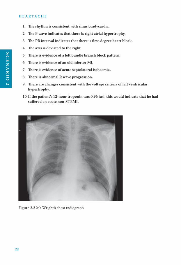
Figure 2.2 Mr Wright’s chest radiograph
H E A R T A C H E
SC
EN
AR
IO 2
23
1 �ere is evidence of left atrial hypertrophy.
2 �e cardiothoracic ratio is increased.
3 �ere is calcification of the aortic valve.
4 �ere is evidence of a right pleural effusion.
5 �e left hilum is abnormal.
6 �e thoracic aorta is ‘unfolded’.
7 �ere is fluid in the horizontal fissure.
8 �ere are multiple Kerley B lines.
9 �ere is evidence of asbestos exposure.
10 �is chest radiograph is consistent with biventricular failure.
Station 4
Prescribing skills 10-minute station Mr Wright is admitted to the CCU awaiting his 12-hour troponin. Using the charts
and the BNF provided please write up Mr Wright’s regular medications
and the hospital ACS protocol.
Details
Mr James Wright; DOB: 14/04/1964; Ward: CCU; Consultant: Dr Agrawal;
Hospital No.: 6172099; Weight: 98 kg; U&Es: normal; RBG: 6.7 mmol/;
Total cholesterol: 8.9 mmol/l
Regular medications
Aspirin 75 mg od; Bisoprolol 5 mg od; GTN spray 2 puffs PRN
ACS protocol
IV access
Cardiac monitor
Oxygen via nasal cannulae or mask (maintain sats at 96–98%)
To be given after ECG confirmation of diagnosis:
Aspirin 300 mg stat (in this case already given); followed by aspirin 75 mg od
(lifelong)
Clopidogrel 300 mg (in this case already given); followed by clopidogrel 75 mg od
(1 year)
Fondaparinux 2.5 mg sc – stat; followed by fondaparinux 2.5 mg od for 48 hours
Lansoprazole 30 mg od

H E A R T A C H E
24
SC
EN
AR
IO
2
Lipids: total cholesterol >6.0 mmol/l; atorvastatin 20 mg od
Blood glucose: start insulin sliding scale if RBG >11 mmol/l
If ongoing or severe pain:
Diamorphine 2.5–5 mg IV with metoclopramide 10 mg IV – stat
Consider addition of enoxaparin, bivalirudin or glycoprotein inhibitor
Discuss with cardiologist regarding transfer for PCI
To write up on PRN side
Diamorphine 2.5–5 mg IV (only)
Metoclopramide 10 mg IV (only)
GTN spray 2 puffs sl
Paracetamol 1 g max (po) qds
Remember: DRUG DRs Don’t Forget Signing O! (page 373)
Station 5
Communication skills 10-minute station You are the FY1 on the cardiology firm under which Mr Wright has been admitted.
"e Nursing Sister on the CCU is concerned that Mr Wright shows little or no
understanding of his medications and condition. She asks you to explain these
issues to him. Investigations have confirmed a troponin <0.05 iu/l, total cholesterol
8.9 mmol/l, RBG 6.7 mmol/l and BMI 31.6. Please explain the diagnosis, possible
further management and the medications that have been prescribed.

H E A R T A C H E
SC
EN
AR
IO 2
25
Answers
Station 1 – History
Patient script
You are Mr James Wright, 47 years old, DOB 14 April 1964. You have been
previously fit and well but ‘never see a doctor and don’t really look after yourself ’.
Over the past 3–4 months you have been suffering with increasingly frequent and
severe episodes of chest pain. Initially you thought the pains were ‘muscle strains’
but they have got slowly worse. Initially the pains lasted less than 20–30 seconds
at a time and came on once or twice a week. �ey would take your breath away
sometimes but were never prolonged and were always when you were exerting
yourself, eg lifting heavy boxes in the factory. More recently the pains have been
lasting for up to 2–3 minutes and come on when you are walking in the park with
your dog. �ey have never come on at rest and have never been prolonged. You get
pains nearly every day – hence the urgent appointment that your wife made for
you today.
You have never had any nausea, vomiting, sweats, clamminess, loss of
consciousness or dizziness, but the pains often make you feel ‘unwell’ and you
have to sit on a park bench or a low wall for a few minutes until they go away.
Cardiovascular risk factors: your grandfather and father both died in their 60s of
‘heart problems’, and your older brother had a ‘heart by-pass’ operation last year
aged 49 years. You smoke 40–50 cigarettes a day and have done so for many years.
You often drink 15–20 pints at the weekend but little during the week. You had a
blood pressure check several years ago at a local supermarket health promotion
day; you were told it was 150 over something but never got it checked again. You
do not know about your cholesterol and have never had symptoms suggestive of a
stroke, peripheral vascular disease or ischaemic heart disease (IHD).
Medications: you take ‘handfuls’ of indigestion tablets, including Zantac , and
occasional paracetamol but no other regular medications; you have no known
allergies.
SHx: you are married with three children, aged 7, 11 and 14 years. You are the
foreman in a large textile factory and are responsible for the day-to-day running
of the entire shop floor. �is is very stressful. You bought a large house at the
end of 2008, but have subsequently had to take a pay cut and have been left with
mounting debts.

H E A R T A C H E
26
SC
EN
AR
IO
2
Ideas and concerns: you think you may have heart problems, as these are the same
symptoms as your father and brother described in the past. You hope you have not
had a heart attack over the last few weeks.
Expectations: you are hoping the doctor can confirm or refute your worries and if
it is a heart problem ‘start you on the right medicines’. You suppose you’ll have to
lose some weight and stop smoking as well.
CONTENT
Identi es key information
Pain – chronological progression, onset, frequency, duration, character, radiation,
relieving and exacerbating factors
Associated features – shortness of breath, nausea and vomiting, dizziness, presyncope
and syncope, feeling unwell, washed out, palpitations
Identi es important negatives, including systemic enquiry
No radiation to the back (thoracic aortic aneurysm)
No features of heart failure – peripheral oedema, orthopnoea, PND, shortness of breath,
wheeze, dry cough, frothy white sputum
No features suggestive of gastrointestinal or hepatobiliary disease
Excludes other systemic symptoms
Identi es key information from rest of the history
Cardiovascular risk – FHx, known IHD, stroke or PVD, diabetes mellitus, hypertension,
smoking, alcohol, others
Relevant facts about employment, housing, social support, life stressors
Completing the patient history
Drug and allergic history: Zantac and indigestion tablets, occasional paracetamol
Allergies: NKDA
Previous medical history: nil known
Social and occupational history: as above
Summarises important areas of the history back to the patient
Invites patient to ask questions and is able to deal with them appropriately
Establishes patient’s ideas, concerns and expectations
A B C D E
PROFESSIONALISM
A B C D E
A B C D E
PROCESS
A B C D E
COMMUNICATION
H E A R T A C H E
SC
EN
AR
IO 2
27
DIAGNOSTIC REASONING
Please present your history
Candidate o�ers a logical, well-structured account of the history
What is your diagnosis?
Candidate o�ers the correct diagnosis and appropriate di�erentials
Diagnosis: unstable angina/IHD – the patient has not su�ered a prolonged or severe
episode in recent days, excluding a diagnosis of ACS or MI
Di�erentials: although the usual di�erentials may be o�ered, they are unlikely in this very
clear-cut history, eg basal pneumonia, upper abdominal causes (gallstones, peptic ulcer
disease)
If you were the GP what immediate therapeutic management would you initiate?
Include aspirin, GTN spray, anti-anginal, eg blocker
What lifestyle changes would you recommend?
Weight loss, exercise, smoking cessation, reduced alcohol intake, address life stressors
(job, money worries)
What other interventions might you consider?
Referral for further investigation, lipids assessment
Demonstrates safe, sensible and appropriate management plan
Demonstrates clear and logical diagnostic reasoning
GLOBAL HISTORY MARK
A B C D E
Station 2– Examination
Patient script
If you are an actor/patient, read the patient history and physical signs fully – when
the candidate comes to an abnormal site in their examination, act out tenderness
and/or volunteer the relevant physical sign.
A B C D E

H E A R T A C H E
28
SC
EN
AR
IO
2
CONTENT
Exposes and positions patient correctly and maintains comfort
Comments on wellbeing of patient, ie well or unwell
‘Feet to face’
Observes, and comments on patient and surroundings from foot of bed – evidence of
previous cardiac surgery, eg sternotomy, JVP, anaemia, colour/perfusion.
Asks for appropriate/relevant clinical observations
BP 164/98 mmHg, RR 14 bpm, O2 sats 97% on air, temperature 36.5 °C, CBG 6.8 mmol/l,
Urinalysis: WCC nil, protein negative, blood negative, nitrites negative, glucose +
Height 1.80 m, weight 98 kg, BMI 98/(1.8)2 = 30.3 kg/m2.
General/systemic examination
Hands and upper limbs: tar staining, perfusion of hands, anaemia, stigmata of
hyperlipidaemia; comments on general signs, eg clubbing, leukonychai
Face and neck: including signs of anaemia, peri-orbital xanthelasma, central cyanosis.
Focused examination
Inspection:
Sternotomy scar, JVP – makes appropriate assessment including correct positioning of
patient, correct technique; comments correctly on JVP.
Palpation:
Carotid pulse – comments on character and presence of bruits
Apex beat – position and character
Assesses and comments on heaves and thrills
Auscultation: listens in correct areas, assesses for radiation, manoeuvres patient
correctly, appropriate use of stethoscope – bell and diaphragm.
Completes examination by identifying relevant additional clinical signs and formally
completing assessment
Signs of left and right heart failure, including bibasal crackles, pleural e!usions,
peripheral oedema; hepatomegaly/ascites
Signs of PVD and generalised atherosclerosis, including AAA, peripheral pulses and
abdominal bruits.
Thanks patient, o!ers assistance, maintains patient’s dignity and privacy until they are
dressed
A B C D E
PROFESSIONALISM
A B C D E
A B C D E
PROCESS
A B C D E
COMMUNICATION

H E A R T A C H E
SC
EN
AR
IO 2
29
DIAGNOSTIC REASONING
Correctly identi!es the relevant physical signs, including important negative !ndings
Demonstrates safe, sensible and appropriate management plan, including
Anti-platelet treatment
Anti-anginals
Lipid-lowering agents, primarily statins
Anti-hypertensives: given his age, race and new-onset IHD, ACEI or blockade should be
considered
Nicotine replacement therapy.
Demonstrates clear and logical diagnostic reasoning
GLOBAL EXAMINATION MARK
A B C D E
Station 3 – Data interpretation1 TRUE – the rate is 300/6 = 50 bpm
2 FALSE – the P wave is slightly notched but is less than 3 mm broad; this is more
suggestive of left atrial compared with right atrial enlargement; if broader this would
be consistent with P mitrale
3 TRUE – the PR interval is 5 mm; borderline !rst-degree heart block
4 FALSE – there is marked left axis deviation (LAD); lead I is positive, lead II is isoelectric,
lead III is negative (ie leads I and III are pointing away from one another = ‘leaving’ one
another = LAD)
5 FALSE – the QRS complex has a normal width (<3 mm) and does not demonstrate any
features suggestive of LBBB
6 FALSE – there are no features suggestive of an old MI, eg Q waves
7 TRUE – there are inverted Q waves from V2 to V6, extending up to a VL
8 TRUE – the R wave should be small in amplitude in V1 and increase in size across the
chest leads to V4 or V5, then drop away slightly to V6; there are relatively tall R waves
from V2 to V5, with equal amplitude
9 FALSE – there are no voltage criteria suggestive of the LVH
10 TRUE – patients presenting with features of ACS are divided !rst by their ECG
!ndings STEMI vs non-STEMI or unstable angina; non-STEMI and unstable angina are
subsequently subdivided by the 12-hour troponin level; a positive result (in context)
is suggestive of a non-STEMI; cardiac (tachyarrhythmias) and non-cardiac pathologies
(chronic renal impairment or PE) may also increase the troponin level
A B C D E

H E A R T A C H E
30
SC
EN
AR
IO
2
GLOBAL DATA INTERPRETATION MARK
A B C D E
Figure 2.2 is a normal CXT
When assessing a CXR, as with all images, it is important to have a structured approach, considering
each aspect in turn, and to know the normal appearance. Always start by checking that the patient
details are correct for your patient. Is it PA (posteroroanterior – X-ray plate against the chest) or AP
– in the latter the heart appears magni!ed, and technically, one should not comment on the cardiac
size. Is there any rotation – in a well-centred !lm the clavicles are horizontal, directly opposing one
another, clearly seen along their entire length, and equally placed on either side of the vertebral
column, which should be perpendicular to them. If the !lm is under-penetrated (the focus of the
X-rays is in front of the plate), the heart and lung markings appear very dense and the vertebrae are
not seen. In over-penetrated views (X rays focussed behind the plate), lung markings and vertebrae
become very prominent (occasionally, used to give lesions in the lung more de!nition).
The left heart border is made up (from superior to inferior) of the aortic knuckle, the left pulmonary
artery, the left atrial appendage and the left ventricle. It is intimately related to the lingual lobe, a
part of the upper lobe, of the left lung - with consolidation of the lingual lobe, the left heart border
becomes hazy and di"cult to de!ne. The right heart border is made up of the superior vena cava
and the right atrium. It is intimately related to the right middle lobe and consolidation within this
lobe causes loss of de!nition of this border. The heart size on a PA !lm should be less than half of
the thoracic cavity at its widest point, ie this cardiothoracic ratio (CTR) should be < 0.50.
The upper lobe of the left lung (incorporating the lingual lobe) lies anteriorly and the lower lobe
posteriorly. The right lung has upper, middle and lower lobes; the horizontal !ssure becomes
visible on X-ray when #uid !lled. The lungs are both divided radiologically into the upper zone
(incorporates the apices and extends from the apex to the 2nd anterior rib), midzone (extends from
the 2nd to 4th anterior ribs) and lower zone (extends from the 4th to the 6th anterior ribs).
Both costodiaphragmatic angles must be included on a CXR: if not, it should be repeated before
commenting on the lungs. The right hemidiaphragm should lie 1–2 cm higher than the left.
Blunting or loss of an angle implies an e$usion. The left hemidiaphragm usually has an underlying
gastric bubble: do not confuse this with a pneumoperitoneum caused by perforation of a viscus.
Two features must be present to diagnose hyperexpansion of the lung !elds: (a) there should be
no more than six anterior ribs seen on a PA !lm (unless a deep inspiration has been taken). The ribs,
in a hyperextended view, often look ‘#attened’ or very horizontal, as do the hemidiaphragms, (b) the
precise way of establishing hyperexpansion is to draw a line between the costodiaphragmatic angle
and the cardiodiaphragmatic angle. In the normal CXR, a perpendicular drawn from the mid-point
of the hemidiaphragm to the original line should be 1.0 cm or more: if less, the hemidiaphragms
are ‘#attened’ and the lung !elds hyperexpanded.
Comment on whether the ribs and other bones look normal or osteopenic, and whether there
is evidence of arthritis, fractures or other abnormality. The rib spacing is greatly reduced with
underlying lung collapse and is termed ‘crowding’. Check the soft tissues for the breast, and other
shadows, air (surgical emphysema) and calci!cation.

H E A R T A C H E
SC
EN
AR
IO 2
31
Station 4 – Prescribing skills
Check: DRUG DRs Don’t Forget Signing O! (page 373)
Allergies, sensitivities and adverse drug reactions Patient details/addressograph
No known allergies Initials AF 007 Gender M / F NHS/ Hospital No: 6172099
Not possible to ascertain Date 21.11.11 Weight (kg) Date
Medicine/substance Reaction & Severity Initials & Date 98 Surname: WRIGHT
HeightFirst name: JAMES
1.80mAlerts Surface area (m2)
Date of birth: 14.04.64
IN-PATIENT MEDICATION PRESCRIPTION AND ADMINISTRATION RECORD
Consultant
AGRAWALTrainee Dr. Name and Bleep no.
FEATHER 007Date of admission
21.11.11Date chart reboarded Estimated date of discharge
This chart is no.
.............................. of ................................
Transcribing Check by Pharmacy
Sign ....................... Date .................
Ward
HDU1. ........................................................................... 2. ...........................................................................
Supplementary Medication charts in use: Other (please specify): 1 .................................................................. 2 .................................................................
Epidural/PCA Syringe driver TPN Chemotherapy Insulin sliding scale
Once only medications – loading doses, pre-medication, PGDs or surgical antibiotic propylaxis
Date Time to
be given
Medicine (approved name) Dose Route Signature and
bleep no.
Pharmacy Time
given
Given by Checked by
21.11 18.00 FONDAPARINUX 2.5mg SC AF 007
H E A R T A C H E
32
SC
EN
AR
IO
2
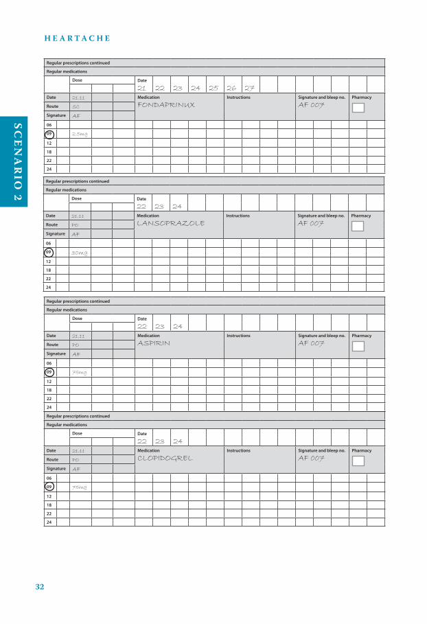
Regular prescriptions continued
Regular medications
Dose Date
21 22 23 24 25 26 27Date 21.11 Medication
FONDAPRINUXInstructions Signature and bleep no.
AF 007Pharmacy
Route SC
Signature AF06
09 2.5mg
12
18
22
24
Regular prescriptions continued
Regular medications
Dose Date
22 23 24Date 21.11 Medication
LANSOPRAZOLEInstructions Signature and bleep no.
AF 007Pharmacy
Route PO
Signature AF06
09 30mg12
18
22
24
Regular prescriptions continued
Regular medications
Dose Date
22 23 24Date 21.11 Medication
ASPIRINInstructions Signature and bleep no.
AF 007Pharmacy
Route PO
Signature AF06
09 75mg
12
18
22
24
Regular prescriptions continued
Regular medications
Dose Date
22 23 24Date 21.11 Medication
CLOPIDOGRELInstructions Signature and bleep no.
AF 007Pharmacy
Route PO
Signature AF06
09 75mg
12
18
22
24

H E A R T A C H E
SC
EN
AR
IO 2
33
Regular prescriptions continued
Regular medications
Dose Date
22 23 24Date 21.11 Medication
BISOPROLOLInstructions Signature and bleep no.
AF 007Pharmacy
Route PO
Signature AF06
09 5mg
12
18
22
24
Regular prescriptions continued
Regular medications
Dose Date
22 23 24Date 21.11 Medication
ATORVASTATINInstructions Signature and bleep no.
AF 007Pharmacy
Route PO
Signature AF06
09 20mg
12
18
22
24
Regular prescriptions
Oral anticoagulation follow the anticoagulation guidelines available on the intranet
Indication Target INR Baseline INR
(if applica-
ble)
Duration of
therapy
Date of anticoagulation
follow-up appointment
(clinic or other)*
Anticoagulant
record book given
or updated. Sign
and date
Date patient coun-
selled and sign
Date therapy
started
* A follow-up appointment must be booked with the anti-coagulant clinic or enhanced provider of primary care services. If not, the TTA will not be dispensed
Initiating warfarin Perform baseline coagulation screen,
LFTs, U&Es and FBC
Prescribe initiation dose as
per guidelines
CHECK INR ON DAY 3 FOLLOW DOSING ALGORITHM IN
GUIDELINE
Continuing warfarin Maintenance therapy FOLLOW MAINTENANCE DOSING ALGORITHM IN GUIDELINE
Do not use the initiation protocol for patients already on warfarin. More frequent INR monitoring may be required for patients on interacting drug(s)
Medication
1
Date
INR
Route Frequency
OD
Time
18.00
Start Dose
Stop Dr sign
Signature Bleep
no.
Phar-
macy
Given
Initiating warfarin – Reduced dosing regimen in red. Refer to anticoagulation policy
Day One Two Three Four and above
INR <1.4 No
test
<2.0 2.0-
2.1
2.2-
2.5
2.6-
2.9
3.0-
3.3
3.4-
4.0
>4.0 <1.4 1.4-
1.5
1.6-
1.7
1.8-
1.9
2.0-
2.3
2.4-
3.0
3.1-
4.0
4.1-
4.5
>4.5
Dose
mg
10 5 10 5 10 5 4 3 2 1 0 9 8 7 6 5 4 3 Miss
1
day
Miss
2
day

H E A R T A C H E
34
SC
EN
AR
IO
2
Thromboprophylaxis please prescribe treatment regimens in the regular medications section
Choice of mechanical prophylaxis and leg(s) to be applied to Enter
Time
Enter details below
Graduated elastic
compression
stockings
Intermittend pneumatic
compression device
(IPC)
Leg
Left Right Both
21/11 Start
Date:
End
Date:
Signature
and Bleep
No.
Start
Date:
End
Date:
Signature
and Bleep
No.
Medication Dose Dose Change Enter
Time
Enter details below
Please ensure
you have
completed the
VTE risk assess-
ment form
Date
Route
Signature Instructions
ON ACS PROTOCOLPharmacy
Bleep no.
Oxygen
Target Saturation 88-92% 94/98% If oxygen saturation falls below target range on prescribed oxygen, patient needs urgent clinical review.
If oxygen saturation is above targent range on prescribed oxygen, ask for review.
Other specify) *Device: N= nasal cannula, SM = simple face mask, V = venturi,
H = humidi!ed, RM = reservoir mask, OTHER = other eg.
NCPAP/NIPPV
Pharmacy
Target Saturation not applicable
Date Changed Date Changed Enter
Time
Enter details below
21/11
DeviceNASAL
CANNULAE
% or L/min (specify a
range eg 1-21 L/min)
1-2L/MIN
Signature and Bleep no. AF
As required medications
Medication
MORPHINEDate
Indication Time
Dose
25-5Mg
Route
IVMaximum
frequency /
dose
as reqd.
Start date
21/11Dose
Stop date Route
Signature
AFBleep no.
007Given
Additional instructions: IV ONLY Pharmacy

H E A R T A C H E
SC
EN
AR
IO 2
35
As required medications
Medication
PARACETAMOLDate
Indication Time
Dose
1gRoute
POMaximum
frequency /
dose
6 hour
Start date
21/11Dose
Stop date Route
Signature
AF Bleep no.
007Given
Additional instructions: Pharmacy
As required medications
Medication
GTN SPRAYDate
Indication Time
Dose
1-2PUFF
Route
S/LMaximum
frequency /
dose
Start date
21/11Dose
Stop date Route
Signature
AF Bleep no.
007Given
Additional instructions: Pharmacy
As required medications
Medication
METOCLOPRAMIDEDate
Indication Time
Dose
10 mg
Route
IVMaximum
frequency /
dose
8 hour
Start date
21/11Dose
Stop date Route
Signature
AF Bleep no.
007Given
Additional instructions: Pharmacy
GLOBAL PRESCRIBING MARK
A B C D E
H E A R T A C H E
36
SC
EN
AR
IO
2
Station 5 – Communication skills
CONTENT
Investigates patient’s present level of understanding of scenario
Asks what, and how much, information they would like to be told
Summarises and con!rms what has happened so far (may be covered above)Patient went to the GP and was told he had a heart disease; he was prescribed medicines but didn’t pick them up (may challenge patient as to why)Admitted a few hours ago from the Emergency Department after prolonged period of chest painTests have shown that cholesterol is raised but heart muscle test (troponin) is negative; ECG abnormal but no acute changes suggesting a heart attack.
Establishes patient’s ideas, concerns and expectations
Explains key important information
Medications: actions, frequency, side-e"ectsAspirin and clopidogrel – anti-platelet; stop blood clots forming in the blood vessels of the heart; take one each daily; SEs – indigestion, abdominal pain and bruising or bleedingBisoprolol – blocker – helps the heart pump and lowers the blood pressure; take one daily; may cause low blood pressure and dizziness, erectile impotence, slow heart beatGTN spray – to spray under the tongue when get pain; SEs – headache and dizzinessAtorvastatin – lowers blood cholesterol; SEs – jaundice and liver problems, muscle aches and painsLansoprazole – proton pump inhibitor; stops acid secretion in the stomach, which in turn stops ulcers and in!ammation in the stomach and small bowel; SEs – may cause upper and lower GI disturbance.
Lifestyle changesWeight loss – will need to see dietitian; low-fat/low-salt dietSmoking cessation – can be booked into hospital or community programmes; may involve nicotine replacement therapyAlcohol – cut down to safe limits (21 units per week)Help with money issues and stressFurther investigations‘Treadmill’ test today; explains procedure and potential outcomesIf inconclusive – may have other test, eg MRA or thallium scanPotential may require PCI – explains purpose, process and potential outcomes, eg angioplasty, stenting and possible CABG.
Invites patient to ask questions as they proceed through the consultation and is able to deal with them appropriately
Summarises important areas of the consultation back to the patient
Formally ends the consultation and ensures appropriate follow-up has been discussed
A B C D E
PROFESSIONALISM
A B C D E
A B C D E
PROCESS

H E A R T A C H E
SC
EN
AR
IO 2
37
DIAGNOSTIC REASONING
What questions might you ask a patient before asking them to do an exercise tolerance
test?
Do they understand and want the test?
Do they understand the possible implications of a positive test?
Are they able to walk and run?
What is their normal exercise tolerance?
Do they have any contraindications to the test, eg severe aortic valve disease, recent MI.
What are the contraindications to a patient having an MRA scan?
Absolute: metallic implants/prostheses of any kind, unable to lie !at because of severe
respiratory or cardiac disease
Relative: claustrophobia.
Demonstrates safe, sensible and appropriate management plan
Demonstrates clear and logical diagnostic reasoning
GLOBAL COMMUNICATION MARK
A B C D E
Scenario 2: Reflection and consolidation
History
Mr Wright is a 46-year-old man who presents today with features suggestive of unstable angina. Of note he
is a life-long smoker, has untreated hypertension and has a very strong family history of IHD and premature
death. He now presents with a 3- to 4-month history of angina-like chest pain. Initially the pains lasted 20–30
seconds and came on once or twice week. They were always associated with exertion such as lifting heavy
objects at work, but the pains were never at rest or prolonged and there were no associated features of note.
More recently the pains have become more frequent – now once a day – and last for a few minutes. They
often come on when he is walking his dog in the park but are always relieved with sitting and resting. No
other signi"cant cardiovascular (eg suggestive of heart failure), respiratory or systemic symptoms.
The patient’s cardiovascular risk factors include:
His brother has just had CABG surgery aged 49 years and his father and grandfather died in their 60s
of IHD.
He is a known heavy smoker – 40–50 cigarettes per day for many years.
He has hypertension – his blood pressure was measured at 150/something at a local health
promotion day, 2 years ago, but he never sought treatment.
He has a very stressful job, and has signi"cant "nancial worries at the present time.
Other risk factors are negative or unknown.
A B C D E
H E A R T A C H E
38
SC
EN
AR
IO
2
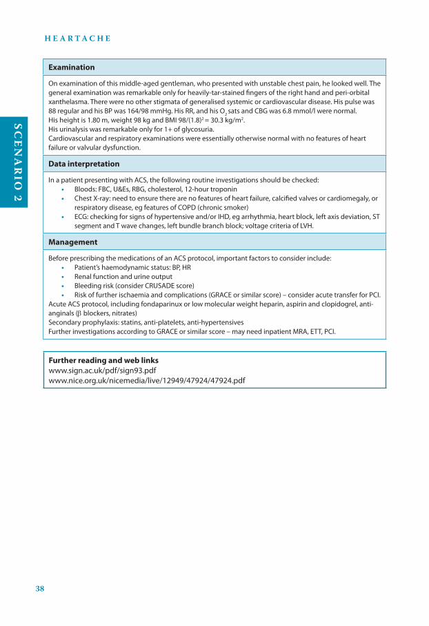
Examination
On examination of this middle-aged gentleman, who presented with unstable chest pain, he looked well. The
general examination was remarkable only for heavily-tar-stained �ngers of the right hand and peri-orbital
xanthelasma. There were no other stigmata of generalised systemic or cardiovascular disease. His pulse was
88 regular and his BP was 164/98 mmHg. His RR, and his O2 sats and CBG was 6.8 mmol/l were normal.
His height is 1.80 m, weight 98 kg and BMI 98/(1.8)2 = 30.3 kg/m2.
His urinalysis was remarkable only for 1+ of glycosuria.
Cardiovascular and respiratory examinations were essentially otherwise normal with no features of heart
failure or valvular dysfunction.
Data interpretation
In a patient presenting with ACS, the following routine investigations should be checked:
Bloods: FBC, U&Es, RBG, cholesterol, 12-hour troponin
Chest X-ray: need to ensure there are no features of heart failure, calci�ed valves or cardiomegaly, or
respiratory disease, eg features of COPD (chronic smoker)
ECG: checking for signs of hypertensive and/or IHD, eg arrhythmia, heart block, left axis deviation, ST
segment and T wave changes, left bundle branch block; voltage criteria of LVH.
Management
Before prescribing the medications of an ACS protocol, important factors to consider include:
Patient’s haemodynamic status: BP, HR
Renal function and urine output
Bleeding risk (consider CRUSADE score)
Risk of further ischaemia and complications (GRACE or similar score) – consider acute transfer for PCI.
Acute ACS protocol, including fondaparinux or low molecular weight heparin, aspirin and clopidogrel, anti-
anginals ( blockers, nitrates)
Secondary prophylaxis: statins, anti-platelets, anti-hypertensives
Further investigations according to GRACE or similar score – may need inpatient MRA, ETT, PCI.
Further reading and web links
www.sign.ac.uk/pdf/sign93.pdf
www.nice.org.uk/nicemedia/live/12949/47924/47924.pdf