osteotomies around the hip
TRANSCRIPT
Osteotomies Around Hip Joint
Dr.Ankit Dr.B.L.Chandrakar
DEFINITION
An osteotomy is a surgical corrective
procedure used to obtain a correct
biomechanical alignment of the extremity so
as to achieve equivocal load transmission,
performed with or without removal of a
portion of the bone.
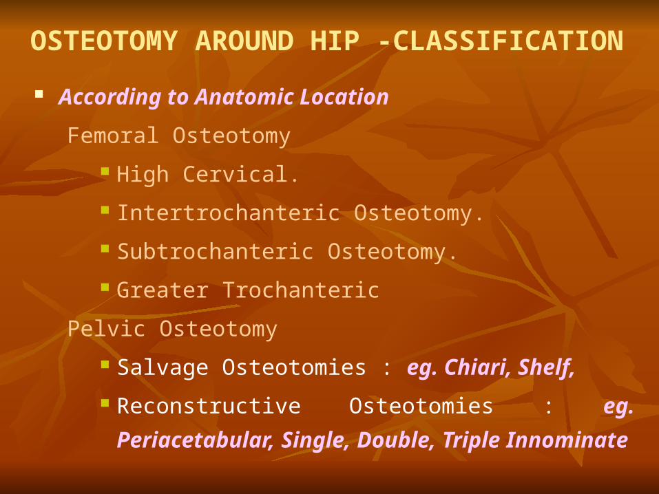
OSTEOTOMY AROUND HIP -CLASSIFICATION
According to Anatomic Location
Femoral Osteotomy
High Cervical.
Intertrochanteric Osteotomy.
Subtrochanteric Osteotomy.
Greater Trochanteric
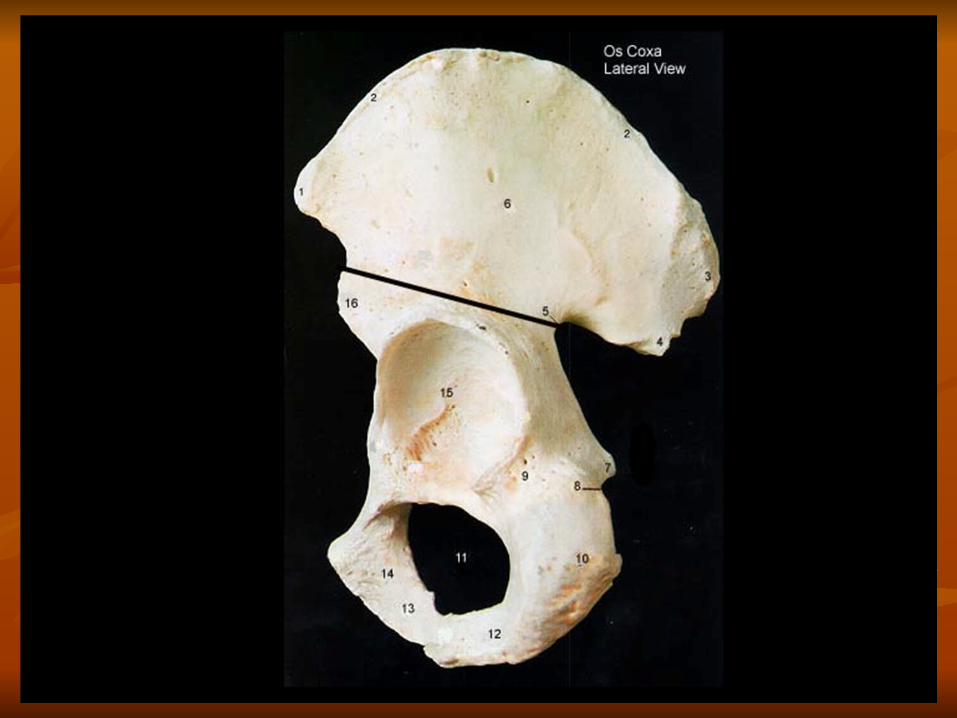
Pelvic Osteotomy
Salvage Osteotomies : eg. Chiari, Shelf,
Reconstructive Osteotomies : eg. Periacetabular, Single,
Double, Triple Innominate
Combined Osteotomy
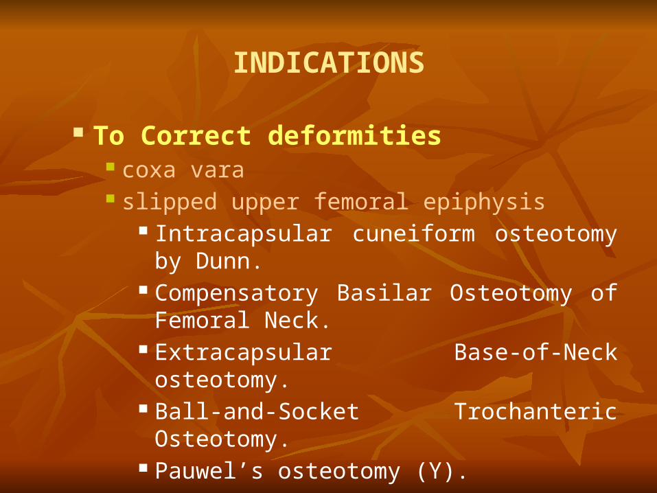
INDICATIONS
To Correct deformities coxa vara slipped upper femoral epiphysis
Intracapsular cuneiform osteotomy by Dunn. Compensatory Basilar Osteotomy of Femoral
Neck. Extracapsular Base-of-Neck osteotomy. Ball-and-Socket Trochanteric Osteotomy. Pauwel’s osteotomy (Y).
To obtain stability old unreduced dislocations.
Lorenz bifurcation osteotomy. Schanz low subtrochanteric.
Relief of pain osteoarathritis.
Pauwel’s type I varus osteotomy. Pauwel’s type II valgus osteotomy.
To obtain union Un-united fractures of femoral neck.
McMurry’s osteotomy. Dickson's high geometric osteotomy. Schanz Angulation Osteotomy.
unstable intertrochanteric fractures. Dimon Hughston Osteotomy. Sarmiento’s Osteotomy
In Osteonecrosis of femoral head
Sugioka’s transtrochanteric osteotomy.
Varus deroation osteotomy of Axer.
- In paralytic disorders of hip.
Varus Osteotomy.
Rotational Osteotomy

In congenital dislocation of hip
Salters innominate osteotomy Pembertons innominate ostetomy Steels triple innominate osteotomy Shelf operation Chiari’s osteotomy

OVERVIEW OF PELVIC OSTEOTOMY


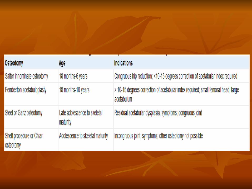
SALTER OSTEOTOMY INDICATIONS-Congruous hip reduction,<10-15 degrees
correction of acetabular index required ,paralytic
disorder,subluxation after septic arthritis PREREQUISITES- femoral head must be positioned opposite
the level of acetabulum,contracture of iliopsoas and adductor
muscles must be released, range of motion of the hip must be
good specially in abduction ,int rotation flexion AGE-18 months-6years AFTERCARE-hip spica for 8 to 12 wks,then partial weight
bearing on crutches ,followed by full weight bearing.result
assessed by center edge angle.

xrays
Salter osteotomy for congenital dislocation of hip. A, Residual acetabular dysplasia and subluxation of right hip in 4-year-old girl in whom open reduction had been performed at 9 months of age. B, One year after repeat open reduction and Salter innominate osteotomy.

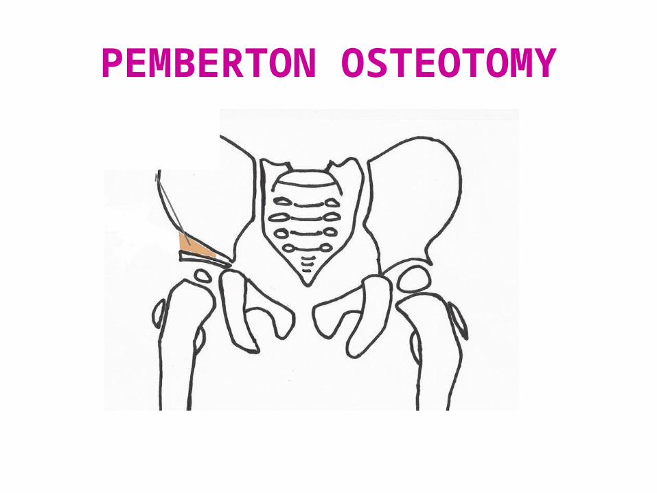
PEMBERTON OSTEOTOMY
PROCEDURE- Pemberton described a pericapsular
osteotomy of the ilium in which the osteotomy is
made through the full thickness of the bone from just
superior to the anteroinferior iliac spine anteriorly to
the triradiate cartilage posteriorly : the triradiate
cartilage acts as a hinge on which the acetabular roof
is rotated anteriorly and laterally.
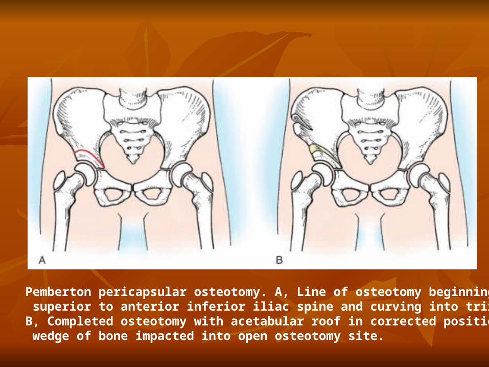
Pemberton pericapsular osteotomy. A, Line of osteotomy beginning slightly superior to anterior inferior iliac spine and curving into triradiate cartilage. B, Completed osteotomy with acetabular roof in corrected position and wedge of bone impacted into open osteotomy site.
PEMBERTON OSTEOTOMY
INDICATION- >10-15 degrees correction of acetabular index required ,small femoral head ,large acetabulum.
ADV- internal fixation not required .greater degree of rotation can be achieved with less rotation of acetabulum
DISADV- Technically more difficult . Alters the configuration and capacity of acetabulum and produces joint incongruity that requires remodelling
AGE-18months- 10 yr AFTERCARE-spica cast for 8 to 12 weeks
PEMBERTON PERICAPSULAR OSTEOTOMY

PERIACETABULAR OSTEOTOMY OF ILIUM (PEMBERTON)
Pemberton acetabuloplasty. A, Symptomatic residual acetabular dysplasia in 8-year-old girl after treatment of congenital dislocation of right hip. B, After Pemberton acetabuloplasty
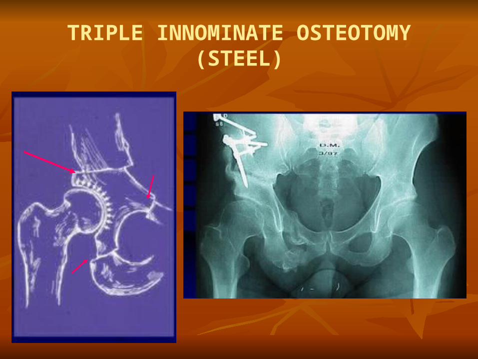
TRIPLE INNOMINATE OSTEOTOMY (STEEL)

STEEL OSTEOTOMY
The ischium, the superior pubic ramus and ilium superior to
the acetabulum are all divided and acetabulum is repositioned
and stabilized by bone graft and metal pins Objective- To establish a stable hip in anatomical position for
dislocation or subluxation of the hip in older children when
this is impossible by any one of the other osteotomies For the operation to be successful, the articular surfaces of the
joint must be congruous or become so when the acetabulum
has been redirected so that a functional, painless range of
motion is achieved and a Trendelenburg gait is absent
STEEL OSTEOTOMY
INDICATIONS-Adolescents and skeletally mature adults with
residual dysplasia and subluxation in whom remodelling of
acetabulum is no longer anticipated ADVANTAGES-Better coverage of femoral head by articular
cartilage. Better hip joint stability,no need of spica cast. DISADVANTAGES- Technically difficult, does not change
size of acetabulum, distorts the hip such that natural child birth
may be impossible in adulthood
STEEL OSTEOTOMY
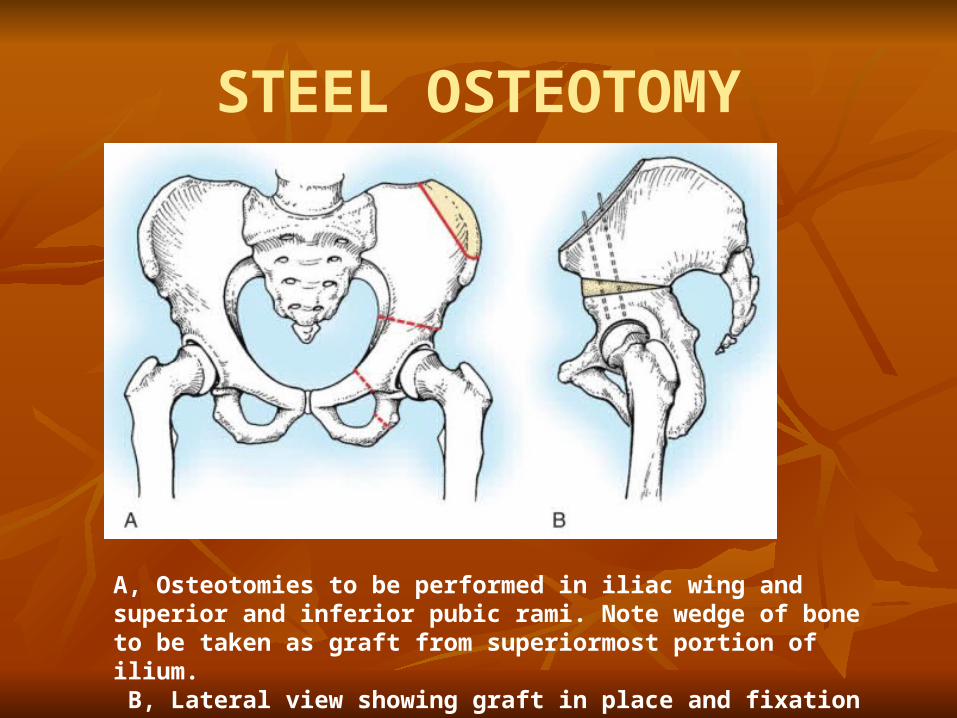
A, Osteotomies to be performed in iliac wing and superior and inferior pubic rami. Note wedge of bone to be taken as graft from superiormost portion of ilium. B, Lateral view showing graft in place and fixation with two Kirschner wires.
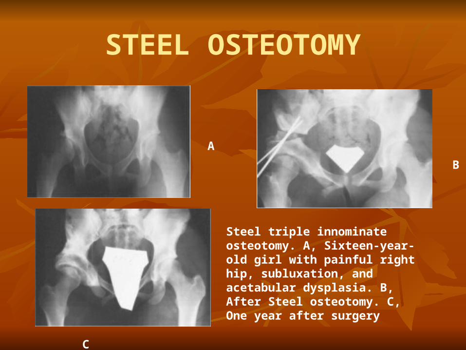
STEEL OSTEOTOMY
Steel triple innominate osteotomy. A, Sixteen-year-old girl with painful right hip, subluxation, and acetabular dysplasia. B, After Steel osteotomy. C, One year after surgery
A
B
C
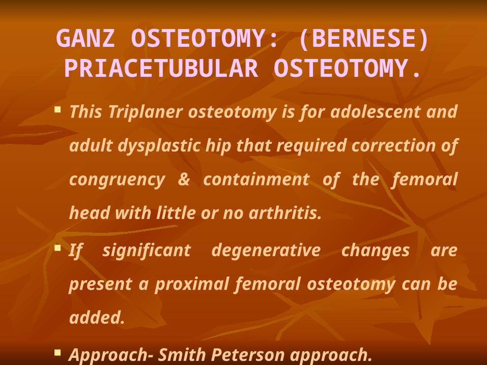
GANZ OSTEOTOMY: (BERNESE) PRIACETUBULAR OSTEOTOMY.
This Triplaner osteotomy is for adolescent and adult
dysplastic hip that required correction of congruency
& containment of the femoral head with little or no
arthritis.
If significant degenerative changes are present a
proximal femoral osteotomy can be added.
Approach- Smith Peterson approach.
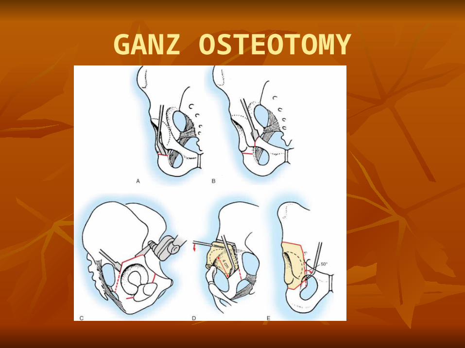
GANZ OSTEOTOMY

GANZ OSTEOTOMY

Advantages : Only one approach is used. A large amount of correction can be obtained in all
directions, including the medial and lateral planes. Blood supply to the acetabulum is preserved. The posterior column of the hemipelvis remains
mechanically intact, allowing immediate crutch walking
with minimal internal fixation. The shape of the true pelvis is unaltered, permitting a
normal child delivery. Can be combined with trochanteric osteotomy if needed.
Contd.
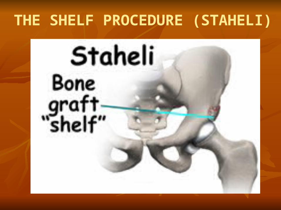
THE SHELF PROCEDURE (STAHELI)
SHELF OPERATION (STAHELI) Have commonly been performed to enlarge the volume of the
acetabulum. The objective is to create a shelf, the size of which is decided by
measuring the “width of augmentation” from the CE angle. The shelf is put just above the acetabular margin. It secures two layers of cancellous grafts bringing the reflected head of rectus femoris forward over the graft and suturing it in its original position.
Best to do after 5 years of age. Indication : A deficient acetabulum that cannot be corrected by
redirectional, osteotomy is the primary indication. Contraindication :
Dysplastic hip with spherical congruity suitable for redirectional osteotomy
Hip requiring open reduction.
The placement of the acetabular slot is the most critical part of the procedure; the slot must be created exactly at the acetabular margin
Approach- Iliofemoral approach using bikini incision parallel and 1cm below the Iliac crest

CENTER EDGE ANGLE/ACETABULAR INDEX
CE ANGLE-measured after 5 yr age, >25 normal, <20 severe dysplasia
AC IND- <27.5 normal, >30 dysplasia
CHIARI OSTEOTOMY PROCEDURE-It is performed at the superior margin of
the acetabulum and the pelvis inferior to the osteotomy
along with the femur is displaced medially.
This is also called as capsular interposition Arthroplasty
as the capsule is interposed between the shelf and the
femoral head.
INDICATIONS-incongruous joint, dysplastic hip with
osteoarthritis ,other osteotomy not possible
DISADVANTAGE-salvage osteotomy only, leaves
anterior acetabulum uncovered,abductor lurch common .

A, Line of osteotomy extending from immediately superior to lip of acetabulum into sciatic notch. Osteotomy can be curved to facilitate femoral head coverage. B, Completed osteotomy with medial displacement of distal fragment for interpositional capsular arthroplasty
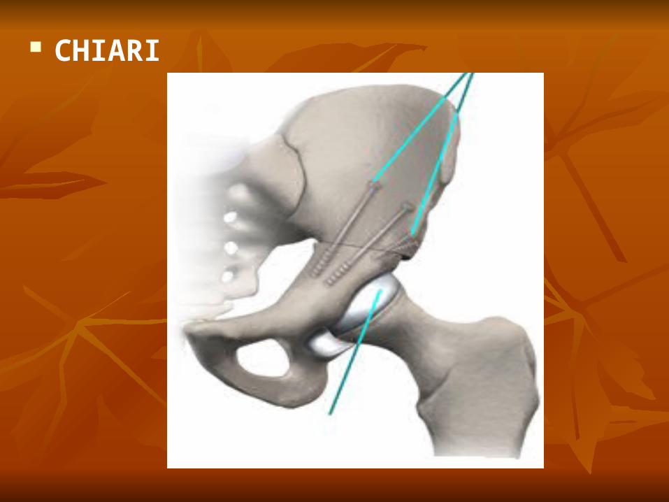
CHIARI

PALLIATIVE OPERATION
Reserve for cases is which reduction is not possible by
either open or closed reduction as in old unreduced
congenital dislocation.
Designed to improve :
Stability.
Decrease lordosis.
Control pain arising from lower back/hip.
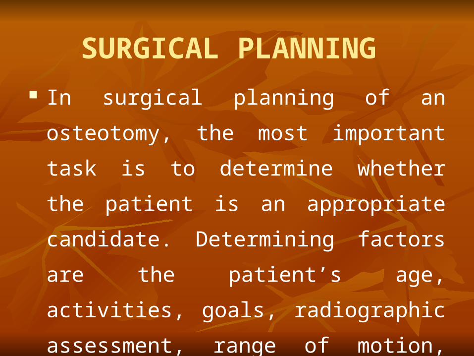
SURGICAL PLANNING
In surgical planning of an osteotomy, the most
important task is to determine whether the
patient is an appropriate candidate.
Determining factors are the patient’s age,
activities, goals, radiographic assessment,
range of motion, and leg lengths and the status
of the knee of same side.
FEMORAL OSTEOTOMY
Primary objective is deflection of wt. bearing by
angulation of femur to bring the axis of the femoral
shaft more in line with the direction of weight
transmission.
The osteotomy performed are Angulation
Osteotomy (Stabilizing osteotomy).
Schanz osteotomy.
Lorenz osteotomy.
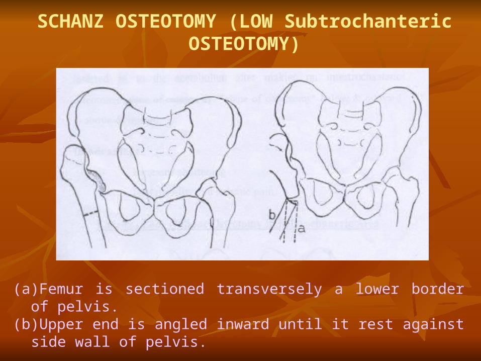
SCHANZ OSTEOTOMY (LOW Subtrochanteric OSTEOTOMY)
(a)Femur is sectioned transversely a lower border of pelvis.(b)Upper end is angled inward until it rest against side wall of pelvis.
Schanz osteotomy : In this osteotomy the deformity flexion, adduction &
external Rotation is corrected by making the osteotomy at tuber ischii level.
Preparation : X-ray are taken with full adduction – to measure
angle medially. Thomas Test - measure degree of flexion to be
corrected. Advantages :
Lurching gait will be diminished. The depression of the trochanter also improves the
leverage of the glutei.
Contraindication : Before 15 years of age, because loss
of angulation during growth period.
Lorenz (Bifurcation osteotomy)
In this upper end of the lower fragment is abducted and
inserted in to the acetabulum after making on
intertrochanteric osteotomy. “Plane of osteotomy” below
& outward to above & inward.
Disadvantage :
Increased shortening.
Less mobility and arthritic pain.
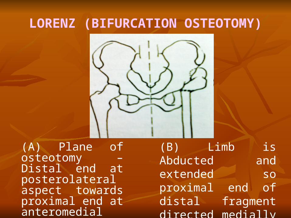
LORENZ (BIFURCATION OSTEOTOMY)
(A) Plane of osteotomy – Distal end at posterolateral aspect towards proximal end at anteromedial aspect.
(B) Limb is Abducted and extended so proximal end of distal fragment directed medially and anteriorly in acetabulum.

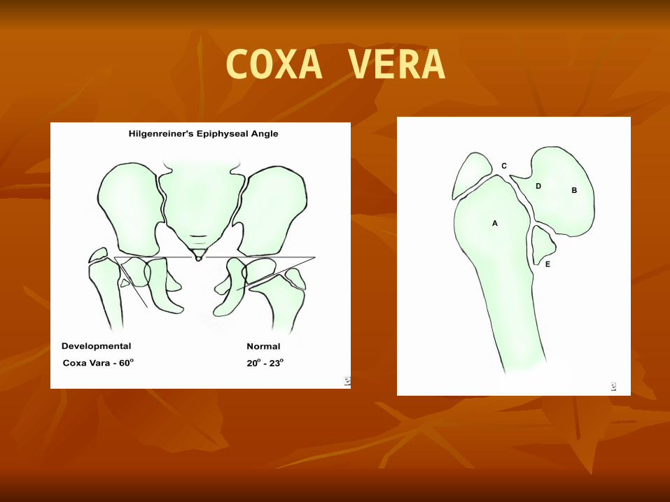
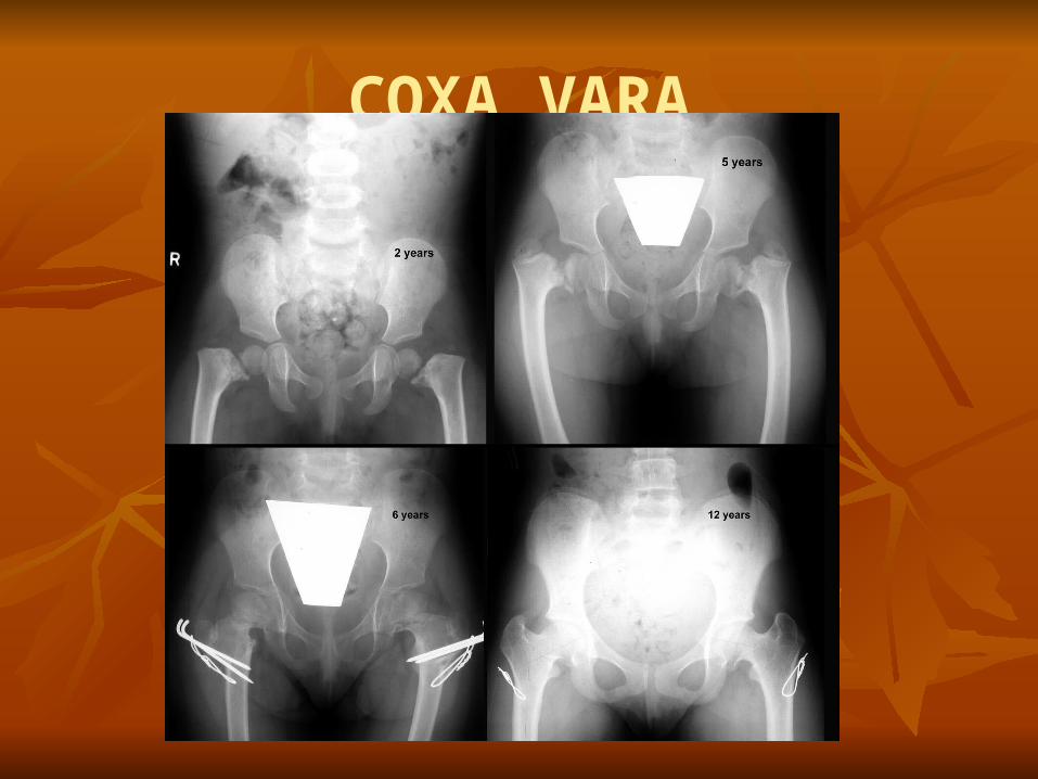
OSTEOTOMY FOR COXA VARA The normal femoral neck shaft angle in infant is 1200 to 1400,
Reduction to a more acute angle constitute a coxa vara deformity.
The goal of treatment are To promote ossification of the defect and correct varus
deformity. Indication for surgery :
Increasing coxa vara Neck shaft angle less than 110°. Painful unilateral or associated with leg length
discrepancy Hilgenreiner - epiphy seal angle of more than 60° .
Surgery performed are
Valgus Subtrochanteric Osteotomy or abduction
osteotomy-with Internal Fixation.
A transverse osteotomy at about the level of lesser
trochanter.
If necessary take a small lateral wedge to correct neck
shaft angle to 135-150.
The surgery may be delayed till child is 4 to 5 year old
to make internal fixation easier.
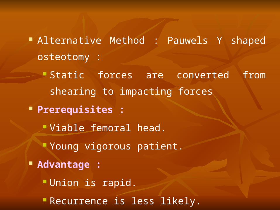
Alternative Method : Pauwels Y shaped osteotomy :
Static forces are converted from shearing to impacting
forces
Prerequisites :
Viable femoral head.
Young vigorous patient.
Advantage :
Union is rapid.
Recurrence is less likely.

PAUWELS Y SHAPED OSTEOTOMY
COXA VERA
COXA VARA
OSTEOTOMY FOR RELIEF OF PAIN IN OSTEOARTHRITIS
Before the onset of osteoarthritis, if normal or near normal
function of the hip can be maintained, reconstructive
osteotomy can prevent or delay the development of
osteoarthritis; if mild or moderate osteoarthritis is present, a
salvage osteotomy can improve function and delay the need
for total hip Arthroplasty.
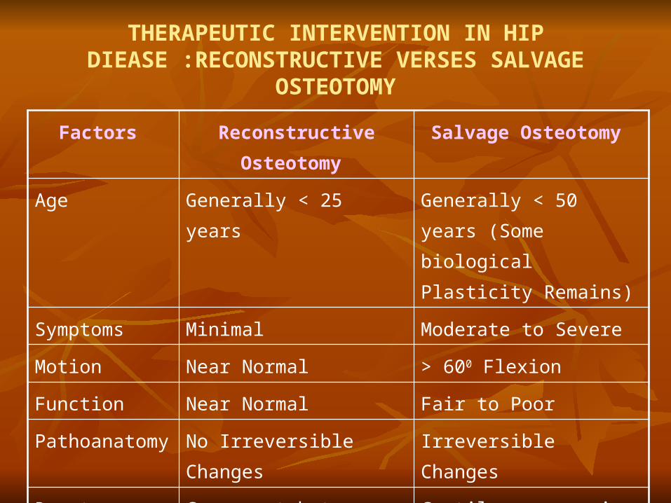
Factors Reconstructive Osteotomy Salvage Osteotomy
Age Generally < 25 years Generally < 50 years (Some biological Plasticity Remains)
Symptoms Minimal Moderate to Severe
Motion Near Normal > 600 Flexion
Function Near Normal Fair to Poor
Pathoanatomy No Irreversible Changes Irreversible Changes
Roentgenography Congruent but Malaligned Surfaces
Cartilage narrowing or incongruity or both
Prognosis if untreated
Poor Poor
THERAPEUTIC INTERVENTION IN HIP DIEASE :RECONSTRUCTIVE VERSES SALVAGE
OSTEOTOMY
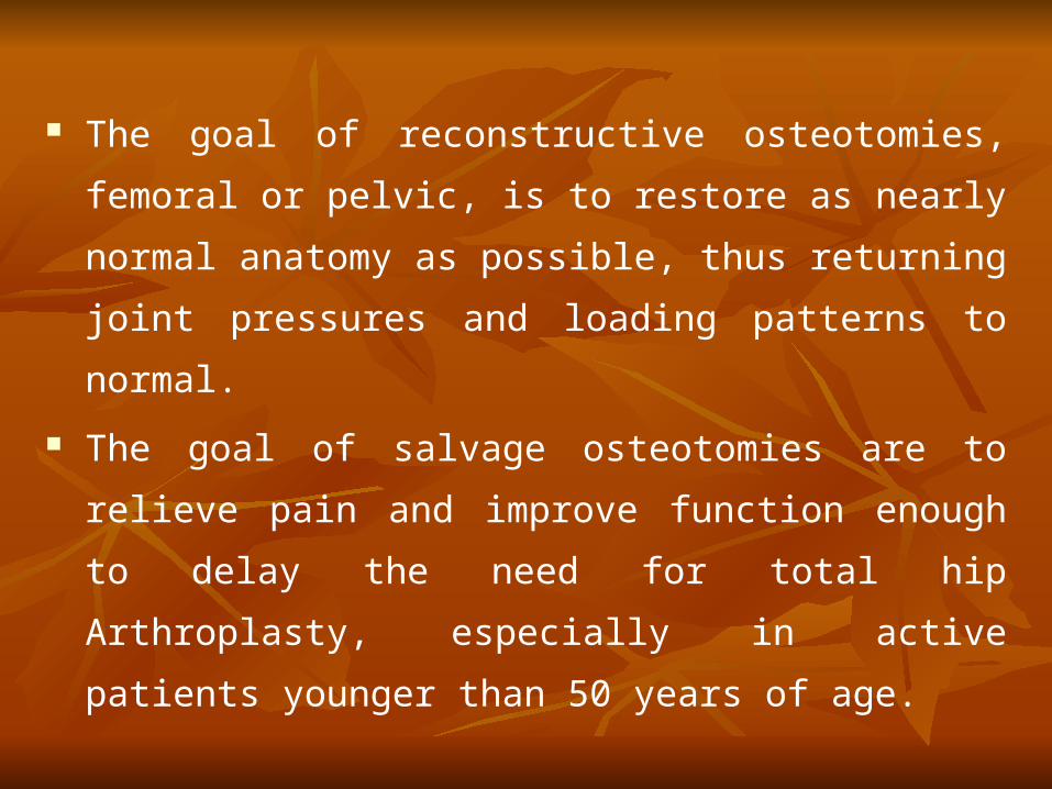
The goal of reconstructive osteotomies, femoral or pelvic, is to
restore as nearly normal anatomy as possible, thus returning
joint pressures and loading patterns to normal.
The goal of salvage osteotomies are to relieve pain and
improve function enough to delay the need for total hip
Arthroplasty, especially in active patients younger than 50
years of age.
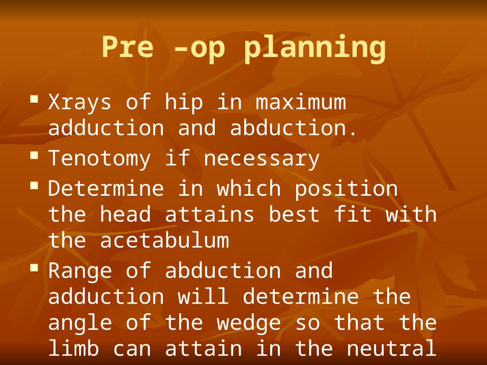
Pre –op planning
Xrays of hip in maximum adduction and abduction.
Tenotomy if necessary Determine in which position the head attains
best fit with the acetabulum Range of abduction and adduction will
determine the angle of the wedge so that the limb can attain in the neutral position

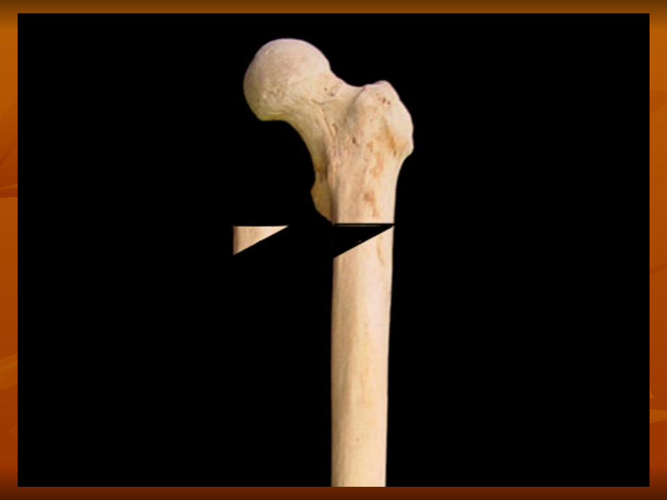
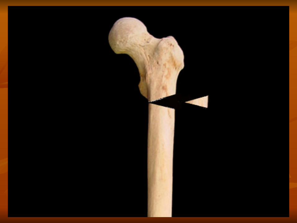
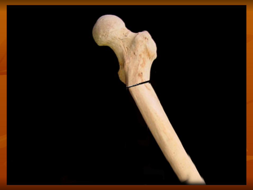
varus osteotomy :- Designed to elevate the greater trochanter and move it
laterally while moving the abductor and psoas muscles medially, to restore joint congruity and decrease muscle forces about the hip.
Indications- patients with a spherical femoral head, little or no acetabular dysplasia center-edge angle of at least 15 to 20 degrees),a valgus neck-shaft angle of more than 135 degrees,fixed abduction deformity.
C/I-fixed ext. rotation >25 deg,flexion of 70 deg Varus osteotomy with medial displacement of the femoral
shaft relaxes the abductor, psoas, and adductor muscles unloads the hip joint, and increases the weight-bearing surface.


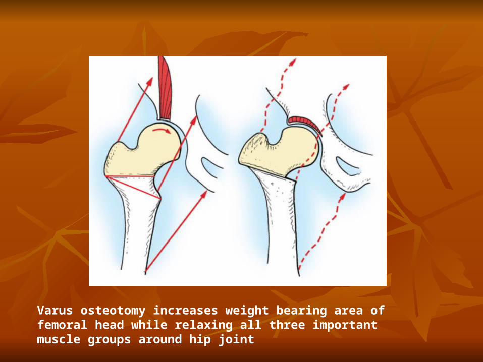
Varus osteotomy increases weight bearing area of femoral head while relaxing all three important muscle groups around hip joint

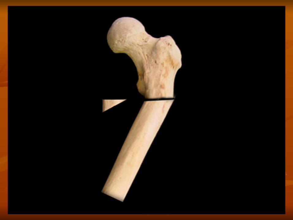
Three types of wedges cut for varus osteotomy. A, Original technique of Pauwels with proximal osteotomy made transversely at distal end of greater trochanter. This type of osteotomy makes it more difficult to correct rotation and to use right-angled blade plate. B, Original Müller technique of excision of wide wedge based medially with distal osteotomy cut transversely across shaft at just above level of lesser trochanter. C, Later technique of Müller using small half wedge cut medially and transposed laterally.
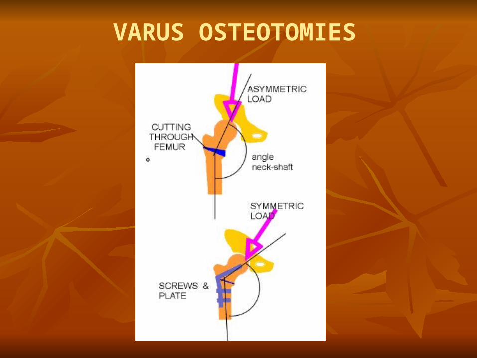
VARUS OSTEOTOMIES
Most authors recommend medial displacement of 10 to
15 mm to keep the ipsilateral knee centered under the
femoral head and to maintain the mechanical axis of the
leg.
Varus osteotomy, however, shortens the limb to some
degree. creates a Trendelenburg gait that may persist for
months after surgery, and increases the prominence of the
greater trochanter.
Limb shortening can be minimized by making a smaller
medial osteotomy and transposing it to the lateral side.


VALGUS INTERTROCHANTERIC FEMORAL OSTEOTOMIES
Valgus Osteotomy - Increase weight bearing area of femur head. It does not produce muscle relaxation. Relaxation obtained by tenotomy of Iliopsos and adductor
muscle. Transfer the center of hip rotation medially from the superior
aspect of the acetabulum to increase joint congruity and the weight-bearing area of the femoral head.
Osteotomy of the greater trochanter often is performed with valgus femoral osteotomy to move the greater trochanter laterally.

INDICATIONS : FIXED ADDUCTION DEFORMITY
CONTRAINDICATIONS : FLEXION OF LESS THAN 60 DEGREES, KNOCK KNEES

Best result were obtained in patients younger than 40 years of age with unilateral involvement, good preoperative range of motion, and a mechanical (secondary) cause.
Most surgeons now advise that all osteotomies be fixed with rigid internal fixation, which offers several obvious advantages: The fragments are maintained in proper position; The danger of limitation of motion of the hip and knee is
greatly decreased;
The patient can be allowed out of bed early; and
Pulmonary, urological, and other medical complications
are decreased. A device frequently used for rigid internal
fixation of intertrochanteric osteotomies is the ASIF, or
right-angled, blade plate.
Nonunion has been a troublesome complication after
Osteotomy, and an incidence as high as 20% has been
reported.
BIOMECHANICAL TREATMENT OF OSTEOARTHRITIS
Therapy must be directed at reducing joint loads. This may
be by reducing the compressive forces directly or by
increasing the weight- bearing area, and thereby reducing
the load per unit area or ideally by combination of the two.
WHILE PERFORMING OSTEOTOMY
The distal cut must be perpendicular to the axis of the shaft
fragment. All cortical wages are taken form the proximal fragment to
avoid loss of apposition when the distal fragment is rotated. General contraindication of femoral osteotomies -
Poor motion Inflamatory joint condition Significant metabolic disease. Severe degenerative joint disease.

OSTEOTOMY TO CORRECT UNSTABLE INTERTROCHANTERIC FRACTURES
Sarmiento Technique
OSTEOTOMY TO CORRECT UNSTABLE INTERTROCHANTERIC FRACTURES
Dimon and Hughston :
Described technique of Trochanteric osteotomy with
valgus nailing and medial displacement to improve
stability there techniques are occasionally useful in some
extremely comminuted fractures.
Recent studies have indicated that anatomical reduction
allow greater load shearing by bone than medial
displacement osteotomy.
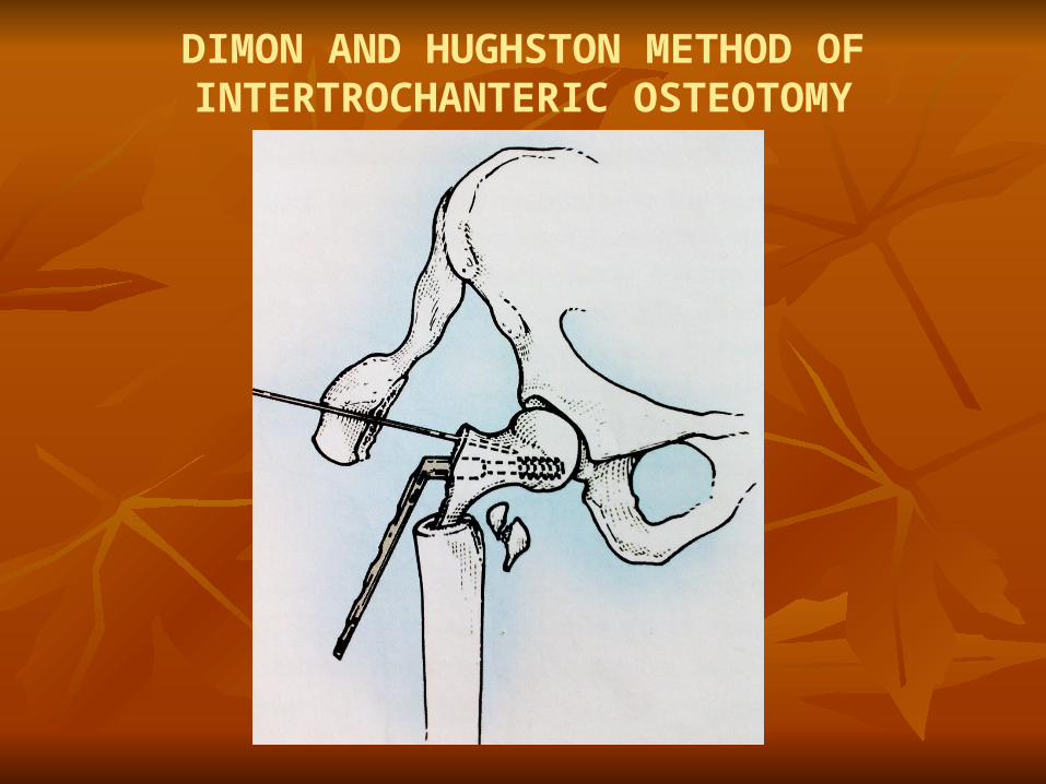
DIMON AND HUGHSTON METHOD OF INTERTROCHANTERIC OSTEOTOMY

SLIPPED CAPITAL FEMORAL EPIPHYSIS (SCFE)
Is a disorder in which there is a displacement of the capital
femoral epiphysis form the metaphysis through the physeal
plate.
By this head is placed in posterior & downward position in
acetabulum.
The goal of treatment is
To prevent further displacement and
To promote closure of physeal plate.

The use of realignment procedure such as lntertrochameric,
Subtrochanteric Osteotomy & osteotomies the around neck is
in those situation in which restricted range of motion impairs
function after plate physeal closure.
Principle of Osteotomy
There are basically three type of Deformity present in SCFE.
These are-
Varus
Hyper extension
Moderate Severe external rotation
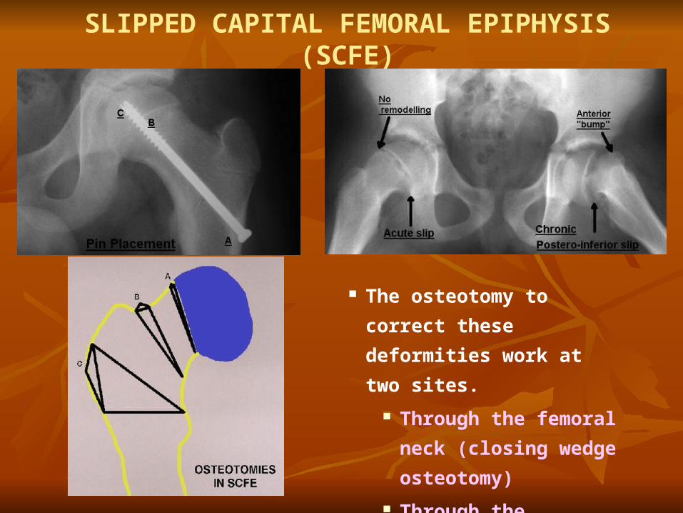
SLIPPED CAPITAL FEMORAL EPIPHYSIS (SCFE)
The osteotomy to correct these
deformities work at two sites.
Through the femoral neck
(closing wedge osteotomy)
Through the trochanteric
area.

EXTRACAPSULAR BASE OF NECK OSTEOTOMY
types of femoral neck osteotomy are - The technique of Dunn - for severe chronic slip with open
physis. Base of the neck osteotomy - Compensatory Basilar most
of femoral neck. (Kramer) - correct the varus and
retroversion component of moderate to severe chronic
SCFE. It is safer than cuniform osteotomy of neck. Further slipping is prevented. Intertrochantric osteotomies
CORRECTIVE OSTOTOMIES
By these osteotomies one can correct angulation, rotation,
flexion, extension Deformity of bones to restore motion for
patient with stiff hip.
Like
Deformities in septic arthritis
Malunion of I/T femurs
Neuromuscular disorder
Cerebral palsy
Poliomyelitis
There are three types of corrective osteotomies Close wedge - transverse closing wedge provide good bony
apposition and is stable, however, it shortens the extremity. Open wedge - simple and lengthens the extremity however.
bony apposition is limited, union is delayed in adults and it is initially unstable.
Ball and Socket type - achieves stability without shortening the extremity; however, extensive dissection is required, and in severe biplame deformities an accurate and stable osteotomy is difficult to perform.
In Ball & socket type of osteotomy concave surface in created in the proximal fragment of convex surface at the distal fragment, at intertrochantaric level & fixed in place by plate.

CORRECTIVE OSTOTOMIES
Brackett ball and socketOsteotomy
Whitman closing wedgeOsteotomy
Gant-opening wedge
Osteotomy

FRACTURE NECK FEMUR In those case which present late (1-5 wks.), are difficult case
to treat because Close reduction is not possible. Open reduction is associated AVN
In young Pt. with viable femoral head & nonunion options are- Mcmurray & Pauwel’s ‘y’ osteotomy Angulation Osteotomy (Schanz) Dickson geometric osteotomy
In old Pt.- Mcmurray Displacement
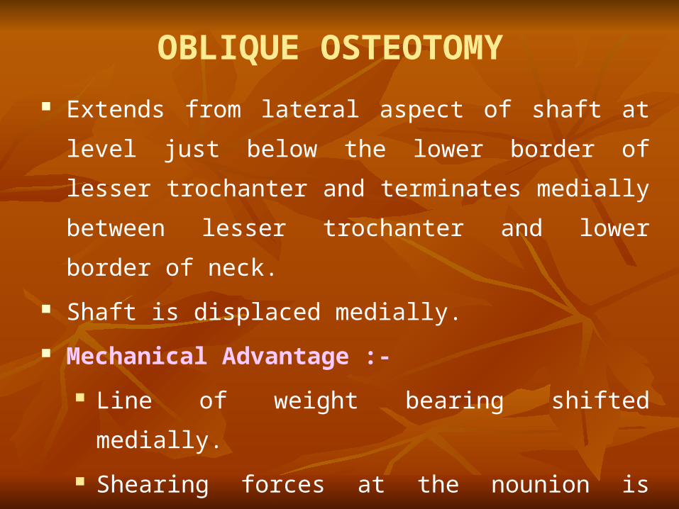
OBLIQUE OSTEOTOMY
Extends from lateral aspect of shaft at level just below the
lower border of lesser trochanter and terminates medially
between lesser trochanter and lower border of neck.
Shaft is displaced medially.
Mechanical Advantage :-
Line of weight bearing shifted medially.
Shearing forces at the nounion is decrease because
fracture surface become more horizontal
These advantages are greater after angulation osteotomy.
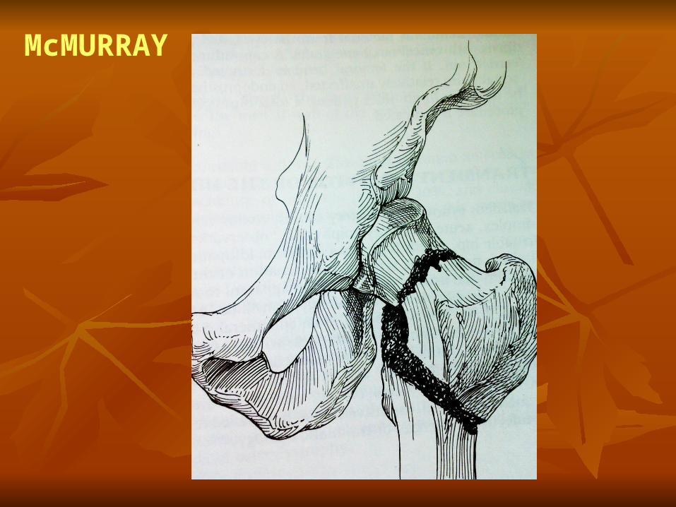
McMURRAY
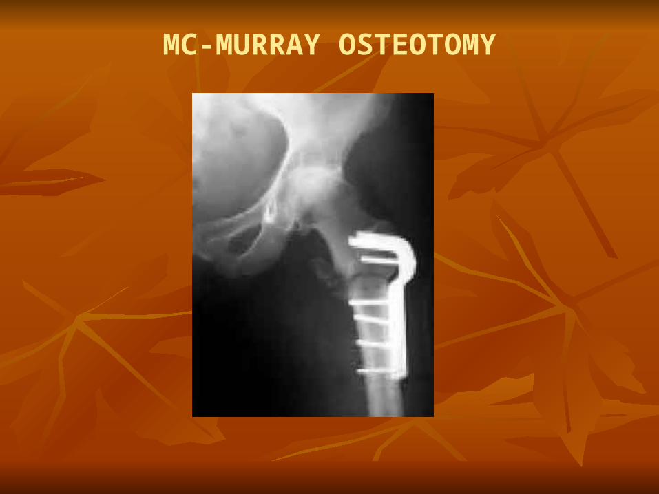
MC-MURRAY OSTEOTOMY
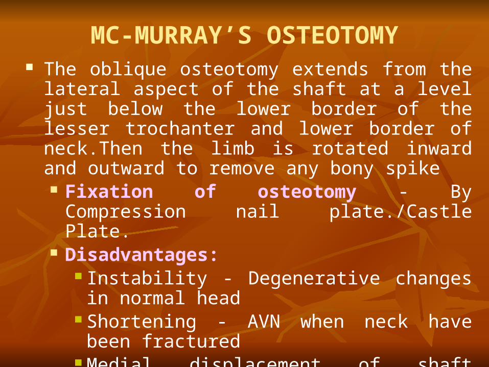
MC-MURRAY’S OSTEOTOMY The oblique osteotomy extends from the lateral aspect of
the shaft at a level just below the lower border of the lesser trochanter and lower border of neck.Then the limb is rotated inward and outward to remove any bony spike Fixation of osteotomy - By Compression nail
plate./Castle Plate. Disadvantages:
Instability - Degenerative changes in normal head Shortening - AVN when neck have been fractured Medial displacement of shaft compromise the
insertion of femoral stem of total hip. Advantage -Changes line of fracture to
horizontal,callus may incarporate fracture
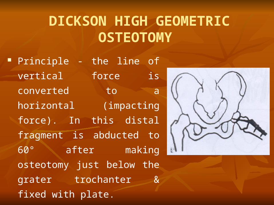
DICKSON HIGH GEOMETRIC OSTEOTOMY
Principle - the line of vertical force
is converted to a horizontal
(impacting force). In this distal
fragment is abducted to 60° after
making osteotomy just below the
grater trochanter & fixed with plate.
High rate of union
Lengthens limb
Improves abductor strength
OSTEOTOMIES – These procedure have achieved best result for small and
medium sized lesion. 1<30% femoral head involvement in young pt.
Intertrochanteric varus/valgus - osteotomies Transtrochantric ant. Rotational osteotomy (Sugioka) -
Technically Demanding procedures. PRINCIPLE:
All osteotomies are designed to transfer the weight bearing forces form the necrotic area to the cartilage on the sound part of the femoral head to allow healing of necrotic area by hyper vascularisation of upper part of femur.
AVN
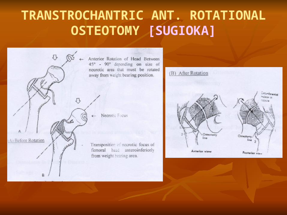
TRANSTROCHANTRIC ANT. ROTATIONAL OSTEOTOMY [SUGIOKA]
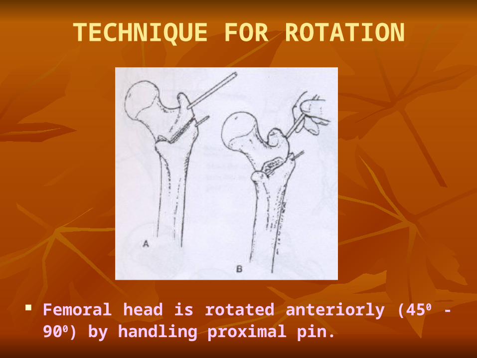
TECHNIQUE FOR ROTATION
Femoral head is rotated anteriorly (450 - 900) by handling proximal pin.
OSTEOTOMY IN PERTHE'S DISEASE Salvage :
Varus Derotational Osteotomy Innominate Osteotomy. MRI / Arthrogram before surgery is mandatory. Varus/derotation osteotomy of this embodies the principle
of “containment” of the diseased femoral head in the treatment of Legg - Calve-Perthes disease.
Guide pin inserted compression screw is placed over guide wire.

Appropriate angled osteotomy is made.
Wedge is removed.
Make osteotomy as proximal as possible just below lag
screw for -
Better Healing
Better correction of deformity.
Reduce the osteotomy and fixed with plate and
compression screw.
SUBTROCHANTERIC DEROTATION AND VARUS OSTEOTOMY
The aim of surgery is to center the whole "plastic" epiphysis inside the joint cavity, keeping it well covered by the roof of the acetabulum and allowing the child to walk so that the redistributed intra-articular pressures will contribute the molding of a more normal joint.
A small 4-hole plate is bent to the desired angle, and a subtrochanteric osteotomy is done followed by derotation and yarns angulation of the shaft. A double hip spica is applied and the removed 2 months later. When the osteotomy site is united, the child is encouraged to walk, then with walking aids and finally without support.

VARUS DEROTATION OSTEOTOMY
The operation is best suited for early stage of Leg-Calve-
Perthes’ disease, preferably those under the age of 7 years.
Axer : Described lateral open wedge osteotomy for children
< 5 years with perthes disease. Defect laterally fills rapidly
in young children > 5 years of age delayed or non union may
occur.
RECONSTRUCTIVE SURGERY
Valgus subtrochanteric osteotomy – for Hinged
Abduction
Shelf Augmentation – Coxa Magna.
Chilectomy - Malformed head in late III Group.
Chiari's Pelvic Osteotomy - Large Malformed
Femoral Head with Subluxation laterally.

THANK YOU