oto rhino laryngo logical surgery
DESCRIPTION
Oto rhino laryngo logical Surgery. ENT SURGERY. Purpose. Ear: improve, restore, preserve hearing Nose: restore or improve breathing/ventilation, ensure drainage of the sinuses, control epistaxis Throat: prevent infection, remove a tumor/mass, perform life-saving procedures. Ear Nose - PowerPoint PPT PresentationTRANSCRIPT
Otorhinolaryngological Surgery
ENT
SURGERY
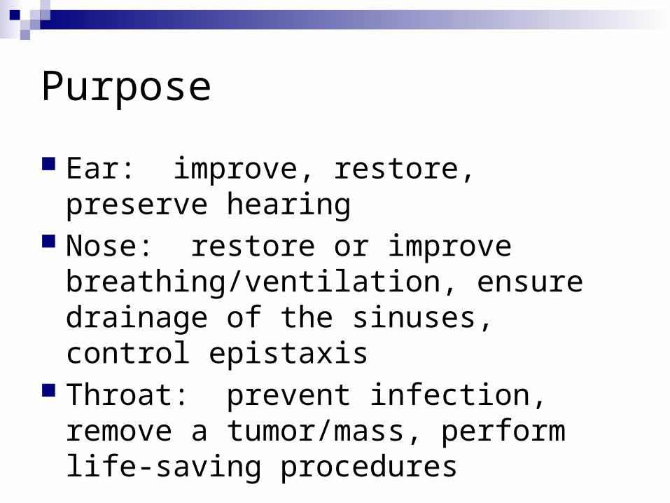
Purpose
Ear: improve, restore, preserve hearing Nose: restore or improve
breathing/ventilation, ensure drainage of the sinuses, control epistaxis
Throat: prevent infection, remove a tumor/mass, perform life-saving procedures
Otorhinolaryngological Surgery
Ear Nose Throat Endoscopy Triple endoscopy Thyroid & Parathyroids Tracheotomy
Terms A & P Pathology Anesthesia & Meds Positioning, Prep, & Draping Supplies, Equipment, & Instrumentation Considerations & Complications

The Ear
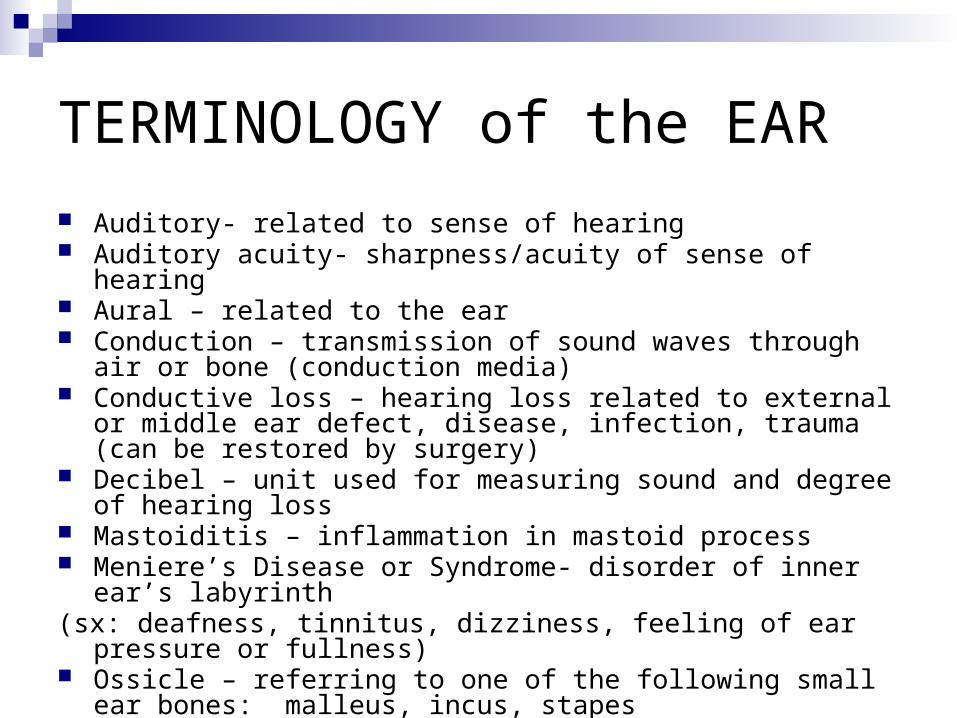
TERMINOLOGY of the EAR
Auditory- related to sense of hearing Auditory acuity- sharpness/acuity of sense of hearing Aural – related to the ear Conduction – transmission of sound waves through air or bone
(conduction media) Conductive loss – hearing loss related to external or middle ear
defect, disease, infection, trauma (can be restored by surgery) Decibel – unit used for measuring sound and degree of hearing loss Mastoiditis – inflammation in mastoid process Meniere’s Disease or Syndrome- disorder of inner ear’s labyrinth(sx: deafness, tinnitus, dizziness, feeling of ear pressure or fullness) Ossicle – referring to one of the following small ear bones: malleus,
incus, stapes

Terminology of the Ear Continued
Otitis media – acute or chronic inflammation of the middle ear Oto – related to the ear Otology – related to the ear Otosclerosis – formation of spongy bone around the oval window
that causes immobility of the stapes resulting in deafness PE Tubes (pressure equalization) – drainage tubes placed in the
eardrum or tympanic membrane allowing drainage of fluid in the middle ear preventing fluid build up that leads to infection
Sensorineural loss – defect in the inner ear from nerve tissue damage that causes hearing loss (surgery does not help)
Tinnitis – a subjective symptom of ringing in the ear Vertigo – sensation of dizziness

Anatomy of the Ear
Outer Ear1. Auricle or pinna2. Auditory meatus extends to the tympanic
membrane Lined with fine hairs Ceruminous glands secrete cerumen Function to collect sound and direct it down a
hole in the temporal bone

Anatomy of the Ear
3. Tympanic Membrane Eardrum Separates outer ear from middle ear Normally pearly grey

Anatomy of the Ear
4. Middle Ear Tympanic cavity Eustasian tube/canal equalizes pressure Auditory ossicles: lateral to medial (from
tympanic membrane in): Malleus (hammer) Incus (anvil) Stapes (stirrup)

Anatomy of the Ear
5. Inner Ear (labyrinth) Bony Membranous Are complex canals and chambers called
the semi-circular canals Equilibrium (Vestibular Apparatus) Hearing (Organs of Corti in the Cochlea)


Physiology of Hearing
Hear a sound>hits auricle>external auditory canal>tympanic membrane (vibration occurs) >malleous connected to tympanic membrane and therefore moves>incus moves>stapes moves>in and out of oval window>pushes on perilymph fluid in vestibule>pushes on vestibular membrane pushes endolymph fluid>pushes against a membrane of the organ of corti housed in the cochlea to move>stimulates axons which become a branch of vestibulo-cochlear nerve>ends in auditory area of cerebrum that interprets sounds

Equilibrium
Semicircular canals (3 per ear) Hollow filled with fluid endolymph Axons form vestibulo-cochlear nerve Fluid when turn or spin stimulates
dendrites and tell body you are moving in a certain direction
Detect 3 planes of movement

Pathology
Hearing Loss Conduction type Sensorineural Central Mixed-Type Functional Congenital Neonatal
Outer Ear Obstruction Exostoses Polyps Infection Abscess

Pathology
Tympanic membrane Perforation Rupture
Middle Ear Trauma Perforation Fluid accumulation Otitis media Otosclerosis

Pathology
Mastoid Mastoiditis Cholesteatoma
Inner Ear Meniere’s syndrome
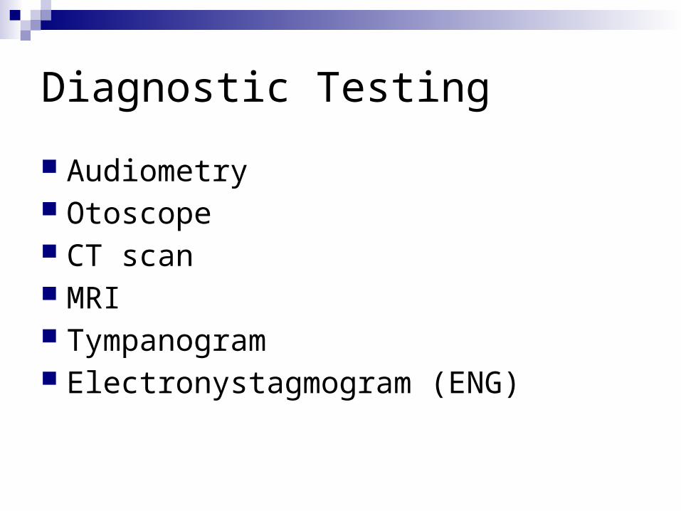
Diagnostic Testing
Audiometry Otoscope CT scan MRI Tympanogram Electronystagmogram (ENG)

Anesthesia
General: Inhalation Intubation

Medications
Local anesthetics (with or without epinephrine)
Gelfoam Bone wax Antibiotics (topical or systemic) Anti-inflammatory agents

Position
Bed reversed to allow operative team to sit with feet under bed
Supine Headrest with operative ear up Arms tucked Pillow under the knees

Prep
Small area may be shaved Hairline to shoulders and from midline of
face to behind operative ear If a solution is used prevent pooling in the
ear or contact with the eyes

Draping
Head wrap Towels Body drape ENT drape

Supplies, Equipment, Instrumentation Moistened cottonoid sponges Burrs Micro Rotating drill Microscope Argon Laser Cautery Speculum Holder Nerve stimulator
Buck (ear) currette Iris scissors Ear speculum Applicator Bayonet forceps Hartman (alligator) forceps Sexton ear knife Frazier suction Baron suction tip Elevator Kerrison ronguer Chisel Mallet

The Nose

Terminology of the Nose
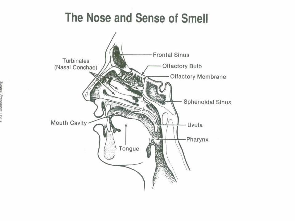
Anosmia-loss of smell Epistaxis-nose bleed Hyperosmia-oversensitive to odors Nares (Naris)-nostrils Nasal-related to the nose Nasal Turbinates-four bony projections or ridges in the nasal cavity
(supreme, superior, middle, inferior) Olfactory- related to smell
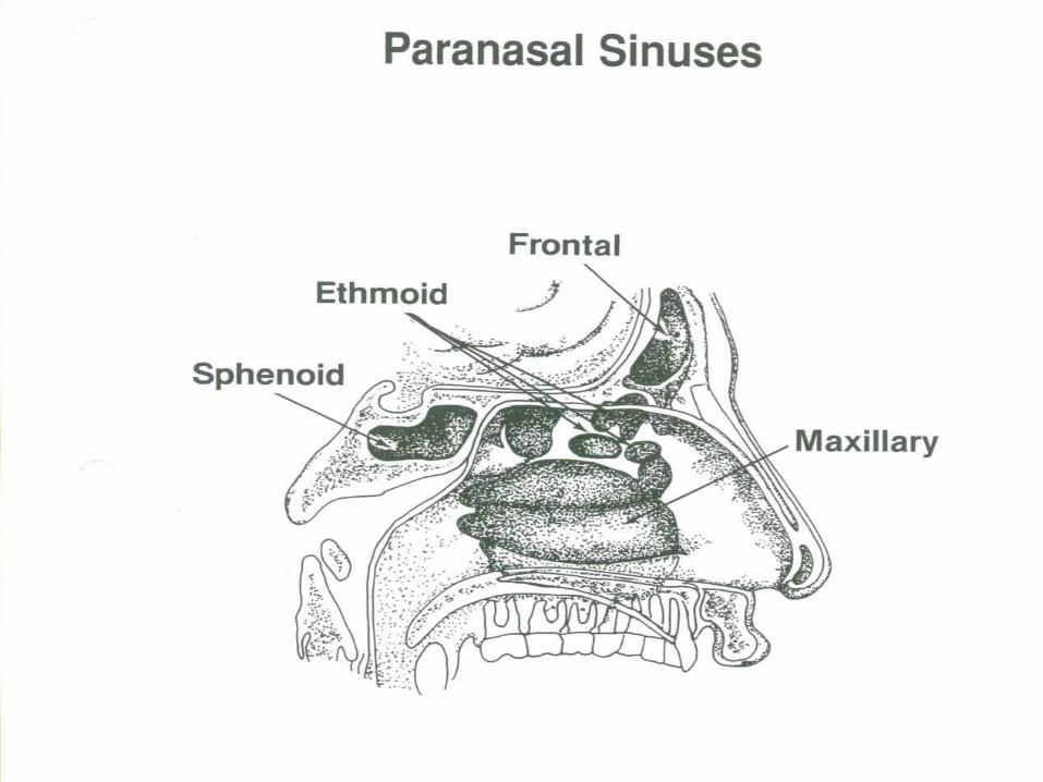
Paranasal sinuses- air cavities in the bone around the nasal cavity lined with mucous membranes (frontal, ethmoid, sphenoid, maxillary)
Parosmia-disorder affecting smell Rhinitis-inflammation of the nasal mucosa Rhino-related to the nose Sinus-cavity in a bone

Anatomy of the Nose
External Nose Internal Nose Paranasal Sinuses
Physiology of Smell
Receptors in upper or superior nasal cavity Bipolar neurons (receptors) pick up a different
chemoreceptor Are about 50 receptors Axons form olfactory nerve (I) These go into cribiform plate End in olfactory bulbs under frontal lobe of
cerebrum

Pathology
Rhinitis Sinusitis Nasal polyps Hypertrophied turbinates Deviated septum Septal perforation Epistaxis
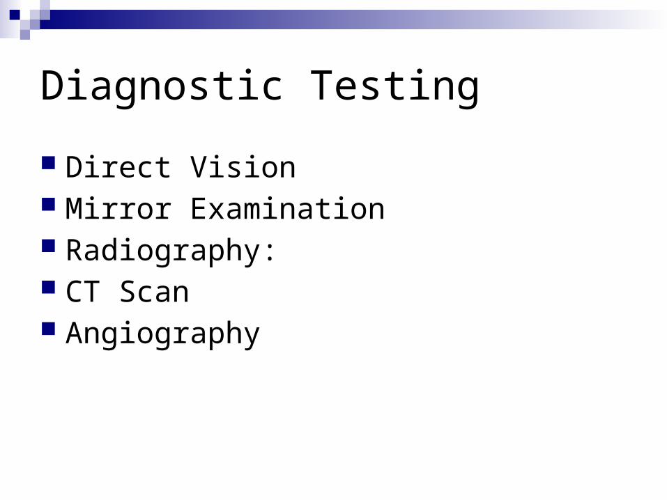
Diagnostic Testing
Direct Vision Mirror Examination Radiography: CT Scan Angiography

Anesthesia
Local Septum and Mucous Membranes General Sinuses
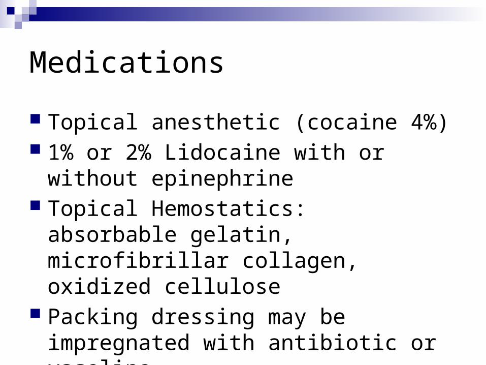
Medications
Topical anesthetic (cocaine 4%) 1% or 2% Lidocaine with or without
epinephrine Topical Hemostatics: absorbable gelatin,
microfibrillar collagen, oxidized cellulose Packing dressing may be impregnated
with antibiotic or vaseline

Positioning
Supine with General Anesthesia Modified Fowler’s with Local Anesthesia Pillow under head Arms tucked or secured across chest Footboard with Fowler’s Safety strap

Prep
Nare hair clipping Eye protection Mild antiseptic on face Cotton tipped applicator nostril cleansing Begins at upper lip, beyond hairline, below
chin Prevent prep solution from entering eyes

Draping
Turban like head wrap 3 triangle folded towels Forehead bar towel or sheet Split sheet Body drape

Supplies, Equipment, Instrumentation Medicine cups 2 local syringes 2” 25 or 27gauge needles Long cotton tipped applicators Packing gauze, cotton, or cottonoids Headlight Microscope
Nasal or septum speculum Bayonet forceps Small scissors (Joseph) Curettes Skin hooks 6, 30, 70° endoscopes Nasal chisel & mallet Nasal dressing forceps Hartman nasal forceps Septal knife (Joseph or Cottle) Ballenger swivel knife Freer elevator Nasal Rasp (Foman) Fine suction tips (irrigate often)

Considerations
Ear and Nasal Surgery not truly sterile surgical procedures, however, aseptic technique imperative to prevent infection

Oral Cavity and Throat
Terminology of the Oral Cavity & Throat
Adenoids-(pharyngeal tonsils if enlarged) lymphatic tissue in nasopharynx (atrophies with age)
Epiglottis-small structure at back of throat, covers larynx when swallowing Fauces-opening of the oropharynx Glottis-space between the vocal cords Larynx-cartilaginous structure above the trachea, houses the vocal cords Palatine tonsils-lymphatic oval masses of tissue in the oropharynx Papilloma-benign epithelial tumor
Pharynx-(throat) begins at internal nares and ends posterior to the larynx where it joins the esophagus
Stomatitis-inflammation of the mouth Thyroid cartilage-(Adam’s apple) Trachea-(airway) cartilaginous tube extending from the larynx to the bronchial tubes Vocal cords-fibrous bands of tissue, stretched across the hollow interior of the larynx
which vibrate to create sound
Anatomy of the Upper Aerodigestive Tract
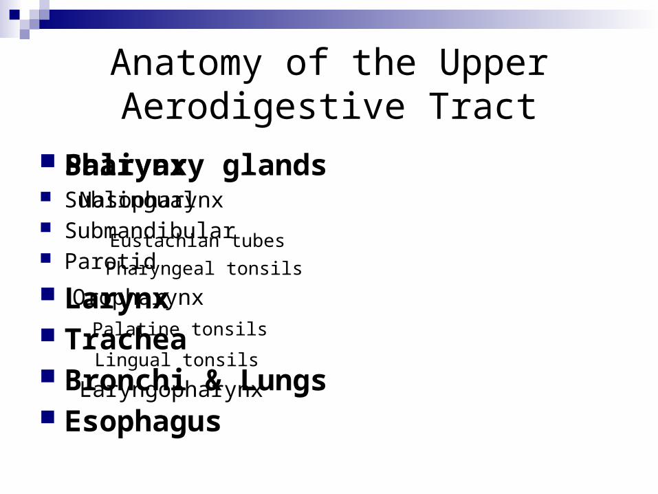
Pharynx Nasopharynx
Eustachian tubes
Pharyngeal tonsils
Oropharynx
Palatine tonsils
Lingual tonsils
Laryngopharynx
Salivary glands Sublingual Submandibular Parotid
Larynx Trachea Bronchi & Lungs Esophagus

Physiology of Taste
Gustatory sense Bipolar neurons in taste buds 4 chemicals detected: sweet, sour, salt, bitter Taste related to smell Taste detected 2/3 anterior taste buds from
facial nerve (VII), 1/3 posterior tongue from glossopharyngeal nerve (IX)
Are most sensitive to bitter Takes a lot of sweet to detect
Pathology of the Upper Aerodigestive Tract
Pharyngitis Epiglottitis Tonsillitis Peritonsillar abscess Sleep apnea Foreign bodies Laryngitis
Polyps Vocal cord nodules Laryngeal neoplasms Tumor Tracheitis Bronchitis Croup

Pathology of the Esophagus
Esophagitis Ulceration Neoplasms Foreign bodies Zenker’s diverticulum
Diagnostic Testing
Direct Visualization Culture & Sensitivity (C&S) CBC X-Ray CT Scan MRI Endoscopy
Anesthesia
General Site of intubation will be opposite that of
operative site (nose verses throat) MAC with IV Sedation Local Anesthesia
Medications
Steroids per anesthesia Water soluble lubricant Lidocaine jelly lubricant Cetacaine spray 4% cocaine (topical) Lidocaine with or without epinephrine Bupivacaine with or without epinephrine Topical hemostatics (surgeon’s preference)

Positioning
Supine Sitting Arms tucked Shoulder roll Head support (donut) Pillow under knees Safety strap

Prep
None Extensive Surgeon’s preference
Draping
Head wrap Towels Impervious drape (Ioban) Fenestrated sheet U-sheet None
Supplies, Equipment, Instrumentation
Basic pack Basin set Raytex Tonsil sponges Small badsin Suction tubing Suction tip (fine) Blade of surgeon choice (#12) Cautery Suction/cautery Plain, vicryl, silk suture or reels Luken’s trap Lubricant Specimen container Tongue depressor
Headlight ECU Microscope Endoscopes (rigid or flexible) Video tower CO2 or Nd:YAG laser Mouth gag Tonsil snare Dental or laryngeal mirror Biopsy forceps Alligator forceps Curettes Fisher tonsil knife Bougies

Post-operative Considerations
Sore throat Hoarse Bleeding Be aware of ET tube as drapes removed Infection
Endoscopies

Endoscopies
Laryngoscopy Microlaryngoscopy Bronchoscopy Esophagoscopy Endoscopes: Rigid – larger viewing surface Flexible – easy insertion and manipulation For: diagnostic or operative use: biopsy, foreign
body removal, bougie insertion
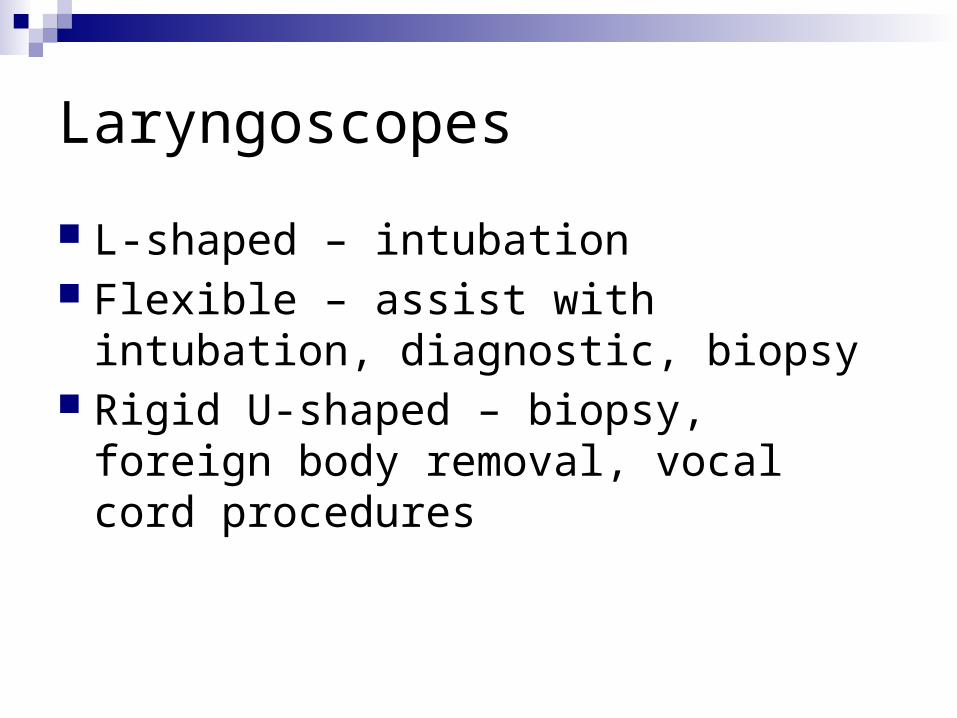
Laryngoscopes
L-shaped – intubation Flexible – assist with intubation,
diagnostic, biopsy Rigid U-shaped – biopsy, foreign body
removal, vocal cord procedures

Microlaryngoscopy
Laryngoscopy Microscope (400mm focal length=40cm focal length) Microlaryngeal instruments (22cm) Laser attached to microscope CO2 single beam, more precise (used with helium-neon
beam to provide red beam for proper aiming) Vocal cord, tracheal, bronchial lesions Nd: YAG Laser tracheal or bronchial lesions

Bronchoscopes
Flexible Rigid Longer than laryngoscopes Adaptor required for oxygenation Nd: YAG (prn)
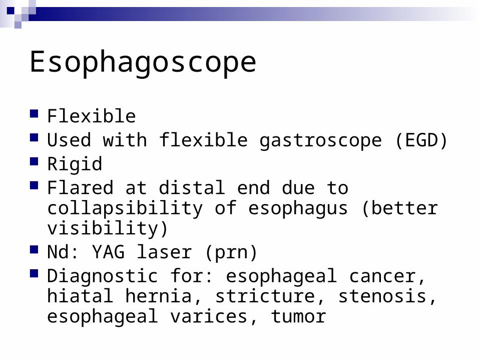
Esophagoscope
Flexible Used with flexible gastroscope (EGD) Rigid Flared at distal end due to collapsibility of
esophagus (better visibility) Nd: YAG laser (prn) Diagnostic for: esophageal cancer, hiatal hernia,
stricture, stenosis, esophageal varices, tumor

Triple Endoscopy/Panendoscopy

Triple Endoscopy or Panendoscopy
Term describes all three procedures combined:
Esophagoscopy Laryngoscopy Bronchoscopy Diagnostic

Thyroid and Parathyroid Glands
Thyroid and Parathyroid Surgery
1° performed by general surgeons

Thyroid Gland
2 lobes Anterior to larynx Connected by isthmus at 2nd tracheal ring H-shaped Two hormonal cell types: Follicular – produce, store, release thyroxine and
triidothyronine, basal metabolic rate regulation hormones Parafollicular - secrete calcitonin, hormone that
maintains calcium homeostasis

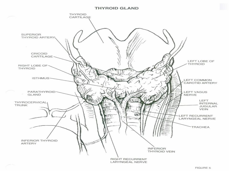
Parathyroid Glands
Numbered 1 to 6 Small, flat, oval dorsal to thyroid gland Produce parathormone, a hormone that
maintains a normal blood and skeletal calcium relationship
Cannot remove all of them = tetany and death
Pathology of Thyroid and Parathyroid Glands Hyperthyroidism: restlessness, fast speech,
tachycardia, palpitations, arrythmias, dyspnea, heat intolerance, diaphoresis, weakness, tremor, hair loss
Hyperparathyroidism: asymptomatic to skeletal damage
Thyroid carcinoma: signs of hyperthyroidism, hypothyroidism, hoarseness, difficulty swallowing, dyspnea

Diagnostic Testing
Physical Exam Serum TSH levels Ultrasound Biopsy CT Scan MRI Laryngoscopy
Anesthesia
General

Medications
Lidocaine with or without epinephrine Bupivicaine with or without epinephrine Antibiotic irrigation

Positioning
Supine Donut headrest Shoulder roll Arms tucked Pillow under knees Safety strap
Prep
Surgeon’s preference: Duraprep, Betadine scrub and/or paint
End of chin to midchest and bedsheet to bedsheet
Draping
Towels Small fenestrated sheet (Pediatric sheet) Thyroid sheet U-Sheet Surgeon’s preference
Supplies, Equipment, Instrumentation
Minor basin Basic pack Blades of choice Suture of choice Silk ties ¼” penrose Bipolar forceps Headlight Minor Tray
Headlight Minor tray

Post-operative Considerations
Will need medical hormonal therapy Potential damage to bilateral laryngeal
nerve with dissection Hemorrhage Infection
Tracheotomy & Tracheostomy

Tracheotomy/Tracheostomy
Tracheotomy temporary opening into the trachea to facilitate breathing
Tracheostomy permanent opening of the trachea and creation of a tracheal stoma
Must place tracheal tube with either Patient will be hooked up to a ventilator Long term tracheostomy may eventually be able
to wean off ventilator, but maintain stoma that will function as their nose did prior to surgery
Indications For Tracheotomy or Tracheostomy Vocal cord paralysis Neck surgery Trauma Prolonged intubation Secretion management Cannot intubate Stridor due to tracheal blockage Sleep apnea

Anesthesia
General Local
Medications
Local anesthetic: Lidocaine or bupivicaine with or without epinephrine
Antibiotic irrigation

Positioning
Supine Shoulder roll Donut headrest Pillow under knees Safety strap

Prep
End of chin to midchest and bedsheet to bedsheet
Prep of choice: Duraprep, betadine scrub and/or paint

Draping
Towels Small fenestrated sheet (Pediatric lap
sheet)

Supplies, Equipment, Instruments
Minor basin Basic pack Pediatric lap sheet Other small fenestrated sheet Blades Suture or ties of surgeon’s choice (prn)
Tracheotomy tray Tracheotomy tube (Shiley) Twill tape
Considerations
Will make sure obturator goes with patient to PACU or ICU
Complications: hemorrhage, infection, damage to other structures

Summary
Ear Nose Throat Endoscopy Triple endoscopy Thyroid & Parathyroids Tracheotomy
Terms A & P Pathology Anesthesia & Meds Positioning, Prep, & Draping Supplies, Equipment, & Instrumentation Considerations & Complications