overcoming presynaptic effects of vamp2 mutations with 4
TRANSCRIPT
Human Mutation. 2020;1–13. wileyonlinelibrary.com/journal/humu © 2020 Wiley Periodicals LLC | 1
Received: 4 June 2020 | Revised: 3 August 2020 | Accepted: 4 September 2020
DOI: 10.1002/humu.24109
R E S E A RCH AR T I C L E
Overcoming presynaptic effects of VAMP2mutations with4‐aminopyridine treatment
Roxanne L. Simmons1 | Haiyan Li2 | Baris Alten3 | Magda S. Santos2 | Ruiji Jiang1 |
Brianna Paul1 | Sanam J. Lalani1 | Audrey Cortesi1 | Kendall Parks1 |
Nitin Khandelwal4 | Bethany Smith‐Packard5 | Malay A. Phoong6 | Michael Chez7 |
Heather Fisher8 | Angela E. Scheuerle9 | Marwan Shinawi10 | Shaun A. Hussain11 |
Ege T. Kavalali3 | Elliott H. Sherr1 | Susan M. Voglmaier2
1Department of Neurology, Weill Institute for Neurosciences and Institute of Human Genetics, School of Medicine, University of California, San Francisco,
San Francisco, California, USA
2Department of Psychiatry, Weill Institute for Neurosciences and Kavli Institute for Fundamental Neuroscience, School of Medicine, University of California,
San Francisco, San Francisco, California, USA
3Department of Pharmacology and Vanderbilt Brain Institute, Vanderbilt University, Nashville, Tennessee, USA
4Department of Neuroscience, UT Southwestern Medical Center, Dallas, Texas, USA
5Department of Pediatrics, Penn State Health Pediatric Specialties, Hershey, Pennsylvania, USA
6Division of Neuroscience, Department of Pediatric Neuropsychology, Sutter Medical Foundation, Sacramento, California, USA
7Neuroscience Medical Group, Sutter Medical Foundation, Sacramento, California, USA
8Department of Genetics, Children's Medical Center of Texas, Dallas, Texas, USA
9Division of Genetics and Metabolism, Department of Pediatrics, UT Southwestern Medical Center, Dallas, Texas, USA
10Division of Genetics and Genomic Medicine, St. Louis Children's Hospital, Washington University School of Medicine, St. Louis, Missouri, USA
11Department of Pediatrics, UCLA Mattel Children's Hospital and Geffen School of Medicine, Los Angeles, California, USA
Correspondence
Elliott H. Sherr, Department of Neurology,
Weill Institute for Neurosciences, School of
Medicine, University of California,
San Francisco, 675 Nelson Rising Ln, 214B,
San Francisco, CA 94158, USA.
Email: [email protected]
Susan M. Voglmaier, Department of Psychiatry,
Weill Institute for Neurosciences, School of
Medicine, University of California, San
Francisco, 401 Parnassus Ave, San Francisco,
CA 94143, USA.
Email: [email protected]
Funding information
National Institute of Neurological Disorders
and Stroke, Grant/Award Number: NS058721;
National Institute of Mental Health,
Grant/Award Numbers: MH066198,
MH083691
Abstract
Clinical and genetic features of five unrelated patients with de novo pathogenic
variants in the synaptic vesicle‐associated membrane protein 2 (VAMP2) reveal
common features of global developmental delay, autistic tendencies, behavioral
disturbances, and a higher propensity to develop epilepsy. For one patient, a cog-
nitively impaired adolescent with a de novo stop‐gain VAMP2 mutation, we tested a
potential treatment strategy, enhancing neurotransmission by prolonging action
potentials with the aminopyridine family of potassium channel blockers,
4‐aminopyridine and 3,4‐diaminopyridine, in vitro and in vivo. Synaptic vesicle re-
cycling and neurotransmission were assayed in neurons expressing three VAMP2
variants by live‐cell imaging and electrophysiology. In cellular models, two variants
decrease both the rate of exocytosis and the number of synaptic vesicles released
from the recycling pool, compared with wild‐type. Aminopyridine treatment in-
creases the rate and extent of exocytosis and total synaptic charge transfer and
desynchronizes GABA release. The clinical response of the patient to 2 years of
Roxanne L. Simmons, Haiyan Li, and Baris Alten contributed equally to this study.

off‐label aminopyridine treatment includes improved emotional and behavioral
regulation by parental report, and objective improvement in standardized cognitive
measures. Aminopyridine treatment may extend to patients with pathogenic var-
iants in VAMP2 and other genes influencing presynaptic function or GABAergic
tone, and tested in vitro before treatment.
K E YWORD S
aminopyridine, neurodevelopmental disorder, synaptic transmission, synaptic vesicle, VAMP2
1 | INTRODUCTION
Fast communication between neurons relies on the precise and
highly regulated fusion of synaptic vesicles (SVs) with the presynaptic
plasma membrane, resulting in neurotransmitter exocytosis (Sudhof,
2013). Fusion of the two membranes is driven by the zippering of
highly conserved hydrophobic side chain soluble N‐ethylmaleimide‐sensitive factor attachment protein receptor (SNARE) motifs of the
vesicular v‐SNARE, vesicle‐associated membrane protein (VAMP2;
MIM #185881) with the plasma membrane target t‐SNAREs, SNAP‐25, and syntaxin‐1, to form the SNARE complex (Jahn & Scheller,
2006). The SNARE complex interacts with accessory proteins, such as
the synaptotagmin family of Ca2+ sensors. Synaptotagmins sense the
local increase in Ca2+ concentration caused by the opening of
voltage‐gated Ca2+ channels (VGCCs) upon action potential (AP)‐induced membrane depolarization. The resultant local increase in
Ca2+ concentration is signaled to the SNARE complex mainly by
synaptotagmin‐1 for synchronous release and by synaptotagmin‐7for asynchronous release. Simultaneously, inward‐rectifier K+ chan-
nels and the Na+/K+‐ATPase repolarizes the membrane, halting
VGCC activity, while active pumps clear local Ca2+ away, terminating
neurotransmitter release. VAMP2 is also vital for the fast en-
docytosis of neuronal membrane to replenish the pool of SVs for
the next round of exocytosis (Deak, Schoch, Liu, Sudhof, &
Kavalali, 2004).
VAMP2 is the most abundant SV protein, with approximately
70 copies per vesicle, while only 2–3 are necessary to mediate fusion
(Mohrmann & Sorensen, 2012; Takamori et al., 2006). Given the high
conservation of the SNARE motif (Fasshauer, Eliason, Brunger, &
Jahn, 1998), a mutation in this region would be expected to disrupt
endocytosis or exocytosis of SVs, impairing neurotransmission and
causing neurological disorders. Indeed, experimental SNARE muta-
tions cause dominant negative disruption of fusion (Grote & Kelly,
1996; Koo et al., 2011), while VAMP2 heterozygosity loss of function
in mice causes only a mild phenotype (Monteggia, Lin, Adachi, &
Kavalali, 2018). Human VAMP2 SNARE mutations can impair fusion
of reconstituted membranes. A clinical phenotype for VAMP2 muta-
tions was recently described in five patients as hypotonia, intellectual
disability, and autistic features (MIM #618760; Salpietro et al., 2019).
Here we report five unrelated patients, doubling the number iden-
tified with pathogenic de novo VAMP2 variants. Currently, there is no
effective treatment targeting the underlying impairment of
neurotransmitter release. Patients likely make wild‐type (WT)
VAMP2 from their functional allele, so we tested a strategy to
overcome exocytosis defects by prolonging the AP by delaying
neuronal repolarization with the potassium channel inhibitors
4‐aminopyridine (4‐AP) and 3,4‐diaminopyridine (DAP) to increase
calcium entry and SV release probability. 4‐AP and DAP have ex-
panding roles in the treatment of multiple sclerosis, cerebellar
ataxias, and Lambert–Eaton and congenital myasthenic syndromes
(Claassen, Teufel, Kalla, Spiegel, & Strupp, 2013; Palace, Wiles, &
Newsom‐Davis, 1991; Strupp et al., 2017). Taken together, this
suggests an enhancement of SV release could improve cognitive
function in patients with single allele VAMP2 pathogenic variants.
2 | METHODS
2.1 | Editorial policies and ethical considerations
Work with animals was conducted under the supervision of the In-
stitutional Care and Use Committees of the University of California,
San Francisco and Vanderbilt University Medical Center. Parents
provided written consent before participation through a University
of California, San Francisco (UCSF) committee on human research
approved protocol.
2.2 | Clinical information
Patients with VAMP2 variants were assessed by chart review and
caretaker phone interviews. Parents provided written consent before
participation. Variants were assessed for clinical significance and
pathogenicity by use of web sources including clinical data obtained
from GeneDx, as well as predicted results from ClinVar, Poly‐Phen2,and gnomAD. Thus, while variants of these patients were initially
reported by GeneDx as “uncertain significance,” they were de-
termined to be pathogenic based on clinical phenotype and predictive
algorithms. Ancillary studies, including electroencephalography
(EEG), electromyography, magnetic resonance imaging (MRI), and
neuropsychologic testing were reviewed by a physician to determine
clinical relevance. After receiving consent from parents and the pa-
tient, Patient 1 was treated with low dose 4‐AP that was gradually
increased over the course of several months. Tolerability of 4‐AP was
2 | SIMMONS ET AL.

assessed by parental report of worsening anxiety or insomnia. The
effects of 4‐AP were measured by qualitative assessments via sub-
jective parental reports, and quantitatively via neuropsychological
testing. Neuropsychological testing pre‐ and posttreatment were
compared by converting scaled scores to Z‐scores.
2.3 | Molecular biology and lentivirus preparation
VAMP2–mOrange2 (mOr2) fusions were constructed by fusing syn-
thetic mOr2 (Shaner et al., 2008) to the C‐terminus of human WT
(RefSeq NM_014232.3) or variant VAMP2 complementary DNAs
(cDNAs) with a linker (SGGSGGTG). Disease‐associated point muta-
tions Arg56Leu (R56L) or Gly73Trp (G73W) were generated in
VAMP2 using Quikchange‐XL2 Site‐Directed Mutagenesis (Agilent)
using the following primer pairs: For Arg56Leu: 5′‐GACAACTTCTGGTCCAGCTCCAGGACCTTGT‐3′ and 5′‐ACAAGGTCCTGGAGCTGGACCAGAAGTTGTC‐3′. For Gly73Trp: 5′‐GGAGGCCCATGCCTGGAGGGCATC‐3′ and 5′‐GATGGCCTCCAGGVATGGGCCTCC‐3′. Tomimic the truncated human VAMP2 Arg56* found in patients, the
Arg56X (R56X) VAMP2–mOr2 construct was made by fusing the
VAMP2 coding sequence for the first 55 amino acids to the N‐terminus of mOr2 with the same linker as above, since introduction
of a stop codon into the full‐length VAMP2 cDNA would not allow
expression of the downstream mOr2. All polymerase chain reaction
(PCR)‐generated VAMP2 sequences were verified by sequencing,
then subcloned into the WT pCAGGS vector by EcoRI and XhoI using
standard molecular biology methods. Synaptophysin‐pHluorin (syp‐pH) was made by inserting pHluorin flanked by a PCR‐generated 5′linker (SGGTGGSGGTGGSGGTGSTSGGSGGTGG) and 3′ linker
(SGGTGGSGGTGGSGGTGGSGGTGGSGGTGGSG) into an en-
gineered Age1 site between amino acids 181T and 182G in the
second luminal loop of rat synaptophysin (gift of R. Edwards, UCSF),
generated by PCR‐mediated mutagenesis, confirmed by sequencing,
and subcloned into a pCAGGS vector.
To overexpress WT as well as variant VAMP2 in primary culture
for electrophysiological experiments, lentiviral constructs carrying the
corresponding cDNA sequences were subcloned into a pFUGW lenti-
viral vector using standard molecular biology techniques and verified
by sequencing. Lentiviral particles were produced by transfecting
HEK293T cells with the corresponding pFUGW transfer vector and
three packaging plasmids (pVSVg, pMdLg/pPRE, and pRSV‐Rev) usingFuGENE 6 transfection reagent (Promega). Twenty‐four hours after
transfection, the HEK293T culture media was replaced by neuronal
growth media. Lentiviral particles were released into the media over
48 h and harvested by low‐speed centrifugation on the day of infection.
2.4 | Primary hippocampal culture, transfection,and lentiviral infection
For live‐cell imaging experiments, hippocampi from embryonic day
19–20 rats of either sex were dissociated as previously described (Li,
Santos, Park, Dobry, & Voglmaier, 2017). Neurons were co-
transfected with 0.8 µg syp‐pH and 0.2 µg VAMP2–mOr2 in
pCAGGS, using the Basic Neuron SCN Nucleofector Kit according to
manufacturer's directions (Lonza). DNA amounts were optimized to
produce relatively equal, moderate expression and punctate locali-
zation consistent with synaptic delivery (data not shown). All co-
transfected VAMP2–mOr2 proteins co‐localize with syp‐pH in
synaptic boutons (data not shown). Cells were maintained in Neu-
robasal media supplemented with 1% heat‐inactivated fetal bovine
serum, B27 (Gibco), 2 mM GlutaMax, 15mM NaCl, and 10 μg/ml
MycoZap antibiotic (Lonza) and imaged at 14–19 days in vitro (DIV).
5‐Fluoro‐2′‐deoxyuridine (10‐μM final concentration) was added at
DIV3–5 as a mitotic inhibitor to control glial growth. This study with
animals was conducted under the supervision of the UCSF Institu-
tional Care and Use Committee.
For electrophysiology experiments, dissociated hippocampal
cultures were prepared using postnatal 2–4 days old Sprague Dawley
rats of either sex as previously described (Kavalali, Klingauf, & Tsien,
1999). On DIV4, neurons were infected by corresponding lentiviral
particles per well in 24‐well plates. Electrophysiology experiments
were performed after DIV14, when synapses reach maturity, and
overexpression of the target protein had plateaued. This study with
animals was conducted under the supervision of the Vanderbilt
University Medical Center Institutional Care and Use Committee.
2.5 | Live cell imaging and data analysis
Assessment of SV recycling by live‐cell imaging was performed es-
sentially as described previously (Li et al., 2017). Coverslips with
transfected hippocampal neurons were mounted in a rapid switching,
laminar‐flow perfusion and stimulation chamber (Warner) on an in-
verted epifluorescence microscope (Nikon) and imaged at room
temperature using a 60X oil objective (NA = 1.45). Cells were imaged
in modified Tyrode's solution pH 7.4 (in mM: 119 NaCl, 10
HEPES–NaOH, 30 glucose, 2.5 KCl, 2 CaCl2, 2 MgCl2) containing
10 μM each of the glutamate receptor inhibitors CNQX and CPP.
Electrical stimulation to elicit APs was applied using an A310 Accu-
pulser (WPI) at 5–100 Hz with 1ms bipolar current pulses through
platinum–iridium electrodes, to yield fields of 5–10 V/cm across the
chamber. Cells were illuminated using a Xenon lamp (Sutter Instru-
ments) with either a 470/40 nm excitation and a 525/50 nm emission
filter for GFP, or a 545/25 nm excitation and 605/70 nm emission
filter for mOr2 (Chroma). Images were acquired on a QuantEM CCD
camera (Photometrics), exposing each fluorophore for 300ms for
images collected every 3 s. MetaMorph software was used to control
data collection and to perform offline analysis (Molecular Devices).
The total pool size was determined using Tyrode's solution with
50mM NH4Cl (NaCl reduced by 50mM). To measure exocytosis
alone, cultures were incubated in modified Tyrode's medium con-
taining 0.5–1 µM bafilomycin A for 30 s before imaging in the same
medium. Neurons were incubated with 500 µM DAP (Sigma) in media
for 30m before imaging in Tyrode's solution containing 200 µM DAP.
SIMMONS ET AL. | 3
MetaMorph software was used to quantify the average fluor-
escence of regions of interest (ROI) at synaptic sites at manually
selected 4 × 4 pixel boxes placed over the center of boutons. The
average fluorescence of three 4 × 4 pixel ROIs without cellular ele-
ments was subtracted as background. Baseline values from the first
five frames (before stimulation) were averaged as initial fluorescence
F0, and the dynamics of fluorescence intensity expressed as fractional
change (ΔF) over initial fluorescence. For normalized measurements,
the average pHluorin fluorescence over individual boutons was
normalized to the total fluorescence visualized by application of
modified Tyrode's solution containing 50mM NH4Cl to alkalinize all
synaptic compartments. Fluorescence measurements from 22 to
145 boutons per coverslip were averaged and the means from 7 to
12 coverslips from at least two independent cultures were averaged.
The fraction of transporter that undergoes exocytosis (recycling pool
[RP]) was measured as the fraction of the total pool that undergoes
exocytosis in response to 10 Hz 90 s stimulation (Foss, Li, Santos,
Edwards, & Voglmaier, 2013). The rate of exocytosis [(ΔF/F0)/s] was
estimated from a linear fit to the increase in pHluorin fluorescence
during the initial 15 s of stimulation in the presence of bafilomycin.
2.6 | Electrophysiology and data analysis
Whole‐cell patch‐clamp recordings were performed on pyramidal cells
using a CV203BU headstage, Axopatch 200B amplifier, Digidata 1320
digitizer, and Clampex 8.0 software (Molecular Devices). Recordings
were filtered at 1 kHz and sampled at 100 s. For external bath solution,
a modified Tyrode's solution containing the following was used: (in mM):
150 NaCl, 4 KCl, 2 MgCl2, 2 CaCl2, 10 glucose, 10 HEPES at pH 7.4. To
isolate mEPSCs, 1 μm TTX, 50 μm PTX (picrotoxin), and 50 μm D‐AP5were added. To isolate evoked IPSCs, 50 μm D‐AP5, and 10 μm CNQX
was added. For evoked IPSC recordings, field stimulation was provided
using a parallel bipolar electrode (FHC) immersed in the external bath
solution, delivering 35mA pulses. The 3–5 MΩ borosilicate glass patch
pipettes were filled with the internal solution contained the following (in
mM): 115 Cs‐MeSO3, 10 CsCl, 5 NaCl, 10 HEPES, 0.6 EGTA, 20
tetraethylammonium‐Cl, 4 Mg‐ATP, 0.3 Na3GTP, and 10 QX‐314 [N‐(2,6‐dimethylphenylcarbamoylmethyl)‐triethylammonium bromide] at
pH 7.35 and 300mOsm. For all recordings included for the analysis,
membrane resistance was greater than 100MΩ, access resistance was
less than 20 MΩ and time constant (τ) was less than 3ms. mEPSC
frequencies and amplitudes were analyzed using Mini Analysis software
(Synaptosoft). Evoked IPSC peak amplitudes and cumulative charge
transfer was analyzed by using Clampfit (Molecular Devices).
2.7 | Statistical analysis
Data are presented as mean ± standard error of the mean. Graphpad
Prism 8 was used for statistical analysis. The effects of variants were
compared against WT VAMP2 by t‐test. The mean difference was
accepted as significant at p < .05.
3 | RESULTS
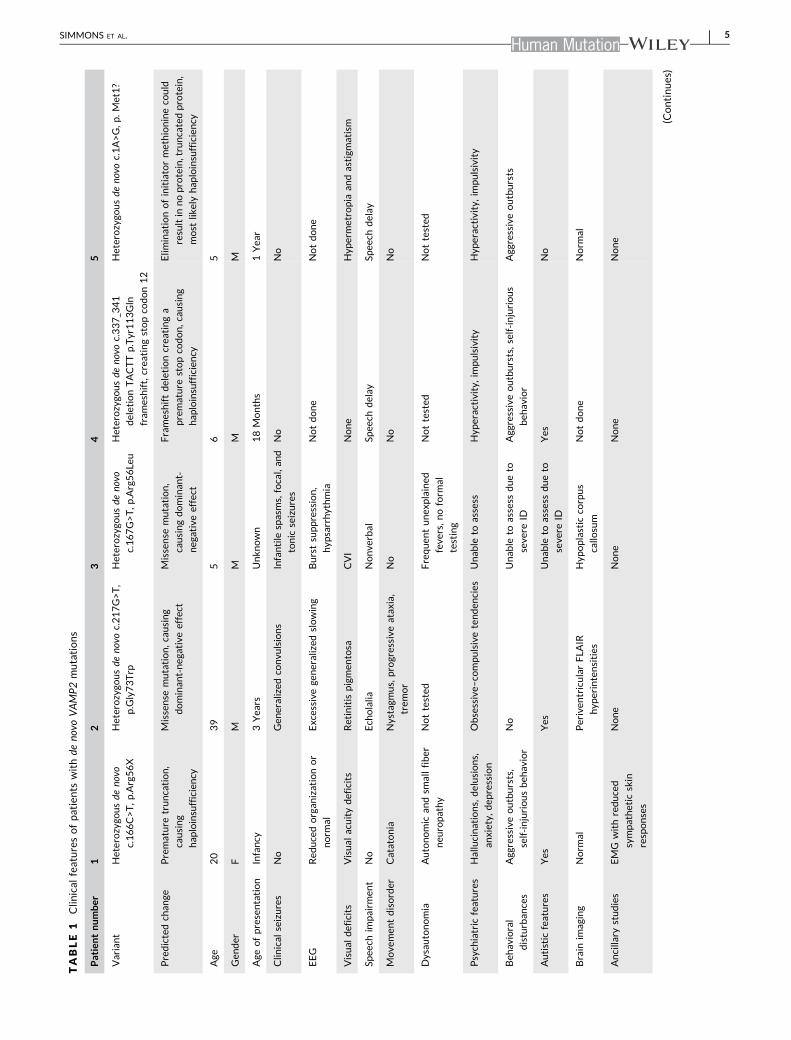
In this study, we describe five previously unreported, unrelated pa-
tients with novel de novo heterozygous VAMP2 pathogenic variants
and their clinical characteristics (Table 1). Patients with these VAMP2
pathogenic variants were assessed by chart review and caretaker
phone interviews. Parents provided written consent before partici-
pation. Common features include global developmental delay, autistic
tendencies, and behavioral disturbances. The SNARE motif VAMP2
variants of Patients 1–3 were studied in vitro. Patients 4 and 5 were
subsequently referred to our study.
3.1 | Case histories
Patient 1 (BRDP ID 2355‐0) exhibited developmental delay and so-
cial impairments beginning in infancy. In adolescence she developed
worsening behavioral problems, aggression, emotional lability, anxi-
ety, hallucinations, and delusions. She was presumptively diagnosed
with Hashimoto's encephalopathy and received empiric treatment
with plasmapheresis, steroids and rituximab, with some degree of
improvement in her mood and function with improved thyroid anti-
body titers, however, she still exhibited slowed psychomotor re-
sponsiveness and cognitive processing. Comprehensive work‐upincluded CSF autoimmune encephalopathy panel (negative) and
neurotransmitter metabolites (normal). MRI showed normal brain
structure. Multiple extended EEGs had either moderate background
slowing or were normal.
She came to UCSF as an adolescent after whole exome se-
quencing revealed a heterozygous de novo VAMP2 mutation
(c.166C>T, p.Arg56X, where c. designates a location in the cDNA and
p. designates a location in the predicted protein) predicting a pre-
mature truncation and haploinsufficiency from nonsense‐mediated
decay. Although not directly tested, this variant could also result in a
dominant‐negative effect. On initial evaluation, she had catatonia and
was largely nonverbal with minimal motor activity. A nerve conduc-
tion study was undertaken to evaluate autonomic nerve function,
where VAMP2 protein is expressed and hence activity is assayable.
Sympathetic skin responses were very low amplitude, with relative
preservation of response latencies, providing confirmation that
VAMP2 haploinsufficiency caused symptoms.
Patient 2 (BDRP 2362‐0) is a 39‐year‐old male with cognitive
impairment, autism spectrum disorder (ASD), epilepsy, and retinitis
pigmentosa. Parents noticed nystagmus during infancy, and he was
evaluated at age 3 years for developmental delay. Whole exome
testing revealed a VAMP2 variant (heterozygous de novo c.217G>T,
p.Gly73Trp). This is predicted to be a missense mutation causing a
dominant‐negative effect. He independently performs some activities
of daily living, such as showering, toileting, and dressing, and parti-
cipates in a sheltered workshop program. He uses basic appliances
and repairs simple things. He speaks in complete sentences and
converses, but he displays obsessive–compulsive tendencies, re-
stricted interests, and echolalia. Progressively worsening ataxia and
4 | SIMMONS ET AL.
TABLE
1Clin
ical
featuresofpatients
withde
novo
VAMP2mutations
Patientnumber
12
34
5
Variant
Heterozygo
usde
novo
c.166C>T,p
.Arg56X
Heterozygo
usde
novo
c.217G>T,
p.Gly73Trp
Heterozygo
usde
novo
c.167G>T,p
.Arg56Le
u
Heterozygo
usde
novo
c.337_341
deletionTACTTp.Tyr113Gln
fram
eshift,crea
tingstopco
don12
Heterozygo
usde
novo
c.1A>G,p
.Met1?
Predictedch
ange
Premature
truncation,
causing
hap
loinsufficiency
Missense
mutation,c
ausing
dominan
t‐neg
ativeeffect
Missense
mutation,
causingdominan
t‐neg
ativeeffect
Frameshiftdeletioncrea
tinga
premature
stopco
don,c
ausing
hap
loinsufficiency
Elim
inationofinitiatormethionineco
uld
resultin
noprotein,truncatedprotein,
most
likelyhap
loinsufficiency
Age
20
39
56
5
Gen
der
FM
MM
M
Age
ofpresentation
Infancy
3Yea
rsUnkn
own
18Months
1Yea
r
Clin
ical
seizures
No
Gen
eralized
convu
lsions
Infantile
spasms,focal,an
d
tonic
seizures
No
No
EEG
Red
ucedorgan
izationor
norm
al
Excessive
generalized
slowing
Burstsuppression,
hyp
sarrhythmia
Notdone
Notdone
Visual
deficits
Visual
acuitydeficits
Retinitis
pigmen
tosa
CVI
None
Hyp
ermetropia
andastigm
atism
Spee
chim
pairm
ent
No
Ech
olalia
Nonve
rbal
Spee
chdelay
Spee
chdelay
Move
men
tdisorder
Catatonia
Nystagm
us,progressive
atax
ia,
trem
or
No
No
No
Dysau
tonomia
Autonomic
andsm
allfiber
neu
ropathy
Nottested
Frequen
tunex
plained
feve
rs,n
oform
al
testing
Nottested
Nottested
Psych
iatric
features
Hallucinations,delusions,
anxiety,
dep
ression
Obsessive–
compulsivetenden
cies
Unab
leto
assess
Hyp
eractivity,impulsivity
Hyp
eractivity,impulsivity
Beh
avioral
disturban
ces
Agg
ressiveoutbursts,
self‐in
juriousbeh
avior
No
Unab
leto
assess
dueto
seve
reID
Agg
ressiveoutbursts,self‐injurious
beh
avior
Agg
ressiveoutbursts
Autistic
features
Yes
Yes
Unab
leto
assess
dueto
seve
reID
Yes
No
Brain
imag
ing
Norm
alPeriven
tricularFLA
IR
hyp
erintensities
Hyp
oplastic
corpus
callo
sum
Notdone
Norm
al
Ancilla
rystudies
EMG
withreduced
sympathetic
skin
responses
None
None
None
None
(Continues)
SIMMONS ET AL. | 5
tremor impair his ability to walk and write. He developed epilepsy at
3 years of age, described as full‐body convulsions, which has been
relatively well‐controlled on oxcarbazepine and more recently la-
motrigine. Parents describe him as mellow, helpful, and cooperative;
he does not have behavioral aggression or hallucinations.
Patient 3 was identified by a clinical laboratory by whole exome
testing. He carries a de novo VAMP2 variant (heterozygous de novo
c.167G>T, p.Arg56Leu) predicted to cause an amino acid missense
mutation predicted to result in a dominant‐negative effect. He is a
6‐year‐old child with refractory infantile spasms and global devel-
opmental delay. EEG showed burst‐suppression and hypsarrhythmia.
MRI of the brain demonstrated a mildly hypoplastic corpus callosum.
Severe intellectual disability confounds any behavioral or autistic
features that might be present.
Patient 4 was born at term without complications after an un-
eventful pregnancy. At 18 months of age, parents noticed issues with
speech and gross motor skills. He was evaluated through their regional
center and began receiving services (speech, occupational, and physical
therapies). Whole exome sequencing at 4 years of age revealed a
variation in VAMP2 (heterozygous de novo c.337_341 deletion TACTT
p.Tyr113Gln frameshift, predicted to create a stop codon 12
[p.Tyr113GlnfsX12]), and resulting haploinsufficiency. Later he was
diagnosed with ASD and attention deficit hyperactivity disorder
(ADHD). He also carries a maternally inherited deletion of unclear
significance (15q13.3 between D‐CHRNA7 to BP5 arr[GRCh37]
15q13.3 [32024132_32509926; size: 486 Kb]). The patient's mother is
cognitively normal. Reports show incomplete penetrance of this dele-
tion, ranging from mildly affected to normal individuals (Shinawi et al.,
2009). While an additive contribution of this deletion to the VAMP2
phenotype in this patient cannot be excluded, it remains uncertain
given that the patient's mother is phenotypically normal. Currently
6 years old, he is making developmental progress in prekindergarten
with services. He can speak in short sentences and can repeat phrases;
however, does not know primary colors or ABCs. For fine motor skills,
he can color. He walks independently but is clumsy and falls frequently.
He exhibits aggressive outbursts and self‐injurious behavior. He is
hyperactive and impulsive, for which he takes stimulant medications.
He does not have a history of seizures or movement disorders.
Patient 5 was born at term, and the pregnancy was complicated
by polyhydramnios and decreased fetal movements. Parents first no-
ticed developmental differences at 1 year of age, mainly involving
speech but eventually including low muscle tone and frequent falls. At
1 year of age, he spoke no words. Audiometry showed a mild hearing
loss, likely due to frequent ear infections, which resolved with tym-
panostomy tubes. His family noticed frequent falls (upwards of 30 falls
per day), trouble climbing stairs, and difficulty transitioning from dif-
ferent positions. With therapies (speech, occupational, and physical),
he saw slow improvement in these domains; however, his mother
estimates he continues functioning several years behind expected
milestones. Currently, 5 years old, he speaks in five‐word sentences
but has trouble understanding multistep commands and is slow to
process verbal information. He can run, walk slowly upstairs, and
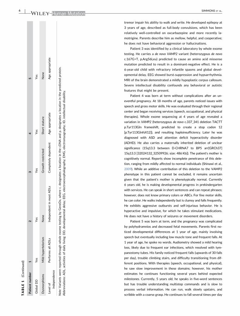
scribble with a coarse grasp. He continues to fall several times per dayTABLE
1(Continued
)
Patientnumber
12
34
5
Global
DD
Yes
Yes
Yes
Yes
Yes
Dysmorphisms
Mild
hyp
ertelorism
None
Growth
restricted
Short
stature
None
Leve
lof
indep
enden
ce
Perform
sallADLs
Indep
enden
tin
most
ADLs
Completely
dep
enden
tAge
appropriate
Age
appropriate
Note:
Variants
werereported
through
whole
exometestingbyGen
eDx,
wherec.
designates
alocationin
thecD
NA
andp.d
esignates
alocationin
thepredictedprotein.
Abbreviations:
ADL,
activities
ofdaily
living;
DD,dev
elopmen
taldelay
;EEG,e
lectroen
cephalograp
hy;
EMG,e
lectromyo
grap
hy;
ID,intelle
ctual
disab
ility.
6 | SIMMONS ET AL.
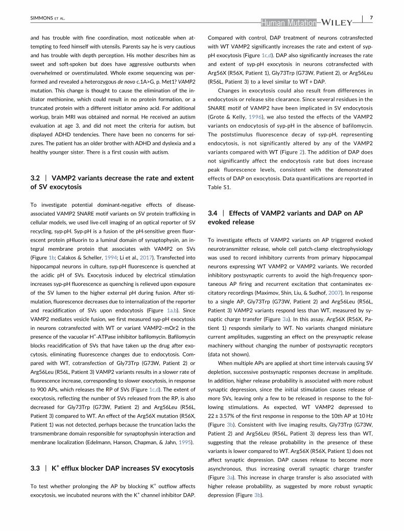
and has trouble with fine coordination, most noticeable when at-
tempting to feed himself with utensils. Parents say he is very cautious
and has trouble with depth perception. His mother describes him as
sweet and soft‐spoken but does have aggressive outbursts when
overwhelmed or overstimulated. Whole exome sequencing was per-
formed and revealed a heterozygous de novo c.1A>G, p. Met1? VAMP2
mutation. This change is thought to cause the elimination of the in-
itiator methionine, which could result in no protein formation, or a
truncated protein with a different initiator amino acid. For additional
workup, brain MRI was obtained and normal. He received an autism
evaluation at age 3, and did not meet the criteria for autism, but
displayed ADHD tendencies. There have been no concerns for sei-
zures. The patient has an older brother with ADHD and dyslexia and a
healthy younger sister. There is a first cousin with autism.
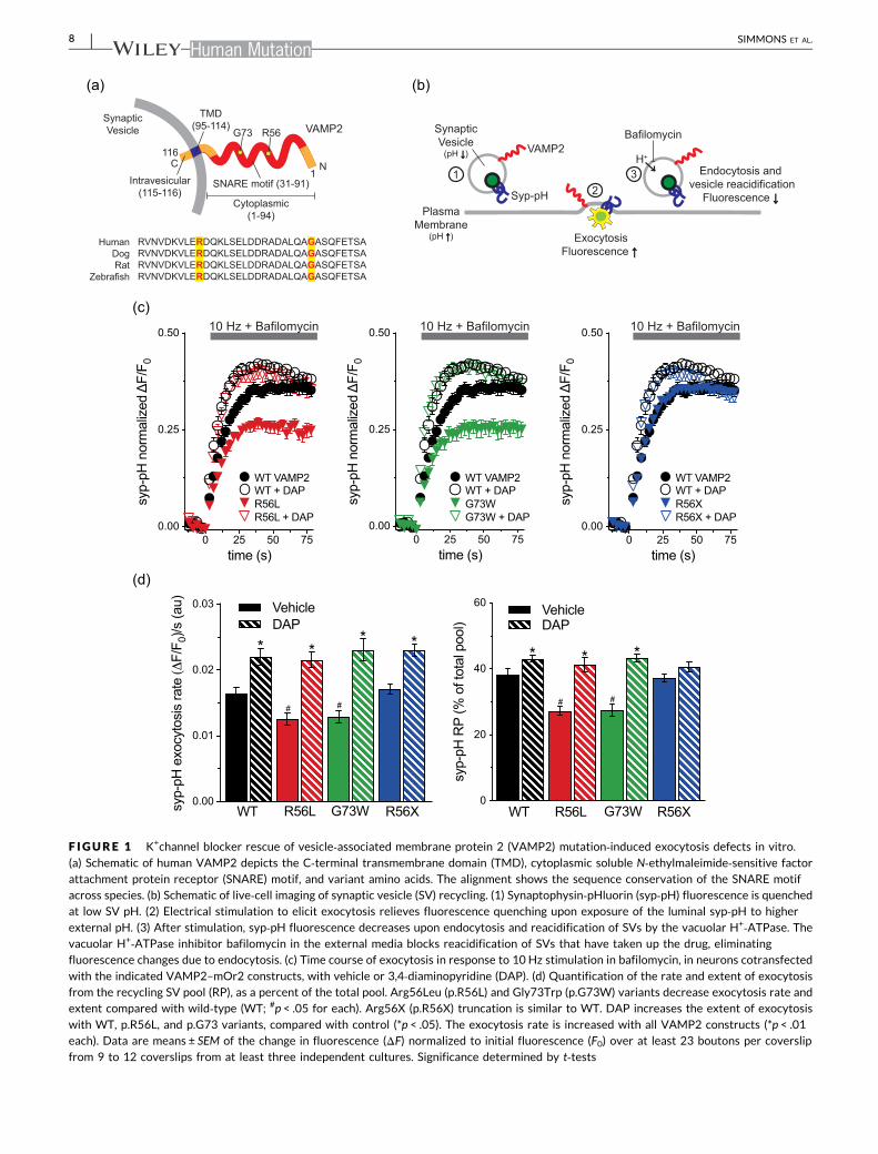
3.2 | VAMP2 variants decrease the rate and extentof SV exocytosis
To investigate potential dominant‐negative effects of disease‐associated VAMP2 SNARE motif variants on SV protein trafficking in
cellular models, we used live‐cell imaging of an optical reporter of SV
recycling, syp‐pH. Syp‐pH is a fusion of the pH‐sensitive green fluor-
escent protein pHluorin to a luminal domain of synaptophysin, an in-
tegral membrane protein that associates with VAMP2 on SVs
(Figure 1b; Calakos & Scheller, 1994; Li et al., 2017). Transfected into
hippocampal neurons in culture, syp‐pH fluorescence is quenched at
the acidic pH of SVs. Exocytosis induced by electrical stimulation
increases syp‐pH fluorescence as quenching is relieved upon exposure
of the SV lumen to the higher external pH during fusion. After sti-
mulation, fluorescence decreases due to internalization of the reporter
and reacidification of SVs upon endocytosis (Figure 1a,b). Since
VAMP2 mediates vesicle fusion, we first measured syp‐pH exocytosis
in neurons cotransfected with WT or variant VAMP2–mOr2 in the
presence of the vacuolar H+‐ATPase inhibitor bafilomycin. Bafilomycin
blocks reacidification of SVs that have taken up the drug after exo-
cytosis, eliminating fluorescence changes due to endocytosis. Com-
pared with WT, cotransfection of Gly73Trp (G73W, Patient 2) or
Arg56Leu (R56L, Patient 3) VAMP2 variants results in a slower rate of
fluorescence increase, corresponding to slower exocytosis, in response
to 900 APs, which releases the RP of SVs (Figure 1c,d). The extent of
exocytosis, reflecting the number of SVs released from the RP, is also
decreased for Gly73Trp (G73W, Patient 2) and Arg56Leu (R56L,
Patient 3) compared to WT. An effect of the Arg56X mutation (R56X,
Patient 1) was not detected, perhaps because the truncation lacks the
transmembrane domain responsible for synaptophysin interaction and
membrane localization (Edelmann, Hanson, Chapman, & Jahn, 1995).
3.3 | K+ efflux blocker DAP increases SV exocytosis
To test whether prolonging the AP by blocking K+ outflow affects
exocytosis, we incubated neurons with the K+ channel inhibitor DAP.
Compared with control, DAP treatment of neurons cotransfected
with WT VAMP2 significantly increases the rate and extent of syp‐pH exocytosis (Figure 1c,d). DAP also significantly increases the rate
and extent of syp‐pH exocytosis in neurons cotransfected with
Arg56X (R56X, Patient 1), Gly73Trp (G73W, Patient 2), or Arg56Leu
(R56L, Patient 3) to a level similar to WT +DAP.
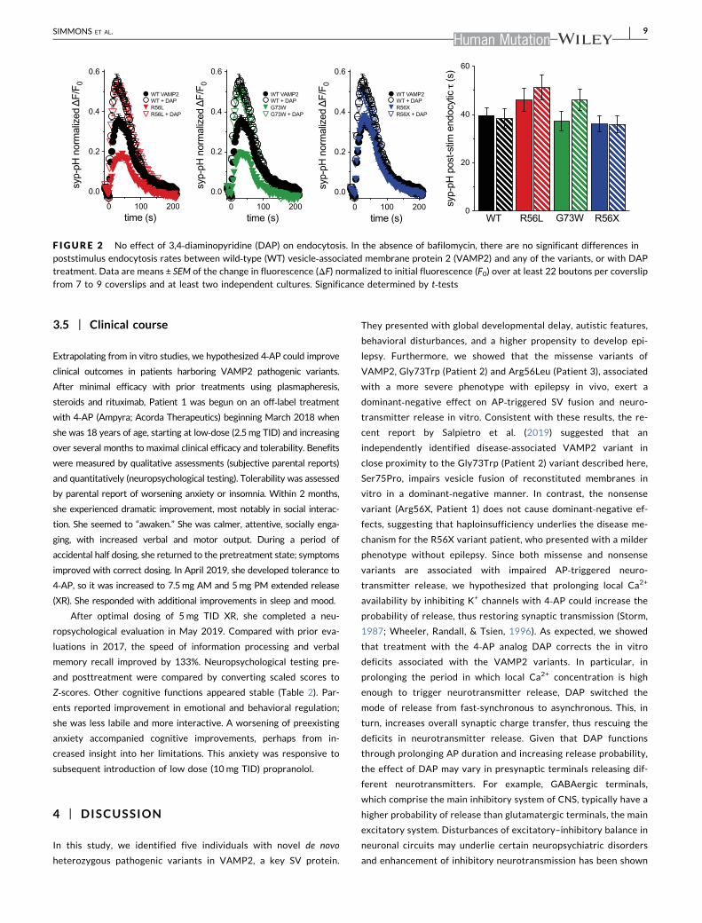
Changes in exocytosis could also result from differences in
endocytosis or release site clearance. Since several residues in the
SNARE motif of VAMP2 have been implicated in SV endocytosis
(Grote & Kelly, 1996), we also tested the effects of the VAMP2
variants on endocytosis of syp‐pH in the absence of bafilomycin.
The poststimulus fluorescence decay of syp‐pH, representing
endocytosis, is not significantly altered by any of the VAMP2
variants compared with WT (Figure 2). The addition of DAP does
not significantly affect the endocytosis rate but does increase
peak fluorescence levels, consistent with the demonstrated
effects of DAP on exocytosis. Data quantifications are reported in
Table S1.
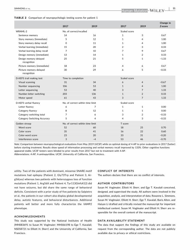
3.4 | Effects of VAMP2 variants and DAP on APevoked release
To investigate effects of VAMP2 variants on AP triggered evoked
neurotransmitter release, whole cell patch‐clamp electrophysiology
was used to record inhibitory currents from primary hippocampal
neurons expressing WT VAMP2 or VAMP2 variants. We recorded
inhibitory postsynaptic currents to avoid the high‐frequency spon-
taneous AP firing and recurrent excitation that contaminates ex-
citatory recordings (Maximov, Shin, Liu, & Sudhof, 2007). In response
to a single AP, Gly73Trp (G73W, Patient 2) and Arg56Leu (R56L,
Patient 3) VAMP2 variants respond less than WT, measured by sy-
naptic charge transfer (Figure 3a). In this assay, Arg56X (R56X, Pa-
tient 1) responds similarly to WT. No variants changed miniature
current amplitudes, suggesting an effect on the presynaptic release
machinery without changing the number of postsynaptic receptors
(data not shown).
When multiple APs are applied at short time intervals causing SV
depletion, successive postsynaptic responses decrease in amplitude.
In addition, higher release probability is associated with more robust
synaptic depression, since the initial stimulation causes release of
more SVs, leaving only a few to be released in response to the fol-
lowing stimulations. As expected, WT VAMP2 depressed to
22 ± 3.57% of the first response in response to the 10th AP at 10 Hz
(Figure 3b). Consistent with live imaging results, Gly73Trp (G73W,
Patient 2) and Arg56Leu (R56L, Patient 3) depress less than WT,
suggesting that the release probability in the presence of these
variants is lower compared to WT. Arg56X (R56X, Patient 1) does not
affect synaptic depression. DAP causes release to become more
asynchronous, thus increasing overall synaptic charge transfer
(Figure 3a). This increase in charge transfer is also associated with
higher release probability, as suggested by more robust synaptic
depression (Figure 3b).
SIMMONS ET AL. | 7
0.00
0.01
0.02
0.03
syp-
pH e
xocy
tosi
s ra
te
F/F 0
)/s (a
u)
VehicleDAP
WT R56L G73W R56X
* ** *
# #
0
20
40
60
syp-
pH R
P (%
of t
otal
poo
l)
VehicleDAP
WT R56L G73W R56X
* * *
# #
0 25 50 750.00
0.25
0.50
time (s)
syp-
pH n
orm
aliz
ed
F/F 0
R56L + DAP
WT VAMP2WT + DAPR56L
0 25 50 750.00
0.25
0.50
time (s)
syp-
pH n
orm
aliz
ed
F/F 0
G73W + DAP
WT VAMP2WT + DAPG73W
0 25 50 750.00
0.25
0.50
time (s)sy
p-pH
nor
mal
ized
F/
F 0
R56X + DAP
WT VAMP2WT + DAPR56X
10 Hz + Bafilomycin 10 Hz + Bafilomycin 10 Hz + Bafilomycin
Syp-pH
VAMP2
SynapticVesicle
(pH ↓↓)
PlasmaMembrane
(pH ↑↑)
H+
ExocytosisFluorescence ↑↑
Endocytosis and vesicle reacidification
Fluorescence ↓↓
12
3
Bafilomycin
TMD(95-114)
C NSNARE motif (31-91)
1
116
Intravesicular(115-116)
Cytoplasmic(1-94)
G73 R56SynapticVesicle
RVNVDKVLERDQKLSELDDRADALQAGASQFETSARVNVDKVLERDQKLSELDDRADALQAGASQFETSARVNVDKVLERDQKLSELDDRADALQAGASQFETSARVNVDKVLERDQKLSELDDRADALQAGASQFETSA
HumanDogRat
Zebrafish
VAMP2
(a) (b)
(c)
(d)
F IGURE 1 K+channel blocker rescue of vesicle‐associated membrane protein 2 (VAMP2) mutation‐induced exocytosis defects in vitro.
(a) Schematic of human VAMP2 depicts the C‐terminal transmembrane domain (TMD), cytoplasmic soluble N‐ethylmaleimide‐sensitive factorattachment protein receptor (SNARE) motif, and variant amino acids. The alignment shows the sequence conservation of the SNARE motifacross species. (b) Schematic of live‐cell imaging of synaptic vesicle (SV) recycling. (1) Synaptophysin‐pHluorin (syp‐pH) fluorescence is quenchedat low SV pH. (2) Electrical stimulation to elicit exocytosis relieves fluorescence quenching upon exposure of the luminal syp‐pH to higher
external pH. (3) After stimulation, syp‐pH fluorescence decreases upon endocytosis and reacidification of SVs by the vacuolar H+‐ATPase. Thevacuolar H+‐ATPase inhibitor bafilomycin in the external media blocks reacidification of SVs that have taken up the drug, eliminatingfluorescence changes due to endocytosis. (c) Time course of exocytosis in response to 10 Hz stimulation in bafilomycin, in neurons cotransfected
with the indicated VAMP2–mOr2 constructs, with vehicle or 3,4‐diaminopyridine (DAP). (d) Quantification of the rate and extent of exocytosisfrom the recycling SV pool (RP), as a percent of the total pool. Arg56Leu (p.R56L) and Gly73Trp (p.G73W) variants decrease exocytosis rate andextent compared with wild‐type (WT; #p < .05 for each). Arg56X (p.R56X) truncation is similar to WT. DAP increases the extent of exocytosis
with WT, p.R56L, and p.G73 variants, compared with control (*p < .05). The exocytosis rate is increased with all VAMP2 constructs (*p < .01each). Data are means ± SEM of the change in fluorescence (ΔF) normalized to initial fluorescence (F0) over at least 23 boutons per coverslipfrom 9 to 12 coverslips from at least three independent cultures. Significance determined by t‐tests
8 | SIMMONS ET AL.
3.5 | Clinical course
Extrapolating from in vitro studies, we hypothesized 4‐AP could improve
clinical outcomes in patients harboring VAMP2 pathogenic variants.
After minimal efficacy with prior treatments using plasmapheresis,
steroids and rituximab, Patient 1 was begun on an off‐label treatment
with 4‐AP (Ampyra; Acorda Therapeutics) beginning March 2018 when
she was 18 years of age, starting at low‐dose (2.5mg TID) and increasing
over several months to maximal clinical efficacy and tolerability. Benefits
were measured by qualitative assessments (subjective parental reports)
and quantitatively (neuropsychological testing). Tolerability was assessed
by parental report of worsening anxiety or insomnia. Within 2 months,
she experienced dramatic improvement, most notably in social interac-
tion. She seemed to “awaken.” She was calmer, attentive, socially enga-
ging, with increased verbal and motor output. During a period of
accidental half dosing, she returned to the pretreatment state; symptoms
improved with correct dosing. In April 2019, she developed tolerance to
4‐AP, so it was increased to 7.5mg AM and 5mg PM extended release
(XR). She responded with additional improvements in sleep and mood.
After optimal dosing of 5mg TID XR, she completed a neu-
ropsychological evaluation in May 2019. Compared with prior eva-
luations in 2017, the speed of information processing and verbal
memory recall improved by 133%. Neuropsychological testing pre‐and posttreatment were compared by converting scaled scores to
Z‐scores. Other cognitive functions appeared stable (Table 2). Par-
ents reported improvement in emotional and behavioral regulation;
she was less labile and more interactive. A worsening of preexisting
anxiety accompanied cognitive improvements, perhaps from in-
creased insight into her limitations. This anxiety was responsive to
subsequent introduction of low dose (10mg TID) propranolol.
4 | DISCUSSION
In this study, we identified five individuals with novel de novo
heterozygous pathogenic variants in VAMP2, a key SV protein.
They presented with global developmental delay, autistic features,
behavioral disturbances, and a higher propensity to develop epi-
lepsy. Furthermore, we showed that the missense variants of
VAMP2, Gly73Trp (Patient 2) and Arg56Leu (Patient 3), associated
with a more severe phenotype with epilepsy in vivo, exert a
dominant‐negative effect on AP‐triggered SV fusion and neuro-
transmitter release in vitro. Consistent with these results, the re-
cent report by Salpietro et al. (2019) suggested that an
independently identified disease‐associated VAMP2 variant in
close proximity to the Gly73Trp (Patient 2) variant described here,
Ser75Pro, impairs vesicle fusion of reconstituted membranes in
vitro in a dominant‐negative manner. In contrast, the nonsense
variant (Arg56X, Patient 1) does not cause dominant‐negative ef-
fects, suggesting that haploinsufficiency underlies the disease me-
chanism for the R56X variant patient, who presented with a milder
phenotype without epilepsy. Since both missense and nonsense
variants are associated with impaired AP‐triggered neuro-
transmitter release, we hypothesized that prolonging local Ca2+
availability by inhibiting K+ channels with 4‐AP could increase the
probability of release, thus restoring synaptic transmission (Storm,
1987; Wheeler, Randall, & Tsien, 1996). As expected, we showed
that treatment with the 4‐AP analog DAP corrects the in vitro
deficits associated with the VAMP2 variants. In particular, in
prolonging the period in which local Ca2+ concentration is high
enough to trigger neurotransmitter release, DAP switched the
mode of release from fast‐synchronous to asynchronous. This, in
turn, increases overall synaptic charge transfer, thus rescuing the
deficits in neurotransmitter release. Given that DAP functions
through prolonging AP duration and increasing release probability,
the effect of DAP may vary in presynaptic terminals releasing dif-
ferent neurotransmitters. For example, GABAergic terminals,
which comprise the main inhibitory system of CNS, typically have a
higher probability of release than glutamatergic terminals, the main
excitatory system. Disturbances of excitatory–inhibitory balance in
neuronal circuits may underlie certain neuropsychiatric disorders
and enhancement of inhibitory neurotransmission has been shown
0 100 200
0.0
0.2
0.4
0.6
time (s)
syp-
pH n
orm
aliz
ed
F/F 0
0 100 200
0.0
0.2
0.4
0.6
time (s)
syp-
pH n
orm
aliz
ed
F/F 0
0 100 200
0.0
0.2
0.4
0.6
time (s)
syp-
pH n
orm
aliz
ed
F/F 0
R56L + DAP
WT VAMP2WT + DAPR56L
G73W + DAP
WT VAMP2WT + DAPG73W
R56X + DAP
WT VAMP2WT + DAPR56X
0
20
40
60
syp-
pH p
ost-s
tim e
ndoc
ytic
(s
)
WT R56L G73W R56X
F IGURE 2 No effect of 3,4‐diaminopyridine (DAP) on endocytosis. In the absence of bafilomycin, there are no significant differences inpoststimulus endocytosis rates between wild‐type (WT) vesicle‐associated membrane protein 2 (VAMP2) and any of the variants, or with DAPtreatment. Data are means ± SEM of the change in fluorescence (ΔF) normalized to initial fluorescence (F0) over at least 22 boutons per coverslip
from 7 to 9 coverslips and at least two independent cultures. Significance determined by t‐tests
SIMMONS ET AL. | 9

to ameliorate some behavioral deficits in mouse models of autism
(Sohal & Rubenstein, 2019).
Extrapolating from in vitro studies, we started 4‐AP (Ampyra)
treatment for Patient 1, the Arg56X affected individual, in 2018.
She responded dramatically, confirming that in vitro studies suc-
cessfully predicted clinical response. With treatment for the last 2
years, Patient 1 shows remarkable improvement in cognitive
processing speed and verbal memory recall. Treatment drastically
improved the quality of life for our patient and her family given
her enhanced ability to interact and decreased emotional lability.
In this study, we demonstrate that augmentation of neuro-
transmitter release by aminopyridines can be a viable treatment
option for VAMP2 associated disorders with impaired neuro-
transmitter release. Most importantly, we showed the first
evidence of clinical improvement upon 4‐AP treatment in a pa-
tient harboring a nonsense variant of VAMP2. 4‐AP could treat
other patients with VAMP2 or other SNARE protein mutations.
Our results are in agreement with recent in vitro studies showing
that DAP also can overcome release deficits associated with
disease‐causing synaptotagmin‐1 variants (Bradberry et al., 2020).
Taken together, these observations confirm and expand our hy-
pothesis that augmentation of release by DAP or 4‐AP would be a
viable treatment option for other SNAREopathies as well (Baker
et al., 2018; Harper, Mancini, van Slegtenhorst, & Cousin, 2017;
Salpietro et al., 2017; Verhage & Sorensen, 2020). This approach
could be tested in vitro before clinical implementation. 4‐APshould be used cautiously in patients with epilepsy given the risk
of lowering the seizure threshold, which can limit its broad‐based
WT
WT + DAP
R56LG73
WR56
X
R56X +
DAP0.0
0.5
1.0
1.5
2.0
Syna
ptic
Cha
rge
Tran
sfer
(nC
)
*
#
#
*
0 500 1000 1500 20000.0
0.5
1.0
Time (ms)
Cum
ulat
ive
Cha
rge
Tran
sfer
WT VAMP2
G73WR56LR56XR56X + DAP
WT + DAP *
*1 nA
100 ms
WT VAMP2
WT + DAP
R56X
R56X + DAP
1 2 3 4 5 6 7 8 9 100
20
40
60
80
100
Stimulation #
Nor
mal
ized
Pea
k Am
plitu
de (%
)
*
* * ** * * * *
1 2 3 4 5 6 7 8 9 100
20
40
60
80
100
Stimulation #
Nor
mal
ized
Pea
k Am
plitu
de (%
)
** * * * * * * *
1 2 3 4 5 6 7 8 9 100
20
40
60
80
100
Stimulation #N
orm
aliz
ed P
eak
Ampl
itude
(%)
** * * * * * * *
#
# # # # # # # #
WT VAMP2R56L G73W
WT VAMP2
R56XR56X + DAP
WT VAMP2WT + DAP
(a)
(b)
F IGURE 3 Effects of vesicle‐associated membrane protein 2 (VAMP2) variants and 3,4‐diaminopyridine (DAP) on evoked release. (a) p.R56Land p.G73W variants decrease the overall synaptic charge transfer, measured as the area under the curve of an evoked inhibitory postsynapticcurrent (eIPSC). DAP treatments increase synaptic charge transfer (wild‐type [WT]: 0.655 ± 0.141 nC, WT +DAP: 1.195 ± 0.128 nC, p.R56L:
0.315 ± 0.055 nC, p.G73W: 0.323 ± 0.045 nC, p.R56X: 0.593 ± 0.088 nC, p.R56X +DAP: 1.287 ± 0.187 nC, *p < .05 for WT vs. p.R56L and WT vs.p.G73W, #p < .05 for WT vs. WT +DAP and p.R56X vs. p.R56X + DAP, two‐tailed t‐test). Variants do not change the synchronicity of the release(*p > .05 for WT vs. p.R56L, p.G73W and p.R56X, Kolmogorov–Smirnov test), but corresponding 3,4‐DAP treatments result in more
asynchronous release (*p < .001 for both WT vs. WT +DAP and p.R56X vs. p.R56X +DAP, Kolmogorov–Smirnov test). (b) Normalized peakamplitudes of eIPSCs in response to 10 consecutive stimulations at 10 Hz. p.G73W and p.R56L variants cause less depression after initialstimulation compared to WT VAMP2 (*p < .05 for WT VAMP2 vs. p.G73W and WT VAMP2 vs. p.R56L, multiple t‐tests for each stimulation).
Although p.R56X responds similarly as WT, corresponding DAP treatments cause more robust depression in response to stimulation (p < .05 for*WT vs. WT +DAP and #p.R56X vs. p.R56X+DAP, multiple t‐tests for each stimulation)
10 | SIMMONS ET AL.
utility. Two of the patients with dominant, missense SNARE motif
mutations had epilepsy (Patient 2, Gly73Trp and Patient 3, Ar-
g56Leu) whereas two patients with heterozygous loss of function
mutations (Patient 1, Arg56X and Patient 4, Tyr113GlnfsX12) did
not have seizures, but did share the same range of behavioral
deficits. Consistent with a prior study of five patients by Salpietro
et al., the patients in our cohort also display global developmental
delay, autistic features, and behavioral disturbances. Additional
patients will better and more fully characterize the VAMP2
phenotype.
ACKNOWLEDGMENTS
This study was supported by the National Institutes of Health
(MH083691 to Susan M. Voglmaier; MH066198 to Ege T. Kavalali;
NS058721 to Elliott H. Sherr) and the University of California, San
Francisco.
CONFLICT OF INTERESTS
The authors declare that there are no conflict of interests.
AUTHOR CONTRIBUTORS
Susan M. Voglmaier, Elliott H. Sherr, and Ege T. Kavalali conceived,
designed, and supervised the study. All authors were involved in the
acquisition, analysis, and interpretation of data. Roxanne L. Simmons,
Susan M. Voglmaier, Elliott H. Sherr, Ege T. Kavalali, Baris Alten, and
Haiyan Li drafted and critically revised the manuscript for important
intellectual content. Susan M. Voglmaier and Elliott H. Sherr are re-
sponsible for the overall content of the manuscript.
DATA AVAILABILITY STATEMENT
The data that support the findings of this study are available on
request from the corresponding author. The data are not publicly
available due to privacy or ethical restrictions.
TABLE 2 Comparison of neuropsychologic testing scores for patient 1
2017 2019 2017 2019Change inZ‐score
WRAML‐2 No. of correct/recalled Scaled score
Sentence memory 14 16 1 3 0.67
Story memory (Immediate) 5 12 1 4 1.00
Story memory delay recall 0 11 1 4 1.00
Verbal learning (immediate) 15 20 2 3 0.33
Verbal learning delay recall 7 10 7 9 0.67
Design memory (immediate) 13 14 1 2 0.33
Design memory delayed
recognition
25 21 5 1 −1.33
Picture memory (immediate) 18 23 4 6 0.67
Picture memory delayed
recognition
30 29 6 5 −0.33
D‐KEFS trail making test Time to completion Scaled score
Visual scanning 31 34 6 4 −0.67
Number sequencing 106 53 1 4 1.00
Letter sequencing 53 40 3 7 1.33
Number‐letter switching 203 136 1 2 0.33
Motor speed 61 43 3 7 1.33
D‐KEFS verbal fluency No. of correct within time limit Scaled score
Letter fluency 6 7 1 1 0.00
Category fluency 8 12 1 1 0.00
Category switching total 7 6 3 2 −0.33
Category Switching Accuracy 5 4 4 3 −0.33
Golden stroop No. of correct within time limit T score
Word score 52 60 18 25 0.70
Color score 35 41 16 22 0.60
Color‐word score 23 20 33 31 −0.20
Interference score 3 −4 53 46 −0.70
Note: Comparison between neuropsychological evaluations from May 2019 (UCSF) while on optimal dosing of 4‐AP to prior evaluations in 2017 (Sutter)
before starting treatment. Results show speed of information processing and verbal memory recall improved by 133%. Other cognitive functions
appeared stable. UCSF testers were blinded to prior results from 2017 but not to treatment with 4‐AP.Abbreviations: 4‐AP, 4‐aminopyridine; UCSF, University of California, San Francisco.
SIMMONS ET AL. | 11
WEB RESOURCES
GeneDx
http://www.genedx.com
ClinVar
http://www.ncbi.nlm.nih.gov/clinvar/
Poly‐Phen2http://genetics.bwh.harvard.edu/pph2/
GnomAD
http://gnomad.broadinstitute.org
ACCESSION NUMBERS FROM CLINVAR FOR VAMP2
VARIANTS
Patient 1: SUB7621453; c.166C>T, p.Arg56X
http://www.ncbi.nlm.nih.gov/clinvar/variation/929460/
Patient 2: SUB7630353; c.217G>T, p.Gly73Trp
http://www.ncbi.nlm.nih.gov/clinvar/variation/929461/
Patient 3: SUB7630360; c.167G>T; p.Arg56Leu
http://www.ncbi.nlm.nih.gov/clinvar/variation/929463/
Patient 4: SUB7630356; c.337_341delTACTT, p.Tyr113GlnfsX12
http://www.ncbi.nlm.nih.gov/clinvar/variation/929464/
Patient 5: SUB7630357; c.1A>G, p. Met1?
http://www.ncbi.nlm.nih.gov/clinvar/variation/929462/
ORCID
Marwan Shinawi http://orcid.org/0000-0003-1329-4100
Susan M. Voglmaier http://orcid.org/0000-0002-1211-8108
REFERENCES
Baker, K., Gordon, S. L., Melland, H., Bumbak, F., Scott, D. J., Jiang, T. J., …
Raymond, F. L. (2018). SYT1‐associated neurodevelopmental disorder:
A case series. Brain, 141(9), 2576–2591. https://doi.org/10.1093/
brain/awy209
Bradberry, M. M., Courtney, N. A., Dominguez, M. J., Lofquist, S. M.,
Knox, A. T., Sutton, R. B., & Chapman, E. R. (2020). Molecular basis for
synaptotagmin‐1‐associated neurodevelopmental disorder. Neuron,
107(1), 57–64.e7. https://doi.org/10.1016/j.neuron.2020.04.003
Calakos, N., & Scheller, R. H. (1994). Vesicle‐associated membrane protein
and synaptophysin are associated on the synaptic vesicle. Journal of
Biological Chemistry, 269(40), 24534–24537.
Claassen, J., Teufel, J., Kalla, R., Spiegel, R., & Strupp, M. (2013). Effects of
dalfampridine on attacks in patients with episodic ataxia type 2: An
observational study. Journal of Neurology, 260(2), 668–669. https://doi.
org/10.1007/s00415-012-6764-3
Deak, F., Schoch, S., Liu, X., Sudhof, T. C., & Kavalali, E. T. (2004).
Synaptobrevin is essential for fast synaptic‐vesicle endocytosis. Nature
Cell Biology, 6(11), 1102–1108.
Edelmann, L., Hanson, P. I., Chapman, E. R., & Jahn, R. (1995). Synaptobrevin
binding to synaptophysin: A potential mechanism for controlling the
exocytotic fusion machine. EMBO Journal, 14(2), 224–231.
Fasshauer, D., Eliason, W. K., Brunger, A. T., & Jahn, R. (1998).
Identification of a minimal core of the synaptic SNARE complex
sufficient for reversible assembly and disassembly. Biochemistry,
37(29), 10354–10362.
Foss, S. M., Li, H., Santos, M. S., Edwards, R. H., & Voglmaier, S. M. (2013).
Multiple dileucine‐like motifs direct VGLUT1 trafficking. The Journal of
Neuroscience, 33(26), 10647–10660. https://doi.org/10.1523/JNEUR
OSCI.5662-12.2013
Grote, E., & Kelly, R. B. (1996). Endocytosis of VAMP is facilitated by a
synaptic vesicle targeting signal. Journal of Cell Biology, 132, 537–547.
Harper, C. B., Mancini, G. M. S., van Slegtenhorst, M., & Cousin, M. A. (2017).
Altered synaptobrevin‐II trafficking in neurons expressing a
synaptophysin mutation associated with a severe neurodevelopmental
disorder. Neurobiology of Disease, 108, 298–306. https://doi.org/10.
1016/j.nbd.2017.08.021
Jahn, R., & Scheller, R. H. (2006). SNAREs—Engines for membrane fusion.
Nature Reviews Molecular Cell Biology, 7(9), 631–643. https://doi.org/10.
1038/nrm2002
Kavalali, E. T., Klingauf, J., & Tsien, R. W. (1999). Activity‐dependent regulationof synaptic clustering in a hippocampal culture system. Proceedings of the
National Academy of Sciences of the United States of America, 96(22),
12893–12900. https://doi.org/10.1073/pnas.96.22.12893
Koo, S. J., Markovic, S., Puchkov, D., Mahrenholz, C. C., Beceren‐Braun, F.,Maritzen, T., … Haucke, V. (2011). SNARE motif‐mediated sorting of
synaptobrevin by the endocytic adaptors clathrin assembly lymphoid
myeloid leukemia (CALM) and AP180 at synapses. Proceedings of the
National Academy of Sciences of the United States of America, 108(33),
13540–13545. https://doi.org/10.1073/pnas.1107067108
Li, H., Santos, M. S., Park, C. K., Dobry, Y., & Voglmaier, S. M. (2017).
VGLUT2 trafficking is differentially regulated by adaptor proteins AP‐1 and AP‐3. Frontiers in Cellular Neuroscience, 11, 324. https://doi.org/
10.3389/fncel.2017.00324
Maximov, A., Shin, O. H., Liu, X., & Sudhof, T. C. (2007). Synaptotagmin‐12,a synaptic vesicle phosphoprotein that modulates spontaneous
neurotransmitter release. Journal of Cell Biology, 176(1), 113–124.
Mohrmann, R., & Sorensen, J. B. (2012). SNARE requirements en route to
exocytosis: From many to few. Journal of Molecular Neuroscience, 48(2),
387–394. https://doi.org/10.1007/s12031-012-9744-2
Monteggia, L. M., Lin, P. Y., Adachi, M., & Kavalali, E. T. (2018). Behavioral
analysis of SNAP‐25 and synaptobrevin‐2 haploinsufficiency in mice.
Neuroscience, 420, 129–135. https://doi.org/10.1016/j.neuroscience.
2018.08.014
Palace, J., Wiles, C. M., & Newsom‐Davis, J. (1991). 3,4‐Diaminopyridine in
the treatment of congenital (hereditary) myasthenia. Journal of
Neurology, Neurosurgery and Psychiatry, 54(12), 1069–1072. https://
doi.org/10.1136/jnnp.54.12.1069
Salpietro, V., Lin, W., Delle Vedove, A., Storbeck, M., Liu, Y., Efthymiou, S.,
… Houlden, H. (2017). Homozygous mutations in VAMP1 cause a
presynaptic congenital myasthenic syndrome. Annals of Neurology,
81(4), 597–603. https://doi.org/10.1002/ana.24905
Salpietro, V., Malintan, N. T., Llano‐Rivas, I., Spaeth, C. G., Efthymiou, S.,
Striano, P., … Houlden, H. (2019). Mutations in the neuronal vesicular
SNARE VAMP2 affect synaptic membrane fusion and impair human
neurodevelopment. American Journal of Human Genetics, 104(4),
721–730. https://doi.org/10.1016/j.ajhg.2019.02.016
Shaner, N. C., Lin, M. Z., McKeown, M. R., Steinbach, P. A., Hazelwood, K. L.,
Davidson, M. W., & Tsien, R. Y. (2008). Improving the photostability of
bright monomeric orange and red fluorescent proteins. Nature
Methods, 5(6), 545–551.
Shinawi, M., Schaaf, C. P., Bhatt, S. S., Xia, Z., Patel, A., Cheung, S. W., …
Stankiewicz, P. (2009). A small recurrent deletion within 15q13.3 is
associated with a range of neurodevelopmental phenotypes. Nature
Genetics, 41(12), 1269–1271. https://doi.org/10.1038/ng.481
Sohal, V. S., & Rubenstein, J. L. R. (2019). Excitation‐inhibition balance as a
framework for investigating mechanisms in neuropsychiatric
disorders. Molecular Psychiatry, 24(9), 1248–1257. https://doi.org/10.
1038/s41380-019-0426-0
Storm, J. F. (1987). Action potential repolarization and a fast after‐hyperpolarization in rat hippocampal pyramidal cells. Journal of
Physiology, 385, 733–759. https://doi.org/10.1113/jphysiol.1987.
sp016517
Strupp, M., Teufel, J., Zwergal, A., Schniepp, R., Khodakhah, K., & Feil, K.
(2017). Aminopyridines for the treatment of neurologic disorders.
Neurology: Clinical Practice, 7(1), 65–76. https://doi.org/10.1212/CPJ.
0000000000000321
12 | SIMMONS ET AL.

Sudhof, T. C. (2013). Neurotransmitter release: The last millisecond in the
life of a synaptic vesicle. Neuron, 80(3), 675–690. https://doi.org/10.
1016/j.neuron.2013.10.022
Takamori, S., Holt, M., Stenius, K., Lemke, E. A., Gronborg, M., Riedel, D., …
Jahn, R. (2006). Molecular anatomy of a trafficking organelle. Cell,
127(4), 831–846.
Verhage, M., & Sorensen, J. B. (2020). SNAREopathies: Diversity in
mechanisms and symptoms. Neuron, 107(1), 22–37. https://doi.org/10.
1016/j.neuron.2020.05.036
Wheeler, D. B., Randall, A., & Tsien, R. W. (1996). Changes in action
potential duration alter reliance of excitatory synaptic transmission
on multiple types of Ca2+ channels in rat hippocampus. Journal of
Neuroscience, 16(7), 2226–2237.
SUPPORTING INFORMATION
Additional supporting information may be found online in the
Supporting Information section.
How to cite this article: Simmons RL, Li H, Alten B, et al.
Overcoming presynaptic effects of VAMP2 mutations with
4‐aminopyridine treatment. Human Mutation. 2020;1–13.
https://doi.org/10.1002/humu.24109
SIMMONS ET AL. | 13